1
OPERATING INSTRUCTIONS OPERATING INSTRUCTIONS
VASCULAR
OBSTETRICS
Clinical Use
Clinical Use
Apply a liberal amount of gel on the site to be
examined. Place the probe at 45° to the skin
surface over the vessel to be examined.
Adjust the position of the probe to obtain the
loudest audio signal. High pitched pulsatile
sounds are emitted from arteries while veins
emit a non-pulsatile sound similar to a rushing
wind.
Apply a liberal amount of gel to the abdomen.
Place the faceplate of the probe flat against the
abdomen above the symphysis pubis. Adjust the
probe to obtain an optimum audio signal ideally
by angling the probe around. Avoid sliding it
over the skin.
In early pregnancy a full bladder may improve
sound detection. In later pregnancy the best
signals are generally located higher on the
abdomen. The fetal heart sounds like a galloping
horse at approximately twice the maternal rate.
A wind-like sound is heard from the placenta.
For best results, keep the probe as still as
possible once the optimum position has been
found. Adjust the audio volume as required.
Waveform Recording (Mini only)
The Mini Dopplex Doppler can be connected to
a chart recorder by using a suitable lead that
plugs into the Waveform socket. (For more
information on the chart recorder, refer to the
manufacturer’s handbook).
The Dopplex units can detect fetal heart sounds
from as early as 9 to 10 weeks gestation.
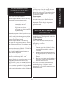
Cal Function
Mini Dopplex only
The baseline and sensitivity of the chart recorder
can be set up using the Cal function. This
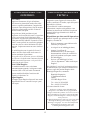
generates a zero velocity baseline and a
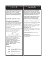
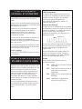
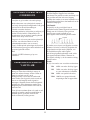
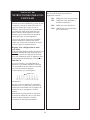
sequence of pulses as shown below:
Obstetric mode is automatically selected when
an obstetric probe (OP2 or OP3) is connected to
the control unit.
Note that early detection (9-12 weeks) is highly
dependent on operator skill and other factors
and may not always be possible.
In this mode the waveform output and Cal
function are disabled.
Cal Pulse
Aqua Dopplex
Aqua Dopplex Doppler has a waterproof probe
for underwater use.
Two levels of calibration pulses are provided.
While Cal is active, calibration tones
synchronised to the calibration pulses are
superimposed on the audio signal.
If the button is pressed and held, then the
calibration sequence is repeated.
When the calibration sequence is complete,
normal operation is resumed.
Probes
Four probes are available for vascular
examinations:
VP4
4MHz for deep lying vessels
VP5
5MHz for deep lying vessels and
oedematous limbs
VP8
8MHz for peripheral vessels
VP10 10MHz for specialist superficial
applications.
2