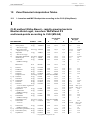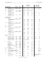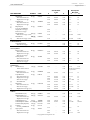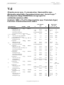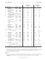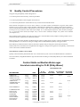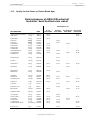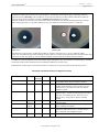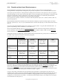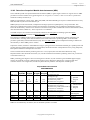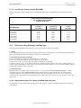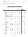
1
User's Guide NEO-SENSITABS ™ SUSCEPTIBILITY TESTING 19th Ed. 2007/2008 TAASTRUPGAARDSVEJ 30 DK-2630 TAASTRUP DENMARK Phone: +45 43 99 33 77 Telefax: +45 43 52 73 74 e-mail: [email protected] DIAGNOSTICA NEO-SENSITABS ™ 09-2007/2008 Page 2 of 170 USER’s GUIDE NEO-SENSITABS™ SUSCEPTIBILITY TESTING 19th Ed. 2007/2008 Contents 1 Susceptibility Testing in General ................................................................................................................................ 6 2 Characteristics of NEO-SENSITABS ......................................................................................................................... 7 2.1 3 Storage of NEO-SENSITABS ....................................................................................................................... 7 NEO-SENSITABS Range ........................................................................................................................................... 9 3.1 Antibacterials ................................................................................................................................................. 9 3.2 Antifungals .................................................................................................................................................. 13 4 Susceptibility Test Media .......................................................................................................................................... 14 5 Primary vs. Pure Culture Susceptibility Testing Early Reading of the Antibiogramme ........................................... 15 6 Inoculum Standardization and Prediffusion Method ................................................................................................. 17 6.1 6.2 Inoculum Standardization: ICS and CLSI (Kirby-Bauer)............................................................................ 17 6.1.1 Inoculum according to ICS ......................................................................................................... 17 6.1.2 Inoculum according to CLSI (Kirby-Bauer) ............................................................................... 17 Prediffusion Method .................................................................................................................................... 18 7 Dispensers and Templates ("Schabelons") ................................................................................................................ 20 8 Representative Antimicrobials to be Tested Routinely ............................................................................................. 22 9 Measuring the Inhibition Zones ................................................................................................................................ 25 10 Zone Diameter Interpretation Tables ........................................................................................................................ 27 10.1 I - Inoculum and MIC Breakpoints according to the CLSI (Kirby-Bauer) .................................................. 27 10.2 II - Interpretations according to MIC Breakpoints of the Dutch CRG ......................................................... 32 10.3 III - Interpretation according to MIC Breakpoints of SRGA in Sweden ..................................................... 37 10.4 IV - Interpretations according to MIC Breakpoints of the Norwegian AFA Group .................................... 44 10.5 V - Danish Blood Agar. Interpretation Valid for Denmark ......................................................................... 52 10.5.1 Interpretations According to MIC Breakpoints of the Danish Reference Group for Susceptibility Testing ................................................................................................................. 57 10.6 VI - Interpretation according to MIC Breakpoints of SFM (France) ........................................................... 65 10.7 VII – Interpretation according to MIC Breakpoints of DIN 58940-4 (Germany)........................................ 73 10.8 VIII – Interpretation according to the MIC break-points recommended by EUCAST ............................... 76 11 Interpretation of Results ............................................................................................................................................ 79 12 Quality Control Procedures ....................................................................................................................................... 80 12.1 Quality Control Flow Chart ......................................................................................................................... 83 12.2 Quality Control Zones on Danish Blood Agar ............................................................................................ 84 13 Detection of Resistant Staphylococci Against Methicillin and Vancomycin ........................................................... 85 13.1 Staphylococcus aureus................................................................................................................................. 85 13.2 Coagulase Negative Staphylococci .............................................................................................................. 88 13.3 Comments Concerning Other Antimicrobials ............................................................................................. 88 13.4 MRSA Quality Control ................................................................................................................................ 90 13.5 VISA / GISA Quality Control ..................................................................................................................... 90 © Copyright Rosco Diagnostica A/S NEO-SENSITABS ™ 09-2007/2008 Page 3 of 170 14 Detection of Resistant Enterococci ........................................................................................................................... 92 14.1 Penicillin/Ampicillin Resistance.................................................................................................................. 92 14.2 Glycopeptide Resistance (VRE) .................................................................................................................. 92 14.3 High-level Aminoglycoside Resistance (HLR) ........................................................................................... 95 14.4 Comments concerning Other Antibacterials ................................................................................................ 95 14.5 Enterococci HLR and VRE Quality Control ............................................................................................... 96 15 Susceptibility Testing of Fastidious or Problem Organisms ..................................................................................... 98 15.1 Susceptibility Testing of Haemophilus influenzae and H. parainfluenzae. ................................................. 99 15.1.1 Quality Control Limits for Haemophilus influenzae ATCC 49247 .......................................... 102 15.2 Susceptibility Testing of Gonococci .......................................................................................................... 104 15.2.1 Quality Control Limits for N. gonorrhoeae ATCC 49226........................................................ 105 15.3 Susceptibility Testing of Meningococci .................................................................................................... 107 15.4 Susceptibility Testing of Moraxella catarrhalis ........................................................................................ 109 15.5 Susceptibility Testing of Pneumococci ...................................................................................................... 110 15.5.1 Quality Control Limits for S. pneumoniae ATCC 49619 ......................................................... 113 15.6 Susceptibility Testing of Beta-haemolytic and Viridans Streptococci....................................................... 115 15.7 Susceptibility Testing of Campylobacter ................................................................................................... 119 15.8 Susceptibility Testing of Vibrio cholerae .................................................................................................. 121 15.9 Susceptibility Testing of Helicobacter pylori ............................................................................................ 122 15.10 Susceptibility Testing of S. maltophilia, B. cepacia, and Acinetobacter spp............................................. 123 16 Detection of Beta-Lactamases ................................................................................................................................. 126 16.1 Extended-Spectrum Beta-Lactamases (ESBL) .......................................................................................... 126 16.2 ESBL Quality Control ............................................................................................................................... 131 16.3 Inducible Cephalosporinases or AmpC Beta-lactamases ........................................................................... 132 16.3.1 Testing / Reporting of Susceptibility to Beta-lactams against Enterobacteriaceae and Non-fermenters ......................................................................................................................... 133 16.4 Plasmid-mediated AmpC Beta-lactamases ................................................................................................ 134 16.4.1 Differentiation of AmpC beta-lactamases in E. coli ................................................................. 135 16.5 Inhibitor Resistant TEM Beta-lactamases (IRT) ....................................................................................... 138 16.6 Carbapenemases ........................................................................................................................................ 138 16.6.1 Detection of acquired carbapenemases Ambler classes A and D ............................................. 138 16.6.2 Detection of acquired Metallo-beta-lactamases (MBL) ............................................................ 141 16.7 Detection of multiple beta-lactamases in one strain .................................................................................. 145 16.8 Detection of ß-lactam Resistance Phenotypes ........................................................................................... 146 17 16.8.1 Detection of ß-lactam Resistance Phenotypes in Enterobacteriaceae ....................................... 146 16.8.2 Detection of ß-lactam Resistance Phenotypes in Non-fermenters ............................................ 147 Detection of Other Resistance Mechanisms ............................................................................................................ 148 17.1 Screening of 16S rRNA Methylases (HLR to Aminoglycosides) ............................................................. 148 17.2 Screening for Plasmid-mediated Quinolone Resistance (QnrA, QnrB, QnrS) in Enterobacteriaceae. Integrons .................................................................................................................................................. 149 17.3 Detection of Resistance Mechanisms (General) ........................................................................................ 151 17.4 Intrinsic (Natural) Resistance .................................................................................................................... 153 © Copyright Rosco Diagnostica A/S NEO-SENSITABS ™ 09-2007/2008 Page 4 of 170 18 Sources of Error in the Diffusion Test .................................................................................................................... 156 19 Limitations of Diffusion Methods. Warning ........................................................................................................... 157 20 Susceptibility Testing of Anaerobes ....................................................................................................................... 158 21 Susceptibility Testing of Yeasts .............................................................................................................................. 161 21.1 Procedure according to CLSI..................................................................................................................... 162 21.1.1 Interpretation Tables for Yeasts (MH-GMB) ........................................................................... 162 21.1.2 Interpretation table fo Local treatment...................................................................................... 163 21.1.3 Candida spp. Quality Control (MH-GMB)............................................................................... 164 21.2 Procedure using Shadomy modified agar .................................................................................................. 164 22 21.2.1 Interpretation Tables for Yeasts (modified Shadomy agar) ...................................................... 164 21.2.2 Candida spp. Quality Control (modified Shadomy Agar) ........................................................ 165 Veterinary Practice CLSI ........................................................................................................................................ 168 © Copyright Rosco Diagnostica A/S NEO-SENSITABS ™ 09-2007/2008 Page 5 of 170 PREFACE The 19th Ed. 2007/2008 of the NEO-SENSITABS User's Guide contains updated text, tables and references, all informative information when using Neo-Sensitabs tablets for susceptibility testing. A table giving the changes from the 19th Ed. 2007/2008 is available at our website. The different interpretation tables following the CLSI (formerly NCCLS) recommendations have been updated according to the latest information of the CLSI described in "Performance Standards for Antimicrobial Susceptibility Testing", 15th Informational Suppl., M100-S17, 2007. Furthermore, the User's Guide includes updated Zone Diameter Interpretative Standards according to CRG, SRGA, AFA, Denmark, SFM, DIN and BSAC. Information and recommendations given by susceptibility testing standardization groups or organizations in different European countries provide supplementary knowledge of specific considerations and decisions made in each country. In the future, the use of common European MIC breakpoints would permit the different countries (and laboratories) to use their separate techniques (including inoculum, media, tablets or disks, automatic methods etc.) and obtain comparable results, because the techniques used would be standardized according to common European MIC breakpoints (1). The European Committee on Antimicrobial Susceptibility Testing (EUCAST) has been reorganized in 2001/2002 giving national breakpoint committees (at present France, Germany, Norway, Sweden, the Netherlands and the UK) a greater role. One of the major tasks of the EUCAST is to harmonize MIC breakpoints across Europe and the process is in progress (2). At the moment MIC-breakpoints for aminoglycosides, cephalosporins, carbapenems, monobactams, fluoroquinolones, glycopeptides, oxazolidinones, tigecycline, and daptomycin have been harmonised though EUCAST. If included in national recommendations, they are included in the interpretation tables I-VIII (3). The User's Guide is available at our website www.rosco.dk and updated information is continuously included. Rosco Diagnostica A/S is welcoming any feedback and questions on susceptibility testing from users directly ([email protected]) or through our representatives. ROSCO DIAGNOSTICA A/S August 2007 References: 1) Kahlmeter G.: Breakpoints in Europe: a future perspective. 42nd ICAAC. Presentation 1022, 2002. 2) Kahlmeter G. et al: European harmonization of MIC breakpoints for antimicrobial susceptibility testing of bacteria. J. Antimicr. Chemother. 52, 145-148, 2003. 3) http://www.srga.org/eucastwt/MICTAB/index.html © Copyright Rosco Diagnostica A/S NEO-SENSITABS ™ 09-2007/2008 Chapter 1 Page 6 of 170 1 Susceptibility Testing in General Susceptibility testing is of great importance to the hospital physician and the practitioner in the treatment of infectious diseases. Knowledge of susceptibility to different antibiotics of an infectious agent, facilitates the choice of the most effective antibacterial agents. In most clinical laboratories an agar diffusion (disk/tablet) method is used routinely for testing the susceptibility of common rapidly growing bacterial pathogens. It is generally accepted that reliable results can be obtained by the diffusion test, when a standardized methodology is used, and the diameter of the inhibition zones has been correlated with minimal inhibitory concentrations (MIC). The interpretation of zone diameters by the diffusion method, should be based on regression lines. A regression line, for a certain antibiotic contained in a disk/tablet, is a graphic expression of the correlation existing between MIC's and the diameters of inhibition zones obtained with a number of bacterial strains. These lines represent a control of the standardization of the method, showing that the interpretative zone diameters correspond to MIC values in agreement with internationally accepted standards. The availability of regression lines makes it possible for the microbiologist to convert the results obtained by the diffusion method (susceptible, intermediate, resistant) into more quantitative results, i.e. approximated MIC values. An accurate diffusion method requires: • A well-standardized technique including control strains (e.g. Staphylococcus aureus ATCC 25923, Escherichia coli ATCC 25922, Pseudomonas aeruginosa ATCC 27853 and Enterococcus faecalis ATCC 29212) enabling control of inoculum and media variations. • Well-traced regression lines, which can be controlled, because there is a correlation between the slope of the regression line and the molecular weight of the antimicrobial tested. • Separate regression lines for rapidly growing bacteria and slow-growing species. Regression lines for Neo-Sensitabs have been prepared using the agar dilution method to determine the MIC values. For the aminoglycosides (amikacin, gentamicin, netilmicin, tobramycin) and polymyxins (colistin) we have also determined MIC's by the broth dilution method in order to correct for the amount of antimicrobial bound to the agar. The above mentioned antimicrobials are strongly bound to the sulphate groups of the agar, and consequently, different MIC values as well as zone sizes may be obtained depending on the purity of the agar used. It is, therefore, important to control the quality of the agar used in the diffusion test, and this may be achieved by controlling zone sizes with control ATCC strains. Separate interpretation tables have been prepared for slow-growing and bacteria with special requirements (Haemophilus, Streptococci, Moraxella, Neisseria, Pneumococci, and Anaerobes). This booklet describes methods, quality control and interpretative criteria recommended in different countries for diffusion susceptibility tests with Neo-Sensitabs. When new problems are recognized or improvements are developed, changes will be incorporated in future editions of the booklet and also distributed as informational supplements. References: 1) Casals J.B, Gylling Pedersen O.: Tablet sensitivity testing: a comparison of different methods. Acta Path. Microbiol. Scand, sect B, 80B, 806-816, 1972. 2) Schumacher H. et al: A procedure for evaluation and documentation of susceptibility test methods using the susceptibility of Klebsiella pneumonae to ciprofloxacin as model. J. Antimicr. Chemother., 48, 493-500, 2001. © Copyright Rosco Diagnostica A/S NEO-SENSITABS ™ 09-2007/2008 Chapter 2 Page 7 of 170 2 Characteristics of NEO-SENSITABS Neo-Sensitabs are produced according to the guidelines of WHO (1). The tablets are 9 mm in diameter and each NeoSensitabs is printcoded for safe identification. The tablets are manufactured with the aid of microbially inert auxiliary substances by a dry process using crystalline antimicrobials. This procedure results in very uniform tablets, homogenous in their content of active ingredients, and with an extraordinary stability (usually not less than 4 years shelf-life at room temperature (2)). Neo-Sensitabs are standardized according to the MIC-breakpoints recommended by "Susceptibility Testing Standardization Groups" in several countries (e.g. Holland, Norway, Sweden, France, Germany, UK and Denmark) including the CLSI (3). All antimicrobials have received new letter codes (see chapter 3), because in order to achieve optimal recognition and zone measurements with automatic instruments a 5-digit code for each Neo-Sensitabs type has been chosen. References: 1) World Health Organization: Expert Committee on Biological Standardization. Requirements for antibiotic susceptibility tests. Requirements for Biological Substances No. 26, Geneva, September 1981. 2) Gylling Pedersen O.: Standardizing, manufacture, and control of Neo-Sensitabs. Acta Clin. Belg., 28, 139-149, 1973. 3) NCCLS: Performance Standards for Antimicrobial Disk Susceptibility Testing. 8th Ed. M2-A8 January 2003. 4) Stes P. et al: Cefepime activity against Ps. aeruginosa: evaluation of Etest and two disc diffusion methods. J. Antimicro. Chemother., 38, 707-711, 1996. 5) Engberg J. et al: Comparison of two agar dilution methods and three agar diffusion methods, including the Etest, for antibiotic susceptibility testing of thermophilic Campylobacter species. Clin. Microbiol. and Infect., 5, 580-584, 1999. 6) Koeth L. et al: Quality control study evaluating the performance of daptomycin and daptomycin/calcium disks and Etest. Clin. Microbiol. Infect., 10, Supp. 3, 541, 2004. 7) Denis O. et al: Polyclonal emergence and importation of CA-MRSA strains harbouring Panton-Valentine leucocidin genes in Belgium. JAC, 56, 1103-06, 2005. 2.1 Storage of NEO-SENSITABS Neo-Sensitabs are stored at room temperature (both unopened and cartridges in use). Very few exceptions (Cefpodoxime, Cefepime, Cefepime+Clavulanate, Temocillin, Ticarcillin, Ticarcillin+Clavulanate, Amphotericin B, Caspofungin) must be stored in the refrigerator. With these Neo-Sensitabs (labelled "STORE AT 2-8°C") the following precautions must be taken: 1) Unopened cartridges (stock): Store in refrigerator, 2-8 °C. (Expiry date on the label and cartridge). 2) Cartridges that have been opened: Store at room temperature for up to 2 months. 3) Cartridges placed in dispenser: Store in dispenser at room temperature for up to 2 months. If an opened cartridge (labelled 2-8°C) is not used up within 2 months, it should be kept in the refrigerator. Each time the cartridge is taken from the refrigerator, it must be allowed time to reach room temperature (30-60 min.) before it is opened, in order to avoid water condensation on the tablets. Cartridges (labelled 2-8°C) that have been opened and are kept in the refrigerator should be used within 6 months. With Neo-Sensitabs stored at room temperature (labelled "store below 25 °C), cartridges that have been opened (or are placed in a dispenser), should be stored at room temperature and used within 12 months of opening date. The stability of antimicrobials in paper disks is decreased compared to Neo-Sensitabs. The CLSI (1) recommends frozen storage of paper disks containing beta-lactam antibiotics. In case of Imipenem, Cefaclor and Clavulanic acid combinations, paper disks should be stored frozen until the day of use. In a comparative stability study between Neo-Sensitabs and Oxoid paper disks (2), it was observed that disks containing Ticarcillin 75 µg , lost activity after 15 days at 4-6 °C, while Ampicillin 10 µg and Amoxycillin + Clavulanate 20+10 µg disks lost activity after one month at 4-6 °C.The corresponding Neo-Sensitabs were stable at room temperature (and at 4-6 °C) during the study period of six months. Steward et al (3) noticed overdetection of imipenem/meropenem resistance in the project ICARE, most probably due to inactivation of the reagents used (Vitek, disk diffusion etc.) and recommended the use of a second independent antimicrobial susceptibility testing method to validate carbapenem-intermediate and resistant strains. © Copyright Rosco Diagnostica A/S NEO-SENSITABS ™ 09-2007/2008 Chapter 2 Page 8 of 170 References: 1) NCCLS: Performance Standards for Antimicrobial Disk Susceptibility Testing, 8th Ed. M2-A8, January 2003. 2) del Cuerpo M. et al: Stability of beta-lactam antibiotics in paper disks and tablets used in the diffusion test. Rev. Esp. Quimioter., 10, nr. 3,1997 (Spanish). 3) Steward C.D. et al: Antimicrobial susceptibility testing of carbepenems: multicenter validity testing and accuracy levels of 5 antimicrobial test methods for detecting resistance in Enterobacteriaceae and Pseudomonas aeruginosa isolates. J. Clin. Microbiol., 41, 351-358, 2003. © Copyright Rosco Diagnostica A/S NEO-SENSITABS ™ 09-2007/2008 Chapter 3 Page 9 of 170 3 NEO-SENSITABS Range 3.1 Antibacterials Identification code Neo-Sensitabs Neo-Sensitabs Diffusible amount of Antimicrobial A. Penicillins (Penams) PENICILLIN LOW AMPICILLIN 33 µg AMPICILLIN 2.5 µg AMOXYCILLIN PEN.L AMP33 AMP.L AMOXY 5 µg 33 µg 2.5 µg 30 µg METHICILLIN OXACILLIN 1 µg OXACILLIN 5 µg CLOXACILLIN 500 µg (AmpC test) (Diatabs) METHI OXA.1 OXA.5 CL500 29 µg 1 µg 5 µg 500 µg MECILLINAM (Amdinocillin) MECIL 33 µg TICARCILLIN TICAR 75 µg PIPERACILLIN PIPER 100 µg TEMOCILLIN TEMOC 30 µg AM+CL 30+15 µg AM+SU TI+CL 30+30 µg 75+15 µg PI+TZ CZ+CL CP+CL IM+ED BORON CAZ+D D.P.A. 100+10 µg 30+10 µg 30+10 µg 15+750 µg 250 µg 30+250 µg 250 µg CEPHALOTHIN CEFACLOR CEFADROXIL CEFAZOLIN CEPHALEXIN CEPHRADINE CLOTN CCLOR CFDRO CFZOL CFLEX CFRAD 66 µg 30 µg 30 µg 60 µg 30 µg 60 µg CEFUROXIME CEFONICID CEFIXIME CEFPODOXIME CEFOTAXIME CEFTAZIDIME CEFTRIAXONE CEFTIZOXIME CEFTIOFUR (Vet.) CEFEPIME CEFUR CFCID CFFIX CFPOX CFTAX CEZDI CETRX CEZOX CFTIO CFEPM 60 µg 30 µg 30 µg 30 µg 30 µg 30 µg 30 µg 30 µg 30 µg 30 µg B. Beta-lactam/ Beta-lactamase Inhibitor Combinations AMOXYCILLIN+CLAVULANATE (Augmentin) AMPICILLIN+SULBACTAM TICARCILLIN+CLAVULANATE (Timentin) PIPERACILLIN+TAZOBACTAM CEFTAZIDIME+CLAVULANATE CEFEPIME+CLAVULANATE IMIPENEM+EDTA BORONIC ACID (Diatabs) CEFTAZIDIME+DIPICOLINIC ACID DIPICOLINIC ACID (Diatabs) C. (1) Cephalosporins (Cephems) © Copyright Rosco Diagnostica A/S NEO-SENSITABS ™ 09-2007/2008 Chapter 3 Page 10 of 170 Neo-Sensitabs CEFPIROME CEFSULODIN CEFQUINOME (Vet.) CEFTOBIPROLE (BAL 9141) Identification code Neo-Sensitabs Diffusible amount of Antimicrobial CFPIR CFSUL CFQUI CFBIP 30 µg 30 µg 30 µg 30 µg CFOXT 60 µg CFBIP CFARL 30 µg 30 µg IMIPM MEROP ERTAP DORIP ----- 15 µg 10 µg 10 µg 10 µg ----- AZTRM 30 µg ST100 ST500 KANAM KA500 NEOMY AMIKA GEN40 GN250 NETIL TOBRA SPECT APRAM ISP30 100 µg 500 µg 100 µg 500 µg 120 µg 40 µg 40 µg 250 µg 40 µg 40 µg 200 µg 40 µg 30 µg C. (2) Cephamycins and Oxacephems CEFOXITIN C. (3) Cephalosporins active against MRSA CEFTOBIPROLE (investigational drug) CEFTAROLINE (investigational drug) D. Penems and Carbapenems IMIPENEM MEROPENEM ERTAPENEM DORIPENEM (investigational drug) FAROPENEM (investigational drug) E. Monobactams AZTREONAM F. Aminoglycosides STREPTOMYCINS 100 µg STREPTOMYCIN 500 µg (HLR) KANAMYCIN 100 µg KANAMYCIN 500 µg (HLR) NEOMYCIN (Framycetin) AMIKACIN GENTAMICIN 40 µg GENTAMICIN 250 µg (HLR) NETILMICIN TOBRAMYCIN SPECTINOMYCIN APRAMYCIN (Vet.) ISEPAMICIN 30 µg G. Tetracyclines TETRACYCLINES 80 µg (Oxytetracycline) TETRACYCLINES 10 µg DOXYCYCLINE MINOCYCLINE TIGECYCLINE (investigational drug) TET80 80 µg TET10 DOXYC MINOC TIG15 10 µg 80 µg 80 µg 15 µg CLR60 CLR10 FFC30 60 µg 10 µg 30 µg H. Chloramphenicol and derivatives CHLORAMPHENICOL 60 µg CHLORAMPHENICOL 10 µg FLORFENICOL (Vet.) I. Macrolides, Lincosamides, Streptogramines, Ketolides and Oxazolidinones ERYTHROMYCIN AZITHROMYCIN CLARITHROMYCIN LINCOMYCIN ERYTR AZITR CLARI LINCO © Copyright Rosco Diagnostica A/S 78 µg 30 µg 30 µg 19 µg NEO-SENSITABS ™ 09-2007/2008 Chapter 3 Page 11 of 170 Neo-Sensitabs CLINDAMYCIN 25 µg SPIRAMYCIN PRISTINAMYCIN VIRGINIAMYCIN QUINUPRISTIN/DALFOPRISTIN TELITHROMYCIN LINEZOLID LINCO-SPECTIN (Vet.) TYLOSIN (Vet.) TILMICOSIN (Vet.) PIRLIMYCIN (Vet.) Identification code Neo-Sensitabs CLIND SPIRA PRIST VIRGI SYN15 TEL15 LINEZ LI+SP TYLOS TILMI PIRLI Diffusible amount of Antimicrobial 25 µg 200 µg 30 µg 30 µg 15 µg 15 µg 30 µg 15+200 µg 150 µg 80 µg 10 µg J. (1) Glycopeptides VANCOMYCIN 5 µg VAN.5 5 µg TPN30 DALBA TELAV 30 µg 30 µg 30 µg J. (2) Lipoglycopeptides TEICOPLANIN DALBAVANCIN (investigational drug) TELAVANCIN (investigational drug) J. (3) Cyclic lipopeptides a) Gram positive spectrum: DAPTOMYCIN (+ Ca) BACITRACIN b) Gram negative spectrum: COLISTIN 10 µg POLYMYXINS 150 µg (Colistin) DAPCa BACIT 30 µg 40 units CO.10 CO150 10 µg 150 µg SULFA TRIME TR+SU 240 µg 5.2 µg 5.2+240 µg NITRO FURAZ 260 µg 50 µg NALID CINOX FLUME OXOLI PIPEM CIP10 CIP.L MOXIF GATIF ENROF LEVOF MARBO NORFX OFLOX PEFLX 130 µg 30 µg 30 µg 10 µg 30 µg 10 µg 0.5 µg 5 µg 5 µg 10 µg 5 µg 5 µg 10 µg 10 µg 10 µg K. Sulphonamides and similars SULPHONAMIDES TRIMETHOPRIM TRIMETHOPRIM+SULFA L. Nitrofurans NITROFURANTOIN FURAZOLIDONE M. Quinolone derivatives NALIDIXAN CINOXACIN FLUMEQUINE (Vet.) OXOLINIC ACID PIPEMIDIC ACID CIPROFLOXACIN 10 µg CIPROFLOXACIN 0.5 µg MOXIFLOXACIN GATIFLOXACIN ENROFLOXACIN (Vet.) LEVOFLOXACIN MARBOFLOXACIN (Vet.) NORFLOXACIN OFLOXACIN PEFLOXACIN © Copyright Rosco Diagnostica A/S NEO-SENSITABS ™ 09-2007/2008 Chapter 3 Page 12 of 170 Neo-Sensitabs Identification code Neo-Sensitabs Diffusible amount of Antimicrobial N. Others FOSFOMYCIN (+G6-P) FUCIDIN METRONIDAZOLE 16 µg MUPIROCIN NOVOBIOCIN 5 µg RIFAMPICIN TIAMULIN (Vet.) PENICILLIN/NOVO (Vet.) FOSFO FUCID MTR16 MUPIR NOV05 RIFAM TIAMU PEN+N © Copyright Rosco Diagnostica A/S 70+40 µg 100 µg 16 µg 10 µg 5 µg 30 µg 30 µg 10 U/30 µg NEO-SENSITABS ™ 09-2007/2008 Chapter 3 Page 13 of 170 3.2 Antifungals Neo-Sensitabs AMPHOTERICIN B CICLOPIROX CLOTRIMAZOLE ECONAZOLE FLUCONAZOLE 5-FLUOROCYTOSINE 10 µg 5-FLUOROCYTOSINE 1 µg GRISEOFULVIN ISOCONAZOLE ITRACONAZOLE KETOCONAZOLE MICONAZOLE NATAMYCIN NYSTATIN TERBINAFINE VORICONAZOLE CASPOFUNGIN (investigational drug) POSACONAZOLE (investigational drug) Identification code Neo-Sensitabs AMPHO CICLO CLOTR ECONZ FLUCZ FLU10 FLU.1 GRISE ISOCN ITRAC KETOC MICON NATAM NYSTA TERBI VOR.1 CASP5 POSAC © Copyright Rosco Diagnostica A/S Diffusible amount of Antimicrobial 10 µg 50 µg 10 µg 10 µg 25 µg 10 µg 1 µg 25 µg 10 µg 8 µg 15 µg 10 µg 50 µg 50 µg 30 µg 1 µg 5 µg 5 µg NEO-SENSITABS ™ 09-2007/2008 Chapter 4 Page 14 of 170 4 Susceptibility Test Media In order to achieve satisfactory results, a proper medium for susceptibility testing must be used. It should fulfil at least two requirements: • The composition of the medium should give sufficiently good growth conditions for the strains to be tested. • The medium must not contain material interfering with the test itself or with any antimicrobial contained in NeoSensitabs. The ideal medium for susceptibility testing has not been found yet, and this is one of the reasons why different media are used in different countries. Mueller-Hinton agar is recommended by the FDA and the CLSI in the U.S. (1) and is routinely used in several European countries. Variations in Mueller-Hinton agar from different manufacturers were first recognized with tetracyclines and aminoglycosides. Variation in Mg and Ca, will affect results of aminoglycoside and tetracycline tests with Ps. aeruginosa. Excess zinc ions may reduce zone sizes of carbapenems. Excessive cation content will reduce zone sizes, whereas low cation content may result in unacceptable large zones of inhibition (1). Ca and Mg should be available in the medium, in the form of soluble salts. Later differences were found in thymidine content affecting testing of trimethoprim and methicillin -resistant staphylococci (2,3). Most agar media contain small amounts of sulphonamide and trimethoprim antagonists that may affect the results of susceptibility testing (especially if blood is not added) when low content disks are used, while Neo-Sensitabs are less affected (4). Susceptibility test media should contain less than 0.03 mg/l thymidine, otherwise small colonies are seen inside the trimethoprim zone. If the medium contains slightly more thymidine than recommended, it is possible to reduce the concentration by adding thymidinephosphorylase: 0.025 to 0.1 IU enzyme/ml medium or 5% haemolyzed horse blood, which contains the same enzyme. Reducing the thymidine content of Mueller-Hinton agar for clearer sulphonamide and trimethoprim zones might have an inhibitory effect on the growth of some staphylococci. This phenomenon may also be related to problems in detecting resistance to oxacillin/methicillin in staphylococci. Consequently, to make sure that a certain MuellerHinton agar is useful for susceptibility testing, it must fulfil the following criteria: • It shows acceptable batch to batch reproducibility for susceptibility testing. • Zone diameters with the 4 control strains must fall within the specified limits. • Ability to detect methicillin-resistance in staphylococci. (Quality control with S. aureus ATCC 43300). • Ability to produce clear inhibition zones within the control limits, around Trimethoprim and Trimethoprim+Sulfa with enterococci. (Quality control with E. faecalis ATCC 29212). Only Mueller-Hinton medium formulations, that have been tested according to, and that meet, the acceptance limits described in the CLSI document M6, should be used (1). Neo-Sensitabs have been standardized with the following susceptibility test media: Mueller-Hinton Agar, Iso-sensitest Agar, and Danish Blood Agar. The pH value of each batch of agar medium should be checked when the medium is prepared. The agar medium should have a pH of 7.2 - 7.4 at room temperature after gelling. If the pH is too low, certain antimicrobials will appear as having lower potency ( e.g. aminoglycosides, macrolides) and the strains will appear as more resistant, while other antimicrobials may appear as having excessive activity (e.g. penicilins) and the corresponding strains will appear as more susceptible. If the pH is too high, the opposite effects can be expected (1). The thickness of the agar must be 4 mm ± 1 mm. References: 1) NCCLS: Performance Standards for Antimicrobial Disk Susceptibility Tests. M2-A8. 8th Ed., January 2003. 2) Pollock H.M., Barry A.L., Gavan T.L., Fuchs P.C., Hansen S., Thornsberry C.L., Frankel H., Forsythe S.B.: Selection of a reference lot of Mueller-Hinton Agar. J. Clin. Microbiol., 24, 1-6, 1986. 3) Hindler J.A., Inderlied C.B.: Effect of source of Mueller-Hinton Agar and resistence frequency on the detection of methicillin resistant Staph. aureus. J. Clin. Microbiol., 21, 205-210, 1985. 4) Casals J.B.: Effects of medium on the results of antimicrobial susceptibility testing. Chemotherapy, 2, 41-46. Proc. 9th International Congress Chemotherapy, London 1975. © Copyright Rosco Diagnostica A/S NEO-SENSITABS ™ 09-2007/2008 Chapter 5 Page 15 of 170 5 Primary vs. Pure Culture Susceptibility Testing Early Reading of the Antibiogramme Direct (or primary) susceptibility tests, in which tablets or disks are applied to plates inoculated from the specimen, are widely used in Denmark and the U.K., but their reliability is often questioned by microbiologists in other countries. The advantages of these tests are: a) speed as results may be available on the following day; b) identification of small numbers of resistant organisms in a predominantly susceptible population and c) their value as selective media in mixed cultures (1). 1) Urine Primary susceptibility tests of urine specimen are particularly successful (5). In urgent clinical situations, physicians may request more rapid information to guide their selection of antimicrobial agents. Therefore, it is common practice in some laboratories to report presumptive susceptibilities after 6-8 hours incubation (9). Midtvedt & Midtvedt (6) found a good correlation between results obtained after 8 hours and 18-24 hours incubation, respectively. In most cases the zones of inhibition were found to be somewhat larger at 18-24 hours than after 8 hours. None of the strains observed to be susceptible after 8 hours incubation was found to be resistant after 18-24 hours. Saha et al (9) using rapid but low-cost susceptibility testing methods (disk diffusion) obtained antibiogram results (8 hours incubation) identical to those obtained by conventional methods. 2) Blood cultures Previous studies have shown an agreement of 94.6 % or higher when comparing direct (primary) susceptibility testing by the diffusion method utilizing positive blood cultures as inoculum and standard diffusion susceptibility test methods (2,3,4). Coyle et al (7) used a combination of direct inoculation from blood cultures and early readings (6 hours) of disk diffusion. 91% of the tests with gram-positive organisms and 86% gram-negatives were in agreement with standard results. Early reading results must be confirmed by re-reading after 18-24 hours incubation or by standard susceptibility testing. In clinical emergencies when the gram strain suggests that the infection is due to a single species, direct susceptibility tests may be performed and the results reported as preliminary (8). Once isolated colonies become available from the initial culture, a follow-up standardized test should be performed. Navon-Venecia et al. (11) inoculated 0.2 ml from incubated aerobic bottles (blood culture) into Mueller-Hinton agar plates and tested for ESBL production by the disk method. High concordance between direct testing and the standard protocol was achieved. Weinbren et al (13) reported ESBL results after 5-6 hours' incubation with E. coli and Klebsiella spp. 3) Faeces For detecting the faecal carriage of antimicrobial-resistant E. coli, a faecal swab was directly plated onto a McConkey plate and antimicrobial disks applied onto the seeded plate. The same swab was tested with a conventional method. The rapid screening method showed a good specificity and good concordance with the conventional method. (Paradisi et al. (12), Kronwall et al. (15)). Bartolini et al. (16) using a direct plating method of fecal samples on McConkey agar studied the status on antimicrobial resistance in commensal E. coli in preschool children from low-resource countries. This inexpensive, sensitive and simple method detected high resistance rates of E. coli to ampicillin (95 %), Trim+Sulfa (94 %), Tetracycline (93 %), and Chloramphenicol (70 %). 35 % of the strains were resistant to Nalidixic acid. 4) Sputum Cercenado et al (10) in patients with ventilator-associated pneumonia evaluated the Gram-strain and direct susceptibility test by the diffusion method (E-test) on respiratory samples. Results of the difusion test correlated with the microdilution method in susceptibility categories in all cases with S. aureus, P. aeruginosa and E. coli. © Copyright Rosco Diagnostica A/S NEO-SENSITABS ™ 09-2007/2008 Chapter 5 Page 16 of 170 For the bacteriological analysis of sputum from cystic fibrosis patiens, colonised by P. aeruginosa, sputum is fluidized with acetylcystein and further diluted 1/20 with saline. Inoculate MH-plate + 5 % blood. Add antimicrobial disks (tablets): Ticarcillin, Ceftazidime, Aztreonam, Imipenem, Tobramycin, Ciprofloxacin, Polymyxins (colistin) and C-390. After incubation overnight, look for suspicious colonies inside the inhibition zones. Identify and perform antibiograms (14). S. maltophilia will show resistance to imipenem, P. aeruginosa resistance to C-390, A. xylosoxydans resistance to tobramycin and B. cepacia resistance to colistin. References: 1) Waterworth P.M., Delpiano M.: Dependability of susceptibility tests in primary culture. J. Clin. Path., 29, 179-184, 1976. 2) Mirrett S., Reller L.B.: Comparison of direct and standard antimicrobial disk susceptibility testing for bacteria isolated from blood. J. Clin. Microbiol., 10, 482-487, 1979. 3) Fay D., Oldfather J.E.: Standardization of direct susceptibility tests for blood cultures. J. Clin. Microbiol., 9, 347-350, 1979. 4) Doern G.V., Scott D.R., Rashad A.L., Kim K.S.: Evaluation of a direct blood culture disk diffusion antimicrobial susceptibility test. Antimicrob. Agents Chemoth., 20, 696-698, 1981. 5) Johnson J.R. et al: Direct antimicrobial susceptiblility testing for acute urinary tract infection in women. J. Clin. Microbiol. 33, 2316-2323, 1995. 6) Midtved K., Midtved T.: Rapid determination of antibiotic susceptibility by a disc diffusion test for urgent clinical situations. Scand. J. Infect. Dis., 17, 131-132, 1985. 7) Coyle M.B., McGonagle L.A., Plorde J.J., Clausen C.R., Schoenknecht F.D.: Rapid antimicrobial susceptibility testing of isolates from blood cultures by direct inoculation and early reading of disk diffusion tests. J. Clin. Microbiol., 20, 473-477, 1984. 8) NCCLS: Performance Standards for Antimicrobial Disk Susceptibility Tests. 8th Ed. M2-A8, 2003. 9) Saha S.K. et al: Rapid identification and antibiotic susceptibility testing of Salmonella enterica serovar tiphi, isolated from blood: implications for therapy. J. Clin. Microbiol., 39, 3583-3585, 2001. 10) Cercenado E. et al: Rapid antimicrobial susceptibility testing in patients with ventilator-associated pneumonia: direct E-test on respiratory samples. 42nd ICAAC, Presentation D-51, 2002. 11) Navon-Venecia S. et al: Direct testing of ESBL producing E. coli and K. pneumoniae from blood cultures. ICAAC 2003, presentation D-204. 12) Paradisi F. et al: Evalutation of a rapid screening method for detection of antimicrobial resistance in the commensal E. coli microbiota. ICAAC 2003, presentation D-248. 13) Weinbren M.J. et al: Rapid detection of ESBL-producing organisms in blood culture. J. Antimicr. Chemother., 55, 131-2, 2005. 14) Zebruh M. et al: Evaluation of a new E-test method for antimicrobial susceptibility testing of P. aeruginosa isolates from cystic fibrosis. Pathologie Biologie, 53, 490-4, 2005 (French). 15) Kronvall G. et al: Extended antimicrobial resistance screening of the dominant faecal E. coli and of rare resistant clones. Intl. J. Antimicrobial Agents, 26, 473-8, 2005. 16) Bartolini A. et al: Multidrug resistant commensal E. coli in children Peru and Bolivia. Emerg. Infect. Dis., 12, 907-913, 2006. © Copyright Rosco Diagnostica A/S NEO-SENSITABS ™ 09-2007/2008 Chapter 6 Page 17 of 170 6 Inoculum Standardization and Prediffusion Method 6.1 Inoculum Standardization: ICS and CLSI (Kirby-Bauer) The specimen must be fully typical of the site of infection, i.e. every effort should be made to obtain a representative sample of the relevant pathogenic bacteria. The density of the inoculum is one of the variables that will have a great influence on the size of the zones of inhibition. With primary cultures, the inoculum may vary to a large degree, i.e. from few colonies to a dense growth. Our studies with Neo-Sensitabs have shown that too sparse an inoculum will give too large zones, and too dense an inoculum will give too small zones compared to the interpretation charts. 6.1.1 Inoculum according to ICS 1) When secondary (pure) cultures are used, the inoculum may be standardized as recommended by the ICS (1) the appropriate inoculum should yield a dense but not completely confluent growth. It is prepared by diluting an overnight broth culture of the organism in broth. Suitable dilutions of fully grown broth cultures of Enterobacteriaceae and most gram-negative rods are usually about 10-4 (1:10,000) while for staphylococci and enterococci a dilution of 10 -3 (1:1,000) is usually appropriate. 2) The inoculum can also be prepared from isolated colonies on agar. Suspend 2 to 3 colonies into 10 ml physiological saline, and thereafter dilute 0.1 ml/10 ml for Enterobacteriaceae and other gram negative rods or 1 ml/10 ml for staphylococci and enterococci. Take 1-2 drops (9 cm plate) or 3 to 4 drops (14 cm plate) of the final suspension and apply onto the agar surface and distribute with a bent glassrod or Drigalski spatula. The open plate is then dried at 35-37°C for 10-15 min. before the Neo-Sensitabs are placed onto the agar surface. When testing slower growing microorganisms, the dilution must be individualized to give semi-confluent growth. The method of inoculation may vary, but it must be such that it gives uniform seeding of the plate and the semiconfluent growth desired. 6.1.2 Inoculum according to CLSI (Kirby-Bauer) When using the technique of Kirby-Bauer, the inoculum is standardized according to the method described by the CLSI (2), which normally results in more dense growth and therefore smaller inhibition zones than for semiconfluent growth. 1) Growth method: a) Select at least 3 to 5 isolated colonies. Touch the top of each colony with a wire loop and transfer to a tube containing 4 to 5 ml of a broth medium such as tryptic soy broth. b) Allow the broth culture to incubate at 35°C until it achieves or exceeds the turbidity of the 0.5 McFarland standard. It usually takes 2 to 6 hours. c) Adjust the turbidity of the broth culture with sterile saline or broth to obtain a turbidity visually comparable to the standard. This results in a suspension containing approx. 1 to 2 x 10⁸ CFU/ml for E. coli ATCC 25922. d) Within 15 minutes, dip a sterile cotton swab into the adjusted suspension and remove inoculum from the swab by exerting firm pressure on the inside of the tube. e) Inoculate the dried surface of the Mueller-Hinton agar plate by streaking the swab over the entire surface. 2) Direct colony suspension method: It is also possible to set up the tests immediately, rather than wait for a broth sub-culture (3). This can be done by suspending several morphologically similar colonies from an 18-24 h agar plate (non selective) into 4-5 ml 0.9% NaCl solution, and then immediately adjusting the turbidity to match that of the BaSO4 standard (0.5 McFarland). Within 15 minutes swabs are used to inoculate the test plates. This method was found to be equivalent to the standard CLSI method and requires less time. This method is used in most laboratories for all types of bacteria. This approach is the recommended for testing the fastidious organisms (Haemophilus spp., gonococci, Moraxella catarrhalis, pneumococci/streptococci) and for testing staphylococci for potential methicillin or oxacillin resistance (2). © Copyright Rosco Diagnostica A/S NEO-SENSITABS ™ 09-2007/2008 Chapter 6 Page 18 of 170 References: 1) Ericsson H.M., Sherris J.C.: Antibiotic susceptibility testing. Report of an International Collaborative Study. Acta Path. Microbiol. Scand. Sec. B, Suppl. No. 217, 1971. 2) NCCLS: Performance Standards for Antimicrobial Disk Susceptibility Tests. M2-A8, 8th Ed., January 2003. 3) D'Amato R.F., Hochstein L.: Evaluation of a rapid inoculum preparation method for agar disk diffusion susceptibility testing. J. Clin. Microbiol., 15, 282-285, 1982. 6.2 Prediffusion Method Prediffusion Method (2 + 18 hours) for Antimicrobials Diffusing Poorly on Agar High molecular weight antimicrobials (vancomycin, teicoplanin, daptomycin, colistin) diffuse poorly on agar media, resulting in difficult to interpret results when using the disc diffusion method. Rosco Diagnostica has developed a 2 + 18 hours prediffusion technique, permitting an easier differentiation between susceptible and resistant strains when testing against these antimicrobials. Procedure One Neo-Sensitabs of the antimicrobial to be tested is placed on an uninoculated plate containing the susceptibility test medium (Mueller-Hinton plain or BHI Agar + 5 % blood). After 2 hours at room temperature the tablet (disc) is removed (by knocking the plate agianst the table) and the plate is maintained at room temperature for further 18 hours (overnight). The plate is now inoculated with the strain to be tested using a McFarland 0.5 inoculum. Additional antibiotic discs (Neo-Sensitabs) may be added now using a dispenser (if MH agar is used) and thereafter the plate is incubated overnight at 35-37 °C. The zones of inhibition are then measured. Zone breakpoints are tentative and for research use only. Notice: In the laboratory, the prediffusion plate can be prepared the day before it is inoculated, in which case there is no loss of time and results are obtained within 24 hours. Interpretation IA) Detection of Visa/GISA/hVISA strains (medium BHI + 5 % blood), inoculum McF. 0.5 VISA/GISA/hVISA strains will show the following zones of inhibition: Teicoplanin 30 µg Vancomycin 5 µg inhibition zone < 20 mm and/or inhibition zone < 22 mm IB) Detection of GISCN/hGISCN strains (medium BHI + 5 % blood), inoculum McF. 0.5 Teicoplanin 30 µg inhibition zone < 20 mm GISCN = Glycopeptide intermediate staphylococci, coagulase negative (2). Strains showing zones of inhibition < 20 mm around Teicoplanin 30 µg should be reported as heteroresistant to both teicoplanin and vancomycin. IC) Detection of vanA, vanB and vanC in enterococci Use vancomyicn 5 µg (2+18 h prediffusion), MH agar and McF. 0.5 inoculum. Susceptible: VanB: VanC: zone > 16 mm (sharp edge) zone < 16 mm (hazy edge) zone < 16 mm (sharp edge) The vanA genotype will show no zone of inhibiton in the current diffusion test with Vancomycin 5 µg Neo-Sensitabs. © Copyright Rosco Diagnostica A/S NEO-SENSITABS ™ 09-2007/2008 Chapter 6 Page 19 of 170 II) Daptomycin testing (medium used Mueller-Hinton plain), inoculum McF. 0.5 a) Staphylococci: Daptomycin 30 µg Neo-Sensitabs: Susceptible zone 22 mm (corresponding to an MIC of 1 µg/ml). b) Enterococci: Daptomycin 30 µg Neo-Sensitabs: Susceptible zone 12 mm (corresponding to an MIC of 4 µg/ml). III) Colistin testing (medium used Mueller-Hinton plain), inoculum McF. 0.5 Colistin 10 µg Neo-Sensitabs: Susceptible zone 15 mm (corresponding to an MIC of 2 µg/ml). I: 14-10 mm, R: no zone (MIC 8 g/ml) References: 1) Nielsen S.V., Casals J.B.: Detection of decreased susceptibility to glycopeptides in S. aureus using tablet (disc) prediffusion. 15th Eur. Cong. Clin. Microbiol. Inf. Dis. (ECCMID), April 2005. 2) Ferreira Nunes AP et al: Heterogeneous resistance to vancomycin in S. epidermidis, S. haemolyticus and S. warneri clinical strains: characterisation of glycopeptide susceptibility profiles and cell wall thickening. Intl. J. Antimicr. Ag., 27, 307-315, 2006. 3) Katz B.D. et al: A new pre-diffusion method for the detection of Daptomycin (DAP) non-susceptible strains using NeoSensitabs. Presentation D-226, ICAAC september 2007, Chicago , USA. 4) Borda N. et al: Comparison of methods: diffusion (DF) prediffusion (PDF) and E-test on isolates of Ac. baumanniicalcoaceticus complex (Abc) against colistin. 2007 (in press). © Copyright Rosco Diagnostica A/S NEO-SENSITABS ™ 09-2007/2008 Chapter 7 Page 20 of 170 7 Dispensers and Templates ("Schabelons") The NEO-SENSITABS Dispensers The Neo-Sensitabs Dispensers have been designed for uniform and accurate simultaneous positioning of the tablets on an agar plate in any combination desired. The application is one operation, easy, accurate, and time saving. Today we have 2 different kinds of models available from Rosco: 1) Neo-Sensitabs Dispensers, mobile These dispensers are to be used with the range of Neo-Sensitabs in cartridges without a spring. Models available: 1) - adaptable to 8-10 cm petri dishes delivering up to 7 Neo-Sensitabs at a time. 2) - for 10 cm petri dishes delivering up to 9 Neo-Sensitabs at a time. 3) - for 14-15 cm petri dishes delivering up to 12 Neo-Sensitabs at a time. 4) - for square petri dishes (12x12 cm) delivering up to 16 Neo-Sensitabs at a time. The mobile Neo-Sensitabs dispensers are made from transparent acrylic plastic, and the agar surface is plainly visible while the tablets are being transferred. The tablets come packed in cartridges (tubes), matching the dispenser top-plate holes. Each cartridge accommodates 50 Neo-Sensitabs. For easy identification the bottom of each tube is labelled with a short name (5-digit alpha-numeric code also marked on each tablet) of the antimicrobial contained in the cartridge. The dispensers ensure uniform tablet location by a pre-determined pattern. The bottom-plate holes are provided with chutes, ensuring that the tablets are correctly placed onto the agar surface. The dispensers are equipped with excentrical legs, which are easily turned to make a perfect fit for the petri dishes. The dispensers may be cleaned with 70 % alcohol, particularly the holes in the top plate, in order to make a good fitting for the cartridges. If one cartridge is found too small, try cleaning of the dispenser top holes to eliminate tablet dust. A mechanized technique for antibiograms has been described using ROSCO dispensers (2). 2) Dispenser 101 and Dispenser 104 for Neo-Sensitabs These dispensers are to be used with Rosco Neo-Sensitabs cartridges with a spring, where the potency of the tablets are according to recommendations of Clinical and Laboratory Standards Institute (formerly NCCLS) (1). Models available from Rosco: 1) - Adaptable to 8-10 cm petri dishes delivering up to 7 Neo-Sensitabs at a time. 2) - Adaptable for square petri dishes (12 x 12 cm) delivering up to 16 Neo-Sensitabs at a time. The Dispenser 101 and Dispenser 104 are made of hard plastic and are operated by pushing the handle down, and the Neo-Sensitabs will be transferred to the agar surface. When using several dispensers, the colour code on the top of the handles can be used for differentiation. The holes in the bottom plate ensure that the tablets are placed onto the agar surface in a pre-determined pattern. The dispensers are easy to disassemble for inside cleaning. They must be cleaned occasionally; wipe with ethanol and hot water to remove dust from the Neo-Sensitabs tablets. The tablets come packed in cartridges (tubes), matching the dispenser top-plate holes. Each cartridge accommodates 50 Neo-Sensitabs and a red block that prevents dispensing when one cartridge is empty. Insert the cartridges gently and carefully one by one through the top-plates holes. For easy identification the bottom of each tube is labelled with a short name (5-digit alpha-numeric code also marked on each tablet) of the antimicrobial contained in the cartridge. Further information and Instructions for use is available on www.rosco.dk References: 1) CLSI: Performance Standards for Antimicrobial Susceptibility Testing 15th Inf. Suppl. M100-S15, 2005. 2) Guinèe P.A. et al: Mechanized technique for phage typing and determination of antibiograms. Zbl. Bakt. Hyg. I Abt. Orig., A246, 276-284, 1980. © Copyright Rosco Diagnostica A/S NEO-SENSITABS ™ 09-2007/2008 Chapter 7 Page 21 of 170 Templates /Schabelons In order to simplify the interpretation of the zone sizes, a reading chart (template) for Neo-Sensitabs has been devised. The template consists of a plate of transparent acrylic plastic with 7, 9, 12 or 16 holes, corresponding to the dispenser types. Each hole can be filled with a tablet and the corresponding zone size interpretation, in the form of self-adhesive circles, is placed around it. Currently a template is prepared for each type of test (or dispenser): i.e. enterobacteriaceae from urine, staphylococci from blood, etc. The template is used by superimposing the susceptibility plate over it, or the template is held on the back of the petri dish illuminated with reflected light. Templates are made to order according to the individual wishes of each laboratory. Ask the ROSCO representative in your country. © Copyright Rosco Diagnostica A/S NEO-SENSITABS ™ 09-2007/2008 Chapter 8 Page 22 of 170 8 Representative Antimicrobials to be Tested Routinely Selection of the most appropriate antimicrobials to test and report is a decision made by each laboratory in consultation with the infectious disease practitioners, the pharmacy and infection control committees. Testing of some agents may be useful for infection control or epidemiological purposes (1). All antimicrobials should be reported by their generic names. To emphasize the relatedness of the currently available drugs, they may be grouped together by drug classes as follows: The penicillin group of antibiotics: With regard to antimicrobial effect, it seems appropriate to classify them as: 1) 2) 3) 4) 5) 6) 7) labile to penicillinase (Penicillin G, Penicillin V), stable to penicillinase (Methicillin, Oxacillin), broad-spectrum penicillins (Ampicillin, Amoxycillin), penicillins active against pseudomonas (Ticarcillin), amdino penicillins (Mecillinam), ureido penicillins (Piperacillin), alpha-methoxypenicillins (Temocillin). 1) Penicillin Low Neo-Sensitabs is the representative of the penicillinase labile penicillins (azidocillin, penicillin V, pheniticillin, propicillin, penicillin G). For pneumococci Oxacillin 1 µg Neo-S is used to detect decreased susceptibility to penicillin. 2) Methicillin and Oxacillin Neo-Sensitabs are the representatives of the penicillinase resistant penicillins (cloxacillin, dicloxacillin, flucloxacillin, nafcillin), and when testing against staphylococci, they are also the representatives of all other beta-lactam antibiotics. 3) Ampicillin 33 µg and Amoxycillin Neo-Sensitabs are the representatives of the group including bacampicillin, epicillin, hetacillin, pivampicillin, and talampicillin. 4) Ticarcillin Neo-Sensitabs is the representative of the group including carindacillin and carfecillin (for UTI only). 5) Mecillinam Neo-Sensitabs is the representative of the group including pivmecillinam. 6) Piperacillin Neo-Sensitabs. 7) Temocillin shows a good activity against Enterobacteriaceae and high stability against beta-lactamases. Beta-lactam/Beta-lactamase inhibitor Combinations: Amoxycillin+Clavulanate, Ampicillin+Sulbactam, Ticarcillin+Clavulanate and Piperacillin+Tazobactam NeoSensitabs, are representatives of combinations of penicillins and betalactamase inhibitors. Ceftazidime+Clavulanate and Cefepime+Clavulanate are useful for detecting ESBLs. Imipenem+EDTA is useful to detect metallo-beta-lactamases (MBL). Cephalosporins: 1) First generation of cephalosporins. Cross-resistance between Cephalothin, Cephalexin, Cefaclor, Cephradine, and Cefadroxil. Cephalothin Neo-Sensitabs is the representative of the group (2). Cefazolin must be tested separately. 2) Second generation of cephalosporins. Cefuroxime and Cefonicid have a similar spectrum of activity being active in vitro against some strains resistant to cephalothin. Cephamycins: cefoxitin has a spectrum of activity (including anaerobe strains) unlike other cephalosporins. 3) Third generation of cephalosporins. They can be divided into cephalosporins for oral use, including cefixime (broad spectrum), and cephalosporins for injectable use that can be subdivided into two groups: A. Narrow spectrum, good activity against pseudomonas: Cefsulodin (non-stock). B. Broad spectrum, which may be divided into two subgroups: a) Cephamycins, oxa-cephems with good activity against anaerobes and low activity against pseudomonas: Cefotetan. b) Moderate activity against anaerobes and b1) good activity against pseudomonas: Ceftazidime, or b2) moderate activity against pseudomonas: Cefotaxime, Ceftriaxone, Ceftizoxime and Cefodizime. 4) Fourth generation cephalosporins: Include Cefepime and Cefpirome Neo-Sensitabs. © Copyright Rosco Diagnostica A/S NEO-SENSITABS ™ 09-2007/2008 Chapter 8 Page 23 of 170 Penems and Carbapenems: Imipenem and Meropenem Neo-Sensitabs have a very broad spectrum of activity and high stability against betalactamases. New members of this group are Ertapenem and Doripenem. Monobactams: Aztreonam is active only against gram-negative aerobic bacteria. Macrolides / Streptogramines / Ketolides: Drugs in the macrolide group (Azithromycin, Clarithromycin, Erythromycin) are closely related and with few exceptions, only Erythromycin may need to be tested routinely. Clindamycin and Lincomycin have a similar spectrum of activity against aerobes and only one needs to be tested routinely. Against anaerobes clindamycin has a broader spectrum of activity than lincomycin. Quinupristin/Dalfopristin (Synercid) Neo-Sensitabs and Telithromycin Neo-Sensitabs (ketolid) should be tested separately. Oxazolidinones: Linezolid Neo-Sensitabs has activity against gram positive bacteria. Aminoglycosides: This class includes members affected by aminoglycoside-inactivating enzymes, which results in some differences in spectrum between the agents. Include Streptomycins 100 µg, Kanamycin 100 µg, Neomycin, Amikacin, Gentamicin 40 µg, Netilmicin, Isepamicin and Tobramycin Neo-Sensitabs. Besides Streptomycins 500 µg, Kanamycin 500 µg and Gentamicin 250 µg Neo-Sensitabs for testing for high level resistance (HLR) with enterococci. Neomycin, Framycetin, and Paromomycin have a close chemical affinity and only one of them should be tested. (Neomycin Neo-Sensitabs). Nitroimidazoles: Cross-resistance between Metronidazole, Ornidazole, and Tinidazole against anaerobes. Only one representative (Metronidazole 16 µg Neo-Sensitabs) needs to be tested routinely. Polymyxin B and Colistin: Very closely related and only one needs to be tested routinely. Colistin 10 µg and Polymyxins 150 µg Neo-Sensitabs (colistin). Quinolones: Broad spectrum antimicrobials including Ciprofloxacin, Gatifloxacin, Levofloxacin, Moxifloxacin, Norfloxacin, Ofloxacin and Pefloxacin. Small differences in spectrum, may require separate testing of the individual agents. Sulphonamides: The Working Group of the W.H.O. expressed the opinion that only one representative sulphonamide needs to be used in sensitivity tests against drugs of the sulphonamide group. (Sulphonamides Neo-Sensitabs) (3). Tetracyclines: Most members of this group (oxytetracycline, tetracycline,chlortetracycline) are closely related and for routine work it is enough to test one tetracycline (Tetracyclines 80 µg Neo-Sensitabs) (4). Doxycycline and Minocycline should be tested separately. Glycopeptides: Close chemical affinity, but with different activity against some species, Vancomycin and Teicoplanin Neo-Sensitabs, should be tested individually. © Copyright Rosco Diagnostica A/S NEO-SENSITABS ™ 09-2007/2008 Chapter 8 Page 24 of 170 Use of the combination of antibiotics in disks was condemned for the reason stated in the W.H.O. report: "Diffusion tests employing either a single disk containing two antibiotics at two disks superimposed are useless, since it has been shown that the effect obtained is only that produced by the antibiotic giving the wider zone of inhibition when acting alone". The combination of trimethoprim and sulphonamides (Trimethoprim+Sulfa Neo-Sensitabs) as well as Amoxycillin+Clavulanate, Ampicillin+Sulbactam, Ticarcillin+Clavulanate, and Piperacillin+Tazobactam are exceptions, because true potentiation or synergism between the two antimicrobials is to be expected in several cases. Antifungals: Neo-Sensitabs representing the polyenes, azoles/imidazoles and caspofungin, fluorcytosine for susceptibility testing of yeast are described in this booklet. References: 1) NCCLS: Performance Standards for Antimicrobial Disk Susceptibility Tests. 8th Ed. M2-A8, January 2003. 2) Barry A.L et al: Reassessment of the "Class concept" of disk susceptibility testing. Am. J. Clin. Path., 70, 909-913, 1978. 3) W.H.O.: Technical report series 210 (1961). Standardisation of methods for conducting microbic sensitivity tests. Second repo rt of the Expert Committee on Antibiotics. 4) Barry A.L. et al: Aerobic and anaerobic susceptibility tests with three tetracyclines. Am. J. Clin. Path., 70, 821-825, 1978. © Copyright Rosco Diagnostica A/S NEO-SENSITABS ™ 09-2007/2008 Chapter 9 Page 25 of 170 9 Measuring the Inhibition Zones After incubation at 35°C overnight, the diameter of the zones of inhibition (tablets included) are measured in millimetres using sliding calipers, a ruler, or a template prepared for this purpose, held on the back of the petri dish illuminated with reflected light. If blood has been added to the agar base, the zones should be measured from the surface illuminated with the cover removed. 1) The diameters of the zones of complete inhibition (as judged by the unaided eye) are measured, including the diameter of the tablet/disk (1). Therefore no zone will be registered as 9 mm. Faint growth of tiny colonies, which can be detected only with a magnifying lens at the edge of the zone of inhibited growth, is ignored. However discrete colonies growing within a clear zone of inhibition should be subcultured, reidentified and retested (resistant mutants or mixed culture). When the zone limits are clear and sharply outlined, ideal readings can be made. 2) Strains of Proteus mirabilis and Proteus vulgaris may swarm into areas of inhibited growth. The veil of swarming growth is ignored. 3) With Chloramphenicol, Erythromycin, and Tetracyclines (bacteriostatic agents) the zones of inhibition will contain a gradient of growth. Zone diameters should be read halfway between the start of inhibition and complete inhibition (CLSI 2006). 4) With Trimethoprim, Sulphonamides and Trimetroprim+Sulfa organisms may grow for several generations before being inhibited, resulting in edges of zones of inhibition containing a large number of small colonies. In this case zones of inhibition are measured up to colonies of normal size (disregard slight growth and measure the more obvious margin). If the test organism is a Staphylococcus or Enterococcus spp., 24 hours of incubation is required and transmitted light (plate held up to light) is used to examine the oxacillin, linezolid and vancomycin zones for light growth (minute colonies) of methicillin, linezolid or vancomycin resistant colonies, respectively, within apparent zones of inhibition. Any discernible growth within the zone of inhibition is indicative of methicillin, linezolid or vancomycin-resistance. Other agents can be read after 16-18 hours incubation (1). With staphylococci and linezolid, read with transmitted light and measure only the clear zone (smallest zone). Serratia marcescens may show a large zone of inhibition around Polymyxins 150 µg (colistin), and within this zone a band of colonies close to the tablet/disk (cocarde). These colonies are polymyxin resistant and consequently S. marcescens is to be considered resistant to polymyxins (2). Brumfitt et al (3) explain the phenomenon of resistance to Amoxycillin+Clavulanate and sensitivity to ampicillin found with a few strains, by the fact that ampicillin in general is more active than amoxycillin against several Enterobacteriaceae. The group of strains: Enterobacter, C. freundii, Hafnia alvei, M. morganii, Providencia, Proteus indole positive and S. marscenscens all produce an inducible chromosomal AmpC beta-lactamase, which is not inhibited by clavulanate. In most cases, there is an antagonism between amoxycillin and clavulanate (smaller zone with the combination than with amoxycillin alone) due to the presence of the inducible beta-lactamase. All these strains should be reported as resistant to ampicillin/amoxycillin and to amoxycillin+clavulanate (except P. vulgaris). The presence of oval zones is in many cases an indication of heterogeneous cultures, which in most cases are resistant to the particular antimicrobial. Oval zones are measured by taking the mean between the two diameters. Due to the above mentioned factors, it is not infrequent that, when different persons measure the same zone of inhibition they arrive to discordant results (variations of 2-3 mm are currently seen). It is, therefore, important to establish rules for the reading of inhibition zones. A good procedure is regularly testing of control organisms (see quality control). © Copyright Rosco Diagnostica A/S NEO-SENSITABS ™ 09-2007/2008 Chapter 9 Page 26 of 170 References: 1) NCCLS: Performance Standards for Antimicrobial Disk Susceptibility Tests, 8th Ed. M2-A8, January 2003. 2) Annear D.I., Hudson J.A.: An unusual zone surrounding colistin discs in sensitivity tests of Serratia marcescens. The Med. J. Austral. i, 840-841, 1970. 3) Brumfitt W. et al: Phenomenon of resistance to Augmentin assosiated with sensitivity to ampicillin: occurrence and explanation. J. Clin. Pathol., 36, 670-673, 1983. © Copyright Rosco Diagnostica A/S NEO-SENSITABS ™ 09-2007/2008 Chapter 10 Page 27 of 170 10 Zone Diameter Interpretation Tables 10.1 I - Inoculum and MIC Breakpoints according to the CLSI (Kirby-Bauer) I CLSI method (Kirby-Bauer) - rapidly growing bacteria Mueller-Hinton agar, inoculum: McFarland 0.5 and break-points according to CLSI (M2-A8) NEO-SENSITABS b) e) d) b) e) d) o) POTENCY Amikacin Amoxycillin* Enterococcus spp. Amoxycillin+Clav. Staphylococcus spp. Ampicillin Enterococcus spp. Ampicillin+Sulbactam Azithromycin Aztreonam (ESBL screening) 40 µg 30 µg Bacitracin* 40 U CODE AMIKA AMOXY 30+15 µg AM+CL 33 µg AMP33 30+30 µg AM+SU 30 µg AZITR 30 µg AZTRM c) c) c) c) BACIT Cefaclor 30 µg Cefadroxil* 30 µg Cefazolin 60 µg Cefepime 30 µg Cefepime+Clav. 30+10 µg 30 µg c) m) Cefixime Cefonicid 30 µg c) 30 µg c) o) Cefotaxime Cefotetan 30 µg c) Cefoxitin 60 µg c) S. aureus and S. lugdunensis t) Coag. neg. staph. t) Cefpirome* 30 µg c) Cefpodoxime 30 µg o) (ESBL screening) Cefsulodin* 30 µg Ceftazidime 30 µg c) (ESBL screening) o) Ceftazidime+Clav. 30+10 µg Ceftizoxime 30 µg c) S. aureus CCLOR CFDRO CFZOL CFEPM CP+CL CFFIX CFCID CFTAX CFTTN CFOXT c) o) c) k) c) k) c) 30 µg CETRX 60 µg 60 µg 30 µg CEFUR CEFUR CFLEX Ceftriaxone (ESBL screening) Cefuroxime (parenteral) Cefuroxime (oral) Cephalexin* CFPIR CFPOX CFSUL CEZDI CZ+CL CEZOX S Zone diameter in mm I R 20 20 20 20 26 20 20 20 18 23 - 19-17 19-17 19-17 19-17 19-17 17-15 22-20 - 16 16 16 16 16 14 19 < 26 16 8 8/4 4/2 8 8/4 2 8 - 32 32 16 32/16 8/4 32 16 32/16 8 32 >1 20 19-17 16 - - 19-17 20 19-17 20 22-20 23 19-17 20 Detection of ESBL 25-23 26 19-17 20 22-20 23 17-15 18 22-20 23 25 28 19-17 20 24-21 25 22-20 23 19-17 20 Detection of ESBL 22-20 23 23 16 16 19 16 8 8 8 8 32 32 32 32 22 16 19 14 19 24 27 16 20 < 20 19 16 < 24 1 8 8 16 8 Oxa S Oxa S 8 2 8 8 - 4 32 32** 64 32 19 20 8 Oxa S 16 < 24 19 20 16 8 8 4 8 20 23 25 20 19-17 22-20 24-21 19-17 © Copyright Rosco Diagnostica A/S Break-points MIC µg/ml S R Mec A pos. Mec A pos. 32 8 >8 32 32 >2 32 Mec A pos. (Oxa R) 32** >2 32 32 32 NEO-SENSITABS ™ 09-2007/2008 Chapter 10 Page 28 of 170 NEO-SENSITABS c) c) a) q) j) s) v) Cephalothin Cephradine* Chloramphenicol Cinoxacin (U) Ciprofloxacin Ciprofloxacin Salmonella spp. Clarithromycin Clindamycin Cloxacillin POTENCY CODE 66 µg 60 µg 60 µg 30 µg 10 µg 0.5 µg CLOTN CFRAD CLR60 CINOX CIP10 ----- 30 µg 25 µg 500 µg CLARI CLIND CL500 Colistin 10 µg CO.10 2+18 h prediffusion Daptomycin 30 µg DAPCa Staphylococcus spp. 2+18 h prediffusion Enterococcus faecalis (Vanco S) 2+18h pred. Doxycycline 80 µg DOXYC S Zone diameter in mm I R 23 23 25 16 20 22-20 22-20 24-21 15-14 19-17 19 19 20 13 16 Break-points MIC µg/ml S R 8 8 8 16 1 32 32 32 64 4 24 23 17-15 18 14 25-23 26 22 Detection of plasmid mediated AmpC beta-lactamases 0.06 2 0.5 0.12 8 4 15 14-11 10 2 8 22 12 20 19-17 16 1 4 4 16 q) c) j) Enrofloxacin (Vet.) Ertapenem Erythromycin 10 µg 10 µg 78 µg ENROF ERTAP ERYTR 23 19 26 22-17 18-16 25-19 16 15 18 0.5 2 0.5 4 8 8 a) l) i) Flumequine* (Vet.) Fosfomycin (U) Fucidin* Furazolidone* 30 µg 70+40 µg 100 µg 50 µg FLUME FOSFO FUCID FURAZ 20 16 28 23 19-17 15-14 27-24 22-20 16 13 23 19 64 1 4 256 4 8 18 23 23 - 17-15 22-20 22-20 - 14 19 19 < 14 2 0.5 4 - 8 2 8 > 500 4 16 8 32 q) r) f) c) f) q) a) c) c) x) c) q) Gatifloxacin 5 µg GATIF Staphylococcus spp. Gentamicin 40 µg GEN40 Gentamicin 250 µg GN250 (Enterococci HLR all amino glycosides) Imipenem Imipenem+EDTA Isepamicin* 15 µg IMIPM 15+750 µg IM+ED 30 µg 19-17 20 16 Detection of metallo-ß-lactamases 19-17 20 16 Kanamycin 100 µg Kanamycin 500 µg (Enterococci HLR amikacin) KANAM KA500 25 - 24-21 - 20 < 14 6 - 25 > 1000 Levofloxacin Staphylococcus spp. Lincomycin Linezolid Enterococcus spp. Staphylococcus spp. 5 µg LEVOF 19 µg 30 µg LINCO LINEZ 16 19 26 15-14 18-16 25-23 13 15 22 2 1 2 8 4 8 23 21 22-21 - 20 20 2 4 8 - Mecillinam (U) Meropenem Pseudomonas spp. Methicillin Minocycline Moxifloxacin Staphylococcus spp. Mupirocin* 33 µg 10 µg MECIL MEROP 29 µg 80 µg 5 µg METHI MINOC MOXIF 10 µg MUPIR 18 18 26 20 22 19 24 14 17-15 17-15 25-21 19-17 21-19 18-16 23-21 - 14 14 20 16 18 15 20 13 8 4 4 8 4 2 0.5 4 32 16 16 16 16 8 2 8 © Copyright Rosco Diagnostica A/S NEO-SENSITABS ™ 09-2007/2008 Chapter 10 Page 29 of 170 NEO-SENSITABS a) q) a) a) q) i) *) q) c) c) a) q) b) e) a) b) h) d) h) s) q) f) a) v) b) h) d) h) u) POTENCY CODE NALID S Zone diameter in mm I Break-points MIC µg/ml S R R 25 - 24-21 - 20 < 25 8 32 Nalidixan (U) Enterobacteriaceae 130 µg Neomycin* Netilmicin Nitrofurantoin (U) Norfloxacin (U) Novobiocin 120 µg 40 µg 260 µg 10 µg 5 µg NEOMY NETIL NITRO NORFX NOVO5 25 20 23 16 16 24-21 19-17 22-20 15-14 15-14 20 16 19 13 13 6 12 32 4 2 25 32 128 16 - Ofloxacin Staphylococcus spp. Oxacillin S. aureus Coag. neg. staph. Oxacillin S. aureus Oxolinic acid* (U) 10 µg OFLOX 18 20 17-15 19-17 14 16 2 1 8 4 1 µg OXA.1 13 18 12-11 - 10 17 2 0.25 4 0.5 16 16 15-14 15-14 13 13 2 4 8 17-15 25-23 19-17 22-20 22-20 19-17 22-20 14 22 < 26 16 19 19 16 19 2 8 5 µg OXA.5 10 µg OXOLI Reduced susceptibility to quinolones Pefloxacin* 10 µg Penicillin Low 5 µg Staphylococcus spp. Enterococcus spp. Pipemidic acid* (U) 30 µg Piperacillin 100 µg Pseudomonas spp. Piperacillin+Tazobactam 100+10µg Pseudomonas spp. Polymyxins* (colistin) 150 µg Pristinamycin* 30 µg PEFLX PEN.L CO150 PRIST 18 26 26 10 20 23 18 23 18 20 23 Quinupristin/Dalfopristin 15 µg SYN15 19 18-16 15 1 4 Rifampicin P. aeruginosa RIFAM 26 23 25-23 22-18 22 17 1 4 4 16 SPECT 20 20 19-17 19-17 16 16 1 16 4 64 SPIRA ST100 ST500 26 26 - 25-23 25-23 - 22 22 < 14 2 6 - 8 25 > 1000 SULFA 23 22-20 19 100 350 TPN30 14 22 18 22 20 16 23 16 13-11 21-19 17-15 21-19 19-17 22-20 - 10 < 20 18 14 18 16 19 - 8 1 16 4 16 64 16/2 64/2 19 19 19 18-15 - 14 - 2 0.5 0.25 30 µg Sparfloxacin* 10 µg Spectinomycin 200 µg (not gonococci) Spiramycin* 200 µg Streptomycin 100 µg Streptomycin 500 µg (Enterococcus spp. HLR) Sulphonamides (U) 240 µg Teicoplanin 30 µg 2+18 h prediffusion (staph.) Telithromycin 15 µg Temocillin* 30 µg Tetracyclines 80 µg Ticarcillin 75 µg Pseudomonas spp. Ticarcillin+Clavulanate 75+15 µg Pseudomonas spp. Tigecycline 15 µg Enterobacteriaceae Staphylococcus spp. Enterococcus spp. PIPEM PIPRA PI+TZ TEL15 TEMOC TET80 TICAR TI+CL 0.1 4 16 64 16/4 64/4 2 2 Beta-Lactamase 16 16 128 128 128/4 128/4 4 8 32 GISA/TRCNS 4 32 16 128 128 128/2 128/2 TIG15 © Copyright Rosco Diagnostica A/S 8 - NEO-SENSITABS ™ 09-2007/2008 Chapter 10 Page 30 of 170 NEO-SENSITABS Tobramycin Trimethoprim (U) Trimethoprim+Sulfa g) v) POTENCY CODE 40 µg TOBRA 5.2 µg TRIME 5.2+240 µg TR+SU Vancomycin 5 µg VAN.5 S. aureus 2+18 hours' prediffusion *** (staph.) 2+18 hours' prediffusion (enterococci) Virginiamycin* 30 µg VIRGI S Zone diameter in mm I Break-points MIC µg/ml S R R 23 20 28 22-20 19-17 27-24 19 16 23 4 4 2/38 8 16 8/152 15 16 23 14-13 15-13 22-20 12 12 < 22 < 16 19 4 2 2 32 16 hVISA/VISA VRE 6 *) Break-points have not been established by the CLSI (M2-A8 (M100-S14)). **) We recommend 32 µg/ml as break-point for resistance because we believe that some resistant strains may be falsely recorded as "Intermediate" when using the 64 µg/ml break-point recommended by the CLSI. ***) See description of this technique on page 86. Remarks: a) For urinary tract infections only. b) Staphylococci resistant to Penicillin Low (beta-lactamase producers) should be reported as resistant to penicillin G, penicillin V, amoxycillin, ampicillin, azlocillin, carbenicillin, piperacillin, and ticarcillin. c) Cefoxitin is preferable to detect methicillin resistance in staphylococci because it is more likely to detect heteroresistance. Strains that are resistant to Cefoxitin Neo-S should be be reported resistant to all other betalactams: penicillins, beta-lactamase inhibitor combinations, cephalosporins, and carbapenems. d) Amoxycillin+Clavulanate and Ampicillin+Sulbactam: For staphylococci it is preferable to use Cefoxitin. Staphylococci resistant to Oxacillin/Methicillin should be reported resistant to all combinations with beta-lactamase inhibitors. e) Enterococci: For treatment of serious infections S refers to high-dose combination therapy. f) When testing enterococci use Gentamicin 250 µg and Streptomycin 500 µg to detect high-level resistance (HLR). Kanamycin 500 µg can be used to detect high-level resistance to amikacin. Strains that are HLR to gentamicin are HLR to all aminoglycosides (including amikacin) except streptomycin. g) When testing enterococci and staphylococci, plates should be incubated for full 24 hours and examined carefully for the presence of a haze or other growth within the zone (indicates resistance) (see chapter 14.2). CLSI recommend a reference MIC method for staphylococcal isolates showing inhibition zones smaller than the limit for susceptible (chapter 13) h) Pseudomonas aeruginosa. Serious infections should be treated with maximum doses of antipseudomonal penicillin or ceftazidine, in combination with an aminoglycoside. According to CLSI: The susceptibility of Ps. aeruginosa isolated from patients with cystic fibrosis can be reliably determined by the diffusion method, but may require extended incubation up to 24 hours (CLSI M100-S17, 2007). i) When blood is added to the medium the interpretation is: Novobiocin 5 µg: S: > 13 mm; I: 12-11 mm; R: < 10 mm, and Fucidin: S: > 26 mm; I: 25-23 mm; R: < 22 mm. Addition of blood should only be used when the strain does not grow well on unsupplemented Mueller-Hinton agar. j) For the detection of Erythromycin resistance phenotypes use the double tablet induction test (see chapter 13.3, page 89). k) Cefuroxime Neo-Sensitabs is used to test both cefuroxime sodium (injectable) (23/19) and cefuroxime axetil (oral) (25/20). The different zone sizes correspond to the recommended MIC break-points. © Copyright Rosco Diagnostica A/S NEO-SENSITABS ™ 09-2007/2008 Chapter 10 Page 31 of 170 l) Fosfomycin: Indicated for use against E. coli and Ent. faecalis only (M100-S15, 2005). m) Morganella morganii should not be tested against Cefixime because false susceptible results may occur. n) All staphylococci with zone diameters < 17 mm with Teicoplanin Neo-S, should be tested using the 2+18 hours' prediffusion test with Vancomycin 5 µg and Teicoplanin 30 µg (page 86 and page 18). o) Strains of Klebsiella spp., Salmonella and E. coli that produce ESBL may be clinically resistant to therapy with penicillins, cephalosporins or aztreonam, despite apparent in vitro susceptibility (M100-S17, 2007). See chapter 16.1 for ESBL screening and confirmatory tests. q) Enterobacteriaceae susceptible to nalidixic acid (NALI) are susceptible to quinolones (MIC ciprofloxacin 0.06 µg/ml). Strains resistant to NALI (zone < 25 mm) show a decreased susceptibility to quinolones (MIC CIPRO 0.125 µg/ml). Therefore Nalidixic acid is a good screening for the detection of decreased fluoroquinolone susceptibility in Salmonella spp. (Hakanen A. et al: J. Clin. Microbiol. 37, 3572-77, 1999) that may be associated with clinical failure (CLSI 2003). According to Hakanen et al. (JCM 43, 577-8, 2005) Salmonella enterica isolates from Southeast Asia may show a new quinolone resistance pattern: NALI susceptible and CIPRO reduced susceptibility (MIC 0.12-0.25 µg/ml). Therefore test both NALI and Ciprofloxacin 0.5 µg Neo-Sensitabs. r) Staphylococci that are resistant to gentamicin, should be reported as resistant against both netilmicin and tobramycin (enzymes APH(2”)+AAC(6’)). s) Rosco Diagnostica has developed a 2+18 hours' prediffusion technique for colistin permitting a clear differentiation between susceptible and resistant strains (see page 18). Colistin 10 µg disk testing without prediffusion can be used as screening test for high level resistance with P. aeruginosa (MIC 128 µg/ml corresponds to no zone of inhibition). t) Cefoxitin should be used to detect MRSA (heterogeneous resistance). Cefoxitin Neo-Sensitabs: S 25 mm (methicillin susceptible) and R 24 mm (methicillin resistant, mecA positive). For coag. neg. staph. except S. lugdunensis use: S 28 mm and R 27 mm. For S. aureus incubate for 18-24 h. For coagulase negative staphylococci incubate for 24 h. Results may be reported at 18 h if resistant. u) Tentative FDA breakpoints (September 2005). v) A 2+18 hours' prediffusion technique has been developed by Rosco Diagnostica. Zone breakpoints are tentative for 1 year. Description of the technique on page 18. x) P. aeruginosa low level resistance to meropenem (MIC 8 µg/ml) cannot be detected by the interpretation recommended by the CLSI. Therefore, we recommend using Rosco's own interpretation: S 22; R < 18 mm. Note: For fecal isolates of Salmonella and Shigella spp., only ampicillin, a quinolone (nalidixic acid), and trimethoprim + sulfa should be tested and reported routinely. In addition, chloramphenicol and a third generation cephalosporin should be tested and reported for extraintestinal isolates of Salmonella spp. Aminoglycosides as well as first and second gen. cephalosporins are not effective clinically (may appear active in vitro). • Second and third generation cephalosporins are associated with emergence of resistance during prolonged therapy for Enterobacter, Citrobacter and Serratia. Susceptible isolates may become resistant within a few days after initiation of therapy. • Pseudomonas aeruginosa may develop resistance during prolonged therapy with all antimicrobials (testing of repeat isolates). The susceptibility of Ps. aeruginosa isolated from patients with cystic fibrosis can be reliably determined by the diffusion method, but may require incubation up to 24 hours (CLSI 2001). • For Enterobacteriacea isolated from the CSF test cefotaxime (or ceftriaxone) instead of cephalothin (or cefazolin). © Copyright Rosco Diagnostica A/S NEO-SENSITABS ™ 09-2007/2008 Chapter 10 Page 32 of 170 10.2 II - Interpretations according to MIC Breakpoints of the Dutch CRG II-a Susceptibility testing of rapidly growing bacteria using Neo-Sensitabs and MIC break-points as recommended by the Dutch CRG (September 2000) Medium Iso-Sensitest. Inoculum according to ICS NEO-SENSITABS a) b) b) c) POTENCY CODE Ampicillin 33 µg Amoxycillin 30 µg Amoxycillin+Clav. 30+15 µg Methicillin 29 µg Oxacillin (MH, McF 0.5) 1 µg S. aureus Staphylococcus spp. Penicillin Low 5 µg Staphylococcus spp. Others Piperacillin 100 µg Piperacillin+Tazobactam 100+10µg Temocillin 30 µg Ticarcillin 75 µg Ticarcillin+Clavulanate 75+15 µg d) AMP33 AMOXY AM+CL METHI OXA.1 S Zone diameter in mm I R 28 28 28 - 27-18 27-18 27-18 - < 18 < 18 < 18 - 2 2 2+0.5 4 > 16 > 16 > 16+4 >4 13 18 - < 13 < 18 2 0.25 >2 0.5 28 28 26 26 20 26 26 27-10 25-20 25-22 19-15 25-20 25-20 < 28 < 10 < 20 < 22 < 15 < 20 < 20 0.25 0.25 16 16+2 8 16 16+4 26 28 26 22 28 30 34 25-20 27-23 25-20 21-18 27-23 29-28 33-32 < 20 < 23 < 20 < 18 < 23 < 28 < 32 4 4 4 4 4 Oxa S Oxa S > 16 > 16 > 16 > 16 > 16 mec A pos. mec A pos. 26-22 25-20 25-20 25-20 27-23 33-23 25-20 27-23 25-20 29-26 25-20 31-30 < 22 < 22 < 20 < 20 < 20 < 23 < 23 < 20 < 23 < 20 < 26 < 20 < 30 Oxa S Oxa S 4 4 4 4 1 4 4 4 1 4 0.25 mec A pos. mec A pos. 16 > 16 > 16 > 16 > 16 > 16 > 16 > 16 >2 > 16 > 0.5 25-22 27-20 25-20 < 22 < 20 < 20 4 2 2 PEN.L PIPRA PI+TZ TEMOC TICAR TI+CL Cefaclor 75+15 µg Cefazolin 60 µg 30 µg e) m) Cefotaxime Cefotetan 30 µg Cefoxitin 60 µg b2 ) S. aureus and S. lugdunensis Coag. neg. staph. Cefoxitin 10 µg b2 ) S. aureus and S. lugdunensis Coag. neg. staph. Cefsulodin 30 µg 30 µg e) m) Ceftazidime 30 µg e) m) Ceftriaxone Cefuroxime (parenteral) 60 µg Cefuroxime (oral) 60 µg d) Cephalexin 30 µg d) Cephalotin 66 µg Cephadrine 60 µg d) Cefixime 30 µg Cefepime 30 µg Cefpodoxime 30 µg TI+CL CFZOL CFTAX CFTTN CFOXT CFSUL CEZDI CETRX CEFUR CEFUR CFLEX CLOTN CFRAD CFFIX CFEPM CFPOX 22 27 26 26 26 28 34 26 28 26 30 26 32 e) m) Aztreonam Imipenem l) Meropenem l) AZTRM IMIPM MEROP 26 28 26 30 µg 15 µg 10 µg Break-points MIC µg/ml S R Beta-Lactamase >4 > 64 > 32+4 > 32 > 64 > 64+4 CFO10 © Copyright Rosco Diagnostica A/S >8 >8 >8 NEO-SENSITABS ™ 09-2007/2008 Chapter 10 Page 33 of 170 NEO-SENSITABS POTENCY CODE S Zone diameter in mm I R Break-points MIC µg/ml S R f) f) f) Amikacin Gentamicin Gentamicin 40 µg 40 µg 250 µg AMIKA GEN40 GN250 26 30 - 25-22 29-23 - < 22 < 23 < 16 4 1 - > 16 >4 > 1000 f) g) f) Kanamycin Kanamycin 100 µg 500 µg KANAM KA500 28 - 27-23 - < 23 < 16 4 - > 16 > 1000 f) f) g) f) Netilmicin Streptomycin Streptomycin 40 µg 100 µg 500 µg NETIL ST100 ST500 28 30 - 27-23 29-23 - < 23 < 23 < 16 2 4 - >8 > 16 > 1000 f) Tobramycin Neomycin 40 µg 120 µg TOBRA NEOMY 30 26 29-23 25-20 < 23 < 20 1 4 >4 >8 h) h) h) h) h) Ciprofloxacin Norfloxacin (U) Ofloxacin Levofloxacin Pefloxacin Pipemidic acid (U) Nalidixan (U) Cinoxacin (U) 10 µg 10 µg 10 µg 5 µg 10 µg 30 µg 130 µg 30 µg CIP10 NORFX OFLOX LEVOF PEFLX PIPEM NALID CINOX 24 24 24 26 24 20 28 22 23-20 23-20 23-18 25-18 23-18 27-23 - < 20 < 20 < 18 < 18 < 18 < 20 < 23 < 22 1 1 1 0.5 1 8 8 8 >2 >2 >4 >4 >4 >8 >8 >8 Bacitracin Clarithromycin Erythromycin Linezolid Fosfomycin 40 U 30 µg 78 µg 30 µg 70+40 µg BACIT CLARI ERYTR LINEZ FOSFO 22 24 28 28 28 21-18 23-18 27-23 27-23 27-23 < 18 < 18 < 23 < 23 < 23 0.5 1 2 16 >2 >2 >8 > 32 Chloramphenicol 60 µg Clindamycin 25 µg Quinupristin/Dalfopristin 15 µg Fucidin 100 µg Nitrofurantoin (U) 260 µg Polymyxins 150 µg Rifampicin 30 µg Sulphonamides 240 µg Teicoplanin 30 µg Tetracyclines 80 µg Doxycycline 80 µg Trimethoprim (U) 5.2 µg Trimethoprim+Sulfa 5.2+240 µg Vancomycin 5 µg Enterococcus spp. Enterococcus spp. (McF 0.5) CLR60 CLIND SYN15 FUCID NITRO CO150 RIFAM SULFA TPN30 TET80 DOXYC TRIME TR+SU VAN.5 28 26 20 32 24 22 28 30 16 28 28 22 28 15 17 15 27-23 25-20 19-16 21-18 29-23 15-13 27-23 27-23 21-18 27-23 14-13 16-14 14-13 < 23 < 20 < 16 < 32 < 24 < 18 < 28 < 23 < 13 < 23 < 23 < 18 < 23 < 13 < 14 < 13 4 1 1 1 32 4 1 32 2 1 1 1 1+19 4 4 4 >8 >4 4 >1 > 32 >8 >1 > 64 >4 >4 >4 >2 > 2+38 >8 >8 >4 (HLR) (HLR) (HLR) j) g) g) i) n) i) © Copyright Rosco Diagnostica A/S NEO-SENSITABS ™ 09-2007/2008 Chapter 10 Page 34 of 170 Remarks: a) Amoxycillin+Clavulanate: Tentative zone limits until further testing has been completed. For staphylococci test Oxacillin 1 µg (see b). b) Methicillin: The MIC-breakpoint is valid only for staphylococci and refers to MICs performed on Mueller-Hinton agar at 30 °C. Methicillin resistance in staphylococci is best detected using Mueller-Hinton agar, inoculum equivalent to McFarland 0.5 (108 CFU/ml), Oxacillin 1µg and incubation at 30-35 °C for full 24 hours for S. aureus (S: 13 mm; R: < 13 mm) and 48 hours for coagulase-negative staphylococci (S: 18 mm; R: < 18 mm). Staphylococci that are resistant to Oxacillin 1 µg (methicillin) should also be considered as resistant to all other beta-lactams: penicillins, beta-lactamase inhibitor combinations, cephalosporins, and carbapenems. b2) Cefoxitin is now the best test to detect mecA positive staphylococci. c) Staphylococci resistant to Penicillin Low should also be considered as resistant to amoxycillin, ampicillin, piperacillin and ticarcillin. d) The relatively high MIC-breakpoint for I/R for the oral cephalosporins is valid only for urinary tract infections. e) When testing Pseudomonas with Cefotaxime, Ceftazidime, Ceftriaxone,and Aztreonam use: S: > 30 mm; I: 29-23 mm; R: < 23 mm. f) When testing enterococci use Gentamicin 250 µg and Streptomycin 500 µg to detect high-level resistance (HLR < 16 mm). Kanamycin 500 µg can be used to detect highlevel resistance to amikacin (HLR < 16 mm). Strains that are HLR to gentamicin are HLR to all aminoglycosides (including amikacin) except streptomycin. g) When testing Pseudomonas with Kanamycin 100 µg, Streptomycins 100 µg, Sulphonamides, and Trimethoprim+Sulfa use: S: 32 mm; I: 31-28 mm; R: < 28 mm. h) When testing Pseudomonas with Ciprofloxacin 10 µg, Norfloxacin, Levofloxacin, Ofloxacin, and Pefloxacin use: S: > 28 mm; I: 27-23 mm; R: < 23 mm. i) When testing enterococci use Vancomycin 5 µg. Plates should be incubated full 24 hours and examined carefully for the presence of a haze or other growth within the zone (indicates resistance). It is preferable to use a heavier inoculum equivalent to McFarland 0.5 (S: > 15 mm; I: 14-13 mm; R: < 12 mm). j) Nalidixan is a good screening for the detection of decreased fluoroquinolone susceptibility in Salmonella spp. (Hakanen A. et al: J. Clin. Microbiol. 37, 3572-77, 1999). Strains resistant to Nalidixan Neo-S (zone < 28 mm) show a decreased susceptibility to quinolones (ciprofloxacin MIC > 0.125 µg/ml). l) When testing Pseudomonas use S > 30 mm, I: 29-23 mm, R < 23 mm. Carbapenem testing on Iso-Sensitest agar may give false susceptibility results for isolates that may harbour metallo-beta-lactamases; testing on Mueller Hinton agar may be preferred (BSAC 2001). m) Strains of Klebsiella, Salmonella and E.coli that produce ESBL may be clinically resistant to therapy with penicillins, cephalosporins or aztreonam, despite apparent in vitro susceptibility. See chapter 16.1 for ESBL screening and confirmatory tests. n) For the detection of staphylococci with reduced susceptibility to vancomycin/teicoplanin (VISA, GISA hVISA) see the chapter in User's Guide on detection of strains with decreased susceptibility to vancomycin page 86. © Copyright Rosco Diagnostica A/S NEO-SENSITABS ™ 09-2007/2008 Chapter 10 Page 35 of 170 II-b Haemophilus spp., S. pneumoniae, N. gonorrhoeae, N. meningitidis and H. pylori. Interpretation according to the MIC break-points recommended by the Dutch CRG (2000) Inoculum McFarland 0.5 or 3 (H. pylori) NEO-SENSITABS c) g) a) a) a) c) h) i) a) d) d) d) POTENCY Penicillin Low S. pneumoniae N. gonorrhoeae N. meningitidis Ampicillin Haemophilus spp. Amoxycillin Haemophilus spp. Amoxycillin+Clav. Haemophilus spp. Oxacillin S. pneumoniae N. gonorrhoeae N. meningitidis 5 µg Cefuroxime Haemophilus spp. N. gonorrhoeae Cefotaxime S. pneumoniae N. gonorrhoeae N. meningitidis Ceftriaxone S. pneumoniae N. gonorrhoeae N. meningitidis Ceftizoxime S. pneumoniae 60 µg CODE S Zone diameter in mm I R Break-points MIC µg/ml S R PEN.L (Use Oxacillin 1 µg) 33-22 34 25-22 26 2.5 µg AMP.L 30 µg AMOXY < 22 < 22 0.06 0.06 >1 >1 22 21-18 < 18 0.5 >2 28 27-24 < 24 1 >2 28 27-24 < 24 1/0.25 > 2/0.5 < 20 < 20 (pen. screening) (pen. screening) - 0.06 (pen) 0.06 (pen) 0.06 (pen) MIC MIC 30+15 µg AM+CL 1 µg OXA.1 20 12 10 CEFUR 30 36 30 µg 30 µg 30 µg 29-26 - < 26 < 36 1 0.25 >2 - (Use Ceftizoxime) 32 31-28 32 < 28 0.25 0.5 >1 (Use Ceftizoxime) 32 31-28 32 < 28 0.25 0.5 >1 0.5 MIC CFTAX CETRX CEZOX 30 - < 30 3rd gen.cepha. e) b) f) b) f) Imipenem S. pneumoniae 15 µg IMIPM Azithromycin Campylobacter spp. Erythromycin S. pneumoniae Clarithromycin H. pylori Ciprofloxacin N. gonorrhoeae Ciprofloxacin N. gonorrhoeae Tetracyclines N. gonorrhoeae H. pylori 30 µg AZITR 78 µg ERYTR 30 µg CLARI 0.5 µg CIP.L 10 µg CIP10 80 µg TET80 34 33-30 < 30 0.12 > 0.5 23 22-19 < 19 1 >2 28 27-25 < 25 0.25 > 0.5 28 27-24 < 24 0.25 >1 20 19-15 < 15 0.06 >1 36 35-30 < 30 0.06 >1 32 30 31-26 - < 26 < 30 1 4 >4 >4 © Copyright Rosco Diagnostica A/S NEO-SENSITABS ™ 09-2007/2008 Chapter 10 Page 36 of 170 NEO-SENSITABS Trimethoprim+Sulfa S. pneumoniae Vancomycin S. pneumoniae Metronidazole H. pylori POTENCY CODE S Zone diameter in mm I R 32 31-28 < 28 16 - 26 - Break-points MIC µg/ml S R 5.2+240 µg TR+SU 5 µg VAN.5 16 µg MTR16 0.5/9.5 > 2/38 - 1 - < 26 8 >8 Remarks: Haemophilus: Beta-lactamase negative, ampicillin resistant strains (BLNAR) are best detected using Ampicillin 2.5 µg NeoSensitabs. BLNAR isolates must be considered resistant to amoxycillin, amoxycillin+clavulanate, as well as first and second generation cephalosporins, no matter the size of the inhibition zone. b) Strains with reduced sensitivity to ciprofloxacin (MIC 0.125 µg/ml) show decreased sensitivity to all quinolones. a) S. pneumoniae: Oxacillin 1 µg is used for the detection of strains with reduced sensitivity to penicillin in pneumococci. Penicillin resistant isolates from the meninges must be considered resistant to ampicillin/amoxycillin, amoxycillin+ clavulanate and first and second generation cephalosporins. d) Cefotaxime and ceftriaxone must not be tested against pneumococci by the diffusion method. A surrogate test is used instead: Ceftizoxime. Ceftizoxime detects reduced sensitivity to third generation cephalosporins. Strains sensitive to ceftizoxime show currently MIC < 0.5 µg/ml towards cefotaxime/ceftriaxone (susceptible), while isolates resistant to ceftizoxime should be tested by an MIC method. e) Erythromycin. Interpretation valid for azithromycin and clarithromycin. c) Gonococci: f) Ciprofloxacin resistant gonococci should presumably be considered resistant to all quinolones. g) A positive beta-lactamase test predicts resistance to penicillin, amoxycillin/ampicillin, piperacillin and ticarcillin. h) Oxacillin 1 µg Neo-Sensitabs are useful to detect beta-lactamase negative gonococci with decreased sensitivity to penicillin due to chromosomal resistance. i) Meningococci: Oxacillin 1 µg is used routinely for the detection of reduced sensitivity to penicillins in meningococci (chromosomal resistance). Helicobacter pylori: For species showing slow growth it may be difficult to estabish good correlation between MIC's and zone sizes. Use an MIC method. References: 1) Interpretation for susceptibility tests and susceptibility criteria for antibacterials in the Netherlands. CRG. Ned. Tijdschr. Med. Microbiol. 8, 79-81, 2000. © Copyright Rosco Diagnostica A/S NEO-SENSITABS ™ 09-2007/2008 Chapter 10 Page 37 of 170 10.3 III - Interpretation according to MIC Breakpoints of SRGA in Sweden III-a Rapidly growing bacteria. Interpretation according to the MIC break-points recommended by the "Swedish Reference Group for Antibiotics" (SRGA). (1) Media: Iso-sensitest. Inoculum acc. to ICS NEO-SENSITABS Beta-lactams: Penicillin Low Staphylococcus spp. Enterococcus spp. Oxacillin S. aureus j) Coag. neg. staph. j) POTENCY 5 µg 1 µg CODE S Zone diameter in mm I Break-points MIC µg/ml S R R 28 26 25-10 < 28 < 10 0.12 0.25 16 20 - < 16 < 20 1 0.25 PEN.L Penicillinase >4 OXA.1 >1 0.5 (CLSI) a) d) c) d) d) j) j) i) Cloxacillin 500 µg CL500 Ampicillin 33 µg AMP33 Enterobacteriaceae Enterococcus spp., Pr. mirabilis Amoxycillin+Clav. 30+15 µg AM+CL Mecillinam 33 µg MECIL Pr.mirabilis Piperacillin 100 µg PIPRA Piperacillin+Tazobactam 100+10µg PI+TZ Cephalothin 66 µg CLOTN Cephalexin 30 µg CFLEX Cefadroxil 30 µg CFDRO Cefaclor 30 µg CCLOR Cefuroxime (inj.) 60 µg CEFUR Cefoxitin 60 µg CFOXT S. aureus and S. lugdunensis Coag. neg. staph. Cefoxitin 10 µg CFO10 S. aureus and S. lugdunensis Coag. neg. staph. Cefpodoxime 30 µg CFPOX Cefotaxime 30 µg CFTAX Enterobacteriaceae Ceftriaxone 30 µg CETRX Enterobacteriaceae Ceftazidime 30 µg CEZDI Enterobacteriaceae Pseudomonas spp./Acinetobacter spp. Ceftazidime+Clav. 30+10 µg CZ+CL Cefepime 30 µg CFEPM Enterobacteriaceae Pseudomonas spp./Acinetobacter spp. Cefepime+Clav. 30+10 µg CP+CL Aztreonam 30 µg AZTRM Enterobacteriaceae Detection of plasmid mediated AmpC 32 26 28 30 24 28 28 34 30 30 30 26 26 30 34 31-18 25-18 27-18 29-20 23-20 27-24 27-24 33-20 29-18 29-18 29-18 25-23 25-20 29-28 33-32 < 18 < 18 < 18 < 20 < 20 < 24 < 24 < 20 < 18 < 18 < 18 < 23 < 20 < 28 < 32 1 2 2+1 1 16 16 1 1 1 1 8 4 Oxa S Oxa S >8 >8 > 8+4 >8 > 16 > 16 >8 >8 >8 >8 >8 >4 Mec A pos. Mec A pos. 22 27 28 26-22 - < 22 21 < 28 Oxa S Oxa S 1 Mec A pos. Mec A pos. 34 32-28 < 28 0.5 >1 34 33-28 < 27 0.5 >1 28 26 27-24 - < 24 < 26 2 8 >4 >8 < 28 < 28 0.5 8 >1 >8 < 28 0.5 >1 Screen ESBL Detection of ESBL 34 28 33-28 22-20 Detection of ESBL 34 33-28 © Copyright Rosco Diagnostica A/S NEO-SENSITABS ™ 09-2007/2008 Chapter 10 Page 38 of 170 NEO-SENSITABS g) g) m) g) POTENCY CODE Imipenem 15 µg IMIPM Enterobacteriaceae/Acinetobacter spp. Pseudomonas spp. Imipenem+EDTA 15+750µg IM+ED Meropenem 10 µg MEROP Enterobacteriaceae Pseudomonas spp. Acinetobacter spp. Ertapenem 10 µg ETP10 Enterobacteriaceae l) n) p) l) n) p) f) h) R 28 26 27-18 25-20 < 18 < 20 Break-points MIC µg/ml S R 1 4 >8 >8 Detection of metallo-ß-lactamases 34 30 30 33-16 29-20 29-20 < 16 < 20 < 20 0.12 2 1 >8 >8 >8 32 31-26 < 26 0.25 >1 24 26 24 30 23-20 25-22 23-20 29-26 < 20 < 22 < 20 < 26 8 8 8 8 > 16 > 16 > 16 > 16 26 28 30 34 25-22 27-24 29-26 - < 22 < 24 < 26 < 34 2 4 1 1 >4 >4 >1 >1 26 28 30 34 25-22 27-24 29-26 - < 22 < 24 < 26 < 34 2 4 1 1 >4 >4 >1 >1 26 28 30 34 25-22 27-24 29-26 - < 22 < 24 < 26 < 34 2 4 1 1 >4 >4 >1 >1 29-26 30 23-20 24 23-20 24 29-26 30 Use Erythromycin < 26 < 20 < 20 < 26 0.5 0.5 0.5 0.5 > 0.5 > 0.5 > 0.5 >2 Fucidin 100 µg FUCID Doxycycline 80 µg DOXYC Tetracyclines 80 µg TET80 Chloramphenicol 60 µg CLR60 Rifampicin 30 µg RIFAM Quinupristin/Dalfopristin 15 µg SYN15 Linezolid 30 µg LINEZ Teicoplanin 30 µg TPN30 2+18 h. prediffusion Vancomycin 5 µg VAN.5 2+18 h. prediffusion (staph.) 2+18 h. prediffusion (enterococci) Nitrofurantoin (U) 260 µg NITRO 32 28 28 26 32 20 26 16 16 24 27-23 27-23 19-16 25-22 15-14 15-14 - < 32 < 23 < 23 < 26 < 32 < 16 < 22 < 14 < 20 < 14 < 22 < 16 < 24 0.5 2 2 8 1 1 4 4 4 32 > 0.5 >2 >2 >8 >1 >2 >4 >8 Nalidixan (U) Enterobacteriaceae 130 µg 28 - 27-23 - < 23 < 28 8 Norfloxacin (U) 10 µg 28 27-24 < 24 0.5 Aminoglycosides: Amikacin 40 µg AMIKA b) Enterobacteriaceae/Acinetobacter spp. Pseudomonas spp. S. aureus Coag. neg. staph. 40 µg GEN40 b) k) Gentamicin Enterobacteriaceae/Acinetobacter spp. Pseudomonas spp. S. aureus k) Coag. neg. staph. k) 40 µg NETIL b) k) Netilmicin Enterobacteriaceae/Acinetobacter spp. Pseudomonas spp. S. aureus Coag. neg. staph. Tobramycin 40 µg TOBRA b) Enterobacteriaceae/Acinetobacter spp. Pseudomonas spp. S. aureus Coag. neg. staph. Others: Erythromycin (staph.) 78 µg ERYTR Azitromycin (staph.) 30 µg AZITR Claritromycin (staph.) 30 µg CLARI Clindamycin (staph.) 25 µg CLIND Telithromycin (staph.) 15 µg TEL15 e) S Zone diameter in mm I NALID NORFX © Copyright Rosco Diagnostica A/S VISA/GISA >8 VISA/GISA VRE > 32 > 16 Reduced susceptibility to quinolones. >1 NEO-SENSITABS ™ 09-2007/2008 Chapter 10 Page 39 of 170 NEO-SENSITABS POTENCY CODE Ciprofloxacin 10 µg CIP10 Enterobacteriaceae/Acinetobacter spp. Pseudomonas spp. Ofloxacin 10 µg OFLOX Levofloxacin 5 µg LEVOF Pseudomonas spp. Moxifloxacin 5 µg MOXIF Staphylococcus spp. p) p) Colistin 2+18 h. prediffusion Daptomycin 2+18 h. prediffusion Staphyloccus spp Enterococcus spp. 10 µg CO.10 30 µg DAPCa d) Sulphonamides (U) Trimethoprim Trimethoprim+Sulfa d) d) Novobiocin (U) 5 µg Mupirocin 10 µg Staphylococcus spp. Tigecycline 15 µg Enterobacteriaceae/enterococci Staphylococcus spp. 240 µg SULFA 5.2 µg TRIME 5.2+240 µg TR+SU NOVO5 MUPIR S Zone diameter in mm I Break-points MIC µg/ml S R R 28 30 28 26 28 28 30 27-24 29-27 27-24 25-23 27-24 27-24 29-26 < 24 < 27 < 24 < 23 < 24 < 24 < 26 0.5/1 0.5 0.5 1 1 0.5 0.5 >1 >1 >1 >2 >2 >1 >1 15 - < 15 2 >2 22 12 - - 1 4 >1 - 22 20 28 19-16 27-23 < 22 < 16 < 23 256 2 16 (1/20) > 256 >4 > 32 16 - < 16 - - 20 - < 20 2 >2 23 25 - < 23 < 25 1 0.5 >2 > 0.5 TIG15 Remarks: Staphylococci do not need to be tested against Ampicillin, Amoxycillin, and Piperacillin (use Penicillin Low). Staphylococci resistant to Penicillin Low (beta-lactamase producers) should be reported as resistant to the above mentioned penicillins. a) Valid for amoxycillin. Including azidocillin with enterococci. Klebsiella and Enterobacter are always reported R to ampicillin. b) For aminoglycosides the intermediate category is only valid in case of UTI. For other tests the intermediate results should be considered as resistant. c) Mecillinam, when using Mueller-Hinton, use: S: > 24 mm; I: 23-16 mm; R: < 16 mm. When testing against P. mirabilis, use: S: > 20 mm, I: 19-16 mm, R: < 16 mm. d) Break-points not yet established by the SRGA. e) If the media contain blood, use: S: > 28 mm; R: < 28 mm. f) Klebsiella, Enterobacter and Proteus spp. should be reported R to nitrofurantoin. g) Carbapenem testing in Iso-sensitest Agar may give falsely susceptibility for isolates that harbour metallo-betalactamases; testing on Mueller-Hinton Agar may be preferred (BSAC 2001). h) Nalidixan is a good screening for the detection of decreased fluoroquinolone susceptibility in Salmonella spp. (Hakanen A. et al: J. Clin. Microbiol. 37, 3572-77, 1999). Strains resistant to Nalidixan Neo-S (zone < 28 mm) show a decreased susceptibility to quinolones (ciprofloxacin MIC > 0.125 µg/ml). i) Strains showing zone < 24 mm with Cefpodoxime Neo-Sensitabs, should be suspected of producing ESBL (E.coli, Klebsiella, Salmonella). For confirmatory tests use Ceftazidime+Clavulanate and Cefepime+Clavulanate compared to Ceftazidime and Cefepime Neo-Sensitabs (see ESBL). © Copyright Rosco Diagnostica A/S NEO-SENSITABS ™ 09-2007/2008 Chapter 10 Page 40 of 170 j) For the detection of methicillin/oxacillin resistance in staphylococci use Mueller-Hinton Agar. Iso-sensitest will not reliably detect resistance in these organisms (BSAC 2001). Cefoxitin is very useful to detect mec A positive strains in staphylococci. k) Staphylococci resistant to gentamicin should be reported as resistant to netilmicin and tobramycin. Gentamicin is recommended as the test drug for staphylococci, because netilmicin might give false sensitive results with coagulase negative staphylococci. Isolates sensitive to gentamicin will currently be sensitive to netilmicin. l) Iso-sensitest cannot be used to detect vancomycin resistant enterococci (van B). Use Mueller-Hinton Agar. m) Iso-sensitest is not recommended for detection of metallo-beta-lactamases. Use Mueller-Hinton Agar. n) For detection of VISA, GISA, hVISA strains, see chapter in User's Guide on detection of staphylococci with decreased susceptibility to vancomycin, page 86. o) When testing enterococci, use Gentamicin 250 µg Neo-Sensitabs to detect HLR. Strains that are HLR to gentamicin are HLR to alle aminoglycosides except streptomycin. May be tested with Streptomycin 500 µg. p) Prediffusion technique (2+18 hours) described on page 18. References: 1) RAF. Referens och Metodbok. Resistensbestämning av bakterier mot Antibiotika, March 1990. 2) Kompletering av RAF's Reference och Metodbok. Juni 1995. 3) RAF. Nyheter 2006. © Copyright Rosco Diagnostica A/S NEO-SENSITABS ™ 09-2007/2008 Chapter 10 Page 41 of 170 III-b Haemophilus spp., Moraxella catarrhalis, S. pneumoniae, Streptococcus spp. and Corynebacterium jeikeium/urealyticum Interpretation according to SRGA 2002 Media: Iso-sensitest + 5 % blood + NAD and 5 % CO2 Inoculum acc. to ICS NEO-SENSITABS a) h) b) c) d) a) d) a) *) e) e) e) POTENCY Penicillin Low (G) Haemophilus spp. Streptococcus spp. Moraxella catarrhalis Penicillin Low (V) Haemophilus spp. Oxacillin S. pneumoniae Streptococcus spp. Ampicillin Haemophilus spp. Streptococcus spp. Moraxella catarrhalis Ampicillin Haemophilus spp. Streptococcus spp. Moraxella catarrhalis Amoxycillin+Clav. Haemophilus spp. CODE 5 µg S Zone diameter in mm I Break-points MIC µg/ml S R R 20 26 BL neg 19-18 25-20 - < 18 < 20 BL pos 30 29-14 < 14 24 17 23 - < 23 < 17 22 24 BL neg 21-19 23-18 - < 18 < 18 BL pos > 0.5 0.5 >1 0.25 Test beta-lact. 34 36 BL neg 33-30 35-28 - < 30 < 28 BL pos > 0.5 0.5 >1 0.25 Test beta-lact. 30 29-26 < 26 2 >4 34 34 33-28 33-28 < 28 < 28 2 2 >2 >2 38 37-28 < 28 0.12 >1 38 37-28 < 28 0.12 >1 32 - < 32 0.5 MIC PEN.L 5 µg PEN.L 1 µg OXA.1 2.5 µg >1 1 > 1 0.25 Test beta-lact. 0.5 0.06 (pen) 0.12 (pen) >4 MIC MIC AMP.L 33 µg AMP33 30+15 µg AM+CL Cefuroxime 60 µg Haemophilus spp. Moraxella catarrhalis Cefotaxime 30 µg Haemophilus spp. S. pneumoniae (use ceftizoxime) Ceftriaxone 30 µg Haemophilus spp. S. pneumoniae (use ceftizoxime) Ceftizoxime 30 µg S. pneumoniae (3rd gen. cephalosporins) CEFUR Aztreonam Haemophilus spp. 30 µg AZTRM Imipenem Haemophilus spp. S. pneumoniae Meropenem Haemophilus spp. S. pneumoniae 15 µg 10 µg CFTAX CETRX CEZOX (Cftax, Cetrx) 40 39-34 < 34 0.5 >1 34 36 33-28 35-28 < 28 < 28 1 0.06 >2 > 0.5 34 36 33-28 35-26 < 28 < 26 1 0.06 >2 > 0.5 IMIPM MEROP © Copyright Rosco Diagnostica A/S NEO-SENSITABS ™ 09-2007/2008 Chapter 10 Page 42 of 170 NEO-SENSITABS f) j) f) f) i) g) g) POTENCY Erythromycin 78 µg S. pneumoniae/strep. Moraxella catarrhalis C. jeikeium/urealyticum Clarithromycin 30 µg S. pneumoniae/strep. Moraxella catarrhalis Azithromycin 30 µg S. pneumoniae/strep. Moraxella catarrhalis Telithromycin µg S. pneumoniae/strep. Moraxella catarrhalis Clindamycin 25 µg S. pneumoniae/strep. Chloramphenicol 10 µg Haemophilus spp. S. pneumoniae Chloramphenicol 60 µg Haemophilus spp. S. pneumoniae Tetracyclines 80 µg Haemophilus spp. S. pneumoniae/strep. Moraxella catarrhalis Rifampicin 30 µg S. pneumoniae/strep. Linezolid 30 µg S. pneumoniae/strep. Quinupristin/Dalfopristin 15 µg S. pneumoniae Vancomycin 5 µg VAN.5 S. pneumoniae/strep. C. jeikeium/urealyticum Teicoplanin 60 µg S. pneumoniae/strep. C. jeikeium/urealyticum Fucidin 100 µg C. jeikeium/urealyticum Norfloxacin 10 µg Pneumococcus spp. Nalidixan 130 µg Haemophilus spp. Ciprofloxacin 10 µg Haemophilus spp. S. pneumoniae Moraxella catarrhalis Ofloxacin 10 µg Haemophilus spp. S. pneumoniae Moraxella catarrhalis Levofloxacin 5 µg Haemophilus spp. S. pneumoniae Moraxella catarrhalis CODE S Zone diameter in mm I Break-points MIC µg/ml S R R 28 32 32 31-22 31-22 < 28 < 22 < 22 0.5 0.5 0.5 > 0.5 >4 >4 test ERY 26 25-17 < 17 0.5 >4 test ERY 26 25-21 < 21 0.5 >2 26 24 25-18 23-18 < 18 < 18 0.125 0.5 >1 >1 32 31-26 < 26 0.5 >2 23 16 22-20 15-14 < 20 < 14 2 8 >2 >8 34 26 33-30 25-23 < 30 < 23 2 8 >2 >8 28 28 28 27-23 27-23 27-23 < 23 < 23 < 23 2 2 2 >2 >2 >2 32 31-28 < 28 1 >1 28 27-23 < 23 2 >4 20 19-17 < 17 1 >2 18 18 17-16 17-16 < 16 < 16 4 4 >4 >4 18 18 17-16 17-16 < 16 < 16 4 4 >4 >4 28 27-24 < 24 0.5 > 0.5 ERYTR CLARI AZITR CLIND CLR10 CLR60 TET80 RIFAM LINEZ SYN15 TEICO FUCID NORFX - - < 16 Decreased susceptibility to quinolones - - < 30 Decreased susceptibility to quinolones 30 36 30 29-26 35-23 29-26 < 26 < 23 < 26 0.5 0.12 0.5 > 0.5 >2 > 0.5 30 36 30 29-26 35-20 29-26 < 26 < 20 < 26 0.5 0.25 0.5 > 0.5 >2 > 0.5 30 22 30 29-26 21-19 29-26 < 26 < 19 < 26 0.5 2 0.25 > 0.5 >2 > 0.5 NALID CIP10 OFLOX LEVOF © Copyright Rosco Diagnostica A/S NEO-SENSITABS ™ 09-2007/2008 Chapter 10 Page 43 of 170 NEO-SENSITABS POTENCY CODE Moxifloxacin 5 µg MOXIF Haemophilus spp. S. pneumoniae Moraxella catarrhalis Trimethoprim+Sulfa 5.2+240 µg TR+SU Haemophilus spp. S. pneumoniae/strep. Moraxella catarrhalis S Zone diameter in mm I Break-points MIC µg/ml S R R 30 26 30 29-26 25-21 29-26 < 26 < 21 < 26 0.5 1 0.5 > 0.5 >2 > 0.5 32 32 32 31-26 31-26 31-26 < 26 < 26 < 26 16 16 16 > 32 > 32 > 32 Remarks: a) Beta-lactamase production in M. catarrhalis can be detected by measuring the zones of inhibition around Amoxycillin and Amoxycillin+Clavulanate Neo-Sensitabs. If the zone around Amoxycillin+Clavulanate is > 5 mm larger than around Amocyxillin alone, the strain produces beta-lactamase (BRO-1, BRO-2). Beta-lactamase positive isolates are resistant to penicillin, amoxycillin, ampicillin, piperacillin and ticarcillin. b) Oxacillin 1 µg is used for the detection of reduced sensitivity to penicillin in pneumococci. Penicillin-resistant isolates from the meninges must be considered resistant to ampicillin/amoxycillin, amoxycillin+clavulanate, first and second generation cephalosporins. c) Oxacillin 1 µg may be used for the detection of viridans streptococci with reduced sensitivity to penicillin. Strains with zones 16 mm should be tested by an MIC method. d) Beta-lactamase negative, ampicillin-resistant strains (BLNAR) are best detected using Ampicillin 2.5 µg NeoSensitabs. BLNAR isolates must be considered resistant to amoxycillin, amoxycillin+clavulanate as well as first and second generation cephalosporins, no matter the size of the inhibition zone. Cephalothin or cefaclor may be used as "surrogate" tablets for screening BLNAR. e) Cefotaxime and ceftriaxone must not be tested against penumococci by the diffusion method (false results). A surrogate test, ceftizoxime, is used instead. Ceftizoxime detects reduced sensitivity to third generation cephalosporins. Strains sensitive to ceftizoxime show currently MIC < 0.5 µg/ml towards cefotaxime/ceftriaxone (susceptible), while isolates resistant to ceftizoxime should be tested by an MIC method. f) Erythromycin is the representative of the macrolide group. The normal population of Haemophilus spp. is categorized as intermediate to erythromycin. g) The break-points are chosen in such a way that the normal population of pneumococci is categorized as intermediate to ciprofloxacin and ofloxacin. h) The break-points are chosen in such a way that the normal population of Haemophilus spp. is categorized as intermediate to penicillin V (oral). i) Strains with reduced sensitivity to ciprofloxacin (MIC 0.125 µg/ml) show decreased sensitivity to all quinolones. j) Erythromycin. Interpretation valid for azithromycin and clarithromycin. *) Break-points not yet established by the SRGA. © Copyright Rosco Diagnostica A/S NEO-SENSITABS ™ 09-2007/2008 Chapter 10 Page 44 of 170 10.4 IV - Interpretations according to MIC Breakpoints of the Norwegian AFA Group IV-a Susceptibility testing of rapidly growing bacteria using Neo-Sensitabs and break-points recommended by the Norwegian AFA Group (January 2006 Version 1.9). Inoculum according to ICS. Media: Mueller-Hinton, Iso-Sensitest NEO-SENSITABS Penicillins: Ampicillin Enterobacteriaceae a) Enterococcus spp. b) Amoxicillin Enterobacteriaceae a) Enterococcus spp. Amoxycillin+Clav. Enterobacteriaceae Penicillin Low Staphylococcus spp. POTENCY CODE 33 µg S Zone diameter in mm I Break-points MIC µg/ml S R R 34 26 33-20 25-18 < 20 < 18 0.5 2 >8 >8 34 28 33-20 27-20 < 20 < 20 0.5 2 >8 >8 34 33-20 < 20 0.5 >8 32 31-28 < 28 0.06 AMP33 30 µg AMOXY 30+15 µg AM+CL 5 µg PEN.L > 0.125 BL pos. c) d) d) e) Enterococcus spp. Oxacillin 1 µg S. aureus Coag. neg. staph. Mecillinam (U) 33 µg Enterobacteriaceae Enterobacteriaceae M-H agar Piperacillin+Tazobactam 100+10µg Enterobacteriaceae Cloxacillin 500 µg Cephalosporins and cephamycins: Cefazolin 66 µg Enterobacteriaceae Cephalexin (U) 30 µg Enterobacteriaceae Cefuroxime 60 µg t) Enterobacteriaceae Cefepime 30 µg Cefepime+Clavulanate 30+10µg Cefotaxime 30 µg Enterobacteriaceae f) Ceftriaxone 30 µg Enterobacteriaceae f) Ceftazidime 30 µg Enterobacteriaceae f) Pseudomonas g) Ceftazidime + Clav. 30+10µg Cefpodoxime 30 µg 12 - no zone 4 >8 16 20 - < 16 < 20 2 0.25 >2 > 0.25 26 22 25-20 21-17 < 20 < 17 2 2 >8 >8 30 29-24 < 24 8 > 16 OXA.1 MECIL PI+TZ CL500 Detection of plasmid mediated Amp C CFZOL 34 33-20 < 20 1 >8 30 29-18 < 18 1 >8 36 35-23 < 23 0.5 >8 CFLEX CEFUR CFEPM CP+CL CFTAX Detection of ESBL 30 29-22 < 22 1 >4 30 29-22 < 22 1 >4 30 30 29-18 29-20 < 18 < 20 1 2 >8 >8 < 28 1 CETRX CEZDI CZ+CL CFPOX Detection of ESBL - © Copyright Rosco Diagnostica A/S - Screen ESBL NEO-SENSITABS ™ 09-2007/2008 Chapter 10 Page 45 of 170 NEO-SENSITABS g) d) POTENCY Cefpirome 30 µg Enterobacteriaceae Pseudomonas Cefoxitin 60 µg Staphylococcus aureus Coag. neg. staph. Cefoxitin 10 µg S. aureus (Iso-Sensitest) S. aureus (Mueller-Hinton) Monobactams: Aztreonam Enterobacteriaceae f) Pseudomonas g) 30 µg CODE Lincosamides: Clindamycin Staphylococcus spp. u) Aminoglycosides: Gentamicin Enterobacteriaceae Staphylococcus spp. k) 30 µg 30 µg 25 µg 40 µg R 30 30 29-18 29-20 < 18 < 20 1 2 30 34 29-28 33-32 < 28 < 32 Oxa S Oxa S mec A pos. mec A pos. 22 18 - < 22 < 18 Oxa S Oxa S mec A pos. mec A pos. 30 30 29-18 29-20 < 18 < 20 1 2 >8 >8 30 28 29-23 27-20 < 23 < 20 0.5 4 >2 >8 32 26 31-24 25-20 < 24 < 20 0.5 4 4 >2 >8 >8 >2 >8 >8 CFOXT CFO10 AZTRM Glycopeptides: Vancomycin 5 µg VAN.5 2+18 h. prediffusion (staph.) i) x) z) 2+18 h. prediffusion (enterococci) Teicoplanin 30 µg TPN30 2+18 h. prediffusion i) x) z) y) Daptomycin 30 µg DAPCa z) 2+18 h. prediffusion Staphylococcus spp. Enterococcus spp. 78 µg Break-points MIC µg/ml S R CFPIR Carbapenems: Meropenem 10 µg MEROP Enterobacteriaceae h) Pseudomonas g) Imipenem 15 µg IMIPM Enterobacteriaceae h) Pseudomonas,Enterococcus faecalis g) Imipenem+EDTA 15+750 µg IMIED Ertapenem 10 µg ETP10 y) Enterobacteriaceae Macrolides: Erythromycin j) Staphylococcus spp., u) Enterococcus spp. Clarithromycin Staphylococcus spp., u) Enterococcus spp. Azithromycin Staphylococcus spp., u) Enterococcus spp. S Zone diameter in mm I Detection of metallo-ß-lactamases 30 - < 30 0.25 16 16 - 15-14 15-14 - < 14 < 22 < 16 < 14 < 20 4 4 - 22 12 - - 1 4 - 28 27-24 < 24 1 >2 22 21-18 < 18 1 >2 22 21-18 < 18 1 >2 26 25-22 < 22 1 >2 26 30 25-22 29-26 < 22 < 26 2 1 >4 >1 >8 VISA/GISA VRE >8 VISA/GISA ERYTR CLARI AZITR CLIND GEN40 © Copyright Rosco Diagnostica A/S NEO-SENSITABS ™ 09-2007/2008 Chapter 10 Page 46 of 170 NEO-SENSITABS l) m) y) z) n) Tetracyclines: Doxycycline Enterobacteriaceae Staphylococcus spp., Enterococcus spp. Tetracyclines Enterobacteriaceae Staphylococcus spp., Enterococcus spp. Quinolones: Nalidixan (U) o) Enterobacteriaceae g) o) q) CODE Netilmicin 40 µg Enterobacteriaceae Staphylococcus spp. Tobramycin 40 µg Enterobacteriaceae Staphylococcus spp. Daptomycin 30 µg 2+18 h. prediffusion Staphylococcus spp. Enterococcus spp. Streptomycin 500 µg Enterococcus spp. (HLR) Gentamicin 250 µg Enterococcus spp. (HLR) Chloramfenicol: Chloramphenicol Enterococcus spp. o) p) POTENCY S Zone diameter in mm I Break-points MIC µg/ml S R R 26 30 25-22 29-26 < 22 < 26 2 1 >4 >1 26 30 25-22 29-26 < 22 < 26 2 1 >4 >1 22 12 - - 1 4 - 18 - < 18 256 > 256 18 - < 18 128 > 128 26 28 - < 26 < 28 8 4 >8 >4 30 28 29-18 27-24 < 18 < 24 1 1 >8 >2 24 28 23-18 27-24 < 18 < 24 4 1 >8 >2 24 - - < 24 < 28 16 > 16 28 27-23 < 23 0.5 >1 28 26 32 28 27-23 25-22 31-18 27-23 < 23 < 22 < 18 < 23 0.5 1 0.125 1 >1 >1 >2 >1 28 30 26 27-23 29-16 25-22 < 23 < 16 < 22 0.5 0.25 1 >1 >4 >1 24 - < 24 32 > 32 26 25-20 < 20 64 > 128 20 28 20 19-16 27-23 - < 16 < 23 < 20 2 2/38 - >4 > 8/152 - 15 - < 15 2 >2 23 25 - < 23 < 25 1 0.5 >2 > 0.5 NETIL TOBRA DAPCa ST500 GN500 60 µg CLR60 80 µg DOXYC 80 µg TET80 130 µg NALID Norfloxacin (U) 10 µg Enterobacteriaceae Ciprofloxacin 10 µg Enterobacteriaceae Staphylococcus spp. Enterococcus spp. Pseudomonas Ofloxacin 10 µg Enterobacteriaceae Enterococcus spp. Staphylococcus spp. (U) Reduced susceptibility to quinolones NORFX CIP10 OFLOX Others: Nitrofurantoin (U) 260 µg Sulfonamides (U) 240 µg Enterobacteriaceae, Staphylococcus spp. Trimethoprim (U) 5.2 µg Trimethoprim+ Sulfa 5.2+240 µg Colistin (Polymyxins) 150 µg Colistin 10 µg y) 2+18 h. prediffusion z) Tigecycline 15 µg y) Enterobacteriaceae Staphylococcus spp. NITRO SULFA TRIME TR+SU CO150 CO.10 TIG15 © Copyright Rosco Diagnostica A/S NEO-SENSITABS ™ 09-2007/2008 Chapter 10 Page 47 of 170 NEO-SENSITABS r) s) POTENCY Fucidin 100 µg Staphylococcus spp. Rifampicin 30 µg Staphylococcus spp., Enterococcus spp. Linezolid 30 µg Staphylococcus spp., Enterococcus spp. Quinupristin-Dalfopristin 15 µg Staphylococcus spp., Enterococcus spp. CODE S Zone diameter in mm I Break-points MIC µg/ml S R R 32 - 32 0.5 > 0.5 32 - < 32 1 >1 26 25-22 < 22 4 >4 18 17-16 < 16 2 >2 FUCID RIFAM LINEZ SYN15 (U) urine Remarks: a) With these MIC breakpoints, the normal population of E. coli is categorized as intermediate to ampicillin/ amoxycillin. Klebsiella spp. should always be reported as resistant to ampicillin, no matter the size of the inhibition zone. b) Reduced sensitivity to ampicillin is common in E. faecium. The MIC distribution of E. faecalis without resistance mechanisms (normal distribution) against ampicillin is 0.125 to 2 µg/ml. c) Staphylococci should be tested (beta-lactams) against penicillin and oxacillin only. Oxacillin-resistant staphylococci should be considered resistant to all available beta-lactam antibiotics. Iso-sensitest agar should not be used for testing staphylococci against oxacillin because of false sensitivity results (BSAC 2002). d) Cefoxitin may be used for the detection of mecA positive staphylococci (oxacillin R). S. aureus resistant to cefoxitin 60 µg (zone 27 mm or less ) are mecA positive. e) Valid for S. epidermidis, S. hominis and S. haemolyticus. Other coagulase negative staphylococci (S. saprophyticus, S. lugdunensis, S. xylosus) should be tested using the zones recommended for S. aureus (16/14) and / or using cefoxitin. f) Isolates being I / R (MIC > 1 µg/ml) should be tested for ESBL production (See Neo-Sensitabs User's Guide chapter 16.1). g) The MIC breakpoints used are tentative. The AFA-group has not yet established MIC breakpoints for pseudomonas. h) Isolates with MIC > 0.5 µg/ml might produce metallo-beta-lactamases (See User's Guide 2003, Imipenem + EDTA) or other broad spectrum beta-lactamases. Carbapenem testing on Iso-sensitest agar may give false sensitive results with isolates able to produce metallo-betalactamases. Testing on Mueller-Hinton agar should be preferred (BSAC 2002). i) Concerning detection of VISA / GISA strains and the detection of vancomycin resistant enterococci (VRE) see Neo-Sensitabs User's Guide chapter 14.2. j) Erythromycin is the group representative for the macrolides and results are valid for clarithromycin and azithromycin. k) Staphylococci and enterococci resistant to gentamicin should be reported as resistant to netilmicin and tobramycin. l) Gentamicin is recommended as test drug for staphylococci because netilmicin might give false sensitive results with coagulase negative staphylococci. Isolates sensitive to gentamicin will currently be sensitive to netilmicin. m) Tobramycin sensitive strains will currently be sensitive to gentamicin and netilmicin. © Copyright Rosco Diagnostica A/S NEO-SENSITABS ™ 09-2007/2008 Chapter 10 Page 48 of 170 n) Gentamicin HLR in enterococci indicates resistance to all aminoglycosides, except streptomycin. Gentamicin sensitive E. faecalis (but not E. faecium) are also sensitive to netilmicin. E. faecium shows intrinsic resistance towards kanamycin, tobramycin and netilmicin due to the production of the enzyme AAC (61). o) Isolates with MICs > 0.125 µg/ml (cipro) or MIC > 0.5 µg/ml (oflox) show reduced sensitivity to ciprofloxacin/ ofloxacin. There is a risk of resistance development during treatment. Nalidixan resistance can be used to detect strains with reduced susceptibility to the fluoquinolones. p) The suggested breakpoint classified the normal population as intermediate. q) Staphylococci may develop resistance during quinolone therapy. There is cross-resistance between quinolones against staphylococci. r) Valid for systemic therapy. s) E. faecalis is resistant to Quinupristin/Dalfopristin. t) With these MIC breakpoints, the normal population of E. coli is categorized as intermediate to cefuroxime. u) For detection of inducible MLSB resistance, we refer to Neo-Sensitabs User's Guide. x) For detection of VISA, GISA, hVISA strains, see chapter in User's Guide on detection of staphylococci with decreased susceptibility to vancomycin, page 86. y) MIC breakpoints taken from SRGA or EUCAST. z) Prediffusion technique (2+18 hours) is described on page 18. © Copyright Rosco Diagnostica A/S NEO-SENSITABS ™ 09-2007/2008 Chapter 10 Page 49 of 170 IV-b Susceptibility testing of Haemophilus spp., Streptococcus pneumoniae, Streptococcus spp., and Moraxella catarrhalis using Neo-Sensitabs and breakpoints recommended by the Norwegian AFA Group (January 2006 Version 1.9) Inoculum according to ICS. Media: HTM*. Besides PDM, MH and ISO (all + 5% blood) and incubation 5-7 % CO2** NEO-SENSITABS POTENCY CODE Penicillins: Penicillin Low (G) 5 µg Haemophilus spp. Beta haem. streptococcus j) Viridans streptococcus j) Moraxella catarrhalis a) Penicillin Low (V) 5 µg Haemophilus spp. Oxacillin 1 µg S. pneumoniae b) Streptococcus spp. j) Ampicillin 2.5 µg Haemophilus spp. c) Ampicillin 33 µg Haemophilus spp. S. pneumoniae (non meningeal) d) Beta haem. streptococcus Viridans streptococcus Moraxella catarrhalis a) Amoxycillin+Clav. 30+15 µg Haemophilus spp. Moraxella catarrhalis Cephalosporins: Cefuroxime 60 µg Haemophilus spp. Moraxella catarrhalis S. pneumoniae (non meningeal) d) Cefotaxime 30 µg Haemophilus spp. e) Moraxella catarrhalis Ceftizoxime 30 µg S. pneumoniae f) (3rd gen cephalosporins) Ceftriaxone 30 µg Haemophilus spp. e) Monobactams: Aztreonam 30 µg Haemophilus spp. Moraxella catarrhalis S Zone diameter in mm I R Break-points MIC µg/ml S R PEN.L 20 25 28 BL neg. 19-14 27-17 30 29-14 < 14 0.5 24 17 - < 24 < 17 0.06 (pen) 0.12 (pen) 20 19-16 < 16 32 36 30 36 BL neg. 31-26 35-29 35-24 < 26 < 29 < 24 BL pos. 30 34 29-26 33-26 < 26 < 26 2 1 >4 >4 34 34 38 33-28 37-32 < 28 < 34 32 2 2 0.5 >4 >2 >1 38 33 37-28 32-29 < 28 < 29 0.125 2 >1 >4 32 - < 32 0.5 MIC < 14 < 17 BL pos. 1 0.12 0.12 >4 >2 Test betalactamase prod. PEN.L >4 OXA.1 - AMP.L 1 >4 1 0.5 0.25 0.25 >4 >2 >4 AMP33 Test betalactamase prod. AM+CL CEFUR CFTAX CEZOX (CFTAX,CETRX) CETRX 38 37-28 28 0.125 >1 40 28 39-32 - < 32 < 28 0.5 4 >2 >4 AZTRM © Copyright Rosco Diagnostica A/S NEO-SENSITABS ™ 09-2007/2008 Chapter 10 Page 50 of 170 NEO-SENSITABS POTENCY Carbapenems: Imipenem 15 µg Haemophilus spp. S. pneumoniae, streptococci Anaerobes Meropenem 10 µg Haemophilus spp. g) S. pneumoniae, streptococci S. pneumoniae (meningit) Anaerobes Glycopeptides: Vancomycin 5 µg S. pneumoniae, streptococci Teicoplanin 30 µg S. pneumoniae, streptococci Macrolides: Erythromycin 78 µg h) Haemophilus spp. S. pneumoniae, streptococci j) Moraxella catarrhalis Lincosamides: Clindamycin 25 µg S. pneumoniae, streptococci j) Chloramphenicol: Chloramphenicol 60 µg Haemophilus spp., Moraxella catarrhalis S. pneumoniae, streptococci Chloramphenicol 10 µg Haemophilus spp., Moraxella catarrhalis Tetracyclines: Tetracyclines 80 µg Tetracyclines 10 µg Doxycycline 80 µg Haemophilus spp. HTM Haemophilus spp. MH/PDM S. pneumoniae, Moraxella catarrhalis Quinolones: Nalidixan 130 µg Haemophilus spp. Ciprofloxacin 10 µg Haemophilus spp, S. pneumoniae i) Moraxella catarrhalis Norfloxacin 10 µg S. pneumoniae CODE S Zone diameter in mm I Break-points MIC µg/ml S R R 34 26 26 25-24 25-20 < 24 < 20 1 2 2 >2 >8 36 30 32 26 29-24 31-24 25-20 < 24 < 24 < 20 0.5 0.5 0.25 2 >2 >2 >8 18 17-16 < 16 4 >4 18 17-14 < 14 4 >4 34 28 32 33-19 - < 19 < 28 < 32 1 0.5 1 >8 > 0.5 >1 36 35-26 < 26 0.25 >2 34 33-30 < 30 2 >4 28 27-23 < 23 4 >4 23 22-20 < 20 2 >4 28 20 27-24 19-17 < 24 < 17 2 2 >2 >2 28 25 26 27-24 24-20 25-23 < 24 < 20 < 23 2 4 1 >2 >4 >2 - - < 30 Reduced susceptibility to quinolones. 30 36 32 29-26 35-22 31-28 < 26 < 22 < 28 0.5 0.125 0.125 - - < 16 Reduced susceptibility to quinolones. IMIPM MEROP VAN.5 TPN30 ERYTR CLIND CLR60 CLR10 TET80 TET10 DOXYC NALID CIP10 NORFX © Copyright Rosco Diagnostica A/S > 0.5 >2 > 0.5 NEO-SENSITABS ™ 09-2007/2008 Chapter 10 Page 51 of 170 NEO-SENSITABS POTENCY CODE Others: Trimethoprim+Sulfa 5.2+240 µg TR+SU Moraxella catarrhalis Linezolid 30 µg LINEZ S. pneumoniae Quinupristin/Dalfopristin 15 µg SYN15 S. pneumoniae, streptococci S Zone diameter in mm I Break-points MIC µg/ml S R R 32 30 31-26 29-26 < 26 < 26 0.5/9.5 1/19 > 2/38 > 2/38 28 27-23 < 23 4 >4 18 - < 18 2 >2 BL pos. = Beta-lactamase producing strains, BL neg. = Non-beta-lactamase producing strains * Haemophilus spp. ** Haemophilus spp. and S. pneumoniae Remarks: a) Beta-lactamase production in M. catarrhalis can be detected by measuring the zones of inhibition around Amoxycillin and Amoxycillin + Clavulanate Neo-Sensitabs. If the zone around Amoxycillin + Clavulanate is > 5 mm larger than around Amoxycillin alone, the strain produces beta-lactamase (BRO-1, BRO-2). Beta-lactamase positive isolates are resistant to penicillin, amoxycillin, ampicillin, piperacillin and ticarcillin. b) Oxacillin 1 µg is used for the detection of reduced sensitivity to penicillin in pneumococci. Penicillin-resistant isolates from the meninges must be considered resistant to ampicillin/amoxycillin, amoxycillin + clavulanate, first and second generation cephalosporins. c) Beta-lactamase negative, ampicillin-resistant strains (BLNAR) are best detected using Ampicillin 2.5 µg NeoSensitabs. BLNAR isolates must be considered resistant to amoxycillin, amoxycillin + clavulanate as well as first and second generation cephalosporins, no matter the size of the inhibition zone. d) To be used with non-meningeal isolates only. e) Breakpoint chosen for therapy of meningitis. f) Cefotaxime and ceftriaxone must not be tested against pneumococci by the diffusion method (false results). A surrogate test, ceftizoxime, is used instead. Ceftizoxime detects reduced sensitivity to third generation cephalosporins. Strains sensitive to ceftizoxime show currently MIC < 0.5 µg/ml towards cefotaxime/ceftriaxone (susceptible), while isolates resistant to ceftizoxime should be tested by an MIC method. g) Breakpoint chosen for therapy of meningitis. h) Erythromycin is the representant of the macrolide group. The normal population of Haemophilus spp. is categorized as intermediate to erythromycin. i) The breakpoints are chosen in such a way that the normal population of pneumococci is categorized as intermediate to ciprofloxacin/ofloxacin. j) For detection of inducible MLSB resistance, we refer to Neo-Sensitabs User's Guide. © Copyright Rosco Diagnostica A/S NEO-SENSITABS ™ 09-2007/2008 Chapter 10 Page 52 of 170 10.5 V - Danish Blood Agar. Interpretation Valid for Denmark Va Danish blood agar. Interpretation valid for Denmark Aflæsningsskema for Neo-Sensitabs (hurtigvoksende bakterier) DIREKTE METODE – Resistensplade (DBA) Semikonfluerende vækst 3 2 0 30 mm 21-29 mm 20 mm i) i) Ciprofloxacin 10 µg Gatifloxacin Imipenem (Pseud. 30/23) Levofloxacin Moxifloxacin Ofloxacin Spiramycin (30/26) Ertapenem (30/26) 3 2 1 0 r) 28 mm 23-27 mm 15-22 mm 14 mm Ampicillin 33 µg Ampicillin 33 µg Cefoxitin 60 µg Cefuroxime Cephalosporins Chloramphenicol 60 µg Clindamycin 25 µg Erythromycin Kanamycin 100 µg Lincomycin Linezolid Methicillin Nalidixan Neomycin Nitrofurantoin Penicillin Low Rifampicin Streptomycin 100 µg Sulphonamides Tetracyclines 80 µg Trimethoprim+Sulfa 3 2 *) 1 0 i) i) i) a) a) b) l) l) k) b) b) g) n) h) i) d) j) 28 mm 21-27 mm 10-20 mm 9 mm Piperacillin Piperacillin+Tazo. Ticarcillin Ticarcillin+Clav. *) høj dosering 1-2 g/time a) a) b) b) b) b) 3 2 1 0 Følsom Moderat følsom Relativ resistent Resistent 3 2 1 0 28 mm 20-27 mm 15-19 mm 14 mm Amoxycillin Amoxycillin+Clav. Aztreonam Cefepime Cefotaxime Cefpirome Cefsulodin Ceftazidime Mecillinam 3 2 1 0 c) p) 22 mm 19-21 mm 13-18 mm 12 mm Vancomycin 5 µg 2+18 t. prædiff. (22/21) staph. Azithromycin Bacitracin Clarithromycin Polymyxins 150 µg Telithromycin (22/16) Trimethoprim Cefoxitin 10 µg (S. aureus 22/22) Daptomycin 2+18 t. prædiff. staph. (22/21) 3 2 0 d) m) m) m) b) c) b) i) © Copyright Rosco Diagnostica A/S o) b) s) b) s) 20 mm 19 mm Mupirocin Cefpodoxime 10 µg (< 20): ESBL 3 2 1 0 18 mm 16-17 mm 13-15 mm 12 mm Fosfomycin S: R: 24 mm 18-23 mm 13-17 mm 12 mm Tigecycline (25/24) Cefpodoxime 30 µg Cefotetan Fucidin (24/22) S: R: s) 20 mm 17-19 mm 16 mm Teicoplanin 30 µg Meropenem Quinupristin/Dalfopristin Teicoplanin 30 µg 2+18 t. prædiff. (20/19) 26 mm 18-25 mm 13-17 mm 12 mm Ceftriaxone Imipenem Norfloxacin Meropenem (Pseud. 26/20) 3 2 1 0 b) b) 26 mm 23-25 mm 15-22 mm 14 mm Amikacin Gentamicin 40 µg Netilmicin Tobramycin 3 2 1 0 b) b) b) a) b) c) c) c) c) 3 2 1 0 16 mm 15 mm Novobiocin 5 µg Oxacillin 1 µg f) Vancomycin 5 µg q) 2+18 t. prædiff.(enterococci) Colistin 10 µg 2+18 t. prædiff. (15/14) Daptomycin 2+18 t. prædiffusion, enterococci (12/11) g) e) s) s) NEO-SENSITABS ™ 09-2007/2008 Chapter 10 Page 53 of 170 Vb Danish blood agar. Interpretation valid for Denmark Aflæsningsskema for Neo-Sensitabs (hurtigvoksende bakterier) DIREKTE METODE – Resistensplade (DBA) Semikonfluerende vækst NEO-SENSITABS a) b) STYRKE KODE 3 Zone diameter i mm 2 1 0 3 MIC µg/ml 2 1 0 30 µg AMOXY 28 30+15 µg AM+CL 28 20-27 20-27 15-19 15-19 14 14 2 2 3-6 3-6 8-32 8-32 > 32 > 32 AMIKA AMP33 AZITR AZTRM 26 28 22 28 23-25 23-27 19-21 20-27 15-22 15-22 13-18 15-19 14 14 12 14 4 2 1.5 2 6-8 3-6 2-4 3-8 12-64 8-32 6-16 16-32 > 64 > 32 > 16 > 32 Chloramphenicol 60 µg j) 30 µg b) c) Cefepime Cefepime+Clav. 30+10µg 30 µg b) c) Cefotaxime 60 µg b) k) Cefoxitin S. aureus og S. lugdunensis Coag. neg. staph. 10 µg b) k) Cefoxitin S. aureus og S. lugdunensis Coag. neg. staph. 30 µg b) c) Cefpirome Cefpodoxime 30 µg o) Cefpodoxime 10 µg CLR60 CFEPM CP+CL CFTAX CFOXT 28 28 23-27 20-27 15-22 15-19 14 14 6 2 8-16 3-8 32-64 16-32 > 64 > 32 3-8 6-8 - 16-31 > 32 12-64 > 64 Mec A pos. Mec A pos. Mec A pos. Mec A pos. 16-32 > 32 8 Screen - Cefsulodin 30 µg 30 µg b) c) Ceftazidime Ceftazidime+Clav. 30+10µg 30 µg b) c) Ceftriaxone Cefuroxime 60 µg b) Cephalothin 60 µg b) Ciprofloxacin 10 µg i) Clarithromycin 30 µg Clindamycin 25 µg Cloxacillin 500 µg Colistin 10 µg 2+18 t prædiffusion s) CFSUL CEZDI CZ+CL CETRX CEFUR CLOTN CIP10 CLARI CLIND CL500 CO.10 a) l) c) Amoxycillin Amoxycillin+ Clavulanate Amikacin Ampicillin Azithromycin Aztreonam 40 µg 33 µg 30 µg 30 µg Påvisning af ESBL 28 28 30 34 20-27 23-27 - 15-19 15-22 - 14 14 27 31 2 3-4 22 27 28 24 - 22-26 20-27 - 15-19 - < 22 21 14 23 < 20 Oxa S 2 4 - 3-8 - 28 28 20-27 20-27 15-19 15-19 14 14 2 2 3-8 3-8 12-32 12-32 > 32 > 32 12 14 14 20 12 14 2 3-4 3-4 0,25 1,5 1 3-8 6-8 6-8 0,5-2 2-4 1,5-2 12-32 12-64 12-64 6-16 4-8 > 32 > 64 > 64 >2 > 16 >8 Oxa S Oxa S CFO10 CFPIR CFPOX CPD10 Oxa S ESBL s) r) Påvisning af ESBL 26 28 28 30 22 28 18-25 23-27 23-27 21-29 19-21 23-27 13-17 15-22 15-22 13-18 15-22 Påvisning af plasmid Amp C 15 - - < 15 2 - - >2 22 12 - - - 1 4 - - - Daptomycin 30 µg 2+18 t prædiffusion Staphylococcus spp. Enterococcus spp. DAPCa Erythromycin Ertapenem 78 µg 10 µg ERYTR ETP10 28 30 23-27 29-26 15-22 - 14 < 26 1 0.5 2-4 - 6-32 - > 32 >1 Fosfomycin Fucidin (3) 70+40 µg FOSFO 100 µg FUCID 18 24 16-17 23 13-15 - 12 22 0,5 1 - 2 © Copyright Rosco Diagnostica A/S NEO-SENSITABS ™ 09-2007/2008 Chapter 10 Page 54 of 170 NEO-SENSITABS i) m) d) STYRKE Gatifloxacin 5 µg Gentamicin 40 µg Enterococcus spp. KODE 3 GATIF GEN40 30 26 - Zone diameter i mm 2 1 0 21-29 23-25 - 15-22 - 20 14 < 20 3 0,25 4 - MIC µg/ml 2 1 0,5-2 6-8 - 12-64 - 0 >2 > 64 > 500 (HLR) b) Imipenem 15 µg IMIPM Pseudomonas spp. Imipenem+EDTA 15+750µg IM+ED 26 30 18-25 24-29 13-17 - 12 23 2 2 3-8 4-8 12-16 - > 16 >8 Påvisning af metallo-ß-lactamase Kanamycin 100 µg KANAM 28 23-27 15-22 14 4 6-16 32-64 > 64 Levofloxacin Linezolid 5 µg 30 µg LEVOF LINEZ 30 28 21-29 23-27 - 20 22 0,25 2 0,5-2 4 - >2 >8 33 µg b) p) Mecillinam (U) Methicillin 29 µg g) Meropenem 10 µg b) Pseudomonas spp. Moxifloxacin 5 µg i) Mupirocin 10 µg MECIL METHI MEROP 28 28 20 26 30 20 20-27 23-27 17-19 21-25 21-29 - 15-19 15-22 - 14 14 16 20 20 19 2 1,5 4 2 0.25 1 3-16 2-6 8-32 4-8 0.5-2 - 32-64 8-32 - > 64 > 32 > 32 >8 >2 >1 n) NALID 28 NEOMY 28 NETIL 26 NITRO 28 NORFX 26 NOVO5 16 23-27 23-27 23-25 23-27 18-25 - 15-22 15-22 15-22 15-22 13-17 - 14 < 26 < 28 < 32 14 14 < 14 12 < 16 15 6-16 32-64 4 Nedsat følsomhed for Kinoloner > 64 6-8 12-64 > 64 4 < 32 32-64 64-128 > 128 1-4 6-16 > 16 0,5 Nedsat følsomhed for Kinoloner >2 2 30 21-29 - 20 0,25 16 20 24 12 - - 15 19 23 9 1 0,25 0,06 (pen) 0,06 (pen) - PIPEM PI+TZ 28 28 28 28 23-27 21-27 21-27 15-22 10-20 10-20 14 27 9 9 0,5 0,25 0,25 32-128 16 32-128 16 CO150 22 19-21 13-18 12 4 8-16 32-128 i) m) h) i) c) g) g) f) f) Nalidixan (U) 130 µg Enterobacteriaceae Haemophilus spp. Gonococcer Neomycin 120 µg Netilmicin 40 µg Nitrofurantoin (U) 260 µg Norfloxacin (U) 10 µg S. pneumoniae Novobiocin 5 µg Ofloxacin 10 µg OFLOX Oxacillin 1 µg OXA.1 S. aureus Koag. neg. staph. S. pneumoniae N. gonorrhoeae (screening) Penicillin Low 5 µg Staphylococcus spp. 100 µg a) b) Piperacillin Piperacillin+ 100+10µg b) Tazobactam Polymyxins 150 µg j) d) MOXIF MUPIR PEN.L 0,5-2 - >2 - >1 > 0,5 0,12 - 1-2 256 256 >2 BL pos. > 256 > 256 > 128 Quinupristin/ Dalfopristin 15 µg SYN15 20 19-17 - 16 1 2-4 - >4 Rifampicin 30 µg RIFAM 28 23-27 15-22 14 0,5 1-2 4-16 > 16 Spiramycin 200 µg Streptomycin 100 µg Enterococcus spp. SPIRA ST100 30 > 28 - 27-29 23-27 - 15-22 - 26 14 < 20 2 8 - 4-8 12-32 - 64-128 - Sulphonamides (U) 240 µg SULFA 28 23-27 15-22 14 64 128-256 512 >8 > 128 > 1000 (HLR) © Copyright Rosco Diagnostica A/S > 512 NEO-SENSITABS ™ 09-2007/2008 Chapter 10 Page 55 of 170 NEO-SENSITABS STYRKE Teicoplanin 30 µg 2 +18 t. prædiffusion Telithromycin 15 µg Tetracyclines 80 µg j) 75 µg a) b) Ticarcillin Ticarcillin+ 75+15 µg b) Clavulanate Tigecycline 15 µg Tobramycin 40 µg m) Trimethoprim (U) 5.2 µg Trimethoprim+ 5.2+240µg Sulfa s) MIC µg/ml 2 1 3 TPN30 TEL15 TET80 TICAR TI+CL 20 22 28 28 28 19-17 21-17 23-27 21-27 21-27 15-22 10-20 10-20 16 < 20 16 14 9 9 2 1 4 16 16 4-8 2 4-12 32-128 32-128 TIG15 TOBRA TRIME TR+SU 25 26 22 28 23-25 19-21 23-27 15-22 13-18 15-22 24 14 12 14 1 4 4 1,6 6-8 12-64 6-8 12-16 1,6-3,2 3,2-12,8 16 15 - 14-13 - - 15 12 < 22 2 4 - - - < 16 - s) e) Zone diameter i mm 2 1 0 KODE Vancomycin 5 µg VAN.5 Enterococcus spp. (Mueller-Hinton) 2+18 t. prædiffusion (Staphylococcus spp.) Enterococcus spp. (M-H) - 3 16-64 256 256 0 > 16 GISA/VISA >2 > 64 > 256 > 256 >2 > 64 > 16 > 12,8 6-16 - - >4 > 16 VISA/GISA - - VRE Bemærkninger til aflæsningsskemaet: a) svares 0 for penicillin-resistente stafylokokker. b) svares 0 for methicillin-resistente stafylokokker. c) Ved resistensbestemmelse af Pseudomonas, samt St. maltophilia og Burkholderia cepacia anvendes: 3 30 mm 2 20-29 mm 0 19 mm d) Streptokokker/enterokokker udviser intrinsisk resistens overfor aminoglykosider og bør kun testes for HLR (high level resistens) overfor Gentamicin 40 µg og Streptomycins 100 µg Neo-Sensitabs. Gentamicin 40 µg: zone < 20 mm HLR (MIC > 500 µg/ml) Streptomycins 100 µg: zone < 20 mm HLR (MIC > 1000 µg/ml) e) For enterokokker anvendes Vancomycin 5 µg og Mueller-Hinton Agar (uden blod) med kraftig inokulum McFarland 0.5). På Dansk Blod Agar kan man ikke påvise VRE (van B). Aflæsning: S 15 mm, I: 14-13 mm, R 12 mm. Gælder også for 2+18 t. prædiffusion. f) Ved testning af pneumokokker med Oxacillin 1 µg anvendes kraftig inokulum (McFarland 0.5). Aflæsning: S 20 mm, I/R 19 mm. Hvis væksten på pladen kun er semikonfluerende anvendes: S 25 mm, I/R 24 mm. g) Ved testning af Methicillin med Staph. aureus anvendes: S 28 mm, R 27 mm. For Koagulase-negative stafylokokker (screening) anvendes kraftig inokulum (McFarland 0.5) og 28 mm, R 27 mm (Methicillin Neo-S). Hvis stammen (KNS) er multi-resistent og methicillin-følsom på DBA, testes den med Oxacillin 1 µg på Mueller-Hinton Agar (uden blod) og kraftigt inokulum (McFarland 0.5): S 18 mm (MIC 0.25 µg/ml). R 17 mm (MIC 0.5 µg/ml). Breakpoints fra NCCLS, M2 A7, 2000. Cefoxitin må testes sideløbende. h) Enterobacter og Proteus spp. bør rapporteres R mod Nitrofurantoin, uanset zonestørrelse. i) Bemærk nye MIC breakpoints for Kinoloner (skandinavisk model). Tidligere MIC breakpoints var fra CLSI. j) P. aeruginosa, St. maltophilia samt Proteus spp. bør rapporteres R mod Tetracyclines Neo-Sensitabs, uanset zonestørrelse. P. aeruginosa bør også rapporteres R mod Chlormaphenicol og Streptomycin. © Copyright Rosco Diagnostica A/S NEO-SENSITABS ™ 09-2007/2008 Chapter 10 Page 56 of 170 k) Cefoxitin 60 µg og 10 µg er bedst til påvisning af methicillin-resistente staphylococcer. Med Cefoxitin 60 µg: S. aureus med zoner < 28 mm er mecA positive. For coag. neg. staph. anvendes < 32 mm. Med Cefoxitin 10 µg: S. aureus med zoner < 22 mm (mec A pos.). Coag. neg. staph: zoner < 27 mm (mec A pos.). l) Klebsiella og Enterobacter rapporteres altid R mod ampicillin. m) Staphylokokker resistente overfor gentamicin bør rapporteres R mod netilmicin og tobramycin. n) Nalidixan er en god screening til påvirkning af nedsat følsomhed overfor kinoloner hos Salmonella spp. Stammer resistente overfor Nalidixan Neo-S (zone < 26 mm) viser nedsat følsomhed ovefor kinoloner (CIPRO MIC 0,125 µg/ml). (1) o) E. coli/Klebsiella/Salmonella stammer der udviser zoner 23 mm overfor Cefpodoxime Neo-Sensitabs, producerer formodentlig ESBL enzymer. Til bekræftelse, test Ceftazidime+Clavulanate og Cefepime+Clavulanate mod Ceftazidime/Cefepime Neo-Sensitabs (se side 13.9.1). p) Mecillinam Neo-Sensitabs kan bruges som screening for beta-lactamase produktion hos staphylococcer. Mecillinam resistente staphylococcer er også penicillin resistente (beta-lactamase produktion). Mecillinam zoner > 22 mm indicerer penicillin følsomhed hos S. aureus (2). q) Til påvisning af hVISA/VISA stammer, se afsnittet i User's Guide "Detection of strains with decreased susceptibility to Vancomycin" side 86. r) Ertapenem må ikke testes over for P. aeruginosa på Dansk Blod Agar, idet det kan resultere i falsk følsomme resultater. s) 2+18 timers prædiffusionsteknikken er beskrevet på side 18. Referencer: 1) Dragsted U.B. et al.: Relapse of multiresistant Salmonella Tiphi after combined therapy with ciprofloxacin and ceftriaxone. Eur. Soc. Clin. Microbiol. Infect. Dis. 6, 167-8, 2000. 2) Bruun B., Gahrn-Hansen B: Mecillinam susceptibility as an indicator of betalactamase production in Staphylococcus aureus. Clin. Microbiol. & Infect. 8, 122-124, 2002. 3) Skov R. et al: Tentative interpretative zone diameters for fusidic acid Neo-Sensitabs on Mueller Hinton Agar and three blood containing media. Int. J. Antimicrob. Ag. 22, 502-7, 2003. © Copyright Rosco Diagnostica A/S NEO-SENSITABS ™ 09-2007/2008 Chapter 10 Page 57 of 170 10.5.1 Interpretations According to MIC Breakpoints of the Danish Reference Group for Susceptibility Testing Vc Aflæsningsskema for hurtigvoksende bakterier. Breakpoints ifølge DSKM Referencegruppe for antibiotikaresistens (DK). Substrat: DBA. Inoculum: Semikonfluerende vækst. NEO-SENSITABS STYRKE Penicillin Low 5 µg Staphylococcus spp. Enterococcus spp. Oxacillin 1 µg l) S. aureus og S. lugdunensis Coag. neg. staph. Cloxacillin 500 µg g) 33 µg l) m) Ampicillin Enterobacteriaceae a) Enterococcus spp. Mecillinam 33 µg Enterobacteriaceae Staphylococci (penicillin) l) n) p) KODE i) n) i) n) i) n) n) f) i) e) f) f) h) Break-points MIC µg/ml S R R 31 26 30-29 25-11 28 10 0,12 0,25 Penicillinase 16 20 - < 16 < 20 1 0,25 >1 > 0,5 1 2 >8 >8 1 >8 PEN.L l) m) m) S Zone diameter i mm I >4 OXA.1 CL500 AMP33 Påvisning af plasmid AmpC 32 26 31-19 25-19 18 18 30 23 29-21 - 20 22 28 28 27-24 27-24 23 23 16 16 > 16 > 16 34 33-29 28 0,5 >1 26 25-23 22 8 >8 34 33-29 28 0,5 >1 34 33-29 28 0,5 >1 28 23 27-24 22-21 23 20 2 8 >4 >8 28 23 < 20 0,5 - 18 20 18 1 4 1 >8 >8 >8 0,12 2 1 >8 >8 >8 MECIL Piperacillin 100 µg PIPRA Enterobacteriaceae Pseudomonas spp./Acinetobacter spp. Aztreonam 30 µg AZTRM Enterobacteriaceae Cefuroxime 60 µg CEFUR Enterobacteriaceae Cefotaxime 30 µg CFTAX Enterobacteriaceae Ceftriaxone 30 µg CETRX Enterobacteriaceae Ceftazidime 30 µg CEZDI Enterobacteriaceae Pseudomonas spp./Acinetobacter spp. Ceftazidime+ Clav. 30+10 µg CZ+CL Cefepime 30 µg CFEPM Cefpodoxime 30 µg CFPOX Cefpodoxime 10 µg CFD10 Cefepime+Clavulanate 30+10 µg CP+CL Imipenem 15 µg IMIPM Enterobacteriaceae Pseudomonas spp. Acinetobacter spp. Imipenem+EDTA 15+750µg IM+ED Meropenem 10 µg MEROP Enterobacteriaceae Pseudomonas spp. Acinetobacter spp. Påvisning af penicilinresistens Påvisning af ESBL 34 33-29 - - >1 Screen ESBL Screen ESBL Påvisning af ESBL 28 26 28 27-19 25-21 27-19 Påvisning af Metallo-beta-lactamases 34 30 30 33-17 29-21 29-21 © Copyright Rosco Diagnostica A/S 16 20 20 NEO-SENSITABS ™ 09-2007/2008 Chapter 10 Page 58 of 170 NEO-SENSITABS STYRKE j2) Ertapenem 10 µg Enterobacteriaceae Cefoxitin 60 µg S. aureus og S. lugdunensis l) - with MH agar: Coag. neg. staph. l) - with MH agar: Cefoxitin 10 µg S. aureus og S. lugdunensis l) Coag. neg. staph. l) Aminoglycosider Amikacin 40 µg Enterobacteriaceae Pseudomonas spp. Acinetobacter spp. Staphylococcus spp. Gentamicin 40 µg s) Enterobacteriaceae / Acinetobacter spp. Pseudomonas spp. Staphylococcus aureus q) Coag. neg. staph. q) Netilmicin 40 µg Enterobacteriaceae / Acinetobacter spp. Pseudomonas spp. Staphylococcus aureus q) Coag. neg. staph. q) Tobramycin 40 µg Enterobacteriaceae / Acinetobacter spp. Pseudomonas spp. Staphylococcus aureus q) Coag. neg. staph. q) Kanamycin 100 µg Staphylococcus spp. Streptomycin 100 µg s) Staphylococcus spp. o) j) *) Erythromycin Staphylococcus spp. Clindamycin Staphylococcus spp. Fucidin Staphylococcus spp. Chloramphenicol Enterobacteriaceae Staphylococcus spp. Tetracyclines Staphylococcus spp. Tigecycline Rifampicin Staphylococcus spp. Enterococcus spp. Linezolid Staphylococcus spp. Enterococcus spp. KODE R 30 29-26 < 26 0.5 30 28 34 32 29-28 27-26 33-32 31-30 < 28 < 26 < 32 < 30 Oxa S Oxa S Oxa S Oxa S Mec A pos. Mec A pos. Mec A pos. Mec A pos. 22 27 26-22 < 22 21 Oxa S Oxa S Mec A pos. Mec A pos. 25-23 26 25-23 26 25-23 26 Test Kanamycin 22 22 22 4 8 4 4 >8 >8 >4 >8 >1 CFOXT CFO10 AMIKA GEN40 28 27-24 23 2 >2 28 30 34 27-24 29-27 - 23 26 33 4 1 1 >4 >1 >1 28 27-24 23 2 >2 28 30 34 27-24 29-27 - 23 26 33 4 1 1 >4 >1 >1 28 27-24 23 2 >2 28 30 34 27-24 29-27 - 23 26 33 4 1 1 >4 >1 >1 26 25-24 23 4 >16 28 - 27 8 >16 30 - 29 0,5 > 0,5 32 31-28 27 0,5 >2 25 - 24 0,5 > 0,5 26 26 - < 26 < 26 8 8 > 16 > 16 32 25 31-29 - 28 24 2 1 >2 >2 35 28 34-31 - 30 < 28 1 1 >2 >2 27 26 26-23 25-23 22 22 4 4 >4 >4 NETIL TOBRA KANAM ST100 ERYTR 25 µg CLIND 100 µg FUCID CLR60 80 µg TET80 15 µg 30 µg TIG15 RIFAM 30 µg Break-points MIC µg/ml S R ETP10 78 µg 60 µg S Zone diameter i mm I LINEZ © Copyright Rosco Diagnostica A/S NEO-SENSITABS ™ 09-2007/2008 Chapter 10 Page 59 of 170 NEO-SENSITABS h2) r) t) h2) r) t) h2) h2) d) b) c) STYRKE KODE Vancomycin 5 µg VAN.5 2+18 t prædiffusion (BHI+blod) Staphylococcus spp. Enterococcus spp. (MH) Teicoplanin 30 µg TPN30 2+18 t prædiffusion (BHI+blod) Staphylococcus spp. Enterococcus spp. (MH) Daptomycin 30 µg DAPCa 2+18 t prædiffusion Staphylococcus spp. Enterococcus spp. Colistin 10 µg CO.10 2+18 t prædiffusion Ciprofloxacin 10 µg CIP10 Enterobacteriaceae Pseudomonas spp./Acinetobacter spp. Staphylococcus spp. Norfloxacin 10 µg NORFX Staphylococcus spp. Moxifloxacin 5 µg MOXIF Staphylococcus spp. Nitrofurantoin (U) 260 µg NITRO Trimethoprim 5,2 µg TRIME Trimethoprim+Sulfa 5,2+240µg TR+SU Sulphonamides (U) 240 µg SULFA Nalidixan (U) 130 µg NALID Enterobacteriaceae Staphylococcus spp. (undtagen S. saprophyticus) Novobiocin (U) 5 µg NOVO5 S Zone diameter i mm I Break-points MIC µg/ml S R R 16 - < 16 2 - - < 21 < 16 - VISA/GISA VRE - - < 20 < 16 - VISA/GISA VISA/GISA VRE 22 12 - - 1 4 - 15 - < 15 2 >2 34 28 24 33-23 27-23 23-18 22 22 17 0,06 1 1 >1 >1 >4 - - < 20 Nedsat følsomhed for kinoloner 29 26 22 28 22 28 - 28-26 21-19 27-24 27-24 - 25 < 26 18 23 < 22 23 < 26 - - ingen zone 16 - < 16 0,25 32 2 16 512 8 >4 > 0,5 > 32 >4 > 32 > 512 32 Nedsat følsomhed for kinoloner Muligvis nedsat følsomhed for kinoloner 2 >2 *) Tentative Bemærkninger til aflæsningsskemaet: a) Enterobacteriaceae Klebsiella og Enterobacter rapporteres altid R mod ampicillin. b) Enterobacter og Proteus spp. bør rapporteres R mod Nitrofurantoin, uanset zonestørrelse. c) Nalidixan er en god screening til påvisning af nedsat følsomhed overfor kinoloner hos Salmonella spp. Stammer resistente overfor Nalidixan Neo-S (zone < 26 mm) viser nedsat følsomhed ovefor kinoloner (CIPRO MIC 0,125 µg/ml). (1) d) Bemærk nye MIC breakpoints for Kinoloner (skandinavisk model). Tidligere MIC breakpoints var fra CLSI. Bemærk at EUCAST anbefaler en følsom breakpoint for ciprofloxacin med Enterobacteriaceae på 0,5 µg/ml (svarende til en zone på 28 mm). e) E. coli/Klebsiella/Salmonella stammer der udviser zoner 23 mm overfor Cefpodoxime Neo-Sensitabs, producerer formodentlig ESBL enzymer. Til bekræftelse, test Ceftazidime+Clavulanate og Cefepime+Clavulanate mod Ceftazidime/Cefepime Neo-Sensitabs (se side 13.9.1). f) Til påvisning af ESBL, se kapitel 16.1: "Extended-spectrum beta-lactamases (ESBL)". g) Til påvisning af plasmid-mediated Amp C beta-lactamases, se kapitel 16.4: "Plasmid-mediated AmpC betalactamases". © Copyright Rosco Diagnostica A/S NEO-SENSITABS ™ 09-2007/2008 Chapter 10 Page 60 of 170 h) Til påvisning af metallo-beta-lactamaser og andre carbapenemaser, se kapitel 16.6: "Carbapenemases". h2) Prædiffusionsmetode. Er beskrevet i detaljer på side 18. Kan med fordel anvendes til påvisning af Colistinresistente P. aeruginosa / A. baumannii, Daptomycinresistente staphylococci/enterococci, samt påvisning af VISA/GISA stammer. Non-fermenters i) Ved resistensbestemmelse af Pseudomonas, samt St. maltophilia og Burkholderia cepacia anvendes: S: 30 mm I: 29-20 mm R: 19 mm j) P. aeruginosa, St. maltophilia samt Proteus spp. bør rapporteres R mod Tetracyclines Neo-Sensitabs, uanset zonestørrelse. j2) Ertapenem må ikke testes over for P. aeruginosa på Dansk Blod Agar, idet det kan resultere i falsk følsomme resultater. k) Til resistensbestemmelse af Acinetobacter spp., St. maltophilia og B. cepacia se kapitel 15.10. l) Staphylococcer Staphylococcer og beta-lactam testning: Til påvisning af beta-lactamase anvendes Penicillin Low Neo-Sensitabs. I tvivlstilfælde kan resultaterne kontrolleres med Mecillinam Neo-Sensitabs. Til påvisning af methicillin/oxacillin resistens anvendes surrogattestning med Cefoxitin 60 µg eller 10 µg samt Oxacillin 1 µg Neo-Sensitabs. Med Cefoxitin 60 µg gælder følgende: S. aureus og S. lugdunensis S 30 mm, I: 29-28 og R < 28 mm Koagulase negative staph. S 34 mm, I: 33-32 og R < 32 mm Med Cefoxitin 10 µg gælder: S. aureus og S. lugdunensis S 22 mm, R < 22 mm Koagulase negative staph. S 27 mm, I: 26-22 og R 21 mm Staphylococcer resistente overfor cefoxitin må betragtes som resistente overfor alle beta-lactam antibiotika uanset zone størrelse. m) svares 0 for penicillin-resistente stafylokokker. n) svares 0 for methicillin-resistente stafylokokker. o) Til påvisning af clindamycin resistens hos erythromycin resistente staphylokokker, se afsnittet "Double tablet induction test (D zone-test)" side 89. p) Mecillinam Neo-Sensitabs kan bruges som screening for beta-lactamase produktion hos staphylococcer. Mecillinam resistente staphylococcer er også penicillin resistente (beta-lactamase produktion). Mecillinam zoner > 22 mm indicerer penicillin følsomhed hos S. aureus (2). q) Staphylokokker resistente overfor gentamicin bør rapporteres R mod netilmicin og tobramycin. r) s) t) Til påvisning af hVISA/VISA stammer, se afsnittet: "Detection of strains with decreased susceptibility to vancomycin (VISA/GISA, hVISA)" side 86. Enterococcer Streptokokker/enterokokker udviser intrinsisk resistens overfor aminoglykosider og bør kun testes for HLR (high level resistens) overfor Gentamicin 40 µg og Streptomycins 100 µg Neo-Sensitabs. Gentamicin 40 µg: zone < 20 mm HLR (MIC > 500 µg/ml) Streptomycins 100 µg: zone < 20 mm HLR (MIC > 1000 µg/ml) For enterokokker anvendes Vancomycin 5 µg og Mueller-Hinton Agar (uden blod) med kraftig inokulum McFarland 0.5). Aflæsning: S 15 mm, I: 14-13 mm, R 12 mm Man kan ikke påvise VanB resistente enterococcer på DBA anvend derfor MH agar. © Copyright Rosco Diagnostica A/S NEO-SENSITABS ™ 09-2007/2008 Chapter 10 Page 61 of 170 Referencer: 1) Dragsted U.B. et al.: Relapse of multiresistant Salmonella Typhi after combined therapy with ciprofloxacin and ceftriaxone. Eur. Soc. Clin. Microbiol. Infect. Dis. 6, 167-8, 2000. 2) Bruun B., Gahrn-Hansen B: Mecillinam susceptibility as an indicator of betalactamase production in Staphylococcus aureus. Clin. Microbiol. & Infect. 8, 122-124, 2002. 3) Skov R. et al: Tentative interpretative zone diameters for fusidic acid Neo-Sensitabs on Mueller Hinton Agar and three blood containing media. Int. J. Antimicrob. Ag. 22, 502-7, 2003. 4) Klaringsrapport. Dansk Selskab for Klinisk Mikrobiologisk Referencegruppe vedrørende Antibiotika. Resistensbestemmelse. 20.04.2004. © Copyright Rosco Diagnostica A/S NEO-SENSITABS ™ 09-2007/2008 Chapter 10 Page 62 of 170 Vd Streptococcus spp., S. pneumoniae, Haemophilus spp., Moraxella catarrhalis, Corynebacterium spp., Listeria spp.n) Breakpoints ifølge DSKM Referencegruppe for antibiotikaresistens (DK). Substrat: DBA + 5% CO2 (Haemophilus spp. Chokolade Agar) Inoculum: Semikonfluerende vækst. NEO-SENSITABS STYRKE KODE S Zone diameter i mm I R Break-points MIC µg/ml S R k) Penicillin Low 5 µg PEN.L Streptococcus spp. Haemophilus spp. Moraxella catarrhalis Corynebacterium spp./ Listeria spp. 1 µg OXA.1 j) k) Oxacillin Streptococcus spp. (penicillin) c) S. pneumoniae (penicillin) e) Amoxycillin+Clav. 30+15 µg AM+CL Haemophilus spp. Ampicillin 2,5 µg AMP.L Streptococcus spp. Haemophilus spp. a) Moraxella catarrhalis m) Ampicillin 33 µg AMP33 Streptococcus spp. Haemophilus spp. (test Amp. 2,5) Corynebacterium spp. Listeria spp. Cefuroxime 60 µg CEFUR Haemophilus spp. Moraxella catarrhalis Corynebacterium spp. Cefotaxime 30 µg CFTAX Streptococcus spp. S. pneumoniae f) Haemophilus spp. Ceftizoxime 30 µg CEZOX S. pneumoniae f) 25-21 26 19-18 20 Test beta laktamase 25-21 26 20 < 18 20 0,25 1 0,25 >1 >1 Penase >1 17 24 < 17 23 < 17 23 0,12 0,06 MIC MIC 30 29-17 26 2 >4 18 18 0,25 0,5 - >1 > 0,5 Penase 23-19 24 21-19 22 Test beta laktamase 36 34 36 34 35-29 33-31 35-29 33-31 28 30 28 30 0,25 0,5 0,25 0,5 1 > 0,5 >1 >1 34 34 40 33-29 33-29 39-31 28 28 30 2 2 0,12 >2 >2 >2 37-27 38 26 Test ceftizoxime som surrogat 37-29 38 28 0,12 0,12 0,12 >2 >1 >1 32 - < 32 0,5 MIC 3. gen. cepha. f) Ceftriaxone Streptococcus spp. S. pneumoniae Haemophilus spp. Aztreonam Haemophilus spp. Imipenem Streptococcus spp. S. pneumoniae Haemophilus spp. 30 µg CETRX 37-27 38 26 Test ceftizoxime som surrogat 37-29 38 28 30 µg AZTRM 15 µg IMIPM 0,12 0,12 0,12 >2 >1 >1 40 39-35 34 0,5 >1 36 36 34 35-24 35-29 33-29 23 28 28 0,06 0,06 1 >2 > 0,5 >2 © Copyright Rosco Diagnostica A/S NEO-SENSITABS ™ 09-2007/2008 Chapter 10 Page 63 of 170 NEO-SENSITABS d) g) d) i) l) b) i) l) b) i) l) h) b) STYRKE KODE Meropenem 10 µg MEROP Streptococcus spp. S. pneumoniae Haemophilus spp. Ertapenem 10 µg ETP10 Streptococcus spp. S. pneumoniae Haemophilus spp./Moraxella catarrhalis Anaerobes Erythromycin 78 µg ERYTR Streptococcus spp./S. pneumoniae Moraxella catarrhalis Corynebacterium spp./Listeria spp. Clindamycin 25 µg CLIND Streptococcus spp./S. pneumoniae Chloramphenicol 60 µg CLR60 S. pneumoniae Haemophilus spp. Tetracyclines 80 µg TET80 Streptococcus spp./S. pneumoniae Haemophilus spp./Moraxella catarrhalis Rifampicin 30 µg RIFAM Streptococcus spp./S. pneumoniae Linezolid 30 µg LINEZ Streptococcus spp./S. pneumoniae Vancomycin 5 µg VAN.5 Streptococcus spp./S. pneumoniae Corynebacterium spp. /Listeria spp. Teicoplanin 60 µg TEICO Streptococcus spp./S. pneumoniae Corynebacterium spp. /Listeria spp. Ciprofloxacin 10 µg CIP10 Haemophilus spp. Moraxella catarrhalis Nalidixan 130 µg NALID Haemophilus spp. Norfloxacin 10 µg NORFX Pneumococcus spp. Moxifloxacin 5 µg MOXIF Streptococcus spp./S. pneumoniae Haemophilus spp./Moraxella catarrhalis Trimethoprim 5,2 µg TRIME Streptococcus spp. Trimethoprim+Sulfa 5,2+240µg TR+SU Streptococcus spp./S. pneumoniae Haemophilus spp./ Listeria spp. S Zone diameter i mm I Break-points MIC µg/ml S R R 36 36 34 35-24 35-27 33-29 23 26 28 0,06 0,06 1 >2 > 0,5 >2 30 30 30 32 29-26 29-26 29-26 31-28 < 26 < 26 < 26 < 28 0.5 0.5 0.5 1 >1 >1 >1 >1 28 32 32 31-22 31-22 27 21 21 0,5 0,5 0,5 > 0,5 >4 >4 32 31-27 26 0,5 >2 26 34 25-23 33-31 22 30 8 2 > 16 >4 28 28 27-24 27-24 23 23 2 2 >2 >2 32 31-29 28 1 >2 28 27-24 23 2 >4 18 18 17-16 17-16 15 15 2 4 >2 >4 24 18 23-17 17-16 16 15 0,5 4 >4 >4 36 36 35-29 35-29 28 28 0,12 0,12 > 0,25 > 0,25 - - < 28 - - < 16 Nedsat følsomhed for kinoloner Nedsat følsomhed for kinoloner 26 33 25-21 32-29 20 28 1 0,25 >2 > 0,5 22 21-19 18 2 >4 32 32 31-27 31-27 26 26 16 16 > 32 > 32 Bemærkninger: Haemophilus Beta-lactamase negative ampicillin resistente (BLNAR) påvises bedst med Ampi 2,5 µg. BLNAR isolater må betragtes som resistente over for amox, amox+clav samt 1. og 2. generations cefalosporiner, uanset zonestørrelse. b) Nalidixanresistente stammer udviser nedsat følsomhed for ciprofloxacin (MIC > 0,125 µg/ml) og må betragtes som nedsat følsomme over for alle kinoloner. a) © Copyright Rosco Diagnostica A/S NEO-SENSITABS ™ 09-2007/2008 Chapter 10 Page 64 of 170 Streptococci Oxacillin 1 µg kan anvendes til påvisning af viridans streptococcer med nedsat følsomhed over for penicillin. Se speciel aflæsning d) Til påvisning af inducerbar clindamycinresistens hos streptococcer se afsnittet "Double tablet induction test (D Zone-test)" side 117. c) S. pneumoniae Oxacillin 1 µg anvendes til påvisning af stammer med nedsat følsomhed over for penicillin. Penicillinresistente isolater fra spinalvæske må betragtes som resistente over for ampicillin/amoxycillin, amoxycillin+clav samt 1. og 2. generations cefalosporiner. Er væksten semikonfluerende, anvendes S 24 mm og I/R 23 mm. Er væksten konfluerende, anvendes S 20 mm og I/R 19 mm f) Cefotaxime og Ceftriaxone må ikke testes over for pneumococcer ved diffusionsmetoder. Anvend surrogat-testen Ceftizoxime. g) Erythromycin. Aflæsningsresultatet gælder også for Azithromycin og Clarithromycin. h) Screening af pneumococcer med nedsat følsomhed overfor kinoloner kan udføres ved at anvende Norfloxacin Neo-S. Hvis hæmningszonen er < 16 mm (eller MIC > 16 µg/ml), er der stor sandsynlighed for udvikling af resistente mutanter in vivo. e) i) j) Gonococcer Ciprofloxacinresistente gonococcer (anvend Nalidixan som surrogat-test) må betragtes som resistente overfor alle kinoloner. Oxacillin 1 µg kan anvendes til påvisning af beta-lactamase negative gonococcer med nedsat følsomhed over for penicillin (kromosomal resistens). Se speciel aflæsning. Meningococcer k) Oxacillin 1 µg kan anvendes til screening af meningococcer med nedsat følsomhed over for penicillin (kromosomal resistens). Se speciel aflæsning. l) Nalidixan er anvendelig til screening af stammer med nedsat følsomhed over for kinoloner. Moraxella catharralis m) Beta-lactamaseproduktion hos M. catharralis kan påvises ved at teste både Amoxycillin og Amoxycillin+Clav Neo-S. Beta-lactamase positive stammer vil udvise zoner > 5 mm med Amox+Clav i forhold til Amoxycillin alene. Listeria spp. n) Listeria er intrinsisk resistent overfor: cephalosporiner, aztreonam, clindamycin, fosfomycin, colistin og nalidixan og må derfor ikke testes med ovennævnte antibakterielle midler. © Copyright Rosco Diagnostica A/S NEO-SENSITABS ™ 09-2007/2008 Chapter 10 Page 65 of 170 10.6 VI - Interpretation according to MIC Breakpoints of SFM (France) VI-a Rapidly growing bacteria *. Interpretation according to the MIC break-points recommended by the "Comité de l'Antibiogramme de la Societé Française de Microbiologie" (Jan. 2006) NEO-SENSITABS a) c) a) b) o) g) POTENCY CODE 40 µg 30 µg 30+15 µg 33 µg AMIKA AMOXY AM+CL AMP33 S Zone diameter in mm I Concentrations critiques S R R 20 23 23 19-17 22-20 22-20 16 19 19 8 4 4/2 > 16 > 16 > 16/2 Amikacin Amoxycillin Amoxycillin+Clav. Ampicillin Enterobacteriaceae Ampicillin+Sulbactam Azithromycin Aztreonam 30+30 µg AM+SU 30 µg AZITR 30 µg AZTRM 23 23 23 25 22-20 22-20 27-17 24-21 19 19 16 20 4 4/8 0.5 4 > 16 > 16/8 >4 > 32 Bacitracin 40 U 20 - 19 2 >2 24-21 2 19-17 16 22-20 19 Detection of ESBL 27-24 28 23 22-20 23 19 22-20 23 19 22-20 23 19 26 25 30 29 22-20 23 19 24 23 2 8 4 >8 > 32 > 32 1 4 4 8 Oxa S Oxa S 4 4 >2 > 32 > 32 > 32 23 23 8 4 > 32 > 32 4 4 8 1 8 8 8 8 0.5 1 1 2 > 32 > 32 > 32 >4 > 32 > 32 > 32 > 16 >1 >2 >4 >2 2 >2 BACIT Cefaclor 30 µg CCLOR Cefadroxil 30 µg CFDRO 30 µg CFEPM o) g) Cefepime Cefepime+Clavulanate 30+10µg CP+CL Cefixime 30 µg CFFIX g) Cefotaxime 30 µg CFTAX g) Cefotetan 30 µg CFTTN Cefoxitin 60 µg CFOXT S. aureus Coag. neg. staph. Cefpirome 30 µg CFPIR g) 30 µg CFPOX g) m) Cefpodoxime (Scr. ESBL) Cefsulodin 30 µg CFSUL Pseudomonas spp. 30 µg CEZDI o) g) Ceftazidime Ceftazidime+Clav. 30+10µg CZ+CL Ceftizoxime 30 µg CEZOX g) Ceftriaxone 30 µg CETRX g) Cefuroxime (parenteral) 60 µg CEFUR Cefuroxime (oral) 60 µg CEFUR Cephalexin 30 µg CFLEX Cephalothin 66 µg CLOTN Cephadrine 60 µg CFRAD Chloramphenicol 60 µg CLR60 Ciprofloxacin 10 µg CIP10 P. aeruginosa + Acinetobacter spp. Clarithromycin 30 µg CLARI Clindamycin 25 µg CLIND Cloxacillin 500 µg CL500 Colistin 10 µg CO.10 t) 2+18 hours' prediffusion **) 25 20 23 22-20 19 22-20 19 Detection of ESBL 22-20 23 19 22-20 23 19 22-20 23 19 29-26 30 25 19-17 20 16 22-20 23 19 22-20 23 19 24-21 25 20 24-22 25 21 22-20 23 19 19-17 20 16 24 24 Detection of plasmid med. Amp C 15 14-11 © Copyright Rosco Diagnostica A/S 10 Mec A pos. Mec A pos. > 32 >4 NEO-SENSITABS ™ 09-2007/2008 Chapter 10 Page 66 of 170 NEO-SENSITABS POTENCY Daptomycin 30 µg **) 2+18 hours' prediffusion Staphylococcus spp. Enterococcus spp. Doxycycline 80 µg r) n) h) q) h) l) p) f) f) S Zone diameter in mm I R DOXYC 22 12 23 22-20 < 20 < 11 19 1 4 4 2 8 >8 CODE Concentrations critiques S R DAPCa Ertapenem Erythromycin 10 µg 78 µg ERTAP ERYTR 23 25 22-21 24-21 20 20 2 1 >4 >4 Fosfomycin Fucidin 70+40 µg FOSFO 100 µg FUCID 16 28 27-21 16 20 32 2 > 32 > 16 Gatifloxacin 5 µg GATIF Gentamicin 40 µg GEN40 Staphylococcus spp. P. aeruginosa + Acinetobacter spp. Gentamicin (HNR) 250 µg GN250 21 24 26 23 14 20-18 23-21 22-20 - 17 20 < 26 19 < 14 1 2 1 4 250 2 >4 >1 8 > 500 4 4 >8 8 8 > 16 Imipenem Pseudomonas spp. Imipenem+EDTA Isepamicin 15 µg Kanamycin Kanamycin (HNR) 100 µg 500 µg IMIPM 15+750µg IM+ED 30 µg 22-20 23 19 27-24 28 23 Detection of metallo-ß-lactamases 19-17 20 16 23 14 22-20 - 19 < 14 8 250 > 16 > 500 Levofloxacin 5 µg LEVOF Enterococcus spp. Lincomycin 19 µg LINCO Linezolid 30 µg LINEZ Staphyloccus spp/Enterococcus spp. 22 21 26 28 24 21-19 20-17 25-23 27-26 - 18 16 22 25 < 24 1 1 2 2 4 >2 >4 >8 >4 >4 Moxifloxacin Enterococcus spp. Mecillinam (U) Meropenem Pseudomonas spp. Minocycline Mupirocin 5 µg MOXIF 33 µg 10 µg MECIL MEROP 80 µg 10 µg MINOC MUPIR 24 21 23 20 26 23 18 23-21 20-18 22-20 19-17 25-23 22-20 - 20 17 19 16 22 19 - 0.5 1 2 4 4 4 2 >1 >2 >8 >8 >8 >8 - Nalidixan (U) Enterobacteriaceae 130 µg NALID 25 - 24-21 - 20 < 25 8 > 16 Neomycin 120 µg NEOMY Netilmicin 40 µg NETIL P. aeruginosa + Acinetobacter spp. Nitrofurantoin (U) 260 µg NITRO Norfloxacin (U) 10 µg NORFX 25 24 23 23 25 24-21 23-21 22-20 22-20 24-23 20 20 19 19 22 8 2 4 32 0.5 > 16 >4 >8 > 128 >1 Ofloxacin Oxacillin 1 µg Staph. aureus Coag. neg. staph. Oxacillin 5 µg Staph. aureus Oxolinic acid (U) 25 24-22 21 0.5 >1 16 20 - < 16 < 20 2 0.25 >2 > 0.5 20 18 17-15 < 20 14 2 2 >2 >4 10 µg 1 µg KANAM KA500 OFLOX OXA.1 5 µg OXA.5 10 µg OXOLI © Copyright Rosco Diagnostica A/S Reduced susceptibility to quinolones NEO-SENSITABS ™ 09-2007/2008 Chapter 10 Page 67 of 170 S Zone diameter in mm I R Pefloxacin 10 µg PEFLX Penicillin Low 5 µg PEN.L Staphylococcus spp. Pipemidic acid (U) 30 µg PIPEM Piperacillin 100 µg PIPRA Enterobacteriaceae Pseudomonas spp../Acinetobacter spp. Piperacillin+Tazobactam 100+10µg PI+TZ Enterobacteriaceae Pseudomonas spp../Acinetobacter spp. Polymyxins (colistin) 150 µg CO150 Pristinamycin 30 µg PRIST 23 22-17 16 1 26 20 19-17 < 26 18 0.25 8 23 20 22-20 19-17 19 16 8 16 > 64 > 64 23 20 20 25 22-20 19-17 24-21 19 18 19 20 8/4 16/4 2 1 > 64/4 > 64/4 >2 >2 Quinu/Dalfopristin 15 µg SYN15 25 24-19 18 0.5 >2 Rifampicin Staphylococcus spp. - other 30 µg RIFAM 28 23 27-24 22-18 23 17 0.5 4 >4 > 16 Sparfloxacin Spiramycin Streptomycin Streptomycin (HNR) Sulphonamides (U) 10 µg 200 µg 100 µg 500 µg 240 µg 23 26 25 14 23 22-20 25-23 24-21 22-20 19 22 20 14 19 1 1 8 250 100 >2 >4 > 16 > 500 > 350 16 - 15-14 - < 14 < 22 4 2 >8 4 NEO-SENSITABS a) e) e) h) POTENCY 30 µg k) s) Teicoplanin 2+18 hours' prediffusion **) CODE SPIRA ST100 ST500 SULFA TPN30 Concentrations critiques S R >4 beta-lactamase > 16 (VISA/GISA) 21 18 23 23 23 19 24 26 23 20 28 20-17 17-15 22-20 22-20 22-20 18-15 23-21 22-20 19-17 27-24 16 14 19 19 19 < 15 20 < 26 19 16 23 0.5 16 4 16 16/2 2 2 1 4 4 2/38 >2 32 >8 > 64 > 64/2 >8 >4 >1 8 >8 > 8/152 5 µg VAN.5 k) s) Vancomycin 2+18 hours' prediffusion (staph.) **) 16 - - < 20 4 - >8 >2 2+18 hours' prediffusion (enterococci) Virginiamycin 30 µg VIRGI 25 24-21 < 16 20 VRE 1 l) a) d) o) i) Telithromycin 15 µg TEL15 Temocillin 30 µg TEMOC Tetracyclines 80 µg TET80 Ticarcillin 75 µg TICAR Ticarcillin+Clavulanate 75+15 µg TI+CL Tigecycline 15 µg TIG15 Tobramycin 40 µg TOBRA Staphylococcus spp. P. aeruginosa + Acinetobacter spp. Trimethoprim (U) 5.2 µg TRIME Trimethoprim+Sulfa 5.2+240 µg TR+SU (VISA/GISA) >2 *) Enterobacteriaceae, Pseudomonas spp., Acinetobacter spp., Staphylococcus spp., Enterococcus spp. **) Special technique for testing high molecular weight antimicrobials (colistin, daptomycin, teicoplanin, vancomycin): 2+18 hours' prediffusion method, permitting a good separation between susceptible and resistant strains. Description of the procedure on page 18. Remarks: a) Klebsiella spp. produces a natural low level beta-lactamase that inactivates amino-, carboxy- and ureido-penicillins. They may appear susceptible in vitro, but they should be reported as Intermediate to carboxy- and ureidopenicillins. © Copyright Rosco Diagnostica A/S NEO-SENSITABS ™ 09-2007/2008 Chapter 10 Page 68 of 170 b) Critical concentrations of ampicillin with a fixed concentration of sulbactam (8 µg/ml). c) Critical concentrations of amoxycillin with a fixed concentration of clavulanic acid (2 µg/ml). d) Critical concentrations of ticarcillin with a fixed concentration of clavulanic acid (2 µg/ml). e) Critical concentrations of piperacillin with a fixed concentration of tazobactam (4 µg/ml). f) For detecting methicillin/oxacillin resistance in staphylococci follow the instructions in User's Guide, chapter 13.1. Test Cefoxitin Neo-Sensitabs. Strains resistant to oxacillin should be reported as resistant to all beta-lactams, even if they appear susceptible in vitro. g) Strains of Klebsiella spp. and E. coli may be clinically resistant to cephalosporins and aztreonam therapy by producing ESBL (extended spectrum beta-lactamase). Read in chapter 16.1 of User's Guide, how to detect ESBL. h) These high content Neo-Sensitabs are used to detect high level resistance to the aminoglycosides. Read in chapter 14.3, how to detect HLR (HNR). i) The interpretation of results is valid for other combinations of Trimethoprim+Sulphonamide. k) Strains showing inhibition zones smaller than the limit for susceptible, should be tested by an MIC method. l) MIC breakpoints (concentrations critiques) not yet established by the Comité de l'Antibiogramme (SFM). m) Strains showing zone < 23 mm with Cefpodoxime Neo-Sensitabs, should be suspected of producing ESBL (E. coli, Klebsiella, Salmonella). For ESBL confirmatory tests see chapter 16.1. n) Results with gentamicin and staphylococci are also valid for netilmicin. Staphylococci that are resistant to gentamicin, should be reported as resistant against both netilmicin and tobramycin (enzymes APH(2”)+AAC(6’)). o) Synergism between TTC (TI + CL) and Aztreonam/Ceftazidime/Cefepime permits the detection of ESBL producing strains in Ps. aeruginosa. p) Nalidixan is useful to detect strains with reduced susceptibility to quinolones in 1)Enterobacteriaceae (Nali zone < 25 mm) 2) Haemophilus influenzae (Nali zone < 25 mm) 3) Gonococci (Nali zone < 28 mm) and 4) Vibrio cholerae (Nali zone < 25 mm). q) With staphylococci, the interpretation is valid for amikacin and isepamicin. r) Do not take into account the presence of colonies inside the zone of inhibition. Resistant strains show homogeneous resistance. s) For detection of VISA, GISA, hVISA strains, see chapter in User's Guide on detection of staphylococci with decreased susceptibility to vancomycin, page 86. t) Zones tentative for 1 year. © Copyright Rosco Diagnostica A/S NEO-SENSITABS ™ 09-2007/2008 Chapter 10 Page 69 of 170 VI-b Haemophilus spp., S. pneumoniae, Streptococcus spp., N. gonorrhoeae, N. meningitidis, Campylobacter spp. and anaerobes. Interpretation according to the MIC break-points recommended by the "Comité de l'Antibiogramme de la Societé Française de Microbiologie" (Jan. 2006) Inoculum, media and incubation conditions acc. to SFM (2006) NEO-SENSITABS e) e) a) d) l) g) a) d) l) g) POTENCY Ampicillin Haemophilus spp. Campylobacter spp. Ampicillin Haemophilus spp. Penicillin Low N. gonorrhoeae Oxacillin S. pneumoniae Streptococcus spp. N. gonorrhoeae N. meningitidis Oxacillin S. pneumoniae Streptococcus spp. N. gonorrhoeae N. meningitidis Amoxycillin Haemophilus spp. Anaerobes (gram pos.) Anaerobes (gram neg.) Amoxycillin+Clav. Haemophilus spp. Campylobacter spp. Anaerobes 33 µg CODE AMP.L 5 µg PEN.L 5 µg 30 µg b) b) R 26 25-22 < 32 < 22 4 >1 > 16 - - < 22 - >1 34 33-23 < 23 0.06 >1 20 15 14 12 - < 20 < 15 < 14 < 12 0.06 (pen) 0.12 (pen) 0.06 (pen) 0.06 (pen) MIC test MIC test MIC test MIC test 24 20 18 18 - < 24 < 20 < 18 < 18 0.06 (pen) 0.12 (pen) 0.06 (pen) 0.06 (pen) MIC test MIC test MIC test MIC test 24 32 23-18 31-28 < 30 < 18 < 28 4 0.5 >1 > 16 >1 28 28 28 27-20 27-20 < 20 < 20 4/2 4/2 4/2 > 16/2 > 16/2 24 23-18 < 18 16/2 > 64/2 28 27-20 < 20 8/4 > 64/4 - - < 26 - 32 - < 32 0.5 > 0.5 < 18 0.25 0.25 4 > 32 OXA.1 OXA.5 AMOXY 30+15 µg AM+CL Ticarcillin+Clavulanate 75+15 µg TI+CL Anaerobes Piperacillin+Tazobactam 100+10µg PI+TZ Anaerobes e) Concentrations critiques S R AMP33 2.5 µg 1 µg S Zone diameter in mm I Cephalothin 66 µg CLOTN Haemophilus spp. Ceftizoxime 30 µg CEZOX S. pneumoniae (valid for 3rd gen. cephalosporins) Cefotaxime 30 µg CFTAX / Ceftriaxone 30 µg CETRX S. pneumoniae N. gonorrhoeae N. meningitidis Campylobacter spp. (use Ceftizoxime) 40 38 23-18 24 © Copyright Rosco Diagnostica A/S > 8 (Amp R) NEO-SENSITABS ™ 09-2007/2008 Chapter 10 Page 70 of 170 NEO-SENSITABS c) n) h) c2) POTENCY Cefoxitin Anaerobes 60 µg Imipenem S. pneumoniae Campylobacter spp. Anaerobes Meropenem S. pneumoniae Ertapenem S. pneumoniae Haemophilus spp. Anaerobes 15 µg CODE MEROP 10 µg ERTAP Tetracyclines 80 µg TET80 Haemophilus spp. S. pneumoniae / Streptococcus spp. N. gonorrhoeae Campylobacter spp. Chloramphenicol 60 µg CLR60 Haemophilus spp. S. pneumoniae / Streptococcus spp. N. gonorrhoeae N. meningitidis Campylobacter spp. Anaerobes Rifampicin 30 µg RIFAM Haemophilus spp. Streptococcus spp. N. meningitidis Anaerobes Norfloxacin 10 µg NORFX Haemophilus spp. N. gonorrhoeae Campylobacter spp. Helicobacter pylori Ofloxacin Haemophilus spp. N. gonorrhoeae 10 µg R - - < 22 - > 32 32 24 24 31-28 23-20 23-20 < 28 < 20 < 20 0.5 4 4 >2 >8 >8 32 31-28 < 28 0.5 >2 28 28 20 27-24 19-18 < 24 17 1 0.5 4 2 >8 28 28 28 27-24 27-24 27-24 < 24 < 24 < 24 1 1 1 >4 >4 >4 26 26 - < 25 < 25 2 2 >2 >2 28 27-25 < 24 2 >4 21 20-17 < 17 0.5 >2 30 28 28 29-26 27-24 27-24 < 26 < 24 < 26 < 24 2 4 1 4 >4 >8 >4 >8 34 30 32 34 30 30 33-30 29-26 31-28 29-26 29-26 < 30 < 26 < 28 < 26 < 26 2 8 4 2 8 8 >4 > 16 > 16 >4 > 16 > 16 26 22 32 22 25-22 21-18 21-18 < 22 < 18 < 18 - - < 12 >4 2 > 16 4 0.25 > 16 4 Reduced susceptibility to quinolones >8 28 36 28 20 (Use nalidixan) < 36 < 23 < 20 0.5 0.06 0.5 1 > 0.12 >1 >1 28 34 (Use nalidixan) < 34 0.5 0.12 > 0.25 IMIPM 10 µg 10 µg Concentrations critiques S R CFOXT Erythromycin 78 µg ERYTR S. pneumoniae / Streptococcus spp. Campylobacter spp. Helicobacter pylori Clindamycin 25 µg CLIND S. pneumoniae / Streptococcus spp. Anaerobes Linezolid 30 µg LINEZ S. pneumoniae / Streptococcus spp. Telithromycin 15 µg TEL15 S. pneumoniae / Streptococcus spp. S. pneumoniae Ciprofloxacin S Zone diameter in mm I CIP10 (Use nalidixan) 27-23 - OFLOX (Use nalidixan) © Copyright Rosco Diagnostica A/S NEO-SENSITABS ™ 09-2007/2008 Chapter 10 Page 71 of 170 NEO-SENSITABS c2) c2) c2) f) k) i) m) POTENCY CODE Levofloxacin 5 µg LEVOF Haemophilus spp. S. pneumoniae Streptococcus spp. Gatifloxacin 5 µg GATIF Haemophilus spp. S. pneumoniae / Streptococcus spp. Moxifloxacin 5 µg MOXIF Haemophilus spp. S. pneumoniae Streptococcus spp. Anaerobes Nalidixan 130 µg NALID Haemophilus spp. N. gonorrhoeae N. meningitidis Campylobacter spp. Trimethoprim+Sulfa 5.2+240 µg TR+SU Haemophilus spp. S. pneumoniae / Streptococcus spp. Gentamicin 40 µg GEN40 Haemophilus spp. Campylobacter spp. Tobramycin 40 µg TOBRA Campylobacter spp. Spectinomycin N. gonorrhoeae Teicoplanin 30 µg TPN30 S. pneumoniae/Streptococcus spp./anaerobes Vancomycin 5 µg VAN.5 S. pneumoniae / Streptococcus spp. Anaerobes Metronidazole 16 µg MTR16 Anaerobes Clostridium difficile (1) S Zone diameter in mm I 24 18 20 (Use nalidixan) 24 21 (Use nalidixan) 28 24 24 21 (Use nalidixan) R Concentrations critiques S R < 18 16 1 2 1 >2 >2 < 18 1 1 >2 23-21 20-18 < 24 20 < 18 0.5 0.5 0.5 1 > 0.5 >1 >2 28 27-24 < 28 < 32 < 28 < 24 36 30 29-26 < 26 0.5/9.5 2/38 > 1/19 > 8/152 26 28 25-23 27-23 < 23 < 23 2 2 >4 >4 28 27-23 < 23 2 >4 22 - < 22 64 > 64 18 - < 18 4 >8 16 18 - < 16 < 18 4 4 >8 >8 32 31-30 < 21 < 30 4 4 > 16 > 16 19-17 20-18 decreased susceptibility to quinolones > 16 8 Remarks: Pneumococci Oxacillin (1 µg or 5 µg) are used for the detection of reduced sensitivity to penicillin in pneumococci. Penicillin resistant isolates from the meninges must be considered resistant to ampicillin/amoxycillin, amox+clav and first and second generation cephalosporins. b) Cefotaxime and ceftriaxone must not be tested against pneumococci by the diffusion method. A surrogate test is used instead: Ceftizoxime, ceftizoxime detects reduced sensitivity to third generation cephalosporins. Strains sensitive to ceftizoxime show currently MIC < 0.5 µg/ml towards cefotaxime/ceftriaxone (susceptible), while isolates resistant to ceftizoxime should be tested by an MIC method. c) Erythromycin: Interpretation valid for azithromycin and clarithromycin. c2) Screening of pneumococci for reduced sensitivity to fluoroquinolones is done using Norfloxacin 10 µg Neo-S. If the inhibition zone is < 12 mm (or the MIC is < 16 µg/ml) there is a high risk of development of resistant mutants in vivo. a) © Copyright Rosco Diagnostica A/S NEO-SENSITABS ™ 09-2007/2008 Chapter 10 Page 72 of 170 Streptococci Penicillin resistant strains of Group A and Group B streptococci have not yet been recognized. Viridans streptococci isolated from blood or CSF should be tested for penicillin or ampicillin susceptiblity using an MIC method. d) Oxacillin (1 µg or 5 µg) are useful for screening for penicillin susceptibility in viridans streptococci. c) Erythromycin: Interpretation valid for azithromycin and clarithromycin. e) f) H. influenzae Beta-lactamase negative, ampicillin resistant strains (BLNAR) are best detected using Ampicillin 2.5 µg NeoSensitabs. Cephalothin Neo-Sensitabs is also useful to detect BLNAR strains (zone < 26 mm). BLNAR isolates must be considered resistant to amoxycillin, amox+clav, as well as first and second generation cephalosporins, no matter the size of the inhibition zone. Strains resistant to nalidixan should be suspected of having reduced susceptibility to quinolones. Strains with reduced susceptibility to ciprofloxacin (MIC 0.125 µg/ml) show decreased susceptibility to all quinolones. Meningococci g) Oxacillin (1 µg or 5 µg) are used routinely for the detection of reduced sensitivity to penicillins, in meningococci (chromosomal resistance). h) Rifampicin: Used for prophylaxis only (not treatment). i) Nalidixan is useful to screen for strains with reduced susceptibility to quinolones. Gonococci k) Nalidixan is useful to detect strains with reduced susceptibility to quinolones. Ciprafloxacin resistant gonococci should presumably be resistant to all quinolones. A positive beta-lactamase test predicts resistance to penicillin, amoxycillin/ampicillin, piperacillin and ticarcillin. l) Oxacillin (1 µg and 5 µg) Neo-Sensitabs are useful to detect betal lactamase negative gonococci with decreased susceptibility to penicillin (chromosomal resistance). Campylobacter For Campylobacter spp. the absence of zone of inhibition around ß-lactams, aminoglycosides, macrolides or quinolones indicates high level resistance. Anaerobes Vancomycin 5 µg, Kanamycin 500 µg and Colistin 10 µg Neo-Sensitabs are very useful for the identification of the most important gram negative bacilli: B. fragilis group are resistant to Vancomycin 5 µg, Kanamycin 500 µg and Colistin 10 µg. Prevotella is resistant to Kanamycin 500 µg and Vancomycin 5 µg (zone < 18 mm), while it is variable to Colistin 10 µg. Porphyromonas is sensitive to Vancomycin 5 µg (zone > 18 mm) and resistant to Kanamycin 500 µg and Colistin 10µg. Fusobacterium is sensitive to Kanamycin 500 µg and Colistin 10 µg and resistant to Vancomycin 5 µg. For species showing slow growth it may be difficult to establish a correlation between MIC's and zone sizes. Use an MIC method. m) Metronidazole: Certain strains may show false resistance to metronidazole if anaerobiosis is not correct. Helicobacter pylori n) Interpretation valid for clarithromycin. References: 1) Barbut F. et al: Antimicrobial susceptibilities and serogroups of clinical strains of Clostridium difficile isolated in France in 1991 and 1997. Antimicr. Ag. Chemother., 43, 2607-11,1999. 2) Communique 2004 de la Societe Francaise de Microbiologie (CA-SFM). 3) Communique Janvier 2006 de la Societe Francaise de Microbiologie (CA-SFM). © Copyright Rosco Diagnostica A/S NEO-SENSITABS ™ 09-2007/2008 Chapter 10 Page 73 of 170 10.7 VII – Interpretation according to MIC Breakpoints of DIN 58940-4 (Germany) VII Rapidly growing bacteria. Interpretation according to the MIC break-points recommended by the German DIN 58940-4 (January 2000) and GENARS (November 2002) Semiconfluent growth (ICS) NEO-SENSITABS a) POTENCY Penicillin Low Staphylococcus spp. CODE 5 µg S Zone diameter in mm I R 28 - 27 Break-points MIC µg/ml S R PEN.L 0.125 0.25 (beta-lactamase) 28 27-17 16 0.125 2 AMP33 AMOXY TICAR PIPRA 16 20 28 28 28 28 15-14 27-21 27-21 27-21 27-21 13 19 20 20 20 20 1 0.25 2 2 8 4 2 0.5 16 16 64 64 i) a) i) a) a) a) other Oxacillin S. aureus Coag. neg. staph. Ampicillin Amoxycillin Ticarcillin Piperacillin b) b) b) b) Ampicillin+Sulbactam 30+30 µg Amoxycillin+Clav. 30+15 µg Ticarcillin+Clavulanate 75+15 µg Piperacillin+Tazobactam 100+10µg AM+SU AM+CL TI+CL PI+TZ 28 28 28 28 27-21 27-21 27-21 27-21 20 20 20 20 2+8 2+2 8+2 4+4 16+8 16+2 64+2 64+4 b) b) b) g) b) b) b) b) b) Cefaclor Cefuroxime (oral) Cefuroxime (parenteral) Cefpodoxime Cefixime Cephalothin Cefazolin Cefoxitin S. aureus Coag. neg. staph Ceftazidime Cefotaxime Ceftizoxime Ceftriaxone Cefepime Cefotetan 30 µg 60 µg 60 µg 30 µg 30 µg 66 µg 60 µg 60 µg CCLOR CEFUR CEFUR CFPOX CFFIX CLOTN CFZOL CFOXT CEZDI CFTAX CEZOX CETRX CFEPM CFTTN 29-24 31-24 27-21 29-24 29-27 29-21 27-21 27-21 29-28 33-32 25-19 27-21 27-21 25-19 25-19 25-19 23 23 20 23 26 20 20 20 27 31 18 20 20 18 18 18 1 1 4 1 1 2 4 4 Oxa S Oxa S 4 2 2 4 4 4 8 8 16 8 4 16 16 16 30 µg 30 µg 30 µg 30 µg 30 µg 30 µg 30 32 28 30 30 30 28 28 30 34 26 28 28 26 26 26 Aztreonam Meropenem Ps. aeruginosa Imipenem Ps. aeruginosa 30 µg 10 µg AZTRM MEROP 15 µg IMIPM 28 26 30 26 30 27-19 25-19 29-24 25-21 29-24 18 18 23 20 23 2 2 2 2 2 32 8 8 8 8 80 µg 80 µg 60 µg TET80 DOXYC CLR60 28 28 26 27-21 27-21 - 20 20 25 1 1 8 8 8 16 b) b) b) b) b) b) b) b) l) b) l) b) j) Tetracyclines Doxycycline Chloramphenicol 1 µg OXA.1 33 µg 30 µg 75 µg 100 µg © Copyright Rosco Diagnostica A/S Mec A pos. Mec A pos. 32 16 16 32 32 32 NEO-SENSITABS ™ 09-2007/2008 Chapter 10 Page 74 of 170 NEO-SENSITABS c) e) Gentamicin Gentamicin Tobramycin Netilmicin Amikacin Streptomycin Kanamycin Staphylococcus spp. e) * m) f) k) k) d) h) POTENCY 40 µg 250 µg 40 µg 40 µg 40 µg 500 µg 100 µg CODE GEN40 GN250 TOBRA NETIL AMIKA ST500 KANAM S Zone diameter in mm I Break-points MIC µg/ml S R R 26 15 26 26 24 15 25-21 25-21 25-21 23-19 - 20 < 14 (HLR) 20 20 18 < 14 (HLR) 1 1 1 4 - 8 > 500 8 8 32 > 1000 23 22-19 18 16 64 Vancomycin 5 µg Teicoplanin 30 µg Fucidin 100 µg Erythromycin 78 µg Azithromycin 30 µg Clindamycin 25 µg Telithromycin 15 µg Linezolid 15 µg Quinupristin/Dalfopristin 15 µg VAN.5 TPN30 FUCID ERYTR AZITR CLIND TEL15 LINEZ SYN15 16 16 30 28 20 28 21 28 22 27-24 19-15 27-24 20-17 27-24 21-18 15 15 29 23 14 23 16 23 17 4 4 2 1 2 1 0.5 2 1 16 16 4 8 8 8 2 8 4 Ofloxacin Ciprofloxacin Norfloxacin Sparfloxacin Gatifloxacin Levofloxacin Moxifloxacin Nalidixan OFLOX CIP10 NORFX SPARF GATIF LEVOF MOXIF NALID 24 24 24 24 26 26 26 28 23-19 23-19 23-19 23-19 25-23 25-23 25-23 27-24 18 18 18 18 22 22 22 23 1 1 1 1 1 1 1 8 4 4 4 4 4 4 4 32 Trimethoprim 5.2 µg TRIME Trim+Sulfa 5.2+240 µg TR+SU Polymyxins (colistin) 150 µg CO150 Pipemidic acid 30 µg PIPEM Nitrofurantoin 260 µg NITRO Fosfomycin 70+40 µg FOSFO Mupirocin 10 µg MUPIR Rifampicin 30 µg RIFAM 20 28 22 22 24 24 18 26 19-17 27-24 23-21 25-23 16 23 21 21 23 20 17 22 2 16 0.5 4 64 32 4 1 8 128 4 8 512 64 8 4 10 µg 10 µg 10 µg 10 µg 5 µg 5 µg 5 µg 130 µg Remarks: Note. This table is valid for rapidly growing bacteria. Fastidious strains and problem organisms have special interpretation tables (haemophilus, neisseria, pneumococci, streptococci etc.) or need special methodologies. See chapter 15 in the User's Guide. a) Staphylococci resistant to Penicillin Low (beta-lactamase producers) should be reported as resistant to penicillin, amoxycillin, ampicillin, azlocillin, piperacillin and ticarcillin. b) Oxacillin resistant staphylococci should be reported as resistant to all other beta-lactams: penicillins, betalactamase inhibitor combinations, cephalosporins and carbapenems. c) Staphylococci resistant to gentamicin, should be reported as resistant to all aminoglycosides, except streptomycin. d) E. coli, Klebsiella and Salmonella strains resistant to Nalidixan Neo-Sensitabs (zone < 28 mm) show a decreased susceptibility to quinolones, (CIPRO MIC 0.125 µg/ml). It may result in treatment failure with quinolones. © Copyright Rosco Diagnostica A/S NEO-SENSITABS ™ 09-2007/2008 Chapter 10 Page 75 of 170 e) When testing enterococci use Gentamicin 250 µg and Streptomycin 500 µg to detect high level resistance (HLR). Strains that are HLR to gentamicin should be reported HLR to all aminoglycosides (except streptomycin). f) When testing enterococci use Vancomycin 5 µg Neo-Sensitabs. Plates should be incubated for full 24 hours and examined carefully for the presence of a haze or other growth within the zone (resistance). g) Strains of E. coli and Klebsiella spp. that produce ESBL, may be clinically resistant to therapy with penicillins, cephalosporins or aztreonam, despite apparent in vitro susceptibility. See chapter 16.1 for ESBL screening and confirmatory tests. Strains showing zone 23 mm with Cefpodoxime Neo-Sensitabs should be suspected of ESBL. h) Klebsiella, Enterobacter and Proteus should be reported R to nitrofurantoin. i) Klebsiella end Enterobacter should be reported R to ampicillin/amoxycillin. j) Ps. aeruginosa, St. maltophilia and Proteus spp. should be reported R to tetracyclines. k) Concerning detection of VISA/GISA strains, see User's Guide, chapters 13.1 and 13.5. m) Interpretation valid for amikacin and isepamicin with staphylococci. References: 1) "Methoden zur Empfindlichkeitsprüfung von mikrobiellen Krankheitserregern gegen Chemotherapeutika" DIN 58940-4 Bbl. 1, Jan. 2000. 2) GENARS: German Network for Antimicrobial Resistance Surveillance, November 2002. © Copyright Rosco Diagnostica A/S NEO-SENSITABS ™ 09-2007/2008 Chapter 10 Page 76 of 170 10.8 VIII – Interpretation according to the MIC break-points recommended by EUCAST VIII Interpretation according to the MIC break-points recommended by the EUCAST Media: Iso-sensitest, Inoculum: Semiconfluent growth (ICS) NEO-SENSITABS POTENCY CODE 1) Aminoglycosides: Amikacin 40 µg AMIKA Coag. neg. staph. Gentamicin 40 µg GEN40 Enterobacteriaceae Pseudomonas spp./Acinetobacter spp. S. aureus Coag. neg. staph. Netilmicin 40 µg NETIL Enterobacteriaceae Pseudomonas spp./Acinetobacter spp. S. aureus Coag. neg. staph. Tobramycin 40 µg TOBRA Enterobacteriaceae Pseudomonas spp./Acinetobacter spp. S. aureus Coag. neg. staph. 2) Quinolones: Nalidixan 130 µg NALID Enterobacteriaceae Haemophilus spp. Ciprofloxacin 10 µg CIP10 Enterobacteriaceae (nal) Pseudomonas spp. Acinetobacter spp./Staphylococci S. pneumoniae H. influenzae/Moraxella spp. (nal) Gonococci/Meningococci (nal) Levofloxacin 5 µg LEVOF Enterobacteriaceae Pseudomonas spp. Streptococcus spp. S. pneumoniae H. influenzae/Moraxella spp. (nal) Moxifloxacin 5 µg MOXIF Enterobacteriaceae (nal) Staphylococci S. pneumoniae H. influenzae/Moraxella spp. (nal) Norfloxacin 10 µg NORFX Enterobacteriaceae (nal) S Zone diameter in mm I R 24 30 23-20 29-26 < 20 < 26 8 > 16 26 28 30 34 25-22 27-24 29-26 - < 22 < 24 < 26 < 34 2 4 1 1 >4 >4 >1 >1 26 28 30 34 25-22 27-24 29-26 - < 22 < 24 < 26 < 34 2 4 1 1 >4 >4 >1 >1 26 28 30 34 25-22 27-24 29-26 - < 22 < 24 < 26 < 34 2 4 1 1 >4 >4 >1 >1 - - < 28 < 30 28 30 26 36 30 36 27-24 29-27 25-22 35-22 29-26 - < 24 < 27 < 22 < 22 < 26 < 36 0.5 0.5 1 0.12 0.5 0.03 >1 >1 >1 >2 > 0.5 > 0.06 26 28 22 22 28 25-23 27-24 21-19 21-19 27-24 < 23 < 24 < 19 < 19 < 24 1 1 1 2 1 >2 >2 >2 >2 >1 28 28 24 30 27-24 27-24 23-22 29-26 < 24 < 24 < 22 < 26 0.5 0.5 0.5 0.5 >1 >1 > 0.5 > 0.5 28 27-24 < 24 0.5 >1 © Copyright Rosco Diagnostica A/S Break-points MIC µg/ml S R Reduced susceptibility to quinolones NEO-SENSITABS ™ 09-2007/2008 Chapter 10 Page 77 of 170 NEO-SENSITABS POTENCY CODE Ofloxacin 10 µg OFLOX Enterobacteriaceae (nal) Staphylococci S. pneumoniae H. influenzae/Moraxella spp. (nal) Gonococci (nal) 3) Glycopeptides: Teicoplanin 30 µg Staphylococci/Enterococci ** Streptococci/S. pneumoniae Vancomycin 5 µg Staphylococci/Enterococci ** Streptococci/S. pneumoniae 4) Oxazolidinones: Linezolid 30 µg Staphylococci/Enterococci Streptococci/S. pneumoniae S Zone diameter in mm I Break-points MIC µg/ml S R R 28 26 36 30 34 27-24 25-22 35-20 29-26 - < 24 < 22 < 20 < 26 < 34 0.5 1 0.12 0.5 0.12 >1 >1 >4 > 0.5 > 0.25 16 18 15-14 17-16 < 14 < 16 4 4 >8 >4 16 18 15-14 17-16 < 14 < 16 4 4 >8 >4 26 28 25-22 27-23 < 22 < 23 4 2 >4 >4 30 28 29-24 Test ceftizoxime 31-28 34-31 < 24 < 28 < 24 32 38 38 38 29-24 Test ceftizoxime 31-28 37-34 37-34 37-34 30 28 29-24 - < 24 < 28 30 < 24 32 36 36 36 29-24 Test ceftizoxime 31-28 35-32 35-32 35-32 26 36 36 36 35-32 35-34 35-30 < 26 < 32 < 34 < 30 4 1 8 1 0.5 0.25 1 1 0.5 0.5 0.12 0.12 0.12 4 1 8 1 1 0.5 0.5 0.12 0.12 0.12 4 8 0.5 0.5 1 >8 >8 >8 >2 > 0.5 > 0.25 >2 >2 >2 > 0.5 > 0.12 > 0.12 > 0.12 >8 >8 >8 >2 >2 >2 > 0.5 > 0.12 > 0.12 > 0.12 >8 >8 >1 > 0.5 >2 32 31-28 < 28 0.5 MIC TPN30 VAN.5 LINEZ 5) Cephalosporins: Cefepime 30 µg CFEPM Enterobacteriaceae Pseudomonas spp. S. pneumoniae Streptococcus spp. H. influenzae/Moraxella catarrhalis Cefotaxime 30 µg CFTAX Enterobacteriaceae S. pneumoniae Streptococcus spp. H. influenzae/Moraxella catarrhalis N. meningitidis N. gonorrhoeae Ceftazidime 30 µg CEZDI Enterobacteriaceae Pseudomonas spp. Ceftriaxone 30 µg CETRX Enterobacteriaceae S. pneumoniae Streptococcus spp. H. influenzae/Moraxella catarrhalis N. meningitidis N. gonorrhoeae Cefuroxime 60 µg CEFUR Enterobacteriaceae S. pneumoniae (non-meningeal) Streptococcus spp. H. influenzae/Moraxella catarrhalis Ceftizoxime 30 µg CEZOX S. pneumoniae (3rd gen. cephalosporins) 32 35 30 © Copyright Rosco Diagnostica A/S < 28 < 31 < 28 < 34 < 34 < 34 < 28 < 32 < 32 < 32 NEO-SENSITABS ™ 09-2007/2008 Chapter 10 Page 78 of 170 NEO-SENSITABS POTENCY CODE 6) Carbapenems: Ertapenem 10 µg ETP10 Enterobacteriaceae Streptococci/S. pneumoniae H. influenzae/Moraxella catarrhalis Anaerobes Imipenem 15 µg IMIPM Enterobacteriaceae Pseudomonas spp. Acinetobacter Streptococci/S. pneumoniae Enterococcus spp. H. influenzae/Moraxella catarrhalis Anaerobes Meropenem 10 µg MEROP Enterobacteriaceae Pseudomonas spp. Acinetobacter Streptococci/S. pneumoniae H. influenzae/Moraxella catarrhalis N. meningitidis Anaerobes 7) Others: Aztreonam 30 µg AZTRM Enterobacteriaceae Pseudomonas spp. Tigecycline 15 µg TIG15 Enterobacteriaceae Staphylococci Streptococcus spp. (non S. pneumoniae) Enterococcus spp. S. pnemoniae Haemophilus spp. Daptomycin 30 µg DAPCa Prediffusion 2+18 h. * Staphylococci Streptococcus spp. (non S. pneumoniae) Enterococci S Zone diameter in mm I R 30 30 30 32 29-26 29-26 29-26 31-28 < 26 < 26 < 26 < 28 26 28 26 28 24 32 28 25-20 27-24 23-18 27-25 23-20 31-28 27-20 26 30 26 28 28 32 32 Break-points MIC µg/ml S R < 20 < 24 < 18 < 25 < 20 < 28 < 20 0.5 0.5 0.5 0.5 1 2 2 4 2 2 4 2 2 >1 >1 > 0.5 > 0.5 >1 >8 >8 >8 >8 >2 >8 >2 >8 25-20 29-24 25-20 27-25 27-24 31-28 31-26 < 20 < 24 < 20 < 25 < 24 < 28 < 26 2 2 2 2 2 0.25 2 >8 >8 >8 >8 >2 > 0.25 >8 30 32 29-24 31-20 < 24 < 20 24 26 25 21 24 28 23-20 24-20 - < 20 < 26 < 20 < 21 < 24 < 28 4 1 1 0.25 1 0.5 0.25 0.25 0.25 0.25 >8 >8 > 16 > 0.5 >2 > 0.5 > 0.5 > 0.5 > 0.5 > 0.25 22 22 12 21-20 21-20 - < 20 < 20 < 12 1 1 4 >1 >1 >4 nal = Use Nalidixan Neo-Sensitabs as surrogate test. Strains resistant to Nalidixan show reduced susceptibility to fluoroquinolones. 5) 6) 7) Zones tentative for 1 year. * Technique described on page 18. ** Prediffusion technique for detecting GISA/VISA strains as well as VRE described on page 18. © Copyright Rosco Diagnostica A/S NEO-SENSITABS ™ 09-2007/2008 Chapter 11 Page 79 of 170 11 Interpretation of Results Susceptible (S): The infection due to the strain tested may be expected to respond to a normal dosage of this antimicrobial. Intermediate (I): The intermediate category implies clinical applicability in body sites where the drugs are concentrated (e.g. urine) or when high dosage of an antimicrobial can be used (e.g. betalactams). The intermediate category also comprises a "buffer zone" which should prevent small uncontrolled technical factors from causing major discrepancies in interpretations; thus, when a zone falls within the intermediate range, the results may be considered equivocal, and if alternative drugs are not available MIC testing may be indicated. Resistant (R): The antimicrobial cannot be recommended for treatment in this case. If only “S” criteria are specified: For some organism/antimicrobial combinations, the absence of resistant strains precludes defining any category other than susceptible. For strains yielding results suggestive of "non susceptible", organism identification and antimicrobial susceptibility test results should be confirmed. Subsequently the isolates should be submitted to a Reference Laboratory for further testing. References: 1) CLSI: Performance Standards for Antimicrobial Disk Susceptibility Testing. 8th ed. M2-A8, 2003. © Copyright Rosco Diagnostica A/S NEO-SENSITABS ™ 09-2007/2008 Chapter 12 Page 80 of 170 12 Quality Control Procedures The goals of a good Quality Control Program are: • To control precision and accuracy of the test procedure. • To control performance of the reagents used in the test. • To control the performance of the staff who carry out the tests and read the results. Stock cultures of Staphylococcus aureus ATCC 25923, E. coli ATCC 25922, Pseudomonas aeruginosa ATCC 27853, and Enterococcus faecalis ATCC 29212 should be obtained from a reliable source. Quality control stock organisms may be obtained from the ROSCO representative in your country. Other strains recommended as quality control strains are Streptococcus pneumoniae ATCC 49619 (see chapter 15.5.1), Haemophilus influenzae ATCC 49247 (see chapter 15.1.1), E. faecalis ATCC 51299 (see chapter 14.5), S. aureus ATCC 43300 (see chapter 13.4), and S. aureus 700788 (see chapter 13.5). Store working control cultures on Tryptic soy agar at 4-8°C and subculture weekly. Replace working cultures once a month from frozen, liophylized or commercial cultures. Before testing, the strains should be streaked onto agar plates to obtain single colonies. Colonies should then be picked and suspended in 0.9 % saline, for testing according to the recommended susceptibility testing procedures. Each laboratory should record the result of consecutive separate analysis using the control cultures and each antimicrobial to be controlled. If an unexplained result suggests a change in the organism's susceptibility, a fresh culture of the control strain should be obtained. Zone Diameters Quality Control Limits Quality control should be performed at least once a week and every time a new lot of Mueller-Hinton is introduced. The control limits, using Mueller-Hinton Agar and inoculum according to CLSI (Kirby-Bauer), have been established as follows: Control limits on Mueller-Hinton agar Inoculum according to CLSI (Kirby-Bauer) Zone diameter in mm NEO-SENSITABS CODE E. coli ATCC 25922 S. aureus ATCC 25923 Ps. aeruginosa ATCC 27853 Ent. faecalis ATCC 29212 AMIKACIN AMOXYCILLIN AMOXYCILLIN+ CLAVULANATE AMPICILLIN AZITHROMYCIN AZTREONAM 40 µg 30 µg 30+15 µg AMIKA AMOXY AM+CL 24-30 21-26 22-27 22-29 28-36 25-31 - 26-32 - 33 µg 30 µg 30 µg AMP33 AZITR AZTRM 22-27 29-36 21-27 - 24-30 25-31 - CEFTOBIPROLE 30 µg CFBIP 30-36 26-34 23-31 - CARBENICILLIN CEFACLOR CEFAZOLIN CEFEPIME CEFPIROME CEFOTAXIME CEFOTETAN CEFOXITIN 115 µg 30 µg 60 µg 30 µg 30 µg 30 µg 30 µg 60 µg CARBE CCLOR CFZOL CFEPM CFPIR CFTAX CFTTN CFOXT 26-32 22-27 26-32 31-38 31-38 30-36 30-36 29-35 25-33 28-36 28-34 15-20 26-34 21-27 26-33 26-32 18-25 - 18-22 - © Copyright Rosco Diagnostica A/S NEO-SENSITABS ™ 09-2007/2008 Chapter 12 Page 81 of 170 Zone diameter in mm NEO-SENSITABS CODE E. coli ATCC 25922 S. aureus ATCC 25923 Ps. aeruginosa ATCC 27853 Ent. faecalis ATCC 29212 CEFSULODIN CEFTAZIDIME CEFTRIAXONE CEFUROXIME CEPHALEXIN CEPHALOTHIN (Cephalosporins) CHLORAMPHENICOL (clear zone) CINOXACIN CIPROFLOXACIN CIPROFLOXACIN CLARITHROMYCIN CLINDAMYCIN COLISTIN (2+18 h. predif.) 30 µg 30 µg 30 µg 60 µg 30 µg 66 µg CFSUL CEZDI CETRX CEFUR CFLEX CLOTN 27-33 29-35 24-30 16-21 21-27 18-23 21-28 29-36 32-40 27-33 22-30 17-24 - 18-23 60 µg CLR60 24-32 23-30 - 21-27 30 µg 10 µg 0.5 µg 30 µg 25 µg 10 µg CINOX CIP10 CIP.L CLARI CLIND Co.10 20-26 30-40 24-34 22-28 21-29 24-31 30-38 - 24-32 17-23 17-24 - DAPTOMYCIN 30 µg DAPCa - 22-28 - - DOXYCYCLINE DORIPENEM 80 µg 10 µg DOXYC DORIP 25-31 28-35 29-35 33-42 29-35 18-23 - ERTAPENEM ERYTHROMYCIN 10 µg 78 µg ERTAP ERYTR 29-36 - 24-31 26-33 13-21 - 19-24 FAROPENEM FOSFOMYCIN FUCIDIN 10 µg 70+40 µg 100 µg FAROP FOSFO FUCID 20-26 25-33 - 27-24 28-38 28-35 - - GATIFLOXACIN GENTAMICIN GENTAMICIN 5 µg 40 µg 250 µg GATIF GEN40 GN250 30-37 25-31 - 27-33 25-32 - 20-28 25-31 - 17-23 IMIPENEM 15 µg IMIPM 29-34 - 23-31 26-31 KANAMYCIN 100 µg KANAM 26-32 25-31 - - LEVOFLOXACIN LINEZOLID 5 µg 30 µg LEVOF LINEZ 30-38 - 26-31 25-31 21-28 - 21-25 MECILLINAM MEROPENEM MINOCYCLINE MOXIFLOXACIN MUPIROCIN 33 µg 10 µg 80 µg 5 µg 10 µg MECIL MEROP MINOC MOXIF MUPIR 27-35 31-38 24-30 28-35 - 32-42 27-33 28-35 21-26 31-38 17-25 - 18-24 - NALIDIXAN NEOMYCIN NETILMICIN NITROFURANTOIN NORFLOXACIN NOVOBIOCIN 130 µg 120 µg 40 µg 260 µg 10 µg 5 µg NALID NEOMY NETIL NITRO NORFX NOV.5 27-33 23-28 24-30 24-30 28-35 - 23-30 25-32 24-30 17-26 18-25 22-27 19-26 - 24-29 12-17 - OFLOXACIN OXACILLIN OXACILLIN OXOLINIC ACID 10 µg 1 µg 5 µg 10 µg OFLOX OXA.1 OXA.5 OXOLI 29-38 30-36 23-29 18-24 22-28 18-24 22-26 - 16-22 - (2+18 h. prediffusion) © Copyright Rosco Diagnostica A/S NEO-SENSITABS ™ 09-2007/2008 Chapter 12 Page 82 of 170 Zone diameter in mm NEO-SENSITABS CODE E. coli ATCC 25922 S. aureus ATCC 25923 Ps. aeruginosa ATCC 27853 Ent. faecalis ATCC 29212 PENICILLIN LOW PIPEMIDIC ACID PIPERACILLIN PIPERACILLIN+ TAZOBACTAM POLYMYXINS 5 µg 30 µg 100 µg 100+10 µg PEN.L PIPEM PIPRA PI+TZ 26-32 26-31 26-31 26-36 - 25-33 25-33 15-21 25-30 - 150 µg CO150 19-24 - 20-25 - QUINUPRISTIN/ DALFOPRISTIN 15 µg SYN15 - 21-28 - - RIFAMPICIN 30 µg RIFAM - 32-40 - - SULPHONAMIDES 240 µg SULFA 18-25 23-33 - - TEICOPLANIN TELAVANCIN TELITHROMYCIN TEMOCILLIN TETRACYCLINES TICARCILLIN TICARCILLIN+ CLAVULANATE TIGECYCLINE TOBRAMYCIN TRIMETHOPRIM TRIMETHOPRIM+ SULFA 30 µg 30 µg 15 µg 30 µg 80 µg 75 µg 75+15 µg TPN30 TELAV TEL15 TEMOC TET80 TICAR TI+CL 18-24 26-32 24-30 25-31 15-21 16-20 24-30 26-34 - 22-28 24-30 15-21 - 15 µg 40 µg 5.2 µg 5.2+240 µg TIG15 TOBRA TRIME TR+SU 20-27 25-30 22-29 30-38 20-25 26-32 19-25 29-38 9-13 28-34 - 21-27 26-32 VANCOMYCIN 5 µg VAN.5 - 16-21 - 15-18 NEO-SENSITABS CODE Zone diameter in mm E. coli ATCC 35218 AMOXYCILLIN+CLAVULANATE 30+15 µg AM+CL 21-26 AMPICILLIN+SULBACTAM 30+30 µg AM+SU 21-26 PIPERACILLIN+TAZOBACTAM 100+10 µg PI+TZ 26-31 TICARCILLIN+CLAVULANATE 75+15 µg TI+CL 25-31 TEMOCILLIN 30 µg TEMOC 20-26 Frequency of Q.C. Testing according to CLSI 1) Daily Testing. When testing is performed daily, for each antimicrobial/organism combination, 1 out of 20 consecutive results may be out of the acceptable range. More than 1 out of control result in 20 consecutive tests requires corrective action. 2) Weekly Testing. Satisfactory performance should be demonstrated for conversion from daily to weekly Q.C. testing. • Test all control strains for 20 consecutive days. • No more than 3 out of the 30 zone diameters for each antimicrobial/organism combination may be out of the acceptable range. • If any of the weekly quality control results is out of the acceptable range, corrective action is required. © Copyright Rosco Diagnostica A/S NEO-SENSITABS ™ 09-2007/2008 Chapter 12 Page 83 of 170 12.1 Quality Control Flow Chart Daily testing Max. 1 result of 20 test out of range > 1 result of 20 tests out of range Test for 30 consecutive days If no more than 3 of 30 results are out of range, go to weekly testing Weekly testing Any result out of range, proceed to corrective action Corrective action Reason obvious Reason not obvious Retest same day Immediate corrective action Result in range, return to normal testing Result out of range Test for 5 days All in range Any result out of range Return to weekly testing Additional corrective action Investigate sources of error © Copyright Rosco Diagnostica A/S NEO-SENSITABS ™ 09-2007/2008 Chapter 12 Page 84 of 170 12.2 Quality Control Zones on Danish Blood Agar Kontrolgrænser på DBA (SSI-substrat) Inokulum: Semi-konfluerende vækst Zone diameter i mm NEO-SENSITABS CODE E. coli ATCC 25922 S. aureus ATCC 25923 Ps. aeruginosa ATCC 27853 Ent. faecalis ATCC 29212 Amoxycillin+Clavulanate Ampicillin 30+15 µg 33 µg AM+CL AMP33 25-30 26-32 - - 30-37 30-38 Cefotaxime Cefoxitin Cefoxitin Ceftazidime Ceftriaxone Cefuroxime Cephalothin Chloramphenicol Ciprofloxacin Clindamycin 30 µg 10 µg 60 µg 30 µg 30 µg 60 µg 66 µg 60 µg 10 µg 25 µg CFTAX CFO10 CFOXT CEZDI CETRX CEFUR CLOTN CLR60 CIP10 CLIND 38-43 - - 35-40 37-42 28-34 24-28 31-35 36-44 - 22-26 30-36 28-34 34-39 32-38 33-39 - 26-31 24-29 - Erythromycin 78 µg ERYTR - 31-37 - - Fucidin 100 µg FUCID - 26-32 - - Gentamicin 40 µg GEN40 28-34 31-36 30-35 18-24 Imipenem 15 µg IMIPM 35-41 - 30-36 - Mecillinam Methicillin 33 µg 29 µg MECIL METHI 34-40 - 30-37 - - Nalidixan Netilmicin Nitrofurantoin 130 µg 40 µg 260 µg NALID NETIL NITRO 28-33 29-34 26-32 33-37 26-31 28-33 - 33-38 Ofloxacin Oxacillin 10 µg 1 µg OFLOX OXA.1 33-38 - 21-27 27-33 - 25-29 - Penicillin Low Piperacillin 5 µg 100 µg PEN.L PIPRA 30-36 34-40 - 34-40 15-19 31-36 Sulphonamides 240 µg SULFA 25-32 - - - Teicoplanin Tetracyclines Ticarcillin Tobramycin Trimethoprim Trimethoprim+Sulfa 60 µg 80 µg 75 µg 40 µg 5.2 µg 5.2+240 µg TEICO TET80 TICAR TOBRA TRIME TR+SU 33-38 28-34 27-31 26-33 38-43 20-24 34-39 31-36 22-26 35-41 28-33 31-35 - 22-27 39-43 42-47 Vancomycin 5 µg VAN.5 - 17-22 - 16-20 © Copyright Rosco Diagnostica A/S NEO-SENSITABS ™ 09-2007/2008 Chapter 13 Page 85 of 170 13 Detection of Resistant Staphylococci Against Methicillin and Vancomycin 13.1 Staphylococcus aureus a) Methicillin resistant Staphylococcus aureus (MRSA) and S. lugdunensis Methicillin-resistant staphylococci are one of the leading causes of nosocomial infections world-wide. Most methicillin resistance is mediated by 1) the mecA-gene determinant, but a resistant phenotype can also be achieved by 2) hyperproduction of beta-lactamase and 3) the presence of PBP's with decreased affinity for beta-lactams (PBP 2a or PBP 2') (1). For susceptibility testing of S. aureus and S. lugdunensis we recommend: • Cefoxitin Neo-Sensitabs • Oxacillin 1 µg Neo-Sensitabs (only S. aureus) • Mueller-Hinton (no NaCl added) • Inoculum from direct colony suspension (McFarland 0.5 standard) • Incubate for full 24 hours at 33-35 °C. • Interpretation: S 25 mm, R 24 mm with Cefoxitin 60 µg Neo-Sensitabs. S 13 mm, I: 12-11 mm, R: 10 mm with Oxacillin 1 µg Neo-Sensitabs. In case of discrepancy (Oxa S and Cefox R), the cefoxitin result should be used. The cefoxitin test is the preferred method for testing S. aureus, S. lugdunensis and coagulase negative staphylococci for resistance to the penicillinase-stable penicillins (CLSI 2005) (43). According to Felten (14) the cefoxitin diffusion method is the best performing test for routine detection of all classes of MRSA. According to the Working Group on staphylococci (NCCLS/CLSI 2005) the results of disk/tablet diffusion using cefoxitin can be used to predict mecA-mediated resistance in stapylococci. Cefoxitin Neo-Sensitabs should be used to detect MRSA (heterogeneous resistance). Isolates of staphylococci that carry the mecA gene or that produce PBP 2a (the mecA gene product) should be reported as oxacillin/methicillin resistant. S. aureus that are multidrug resistant should be suspected of being MRSA and zones of inhibition around Oxacillin 1 µg Neo-Sensitabs should be examined for the presence of a light film of growth within the zone of inhibition (if found report R). Cefoxitin is easier to interpret. Community acquired MRSA (CA-MRSA) infections appear to be an emerging phenomenon worldwide (11,21). They are susceptible to numerous drugs: gentamicin, chloramphenicol, trimethoprim+sulfa, fosfomycin, rifampicin etc. They are mecA positive and can be detected using Cefoxitin Neo-Sensitabs (R). The Vitek 2 system failed to detect MRSA strains having the community acquired MRSA phenotype. Such errors may remain undetected in laboratories exclusively using broth-based methods (vitek 2, Microscan) to test the susceptibility of S. aureus isolates. Disk diffusion and E-test detected methicillin-resistance (26). Warning: Methicillin (Oxacillin) resistant S. aureus and coagulase negative staphylococci (CNS) should be reported as resistant to all beta-lactams, (including beta-lactamase inhibitor combinations and imipenem) regardless of in vitro test results with those agents (2). Do not use cephalosporins or carbapenem disks for susceptibility testing of staphylococci. Use Cefoxitin and Oxacillin 1 µg Neo-Sensitabs. BORSA, Borderline oxacillin resistant S. aureus Some strains of S. aureus with high ß-lactamase activity and mecA negative may show resistance to oxacillin. These strains are not clinically resistant. Testing with Cefoxitin Neo-Sensitabs (zone S 25 mm) will show that the strains are mecA negative (cefoxitin susceptible). © Copyright Rosco Diagnostica A/S NEO-SENSITABS ™ 09-2007/2008 Chapter 13 Page 86 of 170 b) Detection of strains with decreased susceptibility to Vancomycin (VISA/GISA, hVISA) Strains of CNS showing intermediate and resistant MIC's towards vancomycin and teicoplanin have been described (5). Strains of S. aureus with decreased susceptibility to vancomycin (MIC 4 to 8 µg/ml) were reported from Japan (6) in 1997, and shortly thereafter from USA and France. To date all S. aureus strains appear to have developed from MRSA. Automated commercial tests poorly recognize VISA, hVISA and VRSA isolates, which necessitates the use of espensive supplemental screening tests (39). According to Liu (23) classification of strains with reduced susceptibility to vancomycin may be as follows: 1) hVISA: Vancomycin heteroresistant containing a susceptible population and subpopulations of VISA. Stage that precedes the development of VISA. Shows also reduced susceptibility to teicoplanin (Ex Mu 3 strains of Hiramatsu) 2) VISA: Shows MICs to vancomycin of 8 to 16 µg/ml and reduced susceptibility to teicoplanin (Ex Mu 50 from Hiramatsu). 3) VRSA: Shows MICs to vancomycin of > 32 µg/ml. We should be aware of the following: • VISA may not appear on the primary culture plate until day 2 of incubation (48 h). • VISA grows more slowly than typical vancomycin-susceptible S. aureus. • VISA isolates may demonstrate variable colony morphologies. • Some colonies might be present inside the inhibition zone of the glycopeptides. • Vancomycin and teicoplanin inhibition zones show a sharp edge if staphylococci are susceptible. With VISA/GISA strains the edge of the zone of inhibition around Vancomycin 5 µg and Teicoplanin Neo-Sensitabs is currently hazy. • Most VISA lack the ability to hemolyze sheep blood (38). Howden et al (30) showed that the addition of 5 % horse blood or 20 % horse serum to Brain Heart Infusion and incubation for 24-48 hours allowed highly sensitive dectection of clinically important hVISA. The current diffusion method with vancomycin and teicoplanin disks or tablets is not useful for detecting VISA/GISA/ hVISA strains. Consequently a modification of the method is necessary. Using horse blood added to the medium (as recommended by Howden) and a 20 hours' prediffusion technique, Nielsen & Casals (32) obtained a clear separation between VISA/GISA/hVISA strains and VSSA. The technique is described below: Prediffusion procedure for detecting VISA, hVISA a) Screening of suspicious strains MRSA strains showing zones of inhibition < 15 mm around Cefoxitin 60 µg Neo-Sensitabs (indicating high level resistance to oxacillin) in the current diffusion test should be suspected of possibly being VISA/hVISA. b) Confirmation One tablet each of Vancomycin 5 µg and Teicoplanin 30 µg Neo-Sensitabs are placed on an uninoculated plate containing Brain Heart Infusion Agar + 5 % horse blood. After 2 hours at room temperature the tablets are removed (by knocking the plate against the table) and the plate is maintained at room temperature for further 18 hours (overnight). The plate is now inoculated with the suspicious strain using a 0.5 McFarland inoculum and the plate is incubated at 33-35 °C overnight. Reincubation for further 24 hours is seldom necessary. The zones of inhibition are measured and compared to the following: c) Interpretation VISA/GISA/hVISA strains will show the following tentative zones of inhibition: Teicoplanin 30 µg Neo-Sensitabs: Inhibition zone < 20 mm and/or Vancomycin 5 µg Neo-Sensitabs: Inhibition zone < 22 mm Practically all VISA, hVISA (GISA) strains show higher MIC's (and consequently higher resistance) to teicoplanin than to vancomycin. As a consequence, the prediffusion test with Teicoplanin 30 µg Neo-Sensitabs has the highest sensitivity and accuracy identifying these strains. Confirmation of MIC by a reference laboratory is recommended. Recently MSSA-GISA strains have been isolated in France (36). © Copyright Rosco Diagnostica A/S NEO-SENSITABS ™ 09-2007/2008 Chapter 13 Page 87 of 170 VAN.5 TEI30 Plate 13.1-a Staphylococcus aureus ATCC 700698 (VISA). Demonstration of prædiffusion 2+18 hours (see procedure). Vancomycin 5 µg and Teicoplanin 30 µg Neo-Sensitabs have been removed. Note the hazy edge around Vancomycin (VAN.5) and no clear zone for Teicoplanin (TEI30). After prediffusion inoculation and one Vancomycin 5 µg has been placed for comparison of the zone. Incubated at 33-35 °C overnight. Sandberg et al (31) in a screening study for hVISA/VISA in MRSA isolates from Denmark (1/1 2003 to 31/7 2004) reported the presence of VISA/hVISA in Denmark. The clinical significance of hVISA strains is still controversial. It is difficult to evaluate because conventional susceptibility tests including MIC determination cannot detect hVISA (28,42). Clinicians should bear in mind that an inadequate response to vancomycin in the treatment of MRSA infections could be due to heteroresistance to vancomycin. An interesting property of the VISA isolates is the gradually decreasing growth rate of the bacteria, which seemed to parallel the increase in the vancomycin MIC (29). Sakoulas et al. (38) showed that GISA/VISA strains have a decreased hemolysin expression and lack the ability to hemolyze blood on sheep blood agar plates. The MIC of daptomycin is higher for GISA than for susceptible strains, despite a different mechanism of action or resistance between vancomycin and daptomycin. It is attributed to the fact that daptomycin being a large molecule has difficulty in passing through the thickened cell wall (41). Vancomycin resistant S. aureus (VRSA) The first documented case of infection caused by vancomycin resistant S. aureus (VRSA) with vancomycin MIC > 32 µg/ml in a patient in USA has been described (15,17,19). The strain could be detected using the disk diffusion procedure (van A gene). Tenover et al (24) describe the second VRSA strain in the USA (vancomycin MIC 32 µg/ml). The strain was detected using disk diffusion and vancomycin agar screen, while automatic systems (Vitek2 and Microscan) were unable to detect vancomycin resistance. The third VRSA (25) with a van A gene was detected in New York (vancomycin MIC > 64 µg/ml). Commonly used automatic methods (Microscan, Vitek) failed to detect vancomycin resistance in this VRSA strain, while agar-based methods detected vancomycin resistance. The 4th US case of VRSA (Michigan) has been confirmed by the CDC (March 2005). The strains showed an MIC 256 µg/ml by broth microdilution and E-test. Vitek 2 failed to detect resistance with a reported MIC of 1 µg/ml (28). Consequently, additional VRSA infections might have occurred but were undetected by laboratories using only automated methods. © Copyright Rosco Diagnostica A/S NEO-SENSITABS ™ 09-2007/2008 Chapter 13 Page 88 of 170 13.2 Coagulase Negative Staphylococci Concerning methicillin resistance in CNS, the proportion of resistant bacteria in the heterogenous population is lower than for S. aureus, making the detection problems more difficult to solve (special interpretation required) (10). For susceptibility testing of CNS except S. lugdunensis, we recommend: • • • • Direct inoculum (suspension of colonies from an overnight culture in 0.9 % saline, McFarland 0.5). Diffusion test with Cefoxitin Neo-Sensitabs and Oxacillin 1 µg - Neo-Sensitabs incubated at 33-35 °C on MuellerHinton agar (without NaCl added). Incubation for 24 hours, with reincubation for a total of 48 hours, in doubtful cases. Interpretation: For Cefoxitin 60 µg use S 28 mm (oxacillin S) and R 27 mm (mecA positive, oxacillin R). S: 18 mm (MIC 0.25 µg/ml) and R 17 mm (MIC 0.5 µg/ml) for Oxacillin 1 µg. MecA negative isolates that show MICs 4 µg/ml towards oxacillin should be reported as oxacillin resistant (NCCLS 2003). Testing of oxacillin against urine isolates of S. saprophyticus is not recommended, because mecA-negative strains of S. saprophyticus often appear resistant by interpretative criteria used for coagulase-negative staphylococci (M100-S11, NCCLS 2001). In general, routine testing of urine isolates of S. saprophyticus is not advised, because infections respond to antimicrobial agents commonly used to treat acute, uncomplicated urinary tract infections (2). Cefoxitin Neo-Sensitabs can be used to detect mec A positive strains of S. saprophyticus. For S. lugdunensis see recommendations for S. aureus (Chapter 13.1). Methicillin resistant CNS are even more resistant to multiple antibiotics than S. aureus. The observation of multiple resistance should be a clue to the possibility of methicillin resistance. NaCl should not be added in the diffusion test, while MIC tests (broth and agar dilution) give optimal results with Mueller-Hinton added of 2% NaCl and incubation at 33-35 °C for a full 24 hours. Mackenzie et al. found discrepancies in the Oxacillin 1 µg disk test results given by Mueller-Hinton agars from three manufacturers, when testing against S. aureus with low-expression-class methicillin resistance. They recommend to use a low-expression-class methicillin resistant S. aureus as a control for the medium used (3). Coombs et al. (4) had problems detecting low-expression-class methicillin resistance in S. aureus with several batches of Oxoid MuellerHinton agar. Vancomycin-resistant staphylococci (S. haemolyticus, S. epidermidis and S. capitis) have been isolated from healthy carriers in Brazil (33). For detection use the 20 hours' prediffusion technique recommended for VISA/hVISA. 13.3 Comments Concerning Other Antimicrobials Penicillin susceptible staphylococci are also susceptible to other penicillins, cephems and carbapenems. Thus susceptibility or resistance to a wide range of beta-lactams may be deduced from testing only Penicilin Low, Cefoxitin and Oxacillin 1 µg Neo-Sensitabs. Some rare strains of S. aureus produce low levels of ß-lactamase. They may be identified as penicillin resistant because the edge of the zone around Penicillin Low Neo-Sensitabs is sharp. Staphylococcus spp. may develop resistance during prolonged therapy with quinolones. Susceptible isolates may become resistant within 3-4 days after initiation of treatment. Testing of repeat isolates may be warranted (11). Rifampicin and fusidate should not be used alone for therapy (development of resistance) (44). MRSA treatment failures occurred in children when clindamycin was used to treat inducible MLSB resistance (ChavezBueno (20), Siberry et al. (22)). For the detection of Erythromycin resistance phenotypes use the double tablet induction test, also described for streptococci in page 117 (inducible MLSB, constructive MLSB, efflux). Kanamycin/Amikacin: Strains possessing the enzyme AAC (31)-III may show only a minor increase in the MIC of amikacin, but the bactericidal and synergistic activities of amikacin (with beta-lactams or glycopeptides) disappear. Consequently routine testing of kanamycin (not amikacin) against all staphylococci should be performed and the results reported as amikacin (41). © Copyright Rosco Diagnostica A/S NEO-SENSITABS ™ 09-2007/2008 Chapter 13 Page 89 of 170 Double tablet induction test (D Zone-test). Inducible clindamycin resistance Clindamycin and Erythromycin Neo-S are placed approx. 20 mm apart (from edge to edge). Following incubation organisms showing flattening of the clindamycin zone near the Erythromycin tablet indicate inducible clindamycin resistance. Such isolates should be reported as clindamycin resistant (NCCLS 2004). Inducible clindamycin resistance is not detected by current susceptibility test methods including automatic methods. When using Erythromycin 15 µg and Clindamycin 2 µg, the distance between disks must be 15 mm (40). Plate 13.3-a Plate 13.3-b Demonstration of the presence of inducible clindamycin resistens in Staphylococcus aureus (inducible MLSB resistance). Note the flattening of the Clindamycin zones (Plate 13.3-b) near the Erythromycin Neo-Sensitabs (ERYTR) and the big circular zone (Plate 13.3-a) around the Clindamycin Neo-Sensitabs (CLIND) when not induced. CA-MRSA are often susceptible to non-beta-lactam drugs, althoug they may show resistance to macrolides by an efflux mechanism. These strains should be tested for inducible clindamycin resistance (27). Differentiation of phenotypes of macrolide resistance in staphylococci may be possible by the following criteria: Phenotypes of macrolide resistance in staphylococci (34,35) Phenotype Inducible MLSB Inducible MLSB Constitutive MLSB Constitutive MLSB MSB (M) Constitutive L No resistance Genotype Induction CLINDA ERY Remarks test type R Blunted D-shaped clear zone around CLINDA tablet, erm A D S (R) near the ERY tablet. Report CLINDA resistant. R Blunted D-shaped zone around CLINDA tablet near erm C D+ S (R) the ERY tablet and small colonies growing into the CLINDA zone in an otherwise clear zone. Report CLINDA resistant. erm A Hazy D R R Some times 2 zones of growth appear around the CLINDA tablet. An inner zone of light growth and an outer zone of confluent growth. The inner zone is blunted, near to the Ery tablet. Report CLINDA resistant. erm A R R R No hazy zone. Growth up to both the ERY and erm A+C CLINDA tablets. mef (A) negative S R Clear susceptible zone around the CLINDA tablet. msr A Report CLINDA susceptible. lin A negative I/R S Clear susceptible zone around ERY. No zone around CLINDA or reduced zone size (< 20 mm). Report ERY susceptible and CLINDA resistant. none S S S Clear susceptible zone diameter. © Copyright Rosco Diagnostica A/S NEO-SENSITABS ™ 09-2007/2008 Chapter 13 Page 90 of 170 13.4 MRSA Quality Control S. aureus ATCC 43300 NEO-SENSITABS Amoxycillin+Clavulanate b) Cefoxitin Ciprofloxacin Erythromycin Gentamicin Linezolid a) Oxacillin Tetracyclines Trimethoprim Vancomycin Vancomycin a) POTENCY CODE 30+15 µg 60 µg 10 µg 78 µg 40 µg 30 µg 1 µg 80 µg 5.2 µg 5 µg 70 µg AM+CL CFOXT CIP10 ERYTR GEN40 LINEZ OXA.1 TET80 TRIME VAN.5 VAN70 Zone diameter in mm 22-25 16-20 23-24 9 16-22 26-32 9 32-39 24-31 18-23 22-27 (R) (R) (S) (no zone) (I/R) (S) (no zone) (S) (S) (S) (S) There is a zone around Oxacillin 1 µg, but it is overgrown of thin colonies. The zone should be read as 9 (resistant). b) The strain is also resistant to Cefoxitin. 13.5 VISA / GISA Quality Control Using the 20 hours' (2+18 h) prediffusion technique, recommended for detection of VISA/GISA/hVISA strains on Brain Heart Infusion 5 % horse blood and inoculum 0.5 McFarland, Q.C. results should be as follows: Strains: S. aureus ATCC 700788 (VISA) S. aureus ATCC 43300 (VSSA) S. aureus ATCC 70078 S. aureus ATCC 43300 Teicoplanin 30 µg 2+18 h prediffusion MIC µg/ml Zone Diameter 9-12 16 23-27 0.5 Vancomycin 5 µg 2+18 h prediffusion Zone Diameter MIC µg/ml 15-20 4-8 23-27 1 References: 1) Baker C.N. et al: Optimizing testing of methicillin resistant Staphylococcus species. Diagn.Microbiol. Infect. Dis., 19, 167-170, 1994. 2) CLSI: Performance Standards for Antimicrobial Susceptibility Testing 17th Inf. Suppl. M100-S17, 2007. 3) Mackenzie A.M.R. et al: Evidence that the NCCLS disk test is less sensitive than the screen plate for detection of lowexpression-class methicillin resistant S. aureus. J. Clin. Microbiol., 33, 1909-1911, 1995. 4) Coombs G.W. et al: Problems in detecting low-expression-class methicillin resistance in S.aureus with batches of Oxoid Mueller-Hinton agar. J. Antimicr. Chemother., 38, 551-553, 1996. 5) Schalbe R.S. et al: Selection for vancomycin resistance in clinical isolates of S. haemolyticus. J. Infect. Dis., 161, 4551, 1990. 6) Hiramatsu K. et al: Letter. J. Antimicrob. Chemother. 1997, in press. 7) Climo, M.W. et al: Combinations of vancomycin and beta-lactams are synergistic against staphylococci with reduced susceptibilities to vancomycin. Ant. Agents Chemother., 43, 1747-53, 1999. 8) Howe, R.A. et al: Interactions between methicillin and vancomycin in MRSA strains displaying different phenotypes of vancomycin susceptibility. J. Clin. Microbiol. 37, 3068-71, 1999. 9) Hubert, S.K. et al: GISA evaluation of a novel screening method and results of a survey of selected US hospitals. J. Clin. Microbiol., 37, 3590-3, 1999. 10) Hussain, Z. et al: Correlation of oxacillin MIC with MecA gene carriage in coagulasenegative staphylococci. J. Clin. Microbiol., 38, 752-754, 2000. 11) NCCLS: Performance Standards for Antimicrobial Disk Susceptibility Testing, 8th ed. M2-A8, 2003. 12) Patel, R. et al: Frequency of isolation of Staph. lugdunensis among staphylococcal isolates causing endocarditis: a 20 year experience. J. Clin. Microbiol., 38, 4262-4263, 2000. 13) Mougeot C. et al: "Staph. aureus: nouvelle detection de la resistance intrinsique a la methicilline par la methode de diffusion". Pathol.Biol., 49, 199 - 204, 2001. 14) Felten A. et al: Evaluation of 3 techniques for detection of low-level methicillin-resistant S. aureus (MRSA): a disk diffusion method with Cefoxitin and Moxalactam, the Vitek 2 system and the MRSA-screen latex agglutination test. J. Clin. Microbiol., 40, 2766-2771, 2002. © Copyright Rosco Diagnostica A/S NEO-SENSITABS ™ 09-2007/2008 Chapter 13 Page 91 of 170 15) 16) 17) 18) 19) 20) 21) 22) 23) 24) 25) 26) 27) 28) 29) 30) 31) 32) 33) 34) 35) 36) 37) 38) 39) 40) 41) 42) 43) 44) MMWR: Staph. aureus resistant to vancomycin – United States, 2002. MMWR Weekly, July 5, 2002, 51 (26) 565-567, 2002. CDR Weekly: Staph. aureus with reduced susceptibility to vancymycin. 17 May, 2002. Johnson A.P. et al: Glycopeptide-resistant Staph. aureus. J. Antimicrob. Chemoter., 50, 621-3, 2002. Cui L. et al: Cell wall thickening is a commen feature of vancomycin resistance in Staph. aureus. J. Clin. Microbiol., 41, 5-14, 2003. Sievert D.M. et al: Investigation of a van A-positive Vancomycin resistant Staph. aureus infection. 42nd ICAAC, Presentation LB-6 (2002). Chavez-Bueno S. et al: Inducible resistance to clindamycin evidenced by D-test in community-acquired MRSA in children from 1999 to 2002: The Dallas experience. ICAAC 2003, presentation D-246. Vandenesch F. et al: Commonuty-acquired MRSA carrying Panton-Valentine Leucocidin genes: worldwide emergence. Emerg. Infect. Dis., 9, 978-984, 2003. Siberry G. K. et al: Failure of clindamycin treatment of MRSA expressing inducible clindamycin resistance in vitro. Clin. Infect. Dis., 37, 1257-60, 2003. Liu C. et al: S. aureus with heterogeneous resistance to vancomycin: epidemiology, clinical significance and critical assessment of diagnostic methods. Antimicr. Ag. Chemother., 47, 3040-5, 2003. Tenover F.C. et al: Vancomycin-resistant S. aureus isolate from a patient in Pennsylvania. Antimicrob. Ag. Chemother., 48, 275-280, 2004. MMWR Weekly, April 2004. Brief report: Vancomycin resistant Staph aureus New York 2004. Swaber M.J. et al: Failure of broth based tests to detect methicillin-resistant S. aureus in a clinical specimen. Eur. J. Clin. Microbiol. Infect. Dis., 23, 348-51, 2004. Lewis II J.S. et al: Inducible clindamycin resistance in staphylococci: should clinicians and microbiologists be concerned? CID, 40, 280-285, 2005. Jae-Hoon Song et al: Emergence in asian countries of S. aureus with reduced susceptibility to vancomycin. Antimicr. Ag. Chemother., 48, 4926-8, 2004. Sieradzki K. et al: Evolution of VISA strain in vivo: multiple changes in the antibiotic resistance phenotypes of a single lineage of MRSA under the impact of antibiotics administered for chemotherapy. J.Clin. Microbiol, 41, 1687-93, 2003. Howden B.P. et al:Identification of a new agar dilution screening method for the accurate detection of hVISA, 44th Annual ICAAC, presentation D-59, 2004. Sandberg A et al: First report of heterogeneous vancomycin intermediate S. aureus (hVISA/VISA) in Denmark. 44th Annual ICAAC, presentation AR-07, 2004. Nielsen S.V., Casals, J.B.: Detection of decreased susceptibility to glycopeptides in S. aureus using tablet (disc) prediffusion. 15th Eur. Cong. Clin. Microbiol. Infect. Dis. (ECCMID), April 2005. Palazzo I.C.V. et al: First report of vancomycin resistant staphylococci isolated from healty carriers in Brazil. J.Clin. Microbiol., 43, 179-185, 2005. Steward C.D. et al.: Testing for induction of Clindamycin resistance in Erythromycin-resistant isolates of S. aureus. J. Clin Microbiol, 43, 1716-1721, 2005. Novotna G. et al: Prevalence of resistance mechanism against macrolides and lincosamines in methicilin-resistant coagulase negative staphylococci in the Czech Republic and occurrence of an undefined mechanism of resistance to lincosamides. Antimicr. Ag. Chemother., 49, 3586-9, 2005. G.F.W. et al: Prevalence of GISA among S. aureus clinical isolates, including methicillin-susceptible strains (MSSA) in a parisian hospital, 45th ICAAC, presentation D1736, 2005. Wootton M. et al: Evidence for reduction in breakpoints used to determine vancomycin susceptibility to S. aureus. Antimicr. Ag. Chemother., 49, 3982-3, 2005. Sakoulas G. et al: Adaptation of methicillin-resistant S. aureus in the face of vancomycin therapy. CID, 42, (Suppl. 1), 540-550, 2006. Jones R.N.: Microbiological features of vancomycin in the 21st century. MIC creep, bactericidal/static activity and applied breakpoints to predict clinical outcomes or detect resistant strains, CID, 42, (Suppl. 1), 513-524, 2006. O'Sullivan M.V.N.: Influence of disk separation distance on accuracy of the disk approximation test for detection of inducible clindamycin resistance in Staphylococcus spp. J. Clin. Microbiol., 44, 4072-76, 2006. Goldstein F.: The potential clinical impact of low level resistance in S. aureus. J. Antimicrob. Chemother., 59, 1-4, 2007. Tenover F.C. et al: The rationale for revising the CLSI Vancomycin MIC interpretative criteria for S. aureus. Clin. Infect. Dis. 44, 1205-1215, 2007. Swenson J.M. et al: The cefoxitin disk test – what a clinical microbiologist needs to know. Clin. Microbiol. Newsletter, 29, 33-40, 2007. Yazdankhah S. P. et al: Fusidic acid resistance, mediated by fusB, in bovine coagulase negative staphylococci. J. Antimicr. Chemother., 58, 1254-1256, 2006. © Copyright Rosco Diagnostica A/S NEO-SENSITABS ™ 09-2007/2008 Chapter 14 Page 92 of 170 14 Detection of Resistant Enterococci Enterococci should always be tested on plain agar without blood. Use of blood containing media in susceptibility testing of enterococci may result in false susceptibility results with aminoglycosides, cefotaxime and other cephalosporins (1,2,3). 14.1 Penicillin/Ampicillin Resistance Enterococci may be resistant to penicillin/ampicillin because of production of low affinity PBPs or less commonly due to the production of beta-lactamase (sharp edge of the zone of inhibition). The Ampicillin 33 µg Neo-Sensitabs diffusion test accurately detects isolates with altered PBPs. The rare betalactamase-producing strains are best detected using a direct nitrocefin test (4). The majority of E. faecium are resistant to ampicillin (low affinity PBPs). Enterococci (E. faecium) with high level resistance to ampicillin (MIC 64 µg/ml ~ zone 12 mm) may not be susceptible to the synergistic effect with aminoglycosides. Ampicillin susceptibility is used to predict the susceptibility of amoxycillin, acylampicillins, ampicillin+sulbactam, amoxycillin+clavulanate, piperacillin and piperacillin+tazobactam for non-beta-lactamase producing enterococci (crossresistance). An unusual resistance phenotype (16) was recently detected in 33 nosocomial strains of E. faecalis in a Greek hospital. The strains were resistant to penicillin (MIC > 32 µg/ml) and susceptible to ampicillin and amoxycillin+clavulanate (MIC 0.4-1.5 µg/ml). The same resistance phenotype has been observed in Denmark (20). Weinstein (9) recommends the use of ampicillin (surrogate disk) as representative for imipenem, when testing against E. faecalis and E. faecium. Recently, some ampicillin-sensitive, imipenem-resistant strains have been isolated in Europe (10). Consequently sensitivity testing with imipenem should be done when enterococcal infections with this drug is considered for treatment. Ampicillin susceptible (MIC 2 µg/ml) but penicillin (MIC 16 µg/ml) and imipenem (MIC 8 µg/ml) resistant strains of E. faecalis were widely disseminated (31.4 % of the E. faecalis with this phenotype) in a Greek hospital (22). Ono et al (19) reported ampicillin (MIC 8-16 µg/ml ) and imipenem (MIC 4-32 µg/ml) resistant isolates of Van A E. faecalis. Resistance is due to point mutations of PBP4. It should be emphasized that bactericidal therapy of serious enterococcal infections such as endocarditis, can only be achieved with combined drug therapy (high dose penicillin/ampicillin or vancomycin + aminoglycoside) but combined therapy is not required for less serious infections, e.g. UTI (4). 14.2 Glycopeptide Resistance (VRE) Accurate detection of vancomycin-resistant enterococci by the diffusion method, requires the following: • • • • • • Use of Vancomycin 5 µg Neo-Sensitabs on Mueller-Hinton agar without blood and McFarland 0.5 inoculum. Use the 2+18 hours' prediffusion method. Examine carefully the vancomycin zone of apparent inhibition with transmitted light for evidence of small colonies or light film growing within the zone. If found, report resistance to vancomycin. Use the interpretation S: > 16 mm, R: < 16 mm. It is important to examine the zone edges when reading the zone diameters around Vancomycin Neo-Sensitabs. Vancomycin susceptible strains show sharp edges, while vancomycin resistant strains show diffuse edges. See Quality Control Limits with E. faecalis ATCC 51299 (see chapter 14.5). Warning: Vancomycin testing on Iso-sensitest Agar and Danish Blood Agar will not detect vanB resistance and therefore should be avoided. © Copyright Rosco Diagnostica A/S NEO-SENSITABS ™ 09-2007/2008 Chapter 14 Page 93 of 170 Resistance to glycopeptides in enterococci has been detected in different species in many countries. Phenotypically, this resistance has been divided into several phenotypes. See table page 94. The results are interpreted as follows: Compare the test strain with a Reference sensitive strain (E. faecalis ATCC 29212): 1) An inhibition zone smaller than the Reference strain with a sharp edge is typically E. gallinarum / E. casseliflavus. 2) The test strain show a zone of inhibition similar to that of the Reference strain and a sharp edge. It is sensitive to vancomycin. 3) The test strain has a zone of inhibition with a hazy edge i.e. fine growth visible at the edge of the zone. The isolate is VRE with van B type of resistance. Van A: Strains characterized by an inducible high level resistance to both vancomycin (VANCO) and teicoplanin (TEICO). Both vancomycin and teicoplanin are resistance inducers, although vancomycin has a higher inducer activity. These strains show no zone of inhibition around Vancomycin 5 µg Neo-Sensitabs (current diffusion method). The Van A resistance is generally plasmid-mediated and may be transferred by conjugation to susceptible strains. It should be expected that the Van A resistance will spread from enterococci to other microorganisms (5). Van B: Strains characterized by variable levels of resistance to vancomycin with MICs 16-1024 µg/ml. The strains are susceptible to teicoplanin with MICs 0.5-1 µg/ml. Resistance is only induced by vancomycin, but once the strains have been induced, they show resistance to both VANCO and TEICO. The Van B phenotype can also be transferred by conjugation. These strains show with the 2+18 hours' prediffusion method: zone < 16 mm and hazy edge. Van C: Strains characterized by a constitutive low level resistance to vancomycin, but being susceptible to teicoplanin. Strains of phenotype Van C are less frequently encountered species of motile enterococci. They can be detected by performing a motility test. Motile enterococci show either intrinsic low level resistance to vancomycin (zone around Vancomycin 5 µg Neo-Sensitabs) or have acquired high level resistance (no zone around Vancomycin 5 µg NeoSensitabs, Van A phenotype). These strains show with the 2+18 hours' prediffusion method: zone < 16 mm and sharp edge. E. casseliflavus, E. flavescens and E. gallinarum with vancomycin MIC's of 8-16 µg/ml (intermediate) differ from vancomycin resistant enterococci for infection control purposes (NCCLS 2000). Heterogeneous phenotype: Recently, new resistant phenotypes: Van D, Van E have been described (7). Rabiul Alam M. et al (8) describes heteroresistance to Vancomycin in E. faecium. The diffusion test with Vancomycin 5 µg Neo-S, will show growth af subcolonies inside the zone of inhibition. Van B phenotype – Van A genotype E. faecium with heterogeneous expression of glycopeptide resistance have been isolated from a Korean hospital (Vanco MIC 64-128 µg/ml and Teico MIC 4-12 µg/ml) (26,27). Glycopeptide-dependent strains: Glycopeptide-dependent strains require vancomycin/teicoplanin for growth (21). They are currently resistant to vancomycin or to both vancomycin and teicoplanin and appear as colonies growing up to the edge of the vancomycin tablet (disk). When testing Van D isolates comparing semiconfluent growth and McF. 0.5 inoculum respectively, a large difference in zone size will be obtained (10 mm or more) due to the high inocolum effect (11). © Copyright Rosco Diagnostica A/S NEO-SENSITABS ™ 09-2007/2008 Chapter 14 Page 94 of 170 Characteristics (5,7,21) (A) Acquired resistance High Level Phenotype MIC µg/ml Vancomycin Teicoplanin Transfer by conjugation Resistance induced by Vancomycin Teicoplanin Location Microorganisms detected Van A Van B Variable level Van D (11) Van E (13) 64 16 + (4)-16-1024 0.5-1 + (16)-64-128 (0.25)-4-64 0 6-16-32 0.5 0 16 0.5 + + + Plasmidchromosome E. faecium E. faecalis E. durans E. gallinarum (14) + 0 Plasmidchromosome E. faecium E. faecalis E. gallinarum 0 0 Chromosome + 0 Chromosome + 0 Chromosome E. faecium E. faecalis high inoculum effect E. gallinarum E. raffinosus E. faecalis E. faecalis (MIC Vanco 32-128 µg/ml) E. casseliflavus E. mundtii E. avium E. hirae E. raffinosus Characteristics (5,7) (B) Intrinsic resistance Low level Phenotype MIC µg/ml Vancomycin Teicoplanin Transfer by conjugation Resistance induced by Vancomycin Teicoplanin Location Microorganisms detected Van G (15) High level Van C C1 8-16 0.5 0 C2 / C3 4-32 0.5-1 0 0 or + 0 Chromosome E. gallinarum ND ND Chromosome E. casseliflavus E. flavescens 1000 250 0 0 0 Lactobacillus Leuconostoc spp. Pediococcus spp. E. rhusiopathiae Lactococcus spp. If vancomycin is used for serious enterococcal infections, such as endocarditis, combined therapy with an aminoglycoside is usually indicated (4). Hospital outbreaks of VRE (van A, van B) are almost exclusively caused by a specific genogroup of VR E. faecium that can easily be characterized by co-resistance to ampicillin (Vanco I/R, Ampi R) and the presence of the variant esp gene (18). Ampicillin resistance (MIC 16 µg/ml and/or zone < 20 mm with Ampicillin 33 µg Neo-S) appears to be a specific and sensitive marker for this genogroup. In vivo transfer of a Van A resistance gene from an animal isolate of E. facecium into a human isolate of E. faecium has been achieved in the intestines of human volunteers (23). © Copyright Rosco Diagnostica A/S NEO-SENSITABS ™ 09-2007/2008 Chapter 14 Page 95 of 170 14.3 High-level Aminoglycoside Resistance (HLR) High level resistance to aminoglycosides is an indication that an enterococcal isolate will not be affected synergistically by a combination of a penicillin or glycopeptide plus an aminoglycoside (4). Screening for high level gentamicin and streptomycin resistance should be performed on enterococcal isolates from blood or CSF. Special, high-content Neo-Sensitabs: Gentamicin 250 µg, Kanamycin 500 µg and Streptomycin 500 µg are used to screen for this type of resistance. Inhibition zones < 14 mm indicates high level resistance (HLR) and zones 15 mm indicates a lack of HLR. The test is performed on plain Mueller-Hinton agar using McFarland 0.5 inoculum: If the inoculum used results in semiconfluent growth, the zone diameter breakpoint is 17 mm (instead of 14 mm). Aminoglycosides Gentamicin 250 µg Kanamycin 500 µg Streptomycin 500 µg • • • Zone diameters in mm High level resistant (HLR) Equivalent Break-point MIC µg/ml < 14 mm (HLR) < 14 mm (HLR) < 14 mm (HLR) > 500 > 1000 > 1000 If the strain is HLR to streptomycin: This aminoglycoside cannot be used in combination with a penicillin or glycopeptide. If the strain is HLR to kanamycin: Then kanamycin, isepamicin and amikacin cannot be used. If the strain is HLR to gentamicin: Then the strain is HLR to all aminoglycosides (gentamicin, tobramycin, sisomicin, netilmicin, kanamycin, isepamicin and amikacin), except streptomycin. HLR to gentamicin in enterococci is known to be mediated by the bifunctional enzyme AAC(6')-APH(2"). E. faecium is known to harbour a chromosomally mediated enzyme AAC(6')-1 and consequently, is naturally resistant to penicillin-tobramycin, penicillin-netilmicin, penicillin-sisomicin and penicillin-kanamycin synergy (6). For E. faecium report HLR to all aminoglycosides except gentamicin and streptomycin. Recently (17) new aac (6')-Ii-like genes have been characterized in E. hirae and E. durans, which precludes the synergy between tobramycin or kanamycin/amikacin and beta-lactams, even if the strains are not HLR. These results indicate that except for E. faecalis, association of ß-lactams with tobramycin and kanamycin/amikacin should not be recommended in the therapy of enterococcal infections. Penicillin-gentamicin synergy may be obtained even in E .faecium strains exhibiting penicillin/ampicillin resistance, provided that appropriate penicillin concentrations are used (in most cases half of the penicillin MICs) (6). 14.4 Comments concerning Other Antibacterials High level resistance to ciprofloxacin (MIC 64 µg/ml) appears to be associated with ampicillin resistance in genotypically related E. faecium isolates, and favours the hospital adaptation of lineage CC17 of E. faecium (24). New antimicrobials: linezolid, quinupristin/dalfopristin (E. faecium only) show good activity against enterococci. A relatively high rate (40 %) of VR Enterococcus faecium not susceptible (MIC 8 µg/ml) to linezolid was observed recently in Italy, by Bonosa et al. (25) in intensive care unit patients. Because of limited alternatives, chloramphenicol, erythromycin, doxycycline/minocycline and rifampicin may be tested for vancomycin-resistant Enterococci (VRE) and consultation with an infectious disease practitioner is recommended (NCCLS 2001). Cephalosporins, aminoglycosides, clindamycin and trimethoprim+sulfa should not be tested and/or reported against enterococci. Reporting of these results can be dangerously misleading , except for screening for high-level aminoglycoside resistance (4). © Copyright Rosco Diagnostica A/S NEO-SENSITABS ™ 09-2007/2008 Chapter 14 Page 96 of 170 Double tablet induction test (D zone-test) (12) For the detection of macrolide resistance phenotypes in enterococci, use the double tablet induction test described for streptococci in page 117 . Instead of Clindamycin (enterococci are resistant) use Tylosin (16 membered ring) and Erythromycin Neo-Sensitabs at a distance of 10 mm between the tablets. Absence of significant zone of inhibition around the two Neo-Sensitabs indicates: constitutive MLSB resistance (cMLSB). Blunting of the Tylosin or of the Erythromycin zones indicates inducible MLSB resistance (iMLSB). The iMLSB phenotype with blunting of erythromycin zone near the Tylosin tablet indicates reversibly inducible MLSB resistance (riMLSB). 14.5 Enterococci HLR and VRE Quality Control E. faecalis ATCC 51299 (van B) * Zone diameter in mm NEO-SENSITABS AMOXYCILLIN AMPICILLIN CHLORAMPHENICOL DOXYCYCLINE ERYTHROMYCIN GENTAMICIN LINEZOLID PENICILLIN LOW RIFAMPICIN STREPTOMYCIN TEICOPLANIN TETRACYCLINES VANCOMYCIN POTENCY 30 µg 33 µg 60 µg 80 µg 78 µg 250 µg 30 µg 5 µg 30 µg 500 µg 60 µg 80 µg 5 µg CODE AMOXY AMP33 CLR60 DOXYC ERYTR LINEZ PEN.L RIFAM TEICO TET80 VAN.5 E. faecalis ATCC 51299 25-30 25-31 9-14 26-32 no zone no zone 23-29 12-15 21-26 no zone 17-23 24-30 12-14 MIC µg/ml (R) (R) (HLR) (S) > 2000 (HLR) (S) > 2000 (I/R) 16-32 * MH-agar, inoculum McF 0.5, incubation 35 °C 16-18 hours (24 hours for vancomycin). References: 1) Jenkins R.D. et al: False susceptibility of enterococci to aminoglycosides with blood-enriched Mueller-Hinton Agar for disk susceptibility testing. J. Clin. Microbiol., 22, 369-374, 1985. 2) Sahm D.F. et al: Medium dependent zone size discrepancies associated with susceptibility testing of group D streptococci against various cephalosporins. J. Clin. Microbiol., 18, 858-865, 1983. 3) Eliopoulos G.M. et al: Effect of blood product medium supplements on the activity of cefotaxime and other cephalosporins against Enterococcus faecalis. Diagn. Microbiol. Infect. Dis., 12, 149-156, 1989. 4) CLSI: Performance Standards for Antimicrobial Susceptibility Testing 17th Inf. Suppl. M100-S17, 2007. 5) Navarro F.: Mecanismos de resistencia a los glicopeptidos. Enferm. Infecc. Microbiol. Clin., 14, 317-323, 1996. (Spanish) 6) Torres C. et al: Detection of Aminoglycoside-penicillin synergy against Enterococcus faecium using high content aminoglycoside disks. Eur. J. Clin. Microbiol. Infect. Dis., 14, 878-882, 1995. 7) Centinkaya, Y. et al: Vancomycin resistant enterococci. Clin. Microbiol. Reviews, 13, 686-707, 2000. 8) Rabiul Alam et al: Heteroresistance to Vancomycin in Enterococcus faecium. J. Clin. Microbiol. 39, 3379-81, 2001. 9) Weinstein M.P.: Comparative evaluation of penicillin, ampicillin and imipenem MICs and susceptibility breakpoints for vancomycin-susceptible and vancomycin-resistant E. faecalis and E. faecium. J. Clin. Microbiol., 49, 2729-2731, 2001. 10) Amin N. El et al: Ampicillin-sensitive, Imipenem-resistant strains of E. faecium. J. Clin. Microbiol., 40, 738, 2002. 11) Lefort A. et al: Influence of Van D type resistance on activities of glycopeptides in vitro and in experimental endocarditis due to Enterococcus faecium. Antimicr. Ag. Chemother., 47, 3515-8, 2003. 12) Yu-Hong Min et al.: Heterogeneity of macrolide-lincosamide-streptogramin B resistance phenotypes in enterococci. Antimicr. ag. Chemother., 47, 3415-20, 2003. 13) Abadía-Patiño L. et al: Van E-type vancomycin-resistant E. faecalis clinical isolates from Australia. Antimicr. Ag. Chemother., 48, 4882-5, 2004. 14) Corso A. et al: First report of Van A E. gallinarum dissemination within an intensive care unit in Argentina. Intern. J. Antimicr. Ag., 25, 51-6, 2005. 15) Reynolds P.E. et al: Vancomycin resistance in enterococci due to synthesis of precursors terminating in D-alanyl-D-serine. Antimicr. Ag. Chemother., 49, 21-25, 2005. 16) Metzidie E. et al: An unusual resistance phenotype in nosocomial strains of Enterococcus spp. in a Greek hospital. Clin. Microbiol. & Infect., 11, Suppl. 2, 229, 2005. © Copyright Rosco Diagnostica A/S NEO-SENSITABS ™ 09-2007/2008 Chapter 14 Page 97 of 170 17) del Campo R. et al: New aac(61)-I genes in Enterococcus hivae and E. durans: effect on ß-lactam/aminoglycoside synergy. J. Antimicr. Chemoth., 55, 1053-5, 2005. 18) Willems R.J.L. et al: Global spread of vancomycin-resistant E. faecium from distinct nosocomial genetic complex. Emerg. Infect. Dis., 11, 821-8, 2005. 19) Ono S. et al: Mechanisms of resistance to imipenem and ampicillin in Enterococcus faecalis. Antimicr. Ag. Chemother., 49, 2954-8, 2005. 20) Lemming L. et al: Characterization of atypical Enterococcus faecalis resistant to penicillin but susceptible to ampicillin from bacteremic episodes. 45th ICAAC presentation D1651, 2005. 21) Courvalin P.: Vancomycin resistance in gram-positive cocci. CID, 42, (Suppl. 1), 525-534, 2006. 22) Metzidie E. et al: Spread of an unusual penicillin and imipenem-resistant but ampicillin-susceptible phenotype among E. faecalis clinical isolates JAC, 57, 158-9, 2006. 23) Lester C.H. et al: In vivo transfer of the van A resistance gene from an E. faecium isolate of animal origin to an E. faecium isolate of human origin in the intestines of human volunteers. Antimicr. Ag. Chemother., 50, 596-99, 2006. 24) Leavis H.L. et al: High-level ciprofloxacin resistance from point mutations in gyrA and parC confined to global hospitaladapted clonal lineage CC 17 of Enterococcus faecium. J. Clin. Microbiol., 44, 1059-64, 2006. 25) Bonora M.G. et al: Emergence of linezolid resistance in the vancomycin-resistant E. faecium multilocus sequence typing C 1 epidemic lineage. J. Clin. Microbiol., 44, 1153-55, 2006. 26) Lee W.G. et al: Van B phenotype – Van A genotype E. faecium with heterogeneous expression of glycopeptide resistance in a Korean Hospital, 46th ICAAC, Abstract C2-0210, 2006. 27) Jae-Hoon Song et al: High frequency of UR E.faecium isolates with vanB phenotype and van A genotype in Korean Hospitals. Diagn. Microbiol. Infect. Dis., 56, 401-406, 2006. © Copyright Rosco Diagnostica A/S NEO-SENSITABS ™ 09-2007/2008 Chapter 15 Page 98 of 170 15 Susceptibility Testing of Fastidious or Problem Organisms The CLSI (Kirby-Bauer) method and other diffusion tests have been standardized for rapidly growing pathogens. Larger zones of inhibition will result, if the test is performed with organisms that have a slow rate of growth, result ing in erroneous results in the susceptibility test. Consequently, it is important to give optimal growth conditions to the strains being tested. This may be achieved by using: a) A lower incubation temperature It is well-known that methicillin-resistant strains of staphylococci sometimes are reported as susceptible to methicillin by disk testing at 37°C, but as resistant at 30-35°C (or with NaCl added to the medium). This phenomenon is attributed to the heterogeneity of the bacterial population, the resistant part of the population having an optimal growth temperature at 30-35°C, is not detected at 37°C, because of poor (slower) growth. Strains showing a better growth rate at 30°C than at 37°C, should be tested at 30°C (optimal growth temperature). The antibiotic diffusion from Neo-Sensitabs is not affected significantly by using incubation temperatures below or above 37°C. The following species (1) may show a better growth at 30°C: Yersinia spp., Klebsiella ozaenae, certain nonfermentative gram-negative rods: Stenotrophomonas maltophilia, Pseudomonas putida, Pseudomonas fluorescens, some strains of Acinetobacter spp., Burkholderia cepacia, Aeromonas spp., and some Moraxella spp. b) Nutritionally supplemented media (2) Some strains require supplemented media for their growth: 1) Symbiotic streptococci, responsible for bacterial endocarditis require pyridoxine, thiol or Isovitalex. 2) Strains of Enterobacteriaceae which form dwarf colonies on media (e.g. E. coli, Citrobacter spp., Klebsiella spp., Proteus spp., Salmonella spp.) require supplement nutrients for larger colony growth. Among these are the thymineless variants, most likely related to the therapeutic use of trimethoprim. Supplementation of the medium with thymidine is required for correct testing of these organisms. Other strains require CO2, thiamin, glutamic acid etc. 3) Strains of S.aureus that form dwarf colonies in routine media require supplement with thiamin and/or menadione, for normal growth. Some of the supplemental substances may interfere with the activity of the antibiotics, e.g. CO2 affects the activity of aminoglycosides, macrolides, and tetracyclines, in which case a modification of the zone size interpretation may be necessary. Strains that require thymidine must be tested on media supplemented with thymidine, because the effect of thymidine on trimethoprim can be disregarded since such organisms are resistant to trimethoprim in vitro and in vivo. c) Species specific breakpoints When testing the susceptibility of slow growing organisms and/or species with special requirements (Haemophilus spp., Neisseria spp., pneumococci, streptococci) the methods must be modified to fit each organism and special interpretation tables are required (3). Special methodologies may be required to detect problem organisms such as methicillin-resistant staphylococci, resistant enterococci and extended spectrum beta-lactamase producing gram negative bacilli. Methodologies (including interpretation tables) are described in the following pages. References: 1) Manual of Clinical Microbiology. Ed. by Murray, Baron, Pfaller, Tenover & Yolken. 6th Ed. 1995. 2) Antibiotics in Laboratory Medicine. Ed. by V. Lorian, Williams & Williams, Baltimore, 48-50, 1980. 3) NCCLS: Performance Standards for Antimicrobial Disk Susceptibility Tests. 8th Ed. M2-A8, 2003. © Copyright Rosco Diagnostica A/S NEO-SENSITABS ™ 09-2007/2008 Chapter 15 Page 99 of 170 15.1 Susceptibility Testing of Haemophilus influenzae and H. parainfluenzae. The medium recommended for diffusion testing of Haemophilus spp. is Haemophilus Test Medium (HTM). It consist of the following ingredients: Mueller Hinton agar, 15 µg/ml NAD, 15 µg/ml bovine hematin, 5 mg/ml yeast extract + pH adjusted to 7.2 - 7.4. To make HTM: 50 mg bovine hematin is dissolved in 100 ml 0.01 N NaOH with heat and stirring. 30 ml of this solution are added to 1 l MHA with 5 g yeast extract. Autoclave, cool to 45-50 °C and add aseptically 3 ml of NAD sol. (50 mg NAD dissolved in 10 ml H2O and filter sterilized). pH adjusted to 7.2 - 7.4. Procedure (1) Colonies are taken directly from an overnight (20-24 h) chocolate agar culture and a suspension prepared in broth or 0.9% saline. The suspension is adjusted to a 0.5 McFarland standard using a photometer. Within 15 min. after adjusting the turbidity of the suspension, it should be used for plate inoculation. Notice that too high inoculum may lead to false-resistant results with some beta-lactam antibiotics. Apply in general no more than 9 Neo-Sensitabs to the surface of a 140-150 mm plate and no more than 4 Neo-Sensitabs on a 90-100 mm plate. Incubate at 35°C in an atmosphere of 5-7% CO2, for 16-18 h., before measuring the zones of inhibition. The zone margin should be considered as the area showing no obvious growth visible with the unaided eye. Faint growth, or tiny colonies that may appear to fade from the more obvious zone, should be ignored in the measurement (1). Zone diameter interpretative criteria when testing Haemophilus spp. are listed in the table below. Haemophilus spp. HTM-agar, Inoculum: McFarland 0.5, Incubation in 5-7 % CO2, Break-points according to CLSI (M2-A8) NEO-SENSITABS a) a) a) a) b) b) a) b) b) b) b) b) a) * ** b) b) d) b) c) b) d) Amoxycillin+Clav. Ampicillin Ampicillin Ampicillin+Sulbactam Azithromycin Aztreonam Cefaclor Cefepime Cefixime Cefotaxime Cefpirome Cefpodoxime Ceftazidime Ceftriaxone Cefuroxime Cephalothin Chloramphenicol Chloramphenicol Ciprofloxacin Clarithromycin Doxycycline Gatifloxacin Imipenem Levofloxacin Meropenem Moxifloxacin POTENCY CODE 30+15 µg 2.5 µg 33 µg 30+30 µg 30 µg 30 µg 30 µg 30 µg 30 µg 30 µg 30 µg 30 µg 30 µg 30 µg 60 µg 66 µg 10 µg 60 µg 10 µg 30 µg 80 µg 5 µg 15 µg 5 µg 10 µg 5 µg AM+CL AMP.L AMP33 AM+SU AZITR AZTRM CCLOR CFEPM CFFIX CFTAX CFPIR CFPOX CEZDI CETRX CEFUR CLOTN CLR10 CLR60 CIP10 CLARI DOXYC GATIF IMIPM LEVOF MEROP MOXIF S Zone diameter in mm I R 24 20 28 26 16 28 23 28 32 28 28 28 28 28 28 28 20 32 26 13 28 18 22 20 26 18 19-17 27-24 22-20 27-24 19-17 31-27 12-11 27-24 - < 24 16 23 < 26 19 23 23 16 26 10 23 - © Copyright Rosco Diagnostica A/S Break-points MIC µg/ml S R 4+2 1 1 2+1 4 2 8 2 1 2 2 2 2 2 4 8 2 2 1 8 2 1 4 2 0.5 1 8+4 4 4 4+2 32 16 (amp R) 8 8 32 8 - NEO-SENSITABS ™ 09-2007/2008 Chapter 15 Page 100 of 170 NEO-SENSITABS d) Nalidixan b) d) Ofloxacin Penicillin Low Rifampicin Telithromycin (18) Tetracyclines Tetracyclines Trimethoprim+Sulfa Trimethoprim CODE S Zone diameter in mm I 130 µg NALID - - < 25 - 10 µg 5 µg 30 µg 15 µg 10 µg 80 µg 5.2+240 µg 5.2 µg OFLOX PEN.L RIFAM TEL15 TET10 TET80 TR+SU TRIME 22 20 28 15 20 30 28 23 19-17 27-24 14-12 19-17 29-27 27-24 22-20 16 23 11 16 26 23 19 2 1 1 4 2 2 0.5/9.5 1 POTENCY R Break-points MIC µg/ml S R reduced susceptibility to quinolones 4 4 16 8 8 4/76 4 * Special breakpoints - see 4) page 101. ** MIC breakpoints have not been established by the CLSI. a) Beta-lactamase-negative ampicillin-resistant (BLNAR) strains are best detected using Ampicillin 2.5 µg. These strains should be considered resistant also to amoxycillin, amoxycillin+clavulanate, ampicillin+sulbactam, cefaclor, cefonicid, and cefuroxime despite apparent in vitro susceptibility of some BLNAR strains to these agents. Cefaclor and Cephalothin may also be used to detect BLNAR strains. Beta-lactamase negative strains resistant to cefaclor (< 19 mm) and/or cephalothin (< 23 mm) are BLNAR. b) The CLSI has not yet defined other categories than "S" due to the current absence of resistant strains. c) Clinical indications and relevant pathogens include bacterial meningitis and concurrent bacteremia in association with meningitis caused by H. influenzae (2). d) Fluoroquinolone resistant H. influenzae are rare. Non-susceptible strains should be sent to a Reference Laboratory (2). Strains resistant to nalidixan should be suspected of having reduced susceptibility to quinolones. Strains with decreased susceptibility to ciprofloxacin have decreased susceptibility to all quinolones (7). The current CLSI, MIC breakpoints for quinolones will not always detect resistance (16). Note: Only results of testing with ampicillin, one of the third gen. cephalosporins, chloramphenicol, and meropenem should be reported routinely with all blood and CSF isolates of H. influenzae recovered from patients with life-threating infections (e.g. meningitis, bacteremia, epiglottitis and facial cellulitis) (2). A) Beta-lactams When testing the susceptibility of Haemophilus spp. it should be possible to detect 2 different types of resistance towards ampicillin: 1) Beta-lactamase producing strains (plasmidic TEM or ROB-1). Beta-lactamase producing strains are easily detected a) with a rapid beta-lactamase test e.g. Beta-lactamase (acido) Diagnostic Tablets (ROSCO code 455-21), using several colonies, because producers and non-producers may coexist in the sample, b) using Amoxycillin+Clavulanate Neo-Sensitabs compared to Amoxycillin alone. Synergism (larger zone with Amoxycillin+Clavulanate) will be seen in the presence of a beta-lactamase. 2) Chromosomal resistance to ampicillin (due to alteration of PBP's and/or reduction in permeability). Zerva et al (3) showed in a study of 300 H.influenzae strains, that testing with a 2 µg ampicillin disk gave a superior interpretative accuracy than the 10 µg ampicillin disk. The latter miscategorised as susceptible or intermediate 81.3% of the BLNAR strains tested. Kärpänoja P. et al (15) found similar results. Testing of Ampicillin 2.5 µg - Neo-Sensitabs serves better to detect the rare BLNAR strains, than testing of Ampicillin 33 µg or of Amoxycillin+Clavulanate by the diffusion method. Beta-lactamase negative Haemophilus, resistant to ampicillin (amoxycillin) should be reported as resistant to combinations with beta-lactamase inhibitors (Amoxycillin+Clavulanate, Ampicillin+Sulbactam) irrespective of zone size, because beta-lactamase inhibitors have no effect on beta-lactamase negative strains. The prevalence of BLNAR strains is increasing in Europe (8). 17 beta-lactamase positive isolates that were resistant to amoxycillin+clavulanate were detected in a US National Surveillance Study (5). If these strains become common, it would be inappropriate to consider all beta-lactamase positive strains to be uniformly susceptible to AM+CL. © Copyright Rosco Diagnostica A/S NEO-SENSITABS ™ 09-2007/2008 Chapter 15 Page 101 of 170 Matic et al. (13) reported that AM+CL resistance in Haemophilus is due to changes in PBP3, in combination with ß-lactamase production. Cerquetti et al. (19) describe the first heterogeneous resistance to imipenem in H. influenzae using E-test. The standard broth dilution method was unable to detect resistance. Differentiation of strains showing beta-lactam resistance: H. influenzae Sensitive BLNAR Beta-lactamase pos. AM+CL resistant ESBL positive AMP 2.5 Zone > 20 mm Zone < 20 mm No zone No zone No Zone AM+CL Zone S Zone > 28 mm (S) Zone < 28 mm S or R Beta-lactamase neg. neg. pos. pos. pos./neg. Cefpodoxime/Clav. no syn. no syn. no syn. no syn. synergism From South Africa we have the first report of an ESBL in Haemophilus spp. (10,17). Besides, it is important to detect other types of resistance in Haemophilus: B) Chloramphenicol The diffusion method should be able to detect chloramphenicol resistance of Haemophilus. The most common type of chloramphenicol resistance of haemophilus is the production of a plasmidic acetyl transferase (CAT) that inactivates chloramphenicol. CAT can be detected using a "clover-leaf" technique, but when properly used the diffusion method is also able to detect CAT producing strains. The diffusion method using a break-point of 2 µg/ml clearly separates chloramphenicol-susceptible from resistant strains. C) Fluoroquinolones) In Spain (4) ciprofloxacin-resistant H .influenzae have been isolated from patients with cystic fibrosis. Vigilance for quinolone-resistant H.influenzae should be maintained in patients with chronic infections treated with quinolones. Ciprofloxacin resistant H. influenzae have also been isolated in the UK (11), South Africa (12) and Spain (14). The use of Nalidixan (R: < 25 mm) and Ciprofloxacin 0.5 µg Neo-Sensitabs with interpretations: S: 18 mm (MIC 0.12 µg/ml), I: 17-15 mm, R: 14 mm (MIC 1 µg/ml) is recommended in such cases. Current CLSI MIC breakpoints, will not always detect resistance. In a long term care facility in New York, 36 % levofloxacin resistance was found among H. influenzae isolates in 2001 (9). For the detection of decreased susceptibility to fluoroquinolones, due to changes in gyrA and ParC, the following breakpoints should be used (14): NEO-SENSITABS Nalidixan Ciprofloxacin Ciprofloxacin Levofloxacin Gatifloxacin Moxifloxacin Ofloxacin POTENCY 130 µg 0.5 µg 10 µg 5 µg 5 µg 5 µg 10 µg CODE NALID CIP.L CIP10 LEVOF GATIF MOXIF OFLOX S Zone diameter in mm I R 18 32 30 30 30 30 17-15 - < 25 14 - Break-points MIC µg/ml S R 0.12 0.12 0.25 0.25 0.25 0.25 16 1 - D) Telithromycin According to Bogdanovich et al (18) H. influenzae strains with telithromycin MIC's 0.5 µg/ml (zone 25 mm) have efflux present (resistance mechanism). Antimicrobial therapy may be ineffective. © Copyright Rosco Diagnostica A/S NEO-SENSITABS ™ 09-2007/2008 Chapter 15 Page 102 of 170 15.1.1 Quality Control Limits for Haemophilus influenzae ATCC 49247 HTM-agar, Inoculum: McFarland 0.5, Incubation at 35 °C in 5-7 % CO2 NEO-SENSITABS Amoxycillin+Clavulanate Ampicillin Ampicillin Azithromycin Ceftobiprole (BAL 9141) Cefaclor Cefotaxime Ceftazidime Ceftriaxone Cefuroxime Cephalothin Chloramphenicol Chloramphenicol Ciprofloxacin Clarithromycin Doripenem Erythromycin Faropenem Gatifloxacin Iclaprim Levofloxacin Moxifloxacin Nalidixan Quinupristin/Dalfopristin Telithromycin Tetracyclines Tetracyclines Tigecycline Trimethoprim+Sulfa POTENCY CODE 30+15 µg 2.5 µg 33 µg 30 µg 30 µg 30 µg 30 µg 30 µg 30 µg 60 µg 66 µg 10 µg 60 µg 10 µg 30 µg 10 µg 78 µg AM+CL AMP.L AMP33 AZITR ----CCLOR CFTAX CEZDI CETRX CEFUR CLOTN CLR10 CLR60 CIP10 CLARI DORIP ERYTR 5 µg GATIF 5 µg 5 µg 130 µg 15 µg 15 µg 10 µg 80 µg 15 µg 5.2+240 µg LEVOF MOXIF NALID SYN15 TEL15 TET10 TET80 TIG15 TR+SU Zone diameter in mm MIC µg/ml 20-26 10-14 22-27 15-22 28-36 13-18 34-42 29-36 32-39 20-26 20-25 27-34 36-43 34-42 12-18 21-31 21-27 15-22 33-41 24-33 32-40 31-39 36-44 15-21 17-23 9-14 17-23 23-31 30-41 4/2-8/4 2-8 2-8 1-4 0.25-0.5 128 0.12-0.5 0.12-1 0.06-0.25 16 32 0.25-1 0.25-1 0.004-0.03 4-16 0.12 8 0.008-0.016 0.016 0.008-0.03 1 4 2 8-16 8-16 0.12-0.25 0.03-0.6 (as trim) Note: These quality control limits apply only to the tests conducted with H. influenzae ATCC 49247 using Haemophilus Test Medium. References: 1) NCCLS: Performance Standards for Antimicrobial Disk Susceptibility Tests. 8th Ed., M2-A8, section 6.1., page 11-12, 2003. 2) CLSI: Performance Standards for Antimicrobial Susceptibility Testing 17th Inf. Suppl. M100-S17, 2007. 3) Zerva L. et al: Reevaluation of interpretative criteria for H.influenzae by using Meropenem (10µg), Imipenem (10 µg) and Ampicillin (2 and 10 µg) disks. J. Clin. Microbiol., 34, 1970-1974, 1996. 4) Campos J. et al: Long-term persistance of ciprofloxacin-resistant H.influenzae in patients with cystic fibrosis. J.I.D., 174, 13451347, 1996. 5) Doern Gary V. et al: Antibiotic resistance among clinical isolates of H.influenzae in the U.S. in 1994 and 1995 and detection of beta lactamase positive strains resistant to Amoxycillin-Clavulanate: Results of a National Multicenter Surveillance Study. Antimicrob. Agents Chemother., 41, 292-297, 1997. 6) Jacobs M.R. et al: Effect of various test media on the activities of 21 antimicrobial agents against Haem. influenzae. J. Clin. Microbiol., 40, 3269-3276, 2002. 7) Pérez Vázquez M. et al: Activities of 12 quinolones by 3 susceptibility testing methods against a collection of H. influenzae isolates with different levels of susceptibility to ciprofloxacin: evidence of cross-resistance. J. Antimicr. Chemother., 51, 147151, 2002. 8) Dabernat H. et al: Characterization of beta-lactam resistance in Haemophilus influenzae in France. 42nd ICAAC. Presentation C2-1889 (2002). 9) Nazir J. et al: Levofloxacin resistant Haemophilus influenzae in a long term care facility. 42nd ICAAC. Presentation C2-645 (2002). 10) Pitout M.J. et al: Characterization of ESBL activity in Haemophilus parainfluenzae. 42 ICAAC. Presentation C2-645 (2002). 11) Bronwald N.P. et al: Detection of ciprofloxacin resistance in H. influenzae using nalidixic acid and BSAC methodology. JAC, 52, 1311-12, 2003. 12) Elliott E. et al: Fluoroquinolone resistance in H. influenzae. JAC, 52, 734-5, 2003. © Copyright Rosco Diagnostica A/S NEO-SENSITABS ™ 09-2007/2008 Chapter 15 Page 103 of 170 13) Matic V. et al: Contribution of ß-lactamase and PBP aminoacid substitutions to amoxicillin/clavulanate resistance in ßlactamase positive amoxicillin/clavulanate resistant H. influenzae. JAC, 52, 1018-1021, 2003. 14) Perez-Vázquez M. et al: Laboratory detection of H. influeanzae with decreased susceptibility to nalidixic acid, ciprofloxacin, levofloxacin and moxifloxacin due to gyrA and ParC mutations. J. Clin. Microbiol., 42, 1185-91, 2004. 15) Kärpänoja P. et al: Disc diffusion susceptibility testing of Haemophilus influenazae by NCCLS methodology, using lowstrength ampicillin and co-amoxyclav. discs. J. Antimicr. Chemother., 53, 660-3, 2004. 16) Ho P.L. et al: Invasive H. influenzae isolates with decreased levofloxacin susceptibility in Hong Kong. JAC, 57, 366, 2006. 17) Bozdogan B. et al: Combination of altered PBPs and expression of cloned ESBLs confers cefotaxime resistance in H. influenzae. JAC, 57, 747-9, 2006. 18) Bogdanovich T. et al: Effect of efflux on telithromycin and macrolide susceptibility in H. influenaza. Antimicr. Ag. Chemother., 50, 893-8, 2006. 19) Cerquetti M. et al: First characterisation of heterogeneous resistance to imipenem in invasive nontypable H. influenzae isolates. Antimicr. Agents Chemother., 51, 3155-61, 2007. © Copyright Rosco Diagnostica A/S NEO-SENSITABS ™ 09-2007/2008 Chapter 15 Page 104 of 170 15.2 Susceptibility Testing of Gonococci The recommended agar medium for testing gonococci is Gonococcus Agar Base, to which a 1% of a defined growth supplement is added after autoclaving. The use of a cysteine - free growth supplement is not required for diffusion testing. Procedure (1) Use colonies taken directly from an overnight culture (chocolate agar) and make a suspension in broth or 0.9% saline equivalent to the 0.5 McFarland standard. Use the suspension for plate inoculation within 15 min. Not more than 9 Neo-Sensitabs should be placed on a 150 mm agar plate or 4 Neo-Sensitabs onto a 90-100 mm plate. The plates are incubated at 35°C with 5-7% CO2 for 20 to 24 hours and the inhibition zones measured to the nearest millimeter. The susceptibility of the strains tested is determined according to the interpretation table below. Neisseria gonorrhoeae GC Agar Base with Supplements, Inoculum: McFarland 0.5 Incubation in 5-7 % CO2, Break-points according to CLSI (M2-A8) NEO-SENSITABS a) a) a) a) a) b) b) d) c) e) e) a) POTENCY Azithromycin 30 µg Cefepime 30 µg Cefixime 30 µg Cefotaxime 30 µg Cefotetan 30 µg Cefoxitin 60 µg Cefpodoxime 30 µg Ceftazidime 30 µg Ceftriaxone 30 µg Cefuroxime 60 µg Chloramphenicol 60 µg Ciprofloxacin 10 µg Ciprofloxacin 0.5 µg Doxycycline 80 µg Erythromycin 78 µg Levofloxacin 5 µg Nalidixan (quino) 130 µg Ofloxacin 10 µg Oxacillin 1 µg (screening penicillin) Oxacillin 5 µg (screening penicillin) Penicillin Low 5 µg Spectinomycin 200 µg Tetracyclines 80 µg Tetracyclines 10 µg Trimethoprim+Sulfa 5.2+240 µg S Zone diameter in mm I R AZITR CFEPM CFFIX CFTAX CFTTN CFOXT CFPOX CEZDI CETRX CEFUR CLR60 CIP10 CIP.L DOXYC ERYTR LEVOF NALID OFLOX OXA.1 26 32 32 32 26 32 32 32 32 32 30 41 28 36 30 32 32 12 25-21 31-27 31-27 29-27 40-28 27-15 35-27 31-27 31-25 - 25 20 26 26 26 27 14 26 29 26 < 28 24 - 1 0.5 0.25 0.5 2 2 0.5 0.5 0.25 1 4 0.06 0.06 0.25 1 0.25 0.25 0.06 (pen) OXA.5 18 - - 0.06 (pen) PEN.L SPECT TET80 TET10 TR+SU 44 23 42 32 32 43-26 22-20 41-34 31-26 31-27 25 19 33 25 26 CODE Break-points MIC µg/ml S R 0.06 32 0.25 0.25 0.5 (9.5) 2 8 8 4 8 1 1 2 2 1 2 - 2 128 2 2 2 (38) The CLSI has not yet defined other categories than "S" due to the current absence of resistant strains. b) Ciprofloxacin-resistant gonococci should be presumed to be resistant to other quinolones: ofloxacin, moxifloxacin, pefloxacin, norfloxacin and gatifloxacin (2). c) A positive beta-lactamase test predicts resistance to penicillin, ampicillin, amoxycillin, ticarcillin and piperacillin. © Copyright Rosco Diagnostica A/S NEO-SENSITABS ™ 09-2007/2008 Chapter 15 Page 105 of 170 d) Nalidixan is useful to detect strains with reduced susceptibility to quinolones. If the zone is < 28 mm, control MIC’s of CIPRO, OFLOX etc. Two strains of gonococci NALI-susceptible and CIPRO-resistant were recently detected in the UK (7). Test both NALI and CIPRO. e) Gonococci with zonediameters 26 mm (Tetracyclines 80 µg Neo-S) or 18 mm (Tetracyclines 10 µg Neo-S) usually indicate a plasmid mediated tetracycline resistant gonococcus (TRNG). When testing the susceptibility of gonococci towards penicillin, it should be possible to detect 2 types of resistance: 1) Beta-lactamase producing strains (plasmid resistance by a constitutive beta-lactamase TEM-1). Beta-lactamase producing strains of gonococci may be detected a) with a rapid beta-lactamase test e.g. Beta-lactamase (acido) Diagnostic Tablets (ROSCO code 45521), or b) using Amoxycillin+Clavulanate Neo-Sensitabs compared to Amoxycillin alone. Synergism (larger zone with Amox+Clav) will be seen in the presence of a beta-lactamase. 2) Chromosomal resistance (alterations of PBP 1 and PBP 2 or permeability reduction). Chromosomally mediated resistance in N. gonorrhoeae may represent a continuum in the evolution of the resistance of the gonococcus to various antimicrobial agents. In Denmark and Sweden approx.25% of the gonococci show MIC's of 0.25-1 µg/ml towards penicillin and 5% are totally resistant to penicillin with MIC's > 1 µg/ ml. The strains showing decreased sensitivity to penicillin show MIC's > 0.1 µg/ml and are detected using the current diffusion technique with Penicillin Low Neo-Sensitabs. Emergence in Japan of beta-lactamase negative strains, resistant to penicillin (MIC 2-8 µg/ml) and with decreased susceptibility to cefixime (MIC 0.5 µg/ml) and ceftriaxone (MIC 0.125 µg/ml). These strains had a mosaic PBP2 composed of fragments of PBP2 from N. cinerea and N. perflava (7, 8). According to Hoff (3) and confirmed by our own investigations, Oxacillin 1 µg - Neo-Sensitabs is useful to detect beta-lactamase negative gonococci with decreased susceptibility to penicillin (chromosomal resistance), as the case is with pneumococci (S: 12 mm, R: no zone). Oxacillin 5 µg Neo-Sensitabs may be used for the same purpose. Strains of gonococci with decreased susceptibility towards ciprofloxacin have been isolated from patients, who did not respond to ciprofloxacin treatment (4,6). CDC has proposed criteria for the interpretation of susceptibilities of gonococci to the quinolones (5). The use of a disk with lower content of antimicrobial is recommended for detecting these strains. We recommend the use of Ciprofloxacin 0.5 µg Neo-Sensitabs (see table). 15.2.1 Quality Control Limits for N. gonorrhoeae ATCC 49226 G C Agar Base with Supplements, Inoculum: McFarland 0.5, Incubation at 35 °C in 5-7 % CO2 NEO-SENSITABS Azithromycin Ceftriaxone Cefuroxime Ciprofloxacin Gatifloxacin Penicillin Low Spectinomycin Tetracyclines Tigecycline POTENCY 30 µg 30 µg 60 µg 0.5 µg 5 µg 5 µg 200 µg 10 µg 15 µg CODE AZITR CETRX CEFUR CIP.L GATIF PEN.L SPECT TET10 TIG15 Zone diameter in mm MIC µg/ml 29-35 41-53 32-38 34-42 45-56 23-31 27-33 22-28 30-40 0.004-0.016 0.25-1 0.001-0.008 0.004-0.008 0.25-1 8-32 0.25-1 - References: 1) NCCLS: Performance Standards for Antimicrobial Disk Susceptibility Tests. 8th Ed., M2-A8, section 6.2., page 12-13, 2003. 2) CLSI: Performance Standards for Antimicrobial Susceptibility Testing. 15th Inf. Suppl. M100-S15, 2005. 3) Hoff Gerdi.: Microbiol. Dept., Herning Hospital, DK. Personal communication 1990. 4) Gransden W.R. et al: Decreased susceptibility of Neisseria gonorrhoeae to ciprofloxacin. The Lancet 335, 51, 1990. © Copyright Rosco Diagnostica A/S NEO-SENSITABS ™ 09-2007/2008 Chapter 15 Page 106 of 170 5) 6) 7) 8) Knapp. J. et al: Proposed criteria for interpretation of susceptibilities of strains of N.gonorrhoeae to Ciprofloxacin, Ofloxacin, Enoxacin, Lomefloxacin and Norfloxacin. Antimicrob. Agents Chemother., 39, 2442-2445, 1995. Anon.: Dramatic increase in ciprofloxacin-resistant gonorrhoea in England and Wales. CDR Weekly, 10th April 2003. Regunathan P.L. et al: Nalidixic acid susceptible, ciprofloxacin-resistant N. gonorrhoeae strain in the UK. JAC, 56, 437, 2005. Tanaka M. et al: Analysis of mutations within multiple genes associated with resistance in a clinical isolate of N. gonorrhoeae with reduced ceftriaxone susceptibility that shows a multidrug resistant phenotype. Intl. J. Antimicr. Ag., 27, 20-26, 2006. © Copyright Rosco Diagnostica A/S NEO-SENSITABS ™ 09-2007/2008 Chapter 15 Page 107 of 170 15.3 Susceptibility Testing of Meningococci The recommended agar medium is Mueller-Hinton Agar with 5% Blood added. Use colonies taken directly from an overnight culture and make a suspension in broth or 0.9% saline equivalent to the 0.5 McFarland standard. Use the suspension for plate inoculation within 15 min. Not more than 9 Neo-Sensitabs should be placed on a 150 mm agar plate or 4 Neo-Sensitabs onto a 90-100 mm plate. The plates are incubated at 35°C with 5-7% CO2 for 20 to 24 hours and the inhibition zones measured to the nearest millimetre. The susceptibility of the strains tested is determined according to the interpretation table below. The MIC breakpoints, when available, are according to recommendations from CLSI (formerly NCCLS) (8). Interpretative criteria are based upon population distributions of MIC's of various agents, published data on pharmacokinetics and clinical experience in combination with knowledge from different national guidelines. Meningococci Mueller-Hinton + 5 % Blood Inoculum: McFarland 0.5 Incubation in 5-7 % CO2 Break-points according to CLSI NEO-SENSITABS c) c) c) c) c) b) c) a) a) c) c) c) Ampicillin Azithromycin Cefotaxime Ceftriaxone Chloramphenicol Chloramphenicol Ciprofloxacin Doxycycline * Levofloxacin Meropenem Minocycline Nalidixan (screen quino) Ofloxacin * Oxacillin (screen pen) Oxacillin (screen pen) Penicillin Low Rifampicin Sulphonamides Trimethoprim+Sulfa POTENCY CODE S Zone diameter in mm I R Break-points MIC µg/ml S R 2.5 µg 30 µg 30 µg 30 µg 60 µg 10 µg 10 µg 80 µg 5 µg 10 µg 80 µg 130 µg AMP.L AZITR CFTAX CETRX CLR60 CLR10 CIP10 DOXYC LEVOF MEROP MINOC NALID 28 20 36 36 32 20 36 30 35 30 28 - 27-24 31-27 19-17 35-32 29-27 34-33 - 23 19 35 35 26 16 31 26 32 29 27 < 28 10 µg 1 µg OFLOX OXA.1 36 10 35-32 - 31 no zone 5 µg OXA.5 16 - 15 0.06 (pen) MIC PEN.L RIFAM SULFA TR+SU 26 30 28 36 25-23 35-32 22 29 27 31 0.06 0.5 0.5 2 16 32 0.12/2.3 0.5/9.5 5 µg 30 µg 240 µg 5.2+240 µg 0.12 2 0.12 0.12 2 2 0.03 2 0.03 0.25 2 - 2 8 8 0.12 8 0.12 - 0.03 0.12 0.06 (pen) MIC * Breakpoints have not been established by CLSI (NCCLS). a) Oxacillin 1 µg and Oxacillin 5 µg Neo-Sensitabs are useful to screen for beta-lactamase negative meningococci with decreased susceptibility to penicillin (chromosomal resistance). If the zone is < 10 mm (Oxacillin 1 µg) or 15 mm (Oxacillin 5 µg), perform an MIC test for penicillin. b) Nalidixan is useful to screen for strains with reduced susceptibility to quinolones. If the zone is < 28 mm, control MIC's of OFLOX etc. Strains showing decreased susceptibility to ciprofloxacin have been reported in Spain (7). c) Used for prophylaxis only (not treatment) . © Copyright Rosco Diagnostica A/S NEO-SENSITABS ™ 09-2007/2008 Chapter 15 Page 108 of 170 References: 1) Campos J. et al: Detection of relatively penicillin G resistant N. meningitidis by disk susceptibility testing . Antimicrob. Ag. Chemother., 31, 1478-1482, (1987). 2) Struillou L. et al: Rapid emergence of meningococci with reduced susceptibility to penicillin in France: the need for vigilance in meningitis treatment. Clin. Microbiol. Infect, 4, 661-2, 1998. 3) Campos J.: Disc testing of meningococci. J. Clin. Microbiol., 37, 879-880, 1999. 4) Shultz T.R. et al: An invasive isolate of N. meningitidis showing decreased susceptibility to quinolones. Antimicro. Ag. Chemother., 44, 1116, 2000. 5) Richter S.S. et al: Neisseria meningitidis with decreased susceptibility to penicillin: report from the SENTRY antimicrobial surveillance program. North America, 1998-99. Diagn. Microbiol.Infect. Dis., 41, 83 - 88, 2001. 6) Temime L. et al: Bacterial resistance to penicillin G by decreased affinity to penicillin-binding proteins: a mathematical model. Emerg. Inf. Dis., 9, 411-6, 2003. 7) Acala B. et al: N. meningitidis showing decreased susceptibility to ciprofloxacin: first report in Spain. J. Antimicr. Chemoter., 53, 409, 2004. 8) NCCLS/CLSI Performance Standards for Antimicrobial Disk Susceptibility Testing 15th Inf. Suppl. M100-S15, 2005. © Copyright Rosco Diagnostica A/S NEO-SENSITABS ™ 09-2007/2008 Chapter 15 Page 109 of 170 15.4 Susceptibility Testing of Moraxella catarrhalis Zone diameter interpretative standards are as indicated in the table below: Moraxella catarrhalis Mueller-Hinton plain or 5 % blood, Inoculum: McFarland 0.5 NEO-SENSITABS POTENCY CODE a) 30 µg 30+15 µg 33 µg 2.5 µg 30 µg 30 µg 30 µg 30 µg 30 µg 60 µg 60 µg 60 µg 10 µg 10 µg 30 µg 25 µg 80 µg 78 µg 5 µg 40 µg 15 µg 5 µg 10 µg 5 µg 10 µg 5 µg 30 µg 200 µg 80 µg 10 µg 5.2+240 µg AMOXY AM+CL AMP33 AMP.L AZITR CCLOR CFTAX CEZDI CETRX CEFUR CEFUR CLR60 CLR10 CIP10 CLARI CLIND DOXYC ERYTR GATIF GEN40 IMIPM LEVOF MEROP MOXIF OFLOX PEN.L RIFAM SPIRA TET80 TET10 TR+SU a) a) a) a) Amoxycillin Amoxycillin+Clav. Ampicillin Ampicillin Azithromycin Cefaclor Cefotaxime Ceftazidime Ceftriaxone Cefuroxime Cefuroxime (oral) Chloramphenicol Chloramphenicol Ciprofloxacin Clarithromycin Clindamycin Doxycycline Erythromycin Gatifloxacin Gentamicin Imipenem Levofloxacin Meropenem Moxifloxacin Ofloxacin Penicillin Low Rifampicin Spiramycin Tetracyclines Tetracyclines Trimethoprim+Sulfa S Zone diameter in mm I R compare zone to AMC 24 23 compare zone to AMC 25-21 26 20 19-17 20 16 19-17 20 16 30 29 30 29 30 29 27-24 28 23 31-27 32 26 31-27 32 26 19-17 20 16 24 23 19-17 20 16 27-24 28 23 26-23 27 22 27-21 28 20 29-27 30 26 27-24 28 23 25-21 26 20 22 21 23-21 24 20 29-27 30 26 24 22 31-23 32 22 27-24 28 23 25-21 26 20 26-23 27 22 19-17 20 16 31-27 32 26 Break-points MIC µg/ml S R beta-lactamase 4/2 8/4 beta-lactamase 0.12 1 2 8 8 32 2 2 2 4 16 1 4 2 8 2 8 1 2 8 0.5 4 2 8 0.5 8 0.25 0.5 2 8 1 4 2 1 2 0.25 0.5 1 0.06 2 1 4 2 8 2 8 2 8 0.5 (9.5) 2 (38) Beta-lactamase-positive strains should be reported resistant to penicillin, ampicillin, amoxycillin, ticarcillin and piperacillin. These strains may be detected using Amoxycillin+Clavulanate Neo-Sensitabs compared to Amoxycillin Neo-Sensitabs or Ampicillin 33 µg Neo-Sensitabs. Synergism (>5 mm larger zone with Amoxycillin+Clavulanate) will be seen in the presence of a beta-lactamase (BRO-1, BRO-2). The CLSI has not yet defined MIC breakpoints for Moraxella catarrhalis. Interpretative criteria are based upon population distributions of MIC's of various agents, published data on pharmacokinetics and clinical experience in combination with knowledge from different national guidelines. References: 1) Westley Catlin B.: Branhamella catarrhalis: an organism gaining respect as a pathogen. Clinical Microbiology Reviews, Vol. 3, No. 4, 293-320, 1990. 2) Chaibi E.B. et al: Beta lactamases de Branhamella catarrhalis et leurs implications phenotypiques. Res. Microbiol., 146, 761771, 1995. 3) Verduin C.M. et al: Moraxella catarrhalis: from emerging to established pathogen. Clin. Microbiol. Rev., 15, 125-144, 2002. © Copyright Rosco Diagnostica A/S NEO-SENSITABS ™ 09-2007/2008 Chapter 15 Page 110 of 170 15.5 Susceptibility Testing of Pneumococci The recommended medium is Mueller-Hinton agar supplemented with 5% defibrinated sheep blood (1). Growth from an overnight (16-20 h) sheep blood agar plate is suspended in Mueller-Hinton broth or 0.9% saline to a density equivalent to the 0.5 McFarland standard. The suspension is used for plate inoculation within 15 min. The plates are incubated at 35°C in an atmosphere of 5-7% CO2 for 20 to 24 hours, the inhibition zones measured to the nearest millimeter and interpreted according to the table below. 1) Penicillin Results with Oxacillin 1 µg must be reported as susceptibility (S) or resistance (I/R) to penicillin and not to oxacillin as such. Penicillin susceptible strains show MIC 0.06 µg/ml, intermediate or relatively resistant: MIC 0.12-1 µg/ml and penicillin resistant strains MIC 2 µg/ml. Schrag et al. (12) document the emergence of pneumococci with very high level of resistance to penicillin (MIC 8 µg/ml) in the USA. It is associated with high (MIC 8 µg/ml) amoxicillin and cefotaxime MIC's. 2) Cephalosporins Many strains of S. pneumoniae are only intermediate in susceptibility to penicillin and unfortunately some pneumococci are now resistant to cefotaxime (CFTAX) and ceftriaxone (CETRX) and they have been associated with clinical failures. As a consequence, clinical laboratories should consider screening selected isolates for susceptibility to CFTAX and CETRX as well as to penicillin. That is particularly important for blood and CSF isolates. Studies have shown that a surrogate disk (tablet) containing ceftizoxime (CEZOX), can be used to predict CFTAX and CETRX susceptibility of S.pneumoniae (7). This surrogate tablet (Ceftizoxime Neo-Sensitabs) can be used together with Oxacillin 1 µg Neo-Sensitabs, another surrogate that is already in use to test for susceptibility to penicillin. Antimicrobial resistance has clearly emerged as a very serious problem in the United States (3) and other parts of the world. The emergence of pneumococci resistant to broad-spectrum cephalosporins has limited the choice of antibiotics for the treatment of pneumococcal meningitis (4). It appears that the altered PBP's that reduce the susceptibility of S. pneumoniae to penicillin, also adversely influence the potencies of CFTAX, CETRX and cefpirome in vitro (higher MIC values than the susceptible strains). Ceftazidime has poor activity (5). Chiu et al. (22) observed an increasing ceftriaxone resistance in S. pneumoniae from Taiwan. 3) Macrolides/Telithromycin Resistance to macrolides among S. pneumoniae has reached high levels in many countries. There are 2 main types of resistance mechanisms, one (MLSB phenotype) leading to high level resistance and the other (M phenotype) resulting in a lower level of resistance. It is important to be able to detect such strains. Besides the high level macrolides-lincosamides-streptogamin B (MLS) resistance, which may be constitutive or inducible (clindamycin zone distorted in the vicinity of erythromycin), Shortridge et al (6) detected a novel low level macrolide resistance with clindamycin susceptibility in 41% of the erythromycin resistant S. pneumoniae examined. These data suggest that macrolide resistant pneumococci (and streptococci) should not be assumed to be inducible resistant to clindamycin, without performing an induction test (10). Concerning detection of inducible clindamycin resistance, see page 117: double tablet induction test for streptococci. Faccone et al. (16) describe the emergence of an S. pneumoniae clinical isolate with high level telithromycin resistance (MIC 256 µg/ml) and simultaneous resistance to fluoroquinolones (MIC levofloxacin 64 µg/ml). Rantala et al (18) observed in 26 (of 210 erythromycin resistant) erm-B positive isolates, showing heterogeneous resistance to telithromycin, manifested by the presence of colonies inside the inhibition zone. When cultured and tested, these cells showed stabler, homogeneous and high level resistance to telithromycin. 4) Fluoroquinolones As a consequence of the increasing use of fluoroquinolones, resistance has now emerged to this group of compounds (11). © Copyright Rosco Diagnostica A/S NEO-SENSITABS ™ 09-2007/2008 Chapter 15 Page 111 of 170 Pérez-Trallero (11) mentions that using fluoroquinolones to treat a strain that had an exising, but unapparent, first step mutation in the gyr A gene, probably favoured the development of high level resistance to quinolones observed in the later isolates. Current CLSI breakpoints for fluoroquinolones on S. pneumoniae define many isolates as susceptible, even though they harbour QRDR mutations. The proposed microbiological resistance breakpoints (13) is the MIC (zone) in which > 50 % of the isolates carry QRDR mutations (14, 19, 20, 21). Microbiological resistance break-points for fluoroquinolones (13) NEO-SENSITABS Levofloxacin Moxifloxacin Gatifloxacin Norfloxacin POTENCY 5 µg 5 µg 5 µg 10 µg CODE Zone diameter in mm S R LEVOF MOXIF GATIF NORFL - 20 25 24 12 Break-points MIC µg/ml S - R >1 > 0.12 > 0.25 >8 Streptococcus pneumoniae Mueller-Hinton + 5 % blood, Inoculum: McFarland 0.5 Incubation in 5-7 % CO2, Break-points according to CLSI (M2-A8) NEO-SENSITABS c) h) *) d) d) d) g) a) e) b) POTENCY CODE S Zone diameter in mm I R 22 30 21-19 29-26 18 25 Azithromycin 30 µg Ceftizoxime 30 µg (cefotaxime, ceftriaxone, cefepime, cefpirome) Chloramphenicol 60 µg Chloramphenicol 10 µg Clarithromycin 30 µg Clindamycin 25 µg Doxycycline 80 µg Erythromycin 78 µg Fosfomycin 70+40 µg Gatifloxacin 5 µg Imipenem 15 µg Levofloxacin 5 µg Linezolid 30 µg Meropenem 10 µg Moxifloxacin 5 µg Norfloxacin 10 µg AZITR CEZOX CLR60 CLR10 CLARI CLIND DOXYC ERYTR FOSFO GATIF IMIPM LEVOF LINEZ MEROP MOXIF NORFX 28 16 24 28 30 28 24 21 30 18 21 28 18 - 23-21 27-24 29-27 27-24 20-18 29-27 17-15 27-24 17-15 - 27 15 20 23 26 23 23 17 26 14 23 14 12 Ofloxacin 10 µg Oxacillin 1 µg (penicillin) Quinupristin/Dalfopristin 15 µg Rifampicin 30 µg Teicoplanin 60 µg Telithromycin 15 µg Tetracyclines 80 µg Tetracyclines 10 µg Tigecycline 15 µg Trimethoprim+Sulfa 5.2+240 µg Vancomycin 5 µg OFLOX OXA.1 20 20 19-17 19 16 19 SYN15 RIFAM TEICO TEL15 TET80 TET10 TIG15 TR+SU VAN.5 19 28 18 19 30 20 19 32 17 18-16 27-24 18-16 29-27 19-17 31-27 - 15 23 15 26 16 26 - Break-points MIC µg/ml S R 0.5 0.5 2 MIC (CFTAX etc.) © Copyright Rosco Diagnostica A/S 4 4 0.25 0.25 2 0.25 8 1 0.12 2 2 0.25 1 8 8 1 1 8 1 4 1 8 1 4 Reduced susceptibility to quinolones 2 0.06 8 0.12 (pen) (I/R) (pen) 1 1 1 1 2 2 0.25 0.5/9.5 1 4 4 4 8 8 4/76 - NEO-SENSITABS ™ 09-2007/2008 Chapter 15 Page 112 of 170 NEO-SENSITABS POTENCY CODE S Zone diameter in mm I R Break-points MIC µg/ml S R f) non-meningeal criteria (9) f) f) f) f) f) f) f) Amoxycillin Amoxycillin+Clav. Cefepime Cefotaxime Cefpodoxime Ceftriaxone Cefuroxime (oral) 30 µg 30+15 µg 30 µg 30 µg 30 µg 30 µg 60 µg AMOXY AM+CL CFEPM CFTAX CFPOX CETRX CEFUR 26 26 28 28 32 28 30 25-21 25-21 27-24 27-24 31-27 27-24 29-27 20 20 23 23 26 23 26 2 2/1 1 1 0.5 1 1 8 8/4 4 4 2 4 4 Note: Penicillin, cefotaxime (or ceftriaxone) and meropenem should be tested by a reliable MIC method and reported routinely with CSF isolates of S. pneumoniae. Comment: For cefotaxime, use of interpretation criteria for non-meningitis requires doses appropriate for serious pneumococcal infections; e.g. at least 1 g (adults) or 50 mg/kg (children) every 8 hours or more frequently. *) Tentative (17). a) Isolates with Oxacillin 1 µg zone 20 mm, are susceptible (MIC 0.06 µg/ml) to penicillin. A penicillin MIC as well as a cefotaxime/ceftriaxone MIC should be determined on isolates of S. pneumoniae with Oxacillin 1 µg zone sizes 19 mm (penicillin I/R). Penicillin resistant strains from the CSF, should be considered resistant to ampicillin, amoxycillin, amoxycillin + clavulanate and first/second generation cephalosporins. Strains susceptible to Oxacillin 1 µg (penicillin) can be considered susceptible to ampicillin, amoxycillin, amoxycillin+clavulanate, cefaclor, cefepime, cefixime, cefotaxime, cefpirome, ceftibuten, ceftriaxone, cefuroxime, cefpodoxime, ceftizoxime and imipenem for approved indications and these agents need not be tested. b) The CLSI has not yet defined other categories than "S" due to the absence of vancomycin resistant strains. If strains showing smaller zones are observed, they should be submitted to a Reference Laboratory. c) According to the CLSI for CSF isolates, cefotaxime and ceftriaxone should not be tested by the diffusion method. Ceftizoxime Neo-Sensitabs is used to predict the susceptibility of S. pneumoniae to cefotaxime (CFTAX) and ceftriaxone (CETRX). Strains with zones 30 mm are predictably susceptible to CFTAX and CETRX. Strains with zone 29 mm should be tested by an MIC method (7). (S 0.5 µg/ml, R 2 µg/ml). d) Break-points not yet established by the CLSI. e) If reported, laboratories should indicate that rifampicin should not be used alone for therapy. f) For use against S. pneumoniae in acute otitis media, acute sinusitis and comunity acquired pneumonia (nonmeningeal interpretative criteria). Breakpoints have been based primarily on pharmacokinetic and pharmacodynamic considerations (NCCLS 1998, 2001). Amoxycillin results are valid for Penicillin G and ampicillin. g) Norfloxacin is used to screen for fluoroquinolone resistance. Isolates with a zone < 12 mm should be subjected to MIC test. Report results as fluoroquinolone S or R. h) Fosfomycin to be used in combination with ceftriaxone or vancomycin (17). © Copyright Rosco Diagnostica A/S NEO-SENSITABS ™ 09-2007/2008 Chapter 15 Page 113 of 170 15.5.1 Quality Control Limits for S. pneumoniae ATCC 49619 Quality Control Limits for S. pneumoniae ATCC 49619 MH + 5 % blood, Inoculum: McFarland 0.5 Incubation in 5-7 % CO2 NEO-SENSITABS POTENCY Amoxycillin 30 µg Ampicillin 33 µg Ceftobiprole (BAL 9141) 30 µg Cefepime 30 µg Cefotaxime 30 µg Ceftizoxime 30 µg Ceftriaxone 30 µg Cefuroxime 60 µg Chloramphenicol 10 µg Chloramphenicol 60 µg Clindamycin 25 µg Doripenem 10 µg Doxycycline 80 µg Erythromycin 78 µg Faropenem Gatifloxacin 5 µg Iclaprim Levofloxacin 5 µg Linezolid 30 µg Meropenem 10 µg Moxifloxacin 5 µg Ofloxacin 10 µg * Oxacillin 1 µg Penicillin Low 5 µg Quinupristin/Dalfopristin 15 µg Telavancin 30 µg Telithromycin 15 µg Tetracyclines 10 µg Tetracyclines 80 µg Tigecycline 15 µg Trimethoprim+Sulfa 5.2+240 µg Vancomycin 5 µg CODE AMOXY AMP33 ----CFEPM CFTAX CEZOX CETRX CEFU CLR10 CLR60 CLIND DORIP DOXYC ERYTR GATIF LEVOF LINEZ MEROP MOXIF OFLOX OXA.1 PEN.L SYN15 ----TEL15 TET10 TET80 TIG15 TR+SU VAN.5 Zone diameter in mm MIC µg/ml 36-42 36-42 32-39 28-35 33-41 28-34 33-40 32-38 18-24 28-35 29-36 30-38 27-34 28-35 21-29 24-31 21-29 20-25 28-34 30-37 25-31 17-23 12-16 22-28 19-24 17-24 27-33 20-26 28-35 23-29 32-38 19-26 0.06/0.12 0.06/0.12 0.008-0.015 0.12 0.06 0.25 0.06 0.25/0.5 4 4 0.06/0.12 0.06 0.06 0.06/0.12 0.06 0.25 1 1 0.12 0.12 2 0.5 (pen) 0.5 0.5 0.004-0.008 0.008-0.016 0.25 0.25 0.03-0.06 0.25+4.75 0.25 * Deterioration in Oxacillin paper disks content may result in false resistance when testing pneumococci. Therefore the CLSI recommends (8) testing Oxacillin 1µg paper disks against both S. pneumoniae ATCC 49619 (penicillin intermediate) and Staph. aureus ATCC 25923 (penicillin sensitive acceptable zone diameter 18-24 mm). This problem is nonexistent when using Oxacillin 1µg Neo-Sensitabs, due to its high stability. References: 1) NCCLS: Performance Standards for Antimicrobial Disk Susceptibility Tests, 8th Ed. M2-A8, Section 6.3. page 12-13, 2003. 2) Barry A.L., Fuchs P.: Surrogate disks for predicting Cefotaxime and Ceftriaxone susceptibility of S. pneumoniae. J. Clin. Microbiol., 34, 2609-2612, 1996. 3) Doern G. et al: Antimicrobial resistance of S.pneumoniae recovered from outpatients in the U.S. during the winter months of 1994 to 1995.: result of a 30 Center National Surveillance Study. Antimicrob. Agents Chemother., 40, 1208-1213, 1996. 4) Paris M.M. et al. Management of meningitis caused by penicillin resistant S. pneumoniae. Antimicrob. Agents Chemother., 39, 2171-2175, 1995. 5) Barry A.L. et al: In vitro activity of Cefotaxime, Ceftriaxone, Ceftazidime, Cefpirome and Penicillin against S. pneumoniae isolates. Antimicrob. Agents Chemother., 39, 2193-2196, 1995. 6) Shortridge V.D. et al: Novel mechanism of macrolide resistance in S. pneumoniae. Diagn. Microbiol. Infect. Dis., 26, 73-78, 1996. 7) Williams-Bouyer N. et al.: Predicting susceptibility of Streptococcus pneumoniae to ceftriaxone and cefotaxime by cefuroxime and ceftizoxime disk diffusion testing. J. Clin. Microbiol., 37, 3707-3710, 1999. 8) CLSI: Performance Standards for Antimicrobial Susceptibility Testing, 15th Inf. Suppl. M100-S15, 2005. © Copyright Rosco Diagnostica A/S NEO-SENSITABS ™ 09-2007/2008 Chapter 15 Page 114 of 170 9) 10) 11) 12) 13) 14) 15) 16) 17) 18) 19) 20) 21) 22) Sahm D.F. et al: In vitro activities of broad-spectrum cephalosporins against nonmeningeal isolates of St. pneumoniae; MIC interpretation using NCCLs M100-S12 recommendations. J. Clin. Microbiol., 40, 669-674, 2002. Descheemaeker P. et al: Macrolide resistance and erythromycin resistance determinants among Belgian St. pyogenes and S. pneumoniae isolates. J. Antimicr. Chemother., 45, 167-173, 2000. Pérez-Trallero E. et al: Fluoroquinolone and macrolide treatment failure in pneumococcal pneumonia and selection of multidrug resistant isolates. Emerg. Infect. Dis., 9, 1159-1161, 2003. Schrag S.J. et al: Emergence of S. pneumoniae with very high level resistance to penicillin. Antimicr. Ag. Chemother., 48, 3016-23, 2004. Smith H.J. et al: Designing fluoroquinolone breakpoints for S. pneumoniae by using genetics instead of pharmacokinetics/pharmacodynamics. Antimicr. Ag. Chemother., 48, 3630-5, 2004. Lim S. et al: Antimicrobial susceptibility breakpoints and first-step parC mutations in S. pneumoniae: redifining fluoroquinolone resistance. Emerg. Infect. Dis., 9, 833-7, 2003. Wolter N et al: Novel mechanism of resistance to oxazolidinones, macrolides and chloramphenicol in ribosomal protein L4 of the pneumococcus. Antimicr. Ag. Chemother. 49, 3554-7, 2005. Faccone D. et al: Emergence of S. pneumoniae clinical isolate highly resistant to telithromycin and fluoroquinoles. J. Clin. Microbiol., 43, 5800-03, 2005. Ribes S. et al: Evaluation of fosfomycin alone and in combination with ceftriaxone or vancomycin in an experimental model of meningitis caused by 2 strains of cephalosporin resistant S. pneumonaie, JAC, 57, 931-6, 2006. Rantala M. et al: S. pneumoniae isolates resistant to telithromycin. Antimicrob. Ag. Chemother., 50, 1855-8, 2006. Pletz M.W.R. et al: Prevalence of first step mutants among levofloxacin-susceptible invasive isolates of S. pneumoniae in the U.S. Antimicr. Ag. Chemother., 50, 1561-3, 2006. Schurek K.W. et al: Call for the international adoption of microbiological break-points for fluoroquinolones and S. pneumoniae. Intl. J. Antimicr. Ag., 28, 266-69, 2006. Varon E. et al: Non-molecular test for detection of low-level resistance to fluoroquinolones in S. pneumoniae. Antimicr. Ag. Chermother., 50, 572-579, 2006. Chiu C.H. et al: Increasing ceftriaxone resistance and multiple alterations of PBPs among penicillin-resistant S. pneumoniae isolates in Taiwan. Antimicr. Ag. Chemother., 51, 3404-06, 2007. © Copyright Rosco Diagnostica A/S NEO-SENSITABS ™ 09-2007/2008 Chapter 15 Page 115 of 170 15.6 Susceptibility Testing of Beta-haemolytic and Viridans Streptococci The recommended medium is Mueller-Hinton agar supplemented with 5% defibrinated sheep blood. Procedure (1) Growth from an overnight (18-20 h) sheep blood agar plate is suspended in Mueller-Hinton broth or 0.9% saline, to a density equivalent to the 0.5 McFarland standard. Plates must be inoculated within 15 minutes. The current tablet diffusion procedure described for Neo-Sensitabs should be followed, but no more than 9 NeoSensitabs should be placed on a large (150 mm) plate or 4 Neo-Sensitabs on a 90-100 mm plate. Incubate the plate at 35 °C 2 degrees in an atmosphere of 5-7% CO2 for 20-24 hours before reading the inhibition zones. Zone diameter interpretative standards are described in the table below. Special interpretation for beta-haemolytic streptococci with penicillins and cephalosporins (CLSI) NEO-SENSITABS Amoxycillin Ampicillin Cefepime Cefotaxime Ceftriaxone Penicillin Low POTENCY 30 µg 33 µg 30 µg 30 µg 30 µg 5 µg CODE AMOXY AMP33 CFEPM CFTAX CETRX PEN.L S Zone diameter in mm I R 28 28 26 26 26 22 - 27 27 - Break-points MIC µg/ml S R 0.25 0.25 0.5 0.5 0.5 0.12 - Note: The beta-haemolytic group includes the large colony forming pyogenic strains of streptococci with group A (S. pyogenes), C, G antigens and strains with group B (S agalactiae) antigen. Penicillin resistant strains of Group A and Group B streptococci have not yet been recognized. Any strains found to be intermediate or resistant should be referred to a Reference Laboratory for confirmation (NCCLS 2000). Group B streptococci are susceptible to penicillin, ampicillin and cefazolin, but may be resistant to clindamycin and/or erythromycin. When a group B streptococci is isolated from a pregnant woman with severe penicillin allergy, clindamycin and erythromycin should be tested and reported. (See Double-tablet induction test (D Zone-test) page 117). CLSI recommend disk diffusion zone diameter interpretive standards for Ampicillin (Amoxycillin) and Penicillin against Beta-haemolytic Streptococci. Pen Low Neo-Sensitabs: S 22 mm, Ampicillin 33 and Amoxycillin NeoSensitabs: S 28 mm. Strains showing zones less than 22 mm (Pen Low) or less than 28 mm (Ampi 33, Amox) should be sent to a Reference Laboratory (1). CLSI recommend specific zone diameter interpretative criteria for Cefotaxime, Ceftriaxone and Cefepime against Beta Haemolytic Streptococci. Cefotaxime Neo-Sensitabs: S 26 mm, (MIC 0.5 µg/ml), Ceftriaxone Neo-Sensitabs: S: 26 mm (MIC 0.5 µg/ml) and Cefepime Neo-Sensitabs: S: 26 mm (MIC 0.5 µg/ml). Strains showing zones less than 26 mm with any of the 3 cefalosporins should be sent to a Reference Laboratory (1). © Copyright Rosco Diagnostica A/S NEO-SENSITABS ™ 09-2007/2008 Chapter 15 Page 116 of 170 Streptococci (except S. pneumoniae) Mueller-Hinton 5 % blood, Inoculum: McFarland 0.5 Incubation in 5-7 % CO2, Break-points according to CLSI (M100-S15) NEO-SENSITABS POTENCY CODE S Zone diameter in mm I R Break-points MIC µg/ml S R a) a) Amoxycillin (viridans) Ampicillin (viridans) Azithromycin 30 µg 33 µg 30 µg AMOXY AMP33 AZITR 30 30 22 29-21 29-21 21-19 20 20 18 0.25 0.25 0.5 8 8 2 a) a) * a) Cefepime (viridans) Cefotaxime (viridans) Cefpirome Ceftriaxone (viridans) Chloramphenicol Chloramphenicol Clarithromycin Clindamycin 30 µg 30 µg 30 µg 30 µg 60 µg 10 µg 30 µg 25 µg CFEPM CFTAX CFPIR CETRX CLR60 CLR10 CLARI CLIND 26 30 30 28 28 16 24 28 25-23 29-27 29-27 27-24 27-21 15-11 23-21 27-24 22 26 26 23 20 10 20 23 1 1 1 1 4 4 0.25 0.25 4 4 4 4 16 16 1 1 Daptomycin (+Ca) 2+18 h prediffusion Doxycycline Erythromycin Gatifloxacin Imipenem Levofloxacin Linezolid Meropenem Moxifloxacin Ofloxacin Oxacillin (penicillin screening) Oxacillin (penicillin screening) 30 µg DAPCa 22 - - 1 - 80 µg 78 µg 5 µg 15 µg 5 µg 30 µg 10 µg 5 µg 10 µg 1 µg DOXYC ERYTR GATIF IMIPM LEVOF LINEZ MEROP MOXIF OFLOX OXA.1 26 28 21 30 18 21 28 18 20 14 25-23 27-24 20-18 29-27 17-15 17-15 19-17 13 22 23 17 26 14 14 16 13 2 0.25 1 0.25 2 2 0.5 1 2 012 (pen) 8 1 4 1 8 4 8 0.25 (I/R) 5 µg OXA.5 20 19 19 012 (pen) 0.25 (I/R) Penicillin Low (viridans) 5 µg Quinupristin/Dalfopristin 15 µg Rifampicin 30 µg PEN.L SYN15 RIFAM 26 19 28 25-13 18-16 27-24 12 15 23 0.12 1 1 4 4 4 Tigecycline Teicoplanin Telithromycin Tetracyclines Tetracyclines Trimethoprim+Sulfa TIG15 TEICO TEL15 TET80 TET10 TR+SU 19 18 19 26 18 32 18-16 25-23 17-15 31-27 15 22 14 26 0.25 1 2 2 0.5 (9.5) 4 8 8 4 (76) VAN.5 17 - - 1 - e) * e) * b) b) a) d) f) g) c) Vancomycin 15 µg 60 µg 15 µg 80 µg 10 µg 5.2+240 µg 5 µg * Breakpoints have not been established by the CLSI. Note: Viridans streptococci isolated from blood or CSF should be tested for penicillin or ampicillin susceptibility using an MIC method. (1) Small colony-forming beta-haemolytic strains with group A, C, F or G antigens (S. anginosus, previously termed S. milleri) are considered part of the viridans group. The viridans group also includes: S. mitis, S. oralis, S. sanguis, S. salivarius, S. intermedius, S. constellatus, S. mutans and S. bovis. © Copyright Rosco Diagnostica A/S NEO-SENSITABS ™ 09-2007/2008 Chapter 15 Page 117 of 170 a) Strains susceptible to Penicillin Low can be considered susceptible to ampicillin, amoxycillin, amoxycillin+clavulanate, ampicillin+sulbactam, cefaclor, cefepime, cefotaxime, ceftriaxone, cefuroxime, cefpodoxime, ceftizoxime, and imipenem for approved indications, and need not be tested against these agents. Penicillin resistant strains from the CSF should be considered resistant to ampicillin, amoxycillin, amoxycillin+clavulanate and first/second generations cephalosporins. The CLSI recommends only testing of beta-haemolytic streptococci. b) The CLSI recommends only testing of beta-haemolytic streptococci against Ofloxacin/Levofloxacin. c) The CLSI has not yet defined other categories than "S" due to the absence of vancomycin resistant strains. Strains yielding results suggestive of a "non susceptible" category should be submitted to a Reference Laboratory for further testing. d) Rifampicin should not be used alone for chemotherapy. e) If erythromycin-resistant -hemolytic streptococci use D Zone-test to detect resistance phenotype. f) Tentative FDA breakpoints, September 2005. g) Macrolide resistant isolates (erm B) may show heterogeneous resistance to telithromycin (colony growth inside the inhibition zone). Report as resistant to telithromycin (12). Interpretative criteria for streptococci are based on population distributions of various species, pharmacokinetics of the antimicrobials, previously published literature, and the clinical experience of certain members of the CLSI Subcommittee (2). Oxacillin 1 µg Neo-Sensitabs is useful for screening for penicillin susceptibility in streptococci. Strains with inhibition zones 14 mm are susceptible to penicillin, while strains showing zones 13 mm (I/R) should be tested for penicillin susceptibility by an MIC method. Oxacillin 5 µg Neo-Sensitabs is also useful (SFM 2002). Since 1983, there has been several reports disclosing high rates of penicillin resistant viridans streptococci, isolated from clinical significant infections. Penicillin resistance is due to alterations in the PBP's. A study of 410 consecutive viridans streptococcal isolates from blood to 22 beta-lactams was performed in Barcelona (3). 33.6% were found resistant to penicillin (MIC 0.5-8 µg/ml). The most active drug against the resistant strains was imipenem followed by cefpirome, cefotaxime, ceftriaxone and cefepime. Rodriguez-Avial et al. (6) found 49 % erythromycin resistant and 46 % penicillin resistant viridans strains. Decreased activity of erythromycin against S. pyogenes and other beta-haemolytic streptococci has been reported from several countries (4,5,7,8). Horizontal gene transfer of fluoroquinolone resistance from S. dysgalactiae to S. pyogenes is desribed (10). S. dysgalactiae may serve as a resistance gene pool for S. pyogenes. Group B streptococci are susceptible to penicillin, ampicillin and cefazolin, but may be resistant to clindamycin and/or erythromycin. When a group B streptococcus is isolated from a pregnant woman with severe penicillin allergy, clindamycin and erythromycin should be tested and reported (1). Double tablet induction test (D Zone-test) The test is performed to detect the erythromycin resistance phenotypes. Erythromycin resistance is classified on the basis of the double-tablet test with Erythromycin and Clindamycin Neo-Sensitabs (Seppälä, (8)). The tablets are placed approx. 20 mm apart on Mueller-Hinton Agar supplemented with 5 % blood. a) Resistance to both erythromycin and clindamycin indicates constitutive MLSB cross-resistance (ErmB). Report resistance to erythromycin and clindamycin. b) Blunting of the clindamycin zone proximal to Erythromycin Neo-Sensitabs indicates an inducible type of MLSB (ErmA). The strain is reported as resistant to both erythromycin and clindamycin. c) Susceptibility to clindamycin with no blunting of the zone indicates M-phenotype (efflux mechanism mefA). Report erythromycin resistant and clindamycin susceptible. d) Erythomycin susceptibility (S) and Clindamycin I/R indicate the presence of LSA phenotype (lincosamidestreptogramin A) (9,11) © Copyright Rosco Diagnostica A/S NEO-SENSITABS ™ 09-2007/2008 Chapter 15 Page 118 of 170 References: 1) CLSI: Performance Standards for Antimicrobial Susceptibility Testing. 17th Inf. Suppl. M100-S17, 2007. 2) NCCLS: Performance Standards for Antimicrobial Disk Susceptibility Tests, 8th Ed. M2-A8, Section 6.3. page 12-13, 2003. 3) Alcaide F. et al: In vitro activities of 22 beta-lactam antibiotics against penicillin resistant and penicillin susceptible viridans group streptococci isolated from blood. Antimicr. Agents Chemother. 39, 2243-2247, 1995. 4) Po-ran Hsueh et al: Decreased activity of erythromycin against Streptococcus pyogenes in Taiwan. Antimicr. Agents Chemother. 39, 2239-2242, 1995. 5) Jiunn-Jong Wu et al: High incidence of Erythromycin-resistant streptococci in Taiwan. Antimicr. Agents Chemother. 41, 844846, 1997. 6) Rodriguez-Avial et al. "Susceptibility to penicillin and 13 antimicrobial agents in erythromycin-resistant viridans streptococci isolated from blood (spanish)" Rev. Esp. Quimioter. 14, sept. 2001. 7) Descheemaeker P. et al: Macrolide resistance and erythromycin resistance determinants among Belgian S. pyogenes and S. pneumoniae isolates. J. Antimicr. Chemother., 45, 167-173, 2000. 8) Seppälä H. et al: Three different phenotypes of erythromycin resistant Streptococus pyogenes in Finland. J. Antimicr. Chemother., 32, 885-891, 1993. 9) Malbruny B. et al: A new phenotype of resistance to lincosamide and streptogramin A-type antibiotics in S. agalactiae in New Zealand. J. Antimicrob. Chemother., 54, 1040-4, 2004. 10) Pletz M.W.R. et al: Fluoroquinolone resistance in invasive S. pyogenes isolates due to spontaneous mutation and horizontal gene transfer. Antimicr. Ag. Chemother., 50, 943-8, 2006. 11) Jasir A. et al: Emergence of novel clindamycin resistance phenotype among invasive S. pyogenes in Sweden. Clin. Microbiol. Infect., 12, Suppl. 4 , P1594, 2006 12) Rantala M. et al: Telithromycin resistance in pneumococci. Clin. Microbiol. Infect., 12, Suppl. 4, P1277, 2006. © Copyright Rosco Diagnostica A/S NEO-SENSITABS ™ 09-2007/2008 Chapter 15 Page 119 of 170 15.7 Susceptibility Testing of Campylobacter The recommended medium is Mueller-Hinton Agar with 5% blood added. Use colonies taken directly from an overnight culture and make a suspension in broth or 0.9% saline equivalent to the 1.0 McFarland standard. Use the suspension for plate inoculation within 15 min. Not more than 9 Neo-Sensitabs should be placed on a 150 mm agar plate or 4 Neo-Sensitabs onto a 90-100 mm plate. The plates are incubated at 36 °C in Campylobacter atmosphere + catalysator for 42 to 48 hours, or at 42 °C for 20-24 hours (same conditions), and the inhibition zones measured to the nearest mm. The disk diffusion and the Etest have been demonstrated to be reliable and convenient methods (3,5). With the increased resistance it becomes important that susceptibility testing is performed rountinely by laboratories (5). The susceptibility of the strains tested is determined according to the interpretation table below: Interpretative zones for Campylobacter Mueller-Hinton Agar + 5 % Blood. Inoculum: McFarland 1.0 Incubation: Campylobacter atmosphere + catalysator NEO-SENSITABS Amoxycillin Amoxycillin+Clavulanate Ampicillin Azithromycin Cefotaxime Cephalothin (identification) Chloramphenicol Ciprofloxacin (8) Ciprofloxacin (8) Clarithromycin Clindamycin Doxycycline Erythromycin (8) Furazolidone Gentamicin Imipenem Levofloxacin Meropenem Moxifloxacin Nalidixan (identification) - (quinolones) Neomycin Nitrofurantoin Norfloxacin Ofloxacin Tetracyclines Trimethoprim+Sulfa POTENCY CODE S Zone diameter in mm I R 30 µg 30+15 µg 33 µg 30 µg 30 µg 66 µg 60 µg 10 µg 0.5 µg 30 µg 25 µg 80 µg 78 µg 50 µg 40 µg 15 µg 5 µg 10 µg 5 µg 130 µg AMOXY AM+CL AMP33 AZITR CFTAX CLOTN CLR60 CIP10 CIP.L CLARI CLIND DOXYC ERYTR FURAZ GEN40 IMIPM LEVOF MEROP MOXIF NALID 28 28 28 23 28 18 28 24 16 23 28 28 22 28 28 28 23 28 26 18 - 27-24 27-24 27-24 22-20 27-24 27-24 23-21 15-13 22-20 27-24 27-24 21-15 27-24 27-24 27-24 22-20 27-24 25-23 - 23 23 23 19 23 < 16 (ID) 23 20 12 19 23 23 14 23 23 23 19 23 22 < 16 (ID) 27 120 µg 260 µg 10 µg 10 µg 80 µg 5.2+240 µg NEOMY NITRO NORFX OFLOX TET80 TR+SU 30 30 26 26 28 28 29-24 29-27 25-23 25-23 27-24 27-24 23 26 22 22 23 23 Break-points MIC µg/ml S R 2 8 2 8 2 8 1 4 4 16 > 32 4 16 >4 1 >4 1 1 2 1 2 2 8 > 32 8 2 4 >4 2 1 2 >2 1 1 2 >1 0.5 > 128 decreased suscept. to quinolones 8 8 0.5 0.5 4 2/38 32 16 >1 >1 16 8/152 (ID) = ID purposes Please note: Strains resistant to Nalidixan show a decreased sensitivity to the quinolones (CIPRO, NORFX, OFLOX etc.). © Copyright Rosco Diagnostica A/S NEO-SENSITABS ™ 09-2007/2008 Chapter 15 Page 120 of 170 References: 1) Vanhoof R. et al: Disk sensitivity testing for Campylobacter jejuni. Eur. J. Clin. Microbiol., 3, 160-162, 1984. 2) Piddock L.J.V. : Quinolone resistance and Campylobacter. Clin. Microbiol. Infect., 5, 239- 243, 1999. 3) Engberg J. et al: Comparison of 2 agar dilution methods and 3 agar diffusion methods, including E-test, for antibiotic susceptibility testing of thermophilic Campylobacter species. Clin. Microbiol. Infect., 5, 580-584, 1999. 4) Saenz Y. et al: Antibiotic resistance in Campylobacter strains isolated from animals, foods and humans in Spain in 1997-1998. Antimicr. Ag. Chemother., 44, 267-271, 2000. 5) Gaudreau Ch. et al: Antimicrobial resistance of Campylobacter jejuni subsp. jejuni strains isolated from humans in 1998 to 2001 in Montreal, Canada. Antimicr. Ag. Chemother., 47, 2027-9, 2003. 6) Lucero C. et al: Campylobacter spp.: Comparison of disc diffusion and agar dilution methods for susceptibility testing. ICAAC 2003, Presentation D-238. 7) McDermott P.F. et al: Development of a standardized susceptibility test for campylobacter with Q.C. ranges for Ciprofloxacin, Doxycycline, Erythromycin, Gentamicin and Meropenem. Microb. Drug Resist., 10, 124-131, 2004. 8) Gaudreau C. et al: Comparison of disk diffusion and agar dilution methods for erythromycin and ciprofloxacin susceptibility testing of C. jejuni. Antimicr. Agents. Chermother., 51, 1524-26, 2007. © Copyright Rosco Diagnostica A/S NEO-SENSITABS ™ 09-2007/2008 Chapter 15 Page 121 of 170 15.8 Susceptibility Testing of Vibrio cholerae The recommended medium is Mueller Hinton agar. Inoculum is prepared by the growth method or by direct colony suspension in 0.9% saline to a density equivalent to 0.5 Mc Farland standard. Incubate the plates at 35 °C in ambient air for 16-18 hours, before reading the inhibition zones. Zone diameter interpretative standards with breakpoints according to CLSI are as follows: NEO-SENSITABS a) b) b) b) d) c) c) a) POTENCY CODE Ampicillin 33 µg Chloramphenicol 60 µg Ciprofloxacin 10 µg Doxycycline 80 µg Erythromycin 78 µg Nalidixan 130 µg Sulfonamides 240 µg Tetracyclines 80 µg Tetracyclines 10 µg Trimethoprim + Sulfa 5.2+240 µg AMP33 CLR60 CIP10 DOXYC ERYTR NALID SULFA TET80 TET10 TR+SU S Zone diameter in mm I R 20 25 26 20 23 23 16 28 19-17 24-21 19-17 22-20 22-20 15-14 27-24 16 20 25 16 18 < 25 19 19 13 23 Break-points MIC µg/ml S R 8 8 0.5 4 100 4 4 2 (38) 32 32 1 16 16 >8 350 16 16 8 (152) Results for ampicillin, are used to predict susceptibility to amoxycillin. b) Not yet established by the CLSI. c) Results for tetracycline can be used to predict susceptibility to doxycycline. d) Strains resistant to nalidixan show a decreased susceptibility to quinolones (Ciprofloxacin MIC 0.12 mg/l). References: 1) CLSI: Performance Standards for Antimicrobial Susceptibility Testing. 17th Inf. Suppl. M100-S17, 2007. 2) Mukhopadhyay A.K et al: Emergence af Fluoroquinolone resistance in strains of V. cholerae isolated from hospitalized patients with acute diarrhoea in Calcutta, India. Antim. Ag. Chemother., 42, 206-207, 1998. 3) Garg et al: Emergence of fluoroquinolone-resistant strains of Vibrio cholerae 01 Biotype El Tor among hospitalized patients with cholera in Calcutta, India. Antim. Ag. Chemother., 45, 1605-1606, 2001. 4) Lai-King Ng et al: Can E-test be used to determine Vibrio cholerae susceptibility to erythromycin? Antimicr. Ag. Chemother., 47, 1479-80, 2003. © Copyright Rosco Diagnostica A/S NEO-SENSITABS ™ 09-2007/2008 Chapter 15 Page 122 of 170 15.9 Susceptibility Testing of Helicobacter pylori The recommended agar medium is Mueller-Hinton Agar + 10% blood. Use colonies taken directly from a blood agar culture (incubated for 48 hours at 35-37°C under microaerophilic conditions). Control microscopically (with gram-staining) that there are only few cocoid cells in the culture. Prepare a suspension in bouillon or 0.9% saline to an opacity equivalent to McFarland 3 or 4. Not more than 9 Neo-Sensitabs should be placed on a 150 mm plate or 4 Neo-Sensitabs into a 90-100 mm plate. The plates are incubated at 35°C in microaerophilic atmosphere for 72 hours. Helicobacter pylori * Mueller-Hinton + 10 % Blood Inoculum: 3-4 McFarland Incubation: microaerophilic NEO-SENSITABS Amoxycillin Ampicillin Ampicillin Azithromycin Ciprofloxacin Clarithromycin Doxycycline Erythromycin Furazolidone Levofloxacin Metronidazole Tetracycline POTENCY 30 µg 33 µg 2.5 µg 30 µg 0.5 µg 30 µg 80 µg 78 µg 50 µg 5 µg 16 µg 10 µg CODE AMOXY AMP33 AMP.L AZITR CIP.L CLARI DOXYC ERYTR FURAZ LEVOF MTR16 TET10 S Zone diameter in mm I R 32 32 23 30 20 30 32 30 32 26 26 23 31-27 31-27 22-20 29-24 19-17 29-24 31-29 29-24 31-29 25-23 22-20 26 26 19 23 16 23 28 23 28 < 26 22 19 Break-points MIC µg/ml S R 0.5 0.5 0.5 0.25 0.5 0.25 2 1 1 <1 8 2 2 2 2 1 1 1 8 4 4 1 16 8 * MIC breakpoints not yet established by the CLSI. References: 1) Huaxiang X. et al: Standardisation of Disk Diffusion Test and clinical significance for susceptibility testing of Metronidazole against Helicobacter pylori. Antimicr. Ag. Chemother., 38, 2357-2361, 1994. 2) Hachan C.Y. et al: Antimicrobial susceptibility testing of Helicobacter pylory. Diagn. Microbiol. Infect. Dis., 24, 37-41, 1996. 3) Sørberg M. et al: Risk of development of in vitro resistance to Amoxycillin, Clarithromycin and Metronidazole in Helicobacter pylori. Antimicro. Ag. Chemother., 42, 1222-1228, 1998. 4) Megraud F. et al: "Antimicrobial susceptibility testing of Helicobacter pylori in a large multicenter trial: the MACH 2 study". Antimicro. Ag. Chemother. 43, 2747-2752, 1999. 5) Kwon D.H. et al: Isolation and characterization of tetracycline-resistant clinical isolates of Helicobacter pylori. Antimicro. Ag. Chemother., 44, 3203-3205, 2000. 6) Kalach N. et al: High levels of resistance to Metronidazole and Clarithromycin in Helicobacter pylori strains in children. J. Clin. Microbiol., 39, 394-397, 2001. 7) Glupczynski Y. et al: European multicenter survey of in vitro antimicrobial resistance in Helicobacter pylori. EJCMID 20, 820823, 2001. 8) McNulty C. et al: Helicobacter pylori susceptibility testing by disc diffusion. J. Antimicr Chemother., 49, 601-609, 2002. 9) Tankovic J. et al: Single and double mutations in gyrA but not in gyrB are associated with low and high-level fluoroquinolone resistance in H. pylori. Antimicr. Ag. Chemother., 47, 3942-44, 2003. 10) Jung Mogg Kim et al: Distribution of antibiotic MICs for H. pylori strains over a 16 year period in patients from Seoul, South Korea. Antimicr. Ag. Chemother., 48, 4843-7, 2004. 11) Raymond J. et al: Heterogeneous susceptibility to metronidazole and clarithromycin of H. pylori isolates from a single biopsy in adults, is confirmed in children Intl. J. Antimicrobial Agents, 26, 272-8, 2005. © Copyright Rosco Diagnostica A/S NEO-SENSITABS ™ 09-2007/2008 Chapter 15 Page 123 of 170 15.10 Susceptibility Testing of S. maltophilia, B. cepacia, and Acinetobacter spp. The medium recommended is plain Mueller-Hinton Agar. Use colonies taken directly from an overnight culture and make a suspension in 0.9% saline equivalent to the 0.5 McFarland standard. Use the suspension for plate inoculation within 15 min. The plates are incubated in air at 30 °C for 20-24 hours. Acinetobacter spp. at 35 °C. The CLSI Working Group recommends incubation at 35 °C (20-24 hours) for S. maltophilia and B. cepacia. Incubation at higher temperatures (35-37°C) may result in false susceptibility results, particularly with aminoglycosides and polymyxins (7,9,14). S. maltophilia show medium-dependent susceptibility. False susceptibility to beta-lactam antimicrobials is often found on Iso-sensitest Agar (8,14). The susceptibility of the strains tested is determined according to the interpretation table below: Stenotrophomonas maltophilia / Burkholderia cepacia * / Acinetobacter spp. Mueller-Hinton Agar plain. Inoculum Mcfarland 0.5 Incubation at 30 °C for 20-24 hours (S. maltophilia, B. cepacia) Incubation at 35 °C for 16-18 hours (Acinetobacter spp.) NEO-SENSITABS a) a) b) b) POTENCY S Zone diameter in mm I R AMIKA 20 19-17 16 16 32 AM+SU 20 19-17 16 8/4 32/16 AZTRM 23 22-20 19 8 32 CFEPM 20 19-17 16 8 32 20 22 25 26 - 19-17 21-17 24-21 25-21 - 16 16 20 20 - 8 8 8 1 2 32 32 32 4 4 GEN40 15 26 22 23 14-11 25-23 21-19 22-20 10 22 18 19 2 4 4 1 8 16 16 4 IMIPM 23 22-19 18 4 16 2 4 8 16 CODE Amikacin 40 µg (Acinetobacter spp.) Ampicillin+Sulbactam 30+30 µg (Acinetobacter spp.) Aztreonam 30 µg (Acinetobacter spp.) Cefepime 30 µg (Acinetobacter spp.) Ceftazidime ** 30 µg Acinetobacter spp. B. cepacia Chloramphenicol 60 µg Ciprofloxacin 10 µg Colistin 10 µg (P. aeruginosa/Acinetobacter spp.) 2+18 h prediffusion Doxycycline 80 µg Acinetobacter spp. Gentamicin 40 µg (Acinetobacter spp.) Imipenem 15 µg (B. cepacia/Acinetobacter spp.) Imipenem+EDTA 15+750µg Levofloxacin ** 5 µg Meropenem ** 10 µg (B. cepacia/Acinetobacter spp.) Minocycline ** 80 µg Acinetobacter spp. Moxifloxacin 5 µg Ofloxacin 10 µg Piperacillin 100 µg (B. cepacia/Acinetobacter spp.) Piperacillin+Tazo 100+10µg (B. cepacia/Acinetobacter spp.) Polymyxins 150 150 µg (S. maltophilia/Acinetobacter spp.) Rifampicin 30 µg Break-points MIC µg/ml S R CEZDI CLR60 CIP10 CO.10 DOXYC IM+ED LEVOF MEROP detection of metallo-ß-lactamases 22-20 23 19 22-19 23 18 MOXIF OFLOX PIPRA 26 22 23 23 26 25-23 21-19 22-20 22-20 25-21 22 18 19 19 20 4 4 2 2 16 16 16 8 8 128 PI+TZ 26 25-21 20 16/4 128/4 CO150 22 - 21 4 8 RIFAM 23 22-20 19 4 16 MINOC © Copyright Rosco Diagnostica A/S NEO-SENSITABS ™ 09-2007/2008 Chapter 15 Page 124 of 170 NEO-SENSITABS POTENCY S Zone diameter in mm I R TEMOC 18 17-15 14 16 32 TET80 TI+CL 26 22 26 25-23 21-19 25-21 22 20 20 4 4 16/2 16 16 128/2 TIG15 16 - - 2 - TR+SU 30 29-24 23 2/38 8/152 CODE Temocillin 30 µg (B. cepacia) Tetracyclines 80 µg Acinetobacter spp. Ticarcillin+Clavulanate 75+15 µg (S. maltophilia/Acinetobacter spp.) Tigecycline 15 µg (Acinetobacter spp./ Burkholderia spp. (18)) Trimethoprim+Sulfa ** 5.2+240 µg Break-points MIC µg/ml S R * Interpretative zones are Tentative for 1 year. Breakpoints have not yet been established by the CLSI. ** Proposed MIC breakboints by the CLSI Working Group on Stenotrophomonas and Burkholderia (August 2003). The Subcommittee for Antimicrobial Susceptibility Testing has established the Working Group for Susceptibility Testing of S. maltophilia and B. cepacia. CLSI recommends only minocycline, levofloxacin and trim-sulfa to be reported for S. maltophilia isolates and only ceftazidime, meropenem and minocycline to be reported for B. cepacia isolates (13). a) Due to the high molecular weight of colistin, its diffusion on agar is very slow, resulting in small differences in inhibition zones between susceptible and resistant strains (Zones are tentative for one year). Rosco Diagnostica has developed a prediffusion technique for colistin permitting a clear differentiation between susceptible and resistant strains (page 18). Place Colistin 10 µg Neo-Sensitabs on a non-inoculated MH plate and incubate at room temperature for 2 hours. Thereafter eliminate the tablet by knocking the place against the table and leave the plate at room temperature for further 18 hours overnight. Inoculate the plate and incubate overnight. Read the inhibition zones. b) Resistant subpopulations (colonies) were grown within the zone of inhibition around imipenem and meropenem discs with A. baumannii strains in Greece (heteroresistance), showing MIC's of 8-32 µg/ml (imipenem) and meropenem MIC's of 4-16 µg/ml (15). Lesho et al (16) and Jones et al. (17) documented a greater potency and higher susceptibility rates for imipenem compared to meropenem, when Acinetobacter spp. are tested. Reversed for P. aeruginosa, where in isolates from the U.S., meropenem showed a 2 to 4 fold greater potency than imipenem, and a percentage susceptibility advantage of 4-5 %. Because of possible discords when testing carbapenems against non-fermenters, the carbapenem selected for therapy should also be tested by the clinical laboratory. References: 1) Arpi M. et al: In vitro susceptibility of 124 Xanthomonas maltophilia (Stenotrophomonas maltophilia) isolates. APMIS, 104, 108-114, 1996. 2) Denton M et al: Microbiological and clinical aspects of infection associated with Stenotrophomonas maltophilia. Clin. Microbiol. Reviews., 14, 57-80, 1998. 3) Valdezate S. et al: Antimicrobial susceptibilities of unique Stenotrophomonas maltophilia clinical isolates. Antimicr. Ag. Chemother., 45, 1581-1584, 2001. 4) Høiby N.: Prevention and treatment of infections in cystic fibrosis. Intl. J. Antimicr. Agents, 1, 229-238, 1992. 5) Pitt T.L. et al: Type characterisation and antibiotic susceptibility of Burkholderia cepacia isolates from patients with cystic fibrosis in the U.K. and the Republic of Ireland. J.Med.Microbiol., 44, 203-210, 1996. 6) Gowan J.R.W. et al: Burkholderia cepacia: medical, taxonomic and ecological issues. J.Med. Microbiol., 45, 395-407, 1996. 7) Wheat P.F. et al: Effect of temperature on antimicrobial susceptibilities of Ps. maltophilia. J. Clin. Pathol., 38, 1055-58, 1985. 8) Bonfiglio G, Livermore D.M.: Effect of media composition on the susceptibility of X. maltophilia to beta-lactam antibiotics. J. Antimicrob. Chemother., 28, 837-842, 1991. 9) Rahmati-Bahram A. et al: Effect of temperature on aminoglycoside binding sites in Stenot. maltophilia. J. Antimicrob. Chemother., 39, 19-24, 1997. 10) Hsueh Po-Ren et al: Pandrug resistant Acinetobacter baumanii causing nosocomial infections in a University Hospital, Taiwan. Emerg. Infect. Dis., 8, August 2002. 11) Levin A.S.: Severe nosocomial infections with imipenem-resistant Acinetobacter baumanii treated with ampicillin/sulbactam. Intl. J. Antimicrob. Ag., 21, 58-62, 2003. © Copyright Rosco Diagnostica A/S NEO-SENSITABS ™ 09-2007/2008 Chapter 15 Page 125 of 170 12) Garnacho-Montero J. et al: Treatment of multidrug-resistant A. baumannii VAP with intravenous colistin: a comparison with Imipenem-susceptible VAP. Clin. Infect. Dis., 36, 1111-1118, 2003. 13) CLSI: Performance Standards for Antimicrobial Susceptibility Testing 15th Inf. Suppl. M100-S15, 2005. 14) King A.: Susceptibility testing of S. maltophilia: Effect of temperature and medium on results. Clin. Microbiol. Infect., 40, Suppl. 3, 335, 2004. 15) Pournaras S. et al: Heteroresistance to carbapenems in Acinetobacter baumannii. J. Antimicr. Chemother., 55, 1055-6, 2005. 16) Lesho E. et al: Fatal Acinetobacter baumannii infection with discordant carbapenem susceptibility. CID, 41, 758-9, 2005. 17) Jones R. N. et al: Carbapenem susceptibility discords among Acinetobacter isolates. CID, 42, 158, 2005. 18) Thamlikitkul V. et al: In vitro activity of Tigecycline against B. pseudowallei and B. thailandensis. Antimicr. Agents Chemother., 50, 1555-7, 2006. © Copyright Rosco Diagnostica A/S NEO-SENSITABS ™ 09-2007/2008 Chapter 16 Page 126 of 170 16 Detection of Beta-Lactamases 16.1 Extended-Spectrum Beta-Lactamases (ESBL) Screening and Confirmatory Tests for Extended-Spectrum Beta-Lactamases (ESBL) Transferable plasmid-mediated beta-lactamases that produce resistance towards third generation cephalosporins and monobactams (e.g. aztreonam) have been described in strains of Klebsiella pneumoniae, K. oxytoca, E. coli and other Enterobacteriaceae. These enzymes are classified as extended-spectrum beta-lactamases (ESBL) and they have been implicated in clinical resistance to monobactams and broad-spectrum cephalosporins such as ceftazidime (CEZDI), cefotaxime (CFTAX), and ceftriaxone (CETRX). Some ESBLs confer high-level resistance to these beta-lactams and are easily detected as resistant (or intermediate) by disk (tablet) diffusion testing. But the ESBL may provide low levels of resistance (MIC 1-2 µg/ml) to monobactams and third generation cephalosporins that can be easily overlooked by routine susceptibility methods and current interpretative criteria (1). These latter isolates may not reach current CLSI breakpoints for resistance, yet can be clinically resistant to beta-lactam therapy (2). Since some ESBLs are more active on CEZDI, while others are more active on CFTAX, the choice of cephalosporins tested can also affect the ability of laboratories to detect resistant strains (3). Most ESBLs are inhibited by clavulanic acid, tazobactam or sulbactam and can be readily detected by the double-disk (tablet) synergy test (4). Double disk (tablet) synergy test Inoculate a Mueller-Hinton plate as for susceptibility testing and apply Ceftriaxone (CETRX) Neo-Sensitabs, Cefotaxime (CFTAX) Neo-Sensitabs, Ceftazidime (CEZDI) Neo-Sensitabs, Cefepime (CFEPM) Neo-Sensitabs and Aztreonam (AZTRM) Neo-Sensitabs at approximately 20 mm (30 mm from tablet center to tablet center) from a tablet containing Amoxycillin+Clavulanate Neo-Sensitabs (AM+CL) using a dispenser. Incubate overnight at 35 °C. Extension of the zone of inhibition (synergism) towards the tablet containing AM+CL, indicates the presence of an extended spectrum beta-lactamase (ESBL). Plate 16.1-a. Klebsiella pneumoniae (ATCC 700603) producing extended-spectrum beta-lactamases (ESBL). Note the synergy between cefotaxime Neo-Sensitabs (CFTAX), ceftazidim Neo-Sensitabs (CEZDI) and Amoxycillin+Clavulanate Neo-Sensitabs (AM+CL). Another possibility of screening for ESBL is the use of lower MIC break-points for ceftazidime and aztreonam. Livermore et al (5) showed that most ESBL producers were resistant to CEZDI at 2 µg/ml and AZTRM at 1 µg/ml. The corresponding zones with Neo-Sensitabs using McFarland 0.5 inoculum are 24 mm (CEZDI and CETRX) and 26 mm (AZTRM). As a consequence, Klebsiella spp, E. coli and Salmonella spp. showing zones < 24 mm with Ceftazidime, Cefepime and/or Ceftriaxone Neo-Sensitabs and/or < 26 mm with Aztreonam and/or Cefotaxime Neo-Sensitabs, should be suspected of ESBL production. The CLSI has adopted practically all the same MIC breakpoints. Cefpodoxime may also be used in the screening of ESBL. Zones < 20 mm should be suspected of strains with ESBL production (18). Recently, the CLSI changed their Cefpodoxime screening breakpoints for ESBL from 2 to 8 µg/ml (18). In a study comparing several ESBL screening methods, Vercauteren et al. (10) found that the double tablet synergy test using Neo-Sensitabs detected 96.9 % of ESBL producers while the E-test ESBL Screen detected 81.2 %. De Gheldre et al showed that synergism between ceftazidime and cefepime with clavulanate (Neo-Sensitabs) was very useful to detect ESBL in Enterobacter aerogenes from Belgian hospitals (13). © Copyright Rosco Diagnostica A/S NEO-SENSITABS ™ 09-2007/2008 Chapter 16 Page 127 of 170 Confirmatory Tests for ESBL The CLSI (9) recommends the use of Ceftazidime in combination with Clavulanic acid: Ceftazidime+Clavulanate NeoSensitabs, as a phenotypic confirmatory test for the presence of ESBL. Perform the antibiogram using Mueller Hinton Agar and McFarland 0.5 inoculum. Test both Ceftazidime+Clavulanate, Cefepime+Clavulanate and Ceftazidime/ Cefepime Neo-Sensitabs. An increase in zone diameter of 5 mm for the combination Ceftazidime+Clavulanate or Cefepime+Clavulanate compared to Ceftazidime/ Cefepime alone is confirmatory of the presence of an ESBL. Rodriguez-Villalobos et al. (20) and Fluit et al. (21) showed that the double disk (Neo-Sensitabs) synergy test has a higher sensitivity for the detection of ESBL than all combination disks (Oxoid, E-test). Steward et al (12) showed that synergism between cefepime and clavulanate (Cefepime + Clavulanate Neo-Sensitabs) is very useful to detect ESBL in Klebsiella pneumoniae, differentiating strains producing ESBL (synergy between cefepime and clavulanate) from strains producing Amp C or hyperproducers of beta-lactamase. Florijn et al (16) conclude that the use of ceftazidime, ceftriaxone and amoxycillin+clavulanate as Neo-Sensitabs is a cheap and reliable method for detection of E. coli, Klebsiella spp. and P. mirabilis isolates suspected of carrying ESBL. It performs better in a routine setting than the E-test, which often yields a result that cannot be interpreted. Enterobacter, Serratia, Morganella morganii, Providencia, Citrobacter freundii and Pseudomonas aeruginosa produce chromosomally encoded inducible Amp C beta-lactamase. High level expression of Amp C may prevent the recognition of ESBL. Cefepime is practically not affected by Amp C and consequently Cefepime Neo-Sensitabs should be included as an ESBL screening agent when testing Enterobacter, Serratia ect. Synergism between Amox-Clav and Cefepime will indicate ESBL production (11,12,13,17,19). Strains with Cefepime zones < 24 mm should be suspected of ESBL production. Recently Schwaber et al (32) found that the Vitek 2, Advanced Expert System identified the ESBL phenotype in only 62.5 % isolates of Enterobacter spp. and erroneously reported cephalosporin susceptibility in 28 %. Cefepime+Clavulanate (and cefepime) Neo-Sensitabs should be used in the confirmatory tests for ESBL, because they are effective in detecting ESBL in strains of Klebsiella, E. coli etc. that may produce Amp C or are hyperproducers of beta-lactamase (31). Pitout et al. (22) recommend the use of cefepime and piperacillin+tazobactam when testing against strains with high level expression of AmpC beta-lactamases (E. cloacae, E. aerogenes, C. freundii, S. marcescens). Synergism between cefepime and tazobactam indicates presence of an ESBL. Synergism between Ticarcillin + Clavulanate and aztreonam/ceftazidime/cefepime permit the detection of ESBL producing strains of Ps. aeruginosa ( SFM 2001 ). These strains show currently no zone around Ceftazidime NeoSensitabs (14). ESBLs can be obscured by the chromosomal AmpC cephalosporinase in P. aeruginosa (30). Cloxacillin 500 µg or Boronic acid Diatabs can be used to inhibit AmpC, for example by prediffusing (1 hour) one of these compounds on the agar before inoculation and before adding the antibiotic tablets (Neo-Sensitabs), placed on the same spots. With Klebsiella oxytoca, synergism between Amoxycillin+Clavulanate (AM+CL) and Aztreonam or Ceftriaxone but not with ceftazidime indicates the presence of hyperproduction of K-1 chromosomal beta-lactamase (but negative for ESBL). Strains producing ESBL show synergism between AM+CL and ceftazidime (Use Ceftazidime+Clavulanate). The use of cefotaxime, ceftriaxone, cefepime, aztreonam with AM+CL may result in false positive results for ESBL in Klebsiella oxytoca (Vitek). The emergence of ESBL in Salmonellae merits concern. They cause frequently neonatal meningitis in many developing countries and are often already resistant to ampicillin and chloramphenicol (7). Karas et al (8) reports clinical failure due to ESBL, in spite of the organism being susceptible with disk diffusion and MIC test (CFTAX MIC 0.75 µg/ml). The double disk diffusion test indicated the presence of an ESBL, but the test was first performed when therapy with cefotaxime was stopped, due to treatment failure. The laboratory report should indicate that ESBL-producing strains may be resistant clinically to all penicillins, cephalosporins and aztreonam (9). For serious systemic infections, even if the isolate appears susceptible to Amoxycillin+Clavulanate, Ticarcillin+ Clavulanate or Piperacillin+Tazobactam, do not report it as susceptible, because resistant mutants may be selected during therapy. © Copyright Rosco Diagnostica A/S NEO-SENSITABS ™ 09-2007/2008 Chapter 16 Page 128 of 170 For Q.C. use Klebsiella pneumoniae ATCC 700603: zone of Ceftazidime+Clavulanate and Cefepime+Clavulanate is 5 mm larger than Ceftazidime/Cefepime Neo-Sensitabs (see chapter 16.2). Detection of ESBLs using Neo-Sensitabs ENTEROBACTERIACEAE Strains showing cefotaxime and/or ceftazidime MICs 1 µg/ml, showing reduced susceptibility to amoxicillin + clavulanate should be tested further for the presence of ESBLs. Procedure Mueller Hinton agar plates are inoculated with the strain to be tested and Neo-Sensitabs applied onto the agar. Cefotaxime, Ceftazidime and Cefepime Neo-Sensitabs at a distance of 15-20 mm (edge to edge) from Amoxycillin + Clavulanate Neo-Sensitabs, or using their combinations: Cefotaxime + Clavulanate, Ceftazidime + Clavulanate, and Cefepime + Clavulanate Neo-Sensitabs. Interpretation A key hole or ghost zone between Amoxycillin + Clavulanate and any of Cefotaxime, Ceftazidime or Cefepime NeoSensitabs indicates the presence of an ESBL. When using the combination disks, a 5 mm larger zone for any of the combinations compared to the corresponding single antimicrobial indicates the presence of an ESBL. Cefpodoxime and Cefpodoxime + Clavulanate may be used for screening purposes. Klebsiella oxytoca hyperproducing K-1 beta-lactamase may show a false positive result (potentiation of cofotaxime and /or cefepime). Only when the strain is resistant to ceftazidime and shows synergism between ceftazidime and clavulanate should it be reported as ESBL positive. ESBL + AmpC beta-lactamases In strains possessing both chromosomal/plasmidic AmpC beta-lactamases and ESBLs, Boronic acid can be used as inhibitor of the AmpC beta-lactamase (34, 35). For ex. 3 Boronic acid Diatabs are placed on the agar plate and eliminated after 1 hour at room temperature (prediffusion). The plate is now inoculated and the corresponding NeoSensitabs: Cefotaxime, Ceftazidime and Cefepime are placed where the Boronic acid disks were previously. When using the combination disks, the same procedure with Boronic acid Diatabs is followed. An increase in the zones of inhibition of 3 mm (compared to the single drugs prediffused with boronic acid) indicates the presensce of an ESBL. In most cases this procedure is not necessary, because with the use of Cefepime/Cefepime + Clavulanate (almost not affected by AmpC beta-lactamases) and Ceftazidime/Ceftazidime + Clavulanate it is possible to detect ESBLs in the presence of AmpC beta-lactamases. NON-FERMENTERS Here are particularly P. aeruginosa and A. baumannii that may possess several types of beta-lactamases. Nonfermenters showing reduced susceptibility to ceftazidime and/or cefepime and/or aztreonam should be tested for the presence of ESBLs. Procedure Apply Ceftazidime, Cefepime and Aztreonam Neo-Sensitabs. At a distance of approx. 15 mm (edge to edge) apply Ticarcillin + Clavulanate Neo-Sensitabs. Separately apply Ceftazidime + Clavulanate and Cefepime + Clavulanate NeoSensitabs. Interpretation A key-hole zone or ghost zone between Ticarcillin + Clavulanate and any of Ceftazidime, Cefepime or Aztreonam NeoSensitabs indicates the presence of an ESBL. With the combination disks a 5 mm larger zone for Ceftazidime + Clavulanate and/or Cefepime + Clavulanate compared to the single antimicrobials indicates the presence of an ESBL. The prediffusion procedure with Boronic acid may also be used (30) when ESBLs can be obscured by the chromosomal AmpC cephalosporinase in P. aeruginosa. Detection of ESBLs in different strains The presence of ESBLs may be masked by the overexpression of AmpC beta-lactamases or by the induction of AmpC beta-lactamase by clavulanate used in synergy tests. ESBLs may be confused with enzymes such as K. oxytoca chromosomal ß-lactamase (K1). Laboratory staff must be aware of the increasing array of different resistance mechanisms and phenotypes. © Copyright Rosco Diagnostica A/S NEO-SENSITABS ™ 09-2007/2008 Chapter 16 Page 129 of 170 1) E. coli, Klebsiella spp., Salmonella spp., Proteus mirabilis, Shigella sonnei: ESBL Synergism Ceftazidime+Clavulanate and/or Cefepime+Clavulanate. Cefoxitin S. VEB-1 (ESBL) (25) Synergism Ceftazidime+Clavulanate and/or Cefepime+Clavulanate. Synergism Imipenem and Ceftazidime and/or Cefoxitin and Ceftazidime. AmpC plasmid (no ESBL) No synergism Ceftazidime+Clavulanate and Cefepime+Clavulanate. Cefoxitin R, Ceftazidime R. Synergism Cefoxitin and Cloxacillin 500 µg, and/or Ceftazidime and Cloxacillin 500 µg. DHA (Induc. plasmid AmpC) Antagonism Clavulanate (AMC) and 3rd generation. Synergism Cefoxitin/Ceftazidime and Cloxacillin 500 µg. DHA+ ESBL (33) Antagonism Clavulanate (AMC) and 3rd gen. cephalosporins (DHA). Synergism Tazobactam (Piperacillin+Tazobactam) and Ceftazidime/Cefepime. Amp C + ESBL Synergism Cefepime+Clavulanate: ESBL (31). Cefoxitin R, Ceftazidime R. Synergism Cefoxitin or Ceftazidime and Cloxacillin 500 µg: AmpC. DHA + ESBL Synergism Ceftazidime + Clavulanate and/or Cefepime + Clavulanate: ESBL. Antagonism Clavulanate (AMC) and 3rd gen. cephalosporins: DHA/ACT-1. ESBL + Metallo-betalactamases (24,28) Synergism Aztreonam + Clavulanate (Amoxicillin+Clavulanate): ESBL. Synergism Imipenem+EDTA: metallo-beta-lactamases. Synergism Ceftazidime+DPA: metallo-beta-lactamases. ESBL + 16S rRNA methylases (26) Synergism Ceftazidime+Clavulanate and/or Cefepime+Clavulanate. Cefoxitin S. No zone with Amikacin, Gentamicin, Tobramycin Neo-Sensitabs. 2) High level K-1 (Klebsiella oxytoca): (no ESBL) No synergism Ceftazidime+Clavulanate. Ceftazidime S. Synergy is currently observed with third gen. cephalosporins and Clavulanate as well as with Cefepime+Clavulanate. K-1 + ESBL (23) Synergism Ceftazidime+Clavulanate. Ceftazidime I/R. 3a) Enterobacter spp., Serratia spp., Providencia rettgeri, Citrobacter freundii: ESBL Synergism Cefepime and Clavulanate (Amoxycyllin+Clavulanate) and/or Ceftazidime and Clavulanate. ESBL + 16S rRNA methylases Synergism Cefepime and Clavulanate and/or Ceftazidime +Clavulanate. No zone with Amicacin, Gentamicin, Tobramycin Neo-Sensitabs. ESBL + Metallo-betalactamases Synergism Aztreonam (or Cefepime)+Clavulanate: ESBL Synergism Imipenem + EDTA and/or Ceftazidime + DPA: Metallo-beta-lactamases. 3b) Morganella morganii ESBL Synergism Cefepime and Tazobactam (Piperazillin+Tazobactam). Synergism Sulbactam (Ampicillin+Sulbactam) and Ceftazidime or Cefotaxime. © Copyright Rosco Diagnostica A/S NEO-SENSITABS ™ 09-2007/2008 Chapter 16 Page 130 of 170 4) Pseudomonas aeruginosa: ESBL Ceftazidime no zone of inhibition. Ticarcillin resistant. Synergism Ceftazidime+Clavulanate and/or Cefepime+Clavulanate. Synergism Aztreonam/Ceftazidime or Cefepime with Ticarcillin+Clavulanate VEB-1 (ESBL) Synergism between Imipenem and Ceftazidime (or Cefepime) in the presence of Cloxacillin 500 µg. Synergism Ceftazidime+Clavulanate and/or Cefepime+Clavulanate. ESBL + Metallo-betalactamases Synergism Aztreonam (or Cefepime)+Clavulanate: ESBL Synergism Imipenem + EDTA and/or Ceftazidime + DPA:: Metallo-beta-lactamases. 5) Acinetobacter spp. ESBL Synergism Ceftazidime+Clavulanate and/or Cefepime+Clavulanate. Synergism Ceftazidime/Cefepime and Ticarcillin+Clavulanate. VEB-1 (ESBL) Synergism Cefepime and Ticarcillin+Clavulanate (distance 15 mm). Best at 30 °C in the presence of Cloxacillin 500 µg. 6) Achromobacter xylosoxidans VEB-1 (ESBL) Synergy between Ceftazidime and Clavulanate. 7) Haemophilus influenzae: ESBL (27) Compare Cefpodoxime and Cefpodoxime+Clavulanate. Zones: 5 mm larger with the combination. References: 1) Sader H.S. et al: Prevalence of important pathogens and the antimicrobial activity of parenteral drugs at numerous Medical Centers in the U.S.. Diagn. Microbiol. Infect. Dis., 20, 203-208, 1994. 2) NCCLS: Performance Standards for Antimicrobial Disk Susceptibility Tests. 6th Ed., M2-A6, section 6.6., page 14-15, 1997. 3) Rasheed J.K. et al: Evolution of extended-spectrum beta-lactams resistance (SHV-8) in a strain of E.coli during multiple episodes of bacteremia. Antimicr. Agents Chemother., 41, 647-653, 1997. 4) Casals J.B., Pringler N.: Detection in the routine laboratory of new plasmid mediated broadspectrum beta lactamases in Enterobacteriaceae. 7th Mediterr. Congr. Chemother., Barcelona, 1990. 5) Livermore D.M. et al: Antibiotic resistance and production of extended spectrum betalactamases amongst Klebsiella spp. from intensive care units in Europe. J. Antimicrob. Chemother., 38, 409-424, 1996. 6) Cormican M.G. et al: Detection of extended-spectrum beta lactamases (ESBL) producing strains by the E-test ESBL Screen. J. Clin. Microbiol., 34, 1880-1884, 1996. 7) Wahaboglu H. et al: Resistance to extended-spectrum cephalosporins casued by PER-1 beta lactamase in Salm.typhimurium from Istanbul, Turkey. J. Med. Microbiol., 43, 294-299, 1995. 8) Karas J.A. et al: Treatment failure due to extended spectrum beta lactamase. J. Antimicrob. Chemother., 37, 203-204, 1996. 9) CLSI. Performance standards for Antimicrobial Susceptibility Testing. 15th Inf. Suppl. M 100-S15, 2005. 10) Vercauteren E. et al: Comparison of screening methods for detection of Extended-Spectrum Beta Lactamases and their prevalence among blood isolates of E. coli and Klebsiella spp. in a Belgian Teaching Hospital. J. Clin. Microbiol. 35, 21912197, 1997. 11) Thomson K.S.: Controversies about extended-spectrum and AmpC beta lactamases. Emerg. Infect. Dis.7, March/April 2001. 12) Steward C.D. et al: Characterization of clinical isolates of Kl. pneumoniae from 19 laboratories using the NCCLS ESBL detection methods. J. Clin. Microbiol. 39, 2864-2872, 2001. 13) de Gheldre Y. et al: National epidemiologic survey of Enterobacter aerogenes Belgian hospitals from 1996 to 1998. J. Clin. Microbiol. 39, 889-896, 2001. 14) Livermore D.M.: Detection of beta-lactamase-mediated resistance. J. Antimior. Chemother. 48, Suppl S1, 59-64, 2001. 15) Kamile Rasheed J. et al: Characterization of ESBL Reference Strain, Kl. pneumoniae ATCC 700603, which produces the novel enzyme SHV-18. Antimicr. Ag. Chemother. 44, 2382-2388, 2000. 16) Florijn A. et al: Comparison of E-test and double disk diffusion test for detection of extended spectrum Beta-lactamases. Eur. J. Clin. Microbiol. Infect. Dis., 21, 241-243, 2002. 17) Tzelepi E. et al: Detection of ESBL in clinical isolates of Enterobacter cloacae and E. aerogenes. J. Clin. Microbiol., 38, 542-6, 2000. 18) Oliver A. et al: Mechanisms of decreased susceptibility to Cefpodoxime in E. coli. Antimicr. Ag. Chemoter., 46, 3829-3836, 2002. 19) Bolmström A.: Cefepime ± Clavulanic acid in a E-test configuration for investigating non-determinable ESBL results for NCCLS criteria. 42nd ICAAC, Presentation D-527, 2002. © Copyright Rosco Diagnostica A/S NEO-SENSITABS ™ 09-2007/2008 Chapter 16 Page 131 of 170 20) Rodriguez-Villalobos H. et al: Evaluation of the combined disk method for the detection of ESBL in Enterobacteriaceae. ICAAC 2003, presentation D-202. 21) Fluit A. et al: Comparison of phenotypic tests for the detection of ESBLs and AmpC beta-lactamases. ICAAC 2003, presentation D-199. 22) Pitout J.D.D. et al: Modification of the doulbe-disk test for detection of Enterobacteriaceae producing ESBL and AmpC beta lactamases. J. Clin. Microbiol., 44, 3933-5, 2003. 23) Decré D. et al: Outbreak of multiresistant K. oxytoca involving strains with ESBLs and strains with extended-spectrum activity of the chromosomal ß-lactamase. J. Antimicr. Chemother., 54, 881-8, 2004. 24) Lartigue M.F. et al: First detection of a carbapenem-hydrolyzing metalloenzyme in an Enterobacteriaceae isolate in France. Antimicr. Ag. Chemother., 48, 4929-30, 2004. 25) Kim J-Y et al: Nosocomial outbreak by P. mirabilis producing extended-spectrum ß- lactamase VEB-1 in a Korean University Hospital. J. Antimicr. Chemother., 54, 1144-7, 2004. 26) Yan J-J et al: Plasmid mediated 16S rRNA methylases conferring high-level aminoglycoside resistance in E. coli and K. pneumoniae isolates from two Taiwanese hospitals. Antimicr. Chemother., 54, 1007-12, 2004. 27) Tristram S.G. et al: Disc diffusion-based screening tests for ESBL's in H. influenzae. J. Antimicr. Chemother., 55, 570-3, 2005. 28) Lincopan N. et al: First isolation of metallo-ß-lactamase-producing multiresistant K. pneumoniae from a patient in Brazil. J. Clin. Microbiol., 43, 516-9, 2005. 29) Pitout J.D.D. et al: Emergence of Enterobacteriaceae producing ESBLs in the community. J. Antimicr. Chemother., 56, 52-59, 2005. 30) Lee Seungok et al: Prevalence of Ambler class A and D beta-lactamase among clinical isolates of P. aeruginosa in Korea. J. Antimicr. Chermother., 56, 122-27, 2005. 31) Song Wonkeun et al: Failure of cefepime therapy in treatment of K. pneumoniae bacteremia. J. Clin. Microbiol., 43, 4891-94, 2005. 32) Schwaben J.J. et al: Utility of the Vitek 2 Advanced Expert System for identification of ESBL producton in Enterobacter spp. J. Clin. Microbio., 44, 241-3, 2006. 33) Muratani T. et al: Emergence and prevalence of beta-lactamase producing K. pneumoniae resistant to cephems in Japan. Intl. J. Antimicr. Ag., 27, 491-9, 2006. 34) Wonkeun Song et al: Detection of ESBLs by using Boronic acid as an AmpC ß-lactamase inhibitor in clinical isolates of K. pneumoniae and E. coli. J.Clin Microbiol, 45, 1180-84, 2007. 35) Wonkeun Song et al: Clonal spread of both oxyimino-cephalosprin- and cefoxitin-resistant K. pneumoniae isolates coproducing SHV-2a and DHA-1 ß-lactamase at a bruns intensive unit. Int. J. Antimicrob. Agents, 28, 520-24, 2006. 36) Naiemi N et al: Widely destributed and predominant CTX-M ESBLs in Amsterdam, The Netherlands. J. Clin. Microbiol., 44, 3012-3014, 2006. 16.2 ESBL Quality Control Klebsiella pneumoniae ATCC 700603 NEO-SENSITABS Aztreonam Cefepime Cefepime+Clavulanate Cefotaxime Cefpodoxime Ceftazidime Ceftazidime-Clavulanate Ceftriaxone POTENCY 30 µg 30 µg 30+10 µg 30 µg 30 µg 30 µg 30+10 µg 30 µg CODE AZTRM CFEPM CP+CL CFTAX CFPOX CEZDI CZ+CL CETRX Zone diameter in mm MIC µg/ml (15) 11-19 21-26 28-33 20-26 14-21 11-19 24-31 16-24 64 1 0.12 8 16 32 1 16 © Copyright Rosco Diagnostica A/S NEO-SENSITABS ™ 09-2007/2008 Chapter 16 Page 132 of 170 16.3 Inducible Cephalosporinases or AmpC Beta-lactamases Inducible cephalosporinases or AmpC beta-lactamases are produced by Enterobacter cloacae, E. aerogenes, Serratia marcescens, Citrobacter freundii, Hafnia alvei, Providencia stuartii and Morganelli morganii, and they are inhibited by aztreonam, but not by clavulanic acid, sulbactam or tazobactam. Resistant mutants with high beta-lactamase activity are present at a high frequency. As a result therapy with cephalosporins (except fourth generation agents) and monobactams may fail because of selection of such mutants. The tablet approximation test is useful to demonstrate the presence of inducible cephalosporinases, during routine antibiogram testing. Neo-Sensitabs containing an inducer, e.g. cefoxitin (or imipenem) and indicators such as piperacillin+tazobactam, cefotaxime or ceftazidime are placed approx. 20-25 mm apart center to center. A wider spacing (30 mm) may be preferable for e.g. M. morganii and Providencia spp. Following overnight incubation at 35 °C in air, the presence of an inducible beta-lactamase is indicated by the blunting of the zone of inhibition around the indicator drug (piperacillin+tazobactam, cefotaxime/ceftazidime) adjacent to the inducer (cefoxitin/imipenem). Dunne et al (1) have shown that the combination Imipenem and Piperacillin + Tazobactam has the highest sensitivity (97.1 %) followed by Imipenem and Ceftazidime (94.2 %). The result should be reported as R (resistant) for penicillins (except temocillin), penicillin/inhibitor combinations, cephalosporins (except cefpirome and cefepime), cephamycins and monobactams, irrespective of the size of the inhibition zone. Plate 16.3-a Plate 16.3-b Demonstration of the presence of inducible beta-lactamases in Enterobacter cloacae (ATCC 13047). Note the flattened edges of Cefotaxime Neo-Sensitabs (CFTAX) and Ceftazidime Neo-Sensitabs (CEZDI) zones adjacent to Cefoxitin Neo-Sensitabs (CFOXT, Plate 16.3-a) and Imipenem Neo-Sensitabs (IMIPM, Plate 16.3-b), respectively. References: 1) Dunne W.M. et al: Use of several inducer and substrate antibiotic combinations in a disk approximation assay format to screen for AmpC induction in patient isolates of P. aeruginosa, Enterobacter spp., Citrobacter spp. and Serratia spp. J.C.M., 43, 5945-9, 2005. © Copyright Rosco Diagnostica A/S NEO-SENSITABS ™ 09-2007/2008 Chapter 16 Page 133 of 170 16.3.1 Testing / Reporting of Susceptibility to Beta-lactams against Enterobacteriaceae and Non-fermenters Use the table below for testing/reporting of susceptibility to beta-lactams against Enterobacteriaceae and non-fermenters causing serious infections when inducible beta-lactamases are present a): AMP AM+CL CEFUR CFTAX CETRX CEZDI CFEPM CFOXT AZTRM IMIPM MEROP TI+CL PI+TZ E. aerogenes/cloacae C. freundii / S. marcescens R R R R R R T R R T T R R Prov. stuartii/rettgeri Morg. morganii R R R R R R T R T T T R T P. vulgaris/penneri R T R R R T T T R T T T T R T R R R T T T R T T T T Hafnia alvei R R R R R R T R R T T R R Enterobacteriaceae with ESBL (no inducible ß-lactamases) R T b) R R R R R R R T T T b) T b) Enterobacteriaceae with inducible ß-lactamases and ESBL R R R R R R R R R T T R R Aeromonas with A2 c) (most A. sobria) R R T T T T T T T R R R R Aeromonas with A 1 and A 2 R R R R R R T R T R R R R Ps. aeruginosa Burkholderia spp. R R R R R T T R T T T T T S. maltophilia R R R R R T R R R R R T R A. baumannii R T d) R R R T T R R T T T T Klebsiella oxytoca e) AM+CL, Amoxycillin+Clavulanate; AMP, Ampicillin; AZTRM, Aztreonam; CEFUR, Cefuroxime; CETRX, Ceftriaxone; CEZDI, Ceftazidime; CFEPM, Cefepime; CFOXT, Cefoxitin; CFTAX, Cefotaxime, IMIPM, Imipenem; MEROP, Meropenem; PI+TZ, Piperacillin+Tazobactam; TI+CL, Ticarcillin+Clavulanate. A1 Inducible cephalosporinase, the enzyme is usually found in A. hydrophila and A. caviae. These species are considered resistant to cephalosporins and cephamycins. A2 Penicillinase/carbapenemase that hydrolyses imipenem and meropenem. The expression may be heterogeneous. a) R, the microorganism is resistant and may possess a resistance mechanism not always detected by the diffusion method. T, these antimicrobials may be used for testing. b) Test isolates from urine only. Isolates from other sites are considered resistant. c) Test also for Cefazolin. A. caviae does not posses a carbapenemase (A2) and can be tested against imipenem and meropenem. d) Use Ampicillin+Sulbactam. e) K. oxytoca producing a K-1 enzyme are susceptible to ceftazidime. They may show synergism between other third gen. cephalosporins and amoxicillin+clavulanate and may be mistaken as ESBL producers. Adopted from CDS 2005 with modifications. References: 1) Sanders C.C. and Sanders W.E.: Beta-lactam resistance in gram negative bacteria: global trends and clinical impact. Clin. Infect. Dis., 15, 824-39, 1992. 2) Livermore D.M. et al.: Detection of beta-lactamase mediated resistance. J. Antimicr. Chemother., 48, Supp. S1, 59-64, 2001. © Copyright Rosco Diagnostica A/S NEO-SENSITABS ™ 09-2007/2008 Chapter 16 Page 134 of 170 16.4 Plasmid-mediated AmpC Beta-lactamases Plasmid-mediated beta-lactamases represent a new threat, since they confer resistance to aminopenicillins, carboxypenicillins, ureidopenicillins, although they are generally susceptible in vitro to mecillinam and/or temocillin. The enzymes provide resistance to third generation cephalosporins and cefoxitin. The enzymes are also active against aztreonam although for some strains the aztreonam MICs are in the susceptible range. Susceptibility to cefepime is little affected (inoculum effect) and the carbapenems are not affected. The enzymes are not affected by beta-lactamaseinhibitors, except for CMY-8 and CMY-9 that are inactivated by tazobactam. Their expression is generally constitutive, nevertheless inducible plasmid AmpC (ACT-1, DHA-1, DHA-2, CMY-13) have been reported (6). Plasmid-mediated AmpC beta-lactamases have been found most frequently in species naturally negative for AmpC, such as K. pneumoniae, E. coli, K. oxytoca, Salmonella and P. mirabilis. Recently they were also found in Enterobacter spp. (2). The strains with plasmid-mediated AmpC show resistance to cefoxitin (MIC > 16 µg/ml) and ceftazidime (MIC > 16 µg/ml) corresponding to zones of inhibition < 20 mm (McF. 0.5). Strains with plasmid-mediated AmpC do not show antagonism between cefoxitin and 3rd generation cephalosporins (are not inducible), while inducible plasmid-mediated AmpC (ACT-1, DHA-1, DHA-2, CMY-13) show antagonism between cefoxitin (or imipenem) and third generation cephalosporins. Isolated that coproduce an ESBL and a plasmid mediated AmpC beta-lactamase may yield a positive confirmatory test for ESBL using cefepime and cefepime+clavulanate (synergism). Characteristics of AmpC beta-lactamases: Chromosonally mediated AmpC (partially derepressed AmpC mutants) Plasmidmediated AmpC (derepressed AmpC mutants) Inducible plasmidmediated AmpC ACT-1, DHA-1, DHA-2, CFE-1, CMY-13 AmpC overexpression P. aeruginosa Cefepime+Clavulanate and/or Ceftazidime+Clavulanate No synergism No synergism (except MOX-1, MOX-2) No synergism No synergism Cefoxitin, Imipenem or Amoxycillin+Clavulanate (7) Cefoxitin R (zone < 20 mm) Antagonism with 3rd gen. cepha. Cefoxitin R (zone < 20 mm) No antagonism with 3rd gen cepha. Cefoxitin R (zone < 20 mm) Antagonism with 3rd gen. cepha. No antagonism Imipenem / 3rd gen cepha. SR S R (zone < 20 mm) S SR S Ceftazidime R Synergism Cloxacillin-cefoxitin Synergism Cloxacillin+cefoxitin Cloxacillin+ceftazidime Synergism Cloxacillin+cefoxitin Cloxacillin+ceftazidime Synergism Cloxacillin-ceftazidime Ceftazidime Cefepime Cloxacillin 500 µg Boronic acid Boronic acid-ceftazidime Boronic acid–ceftazidime Boronic acid–ceftazidime Boronic acid-ceftazidime Enterobacter spp., C. freundii, M. morganii, Hafnia alvei, Providencia spp., Proteus indole positive and Serratia marcescens, all produce an inducible chromosomal AmpC beta lactamase, which is not inhibited by clavulanate. There may be seen an antagonism between amoxycillin and clavulanate (smaller zone with the combination that with amoxycillin alone) due to the presence of the inducible beta-lactamase. All these strains should be reported as resistant to ampicillin/amoxycillin and to amoxycillin+clavulanate (except P. vulgaris). Using an Amoxycillin+Clavulanate disc (Neo-Sensitabs) better performance is obtained due to the dual action of clavulanic acid: 1) induces expression of inducible plasmid mediated AmpC beta-lactamases (antagonism with 3rd gen. cephalosporins) and 2) permits the detection of an ESBL by enlarging inhibition zones of 3rd gen. cephalosporins (synergism) (7). In the presence of an ESBL + an inducible plasmid-mediated AmpC, both antagonism and synergism can be detected in the same plate (7). © Copyright Rosco Diagnostica A/S NEO-SENSITABS ™ 09-2007/2008 Chapter 16 Page 135 of 170 16.4.1 Differentiation of AmpC beta-lactamases in E. coli Mirelis et al (9) found a simple phenotypic method for the differentiation between plasmid-mediated and chromosomal AmpC-ß-lactamases in E. coli using Cloxacillin 500 µg Neo-Sensitabs and by visual examination of the antibiogram plates. The presence of scattered colonies located near the edge of the zone of inhibiton of cefoxitin, cefotaxime, ceftazidime and aztreonam indicated the presence of plasmid-mediated AmpC beta-lactamases. Cloxacillin 500 µg alone do not distinguish between chromosomal or plasmidic AmpC beta-lactamases. E. coli (AMC I/R, Cefotaxime I/R, Ceftazidime I/R Cefoxitin: most I/R) Plasmid AmpC Inducible plasmid AmpC Chromosomal AmpC hyperprod. Not AmpC Cloxacillin 500 µg Synergy with ceftazidime or cefoxitin Synergy with ceftazidime or cefoxtin Synergy with ceftazidime or cefoxitin No synergy Boronic acid Synergy with Synergy with Synergy with Ceftazidime+Clav. Ceftazidime+Clav. Ceftazidime+Clav. and/or Cefotaxime+Clav and/or Cefotaxime+Clav and/or Cefotaxime+Clav - Cefoxitin Imipenem No antagonism with 3rd gen. cephalosporins Antagonism with 3rd gen. cephalosporins Antagonism or not depending on the degree of derepression - Antibiogram Scattered colonies (resistant mutants) near the edge of the zone of cefoxitin, cefotaxime, ceftazidime and aztreonam Scattered colonies Well defined edge of zone - The same procedure will be appropriate for K. pneumoniae and P. mirabilis strains. Plasmid-mediated AmpC beta-lactamases MIC µg/ml Beta-lactamases AAC-1 ACT-1 BIL-1 (CMY-2) CMY-1 CMY-2 CMY-3 CMY-4 CMY-5 CMY-6 CMY-7 CMY-8 CMY-9 CMY-10 CMY-11 CMY-12 CMY-13 CMY-14 CMY-15 CMY-16 CMY-19 CMY-21 Cefoxitin 4-8 Ceftazidime Aztreonam Cefepime Imipenem Meropenem Microorganisms 1 0.25 0.125-0.8 0.03 E. coli, K. pneumoniae, P. mirabilis, 32 Salmonella, C. freundii > 256 4 - > 128 4 > 128 0.06-8 1 · E. coli, K. pneumoniae, E. cloacae (inducible) R > 16 4-16 1 0.5 0.06 E. coli 256 4-128 32 0.25-4 0.06 K. pneumoniae 0.5 32-256 32-128 16-64 0.5-4 0.06 E. coli, Salmonella, K. pneumoniae 0.5 128 64 32-256 1 0.25 0.03 P. mirabilis 8 - > 256 8-256 0.5-32 0.06-4 0.25 0.125 E. coli, Salmonella, P. mirabilis R 256 64 0.5-1 K. oxytoca 256 256 64 0.5 0.25 0.06 E. coli R > 32 · I 0.25 <2 E. coli, Salmonella > 256 32-64 · · 0.25-0.5 K. pneumoniae > 128 128 8 0.25 0.5 0.06 E. coli > 128 8-64 4-128 0.12-0.5 0.25-0.5 E. coli, E. aerogenes, K. pneumoniae 0.125 > 256 256 128 · · · E. coli 256 128 8-32 16 0.25-4 0.5 P. mirabilis 512 256 64 1 0.25 E. coli (inducible) 0.03 128 128 16-32 0.5-32 0.25-2 0.06 P. mirabilis 512 128 8-32 0.25-8 0.25-16 4 P. mirabilis 1 2 2 0.05 P. mirabilis / Synergy TAZO-FEP 32 32 > 128 16 4 0.25 K. pneumoniae (8) 128 0.06 > 64 64 32 0.5 0.25 0.06 E. coli © Copyright Rosco Diagnostica A/S NEO-SENSITABS ™ 09-2007/2008 Chapter 16 Page 136 of 170 Beta-lactamases CFE-1 DHA-1 Cefoxitin R 128-512 DHA-2 FOX-1 FOX-2 FOX-3 FOX-4 FOX-5 FOX-7 LAT-1 LAT-2 (CMY-2) LAT-3 (CMY-6) LAT-4 (CMY-1) MIR-1 MOX-1 MOX-2 16 128 256 64 > 512 512 64-256 256 256 64-256 256 R 128 Ceftazidime Aztreonam Cefepime Imipenem Meropenem Microorganisms 64 8 0.25 0.25 · E. coli (inducible) · E. coli, K. pneumoniae, Salmonella, 8-64 1-16 0.125-2 0.125 P. mirabilis (inducible) - 0.5 8 2 0.03 0.25 · K. pneumoniae (inducible) 8 1 1 0.25 E. coli, K. pneumoniae 0.03 32 2 0.13 0.5 0.03 E. coli 16 1 0.12 E. coli, K. oxytoca 0.06 > 128 64 2 0.5 0.12 E. coli 128 8-16 0.5 0.5 K. pneumoniae, E. coli E. coli, K. pneumoniae, E. cloacae > 128 64 1 0.25-2 0.06 K. pneumoniae > 256 64-256 E. coli, K. pneumoniae, E. aerogenes 128 64 0.5 0.25 0.06 E. coli 8-256 8-128 0.125-1 0.25 0.125 E. coli 128 128 1 1 0.125 E. cloacae, K. pneumoniae 16 0.5 K. pneumoniae, 16 R 4-256 · 0.25-4 K. pneumoniae 0.125 Gupta et al. (3) describes isolation of multiresistant Salmonella, with plasmid-mediated AmpC beta-lactamase, from cattle and humans in the USA. Three cases of invasive infections caused by Salmonella enterica serotype cholerasuis found in Taiwan (5). The strains were resistant to ciprofloxacin (mutations gyrA and ParC) and to ceftriaxone (presence of plasmid-mediated CMY-2 beta-lactamase). Recent studies (4) show that the outcome of cephalosporin treatment in serious infections due to AmpC beta-lactamase producing K. pneumonia isolates was poor. A standard test for detection of plasmid-mediated AmpC beta-lactamases is needed. Emergence of cefepime-hydrolyzing CMY-19 in Japan (8). Detection of Plasmid-mediated-AmpC beta-lactamases Strains suspicious of possessing plasmid-mediated AmpC beta-lactamases are cefoxitin resistant (except AAC-1) and have reduced susceptibility to ceftazidime, while currently they are susceptible to cefepime and the carbapenems. Procedure Apply Cefotaxime+Clavulanate and Ceftazidime+Clavulanate Neo-Sensitabs. At a distance of 10 mm (edge to edge) of each apply Boronic acid Diatabs. Apply Ceftazidime and Cefoxitin Neo-Sensitabs. At a distance of 5 to 10 mm (edge to edge) apply Cloxacillin 500 µg Diatabs. Interpretation A keyhole or ghost zone (synergism) between Boronoic acid and any of Cefotaxime+Clavulanate or Ceftazidime+ Clavulanate indicates the presence of a AmpC beta-lactamase. A keyhole or ghost zone (synergism) between Cloxacillin 500 µg and any of Ceftazidime or Cefoxitin indicates the presence of an AmpC beta-lactamase. Inducible AmpC beta-lactamases (both chromosomal and plasmidic (DHA-1, DHA-2)) will show antagonism (distorted zone) between Cefoxitin and Ceftazidime. Strains of Klebsiella spp., Salmonella spp. and P. mirabilis that show synergism using Boronic acid and/or Cloxacillin 500 µg possess presumptively plasmid mediated AmpC beta-lactamases. (10). Strains of E. coli showing synergism with Boronic acid and/or Cloxacillin 500 µg may possess plasmidic or chromosomal AmpC-bata-lactamases. In order to differentiate plasmidic from chromosomal AmpC beta-lactamases in E. coli (9), look at the zones around Cefotaxime, Ceftazidime and Aztreonam Neo-Sensitabs. The presence of scattered colonies (resistant mutants) at the edge of the zones indicates plasmid-mediated AmpC, while the presence of well-defined zone-edges indicates chromosomal AmpC beta-lactamase. © Copyright Rosco Diagnostica A/S NEO-SENSITABS ™ 09-2007/2008 Chapter 16 Page 137 of 170 References: 1) Smith Moland E. et al: Occurrence of newer beta-lactamases in Klebsiella pneumoniae isolates from 24 US Hospitals. Antimicr. Ag. Chemother., 46, 3837-3842, 2002. 2) Philippon A. et al: Plasmid-determined AmpC-type beta-lactamases. Antimicr. Ag. Chemother., 46, 1-11, 2002. 3) Gupta et al: Emergence of multidrug resistant Salmonella enterica serotype Newport resistant to expanded-spectrum cephalosporins in the US. J. Infect. Dis., 188, 1707-16, 2003. 4) Hyunjeo Pai et al: Epidemiology and clinical features of bloodstream infections caused by AmpC type beta-lactamaseproducing K. pneumoniae. Antimicr. Ag. Chemogher., 48, 3720-28, 2004. 5) Wen-Chien Ko et al: A new therapeutic challenge for old pathogens: community acquired invasive infections caused by ceftriaxone resistant Salmonella enterica serotype Cholerasuis. CID, 40, 315-8, 2005 6) Nakano R et al: CFE-1, a novel plasmid-encoded beta-lactamse with an ampR gene originating from C. freundii. Antimicr. Ag. Chemother., 48, 1151-8, 2004. 7) Kyungwon Lee et al: Evaluation of phenotypic screening methods for detecting plasmid mediated AmpC beta-lactamasesproducing isolates of E. coli and K. pneumoniae. Diagn. Microbiol. Infect. Dis., 53, 319-323, 2005. 8) Wachino Jun-ichi et al: Horizontal transfer of blaCMY-bearing plasmids among clinical E. coli and K. pneumonia isolates and emergence of cefepime-hydrolyzing CMY-19, AAC, 50, 534-541, 2006. 9) Mirelis B. et al: A simple phenotypic method for the differentiation between acquired and chromosomal AmpC beta-lactamases in E. coli. Enferm. Infecc. Microbiol. Clin., 24, 370-2, 2006. 10) Wonkeun Song et al: Use of boronic acid methods to detect the combined expression of plasmid mediated AmpC betalactamases and ESBLs in clinical isolates of Klebsiella spp., Salmonella spp. and P. mirabilis. Diagn. Microbiol. Infect. Dis., 57, 315-18, 2007. 11) Prof. P. Nordmann: Evaluation de tests phenotypiques de detection de cephalospinases integrant l'utilisation des disques de cloxacilline et d'acide boronique. Oct. 2006. Internal study. 12) Ruppé E. et al: First detection of the Ampler Class C1 AmpC beta-lactamase in Citrobacter freundii by a new, simple doubledisk synergy test. J. Clin. Microbiol., 44, 4204-4207, 2006. © Copyright Rosco Diagnostica A/S NEO-SENSITABS ™ 09-2007/2008 Chapter 16 Page 138 of 170 16.5 Inhibitor Resistant TEM Beta-lactamases (IRT) Strains with this phenotype give patterns of antibiotic resistance similar to TEM 1 or 2 or SHV 1 beta-lactamases, but they are resistant to amoxicillin + clavulanate. IRT are found mainly in E. coli and Klebsiella pneumoniae. Are R (resistant) to amoxicillin + clavulanate. Zone diameter for Amoxicillin+Clavulanate Neo-Sensitabs < 19 mm. Are S (generally susceptible) to cephalosporins: cephalothin, cefoxitin, cefotaxime. References: 1) Reguera J.A. et al: Factors determining resistance to beta-lactams combined with beta-lactamase inhibitiors. J. Antimicr. Chemother. 27, 569-75, 1991. 2) Lemozy. J. et al: First characterization of inhibitor resistant TEM (IRT) beta-lactamases in Klebsiella pneumoniae strains. Antimicr. Ag. Chemother., 33, 2580-2, 1995. 16.6 Carbapenemases Carbapenemases are beta-lactamases that significantly hydrolyze at least imipenem and/or meropenem. Carbapenemases involved in acquired resistance are of Ambler classes A, B and D. They may be plasmid or chromosomally encoded. Because several of these carbapenemases confer only reduced susceptibility to carbapenems in Enterobacteriaceae, they may remain underestimated, because they are not detected in the laboratory. Acquired carbapenemases are increasingly reported worldwide and consequently it is important to be able to detect them in the laboratory. Carbapenemases classification (1) MICs µg / ml Ambler classification A B Metallo-betalactamases D Oxacillinases Enzymes NmcA Sme-1 to Sme-3 IMI-1 to IMI-2 KPC-1 to KPC-4 GES-2 to GES-5 IMP 1-16 VIM 1-12 SPM-1 GIM-1 SIM-1 OXA 23-27 OXA 40-48 OXA 54-55 OXA-60 OXA-58 3rd gen cepha S S S 32 32 32 64 256 16-32 256 > 256 SR S S 4-128 AZTRM 4 4-64 S 64 16R SR SR 4 8-16 128 > 256 SR S R 32 IMIPM 16 16 64 416 0.2516 0.5-128 1R R >8 8-16 4-64 2-64 4 0.5 3-32 Inhibited by MEROP 2-8 0.25-8 4-32 416 0.5-16 0.25R 0.5R R >8 16 4-128 0.25-64 0.25 2 264 CLAV ± wk ± wk + + or wk + or 0 no no no no no ± wk wk wk no no EDTA no no no no no yes yes yes yes yes no no no no no Inducible yes yes yes no no no no no no no · no · yes no wk = weak References: 1) Nordmann P et al: Emerging carbapenemases in gram-negative aerobes. Clin Microbiol. Infect 8, 321-331, 2002. 16.6.1 Detection of acquired carbapenemases Ambler classes A and D Class A carbapenemases are penicillinases with greater activity against imipenem than meropenem and they also give resistance to penicillins, cephalosporins and aztreonam. Clavulanate is an inhibitor of class A carbapenemases and therefore synergy with imipenem is useful to detect these enzymes (1,2,3,4,5). The KPC family of enzymes confer greater resistance to third gen cephalosporins than to carbapenems (3,5). Carbapenemase IMI-2 is the first inducible and plasmid-encoded carbapenemase. Class D carbapenemases correspond to the enzymes classified as OXA-types (oxacillinase activity). They hydrolyze imipenem and meropenem weakly and do not hydrolyze third gen cephalosporins and aztreonam (although MICs against the later drugs are often increased due to the presence of other beta-lactamases). © Copyright Rosco Diagnostica A/S NEO-SENSITABS ™ 09-2007/2008 Chapter 16 Page 139 of 170 Clavulanate is a progressive inhibitor of most OXA carbapenemases, but not all. The synergy test (clavulanate and imipenem) may have value for the detection of these enzymes. KPC possessing Enterobacter spp. and K. pneumoniae were reported as falsely susceptible to carbapenems using automated systems (Vitek). MIC microdilution using standard inocula of 104 or 105 CFU/ml did not detect carbapenem resistance, while diffusion methods (E-test) using inocula of 108 CFU/ml detected resistance (5,7,12). K. pneumoniae intermediate or resistant to ertapenem or meropenem should be considered resistant to all carbapenems (7). Clinicians should be aware of the potential for clinical failure (Class D, OXA-55 carbapenemase) when imipenem is used for treatment of serious infections caused by S. algae (9). Acquired carbapenemases Ambler class A and D Ambler class. Enzymes A NmcA Sme-1 to Sme-3 IMI-1 IMI-2 KPC-1 KPC-2 IMIPM 16 26 IMIPENEM + CLAV MEROP (synergy) Inducible Organisms 2-8 ± wk + E. cloacae 0.25-8 ± wk + S. marcescens > 256 16128 > 128 64 1-2 0.1-2 32 8 8 4-8 > 64 > 16 > 64 64 16 816 4 4-32 16 16 + + + + + + no no KPC-3 256 256 256 > 256 > 256 256 >4 256 >4 (+) (+) no no KPC-4 GES-2 128 32 16 > 16 416 > 16 4-16 (+) + no GES-3 128 64256 64 0.25 0.5 + no GES-4 GES-5 OXA-23 to OXA-27 OXA-40 128 R > 256 R R > 256 R > 256 8 8 4-64 8 8 4128 (+) + ± wk no · R 4128 4128 > 32 32 wk no OXA-48 OXA-54 OXA-55 OXA-58 OXA-60 OXA-62 8R 32 S 256 S SR SR S S 4-128 S SR SR S S 32 R SR 264 1 1-4 2-32 0.5 264 0.2564 0.12 0.25 264 2 64 128 wk wk no wk no no no no · · no + no 832 8>32 (11) KPC-2 D PIPER S S MICs µg / ml 3rd gen cepha AZTRM S (0.25-2) 4 0.25-0.5 4-64 OXA-23, 27, 49 Genetic Location Chromosomal Chromosomal E. cloacae E. asbariae K. pneumoniae K. pneumoniae /oxytoca Salmonella Enterobacter P. aeruginosa K. pneumoniae Enterobacter E. coli Enterobacter E. cloacae P. aeruginosa K. pneumoniae Chromosomal Plasmid Plasmid Plasmid Ac. baumannii Chromosomal Ac. baumannii Chromosomal Ac. baumannii Plasmid (only 58) Plasmid/Chrom. Plasmid Plasmid, integron Plasmid, CEFOX R K. pneumoniae CEFOX R P. aeruginosa Integron A. baumanni Chromosomal ± integron Ac.haemolyticus Plasmid K. pneumoniae P. aeruginosa K. pneumoniae Plasmid Sh. putrefaciens Not integron Sh. algae (9) Chromosomal A. baumanii Plasmid R. pickettii Chromosomal Pandorea (10) Chromosomal pnomenusa Ac. baumannii Plasmid (only 23) (subgroup 1) D (8) OXA-24, 25, 26, 40 (subgroup 2) OXA-51 + OXA-64-66, 68-71, 78-82. OXA-51-like (subgroup 3) 1 1 OXA-58 (subgroup 4) Bold = involved in outbreaks © Copyright Rosco Diagnostica A/S NEO-SENSITABS ™ 09-2007/2008 Chapter 16 Page 140 of 170 Procedure for detection of class A and D carbapenemases (14). Strains with reduced susceptibility to imipenem/meropenem (MIC > 1 ug/ml), I/R to ertapenem (disk) and highly resistant to ceftazidime (KPC and GES enzymes) should be suspected of possessing carbapenemases (particularly KPC). It is important to recognize small resistant colonies growing inside the ertapenem disk zone (13, 24). Besides Class D enzymes, such as oxacillinases (OXA-25, OXA-34, OXA-58, OXA-64/66) are isolated mainly from Acinetobacter baumannii outbreaks. Strains producing Class A enzymes are isolated more frequently (particularly KPC enzymes). 1) Synergy test between Imipenem and Amox+Clav Neo-Sensitabs (distance 10 mm from edge to edge). The addition of 5 % NaCl to the medium provide larger inhibition zones, making the test more sensitive, 2) Synergy test between Imipenem and Imipenem + EDTA Neo-Sensitabs (>6 mm larger zone indicates synergy) . 3) Induction test between Cefoxitin/Imipenem Neo-Sensitabs and Ceftazidime+Dipicolinic acid and Ceftazidime (distance 15 mm from edge to edge). 4) Carbapenem inactivation assay (carbapenemase test) page 140. Results Carbapenemases type A will show in most cases a synergism between Imipenem and Clavulanate (KPC/GES). Besides no synergism between Imipenem and EDTA and/or Ceftazidime+Dipicolinic acid (metallo-ß-lactamase negative). Carbapenemase test is positive. Inducible carbapenemases (Sme-1 to Sme-3, NmcA, IMI-1 and IMI-2, and OXA-60) are induced by cefoxitin and imipenem and will show antagonism between Cefoxitin/Imipenem and Ceftazidime. Carbapenem inactivation assay. Carbapenemase test (2) In order to determine whether resistance to carbapenems is caused by a carbapenemase, a tablet (disk) diffusion bioassay may be performed. A M-H agar plate is inoculated with the susceptible strain E. coli ATCC 25922 as for disk diffusion (Mc Farland 0.5 inoculum). Then Imipenem and/or Meropenem Neo-Sensitabs are applied onto the plate. A suspension of the microorganism to be tested for the presence of carbapenemase is adjusted to McF 0.5 Standard and a loop is used to make a 15-20 mm streak on each side of the Imipenem and/or Meropenem Neo-Sensitabs (starting at the tablet edge). Thereafter incubation for 18-24 hours at 35 – 37 °C. Alterations in shape of the zones of inhibition around the test organism is considered indication of carbapenemase activity. References: 1) Pottumarthy S et al: NmcA carbapenem-hydrolyzing enzyme in E. cloacae in North America.Emerg Infect Dis 9, 999-1002, 2003. 2) Yigit H et al: Novel carbapenem-hydrolyzing beta-lactamase KPC-1 from a carbapenem-resistant strain of K. pneumoniae. Antimicr. Ag. Chemother. 45, 1151-61, 2001. 3) Smith-Moland E et al: Plasmid-mediated carbapenem-hydrolyzing beta-lactamase KPC-2 in K. pneumoniae isolates. J.Antimicr.Chemother. 51, 711-14, 2003. 4) Aubron C et al: Carbapenemase-producing Enterobacteriaceae, U.S. rivers.Emerg.Infect.Dis. 11, 260-4, 2005. 5) Bratu S. et al: Detection of KPC carbapenem-hydrolyzing enzymes in Enterobacter spp from Brooklyn, N.Y. Antimicr.Ag.Chemother. 49, 776-8, 2005. 6) Lopez-Otsoa F et al: Endemic carbapenem resistance associated with OXA-40 carbapenemase among A. baumanni isolates from a hospital in Northern Spain. J.Clin.Microbiol 40, 4741-3, 2002. 7) Bratu S. et al: Emergence of KPC-possessing K. pneumoniae in Brooklyn, N.Y.: epidemiology and recommendations for detection. Antimicr. Ag. Chemother., 49, 3018-20, 2005. 8) Brown S., Amyes S.: OXA beta-lactamases in Acinetobacter: the story so far. JAC, 57, 1-3, 2006. 9) Dong-Min K. et al: Treatment failure due to emergence of resistance to carbapenem during therapy for Shewanella alga bacteremia. J.C.M., 44, 1172-4, 2006. 10) Schneider I. et al: Novel carbapenem-hydrolyzing oxacillinase OXA-62 from Pandoraea pnomenusa. Antimicr. Ag. Chemother., 50, 1330-35, 2006. 11) Villegas M.V. et al: First identification of P. aeruginosa isolates producing a KPC-type carbapenem hydrolysing ß-bactamase. Antimicr. Ag. Chemother., 51, 1553-55, 2007. 12) Tenover F.C. et al: Carbapenem resistance in K. pneumoniae not detected by automated susceptibility testing. Emerg. Inf. Dis., 12, 1209-1213, 2006. 13) Sundin D.R.: Rapid screening for KPCs. Presentation D 1555, ICAAC september 2007, Chigago, USA. 14) Queenan A.M. et al: Carbapenemases: the versatile ß-lactamases. Clin. Microbiol. Reviews, 20, 440-458, 2007. © Copyright Rosco Diagnostica A/S NEO-SENSITABS ™ 09-2007/2008 Chapter 16 Page 141 of 170 16.6.2 Detection of acquired Metallo-beta-lactamases (MBL) The worldwide spread of acquired metallo-beta-lactamases (MBL) in gram-negative aerobes is of great concern. MBL production in clinical isolates of key gram-negatives: P. aeruginosa, E. cloacae, S. marcescens and K. pneumoniae should be carefully monitored (5). MLBs are classified into 5 major types: IMP, VIM, SPM, and GIM and SIM type enzymes. In Enterobacteriaceae only IMP and VIM enzymes have been found as yet. MBLs hydrolyze most beta-lactams (carbapenems and large expectrum cephalosporins), except aztreonam. This phenotype of multiple beta-lactam resistance and aztreonam susceptibility may be helpful for identification of these strains in the laboratory. If the strain is resistant to aztreonam it may be due to additional resistance mechanisms (efflux, other beta-lactamases, ESBL etc.). Their expression is not inducible. The MBL enzymes are resistant to beta-lactamase inhibitors and susceptible to chelating agents like EDTA, 2-mercaptopropionic acid (2-MPA) and Dipicolinic acid (DPA). Early detection of MBL-producing microorganisms is essential to prevent dissemination of these organisms. The enclosed tables, including strains of Enterobacteriaceae and Non-fermenters producing MBLs, show that MPLproducers (particularly in Enterobacteriaceae) may show low MIC values against carbapenems making it difficult for the laboratory to detect MBL-positive isolates. Suspicious isolates (resistant to ceftazidime showing no synergy between clavulanate and third gen. cephalosporins and possibly showing reduced susceptibility to carbapenems) should be tested for carbapenemase activity using Imipenem, Meropenem and EDTA and Dipicolinic acid tests. The first metallo-beta-lactamase producing strain of E. coli (in Spain) has been detected in Barcelona, using Imipenem+EDTA Neo-Sensitabs and E-test (3,8). The first metallo-beta-lactamase producing strain of K. pneumoniae was found in France (4). MBL- producing gram-negatives have now emerged in Australia (15).The resistance gene bla-IMP4 appears highly mobile, this outbreak involved 5 different gram-negative genera. Diagnostic laboratories in Australia and other countries must be now in high alert, because early detection may limit the wide dispersal of MBL-genes. Acquired Metallo-beta-lactamases NON-FERMENTERS MBL 3rd gen. AZTRM IMIPM MEROP Microorganisms Genetic location Cepha MIC MIC µg/ml MIC µg/ml MIC µg/ml IMP 1-11 Pseudomonas spp. 128 8/16 8 8 Chromosomal Alcaligenes spp. plasmid Acinetobacter baumannii integron 32 32 128 Pseudomonas putida IMP 12 128 IMP 13-16 4-128 Pseudomonas aeruginosa Integron 256 64 64 VIM 1-3 R SR 2-128 1-128 Achromobacter xylosoxidans Chromosomal Pseudomonas aeruginosa plasmid Pseudomonas putida (VIM 2 and 4) integron Acinetobacter baumannii VIM 4-11 > 256 SR 32-256 32-256 Pseudomonas aeruginosa SPM-1 4 R R Pseudomonas aeruginosa Plasmid 256 (not integron) GIM-1 16 32 8-16 >8 >8 Pseudomonas aeruginosa Integron SIM-1 128 8-16 16 Acinetobacter baumannii Integron 256 IND-5 1-32 32 32 16 Chryseobact indologenes Chromosomal (23) MBL are not inhibited by clavulanate, but are inhibited by EDTA or 2-MPA. © Copyright Rosco Diagnostica A/S NEO-SENSITABS ™ 09-2007/2008 Chapter 16 Page 142 of 170 Acquired Metallo-beta-lactamases ENTEROBACTERIACEAE MBL IMP-1 IMP-1 IMP-3 IMP-4 IMP-6 IMP-6 IMP-8 VIM-1 VIM-1 VIM-2 VIM-2 VIM-2 VIM-4 VIM-12 VIM-12 VIM-2 + GES7 3rd gen. AZTRM IMIPM MEROP Microorganisms Cepha MIC MIC µg/ml MIC µg/ml MIC µg/ml < 0.5 2 0.5 E. coli 32 0.5 R 4-128 4-128 S. marcescens, K. pneumoniae, 32 K. oxytoca, E. cloacae / E. aerogenes, Cit. freundii, P. rettgeri, M. morganii, 64 0.5 1 · Shigella flexneri 256 · 3 6 Citrobacter youngae > 128 0.25 2-8 64 E. coli > 128 128 32 > 128 Serratia marcescens R SR 0.5-8 0.25-4 Enterobacter cloacae, Klebsiella pneumoniae R 8 8 2 E. coli, P. mirabilis (integron) C. koseri 16-128 SR 1-64 1-32 Klebsiella pneumoniae, E. cloacae SR 0.5 > 2 Citrobacter freundii / E.cloacae 32 1 32 16-64 8-64 Serratia marcescens, P. rettgerii 128 8 16 4 0.1 (S) Klebsiella oxytoca 4R 2-4 0.5-1 K. pneumoniae / E. cloacae 32 16 8 4 K. pneumoniae 128 > 32 1 1 0.25 E. coli E. coli MBL are not inhibited by clavulanate, but are inhibited by EDTA or 2-MPA. © Copyright Rosco Diagnostica A/S Genetic location Integron Plasmid Plasmid Plasmid (integron) Plasmid Integron Plasmid (integron) Plasmid Plasmid (16) Plasmid (22) Integron NEO-SENSITABS ™ 09-2007/2008 Chapter 16 Page 143 of 170 Procedure for MBL detection Some resistance profiles may suggest MBL production, for example: a) Pseudomonas aeruginosa, Pseudomonas. spp. and Acinetobacter spp. All isolates non-susceptible to carbapenems and resistant to either ticarcillim, ticarcillin+clavulanate or ceftazidime should be tested for MBL production. b) Enterobacteriaceae For E. coli, Klebsiella spp., P. mirabilis, Salmonella spp. and Shigella spp.: All carbapenem S-I-R isolates that are resistant to cefoxitin and amoxicillin+clavulanate and are non-susceptible to ceftazidime (inhibition zone < 18 mm) should be tested for MBL production. In all other cases all isolates are non-susceptible to carbepenems (18). Procedure Apply an EDTA Diatabs on the plate. At 10 mm distance apply Imipenem Neo-Sensitabs at one side and Meropenem at the other side (10 mm distance). In case the strains shows no zone around Imipenem/Meropenem the distance to EDTA should be reduced to 5 mm. Apply Ceftazidime Neo-Sensitabs and Dipicolinic acid (DPA) Diatabs at a distance of 5 mm from each other. Apply Imipenem+EDTA and Ceftazidime+DPA separate from the others. Interpretation The use of 2 chelating agents (EDTA and DPA) will enhance the detection of metallo-beta-lactamases in the clinical laboratory (25). A key-hole or ghost zone (synergism) between EDTA and Imipenem and/or Meropenem indicates the presence of a MBL (19). A key-hole or ghost zone between Ceftazidime and Dipicolinic acid indicates the presence of a MBL. A difference of 5 mm larger zone with Imipenem+EDTA compared to Imipenem and EDTA alone indicates the presence of a MBL. False MBL-positive results (9,15) may be obtained if EDTA alone is not used for comparative zone results with Imipenm+EDTA (due to the intrinsic antibacterial activity of EDTA). A difference of 5 mm larger zone with Ceftazidime+DPA compared to Ceftazidime alone indicates the presence of a MBL. Strains showing no zone of inhibition around Ceftazidime Neo-Sensitabs and a zone of inhibition 13 mm around Ceftazidime+DPA Neo-Sensitabs (synergism) should indicate the presence of a MBL. Plate 16.6-a Pseudomonas aeruginosa CCUG 51971 producing metallo-beta-lactamases. Note the large zone around Imipenem+EDTA Neo-Sensitabs (IM+ED) reflecting the production of metallo-beta-lactamases and no zone around Imipenem Neo-Sensitabs (IMIPM); visible at the left and right side of IM+ED there is synergism between both Imipenem Neo-Sensitabs (different potencies) and the EDTA component of the combination of IM+ED. Plate 16.6-b Plate 16.6-c Demonstration of the presence of metallo-beta-lactamases in Pseudomonas aeruginosa CCUG 51971 using discs with dipicolinic acid (D.P.A.); the combination ceftazidime plus D.P.A. or D.P.A. alone. Plate 16.6-b: Note the large zone around Ceftazidime+D.P.A. (CAZ+D) and no zone around Ceftazidime (CAZ30) reflecting the production of metallo-beta-lactamases. Plate 16.6-c: Note the synergism between Ceftazidime (CAZ30) and D.P.A. and no zone around Ceftazidime (CAZ30) or D.P.A. reflecting the production of metallo-beta-lactamases. © Copyright Rosco Diagnostica A/S NEO-SENSITABS ™ 09-2007/2008 Chapter 16 Page 144 of 170 References: 1) Walsh T.R. et al: Evaluation of new E-test for detecting metallo-beta-lactamases in routine clinical testing. J. Clin. Microbiol., 40, 2755-59, 2002. 2) Jong D. et al: Imipenem-EDTA disk method for differentiation of metallo-beta-lactamase producing clinical isolates of Pseudomonas spp. and Acinetobacter spp. J. Clin. Microbiol., 40, 3798-3801, 2002. 3) Larrosa M. N. et al: E. coli multi-resistente productora de una metalo-beta-lactamasa (Spanish). SEIMC Congress, Bilbao, 1619th May, 2004. 4) Lartigue MF et al: First detection of a carbapenem-hydrolyzing metalloenzyme in an Enterobacteriaceae isolate in France. Antimicr. Ag. Chemother., 48, 4929-30, 2004. 5) Castanheira M et al: Molecular characterization of a beta-lactamase gene blaGIM-1, a new subclass of metallo-beta-lactamases. Antimicr. Ag. Chemother., 48, 4654-61, 2004. 6) Jing-Jou Yan et al: Comparison of the double-disk, combined disk and E-test methods for detecting metallo-beta-lactamases in gram-negative bacilli. Diagn. Microbiol. Infect. Dis., 49, 5-11, 2004. 7) Walsh T.R. et al: Metallo-beta-lactamases: The quite before the storm? Clin. Microbiol. Reviews, 18, 306-325, 2005. 8) Tortola M.T. et al: First detection of a carbapenem-hydrolyzing metalloenzyme in two Enterobacteriaceae isolates in Spain. Antimicr. Ag. Chemother., 49, 3492-4, 2005. 9) Chu Y.W. et al: EDTA susceptibility leading to false detection of metallo-beta-lactamase in P. aeruginosa by E-test and an Imipenem-EDTA disk method. Intl. J. Antimicr. Agents, 26, 340-341, 2005. 10) Lincopan N. et al: First isolation of metallo-ß-lactamase producing multiresistant K. pneumoniae from a patient in Brazil. J. Clin. Microbiol., 43, 516-9, 2005. 11) Pitout J.D.D. et al: Detection of P. aeruginosa producing metallo-ß-lactamases in a large centralized laboratory. J. Clin. Microbiol., 43, 3129-35, 2005. 12) Marqué S. et al: Regional occurrence of plasmid-mediated carbapenem-hydrolyzing oxacillinase OXA-58 in Acinetobacter spp. in Europe. J.Clin. Microbiol., 43, 4885-8, 2005. 13) Choi Y.S. et al: Evaluation of Imipenem Disk Hodge Test and double disk synergy tests to detect SIM-1 type metallo-ßlactamase-producing Acinetobacter isolates. Presentation D-1705, 45th ICAAC, 2005. 14) Lee Kyungwon et al: Novel acquired metallo-ß-lactamase gene blaSIM-1 in class 1 integron from Ac. baumannii clinical isolates from Korea. Antimicr. Ag. Chemother., 49, 4485-91, 2005. 15) Peleg A.Y.: Dissemination of the metallo-↓-lactamase gene blaIMP-4 among gram-negative pathogens in a clinical setting in Australia.CID, 41, 1549-56, 2005. 16) Pournaras et al: VIM-12 a novel plasmid-mediated metallo-ß-lactamase from K. pneumoniae that resembles a VIM-1/VIM-2 hybrid. Antimicr. Ag. Chemother., 49, 5153-6, 2005. 17) Berges L. et al: Prospective evaluation of Imipenem/EDTA combined disk and E-test for detection of MBL producing P. aeruginosa. Clin. Microbiol. Infect., 12, Suppl. 4, P1451, 2006. 18) Gornaglia G. et al: Metallo-ßlactamases as emerging resistance determinants in Gram-negative pathogens; open issues. Int. J. Antimicr. Ag., 29, 380-88, 2007. 19) Franklin C. et al: Phenotypic detection of carbapenem-susceptible metallo-ß-lactamase-producing Gram-negative bacilli in the clinical laboratory. J. Clin. Microbiol., 44, 3139-3144, 2006. 20) Moland E.S. et al: Prevalence of newer ß-lactamases in gram-negative cllinical isolates collected in the U.S. from 2001 to 2002. J. Clin. Microbiol., 44, 3318-3324, 2006. 21) Deshpande L.M. et al: Emergence of serine carbapenemases (KPC and SME) among clinical strains of Enterobacteriaceae in the U.S. Medical Centers: Report from the MYSTIC Program (1999-2005). 22) Iconomidis A. et al: First occurrence of an E. coli clinical isolate producing theVIM1/VIM2 hybrid metallo-ß-lactamase VIM12. Antimicr. Ag. Chemother., 51, 3038-39, 2007. 23) Perilli M. et al: Identification and characterisation of a new metallo-ß-lactamase from a clinical isolate of Chryseobacterium indologenes. Antimicr. Ag. Chemother., 51, 2988-2990, 2007. 24) Anderson K.F. et al: Evaluation of methods to identify the K. pneumoniae carbapenemase in Enterobacteriaceae. J. Clin. Microbiol, 45, 2723-25, 2007. 25) Kim Soo-Young et al: Convenient test using a combination of chelating agents for the detection of metallo-beta-lactamases in the clinical laboratory. J. Clin. Microbio., 45, 2798-2801, 2007. © Copyright Rosco Diagnostica A/S NEO-SENSITABS ™ 09-2007/2008 Chapter 16 Page 145 of 170 16.7 Detection of multiple beta-lactamases in one strain Diagnostic problems posed by coexistance of different classes of beta-lactamases in a single bacterial isolate could be solved by the combined use of various phenotypic detection methods. See below example with multiresistant K. pneumoniae from Taiwan and USA. Neo-Sensitabs K. pneumoniae producing: Cefoxitin Cefepime Ceftazidime+ Clavulanate or Cefepime+ Clavulanate synergy D.P.A. + Ceftazidime or Imipenem+ EDTA synergy AmpC R S negative negative Boronic 250 µg Cefoxitin+Clav/ Cefepime+ Clav. or Cloxacillin 500 µg Cefoxitin/Ceftazidime synergy POSITIVE ESBL S (V) I/R POSITIVE negative negative AmpC + ESBL R I/R POSITIVE negative POSITIVE AmpC + metallo-ßlactamases AmpC + ESBL + metallo-ß-lactamases R I/R negative POSITIVE POSITIVE R I/R POSITIVE POSITIVE POSITIVE References: 1) Jing-Jou Yan et al: Complexity of Klebsiella pneumoniae isolates resistant to both cephamycins and extended spectrum cephalosporins at a teaching hospital in Taiwan. J. Clin. Microbiol., 42, 5337-40, 2004. 2) Smith Moland E. at al: Klebsiella pneumoniae isolate producing at least 8 different beta-lactamases, including AmpC and KPC beta lactamases. Antimicr. Ag. Chemother., 51, 800-801, 2007. © Copyright Rosco Diagnostica A/S NEO-SENSITABS ™ 09-2007/2008 Chapter 16 Page 146 of 170 16.8 Detection of ß-lactam Resistance Phenotypes 16.8.1 Detection of ß-lactam Resistance Phenotypes in Enterobacteriaceae AMP AMC CLOT CAZ FEP FOX CAZ+CLAV FEP+CLAV IMI IMI+ CLOXA EDTA 500 CAZ+DPA Boronic 1) E. coli / P. mirabilis / Salmonella spp. / Shigella spp. / Klebsiella spp., /C. diversus Penicillinase low I/R S/I S S S S S (E. coli) Penicillinase high R R R S S S S Cephalosporinase low S/I S/I I/R S S S S AmpC high / plasmid R R R I/R S I/R S AmpC + ESBL R R R R S/R IRT R R S/I S Oxacillinase R R S/I S > CAZ (zone) CAZ (zone) Penicillinase + Cephalosporinase Chromosomal K-1 high (Klebsiella oxytoca) ESBL Metallo-ß-lactamase Carbapenemase class A R R R S/I S I/R R I/R R S S/I S false synergy FEP+CLAV S R R R V/R R S/R I/R R R S/R R R synergy - S S/R I/R V/R V/R I/R I/R I/R I/R R Synergy Comments synergy FOX antagonism CAZ+ with CAZ or FEP CLAV indicates inducible synergy CAZ+ CLAV S S S S AZT SR Synergy (syn CLAV) 2) Enterobacter spp. / C. freundii Penicillinase R R AmpC derepressed R R R R S R S S R R AmpC + ESBL R R S/R R S S R I/R S S S S S S S S ESBL, Metallo-ß-lactamase and Carbapenemases R R Synergy S syn FOX syn CAZ + CLAV syn FOX syn CAZ + CLAV As for E. coli etc. 3) M. morganii / Providencia spp. / Serratia spp. Penicillinase R R R S Amp C derepressed R R R R ESBL, Metallo-ß-lactamase and Carbapenemases S S syn FOX syn CAZ + CLAV As for E. coli etc. 4) P. vulgaris / P. penneri Penicillinase R Chromosomal ß-lactamase R derepressed S S R R S R AMP; Ampicillin, AMC; Amoxicillin – clavulanic acid, CLOT; Cephalothin, CAZ; Ceftazidime, FEP; Cefepime, FOX; Cefoxitin, CAZ+CLAV; Ceftazidime + clavulanic acid, FEP+CLAV; Cefepime + clavulanic acid, IMI; Imipenem, IMI+EDTA; Imipenem+EDTA, Cloxa 500; Cloxacillin 500 µg (High), AZT; Aztreonam. © Copyright Rosco Diagnostica A/S NEO-SENSITABS ™ 09-2007/2008 Chapter 16 Page 147 of 170 16.8.2 Detection of ß-lactam Resistance Phenotypes in Non-fermenters TIC TCC PIP CAZ 1) Pseudomonas aeruginosa Penicillinase R S/I (TEM 1-2, PSE 1-4) Amp C partial derep. S/I any Amp C derepressed I/R R Oxacillinase I/R I/R FEP IMI MERO I/R S S S S I I/R I R S/I I/R S S S S any S S/R S S S S S S CAZ+CLAV FEP+CLAV IMI+EDTA CAZ+DPA CAZ < AZT TIC > CTAX FEP CAZ TIC > CTAX (CAZ) OXA-31, OXA 1, 4. Mex XY-OprM Efflux I/R (1) PER 1-2, VEB-1 (ESBL) I/R ESBL Metallo-ß-lactamase Carbapenemase Class A I/R any R R R S/I Increased efflux Loss of Opr porin I/R S R I/R S any S S I/R I/R S/I R R R R R I/R R I/R R R I/R S I/R I/R S I/R I/R I/R S I/R S I/R S SR I/R I S/I R I R R R R R R R R S R R R IMI S MERO S S S S S S S S S Remarks CAZ FEP Phenotype PIP S, CAZ R indicates PER-1 enz. S (PER-1) S (PER-1) Synergy I/R (VEB-1) I/R (VEB-1) Synergy Synergy AZT, SR AZT, SR Syn. IMI+CLAV I/R S 2) Stenotrophomonas maltophilia Beta-lactamase L-1 R R Beta-lactamase L-2 S Beta-lactamase L-1+L-2 R R 3) Acinetobacter baumannii TIC PIP PTZ CAZ FEP Penicillinase R R S S S (TEM 1-2) Oxacillinase R R S S S (CAZ) (OXA 21, 37) Amp C partial derep. I I S/I I S/I Amp C derepressed R R I/R R I/R PER-1, VEB-1 (ESBL) R any S R R ESBL Metallo-ß-lactamase Carbapenemase Class D (Oxa 23-27, 40, 51, 58) Loss of porins R R R R R R any R I/R R R I/R R R I/R S I/R S/I/R S I/R S/I/R S S S S S I/R S/I CAZ > AZT - Synergy Synergy AZT, S AZT, R AZT, R FEP CAZ Syn. TIC+CLAV Syn. PIP+TAZO Synergy Synergy Synergy AZT, SR AZT, SR TIC; Ticarcillin, TCC; Ticarcillin – clavulanic acid, PIP; Piperacillin, PTZ; Piperacillin – tazobactam, CAZ; Ceftazidime, FEP; Cefepime, IMI; Imipenem, MERO; Meropenem, CAZ+CLAV; Ceftazidime + clavulanic acid, FEP+CLAV; Cefepime + clavulanic acid, IMI+EDTA; Imipenem + EDTA, AZT; Aztreonam. References: 1) Hocquet D. et al: Involvement of the MexXY-OprM Efflux system in emergence of Cefepime resistance in clinical strains of Ps. aeruginosa. Antimicr. Ag. Chemother., 50, 1347-51, 2006. © Copyright Rosco Diagnostica A/S NEO-SENSITABS ™ 09-2007/2008 Chapter 17 Page 148 of 170 17 Detection of Other Resistance Mechanisms 17.1 Screening of 16S rRNA Methylases (HLR to Aminoglycosides) Unlike aminoglycoside-modifying enzymes that vary in their substrate profile, the acquired 16S rRNA methylases confer high level resistance (HLR) to almost all clinically important aminoglycosides. They have been identified in several nosocomial pathogens, including P, aeruginosa, Serratia marcescens, E. coli, P. mirabilis, K. pneumoniae and Acinetobacter spp., Enterobacter cloacae, Citrobacter freundii (8,9). These enzymes (RmtA, RmtB, RmtC, ArmA) are capable of conferring very high levels of resistance (MIC > 512 µg/ml) against amikacin, gentamicin, isepamicin, netilmicn and tobramycin, while apramycin, neomycin and streptomycin are not affected. The responsible genes ArmA, RmtA, RmtB and RmtC are located in self-tramsmissible plasmids (7). Screening method A high-level amikacin resistance (MIC > 512 µg/ml) corresponding to no-zone of inhibition around Amikacin 40 µg Neo-Sensitabs may be used as a marker for screening the 16S rRNA methylase producing strains. The diffusion test is performed on MH-agar using a 0.5 McF inoculum and incubation at 35-37 °C overnight. Strains of Enterobacteriaceae and non-fermenters (P. aeruginosa and Acinetobacter spp.) showing no-zone of inhibition around Amikacin 40 µg Neo-Sensitabs should be suspected of possessing 16S rRNA methylases. a) Enterobacteriaceae 16S rRNA methylase positive strains will show: Amikacin 40 µg: No zone of hibition Gentamicin 40 µg: No zone of hibition Netilmicin 40 µg: No zone of hibition Tobramycin 40 µg: No zone of hibition Neomycin 120 µg: Zone of inhibition 20 mm or no zone Apramycin 40 µg Zone of inhibition 20 mm (6) (S) b) Non-fermenters 16S rRNA methylase positive strains will show: Amikacin 40 µg: No zone of hibition Gentamicin 40 µg: No zone of hibition Netilmicin 40 µg: No zone of hibition Tobramycin 40 µg: No zone of hibition Neomycin 120 µg No zone or small zone Streptomycin 100 µg: Small zone in most cases Galimand et al (5) found in 12 clinical isolates of Enterobacteriaceae the armA gene associated with ESBL betalactamase CTXM-3 (cefotaxime zone < ceftazidime zone) on a conjugative plasmid. Bogaerts et al (9) investigated the presence of 16 SrRNA methylase mediated high level resistance to aminoglycosides in clinical isolates of Enterobacteriaceae from 2 University Hospitals in Belgium. They screened for HLR to gentamicin, tobramycin and amikacin resistance and deleted by PCR, ArmA genes in 18 K. pneumoniae, E. coli, E. aerogenes, E. cloacae, and C. amaloneticus, whereas RmtB was detected in a single E. coli isolate. These strains were susceptible to Apramycin and Neomycin Neo-Sensitabs (except 2 strains). All 16 SrRNA methylase positive strains produced ESBL's predominantly type CTX-M3. The emergence of 16S rRNA methylases in Enterobactericaceae and non-fermenters (P. aeruginosa, Acinetobacter spp.) in strains that already are ESBL positive, may result in the spread of multidrug-resistant isolates producing both ESBLs and 16S rRNA methylases becoming an important clinical problem. References: 1) Galimand M et al: Plasmid-mediated high-level resistance to aminoglycosides in Enterobacteriaceae due to 16S rRNA methylation. Antimicr. Ag. Chemother., 47, 2565-2571, 2003. 2) Yohei Doi et al: Plasmid-mediated 16S rRNA methylase in Serratia marcescens conferring HLR to aminoglycosides. Antimicr. Ag. Chemother., 48, 491-6, 2004. 3) Jing-Jou Yan et al: Plasmid-mediated 16S rRNA methylases conferring HL aminoglycoside resistance in E. coli and K. pneumoniae isolates from 2 Taiwanese hospitals. J. Antimicr. Chemother., 54, 1007-1012, 2004. 4) Kumkazu Yamane et al: Global spread of multiple aminoglycoside resistance genes. Emerg. Infect. Dis, 11, 951-953, 2005. © Copyright Rosco Diagnostica A/S NEO-SENSITABS ™ 09-2007/2008 Chapter 17 Page 149 of 170 5) 6) 7) 8 9) Galimand M. et al: Worldwide disseminated armA aminoglycoside resistance methylase gene is borne by composite transposon Tn1548. Antimicr. Ag. Chemother., 49, 2949-53, 2005. González-Zorn B. et al: Genetic basis of dessemination of armA. J.Antimicr. Chemother., 56, 583-5, 2005. Wachino Jun-idri et al: Novel plasmid mediated 165 rRNA methylase RmtC found in a P. mirabilis isolate demonstrating extraordinary high-level resistance against various aminoglycosides. Antimicr. Ag. Chemother., 50, 178-184, 2006. Lee S. et al: Dissemination of 16S rRNA methylase in AmpC producing E. cloacae, C. freundii and S. marcescens in Korea. Clin Microbiol. Infect., 12, Suppl. 4, P1274, 2006. Bogaerts P. et al: Emergence of ArmA and RmtB aminoglycoside resistance rRNA methylases in Belgium., JAC, 59, 459-464, 2007. 17.2 Screening for Plasmid-mediated Quinolone Resistance (QnrA, QnrB, QnrS) in Enterobacteriaceae. Integrons The plasmid gene responsible for quinolone resistance (QnrA, QnrB, QnrS) is carried on class 1 integrons of the In 4 family, an efficient mechanism for rapid horizontal and vertical dissemination og antibiotic resistance determinants among bacteria. The plasmid mediated mechanisms have led to resistance to almost all clinical important antimicrobials, such as -lactams, aminoglycosides, macrolides, phenicols, sulphonamides and trimethoprim. The identification in the US of qnr in clinical strains of K. pneumoniae isolates besides producing plasmidic -lactamases and ESBL's (7) and its discovery in strains of E. coli from Southeast Asia and Salmonella in Hong Kong indicates the emergence of this new mechanism of quinolones resistance in clinical strains. It is important to indicate that a significant relation exists between quinolone resistance and resistance to 3. gen. cephalosporins (co-resistance): ESBL and/or plasmid mediated AmpC (6,14). Poirel et al (10) have shown in vivo selection of fluoroquinolone-resistant E. coli isolates expressing plasmid mediated quinolone resistance and ESBL and physical linkage between ESBL and QnrA-encoding genes in the same integron. Although QnrA, QnrB, QnrS produce low levels of quinolone resistance, it facilitates selection for a high level of quinolone resistance. QnrB, another plasmid-mediated gene for quinolone resistance has been discovered in plasmids encoding the ESBL: CTA-15 from a K. pneumoniae. These strains show low-level resistance to quinolones and MIC of 16 µg/ml towards nalidixic acid, and show similar multiresistance phenotypes as Qnr A containing strains (11). Lavigne et al (12) screened for qnr genes 112 clinical isolates of ESBL-producing E. coli from French hospitals in 2004. 7.7 % of CTX-M-producing E. coli presented a plasmid-mediated resistance to quinolones. All strains harboured a qnrA gene located on a class 1 integron. Poirel et al (13) listed 186 ESBL positive Enterobacteriaceae. From them 2.2 % and 1.6 % carried a QnrA1 and a QnrS1 determinant respectively. The association of the QnrA gene with class 1 integrons was confirmed. Hyunjoo Pai et al (14) screened E. coli and K. pneumoniae producing ESBLs or plasmid mediated AmpC beta-lcatamases for the presence of qnrA and qnrB genes. QnrB was present in 54 of 54 DHA-1 producing K. pneumoniae isolates and 10 of 45 SHV-12 producing isolates. It is possible that qnrB contributes to the widespread sistribution of DHA-1 (plasmid mediated AmpC) in areas, where 3rd generation cephalosporins and fluoroquinolones are widely used. © Copyright Rosco Diagnostica A/S NEO-SENSITABS ™ 09-2007/2008 Chapter 17 Page 150 of 170 Screening procedure Perform antibiogram as usual (standard procedure): MH agar, inoculum McF 0.5, incubation at 35-37 °C for 18-24 hours. Strains of Enterobacteriaceae should be suspected of plasmid-mediated quinolone resistance when showing unusual multiresistance phenotypes such as: Neo-Sensitabs Ampicillin 33 µg Sulphonamides Trimethroprim Trimethroprim+Sulfa Streptomycins Nalidixan Ceftazidime Chloramphenicol Gentamicin Tetracyclines - no zone (HLR) - no zone (HLR) - no zone (HLR) - no zone (HLR) - no zone (HLR) - no zone or zone < 20 mm - zone < 20 mm - may show resistance - may show resistance - may show resistance Suspected strains can be tested for the presence of the Qnr gen by PCR. It should be noted that strains showing the above-mentioned resistance phenotypes are most probably integron-carrying. Enterobacteriaceae and barrier precaution should be established to prevent further spread. In a selected group of ciprofloxacin and ceftazidime-resistant Enterobacteriaceae (mainly K. pneumonae and E. cloacae), carriage of QnrA gene was 32 % (9). From those 73 % were ESBL-positive. References: 1) Minggui Wang et al: Emerging plasmid-mediated quinolone resistance associated with the qnr gene in K. pneumoniae clinicial isolates in the U.S. Antimicr. Ag. Chemother., 48, 1295-9, 2004. 2) Rodriguez-Martinez J.M.: Mechanisms of plasmid-mediated resistance to quinolones. Enferm. Infecc. Microbiol. Clin., 23, 2531, 2005. 3) Hedi Mammeri et al: Emergence of plasmid-mediated quinolone resistance in E. coli in Europe. Antimicr. Ag. Chemother., 49, 71-6, 2005. 4) Xian-Zhi Li: Quinolone resistance in bacteria: emphasis on plasmid-mediated mechanisms. Intl. J. Antimicrob. Ag., 25, 453-63, 2005. 5) Jin-Yong Jeong et al: Detection of qnr in clinical isolates of E. coli from Korea. Antimicr. Ag. Chemother., 49, 2522-4, 2005. 6) Poirel L. et al: Association of plasmid-mediated quinolone resistance with ESBL: VEB-1. Antimicr. Ag. Chemother., 49, 30914, 2005. 7) Robicsek A. et al: Broda distribution of plasmid-mediated quinolone resistance in the U.S. Antimicr. Ag. Chemother., 49, 30013, 2005. 8) Nordmann P, Poirel L: Emergence of plasmid-mediated resistance to quinolones in Enterobacteriaceae. J. Antimicr. Chemother., 56, 463-9. 9) Corkill J.E. et al: High prevalence of the plasmid mediated quinolone resistance determinant anrA in multidrug-resistant Enterobacter from blood cultures in Liverpool, UK. JAC, 56, 1115-7, 2005. 10) Poirel L. et al: In vivo selection of fluoroquinolone-resistant E. coli isolates expressing plasmid-mediated quinolone resistance and ESBL. Antimicr. Ag. Chemother., 50, 1525-27, 2006. 11) Jacoby g.A. et al: QnrB, another plasmid-mediated gene for quinolone resistance. Antimicr. Ag. Chemother., 50, 1178-82, 2006. 12) Lavignes P. et al: qnrA in CTX-M producing E. coli isolates from France. Antimicr. Agents Chemother., 50, 4224-28, 2006. 13) Poirel L. et al: Prevalence and genetic analysis of plasmid mediated quinolone resistance determining 5 QnrA and QnrS in Enterobacteriaceae from a French University Hospital. Antimicr. Ag. Chemother., 50, 3992-97, 2006. 14) Hyundoo Pai et al: Association of QnrB determinants and production of ESBL's or plasmid-mediated AmpC beta-lactamases in clinical isolates of K. pneumoniae. Antimicr. Ag. Chemother., 51, 366-68, 2007. © Copyright Rosco Diagnostica A/S NEO-SENSITABS ™ 09-2007/2008 Chapter 17 Page 151 of 170 17.3 Detection of Resistance Mechanisms (General) Mechanisms of resistance include production of inactivating enzymes, alteration of drug targets, and altered drug uptake or efflux. Find enclosed in the table below the antibiotics recommended to detect certain resistance mechanisms (1). Antibiotic (Neo-Sensitabs) 1) Beta-lactams Penicillin + pH indicator (Beta-lactamase - D.T.) Oxacillin 1 µg Cefoxitin 60 µg (zone < 24 mm) Ampicillin 33 µg (zone < 20 mm) Oxacillin 1 µg (zone < 20 mm) (zone < 14 mm) (zone < 12 mm) Ceftizoxime (zone < 26 mm) Ampicillin 2.5 µg (zone < 20 mm) Phenotype Mechanism of resistance Penicillin resistance Penicillinase Resistance to all beta-lactams Resistance to all beta-lactams Additional PBP mecA Staphylococci, Haemophilus, Gonococci Staphylococci Staphylococcus aureus a) Resistance to penicillins and beta-lactams inhibitor comb. Penicillin resistance Altered PBPs Enterococci Bacteria PBP alteration PBP alteration PBP alteration Haemophilus Amoxycillin and Amoxycillin+Clavulanate Ampicillin 33 µg and Amoxycillin+Clavulanate Ceftazidime, Ceftriaxone (zone < 24 mm) Resistance to third generation cephalosporins Resistance to AMP, AMOXY, AM+CL, CCLOR, CEFUR (BLNAR strains) b) Penicillin resistance AM+CL synergy Penicillin resistance and AM+CL susceptibility Resistance to all cephems and aztreonam Pneumococci, Streptococci, Gonococci Pneumococci Beta-lactamase (BRO-1, BRO-2) Penicillinase Moraxella catarrhalis Screening ESBL Cefpodoxime (zone < 20 mm) Resistance to all cephems and aztreonam Screening ESBL Cefotaxime, Ceftriaxone Ceftazidime/Cefepime and Amoxycillin+Clavulanate c) Synergy between CFTAX, CETRX, CEZDI and AM+CL, CP+CL, (double disk synergy) Extended spectrum beta-lactamase (ESBL) Klebsiella spp, E. coli, Salmonella E. coli, Klebsiella Salmonella Enterobacteriaceae d) Ceftazidime+Clavulanate CZ+CL zone 5 mm than CEZDI alone Synergy between CFEPM and AM+CL ESBL Enterobacteriaceae ESBL Enterobacter, Serratia, Citrobacter freundii Enterobacteriaceae Cefepime and Amoxycillin + Clavulanate CP+CL zone 5 mm than CFEPM alone Cefoxitin + Cephalosporins Antagonism, indicates Imipenem + Cephalosporins cephalosporin resistance Enterobacteria Cefepime+Clavulanate ESBL (confirmatory) Enterobacteriaceae Cefepime+Clavulanate Cefoxitin/3rd gen. cepha. Inducible cephalosporinase AmpC Plasmid mediated AmpC Inhibitor resistant TEM ß-lactamase E. coli Klebsiella Amoxycillin+Clavulanate (Zone < 19 mm) Cefazolin No synergy No antagonism Cefoxitin R, Ceftazidime R Amoxycillin+Clavulanate R Cefazolin S © Copyright Rosco Diagnostica A/S Enterobacteriaceae NEO-SENSITABS ™ 09-2007/2008 Chapter 17 Page 152 of 170 Antibiotic (Neo-Sensitabs) Phenotype Mechanism of resistance Cefoxitin Antibiotic resistance Porin alteration Aztreonam, Ceftazidime, Cefepime and Ticarcillin + Clavulanate Imipenem+EDTA Synergy between TC+Cl and AZTRM, CFEPM, CEZDI. ESBL Synergy between Imipenem and EDTA Metallo-ß-lactamase Cloxacillin 500 µg Dipicolinic acid Boronic acid 2) Aminoglycosides Kanamycin 100 µg (zone < 25 mm) Gentamicin 40 µg (zone < 23 mm) Kanamycin 500 µg (zone < 14 mm) Gentamicin 250 µg (zone < 14 mm) Streptomycin 500 µg (zone < 14 mm) Amikacin + Tobramycin Netilmicin + Tobramycin 3) Others Erythromycin + Clindamycin Nalidixan (zone < 25 mm) Nalidixan (zone < 28 mm) Ciprofloxacin 0.5 µg (zone < 20 mm) Vancomycin 5 µg (Teicoplanin 30) Metronidazole a) Synergy between cefoxitin/ AmpC beta-lactamase ceftazidime and cloxacillin 500 µg Synergism with Ceftazidime Metallo-ß-lactamase and/or Imipenem Synergism with Cefotaxime+Clav. AmpC beta-lactamase and/or Ceftazidime+Clav. Amikacin and Isepamicin resistance Resistance to aminoglycosides except streptomycin HLR to amikacin (no synergy with penicillins) HLR to all aminoglycosides Bacteria E. coli Klebsiella Ps. aeruginosa Ps. aeruginosa Acinetobacter E. coli Enterobacteriaceae Enterobacteriaceae Non-fermenters Enterobacteriaceae APH(3'), ANT(4') Staphylococci APH(2")-AAC(6') Staphylococci APH(3'), ANT(4') Enterococci (HLR) APH(2")-AAC(6') Enterococci (HLR) Streptomycin resistance Enterococci (HLR) Resistance to aminoglycosides Resistance to aminoglycosides APH(3')-VI AAC(3) Acinetobacter Pseudomonas Inducible MLS resistance (antagonism) Reduced sensitivity to quinolones Ribosomal methylation DNA gyrase Reduced sensitivity to quinolones Quinolone resistance DNA gyrase DNA-gyrase Vancomycin resistance 2+18 hours' prediffusion Imidazole resistance Van A, Van B VISA, hVISA Reductase Staphylococci, Streptococci, Enterobacteriaceae Vibrio cholerae Haemophilus Gonococci Meningococci Gonococci, Haemophilus Enterococci, Staphylococci Anaerobes for non beta-lactamase producing enterococci. b) Synergy: AM+CL zone > 5 mm larger than AMOXY (resistance to penicillin, amoxycillin and ampicillin, susceptible to amoxycillin+clavulanate). c) Beta-lactam resistance (except cephamycins and carbapenems). d) Except Proteus penneri and P. vulgaris. Note: The mentioned zone sizes are valid for McFarland 0.5 inoculum. © Copyright Rosco Diagnostica A/S NEO-SENSITABS ™ 09-2007/2008 Chapter 17 Page 153 of 170 References: 1) Clinical Microbiology and Infection. Vol 2. Suppl. 1. December 1996. 2) Hakonen A. et al: Detection of decreased fluoroquinolone susceptibility in Salmonellas and validation of Nalidixic acid screening test. J. Clin. Microbiol., 37, 3572-77, 1999. 17.4 Intrinsic (Natural) Resistance Antimicrobial resistance can be classified as either intrinsic or acquired. Intrinsic resistance may be related to inherent or natural characteristics in a bacteria and may be used for recognition of a bacterial species, and results of in vitro susceptibility testing is not relevant to report as treatment options. The most relevant drug related natural resistance in a group or species is listed below: BACTERIA NATURAL RESISTANCE Enterobacteriaceae Enterobacteriaceae in general Enterobacteria, group 2 K. pneumoniae, K oxytoca, C. diversus, Esch. hermannii Enterobacteria, group 3 E. cloacae, E. aerogenes, C. freundii, S. marcescens, M. morganii, Prov. rettgeri, P stuartii, Hafnia alvei (except P. vulgaris AMC S) Citrobacter freundii Citrobacter koseri (diversus) Enterobacter aerogenes, E cloacae Klebsiella pneumoniae, K. oxytoca Morganella morganii Proteus mirabilis Proteus vulgaris, P. penneri Providencia rettgeri Providencia stuartii Salmonella spp. Serratia marcescens Shigella spp. Yersinia enterocolitica Gram positive cocci in general Staphylococci Staphylococcus spp. in generel S. saprophyticus Penicillin resistant staphylococci (Oxa S) Methicillin resistent staphylococci Micrococcus spp. Penicillinase stable penicillins, Macrolides, Fucidin, Rifampicin, Glycopeptides Aminopenicillins, Carboxypenicillins Aminopenicillins, Amoxicillin+Clavulanate, 1st gen Cephalosporins Aminopenicillins, Amoxicillin+Clavulanate, 1st gen Cephalosporins, Cefoxitin, Aminopenicillins, Carboxypenicillins Aminopenicillins, Amoxicillin+Clavulanate, Cefoxitin, 1st gen Cephalosporins, Nitrofurantoin Aminopenicillins, Carboxypenicillins Aminopenicillins, Amoxicillin+Clavulanate, 1st and 2nd gen Cephalosporins, Cefoxitin, Polymyxins, Tetracyclines, Nitrofurantoin Polymyxins, Tetracyclines, Nitrofurantoin Aminopenicillins, Carboxypenicillins Cefuroxime, Polymyxins, Tetracyclines, Nitrofurantoin Aminopenicillins, Polymyxins, Tetracyclines, Nitrofurantoin, Amoxycillin+Clavulanate. Aminopenicillins, Amoxicillin+Clavulanate, Polymyxins, Tetracyclines, Nitrofurantoin, Gentamicin, Tobramycin, Netilmicin 1st and 2nd gen Cephalosporins, Cefuroxime (active in vitro, not active in vivo) Aminoglycosides (in vivo) Aminopenicillins, Amoxicillin+Clavulanate, 1st and 2nd gen Cephalosporins Polymyxins 1st and 2nd gen Cephalosporins, Aminoglycosides (in vivo) Aminopenicillins, Carboxypenicillins, Amoxicillin+Clavulanate, 1st and 2nd gen Cephalosporins, Cefoxitin Aztreonam, Nalidixic acid, Polymyxins Nalidixic acid, Polymyxins Novobiocin, Fosfomycin Penicillin, Aminopenicillins, Ureidopenicillins, Carboxypenicillins All beta-lactams Nitrofurantoin, Mupirocin © Copyright Rosco Diagnostica A/S NEO-SENSITABS ™ 09-2007/2008 Chapter 17 Page 154 of 170 BACTERIA Streptococci/enterococci Streptococcus spp. Enterococcus faecalis E. faecium E. gallinarum/casseliflavus Arcanobacterium spp. Pediococcus/Leuconostoc Lactobacillus/Erysipelothrix Non fermenters Acinetobacter baumanii/calcoaceticus Achromobacter xylosoxidans Alc denitrificans Burkholderia cepacia Chryseobacterium meningosepticum Ochrobactrum anthropi Pseudomonas aeruginosa Stenotrophomonas maltophilia Listeria Neisseria/Branhamella Branhamella catarrhalis Gonococci, meningococci Campylobacter/Helicobacter Campylobacter spp. Helicobacter pylori NATURAL RESISTANCE Polymyxins, Nalidixic acid, Aminoglycosides (low level) Cephalosporins, Clindamycin, Mupirocin, Aminoglycosides (low level – HLR test), Novobiocin, Trim+Sulfa (in vivo) Cephalosporins, Aminoglycosides (low level – HLR test), Nitrofurantoin, Trim+Sulfa (in vivo) Vancomycin (MIC 4-16 µg/ml) Bacitracin, Mupirocin, Optochin Glycopeptides Aminopenicillins, 1st and 2nd gen Cephalosporins, Chloramphenicol, Trimethoprim, Fosfomycin, Nitrofurantoin Aminopenicillins, 1st and 2nd gen Cephalosporins, Aminoglycosides, Aztreonam Cefotaxime Aminopenicillins, Ureidopenicillins, Carboxypenicillins, Amoxicillin+Clavulanate, 1st and 2nd gen Cephalosporins, Quinolones, Aminoglycosides, Polymyxins, Nitrofurantoin, Fosfomycin, Chloramphenicol Aminoglycosides, Carboxypenicillins, 1st, 2nd and 3rd gen. Cephalosporins, Polymyxins, Tetracyclines, Chloramfenicol, Ticarcillin+Clavulanate, Quinolones Ureidopenicillins, Carboxypenicillins, Ticarcillin+Clavulanate, 3rd gen. Cephalosporins, Aztreonam Aminopenicillins, Amoxicillin+Clavulanate, 1st and 2nd gen. Cephalosporins, Cefotaxime, Ceftriaxone, Chloramfenicol, Nalidixic acid, Trim+Sulfa, Tetracyclines, Nitrofurantoin Ureidopenicillins, Carboxypenicillins, 1st and 2nd gen Cephalosporins, Imipenem, Cefotaxime, Aztreonam, Aminoglycosides, Tetracyclines (except Minocycline) Fosfomycin Oxacillin, Cephalosporins, Aztreonam, Polymyxins, Nalidixic acid, Clindamycin, Fosfomycin Lincomycin, Clindamycin, Trimethoprim Lincomycin, Clindamycin, Polymyxins, Trimethoprim, Vancomycin Vancomycin, Trimethroprim, Polymyxins, Lincomycin, Novobiocin, Aztreonam Vancomycin, Polymyxins, Nalidixic acid, Trimetoprim, Sulfonamides Corynebacterium in general C. jeikeium/urealyticum Fosfomycin, Mupirocin, Polymyxins, Nalidixic acid All Penicillins, 1st, 2nd and 3rd gen. Cephalosporins, Amoxicillin+Clavulanate., Imipenem, Meropenem, Aminoglycosides, Chloramphenicol, Nalidixic acid, Trim+Sulfa, Polymyxins, Fosfomycin, Mupirocin, Macrolides Anaerobes in generel Aminoglycosides, Aztreonam (exept Fusobacteria), Trimethoprim, Nalidixic acid Aminoglycosides, Vancomycin, Aminopenicillins, 1st, and 2nd gen. Cephalosporins, Polymyxins, Glycopeptides, Fosfomycin, Aztreonam, Oxgall Kanamycin, Trimethrprim, Aztreonam, Polymyxins, Fosfomycin Nalidixic acid, Vancomycin, Macrolides (low level) Polymyxins, Fosfomycin, Aminoglycosides Glycopeptides, Fosfomycin, Aminoglycosides Bacteroides fragilis group Clostridium spp. Fusobacteria spp. Porphyromonas spp Prevotella spp. © Copyright Rosco Diagnostica A/S NEO-SENSITABS ™ 09-2007/2008 Chapter 17 Page 155 of 170 BACTERIA NATURAL RESISTANCE Actinomyces/Propionibacterium Mobiluncus spp. Peptostreptococcus/Eubacterium Veillonella spp. 1st, and 2nd gen. Cephalosporins, Polymyxins, Metronidazole Metronidazole Polymyxins, Fosfomycin Macrolides (low level), Glycopeptides References: 1) Livermore DM et al. Interpretative reading: recognizing the unusual and inferring resistance mechanisms from resistance phenotypes. J Antimicrob Chemother. 2001; 48 Suppl 1:87-102. 2) Comminique January 2005: Societé Francaise de Microbiologie (CA-SFM) © Copyright Rosco Diagnostica A/S NEO-SENSITABS ™ 09-2007/2008 Chapter 18 Page 156 of 170 18 Sources of Error in the Diffusion Test The following common sources of error should be considered, when a zone diameter is outside the indicated control limits with ATCC Control Strains: • Variability in the agar test medium (lot variation). • pH value of medium too high or too low. If the pH value is too low: Zones of inhibition will be smaller with aminoglycosides, macrolides and quinolones. If the pH value is too high: Zones of inhibition will be too small for penicillins and tetracyclines. • Ca++ and Mg++ content of media. - content too low: - content too high: Zone too small with daptomycin Zone too large with P. aeruginosa and aminoglycosides Zone too small with P. aeruginosa and aminoglycosides Zone too small with Tetracyclines • Inoculum used, too heavy or too light. • Inoculum for the test prepared from a plate incubated for too long time (more than 24 hours old). • Incorrect incubation temperature and/or atmosphere used. • Contamination of the control strain. • Deteriorated McFarland standard or failure in the vortex procedure of the BaS0 4 turbidity standard. • Loss of disk potency, which is unlikely with Neo-Sensitabs. • Error in reading the inhibition zones. • Error in transcribing the data. Some errors can be readily resolved by examining the test plates. If no obvious deviations are found, daily control tests should be performed for at least 5 consecutive test days or until the problem is resolved (1). References: 1) NCCLS. Performance Standards for Antimicrobial Disk Susceptibility Tests. 8th Ed. M2-A8, 2003. © Copyright Rosco Diagnostica A/S NEO-SENSITABS ™ 09-2007/2008 Chapter 19 Page 157 of 170 19 Limitations of Diffusion Methods. Warning Studies are not yet adequate to develop reproducible standards to interpret diffusion tests with other microorganisms not listed in the tables of this booklet. Dangerously misleading results can occur when certain antimicrobials are tested against specific microorganisms (1,2). These combinations include the following: • • • • • • All beta-lactam antibiotics (except oxacillin, methicillin and cefoxitin) against methicillin-resistant staphylococci. Cephalosporins, aminoglycosides (except testing for high level resistance), clindamycin, and trimethoprim + sulfa against enterococci. First and second-generation cephalosporins, cephamycins and aminoglycosides against Salmonella and Shigella species. Cephalosporins against Listeria spp. Glycopeptides against S. aureus with reduced susceptibility to vancomycin. Penicillins, Cephalosporins and aztreonam against ESBL-producing E. coli, K. pneumoniae, P. mirabilis (except Temocillin). Routine reporting of results from strains isolated from the CSF could be dangerously misleading for patient care in the following cases: • • • • • • Agents administered orally only 1st and 2nd generation cephalosporins (except Cefuroxime sodium) Clindamycin Macrolides Tetracyclines Fluoroquinolones Some antimicrobials are associated with the emergence of resistance during prolonged therapy. As a consequence, isolates initially susceptible may become resistant within a few days after initiation of treatment. This occurs most frequently in: • • • Enterobacter, Citrobacter and Serratia spp. with third generation cephalosporins and aztreonam. Pseudomonas aeruginosa with most antimicrobials. Staphylococci with quinolones. Repeat testing in 3-5 days should be considered. Essentially all isolates of Enterobacter aerogenes, E. cloacae, Citrobacter freundii, Providencia spp., Proteus spp. (except P. mirabilis), Serratia marcescens, Pseudomonas aeruginosa, possess the genes for Group I beta-lactamase production. Therefore, there is no useful information from demonstrating in vitro induction of the enzyme. Laboratories should focus on repeat testing (every 3-4 days or earlier depending on the patient's condition) of isolates repeatedly recovered from infected patients during therapy to detect selection of clones that constitutively produce Group I beta-lactamase (1). References: 1) NCCLS. Performance Standards for Antimicrobial Disk Susceptibility Tests. 8th Ed. M2-A8, 2003. 2) CLSI. Performance Standard for Antimicrobial Susceptibility Testing 17th Inf. Suppl. M100-S17, 2007. © Copyright Rosco Diagnostica A/S NEO-SENSITABS ™ 09-2007/2008 Chapter 20 Page 158 of 170 20 Susceptibility Testing of Anaerobes Increasing resistance to clindamycin and some beta-lactam agents among the Bacteroides group has been found at some hospitals. Organisms recognized as virulent or commonly resistant, should be considered for testing. These include the Bacteroides fragilis group, Prevotella and Porphyromonas spp., Clostridium perfringens, Cl. ramosum Cl. septicum and Cl. difficile (7). A hypervirulent strain of Cl. difficile ribotype 027 reported as cause of outbreaks in Holland, Canada, USA and the UK. The strain has a characteristic pattern, since it is resistant to CIPRO and ERY, but susceptible to Clindamycin and Metronidazole (7). Cl. ramosum shows intrinsically low level resistance to vancomycin and linezolid. Cl. innocuum shows intrinsically low level resistance to vancomycin and daptomycin. Cl. clostridioforme shows intrinsically low level resistance to teicoplanin. The recommended procedure for susceptibility testing of anaerobes by the diffusion method is the following: • Use supplemented Brucella blood agar, it supports good growth for essentially all anaerobes. Brucella agar base is supplemented with 5 µg/ml haemin, 5% defibrinated sheep blood and 1 µg/ml vitamin K1 (haemin and vitamin K1 may be added before sterilisation). • Direct suspension of colonies in broth to achieve a turbidity equivalent to a 1.0 McFarland standard (3x10⁸ CFU/ml). Streak the surface of the agar with a cotton swab. • Allow the inoculated plate to remain at room temperature (5-10 min.) until the surface of the agar looks dry. For some fastidious isolates that do not grow on control plates, pre-reduction of plates in an anaerobic environment may be necessary. Apply Neo-Sensitabs tablets. • Invert the inoculated plate and incubate at 35°C in an anaerobic jar or alternative anaerobic environment, for 24-48 hours. Zone diameter interpretative standards are correlated to the MIC break points recommended by the CLSI for anaerobic bacteria (1): Anaerobes: Supplemented Brucella blood agar. Inoculum: McFarland 1.0 Incubation in anaerobic environment, MIC break-points according to CLSI (M11-A7) NEO-SENSITABS a) c) S Zone diameter in mm I b) R Break-points MIC µg/ml S R 33 µg 30+30 µg 30+15 µg 30 µg 60 µg 60 µg AMP33 AM+SU AM+CL CFTTN CFOXT CLR60 32 26 28 20 24 26 31-26 25-22 27-23 19-16 23-18 25-22 < 26 < 22 < 23 < 16 < 18 < 22 0.5 8 (4) 4 (2) 16 16 8 2 32 (16) 16 (8) 64 64 32 Clindamycin 25 µg Doxycycline 80 µg Ertapenem 10 µg Fucidin (Clostridia) * 100 µg Imipenem 15 µg Imipenem+EDTA 15+750 µg Linezolid 30 µg CLIND DOXYC ERTAP FUCID IMIPM IM+ED LINEZ 28 26 23 30 24 27-23 25-22 22-19 23-20 < 23 < 22 < 19 29 < 20 2 4 4 2 4 8 16 16 4 16 4 - Ampicillin Ampicillin+Sulbactam Amoxycillin+Clav. Cefotetan Cefoxitin Chloramphenicol Detection of metallo-beta-lactamases 21 © Copyright Rosco Diagnostica A/S - - NEO-SENSITABS ™ 09-2007/2008 Chapter 20 Page 159 of 170 NEO-SENSITABS d) a) Meropenem 10 µg Metronidazole 16 µg Moxifloxacin 5 µg Penicillin Low 5 µg Piperacillin 100 µg Piperacillin+Tazobactam 100+10µg Tetracyclines 80 µg Ticarcillin 75 µg Ticarcillin+Clavulanate 75+15 µg Tigecycline 15 µg Vancomycin 5 µg MEROP MTR16 MOXIF PEN.L PIPRA PI+TZ TET80 TICAR TI+CL TIGEC VAN.5 S Zone diameter in mm I b) R 22 22 23 24 24 24 28 24 24 18 18 21-18 21-18 22-19 23-18 23-20 23-20 27-23 23-20 23-20 17-15 - < 18 < 18 < 19 < 18 < 20 < 20 < 23 < 20 < 20 < 15 - Break-points MIC µg/ml S R 4 8 2 0.5 32 32 (4) 4 32 32 (2) 4 4 16 32 8 2 128 128(4) 16 128 128(2) 16 - * Break-points have not been established by the CLSI. a) Members of the Bact. fragilis group are resistant. Other gram negative anaerobes should be screened for betalactamase with nitrocefin, and if positive reported as penicillin/ampicillin resistant. b) The intermediate range is established because of the difficulty of reading zone endpoints with some bacteriaantibiotic combinations and because of clustering of MICs at breakpoint concentrations. c) Imipenem shows typically MIC´s of 0.12 µg/ml against Bact. fragilis. (Imipenem Neo-S zone > 30 mm). Strains showing higher imipenem MIC´s (2-4 µg/ml) may represent up to 7% of Bact. fragilis isolates (2) and although susceptible to the breakpoint of 4 µg/ml, they may sometimes conduct to therapeutic failure. d) Resistance among strict anaerobes to metronidazole, in most cases represent laboratory error (3). Strict anaerobiosis is critical for accurate metronidazole susceptibility testing and this environmental failure is the most likely source of false-resistant results. The SFM recommends Metronidazole Neo-Sensitabs for sensitivity testing of anaerobes (5). Beta-lactamase testing has limited utility in detecting resistance to certain beta-lactam agents among certain anaerobes. A positive nitrocefin test can predict resistance to penicillin and ampicillin. Other types of tests are not suitable for anaerobes (1). Imipenem resistant and metronidazole resistant Bact. fragilis group strains have been detected in fecal samples using the diffusion method (4). Detection of carbapenemase (metallo-ß-lactamase) production in the B. fragilis group is performed using Imipenem and Imipenem+EDTA as well as Meropenem/EDTA. Positive strains show inhibition zones 5 mm larger with Imipenem+EDTA compared to Imipenem and/or synergism between Meropenem and EDTA (8). Since most anaerobic infections involve mixed aerobic-anaerobic flora with several different strains, it may be difficult to develop reliable predictive values for patient management. When new problems are recognised or improvements in these criteria are developed, changes will be incorporated into future editions of this booklet and also distributed as informational supplements. © Copyright Rosco Diagnostica A/S NEO-SENSITABS ™ 09-2007/2008 Chapter 20 Page 160 of 170 Quality control limits for anaerobes: Suppl. Brucella Blood Agar Inoc.: McFarland 1.0. Anaerobic incubation Zone diameter in mm NEO-SENSITABS Amoxycillin+Clavulanate Cefoxitin Chloramphenicol Clindamycin Imipenem Linezolid Meropenem Metronidazole Tetracyclines Penicillin Low Piperacillin Piperacillin+Tazobactam CODE 30+15 µg 60 µg 60 µg 25 µg 15 µg 30 µg 10 µg 16 µg 80 µg 5 µg 100 µg 100+10µg AM+CL CFOXT CLR60 CLIND IMIPM LINEZ MEROP MTR16 TET80 PEN.L PIPRA PI+TZ Bact. fragilis ATCC 25285 MIC µg/ml B. thetaiotaomicron ATCC 29741 MIC µg/ml 32 – 38 27 – 33 29 – 34 30 – 37 33 – 40 0.5 8 8 1 0.06 4 0.12 0.5 0.25 16 4 (8) 0.25/4 35 – 41 22 – 27 32 – 37 25 – 30 34 – 41 0.5 16 16 4 0.12 4 0.12/0.25 1 16 16 8 (16) 4/4 29 – 35 29 – 36 33 – 39 9 – 14 22 – 27 26 – 32 31 – 36 30 – 36 22 – 26 9 – 14 22 – 28 26 – 32 References: 1) NCCLS Methods for Antimicrobial Susceptibility Testing of Anaerobic Bacteria, 7th Ed. Approved Standard, M11-A7, 2007. 2) Edwards R.et al: Mechanism responsible for reduced susceptilility to imipenem in Bacteroides fragilis. J. Antimicrob. Chemother., 38, 941 - 951, 1996. 3) Cormican M.G. et al: False resistance to metronidazole by E-test, among anaerobic bacteria. Diagn. Microbiol. Infect. Dis., 24, 117 - 119, 1996. 4) Fang H. et al: Detection of imipenem-resistant and metronidazole-resistant Bact. fragilis group strains in fecal samples. Clin. Microbiol. Infect., 5, 753-8, 1999. 5) SFM. Comite de l'Antibiogramme de la SFM. Communiqué 2006. 6) Aldrige K.E.: Detection of carbapenemase production in the B. fragilis group using the MBL E-test strip: correlation with broth microdilution testing. Clin. Microbio. Inf., 11, Suppl. 2, 625, 2005. 7) Kuijper E.J. et al: Clostridium difficile ribotype 027, toxinotype III, The Netherlands. Emerg. Infect. Dis., 12, 827-830, 2006. 8) Bogaerts P. et al: Phenotypic and genotypic analysis of carbapenem-resistant B. fragilis strains isolated in Belgium. Presentation d-246, ICAAC 2007, Chicago, USA. © Copyright Rosco Diagnostica A/S NEO-SENSITABS ™ 09-2007/2008 Chapter 21 Page 161 of 170 21 Susceptibility Testing of Yeasts The CLSI M27-A2 reference broth microdilution method requires 48 hours' incubation and can be cumbersome to perform for most laboratories. Therefore there is interest in the validation of the agar-based methods of disk diffusion for the antifungal susceptibility testing of yeasts. Approximately 10 years ago Rosco Diagnostica developed the first disk diffusion method using modified Shadomy agar with Amphothericin B, Fluconazole, Fluorocytosine, Itraconazole and Ketoconazole Neo-Sensitabs. Studies with fluconazole and ketoconazole have, however, shown a good correlation between in vitro and in vivo results using animal models (1). In vitro resistant strains isolated from clinical treatment failure were clearly more difficult to treat in the animal models than typical susceptible strains. Troillet et al (1993) demonstrated a good correlation between MICs and zone diameters using a fluconazole disk on Yeast Nitrogen agar. Barry et al (2) defined a simple disk diffusion test for rapid determination of the susceptibility of Candida spp. to fluconazole, using RPMI-1640-glucose agar. Quindós et al (3) in a multicenter survey of antifungal resistance using Neo-Sensitabs antifungals, found a good correlation between the 12 Spanish laboratories involved in the survey. Other papers about the use of antifungal NeoSensitabs have been presented or published elsewhere (4,5,6,7,14,15,18,19,20,21,24,27,33,34,36,37,39,40,44). Subsequent studies modified the procedure to use Mueller-Hinton Agar supplemented with 2 % glucose, because this medium is more readily available in the microbiological laboratory. Further studies showed that the addition of 0.5 µg/ml methylene blue made the zones of inhibition clearer and easier to read, particularly with the azole drugs. Finally this technique was approved by the CLSI (formerly NCCLS) as the disk diffusion method of choice for performing antifungal susceptibility testing of yeasts (CLSI document M44-A). Lee (25 and Barry (26) using the CLSI technique could read the inhibition zones after 24 hours' incubation. 97 % of results were in agreement with those of the reference test. Vandenbosche et al. (24) showed that the disk diffusion method using Neo-Sensitabs had the best agreement with the CLSI reference method (better than Etest). In a study using 282 Candida spp. Rementeria et al. (37) comparing paper disks and Neo-Sensitabs containing Fluconazole and Voriconazole found that the results were equivalent, but the zones of inhibition were best defined and easier to read with Neo-Sensitabs. In a recent study by Espenel-Ingroff (50), comparing the Neo-Sensitabs disk method to both CLSI-reference broth microdilution and disk diffusion for testing susceptibilities of 10-20 isolates each of Candida (8 species) and C. neoformans to 5 antifungal agents: amphothericin B, caspofungin, fluconazole, itraconazole and voriconazole, found that agreement by breakpoint category of Neo-Sensitabs results with CLSI method M27-A2 was 95.5 %. Even though Amphotericin testing was found to work on MH-GMB agar (38), results with amphothericn B might be diffucult to assess by the current diffusion method. Consequently Rosco Diagnostica is developing a prediffusion method with Amphothericin B Neo-Sensitabs that we expect will detect all strains with reduced susceptibility to amphothericin B. According to Pfaller et al. (31) the fluconazole disk diffusion method using MHGMB agar performed acceptably for testing C. neoformans, when compared to the reference microdilution method. Recently Carillo et al. (51) tested Caspofungin and Voriconazole Neo-Sensitabs against 184 clinical isolates of Candida spp. and other medically important yeasts, finding the expected results. Diekema et al. (52) evaluated Etest and disk diffusion against the broth microdilution reference method using 2171 isolates of Candida spp. from 60 medical centres worldwide. Posaconazole results with disk diffusion correlated well with the CLSI broth microdilution method demonstrating categorial agreements of 98 % (interpretations S 17 mm and R 13 mm). © Copyright Rosco Diagnostica A/S NEO-SENSITABS ™ 09-2007/2008 Chapter 21 Page 162 of 170 21.1 Procedure according to CLSI Colonies may be taken from CHROM agar Candida medium or from non-differential media as potato dextrose agar. 1) Inoculum density and inoculation It is important to standardize the inoculum. Therefore, use a McFarland 0.5 inoculum. Plates may be dried for 20 minutes at 35 °C before inoculation. Inoculate Mueller-Hinton agar plates supplemented with 2 % dextrose (glucose) and 0.5 µg/ml methylene blue with the undiluted 0.5 McFarland Standard, using a sterile cotton-wab dipped into the suspension, removing excess fluid by pressing aginst the tube. Inoculate the agar by streaking while rotating the plate, swabbing over the entire agar surface. 2) Incubation time Reading of the test should be made as soon as possible - i.e. for yeasts after not more than 18-24 hours. A longer incubation time may result in false resistance against imidazoles/azoles. Plates should always be examined after overnight incubation - and if inhibition zones are visible, they must be measured. If growth is not yet visible with particular strains, the plates may be reincubated for up to 24 hours more. For Cryptococcus spp. incubate at 30 °C for 42-48 hours. 3) Criteria for measuring inhibition zones For the polyenes (Amphotericin B, Nystatin) the clear zone with no visible growth is measured. Colonies inside the zone of inhibition must be considered resistant mutants. For azoles/imidazoles, Caspofungin and Terbinafine (Candida) measure the zones must be measured up to colonies of normal size. There is often a zone of growth of partially inhibited colonies (smaller) at the edge of the real zone. They are not resistant mutants. Fluorocytosine cannot be tested with MH-Glucose, Methylene Blue agar, because of the presence of antagonists in the agar. It may be testing using Shadomy agar or similar agars free of antagonists. Individual large colonies inside the zone are usually resistant mutants. 21.1.1 Interpretation Tables for Yeasts (MH-GMB) 21.1.1.1 Interpretation according to CLSI breakpoints When using the procedure recommended by the CLSI for the diffusion test (M-H agar + 2 % glucose + 0.5 µg/ml methylene blue and McF. 0.5 inoculum) the interpretation will be as follows (25,26,29,30,44,47,48,49,50): MH Glucose Methylene Blue Agar Inoculum McFarland 0.5 undiluted MIC breakpoints according to CLSI (M44-A) NEO-SENSITABS Amphotericin B Caspofungin Fluconazole * Itraconazole (50) Ketoconazole Posaconazole (52) Voriconazole POTENCY 10 µg 5 µg 25 µg 8 µg 15 µg 5 µg 1 µg CODE AMPHO CASP5 FLUCZ ITRAC KETOC POSAC VOR.1 S 15 15 19 23 28 17 17 Zone diameter in mm I 14-10 14-12 18-15 (DD) 22-14 (DD) 27-21 16-14 16-14 R < 10 11 14 13 20 13 13 DD = Dosis Dependent * C. krusei should always be reported as resistant to fluconazole (no matter the zone size). © Copyright Rosco Diagnostica A/S Break-points MIC µg/ml S R 1 2 8 0.12 0.12 1 1 2 >2 64 1 0.5 4 4 NEO-SENSITABS ™ 09-2007/2008 Chapter 21 Page 163 of 170 Azole resistance Development of azole resistance may be a progressive phenomenon that may be related to the accumulation of stepwise mutations, with MIC of fluconazole being the first to increase after the selective presure made by any azole (13). Isolates with fluconazole MIC > 4 µg/ml are in general less susceptible to other azole drugs, but different patterns of decreased susceptibility are found. On the other hand, isolates with fluconazole MICs 2 never show decreased susceptibility to other azole drugs (13). C. glabrata, C. tropicalis, C. albicans and C. parapsilosis showing fluconazole MICs 64 µg/ml (R) corresponding to zones of inhibition 14 mm with Fluconazole Neo-Sensitabs showed cross-resistance with voriconazole (MICs of 2 µg/ml) (46). On the other hand C. Krusei (fluconazole R) are currently voriconazole susceptible (no crossresistance). EUCAST is working on the harmonization of breakpoints, however, there is no agreement with MIC breakpoints from CLSI. In studies comparing methods, interpretive categories misclassification may be seen caused by differences in MIC breakpoints (41,43). 21.1.1.2 Interpretation according to EUCAST MIC breakpoints: MH Glucose Methylene Blue Agar Inoculum McFarland 0.5 undiluted MIC breakpoints according to EUCAST (tentative) NEO-SENSITABS Amphotericin B Fluconazole * Itraconazole (50) Ketoconazole Posaconazole (52) Voriconazole POTENCY 10 µg 25 µg 8 µg 15 µg 5 µg 1 µg CODE AMPHO FLUCZ ITRAC KETOC POSAC VOR.1 Zone diameter in mm I S 18 28 23 30 22 22 17-13 27-23 22-14 29-23 21-16 21-16 R 12 22 13 22 15 15 Break-points MIC µg/ml S R 0.25 2 0.12 0.12 0.25 0.25 2 >4 1 1 2 2 All MIC breakpoints from EUCAST are tentative (41). * C. krusei should always be reported as resistant to fluconazole. 21.1.2 Interpretation table fo Local treatment In local treatment of fungal infections a high concentration of antifungal is placed at site of the infection. Consequently other MIC breakpoints and zone interpretations should be used in those cases. Local Treatment MH Glucose Methylene Blue Agar or Shadomy McFarland 0.5 inoculum Susceptible Intermediate Resistant 20 mm 12-19 mm 11 mm Ciclopirox Clotrimazole Econazole Fluconazole Isoconazole Ketoconazole Miconazole Tioconazole Terbinafine 15 mm 10-14 mm no zone Natamycin Nystatin Itraconazole © Copyright Rosco Diagnostica A/S 10 mm no zone Griseofulvin NEO-SENSITABS ™ 09-2007/2008 Chapter 21 Page 164 of 170 21.1.3 Candida spp. Quality Control (MH-GMB) Quality control limits using Candida strains on modified Shadomy agar or MH-GMB agar are included in the table below: Mueller-Hinton Glucose Methylene Blue Inoculum McFarland 0.5 undiluted Incubation 18-24 hours NEO-SENSITABS C. albicans ATCC 90028 Zone diameters (mm) C. parapsilosis ATCC 22019 C. krusei ATCC 6528 Amphotericin B Caspofungin Fluconazole Itraconazole Ketoconazole Posaconazole Voriconazole 18-23 15-22 28-39 21-30 31-42 24-34 31-42 20-26 13-23 22-33 19-26 26-35 25-36 25-36 15-21 16-22 16-22 22-29 23-31 23-31 21.2 Procedure using Shadomy modified agar This technique developed by Rosco approx. 10 years ago (8) uses the following procedure: 1) Media The modified Shadomy Agar contains Yeast Nitrogen Base, glucose and asparagine, and is buffered with phosphate to pH 7.0. Chloramphenicol has been added to avoid bacterial contamination. 2) Inoculum and inoculation technique Prepare a McFarland 0.5 suspension and ilute further 1:1 with saline. For C. krusei use a McFarland 0.5 suspension diluted 1:10 with saline. For Cryptococcus spp. use an inoculum equivalent to McFarland 1.0 undiluted. 0.5 ml (9 cm plate) or 1 ml (14 cm plate) of the above suspensionis poured onto the agar surface (flooding) and excess liquid is removed immediately with a pipette. Thereafter the open plate is dried at 35 °C for 10 minutes before applying the Neo-Sensitabs disks on the agar surface. 3) Incubation time Reading of the test should be made as soon as possible - i.e. for yeasts after not more than 18-24 hours. A longer incubation time may result in false resistance against imidazoles/azoles. Plates should always be examined after overnight incubation - and if inhibition zones are visible, they must be measured. If growth is not yet visible with particular strains, the plates may be reincubated for up to 24 hours more. For Cryptococcus spp. incubate at 30° C for 42-48 hours. 21.2.1 Interpretation Tables for Yeasts (modified Shadomy agar) The following zone size interpretation should be used with using this procedure with MIC breakpoints according the CLSI M44-A. © Copyright Rosco Diagnostica A/S NEO-SENSITABS ™ 09-2007/2008 Chapter 21 Page 165 of 170 21.2.1.1 Interpretation according to CLSI breakpoints Shadomy Modified Agar Inoculum McF. 0.5 diluted 1:1, flooding MIC breakpoints according to CLSI M44A NEO-SENSITABS POTENCY Amphotericin B Caspofungin Fluconazole * Fluorocytosine Fluorocytosine Itraconazole Ketoconazole Posaconazole Voriconazole 10 µg 5 µg 25 µg 1 µg 10 µg 8 µg 15 µg 5 µg 1 µg CODE AMPHO CASP5 FLUCZ FLU.1 FLU10 ITRAC KETOC POSAC VOR.1 Zone diameter in mm I S 15 15 22 20 30 23 30 17 17 14-10 14-12 21-15 (DD) 19-12 29-23 22-14 (DD) 29-23 16-14 (DD) 16-14 (DD) R < 10 11 14 11 22 13 22 13 13 Break-points MIC µg/ml S R 1 2 8 4 4 0.12 0.12 1 1 2 >2 64 32 32 1 0.5 4 4 DD: Dosis dependent. 21.2.1.2 Interpretation according to EUCAST MIC breakpoints MIC breakpoints according to EUCAST Shadomy Modified Agar Inoculum McF. 0.5, diluted 1:1, flooding NEO-SENSITABS POTENCY Amphotericin B Caspofungin Fluconazole * Fluorocytosine Voriconazole 10 µg 5 µg 25 µg 1 µg 1 µg CODE AMPHO CASP5 FLUCZ FLU.1 VOR.1 Zone diameter in mm I S 18 15 * 28 26 22 - R - Break-points MIC µg/ml S R 0.25 1 2 1 0.25 - * Tentative 21.2.2 Candida spp. Quality Control (modified Shadomy Agar) Quality control limits using ATCC strains on modified Shadomy agar are included in the table below: NEO-SENSITABS Amphotericin B Caspofungin Fluconazole Fluorocytosine 1 µg Itraconazole Ketoconazole Posaconazole Voriconazole C. albicans ATCC 64548 C. albicans ATCC 64550 C. parapsilosis ATCC 22019 C. krusei ATCC 6528 18-23 15-22 36-42 34-40 25-31 36-44 28-39 31-42 19-24 11-17 26-33 9 21-29 - 20-26 13-23 30-36 35-43 28-35 41-49 25-36 28-37 15-21 15-22 10-16 9 16-22 20-26 23-31 16-25 © Copyright Rosco Diagnostica A/S NEO-SENSITABS ™ 09-2007/2008 Chapter 21 Page 166 of 170 References: 1) Galgiani J.N.: Susceptibility of Candida albicans and other yeasts to fluconazole: Relation between in vitro and in vivo stud ies. Rev. Infect. Dis., 12, Suppl. 3, S272-S275, 1990. 2) Barry A.L. et al: Fluconazole disk diffusion procedure for determining susceptibility of Candida species. J. Clin. Microbiol., 34, 2154-2157, 1996. 3) Quindós G. et al: Multicenter survey of in vitro antifungal resistance in yeasts of medical importance isolated from Spanish patients. Rev. Iberoam. Micol., 16, 97-100, 1999. 4) Carrillo-Munoz A.J. et al: Abstract P.02.72. In vitro antifungal susceptibility testing standardization. Contribution of the Committee of the Spanish Society for Mycology. XII congress of the Intl. Soc. for Human and Animal Mycology. Adelaide (Australia), 1994. 5) Quindos G. et al: Poster I-217. In vitro antifungal susceptibility of Candida isolates from HIV-infected patients with oral candidiasis treated with Fluconazole and/or Nystatin. ICAAC Orlando (USA), 1994. 6) Carrillo - Munoz A.J. et al: Evaluation of an agar diffusion method for in vitro antifungal susceptibility testing. Rev. Esp. Quimioterap. 8, 221-228, 1995 (Spanish). 7) Carrillo-Munoz A.J. et al: In vitro antifungal activity of sertaconazole, bifonazole, ketoconazole and miconazole against yeast of the Candida genus. J Antimicrob. Chemother., 37, 815-819, 1996. 8) Casals J.B.: Tablet sensitivity testing of pathogenic fungi. J. Clin. Path., 32, 719-722, 1979. 9) Van Eldere J. et al: Fluconazole and Amphotericin B antifungal susceptibility testing by NCCLS broth microdilution method compared with E-test and semiautomated broth microdilution test. J. Clin. Microbiol., 34, 842-847, 1996. 10) Wanger A. et al: Comparison of E-test and NCCLS broth microdilution method for antifungal susceptibility testing: enhanced ability to detect Amphotericin B resistant Candida isolates. Antimicr. Agents Chemother., 39, 2520-2522, 1995. 11) Casals J.B., Pringler N.: Sensitivity testing of yeast against Ketoconazole, Itraconazole and Fluconazole by an agar diffusion method. Scand. Meet. Bact. Gothenburg, May 1989. 12) Casals J.B., Pringler N.: Improved antifungigramme using Ketoconazole, Itraconazole and Fluconazole Neo-Sensitabs. Joint Meet. Brit. Scand. Soc. Mycopath. and Swed. Soc. Clin. Mycology, Gothenburg, Sept. 1989. 13) Martinez-Suarez J.V., Rodriguez-Tudela J.L.: Patterns of in vitro activity of Itraconazole and Imidazole antifungal agents against C.albicans with decreased susceptibility to Fluconazole in Spain. Antimicr. Agents Chemother., 39, 1512-1516, 1995. 14) Sandven P.: Detection of Fluconazole resistant Candida strains by a disc diffusion screening test. J. Clin. Microbiol., 37, 3856-9, 1999. 15) Carrillo-Muñoz A.J.: Multicenter evaluation of Neo-Sensitabs, a standardized diffusion method for yeast susceptibility testing. Rev. Iberoam. Micol., 16, 92-96, 1999. 16) Ceballos A. et al: In vitro antifungal resistance in Candida albicans from HIV-patients with and without oral candidosis. Rev. Iberoam. Micol., 16, 194-7, 1999. 17) Carrillo Muñoz AJ et al: In vitro resistance to fluconazole and itraconazole in clinical isolates of Candida spp. and Cryptococcus neoformans.Rev Iberoam.Micol., 14, 50-54, 1997 (Spanish). 18) Carrillo Muñoz AJ: Multicenter evaluation of the reproducibility of Neo-Sensitabs antifungal sensitivity testing. Rev Iberoam.Micol., 11, 56, 1994 (Spanish). 19) Carrillo Muñoz AJ et al: Comparison of two methods for sensitivity testing in vitro to sertaconazole in clinical isolates of yeast. Rev. Esp.Quimioter., 12, march 1999 (Spanish). 20) Carrillo Muñoz A.J. et al: Sertaconazole: in vitro antifungal activity against vaginal and other superficial yeast isolates. J. of Chemother., 13, 555-562, 2001. 21) Carrillo-Muñoz A.J. et al: Ciclopiroxolamine: in vitro antifungal activity against clinical yeast isolates. Intl. J. Antimicr. Ag., 20, 375-379, 2002. 22) Mycology online. Antifungal susceptibility testing. Disk diffusion and E-Test methods. www. mycology.adelaide.edu.au/mycology. 23) Cuenca-Estrella M. Personal communication 2002. 24) Vandenbossche I. et al: Susceptibility testing of fluconazole by the NCCLS broth macrodilution method. E-Test and Disk diffusion for application in the routine laboratory. J. Clin. Microbiol, 40, 918-921, 2002. 25) Lee S. et al: Fluconazole disk diffusion test with methylene blue and glucose-enriched Mueller-Hinton Agar for determining susceptibility of Candida species. J. Clin. Microbiol, 39, 1615-7, 2001. 26) Barry A.L. et al.: Precision and accuracy of Fluconazole susceptibility testing by broth microdilution, E-Test and disk diffusion methods. Antimicr. Ag. Chemoter., 46, 1781-4, 2002. 27) McCullough M. et al: A longitudinal study of the change in resistance patterns and genetic relationship of oral Candida albicans from HIV-infected patients. J. Med. Vet. Mycol., 33, 33-37, 2002. 28) NCCLS 2002. Reference method for broth dilution antifungal susceptibility testing of yeasts. Approved Standard M27-A2. 29) NCCLS 2004. Method for antifungal disk diffusion susceptibility testing of yeasts. Approved guideline M44-A. 30) Pfaller M.A. et al: Evaluation of the Etest and disk diffusion methods for determining susceptibilities of 235 bloodstream isolates of C. glabrata to Fluconazole and Voriconazole. J. Clin. Microbiol., 41, 1875-80, 2003. 31) Pfaller M.A. et al: Evaluation of the NCCLS M44-P Disk diffusion method for determining Fluconazole susceptibility of 276 clinical isolates of Cryptococcus neoformans. ICAAC 2003, presentation M-1204. 32) Barry A. et al: Quality Control for Fluconazole disk susceptibility tests on Mueller-Hinton Agar with glucose and methylene blue. J.Clin.Microbiol., 41, 3410-12, 2003. 33) Carrillo-Muñoz A.J. et al: In vitro antifungal activity of voriconazole against dermatophytes and superficial isolates of S. brevicaulis. Rev. Iberoam. Micol., 22, 110-3, 2005 (Spanish). 34) Defontaine A. et al: In vitro resistance to azoles associated with mitochondrial DNA deficiency in Candida glabrata. J. Med. Microbiol., 48, 663-670, 1999. 35) Pfaller M.A. et al: Q.C. limits for voriconazole disk susceptibility tests on MH agar with glucose and methylene blue. J. clin. Microbiol, 42, 1716-8, 2004. 36) Carrillo Muñoz A.J. et al: Is Amphotericin B active against dermatophites and Scopuloriosis brevicaulis? Rev. Esp. Quimiot, 17, 244-9, 2004 (Spanish). © Copyright Rosco Diagnostica A/S NEO-SENSITABS ™ 09-2007/2008 Chapter 21 Page 167 of 170 37) Rementeria A. et al: Utility of Neo-S. tablets of Fluconazole and Voriconazole for in vitro susceptibility testing of Candida spp. with the NCCLS M44-P method of diffusion on agar. VII Congress Mycology. Salamanca, July 2004 (Spanish). 38) Pfaller M. et al: Evaluation of the E-test method using MH-GMB agar for determining Amphothericin B MIC's for 4936 clinical isolates of Candida spp. J. Clin. Microbiol., 42, 4977-9, 2004. 39) Espinel-Ingroff A. et al: Correlation between Neo-S. tablets on 3 media, NCCLS reference disk diffusion and broth microdilution methods for testing Candida spp. and Cryptococcus neoformans with fluconazole and voriconazole. Abstract, ECCMID 2005. 40) Carrillo-Muñoz A. J. et al: In vitro antifungal activity of Sertaconazol, compared with 9 other drugs against 250 clinical isolates of dermatophites and S. brevicaulis. Chemotherapy, 50, 308-313, 2004. 41) Cuenca-Estrella M. et al: Correlation between the procedure for antifungal susceptibility testing for Candida spp. of the European Committee on Antibiotic Susceptibility Testing (EUCAST) and four commercial techniques. Clin. Microbiol Infect., 11(6), 486-92, 2005 Jun. 42) Wanger A: Comparison of E-test and Yeast one to broth microdilution for MIC testing of 7 antifungal agents using challenge Candida strains. Abstract C-236. ASM meeting June 2005. 43) Espinel-Ingroff A. et al: International and multicenter comparison of EUCAST and CLSI M27-A2 broth microdilution methods for testing susceptibilities of Candida spp. to fluconazole, itraconazole, posaconazole and voriconazole. J. Clin. Microbiol., 43, 3884-9, 2005. 44) Espinel-Ingroff A., Canton E.: Comparison of three antifungal susceptibility methods for testing Candida spp. /Cryptococcus neoformans with Caspofungin (CAS) and Amphothericin B (AMB). Abstract, presentation M1604, 45th ICAAC, 2005. 45) Espinel-Ingroff A. et al: Quality Control and reference guidelines for CLSI broth microdilution susceptibility method (M38-A document) for amphothericin B, itraconazole, posaconazole and voriconazole. J. Clin. Microbiol., 43, 5243-6, 2005. 46) Magill S.S. et al: Triazole cross-resistance among Candida spp.: case report, occurrence among bloodstream isolates, and implications for antifungal therapy. J. Clin. Microbiol., 44, 529-535, 2006. 47) Pfaller M.A. et al: Correlation of MIC with outcome for Candida spp. tested against viroconazole: analysis and proposal for interpretative breakpoints. J. Clin. Microbiol., 44, 819-26, 2006. 48) Sims C.R. et al: Correlation between microdilution, E-test and disk diffusion methods for antifungal susceptibility testing of Posaconazole against Candida spp. J. Clin. Microbiol., 44, 2105-8, 2006. 49) Espinel-Ingroff A: Comparison of 3 commercial assays and a modified disk diffusion assay with 2 broth microdilution Reference Assays for testing zygomycetes, Aspergillus spp., Candida spp. and Cryptococcus neoformans with Posaconazole and Amphtohericin B. J. Clin. Microbio., 44, 3616-22, 2006. 50) Espinel Ingroff A. et al: Correlation of Neo-Sensitabs Tablet diffusion assay on 3 media, with CLSI broth microdilution M27A2 and Disk Diffusioin M44A methods for susceptibilty testing of Candida spp. and Cryptococcus neoformans with Amphothericin B, Caspofungin, Fluconazole, Itraconazole and Voriconazole – In Press. 51) Carrillo-Munoz A.J. et al: Activity of Caspofungin and Voriconazole against clinical isolates of Candida and other medically important yeasts by the CLSI M44A disk diffusion method with Neo-Sensitabs tablets. Chemotherapy 2007 (in press). 52) Espenel-Ingroff A. et al: Multicenter evaluation of a new disk agar diffusion method for susceptibility testing of filamentous fungi with Voriconazole, Posaconazole, Itraconazole, Amphothericin B and Caspofungin. J. Clin. Microbiol., 45, 1811-20, 2007. 53) Espinel-Ingroff A. et al: Standardized disk diffusion method for yeasts. Clin. Microbiol. Newsletter, 29, 97-100, 2007. © Copyright Rosco Diagnostica A/S NEO-SENSITABS ™ 09-2007/2008 Chapter 22 Page 168 of 170 22 Veterinary Practice CLSI Interpretation of the Antibiogramme with Neo-Sensitabs MIC break-points according to CLSI (M31-A3) 2007 Inoculum according to Kirby-Bauer / confluent colonies NEO-SENSITABS c) c) c) e) h) e) h) f) a) c) h) h) c) h) h) c) h) c) e) h) c) e) h) i) c) i) c) POTENCY CODE Amoxicillin+Clav. 30+15 µg AM+CL Staphylococcus spp. Other organisms Amikacin 40 µg AMIKA Amoxycillin 30 µg AMOXY Enterococcus spp. Other organisms Ampicillin 33 µg AMP33 Enterobacteriaceae Staphylococcus spp. (use Pen Low) Enterococcus spp. Streptococcus spp. (not S. pneumoniae) Listeria monocytogenes Mannheimia haemolytica Apramycin 40 µg APRAM Bacitracin 40 U BACIT Cefadroxil 30 µg CFDRO Cefazolin 60 µg CFZOL Cefoxitin 60 µg CFOXT S. aureus Coag. neg. staph. Ceftriaxone 30 µg CETRX (Cefoperazone) Cefpodoxime 30 µg CFPOX Cefquinome 30 µg CFQUI Ceftiofur 30 µg CFTIO Cefuroxime 60 µg CEFUR Cephalexin 30 µg CFLEX Cephalothin 60 µg CLOTN Chloramphenicol 60 µg CLR60 S. pneumoniae Streptoccoccus spp. Other organisms Clindamycin 25 µg CLIND Staphylococcus spp. Doxycycline 80 µg DOXYC Enrofloxacin 10 µg ENROF Erythromycin 78 µg ERYTR Streptococcus spp. Other organisms Florfenicol 30 µg FFC30 Cattle Swine Flumequine 30 µg FLUME S Zone diameter in mm I R 26 20 20 19-17 19-17 25 16 16 4/2 8/4 16 8/4 32/16 64 20 20 19-17 19 16 8 8 16 32 20 19-17 16 8 32 20 30 26 32 23 20 23 23 29-21 31-26 22-20 19-17 22-20 22-20 19 20 25 25 19 16 19 19 8 0.25 2 0.25 4 8 8 16 8 4 1 16 32 32 25 28 20 19-17 24 27 16 OxaS OxaS 8 MecA pos MecA pos 32 25 23 23 23 20 23 24-21 22-20 22-20 22-20 19-17 22-20 20 19 19 19 16 19 2 2 2 8 8 8 8 8 8 32 32 32 28 28 25 27-21 24-21 27 20 20 4 4 8 8 16 32 26 23 23 25-23 22-20 22-17 22 19 16 0.5 4 0.5 4 16 4 28 26 27-24 25-23 23 22 0.25 0.5 1 8 20 22 20 19-17 21-19 19-17 16 18 16 2 2 2 8 8 4 © Copyright Rosco Diagnostica A/S Break-points MIC µg/ml S R NEO-SENSITABS ™ 09-2007/2008 Chapter 22 Page 169 of 170 NEO-SENSITABS c) c) c) i) c) i) i) c) b) c) c) c) c) c) f) c) c) c) POTENCY S Zone diameter in mm I R FURAZ GEN40 KANAM LINCO LIN+N 28 26 23 25 23 26 20 27-24 25-23 22-20 24-21 22-20 25-23 19-17 23 22 19 20 19 22 16 1 1 4 2 16 2 2/4 4 4 8 8 64 8 5/16 LI+SP IMIPM MAR.5 MTR16 20 20 20 28 19-17 19-17 19-15 27-24 16 16 14 23 4/32 4 1 4 16/64 16 4 8 N+P+S NEOMY NITRO 20 23 23 19-17 22-20 22-20 16 19 19 1/1/4 6 32 2/2/16 25 128 13 16 12-11 15-14 10 13 2 2 4 4 18 13 20 20 12-11 19-17 17 10 19 16 0.25 2 0.06 4 0.5 4 0.12 8 18 17 17-15 - 14 16 1/2 1/2 4/8 4/8 26 26 26 18 18 25-13 17-11 - 25 12 10 17 0.12 0.12 0.12 1 2 0.25 4 4 4 28 26 18 27-24 25-23 17-15 23 22 14 1 1 32 4 4 128 26 25-23 22 2 8 32 26 23 25-23 22-20 31 22 19 6 6 256 25 25 512 26 30 23 25-23 29-27 22-20 22 26 19 2 0.5 4 8 2 16 28 11 16 27-24 15-14 23 no zone 13 1 16 8/4 4 32 16/8 16 20 19-17 15 16 64 16 128 128 CODE Fucidin 100 µg Plain agar Blood agar Furazolidone 50 µg Gentamicin 40 µg Kanamycin 100 µg Lincomycin 19 µg Lincomycin/Neomycin 15+60 µg (Albiotic Forte) Linco-spectin 15+200µg Imipenem 15 µg Marbofloxacin 5 µg Metronidazole 16 µg (anaerobes) Naf-Pen-Strep 5+2+20 µg Neomycin 120 µg Nitrofurantoin 260 µg (nitrofurans) Novobiocin 5 µg Blood agar Plain agar Oxacillin 1 µg Coag. neg. staph. S. aureus S. pneumoniae (penicillin) Oxolinic acid 10 µg Penicillin/Novo 10 U + 30 µg Mastitis Other organisms Penicillin Low 5 µg Staphylococcus spp. Streptococcus viridans Beta haemolytic Other organisms Pirlimycin 10 µg Rifampicin 30 µg S. pneumoniae Enterococci Spectinomycin 200 µg (Pasteur., Haemoph.) Spiramycin 200 µg Streptomycins 100 µg Pseudomonas spp. Other organisms Sulphonamides (U) 240 µg Tetracyclines 80 µg Streptococcus spp. Cattle Swine Other organisms Tiamulin 30 µg Spirochaetae Actinobacillus Tiamulin+Tetra 30+15 µg Ticarcillin 75 µg Ps. aeruginosa Gram neg. ent. Break-points MIC µg/ml S R FUCID NOVO5 OXA.1 OXOLI PEN+N PEN.L PIRLI RIFAM SPECT SPIRA ST100 SULFA TET80 TIAMU TICAR © Copyright Rosco Diagnostica A/S NEO-SENSITABS ™ 09-2007/2008 Chapter 22 Page 170 of 170 NEO-SENSITABS c) g) c) POTENCY CODE Ticarcillin+Clavulanate 75+15 µg Ps. aeruginosa Gram neg. enteric organisms Tilmicosin 80 µg Bovine RD Swine RD Trimethoprim 5.2 µg Trimethoprim+Sulfa 5.2+240 µg S. pneumoniae Systemic infection Urine Tylosin 150 µg Vancomycin 5 µg S Zone diameter in mm I Break-points MIC µg/ml S R R 16 20 19-17 15 16 64/2 16/2 128/2 28/2 18 15 20 17-15 19-17 14 14 16 8 16 4 32 32 16 32 32 28 26 16 31-27 31-27 27-24 25-23 15-14 26 26 23 22 13 0.5/9.5 0.5/9.5 2/38 4 4 4/76 4/76 4/76 16 32 TI+CL TILMI TRIME TR+SU TYLOS VAN.5 Remarks: a) Results with Ceftriaxone are valid for Cefoperazone. b) Metronidazole 16 µg is the representative of the nitroimidazole-group, including ronidazole, ornidazole, ipronidazole, and moxnidazole. Results obtained with metronidazole are applicable to the others. c) MIC break-points have not yet been given by the CLSI. d) From August 2005, the FDA no longer allow the use of enrofloxacin for treating infections in poultry (to avoid development of resistance in Campylobacter spp.). e) Results of cephalothin susceptibility tests are used to predict susceptibility to the first generation cephalosporins, such as cephadroxil and cephalexin. f) Results of Cefoxitin and Oxacillin with staphylococci are used to predict susceptibility to cloxacillin. Cefoxitin resistant staphylococci should be reported as resistant to all beta-lactams. In case of discordant results between cefoxitin and oxacillin, report the strains as resistant (R). g) The results of Trimethoprim+Sulfa can be used to predict the susceptibility of other potentiated sulphomanides with thrimethoprim. h) For detection of ESBL (CTX-M) and AmpC beta-lactamases (CMY) in Salmonella spp. see Chapter 16 on "Detection of Beta-Lactamases" (7). i) Routine screening of clindamycin inducible resistance in staphyloccci/streptococci should be performed (double disk/induction test, page 89) (6). Results are also valid for lincomycin. References: 1) 2) 3) 4) 5) 6) 7) 8) M37-A2 Development of in vitro susceptibility testing criteria and quality control parameters for veterinary antimicrobial agents; approved guideline 2002. M31-A2 Performance standards for antimicrobial disk and dilution susceptibility tests for bacteria isolated from animals; approved standard, 2002, and M31-S1 (Informational Supplement 2004). Jones R. N. et al: Tiamulin activity against fastidious and non-fastidious veterinary and human bacterial isolates: initial development of in vitro susceptibility test methods. J. Clin. Microbiol., 40, 461-5, 2002. Petersen A et al: Harmonization of antimicrobial susceptibility testing among veterinary diagnostic laboratories in the five nordic countries. Microbial Drug Resistance, 9, 381-388, 2003. Gray J.T. et al: Antibiotic susceptibility testing of bacteria isolated from animals. Clin. Microbiol. Newsletter, 27, 131-5, 2005. Rich M. et al: Clindamycin-resistance in MRSA isolated from animals. Veterinary Microbiology, 111, 237-40, 2005. Xian-Zhi Li: Beta lactam resistance and beta-lactamases in bacteria of animal origin. Vet. Microbiol., 121, 197-214, 2007. Lüthje P. et al: Molecular basis of resistance to macrolides and lincosamines among staphylococci and streptococci from various animal sources collected in the resistance monitoring program B of T-Germ. Vet. Int. J. Antimicr. Agents, 29, 528-535, 2007. © Copyright Rosco Diagnostica A/S