1
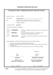
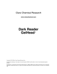
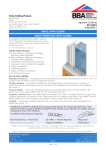
Patent Pending amount of reducing agent required. White screens are supplied to aid visualization during running. See fitting instructions for details. Protocol: Standard Step-1: non-reduced or <10mM BME/DTT 1. Add 5µl Realtime stain to 5µl sample (add up to 10mM BME or DTT if required) 2. User Guide Version 1.2 Heat for 10minutes at 100ᵒC (Quick pulse in a microfuge) Step-2: optional for >10mM BME/DTT 3. Add required amount of reducing agent (0.5µl of 1-2M BME or DTT final concentration 50-100mM) Introduction Realtime stain is a revolutionary dual use protein visualization and sample loading buffer specially formulated for SDS polyacrylamide gel electrophoresis (PAGE). Realtime stain binds specifically to amine groups in proteins offering a unique alternative to the current poststaining techniques. Simply add the Realtime stain to your protein sample (in a 1:1 ratio) and heat then watch your protein visibly running directly down the gel in Realtime. The simple protocol offers you the flexibility to optimize and customize the labelling efficiency with the capability to generate your own pre-stained molecular weight standards. Realtime stain is currently optimized for pure and partially-pure protein samples, recombinant protein fragments, FPLC fractions and antibodies. Storage conditions & shelf life Realtime stain should be stored at room temperature (18-24ᵒC) and used within one year of the production date (see expiry date for details). Solution may solidify and require warming (30-40ᵒC for 3060seconds) prior to use if stored at a lower temperature. Lyophilized Bovine serum albumin (BSA) should be stored at room temperature and used within the expiry date. When resuspended, the BSA (25mg/ml) should be aliquoted and stored at -20ᵒC. Contents 2ml or 0.2ml Realtime stain solution (blue cap) 5mg Bovine serum albumin (red cap) 1x White screen (10x10cm gel) 1x White screen (8x10cm gel) 1x Vivaspin 20, 10,000Da MWCO (2ml Realtime stain only) 1x User Guide 1x Easy Guide Protocol requirements (not supplied) Dry heat block Reducing agent (step-1 and step-2) Microcentrifuge Refrigerated bench top centrifuge (for custom molecular weight standards only) Sample loading buffer (for custom molecular weight standards only) SDS PAGE gel system Procedures Realtime stain is formulated as a 2x solution. Simply add the Realtime stain to your sample in a 1:1 ratio (5 + 5µl is recommended for most applications) then follow the protocol according to the 4. Heat for additional 3minutes at 100ᵒC (Quick pulse in a microfuge) Load and run sample 5. Load 5µl on to SDS PAGE gel 6. Run gel according to manufacturer’s instructions Note: Labelling efficiency can be optimised by varying the time and temperature of ‘step-1’ if required. See ‘performance and & technical data’ for more information. Note: Gels can also be post-stained with Quick Coomassie, if required. Buffer compatibility testing 5mg of BSA is supplied for direct buffer compatibility testing and to serve as a positive control for your SDS PAGE. Some amine containing buffers can affect labelling efficiency (see compatibility table 1 below for further details). If you do not know your buffer composition or your buffer is likely to contain incompatible components then Generon recommends using the following protocol for buffer testing prior to sample use: 1. Resuspend 5mg BSA in 200µl deionised water (final concentration 25mg/ml) (Note: once dissolved, aliquot and store at -20ᵒC for future use) 2. 3. Add 1µl of 25mg/ml BSA to 9µl buffer, and 10µl Realtime stain. Repeat with PBS or 20mM Tris pH 7.5 Heat for 10minutes at 100ᵒC (Quick pulse in a microcentrifuge) 4. Load 4µl on to an SDS-PAGE gel Any incompatible components will significantly inhibit labelling efficiency and reduce BSA band intensity. If present, then we recommend diluting out 2-5 fold with deionised water or a compatible buffer, or remove the incompatible component via buffer exchanging prior to using the Realtime stain. Re-run the protocol using a dilution series if required. Table1. Realtime buffer compatibility. Buffer component Conc. Tris NaCl <50mM 0.5M >100mM BME & DTT <10mM (Step-1) >10mM (Step-2) TCEP <5mM (Step-1) >5mM (Step-2) Imidazole Urea Glycine <50mM 4M 50mM >100mM Compatible? Conc. Compatible? Protocol: Molecular weight standards 1. Add 20µl protein to 20µl Realtime stain, (Add up to 10mM BME or DTT, if required) 2. repeat separately for each individual protein standard Heat for 10minutes at 100ᵒC (Quick pulse in a microcentrifuge) 3. 4. 100 75 50 25 0 0 2 3 4 5 6 1B Labelling efficiency of non-reduced BSA with increasing time and temperature Add sample loading buffer (not supplied) Load 5-10µl on to an SDS PAGE gel For the best results: label each protein individually before combining in equal ratio and removing the unreactive dye. Aim for a final protein concentration, after buffer exchange, of 1-2mg/ml per protein. Note: This protocol is designed for removing unreactive dye from the protein solution, reducing the running dye front, and can be used for small (<10-15kDa) proteins which would normally be masked by the dye front. 100 Band Intensity (%) Heat 5-10µl for 3minutes at 100ᵒC (Quick pulse in a microcentrifuge) 7. 1 Protein load (µg) (Note: once made, aliquot and store at -20ᵒC for future use) 6. Labelling efficiency of non-reduced BSA at 10minutes, 100ᵒC Combine the protein samples together in equal ratio Buffer exchange with 20mM Tris pH 7.5 or PBS pH 7.4 using a Vivaspin 20, 10,000Da MWCO (supplied with 2ml Realtime stain) and a refrigerated bench top centrifuge at 4ᵒC (200 fold or 2x5ml buffer exchanges are required, with a final volume equal to the initial protein start volume: 20µl) 5. 1A Band Intensity (%) Designed for the production of customised pre-stained molecular weight standards and ladders. Labelling efficiency should be tested using the standard protocol above prior to molecular weight standard production. 75 50 25 0 0 1C 10 15 20 25 30 Labelling efficiency of reduced BSA during step-1 labelling 100 Band Intensity (%) Realtime stain has been tested with Tris-Glycine, Tris-HEPES, and BisTris pre-cast and cast SDS polyacrylamide protein gels. We are not currently aware of any issues with other gel types not described above, however, these have not currently been tested using the Realtime stain. 5 Time (minutes) Note: Standards made using this protocol are compatible with all SDS PAGE gel types if compatible sample loading buffers and SDS PAGE gels are used. SDS polyacrylamide gel compatibility 100oC 80oC 60oC BME DTT TCEP 75 50 25 0 0 5 10 15 20 25 30 35 40 45 Reducing agent (mM) Performance & technical Data Labelling efficiency can be optimized for individual proteins by varying the labelling parameters (time, temperature, and reducing agent). Figure 1 shows performance data generated using the supplied BSA control. 1D Labelling efficiency of reduced BSA during step-1 and step-2 labelling (50mM reducing agent) 75 50 25 TCEP step-2 TCEP step-1 DTT step-2 DTT step-1 BME step-2 BME step-1 0 Non-reduced step-2 All samples were prepared in accordance with the protocol and run on NuSep 4-20% Tris-Glycine SDS PAGE pre-cast gels at 170volts (5µg BSA load unless otherwise stated). Each gel was dried and scanned using an over-head light source and the band intensity measured using Image J. (A) Non-reduced labelling efficiency at 10minutes, 100ᵒC. (B) Non-reduced labelling efficiency with increasing time and temperature, recommended 10minute, 100ᵒC labelling highlighted. (C) Effect of reducing agents upon step-1 labelling efficiency at 10minutes, 100ᵒC. (D) Effect of 50mM reducing agent upon step-1 and step-2 labelling efficiency. Non-reduced step-1 Figures 1A-D: Labelling efficiency of the Realtime stain with BSA. Band Intensity (%) 100 50 White screen: Fitting instructions Troubleshooting assistance Optional 8x10cm and 10x10cm gel white screens can be used to aid visibility during electrophoresis. Fitting instructions: Life Technologies XCell SureLock® Mini-Cell: Setup the gel and tank as per manufacturer’s instructions. Then simply slide the appropriately sized white screen (8x10 or 10x10cm gel) in-between the gel cassette and the electrode assembly (1) prior to loading the samples (figure 2). Problem Possible Cause Solution No visible or very faint bands >10mM reducing agent used during step-1 labelling Add reducing agent at step-2 (see protocol and figures 1C and 1D for details) See figure 1C and 1D for performance details. Recommend using BME or DTT at the appropriate step Use a compatible reducing agent such as BME or DTT. See figure 1D for details Increase sample loading volume to 10µl Optimize staining by increasing the labelling time (see figure 1B) Concentrate protein sample prior to adding the Realtime stain Increase the labelling time for proteins with low numbers of Arginine and Lysine residues to compensate Increase the volume of sample for longer labelling times. For example: add 20µl protein sample to 20µl Realtime stain when labelling for 10minutes, 100ᵒC Test for incompatible components using the ‘buffer compatibility test’ Dilute out the incompatible component (2-5 times) or buffer exchange out the component before use Check the temperature of the heat block Use the ‘molecular weight standards’ protocol to remove excess stain Reduce the load volume to <10µl. Increase concentration of the protein if required Non-recoverable effect caused by the heating up of the Realtime stain >5mM TCEP reducing agent used during step-1 labelling Bio-Rad mini-PROTEAN: Place the 8x10cm gel white screen on to the electrode assembly (1), so it sits within the gasket seal. Then lock the gel cassette into place (2) taking care not to disrupt the gasket seal in the process (figure 3). >50mM TCEP reducing agent used during step-2 heating Low protein concentration White screen Electrode assembly 1 Limited availability of amine groups in the protein SDS PAGE Gel Sample dried out during labelling. Can occur when using extended labelling times and small sample volumes Figure 2. Insertion procedure for the 8x10cm or 10x10cm gel white screen into the XCell SureLock® Mini-Cell tank (image courtesy of Life Technologies). Incompatible buffer component. See table 1 for details White screen (8x10cm gel) 1 SDS PAGE Gel Electrode assembly 2 Figure 3. Insertion procedure for the 8x10cm gel white screen into the Bio-Rad mini-PROTEAN tank (image courtesy of Bio-Rad). Unable to view low molecular weight protein (<10-15kDa) Broad dye front Possible heat block failure Protein runs within the dye front Large load volume (>10µl) Continual heating of Realtime stain at high temperatures (100ᵒC) prior to use causes the dye front to broaden Troubleshooting assistance continued … Problem Possible Cause Solution No visible or very faint bands seen with the BSA control during ‘buffer compatibility testing’ Possible heat block failure Excess freezing and thawing of the stock solution Poor band resolution of custom made molecular weight standards Heating of the sample during centrifugation Poor labelling efficiency Check the temperature of the heat block Purchase new BSA control sample. See ‘ordering information’ for further details Purchase new BSA control sample. See ‘ordering information’ for further details. Resuspend in deionised water or compatible buffer (PBS pH 7.4 or 20mM Tris pH 7.5) Repeat using a refrigerated bench top centrifuge at 4ᵒC Optimize labelling efficiency (time and temperature) using the ‘standard’ protocol prior to making the standards Test for incompatible components using the ‘buffer compatibility test’ Warm Realtime stain gently (30-40ᵒC) for a short period and mix well before use. It is not recommended to heat Realtime stain above this prior to use Use standard sample loading buffer and stain with Quick Coomassie Incompatible resuspension buffer Incompatible buffer component Solidified Realtime stain Low temperature storage (2-4ᵒC) Poor definition of lysate/serum bands Not currently optimized for use with complex protein mixtures Ordering Information Product Order number 2ml Realtime stain GEN-RT-STAIN-2000 0.2ml Realtime stain GEN-RT-STAIN-200 White screen (10x10cm gel) GEN-RT-SCREEN-1010 White screen (8x10cm gel) GEN-RT-SCREEN-810 Vivaspin 20, 10,000Da MWCO (12 pack) VS2001 10g Bovine Serum Albumin (>99%) GEN-BSA-10 1g Bovine Serum Albumin (>99%) GEN-BSA-1 Technical Support For the latest technical support information: Go to www.generon.co.uk and search for ‘Realtime stain’ Submit a question to our support team at [email protected] Or contact our telephone sales and support team on +44 (0)1753 866-511 Materials Safety Data Sheet (MSDS) Disclaimer This product is designed for research purposes only. No right to resell this product or any of its components is conveyed. Please contact Generon Ltd for further information. Materials Safety Data Sheets are available from Generon upon request. Certificate of Analysis The Certificate of Analysis provides detailed quality control and product qualification information for each product. Certificates of Analysis are available upon request from Generon by quoting the product catalogue number and lot number printed on the tin. 12 Rawcliffe House, Howarth Road, Maidenhead, Berkshire, SL6 1AP, UK Tel: +44 (0)1753 866-511 Fax: +44 (0)1753 208-899 Email: [email protected] Web: www.generon.co.uk