1
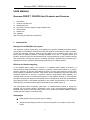
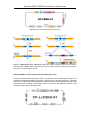
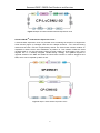
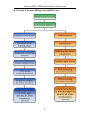
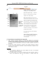
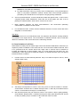
Genome-CRISP™ CRISPR-Cas9 Products and Services User Manual GeneCopoeia, Inc. 9620 Medical Center Drive, #101 Rockville, MD 20850 USA 301-762-0888 866-360-9531 [email protected] www.genecopoeia.com © 2014 GeneCopoeia, Inc. Genome-CRISP™ CRISPR-Cas9 Products and Services USER MANUAL Genome-CRISP™ CRISPR-Cas9 Products and Services I. II. III. IV. V. VI. Introduction Products and Services Related Services Overview of Genome Editing Using CRISPR-Cas9 Critical Steps References VII. Appendix VIII. Licensing and Warranty Statement I. Introduction Background of CRISPR-Cas9 system The clustered, regularly interspaced, short palindromic repeats (CRISPR)-associated protein (Cas) systems are adaptive mechanisms evolved by bacteria and archaea to destroy invading viruses and plasmids. Recently, efficient genome editing by the CRISPR-Cas9 system has been shown in multiple organisms, including zebrafish, mice, rats, C. elegans, plants, and bacteria. Several groups have demonstrated that, compared with zinc finger nucleases (ZFNs) and transcription activator-like effector nucleases (TALENs), CRISPR-Cas9–mediated gene targeting has similar or greater genome editing efficiencies in multiple systems. Efficient and flexible targeting In the CRISPR-Cas9 system, the complex of a CRISPR RNA (crRNA) annealed to a trans-activating crRNA (tracrRNA) is sufficient to guide the Cas9 endonuclease to a specific genomic sequence to execute gene-editing functions, such as gene knockout, knockin (with donor plasmid), modification and more. This system can be simplified by fusing the crRNA and tracrRNA sequences to produce a synthetic chimeric single-guided RNA (sgRNA). The selected target consists of a 20bp DNA sequence in the sgRNA, followed by a trinucleotide (5'-NGG-3') protospacer adjacent motif (PAM) which is recognized by Cas9 and is essential for gene editing functions. The sgRNA hybridizes to the strand complementary to the PAM site. Subsequently, the Cas9 nuclease cleaves both strands of the DNA (Figure 1). This RNA-guided DNA recognition mechanism of CRISPR-Cas9 provides a simple but powerful tool for precise genome engineering. One of the most important advantages of CRISPR-Cas9 systems is that the Cas9 protein can be guided by individual sgRNAs to modify multiple genomic target loci simultaneously. Advantages RNA-guided sequence-specific genome editing Simple and fast design process. No need to reengineer the nuclease for each new target 2 Genome-CRISP™ CRISPR-Cas9 Products and Services Efficient RNA-guided DNA recognition regardless of the methylation state of the target site Flexible and robust multiplexing – edit multiple genome sites simultaneously Figure 1. Illustration of CRISPR-Cas9-mediated genome editing II. Products and Services Genome-CRISPTM Cas9 Nuclease Expression Clone (Cat# CP-C9NU-01) (Figure 2) A Cas9 nuclease expression clone is a premade clone containing the sequence of a mammalian codon-optimized CRISPR-associated (Cas) 9 gene from Streptococcus pyogenes. Co-expression of Cas9 with an sgRNA guides the Cas9 nuclease to create a site-specific double-strand break (DSB) in the host genome. DSB repair mediated by non-homologous end joining (NHEJ) can then reconnect the DNA and induce insertion or deletion mutations at the site of the break. Alternatively, specific sequences, genes, or base pair changes can be introduced into the DSB site by homologous recombination with an exogenous double-stranded donor DNA fragment. 3 Genome-CRISP™ CRISPR-Cas9 Products and Services Figure 2. Map of Cas9 Nuclease Expression Clone Figure 3. sgRNA-guided gene engineering. Left: DSB created by sgRNA-guided Cas9 nuclease is repaired by NHEJ. Right: DSB is repaired by the insertion of GOI & selection markers (or other genetic elements) from a donor plasmid via HR. Genome-CRISPTM Cas9 nuclease lentiviral expression clone A Cas9 nuclease lentiviral expression clone is a premade clone containing the sequence of engineered CRISPR-associated (Cas) gene 9 constructed in a lentiviral backbone. A lentiviral system is very effective at delivering genetic material to whole model organisms and almost all mammalian cells, including non-dividing, inactive or growing, and difficult-to-transfect cells including neuron, primary and stem cells. 4 Genome-CRISP™ CRISPR-Cas9 Products and Services Figure 4: Maps of Cas9 nuclease lentiviral expression clone Genome-CRISPTM Cas9 Nickase Expression Clone A Cas9 nickase expression clone is a premade clone containing the sequence of engineered Cas9 nickase (Figure 4). Wildtype Cas9 has two catalytic domains – one cuts the binding strand and the other cuts the complementary strand. The Cas9 D10A nickase contains an Aspartate to Alanine amino acid substitution at position 10. This mutation makes the Cas9 nuclease able to cut only the binding strand, thereby creating a single-strand “nick” at the targeted site. In a carefully designed “double-nicking” strategy, two sgRNAs targeted to opposite strands of the DNA will enable the Cas9 D10A nickase to create a staggered end DSB, which can be repaired by NHEJ or HR. Figure 5: Maps of Cas9 Nickase Expression Clone 5 Genome-CRISP™ CRISPR-Cas9 Products and Services Figure 6: Illustration of the “double nicking” strategy using the Cas9 D10 nickase mutant. Genome-CRISPTM sgRNA Design and Cloning Services sgRNA clones express a single-stranded chimeric sgRNA. In the presence of the Cas9 endonuclease, an sgRNA guides the Cas9 nuclease to create a DSB at the target site. Multiple sgRNA clones can be co-expressed with one Cas9 clone to enable simultaneous editing of several sites in the genome, offering great efficiency and flexibility. Vector types Vector Promoter sgRNA Cas9 Nuclease Selection Marker/ Reporter Gene Host pCRISPR-SG01 U6 1 Sold separately Hygromycin Mammalian pCRISPR-CG01 U6 1 CMV-driven Cas9 in Neomycin / mCherry Mammalian the same vector pCRISPR-CG02 U6 1 CBh-driven Cas9 in N/A Mammalian Puromycin / Mammalian/ mCherry Lentiviral the same vector pCRISPR-LvSG02 U6 1 Sold separately 6 Genome-CRISP™ CRISPR-Cas9 Products and Services (A) (B) (C) (D) Figure 7. Maps of sgRNA clones 7 Genome-CRISP™ CRISPR-Cas9 Products and Services III. Related Product and Services Services Surrogate reporter assay Validation T7 endonuclease I assay Description Plasmid-level functional validation. Detects activities of genome editing tools by observing the expression level of a surrogate reporter gene. Chromosomal-level functional validation. Detects the presence of indels created by CRISPR-Cas9 mediated NHEJ repair at the specific target site of the chromosome. Customized plasmids designed to specifically transfer your gene of interest, selection marker or other genetic Donor clone Donor clone services (see design and appendix) construction elements into targeted sites through homologous recombination (HR) induced by our CRISPR-Cas9. We offer various donor vector choices with different selection markers and genetic elements built in for your experiment purpose. Stable cell Monoclonal Monoclonal stable cell line with CRISPR-Cas9-mediated line services colony genome modifications. (see appendix) Transgenic mouse services Cell bank Creation of a cell bank of monoclonal stable cell line with CRISPR-Cas9-mediated genome modifications. Transgenic Transgenic mice with CRISPR-Cas9-mediated genome mouse modifications. 8 Genome-CRISP™ CRISPR-Cas9 Products and Services IV. Overview of Genome Editing Using CRISPR-Cas9 9 Genome-CRISP™ CRISPR-Cas9 Products and Services V. Critical Steps A. Plasmid propagation We recommend propagating the plasmids provided before your gene targeting experiment. Plasmids can be transformed using standard conditions suitable in any RecA- and EndAE.coli competent cells. For transformation of CRISPR-Cas9 product plasmids, we suggest plating 50-200µl of transformed cells on fresh LB-Ampicillin plates (50µg/ml). Incubate the plates at 37°C overnight. Inoculate colonies from the transformation and grow them at 37°C overnight in ~200ml of LB media containing 50µg/ml Ampicillin. Use an endotoxin-free plasmid DNA maxiprep kit to extract plasmid DNA after overnight growth. To confirm the integrity of the amplified plasmids, we recommend restriction digestion analysis or direct sequencing. B. CRISPR chromosomal validation CRISPR-modified DNA will have a few bases of sequence inserted or deleted near the Cas9 cut site due to NHEJ exonuclease activity. We recommend using the Surveyor mutation detection kit for standard gel electrophoresis (Transgenomic, cat. no. 706025) for this assay. Alternatives include the Cel1, T7, mung bean and S1 nucleases. For an example, see Figure 5. The Surveyor procedure is carried out according to the manufacturer’s instructions and is described in greater detail in the Surveyor manual. We provide brief details here. 1. 24 or 48 hr post-transfection, collect cells to extract genomic DNA. 2. PCR amplify the region surrounding the sgRNA target site. 3. Check the PCR result by running 5 μl of PCR product on a 2% agarose gel. For all templates, it is important to make sure that there is only a single band corresponding to the intended product for the primer pair. The size of this band should be the same as calculated from the distance between the two primer annealing sites in the genome. CRITICAL STEP: If multiple amplicons are generated from the PCR, redesign the primers and reoptimize the PCR conditions to avoid off-target amplification. In difficult cases in which a single-band product cannot be achieved, it is acceptable to gel-extract the correct-length band before proceeding with heteroduplex reannealing and Surveyor nuclease digestion. 1) 2) DNA heteroduplex formation. At this point, the amplified PCR product includes a mixture of both CRISPR-modified and unmodified genomic DNA. Place 300 ng of the PCR product in a thermocycler tube and perform the cross-hybridization. Surveyor Nuclease S digestion. Treat the cross-hybridized homo- and heteroduplexes with Surveyor Nuclease S to determine TALEN cleavage efficiency. 10 Genome-CRISP™ CRISPR-Cas9 Products and Services (A) (B) Figure 8. Mutagenesis of the human HUWE gene using CRISPR-Cas9. (A) HUWEI sgRNA design. Expected bands of T7 endonuclease I (T7 ENI) digestion are 330bp+190bp; (B) HUWEI-sgRNA/Cas9 clones were transfected into HEK293T cells in a 6-well plate. The medium was changed at 16h post transfection. Cells were harvested at 40h post transfection. Genomic DNA was then extracted for PCR. PCR products were gel purified. 8uL purified product was mixed with 1uL 10x NEB buffer 2, followed by annealing in a PCR machine programmed as follows: 94 °C10min, (93°C-25s, 92°C-25s)x35, 20°C5min, 4°C After annealing, the products were cut with 2U T7 Endonuclease I for 60mins at 37°C, and analyzed by agarose gel electrophoresis. C. Co-transfection (or transfection) into target cells 1. 2. Plate ~100,000 to 300,000 cells/well in a 6-well plate following the recommended conditions for the cell type(s) being transfected. Scale up and down the culture if needed. On the day before transfection, trypsinize and count the cells. The number of cells plated in each well should be determined so that they are 70-80% confluent at the time of transfection. The next day, prepare transfection complexes using suitable transfection reagents according to the manufacturer’s instructions. Leave the transfection complexes on the cells to react for >6 hours. Tech Notes: 1) Since transfection efficiencies vary across different cell lines, we recommend optimizing the input for best results. 2) For optimal results, we recommend complexing DNA with transfection reagent in serum- and antibiotic-free media and cells growing in complete media (e.g. 11 Genome-CRISP™ CRISPR-Cas9 Products and Services DMEM/F12+10% FBS w/o antibiotics). 3) For hard-to-transfect cells (e.g. primary, stem, hematopoietic), it may be advisable to utilize a non-passive transfection method. Please follow recommended guidelines provided by the manufacturer for the specific cell type(s) being transfected. 3. 24 hours post-transfection, remove transfection media and split the cells 1:10 and 1:20 in complete growth media w/antibiotics. Plate cells into 6-well plates and save a set of plate(s) for characterization. Allow cells to recover for 24 hours. 4. Begin antibiotic selection 48 hours post-transfection. We recommend optimizing concentration of antibiotic for best results. 5. For HR-based knockin,. another round of antibiotic selection to enrich clones with donor integration is recommended. Tech Note: Establishing a kill-curve on untransfected cells can determine the effective working antibiotic concentration for a target cell line. The concentration of antibiotic that kills >90% of cells after 48hours of selection is the correct dose for the cells being selected. D. Clonal isolation of cell lines Serial dilution is widely used to isolate single clones with desired modifications, followed by an expansion period to establish a new clonal cell line. Like most clonal isolation methods, there is no guarantee that the colonies arose from single cells. A second round is advised to increase the likelihood of clonal isolation. Also, it is worth noting that cell types can vary substantially in their responses to single-cell isolation, so literature specific to the cell type of interest should be consulted. 1. Fill each well of a sterile 96-well plate with 100 µl of medium except for well A1, which should remain empty. 2. Add 200µl cell suspension to well A1. Mix 100 µl from A1 with the medium in well B1. Avoid bubbles. Continue this 1:2 dilution through column 1. Add 100 µl of medium back to column 1 so that wells A1 through H1 contain 200 µl. Mix cells and transfer 100 µl of cells from column 1 into column 2. Mix by gently pipetting. Avoid bubbles. Repeat these 1:2 dilutions through the entire plate. Bring the final volume to 200 µl by adding 100 µl of medium to all but the last column of wells. 3. 12 Genome-CRISP™ CRISPR-Cas9 Products and Services 4. Incubate plates undisturbed at 37℃. 5. Cells will be observable via microscopy over 3 days and be ready to score in 5-8 days, depending on the growth rate of cells. Mark each well on the cover of the plate indicating which well contains a single colony. These colonies can later be subcultured from the well into larger vessels. Tech Note: 1) Adding 4000 cells in well A1 (2×10⁴ cells/ml) is a good starting concentration. Increase the concentration for more difficult to grow cell lines. 2) If the reporter gene is fluorescent, determine which of these colonies express it. If the reporter gene is not observable you will have to wait until later in the culture process. 3) Label each well with a single colony using a unique identification number and record this number on the plate and in your notebook. E. Validation of CRISPR-Cas9 modified and HR recombinant cells 1. To confirm donor vector integration specifically at a target site, junction-PCR can be performed using PCR primer pairs that flank the 5’ homology arm and 3’ homology arm. 2. Protocol for Junction-PCR 1) Primers should be diluted to 10μM before use. Validation of either the 5’ or 3’ homology arms for donor integration is usually sufficient; however, both arms can be done for additional confirmation. 2) Protocol details for junction-PCR assay: a) Isolate genomic DNA from positive control cells or test sample cells using a suitable genomic DNA miniprep kit. Please follow the protocol recommended by the manufacturer. b) Perform junction-PCR (PCR reaction below) Genomic DNA(60~100ng/µl) CRISPR-Cas9 cut+ positive control donor 1μl Positive control donor only 1μl 10μM 5' (or 3’) PCR Primer Mix 1μl 1μl 5×UltraPFTM Buffer (Mg2+ free) 5μl 5μl 10 mM dNTPs 0.5μl 0.5μl 20mM MgSO4 2.5μl 2.5μl UltraPF(5U/μl) 0.25μl 0.25μl PCR-grade distilled water 14.75μl 14.75μl Total 25μl 25μl Reagent 13 Genome-CRISP™ CRISPR-Cas9 Products and Services 98°C, 5min 98°C, 20sec 55°C, 30sec 35 cycles 72°C, 1min 72°C, 7min Hold at 4~16°C Run the PCR reaction out on the 1% Agarose/EtBr gel in 1X TAE buffer to confirm the junction-PCR result. Sample results for 5’ and 3’ junction-PCR assay depend on design. Tech Note: 1) 2) 3) The 3’ junction PCR band and 5’ junction PCR band may differ in brightness because the amplification efficiency may be different due to the nature of the chromosomal structure, modification and sequence around that region. One positive in junction PCR is sufficient to confirm the integration. Though rare, it is possible that random integration can coexist with site-specific integration. Negative selection can be used to detect coexisting random integration. VI. References 1. Horvath P, Barrangou R (January 2010). "CRISPR/Cas, the immune system of bacteria and archaea". Science 327 (5962): 167–70. 2. Marraffini LA, Sontheimer EJ (February 2010). "CRISPR interference: RNA-directed adaptive immunity in bacteria and archaea". Nat Rev Genet 11 (3): 181–190. 3. Hale CR, Zhao P, Olson S, et al. (November 2009). "RNA-Guided RNA Cleavage by a CRISPR RNA-Cas Protein Complex". Cell 139 (5): 945–56. 4. van der Oost J, Brouns SJ (November 2009). "RNAi: prokaryotes get in on the act". Cell 139 (5): 863–5. doi:10.1016/j.cell.2009.11.018. 5. Jinek, M., Chylinski, K., Fonfara, I., Hauer, M., Doudna, J.A., and Charpentie E. (2012). A programmable dual-RNA-guided DNA endonuclease in adaptiv bacterial immunity. Science 337, 816–821. 6. Jiang, W., Bikard, D., Cox, D., Zhang, F., and Marrafni, L.A. (2013). RNA-guided editing of bacterial genomes using CRISPR-Cas systems. Nat.Biotechnol. 31, 233–239. 7. Hsu, P.D., Scott, D.A.,Weinstein, J.A., Ran, F.A., Konermann, S., Agarwala, V.,Li, Y., Fine, E.J., Wu, X., Shalem, O., et al. (2013). DNA targeting specicity of RNA-guided Cas9 nucleases. Nat. Biotechnol. Published online July 21, 2013. 14 Genome-CRISP™ CRISPR-Cas9 Products and Services VII. Appendix Donor services GeneCopoeia offers customized donor clone design and construction services. Donor clones are customized plasmids designed to specifically transfer your gene of interest, selection marker or other genetic elements into a target site via HR-mediated repair of DSBs induced by site-specific genome editing tools. Donor vectors are available with several options for selection markers and genetic elements to meet your experimental needs. Donor vector types Vector Promoter Reporter Selection Gene Marker LoxP Site pDonor-D01 EFa1 copGFP Puromycin N/A pDonor-D02 CMV copGFP Neomycin N/A pDonor-D03 CMV N/A Neomycin N/A pDonor-D04 CMV N/A Puromycin N/A pDonor-D05 EFa1 N/A Neomycin N/A pDonor-D07 EFa1 copGFP Puromycin/TK Loxp pDonor-D08 CMV copGFP Neomycin/TK Loxp pDonor-D09 EFa1 N/A Puromycin/TK Loxp pDonor-D10 CMV N/A Neomycin/TK Loxp 15 Genome-CRISP™ CRISPR-Cas9 Products and Services Figure 9. Maps of donor vectors 16 Genome-CRISP™ CRISPR-Cas9 Products and Services Stable cell line services GeneCopoeia offers monoclonal stable cell line service with customized CRISPR-Cas9-mediated genome modifications. Cell banking service is also available. 17 Genome-CRISP™ CRISPR-Cas9 Products and Services VIII. Limited Use License and Warranty Limited Use License Following terms and conditions apply to use of the Genome-CRISPTM Product and services (the Product). If the terms and conditions are not acceptable, the Product in its entirety must be returned to GeneCopoeia within 5 calendar days. A limited End-User license is granted to the purchaser of the Product. The Product shall be used by the purchaser for internal research purposes only. The Product is expressly not designed, intended, or warranted for use in humans or for therapeutic or diagnostic use. The Product must not be resold, repackaged or modified for resale, or used to manufacture commercial products or deliver information obtained in service without prior written consent from GeneCopoeia. This Product should be used in accordance with the NIH guidelines developed for recombinant DNA and genetic research. Use of any part of the Product constitutes acceptance of the above terms. Limited Warranty GeneCopoeia warrants that the Product meets the specifications described in the accompanying Product Datasheet. If it is proven to the satisfaction of GeneCopoeia that the Product fails to meet these specifications, GeneCopoeia will replace the Product. In the event a replacement cannot be provided, GeneCopoeia will provide the purchaser with a refund. This limited warranty shall not extend to anyone other than the original purchaser of the Product. Notice of nonconforming products must be made to GeneCopoeia within 30 days of receipt of the Product. GeneCopoeia’s liability is expressly limited to replacement of Product or a refund limited to the actual purchase price. GeneCopoeia’s liability does not extend to any damages arising from use or improper use of the Product, or losses associated with the use of additional materials or reagents. This limited warranty is the sole and exclusive warranty. GeneCopoeia does not provide any other warranties of any kind, expressed or implied, including the merchantability or fitness of the Product for a particular purpose. GeneCopoeia is committed to providing our customers with high-quality products. If you should have any questions or concerns about any GeneCopoeia products, please contact us at 301-762-0888. © 2014 GeneCopoeia, Inc. For Research Use Only. © 2014 GeneCopoeia, Inc. Trademark: Genome-CRISPTM, EndoFectinTM, GeneCopoeiaTM (GeneCopoeia, Inc.) 18 CP-010214