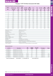
1
BG01V/hOG Cells Variant hESC hOct4-GFP Reporter Cells Catalog no. R7799-105 A10022 Version D 30 September 2008 Corporate Headquarters Invitrogen Corporation 1600 Faraday Avenue Carlsbad, CA 92008 T: 1 760 603 7200 F: 1 760 602 6500 E: [email protected] For country-specific contact information visit our web site at www.invitrogen.com User Manual ii Table of Contents Contents and Storage........................................................................................... iv Accessory Products............................................................................................... v Introduction ............................................................................................................1 Methods............................................................................................... 3 General Information ..............................................................................................3 Preparing a Feeder Cell Layer..............................................................................4 Thawing and Establishing Cells ..........................................................................6 Culturing on MEF Feeders ...................................................................................8 Culturing on STEMPRO® hESC SFM ...................................................................10 Freezing Cells Cultured on MEF Feeders ........................................................16 Freezing Cells Cultured on SFM........................................................................18 Expected Results...................................................................................................20 Troubleshooting ............................................................................... 22 Appendix ........................................................................................... 24 Generating Mitomycin C Treated MEFs ..........................................................24 Detecting Fluorescence........................................................................................25 Technical Support ................................................................................................26 Purchaser Notification.........................................................................................27 References..............................................................................................................30 iii Contents and Storage Shipping and Storage This manual is shipped with BG01V/hOG Cells. BG01V/hOG cells are shipped on dry ice. Upon receipt, store in liquid nitrogen. Contents Storage conditions: Liquid nitrogen Amount supplied: One vial containing ~2 × 10⁶ cells Composition: 1 ml of cells in Freezing medium (see page 10 for composition). Handle as potentially biohazardous material under at least Biosafety Level 1 containment. This product contains Dimethyl Sulfoxide (DMSO), a hazardous material. Review the Material Safety Data Sheet before handling. iv Accessory Products Additional Products For more information about the following products, refer to our Web site (www.invitrogen.com) or call Technical Support (see page 24). Item Quantity Collagenase Type IV DMEM/F12* containing GLUTAMAX™ (2mM) Knockout™ Serum Replacement (KSR) MEM Non-Essential Amino Acids Solution, 10 mM (100X) FGF-Basic (bFGF) STEMPRO® hESC SFM 2-Mercaptoethanol, 1,000X (55 mM) Dulbecco's Modified Eagle Medium (D-MEM) high glucose with L-glutamine and sodium pyruvate Fetal Bovine Serum, ES Cell-Qualified (US) Bovine Albumin Fraction V Solution (7.5%) Geltrex™ DPBS Antibiotic-Antimycotic (100X), liquid Hygromycin B StemPro® EZPassage™ Disposable Stem Cell Passaging Tool StemPro® EZChek™ Human Tri-Lineage Multiplex PCR Kit Water, distilled Mitomycin C Treated MEFs 1g 500 ml 500 ml 100 ml Catalog no. 17104-019 10565-018 10828-028 11140-050 50 μg 1 kit 50 ml 500 ml PHG0026 A1000701 21985 11995-065 500 ml 100 ml 5 ml 500 ml 100 ml 20 ml 10 disposable tools 100 reactions 16141-079 15260-037 12760 14190 15240-062 10687-010 23181-010 23191-050 500 ml 15230-162 Mitomycin C-treated, Hygromycin-resistant MEFs (DR4) are available from ATCC ( Cat. no. SCRC-1045.2). Hygromycinresistant MEFs (DR4) are also available separately from ATCC (Cat. no. SCRC-1045); Mitomycin C is available separately from Sigma, St. Louis (Cat. no. M0503). Continued on next page v Accessory Products, Continued Fetal Bovine Serum, ES Cell-Qualified Invitrogen also provides ES Cell-Qualified Fetal Bovine Serum originating from countries other than the U.S. (go to www.invitrogen.com for information). These can be more appropriate for your situation, and may be used to grow BG01V/hOG Cells. Porcine Skin Gelatin Porcine Skin Gelatin can be obtained from Sigma, St. Louis (Cat no. G1890). vi Introduction Introduction BG01V/hOG human embryonic stem cells (hESCs) are engineered to enable monitoring the pluripotency of hESCs without sacrificing cells. When pluripotent, these cells express Emerald Green Fluorescent Protein (emGFP). Use these cells to assess whether your culture conditions are adequate to keep hESCs undifferentiated, or determine the efficiency of your differentiation assays. Characteristics BG01V/hOG cells have the following characteristics: of BG01V/hOG Prepared from low-passage (passage 17) parental BG01V cells cultured on murine embryonic feeders (MEFs) Cells Pluripotent: can differentiate to representatives of the three primary germ layers Express EmGFP when pluripotent; lose EmGFP expression upon differentiation. BG01V Parental Cell Line BG01V/hOG Cells are derived from the BG01V hESC line (ATCC No. SCRC-2002). BG01V cells in turn are a variant with abnormal karyotype of the wild-type, parental hESC line BG01 (Mitalipova et al., 2003; Plaia et al., 2006). BG01V cell colonies grow on murine embryonic feeders (MEFs) with uniform morphology, and are easy to culture at a predictable growth rate. BG01V cells stain positive for pluripotency markers and alkaline phosphatase activity. BG01V cells are pluripotent and can differentiate to representatives of all three primary germ layers. Continued on next page 1 Introduction, Continued Generation of BG01V/hOG Cells We constructed an integration vector containing EmGFP expressed under the direction of the human Oct4 promoter, and followed by the HSV-TK polyadenylation signal. This plasmid was stably integrated into the genome of BG01V cells. We selected a clone expressing EmGFP, which was tested extensively to make sure it was pluripotent and able to differentiate to representatives of the three primary germ layers The resulting cell line was called BG01V/hOG. Note: Since the integration vector used contains a Hygromycin resistance gene (HygR), BG01V/hOG Cells are resistant to Hygromycin B. If you want to stably integrate more genes in these cells, do not use the Hygromycin resistant gene as your selection marker. GFP Expression Indicates Pluripotency 2 Engineered cells will express EmGFP under the appropriate conditions when the Oct4 promoter is active. This promoter is only active when hESCs are in the pluripotent state and therefore acts as a sensitive indicator of differentiation (Pan et al., 2002). This tool can be used as a reporter of the cell culture’s response to external stimuli including media composition and stress. Methods General Information General Cell Handling Important Follow the general guidelines below to grow and maintain BG01V/hOG Cells. All solutions and equipment that come in contact with the cells must be sterile. Always use proper sterile technique and work in a laminar flow hood. BG01V/hOG Cells may be cultured on a feeder layer of mitotically inactivated mouse embryonic fibroblasts (MEFs) or may be thawed on MEFs and then transitioned to MEF-conditioned media (MEF-CM) or STEMPRO® hESC SFM. Make sure to start preparing the feeder layer two days before thawing BG01V/hOG Cells. Before starting experiments, be sure to have cells established (at least 5 passages) and also have some frozen stocks on hand. We recommend using earlypassage cells for your experiments (below 30 passages). Upon receipt of the cells from Invitrogen, grow and freeze multiple vials of the BG01V/hOG cells to ensure that you have an adequate supply of early-passage cells. For general maintenance of cells, pass BG01V/hOG Cells before colonies start contacting each other. When thawing or subculturing cells, transfer cells into pre-warmed medium. 10 ml/L of Antibiotic-Antimycotic containing penicillin, streptomycin, and amphotericin B may be used if required (see page v for ordering information). It is very important to strictly follow the guidelines for culturing BG01V/hOG Cells in this manual to keep them undifferentiated. As with other human cell lines, when working with BG01V/hOG cells, handle as potentially biohazardous material under at least Biosafety Level 1 containment. 3 Preparing a Feeder Cell Layer Introduction BG01V/hOG cells are frozen from cultures grown on mouse embryonic fibroblast (MEF) feeder cells, and should be thawed on feeders. Follow the protocol below to prepare the feeder-layer matrix. Use mitotically inactivated MEFs to prevent overgrowth of the hESCs. Both Mitomycin C and irradiation methods can be used to mitotically inactivate your MEFs. Materials Needed Have the following reagents on hand before beginning (see page v for ordering information): Mitotically inactivated, Hygromycin resistant MEFs. Order from ATCC (SCRC-1045.2), or generate them as described in Generating Mitomycin C Treated MEFs (page 24). Dulbecco's Modified Eagle Medium (D-MEM) high glucose with L-glutamine and sodium pyruvate. Fetal Bovine Serum, ES Cell-Qualified. MEM Non-Essential Amino Acids Solution 10 mM (100X) (NEAA). 2-Mercaptoethanol, 1,000X ™ DMEM/F12 with GLUTAMAX (2 mM) Knockout™ Serum Replacement (KSR) bFGF. Reconstitute lyophilized human bFGF in sterile, DMEM/F12 containing 0.1% BSA to 10 μg/ml. Divide stock solution into working aliquots and store at ≤-20°C. Porcine skin gelatin. Prepare 0.1% (w/v) porcine skin gelatin (Sigma Cat no. G1890) in sterile, distilled water, and sterilize by filtration using a 0.2 μm filter. Store up to 1 year at 4°C. 37°C incubator with a humidified atmosphere of 5% CO2 Continued on next page 4 Preparing a Feeder Cell Layer, Continued Preparing MEF Medium To prepare 500 ml MEF medium, mix the following reagents: Preparing hESC Medium To prepare 100 ml hESC Medium, mix following reagents: Preparing Gelatin Coated Plates Coat plates for 20–60 minutes at room temperature with 0.1% gelatin in dH2O. Plating Feeder Layer 1. Two days before hESC coculture, plate 30,000/cm2 of mitotically inactivated mouse embryonic fibroblasts on a 0.1% gelatin-coated culture plate in MEF medium. 2. One day before hESC coculture, replace medium with hESC Medium 3. Next day, the feeder layer is ready to be seeded with BG01V/hOG Cells in fresh hESC Medium. Volume 445 ml 50 ml 5 ml 500 μl Reagent Final Concentration DMEM 1x FBS 10% NEAA (10 mM) 0.1 mM 55 μM 2-Mercaptoethanol, 1,000X (55 mM) Filter through 0.22 μM filtration unit. Pre-heat the medium to 37°C before use. Volume 79 ml Reagent Final Concentration 1x DMEM/F12 with GLUTAMAX™ 20% 20 ml Knockout™ Serum Replacement (KSR) 1 ml NEAA (10 mM) 0.1 mM 100 μl 55 μM 2-Mercaptoethanol, 1,000X (55 mM) 40 μl bFGF (10 μg/ml) 4 ng/ml If stored at 4°C, hESC Medium can be kept for up to 1 week. Pre-heat the medium to 37°C before use. 5 Thawing and Establishing Cells Introduction BG01V/hOG cells are supplied in a vial containing 1 ml of cells at 2 × 10⁶ viable cells/ml in freezing medium. They are frozen from cultures grown on MEF feeder cells, and must be thawed on MEF feeders. Follow the protocol below to thaw BG01V/hOG Cells directly into hESC Medium in a 35mm dish. Materials Needed You will need to have the following reagents on hand before beginning (see page v for ordering information): Thawing Procedure BG01V/hOG cells (store frozen cells in liquid nitrogen until ready to use) hESC Medium (see page 5 for composition); pre-warm to 37°C before use) Feeder layer plates with mitotically inactivated MEFs – prepare at least two days in advance (see page 4) Disposable, sterile 50 ml tubes. 37°C incubator with humidified atmosphere of 5% CO2 Store frozen cells in liquid nitrogen until ready to use. To thaw and establish BG01V/hOG Cells: 1. Remove the cryovial of cells from the liquid nitrogen and thaw quickly in a 37°C water bath (to prevent crystal formation). 2. When thawed, immediately transfer cells into 50-ml tube and add warm hESC Medium dropwise up to 10 ml. 3. Spin cells down for 4 minutes at 200 × g. 4. Aspirate supernatant 5. Resuspend cells in hESC Medium (2 ml for a 35-mm dish) 6. Aspirate feeder layer plates, and plate resuspended BG01V/hOG Cells on the prepared MEFs. 7. Grow cells in a 37°C incubator with a humidified atmosphere of 5% CO2. Change the medium every day. Following thawing, you can continue to culture cells on MEF feeders as described starting on page 8, or you can transition the cells into StemPro® hESC SFM as described starting on page 10. Continued on next page 6 Thawing and Establishing Cells, Continued Judging Thawed Cells Observe colonies recovered at day 5 after thawing, to assess growth rate and differentiation state, keeping the following in mind: Cells should be undifferentiated. They should express EmGFP (if you are unfamiliar with fluorescence microscopy, see page 25), and grow as colonies (see image below for examples of undifferentiated BG01V/hOG Cells). If not, refer to the Troubleshooting section (page 22). Before colonies start contacting each other, they should be passed (see next page). 7 Culturing on MEF Feeders Introduction Important Materials Needed Important Collagenase Preparation Follow the protocol below to culture BG01V/hOG Cells on feeder layer plates. For culturing cells in serum-free medium without feeders, see page 10. Before starting experiments, we recommend that you first prepare ample cell stocks, as described in Freezing Cells (page 16). You will need to have the following reagents on hand before beginning (see page v for ordering information): Plates with BG01V/hOG Cells hESC Medium (see page 5 for composition); pre-warm to 37°C before use) DMEM/F12 with GLUTAMAX™ Feeder layer plates with mitotically inactivated MEFs – prepare at least two days in advance (see page 4) Collagenase Type IV Hygromycin B Disposable, sterile 15-ml tubes. 37°C incubator with humidified atmosphere of 5% CO2 BG01V/hOG Cells should be cultured in the presence of 50 μg/ml Hygromycin B, on order to prevent losing the GFP expression cassette during prolonged culturing. Prepare 1-mg/ml aliquots of collagenase IV in DMEM/F-12. Filter to sterilize and freeze at –20°C. Continued on next page 8 Culturing on MEF Feeders, Continued Passaging Cells 1. Aspirate culture medium and add 1 ml of 1 mg/ml collagenase solution for every 10 cm2 of culture vessel surface area. (Alternatively, use the StemPro® EZPassage™ Disposable Stem Cell Passaging Tool to cut the cell colonies into pieces; follow the protocol provided with the tool and then proceed to step 4.) 2. Incubate in a 37°C incubator until the edge of colonies curl up (usually less than an hour). 3. Aspirate collagenase solution. 4. Add hESC Medium or 0.1% BSA in DMEM/F12. 5. Gently scrape dish using a 5-ml serological pipette and transfer clumps into a 15-ml tube. Do not make the clumps too small; there should be >100 cells per clump. See below for an example of acceptable clumps. 6. Spin cells down for 2 minutes at 200 × g at room temperature. 7. Gently aspirate media and resuspend the BG01V/hOG Cells in hESC Medium. 8. Aspirate feeder layer plates, and plate resuspended BG01V/hOG Cells on the prepared MEFs (passage ratio 1:3 or 1:4). 9. Add a final concentration of 50 μg/ml Hygromycin B (1:1,000 dilution of 50 mg/ml Hygromycin B stock). 10. Grow cells in a 37°C incubator with a humidified atmosphere of 5% CO2. Change the medium everyday. Feed cells every day and passage by the above protocol whenever required (before colonies start contacting each other; typically every 4–7 days). 9 Culturing on STEMPRO® hESC SFM Introduction STEMPRO® hESC SFM allows you to culture BG01V/hOG Cells in a serum-free medium (SFM) without feeder cells. Follow the protocol in this section to culture BG01V/hOG Cells using STEMPRO® hESC SFM. BGO1V/hOG Cells are initially frozen down from cells grown on MEF feeders and should be thawed on feeders as described on pages 6–7 before transferring into MEFconditioned medium (MEF-CM) and then into STEMPRO® hESC SFM, as described in this section. Frozen stocks may then be prepared from the cells grown on STEMPRO® hESC SFM. Important Before starting experiments, we recommend that you first prepare ample cell stocks, as described in Freezing Cells (page 10). Features of the STEMPRO® hESC SFM has been extensively tested and proven to have the following characteristics: Medium Supports hESC growth for up to 80 passages, maintaining the ability of hESCs to differentiate into all three germ line lineages without any signs of karyotypical abnormalities. Maintains pluripotency in multiple hESC lines, including BG01V cells. Supports scale-up production of hESCs to over 1 × 10⁹ cells while maintaining pluripotency. No need to maintain feeder cells or produce feederconditioned medium. More reproducible results due to steady growth factor levels. Continued on next page 10 Culturing on STEMPRO® hESC SFM, Continued Guidelines for SFM Culture To prevent differentiation and slow growth of BGO1V/hOG cells grown in STEMPRO® hESC SFM, follow these guidelines: Starter culture: This must be a high-quality culture, with a high density of cells, and primarily undifferentiated. The starter culture should be cells maintained on Geltrex™ in Mouse Embryonic Fibroblast-Conditioned Medium (MEF-CM). See the protocol in this section for transferring cells from MEF feeders to MEF-CM and then into STEMPRO® hESC SFM. Passaging: It is critical to achieve high plating/survival of colony pieces. The pieces must be a bit smaller than typical collagenase passaging on Geltrex™/ MEF-CM. Some cell death at passaging is normal, but wide-scale cell death (i.e., <20% survival) indicates poor passaging. Timing of passaging. Critical: the cultures need to grow to near-confluence, i.e., a day or two after the colonies are just touching, cultures should be harvested. This usually results in a cell density of 2.5 to 4 × 10⁵ cells/cm2 at time of harvest. Do not over-expose cells to collagenase; we recommend 3 minutes at most, even with lower amounts of collagenase. Density: The cultures must be maintained at a high density (200+ colonies in a 60-mm dish). hESCs grown in culture are always under selection pressure of proliferation vs. differentiation. The cultures should be fed every day; do not exhaust medium by not feeding. Scrape clearly differentiated areas out with a 21½-gauge needle. Continued on next page 11 Culturing on STEMPRO® hESC SFM, Continued Materials Needed You will need to have the following materials on hand before beginning (see page v for ordering information): STEMPRO® hESC SFM, which includes: DMEM/F-12 with GLUTAMAX™ STEMPRO® hESC Supplement Bovine Serum Albumin 25% (BSA) FGF-basic, 10 μg/ml, prepared as described below Collagenase Type IV, prepared as described on next page Geltrex™ DPBS 2-Mercaptoethanol Hygromycin B Culture dishes (60-mm dishes recommended) An incubator at 37°C, humidified atmosphere of 5% CO2 in air Preparing FGF-basic Prepare 10 μg/ml FGF-basic in DMEM/F-12 with 0.1% BSA. Aliquot 80 μl per tube and freeze at –20°C. Preparing Geltrex™ Aliquots Thaw the Geltrex™ bottle at 2–8°C overnight and prepare 1-ml aliquots of Geltrex™ in 50-ml conical tubes. Store the tubes at –20°C. Coating Plates with Geltrex™ 1. Thaw a 1-ml tube of Geltrex™ at 2–8°C. 2. Remove DMEM/F-12 from 2–8°C storage and add 29 ml of cold DMEM/F-12 to the 1 ml of Geltrex™. Mix gently. 3. Cover the whole surface of each culture plate with the Geltrex™ solution (1 ml for a 35-mm dish, 1.5 ml for a 60-mm dish). 4. Seal each dish with parafilm to prevent drying, and incubate 1 hour at 37°C. 5. Transfer each dish to a laminar flow hood and allow it to equilibrate to room temperature (about 1 hour) before using. The Geltrex™-coated dish may be stored at 2–8°C for up to 1 week. Continued on next page 12 Culturing on STEMPRO® hESC SFM, Continued Prepare 1-mg/ml and 10-mg/ml aliquots of collagenase IV Preparing Collagenase IV in DMEM/F-12. Filter to sterilize and freeze at –20°C. Preparing MEFConditioned Medium (MEF-CM) Preparing Complete STEMPRO® hESC SFM 1. Prepare mitotically inactive MEF cells as described on pages 4–5. 2. Change the MEF medium to hESC medium (prepared on as page 5) after a 24-hour incubation 3. Collect medium from the cells every 24 hours and supplement with FGF-basic (prepared as described on the previous page) at a final concentration of 4 ng/ml before using. Thaw STEMPRO® hESC Supplement in 37°C water bath (minimize dwell time), and prepare according to the table below: Component DMEM/F-12 with GLUTAMAX™ (1X) STEMPRO® hESC SFM Growth Supplement (50X) 25% BSA FGF-basic (10 μg/ml) 2-Mercaptoethanol (55 mM) Final conc. For 500 ml For 100 ml 1X 454 ml 90.8 ml 1X 10 ml 2 ml 1.8% 8 ng/ml 0.1 mM 36 ml 400 μl 909 μl 7.2 ml 80 μl 182 μl Storing Complete Medium Store complete STEMPRO® hESC SFM at 2–8°C in the dark for up to 7 days. Add 2-Mercaptoethanol daily during storage, at volumes listed in the table above. Preparing Wash Medium For the wash medium, prepare 0.1% BSA in DMEM/F-12 with GLUTAMAX™ (use the 25% BSA provided in the kit). Important BG01V/hOG cells should be cultured in the presence of 50 μg/ml Hygromycin B to prevent losing the GFP expression cassette during prolonged culturing. Continued on next page 13 Culturing on STEMPRO® hESC SFM, Continued Transferring Cells into STEMPRO® hESC SFM After thawing and establishing BGO1V/hOG cells on MEF feeders as described on pages 6–7, use the following procedure to transfer the cells into STEMPRO® hESC SFM. 1. Aspirate medium from the cells and add 1 ml of 1 mg/ml collagenase solution for every 10 cm2 of culture vessel surface area. 2. Incubate in a 37°C incubator until the edge of colonies curl up (usually it is less than an hour to see this curling). 3. Aspirate collagenase solution and add MEF-CM supplemented with FGF-basic, as described on page 13. 4. Gently scrape the dish using a 5-ml serological pipette and transfer clumps into a 15-ml tube. Try not to make the clumps too small; there should be > 100 cells per clumps. 5. Remove the Geltrex™ from a Geltrex™-coated plate by tipping the plate slightly and aspirating the solution. Wash the plate once with DMEM/F12. Take care to avoid drying of the plate surface before plating. 6. Seed the cells onto the plate. 7. Add a final concentration of 50 μg/ml Hygromycin B (1:1,000 dilution of 50 mg/ml Hygromycin B stock). 8. Place the plate with the cells in the incubator. Shake the plate gently to evenly distribute the cells. 9. The next day, feed the cells once more with MEF-CM supplemented with FGF-basic. 10. The following day (Day 3), feed the cells with an equivalent amount of complete STEMPRO® hESC SFM and a final concentration of 50 μg/ml Hygromycin B. 11. Thereafter, continue feeding cells daily with complete SFM and 50 μg/ml Hygromycin B. Continued on next page 14 Culturing on STEMPRO® hESC SFM, Continued Passaging Cells 1. In a 37°C water bath, warm appropriate amounts of 10-mg/ml collagenase solution, complete STEMPRO® hESC SFM, and wash medium (prepared as described on page 13). Minimize dwell time. 2. Set up the plate with BGO1V/hOG cells on a dissecting microscope in a biosafety cabinet or laminar flow to comfortably observe colonies. 3. Cut out and remove any overtly differentiated colonies with a 21½-gauge needle. 4. Aspirate the medium and gently add 1–2 ml of collagenase solution. (Alternatively, use the StemPro® EZPassage™ Disposable Stem Cell Passaging Tool to cut the cell colonies into pieces; follow the protocol provided with the tool and then proceed to step 7.) 5. Leave for 3 minutes to dislodge cells from the substrate. 6. Remove collagenase and rinse with DPBS 7. Add 3 ml of wash medium ( 0.1% BSA in DMEM/F-12 with GLUTAMAX™; see page 13). 8. Gently scrape the dish using a sterile 1000-μl pipette tip. 9. Gently transfer the cell clumps using a 5-ml pipette and place into a 15-ml tube. 10. Wash plate with 3 ml of wash medium and add to the tube. 11. Spin cells at 200 × g for 2 minutes at room temperature. 12. Gently aspirate the media and flick the tube to loosen cells. 13. Gently resuspend the cells in warm complete STEMPRO® hESC SFM using a 1-ml or 5-ml serological pipette. 14. Remove the Geltrex™ from a Geltrex™-coated plate by tipping the plate slightly and aspirating the solution. Immediately plate the cells. Do not allow the surface to dry out before plating. 15. Mix plates gently to evenly spread out clumps and place the plate in a 37°C incubator at with 5% CO2 in air. 16. Each day, gently change the media to remove excess cells and provide fresh nutrients. 17. Observe cells every day and passage by the above protocol whenever required (about every 5–7 days). 15 Freezing Cells Cultured on MEF Feeders Introduction When freezing BG01V/hOG Cells that are cultured on MEF feeder cells, we recommend the following: Freeze cells at a density of 2.5 × 10⁶ viable cells/ml For every 20 cm2 of cells (one 60-mm dish), prepare 1 ml of MEF Freezing Medium A and 1 ml of MEF Freezing Medium B (see below) Bring BG01V/hOG Cells into freezing medium in two steps, as described in this section Guidelines for preparing freezing medium and freezing cells are provided in this section. Materials Needed Preparing Freezing Medium You will need to have the following reagents on hand before beginning (see page v for ordering information): Plates with BG01V/hOG Cells on MEF feeders hESC Medium (see page 5 for composition) Collagenase Type IV working solution (1 mg/ml) in DMEM/F12 (see page 8) Fetal Bovine Serum, ES Cell-Qualified DMSO (use a bottle set aside for cell culture; open only in a laminar flow hood) Disposable, sterile 15-ml conical tubes. Sterile freezing vials Prepare Freezing Medium A and B immediately before use. Discard any unused medium. 1. In a sterile 15-ml tube, mix together the following for every 1 ml of Freezing Medium A needed. hESC Medium Fetal Bovine Serum, ES Cell-Qualified 0.5 ml 0.5 ml 2. In another sterile 15-ml tube, mix together the following for every 1 ml of Freezing Medium B needed: hESC Medium DMSO 0.8 ml 0.2 ml 3. Place tube with Freezing Medium B on ice and leave Freezing Medium A at room temperature. Continued on next page 16 Freezing Cells Cultured on MEF Feeders, Continued Freezing Cells Cultured on MEF Feeders 1. Aspirate culture medium from the cells and add 1 ml of 1 mg/ml collagenase solution for every 10 cm2 of culture vessel surface area. (Alternatively, use the StemPro® EZPassage™ Disposable Stem Cell Passaging Tool to cut the cell colonies into pieces; follow the protocol provided with the tool and then proceed to step 4.) 2. Incubate in a 37°C incubator until the edge of colonies curl up (usually less than an hour). 3. Aspirate collagenase solution 4. Add hESC Medium (see page 5) or 0.1% BSA in DMEM/F12. 5. Gently scrape dish using 5 ml serological pipette and transfer clumps into a 15-ml tube. Try not to make the clumps too small; there should be > 100 cells per clumps (see page 9 for an example). 6. Spin cells down for 2 minutes at 200 × g at room temperature. 7. Gently aspirate media and resuspend BG01V/hOG cells in Freezing Medium A (e.g., resuspend cells from one 60-mm dish in 1 ml of freezing medium). 8. Add the same volume of Freezing Medium B to cells in a dropwise manner, swirling the tube after each drop. 9. Resuspend the cells by gently pipetting 2–3 times. Aliquot 1 ml of the cell suspension to each freezing vial and store at –80°C overnight in isopropanol chamber. 10. Transfer frozen vials to liquid nitrogen tank for long-term storage. Note: You may check the viability and recovery of frozen cells 24 hours after storing cryovials in liquid nitrogen. 17 Freezing Cells Cultured on SFM Introduction When freezing BG01V/hOG Cells cultured on STEMPRO™ hESC SFM, we recommend the following: Freeze cells at a density of 2.5 × 10⁶ viable cells/ml. For every 20 cm2 of cells (one 60-mm dish), prepare 0.5 ml of SFM Freezing Medium 1 and 0.5 ml of SFM Freezing Medium 2. Bring BG01V/hOG Cells into freezing medium in two steps, as described in this section. Guidelines for preparing freezing medium and freezing cells are provided in this section. Materials Needed You will need to have the following reagents on hand before beginning (see page v for ordering information): Plates with BG01V/hOG Cells in SFM Wash Medium (see page 13) DMEM/F-12 with GLUTAMAX™ Preparing SFM Freezing Medium Collagenase Type IV working solution (10 mg/ml) in DMEM/F12 ™ Knockout Serum Replacement (KSR) DMSO (use a bottle set aside for cell culture; open only in a laminar flow hood) Disposable, sterile 15-ml conical tubes. Sterile freezing vials Prepare SFM Freezing Medium 1 and 2 immediately before use. Discard any unused medium. 1. In a sterile 15-ml tube, mix together the following for every 0.5 ml of SFM Freezing Medium 1 needed: DMEM/F12 with GLUTAMAX™ Knockout™ Serum Replacement (KSR) 0.5 ml 0.5 ml 2. In another sterile 15-ml tube, mix together the following for every 0.5 ml of SFM Freezing Medium 2 needed: DMEM/F12 with GLUTAMAX™ DMSO 0.8 ml 0.2 ml 3. Place tube with SFM Freezing Medium 2 on ice and leave SFM Freezing Medium 1 at room temperature. Continued on next page 18 Freezing Cells Cultured on SFM, Continued Freezing Cells 1. Cultured on STEMPRO® hESC SFM Aspirate serum-free culture medium from the cells and gently add 1–2 ml of 10 mg/ml collagenase solution. (Alternatively, use the StemPro® EZPassage™ Disposable Stem Cell Passaging Tool to cut the cell colonies into pieces; follow the protocol provided with the tool and then proceed to step 4.) 2. Leave for 3 minutes to dislodge cell colonies from the substrate. 3. Remove collagenase and rinse with DPBS. 4. Add 3 ml of wash medium ( 0.1% BSA in DMEM/F-12 with GLUTAMAX™; see page 13). 5. Gently scrape the dish using a sterile 1000-μl pipette tip. 6. Gently transfer the cell clumps using a 5-ml pipette and place into a 15-ml tube. 7. Wash plate with 3 ml of wash medium and add to the tube. 8. Spin cells down for 2 minutes at 200 × g at room temperature. 9. Gently aspirate media and resuspend the BG01V/hOG cells in SFM Freezing Medium 1 at room temperature (use 0.5 ml of Freezing Medium 1 for one 60-mm dish). 10. Add the same volume of cold SFM Freezing Medium 2 to cells in a dropwise manner, swirling the tube after each drop. 11. Resuspend the cells by gently pipetting 2–3 times. Aliquot 1 ml of the cell suspension to each freezing vial and store at –80°C overnight in isopropanol chamber. 12. Transfer frozen vials to liquid nitrogen tank for long-term storage. Note: You may check the viability and recovery of frozen cells 24 hours after storing cryovials in liquid nitrogen. 19 Expected Results Introduction Pluripotent BG01V/hOG Cells express EmGFP, but upon differentiation, these cells lose EmGFP expression. In this section, some typical examples are shown to help you interpret your experiments. Note: Suggestions for differentiation protocols are available from www.invitrogen.com/stemcells; click on the section protocols. FACS Analysis BG01V/hOG human embryonic stem cells differentiate when induced to form embryoid bodies. Embryoid bodyderived BG01V/hOG Cells hardly contain any EmGFP positive cells, as determined by FACS analysis. Continued on next page 20 Expected Results, Continued Fluorescence Microscopy EmGFP expression is lost upon embryoid body induced differentiation of BG01V/hOG Cells, as seen in the green fluorescence image below. Note: If you are unfamiliar with fluorescence microscopy, see page 25. 21 Troubleshooting Culturing The table below lists some potential problems and solutions that help you troubleshoot your cell culture problems. Cells Problem No viable cells after thawing stock Cause Solution Stock not stored correctly Order new stock and store in liquid nitrogen. Keep in liquid nitrogen until thawing. Home-made stock not viable Freeze cells at a density of 2.5 × 10⁶ viable cells/ml. Use low-passage cells to make your own stocks. Follow the freezing procedure for your type of cell culture (starting on page 16) exactly. Slow freezing and fast thawing are key. Add the cold freezing medium in a dropwise manner (slowly), swirling the tube after each drop. At the time of thawing, thaw quickly and do not expose vial to the air but quickly change from nitrogen tank to 37°C water bath. Obtain new BG01V/hOG Cells. MEFs overgrow plate Thawing medium not correct Use specified medium. Cells too diluted Generally, we recommend thawing one vial in a 35-mm dish. If you need to concentrate cells, spin down the culture for 4 minutes at 200 × g at room temperature and dilute the cells at higher density. MEFs sub optimal and do not support recovery of BG01V/hOG Cells Purchase (see page v) or make (see page 24) a new batch of mitotically inactivated MEFs. MEFs not inactivated Inactivate mitosis in MEFs as described on page 24, or purchase inactivated MEFs (see page v) Continued on next page 22 Troubleshooting, Continued Culturing Cells The table below lists some potential problems and solutions that help you troubleshoot your cell culture problems. Problem Cause Cells grow slowly Cells differentiated No fluorescence signal detected Solution Growth medium not correct Use correct growth medium. bFGF inactive bFGF is not stable when frequently warmed and cooled Add bFGF to medium just before use, or store medium with bFGF in aliquots at –20°C. Cells too old Use healthy BG01V/hOG cells, under passage 30; do not overgrow. Cells too diluted Spin down cells for 4 minutes 200 × g at room temperature; aspirate media and dilute cells at higher density Clump size is to small and differentiated Be gentle at time of passage so the clumps of cells don’t get too small. Mycoplasma contamination Discard cells, media and reagents, and use early stock of cells with fresh media and reagents Cells not thawed and established on feeder layers Thaw and culture a fresh vial of BG01V/hOG cells. Make sure to thaw on feeder layers as described on page 4. Suboptimal quality of feeder layer. Check the concentration of feeder cells used. Purchase (see page v) or make (see page 24) new batch of mitotically inactivated MEFs. if necessary. Use Hygromycin resistant MEFs. Culture conditions not correct Thaw and culture fresh vial of BG01V/hOG Cells. Follow thawing instructions (page 6) and subculture procedures (page 8) exactly. Incorrect filters used to detect fluorescence Be sure to use the recommended filter sets for detection of fluorescence (see page 25). Be sure to use an inverted fluorescence microscope for analysis. Cells lost GFP expression cassette Thaw and culture fresh vial of BG01V/hOG Cells. Culture cells in presence of 50 μg/ml Hygromycin B. Cells differentiated See points above 23 Appendix Generating Mitomycin C Treated MEFs Mitomycin C is highly toxic. Read and understand the MSDS and handle accordingly. Preparing Gelatin Coated Plates Prepare 0.1% (w/v) porcine skin gelatin (Sigma Cat no. G1890) in sterile, distilled water, and sterilize by filtration using a 0.2 μm filter. Store up to 1 year at 4°C. Coat plates for 20 to 60 minutes at room temperature with 0.1% gelatin in dH2O. Preparing Mitomycin C Prepare 10 μg/ml mitomycin C in MEF medium; filter sterilize and store at -20C until use. Good for 2 weeks at 4C. Obtaining MEFs Obtain Hygromycin resistant MEFs (DR4) from ATCC (Cat. no. SCRC-1045). Mitomycin C Treatment Use the procedure below to generate mitotically inactivated MEFs (DR4 strain): 1. Culture MEFs in MEF medium (see page 5) 2. Inactivate by treating MEFs with 10 μg/ml mitomycin C for 2 to 3 hours at 37°C. 3. Wash cells four times with Dulbecco's PhosphateBuffered Saline (D-PBS) (Cat. no. 14190-144) 4. Trypsinize cells with 0.05% Trypsin-EDTA (Cat no. 25300-054) 5. Plate MEFs at a density of 3 x 10⁴ cells / cm2 of culture surface area in MEF medium (see page 5) with 2.5 ml per well of a gelatin-coated 6-well dish. 6. Freeze the cells for later use, or use within 2 to 5 days after plating for hESC cell culture. The medium should be changed every other day if they are not used immediately. 24 Detecting Fluorescence Introduction You may detect EmGFP protein expression directly in undifferentiated BG01V/hOG Cells by fluorescence microscopy or other methods that use light excitation and detection of emission. See below for recommended fluorescence microscopy filter sets. Filters for Use with EmGFP The EmGFP can be detected with standard FITC filter sets. However, for optimal detection of the fluorescence signal, you may use a filter set which is optimized for detection within the excitation and emission ranges for the fluorescent protein such as the Omega XF100 filter set for fluorescence microscopy. The spectral characteristics of EmGFP are listed in the table below: Fluorescent Protein EmGFP Excitation (nm) 487 Emission (nm) 509 For information on obtaining these filter sets, contact Omega Optical, Inc. (www.omegafilters.com) or Chroma Technology Corporation (www.chroma.com). Fluorescence Microscope You may view the fluorescence signal of EmGFP in cells using an inverted fluorescence microscope with FITC filter or Omega XF100 filter (available from www.omegafilters.com ) for viewing cells in culture or a flow cytometry system. Color Camera If desired, you may use a color camera that is compatible with the microscope to photograph the cells. We recommend using a digital camera or high sensitivity film, such as 400 ASA or greater. What You Should See See the Expected Results Section, page 21. 25 Technical Support Web Resources Contact Us Visit the Invitrogen Web site at www.invitrogen.com for: Technical resources, including manuals, vector maps and sequences, application notes, MSDSs, FAQs, formulations, citations, handbooks, etc. Complete Technical Support contact information Access to the Invitrogen Online Catalog Additional product information and special offers For more information or technical assistance, call, write, fax, or email. Additional international offices are listed on our Web page (www.invitrogen.com). Corporate Headquarters: Invitrogen Corporation 5791 Van Allen Way Carlsbad, CA 92008 USA Tel: 1 760 603 7200 Tel (Toll Free): 1 800 955 6288 Fax: 1 760 602 6500 E-mail: [email protected] Japanese Headquarters: Invitrogen Japan LOOP-X Bldg. 6F 3-9-15, Kaigan Minato-ku, Tokyo 108-0022 Tel: 81 3 5730 6509 Fax: 81 3 5730 6519 E-mail: [email protected] European Headquarters: Invitrogen Ltd Inchinnan Business Park 3 Fountain Drive Paisley PA4 9RF, UK Tel: 44 (0) 141 814 6100 Tech Fax: 44 (0) 141 814 6117 E-mail: [email protected] Material Safety Data Sheets (MSDSs) MSDSs (Material Safety Data Sheets) are available on our website at www.invitrogen.com/msds. Certificate of Analysis The Certificate of Analysis provides detailed quality control information for each product. Certificates of Analysis are available on our website. Go to www.invitrogen.com/support and search for the Certificate of Analysis by product lot number, which is printed on the box. 26 Purchaser Notification Information for European Customers BG01V/hOG Cells (variant hESC hOct4-GFP Reporter Cells) are genetically modified and carry a GFP reporter and a Hygromycin Resistance gene. The paternal human embryonic stem cells were derived March 2001 from a supernumerary IVF embryo that would have otherwise been discarded, and was obtained with informed consent. As a condition of sale, this product must be in accordance with all applicable local legislation and guidelines including EC Directive 90/219/EEC on the contained use of genetically modified organisms. Limited Warranty Invitrogen is committed to providing our customers with high-quality goods and services. Our goal is to ensure that every customer is 100% satisfied with our products and our service. If you should have any questions or concerns about an Invitrogen product or service, contact our Technical Support Representatives. Invitrogen warrants that all of its products will perform according to specifications stated on the certificate of analysis. The company will replace, free of charge, any product that does not meet those specifications. This warranty limits Invitrogen Corporation’s liability only to the cost of the product. No warranty is granted for products beyond their listed expiration date. No warranty is applicable unless all product components are stored in accordance with instructions. Invitrogen reserves the right to select the method(s) used to analyze a product unless Invitrogen agrees to a specified method in writing prior to acceptance of the order. Invitrogen makes every effort to ensure the accuracy of its publications, but realizes that the occasional typographical or other error is inevitable. Therefore Invitrogen makes no warranty of any kind regarding the contents of any publications or documentation. If you discover an error in any of our publications, please report it to our Technical Support Representatives. Invitrogen assumes no responsibility or liability for any special, incidental, indirect or consequential loss or damage whatsoever. The above limited warranty is sole and exclusive. No other warranty is made, whether expressed or implied, including any warranty of merchantability or fitness for a particular purpose. Continued on next page 27 Purchaser Notification, Continued Limited Use Label License 5 Invitrogen Technology The purchase of this product conveys to the buyer the nontransferable right to use the purchased amount of the product and components of the product in research conducted by the buyer (whether the buyer is an academic or for-profit entity). The buyer cannot sell or otherwise transfer (a) this product (b) its components or (c) materials made using this product or its components to a third party or otherwise use this product or its components or materials made using this product or its components for Commercial Purposes. The buyer may transfer information or materials made through the use of this product to a scientific collaborator, provided that such transfer is not for any Commercial Purpose, and that such collaborator agrees in writing (a) not to transfer such materials to any third party, and (b) to use such transferred materials and/or information solely for research and not for Commercial Purposes. Commercial Purposes means any activity by a party for consideration and may include, but is not limited to: (1) use of the product or its components in manufacturing; (2) use of the product or its components to provide a service, information, or data; (3) use of the product or its components for therapeutic, diagnostic or prophylactic purposes; or (4) resale of the product or its components, whether or not such product or its components are resold for use in research. For products that are subject to multiple limited use label licenses, the most restrictive terms apply. Invitrogen Corporation will not assert a claim against the buyer of infringement of patents owned or controlled by Invitrogen Corporation which cover this product based upon the manufacture, use or sale of a therapeutic, clinical diagnostic, vaccine or prophylactic product developed in research by the buyer in which this product or its components was employed, provided that neither this product nor any of its components was used in the manufacture of such product. If the purchaser is not willing to accept the limitations of this limited use statement, Invitrogen is willing to accept return of the product with a full refund. For information on purchasing a license to this product for purposes other than research, contact Licensing Department, Invitrogen Corporation, 5791 Van Allen Way, Carlsbad, California 92008. Phone (760) 6037200. Fax (760) 602-6500. Email: [email protected] Continued on next page 28 Purchaser Notification, Continued Limited Use Label License 127: GFP with Heterologous Promoter This product and its use is the subject of one or more of U.S. Patent Nos. 5,491,084 and 6,146,826, and foreign equivalents. This product is sold under license from Columbia University. Rights to use this product are limited to research use only, and expressly exclude the right to manufacture, use, sell or lease this product for use for measuring the level of toxicity for chemical agents and environmental samples in cells and transgenic animals. No other rights are conveyed. Not for human use or use in diagnostic or therapeutic procedures. Inquiry into the availability of a license to broader rights or the use of this product for commercial purposes should be directed to Columbia Innovation Enterprise, Columbia University, Engineering Terrace-Suite 363, New York, New York 10027 Limited Use Label License 198: Fluorescent Protein Products This product and its use is the subject of one or more of U.S. Patent Nos. 5,777,079, 6,066,476, and 6,319,669 and foreign equivalents. Any use of this product by a commercial entity requires a separate license from either GE Healthcare or Invitrogen Corporation. For information on obtaining a commercial license to use this product, please refer to the contact information located at the bottom of this statement. No rights are conveyed to modify or clone the gene encoding GFP contained in this product. For information on licensing, contact Licensing Department, Invitrogen Corporation, 5791 Van Allen Way, Carlsbad, California 92008. Phone (760) 6037200. Email: [email protected]. Limited Use Label License 267: Mutant GFP Products This product and its use is the subject of one or more of U.S. Patent Nos. 6,090,919, 5,804,387, 5,994,077, and foreign equivalents. Limited Use Label License 328: Phi C31 Recombinase Technology This product and its use are the subject of one or more of U.S. Patent Nos. 6,632,672 and 7,361,641 and foreign equivalents. 29 References Mitalipova, M., Calhoun, J., Shin, S., Wininger, D., Schulz, T., Noggle, S., Venable, A., Lyons, I., Robins, A., and Stice, S. (2003) Human embryonic stem cell lines derived from discarded embryos. Stem Cells 21, 521-526 Pan, G. J., Chang, Z. Y., Scholer, H. R., and Pei, D. (2002) Stem cell pluripotency and transcription factor Oct4. Cell Res 12, 321-329 Plaia, T. W., Josephson, R., Liu, Y., Zeng, X., Ording, C., Toumadje, A., Brimble, S. N., Sherrer, E. S., Uhl, E. W., Freed, W. J., Schulz, T. C., Maitra, A., Rao, M. S., and Auerbach, J. M. (2006) Characterization of a new NIH-registered variant human embryonic stem cell line, BG01V: a tool for human embryonic stem cell research. Stem Cells 24, 531-546 ©2007–2008 Invitrogen Corporation. All rights reserved. For research use only. Not intended for any animal or human therapeutic or diagnostic use. 30 Notes: Notes: Corporate Headquarters Invitrogen Corporation 5791 Van Allen Way Carlsbad, CA 92008 T: 1 760 603 7200 F: 1 760 602 6500 E: [email protected] For country-specific contact information, visit our web site at www.invitrogen.com User Manual