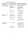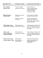
1
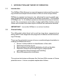
DiffSpin
_____________________________________________
®
Operator's Manual
Operator’s Manual
for
DiffSpin® Slide Spinner
Model Number
M700-10
M700-12
M700-22
Covered under one or more of the following US patents:
StatSpin and accessories:
DiffSpin Slide Spinner:
#4,846,974
#4,981,585
#5,326,398
Other patents applied for.
StatSpin is a registered trademark of StatSpin, Inc. an IRIS Company (International Remote Imaging Systems).
Copyright 2002
Printed in U.S.A.
TABLE OF CONTENTS
1.
2.
3.
4.
INTRODUCTION AND THEORY OF OPERATION
1.1
Introduction
1.2
Clinical Utility
1
1
UNPACKING AND INSTALLATION
2.1
Check for damage sustained
during transport
2.2
Check for complete packaging
2.3
Installing the DiffSpin slide spinner
2.4
Power connection
3
3
3
3
WORKING WITH DIFFSPIN
3.1
Controls and indicators
3.2
Safety features and precautions
4
6
INSTRUCTIONS FOR USE
4.1
Collection and preparation of blood sample
4.2
Operation
4.3
Sample Volume
4.4
Analysis of the slide
4.5
Typical results
4.6
Slides
7
8
9
12
12
12
MAINTENANCE
5.1
Cleaning instructions
13
6.
SERVICE
14
7.
TECHNICAL DATA
14
8.
NOTES
8.1
Safety Warning
8.2
General Note
15
15
9.
TROUBLESHOOTING GUIDE
16
10.
REFERENCES
18
11.
WARRANTY FOR THE DIFFSPIN
Warranty Card
5.
19
Inside back cover
UNIVERSAL PRECAUTIONS
Universal Precautions should be followed on all specimens,
regardless of whether a specimen is known to contain an
infectious agent. Universal Precautions have been stated by
Center for Disease Control in 1987 and updated in 1988 and
are reprinted in National Committee for Clinical Laboratory
Standards proposed guidelines, “Protection of Laboratory
Workers From Occupationally Acquired Infections”, Approved
Standard, NCCLS Document M29-A2, 2001 (NCCLS, National
Office, 940 West Valley Road, Suite 1400, Wayne, PA 190871898, USA).
1. INTRODUCTION AND THEORY OF OPERATION
1.1
Introduction
The DiffSpin Slide Spinner is a special purpose instrument for producing uniform monolayer blood smears or films on microscope slides.
DiffSpin is compact and easy to use, allowing the user to rapidly prepare reliable blood smears that are technique insensitive. The benefits
of DiffSpin are: 1) it’s ability to produce consistent monolayer smears,
and 2) the convenience and safety of disposable Slide Holders which
collect and contain the excess spun-off blood and minimize aerosols.
IMPORTANT: Insure the DiffSpin is on a level surface.
1.2
Clinical Utility
The differential white blood cell count has long been recognized as
being useful in the diagnosis and prognosis of disease and the monitoring of therapy.
There are five potential sources of error or methodological variables in
making the differential count.
1.
2.
3.
4.
Errors in interpretation or classification of the cells.
Statistical sampling errors.
Non-random or non-uniform distribution of cells on the slide.
Distortion of blood cell morphology by inappropriate
anticoagulant, blood storage and handling.
5. Blood film preparation technique.
The spinner technique addresses the third and fifth sources of these
potential errors. A more complete explanation follows.
1
1.2
Clinical Utility (cont.)
The traditional technique to prepare the microscope slide, the manual
wedge smear, introduces a source of error as it causes non-random
distribution of cells which is dependent on cell size (1). This generally results in an under-reporting of monocytes (7) and a slight overreporting of lymphocytes. This phenomenon is not surprising with
the visually apparent non-uniform distribution gradient established using
the wedge technique.
The slide spinner technique addresses both the non-uniform distribution of cells and preparation technique. By using a slide spinner, all
areas covered by cells are good; no searching for a "good" area.
Total time to achieve accurate results is less. Indeed, the results
achieved with spun slides are more accurate than those prepared
using the wedge smear technique (6).
Slide spinners were first developed in association with automated
differential counters to produce uniform monolayers independent of
operator skill (1). Using these early generation slide spinner instruments produced superior quality slides, however, the advantages were
offset by the hazards associated with aerosol generation.
The DiffSpin was developed to eliminate aerosol formation while providing the higher quality of a spun slide. Using the DiffSpin, when the
slide rotates, the excess blood is captured by the slide holder. When
used according to the instructions, independent studies indicate there
is no detectable aerosol generation (5). In addition, because of the
eccentric spinning platform, slides prepared with the DiffSpin are not
completely covered with blood. Therefore, prepared specimen slides
are readily handled and easily labeled for identification.
When the slide rotates, the excess blood is thrown off and captured
by the Slide Holder. The remaining blood wets the slide and the
thickness of the layer left behind depends on the sample’s viscosity,
cell content and surface tension. Though the density of the cellular
layer may depend on the sample's viscosity, cell content and surface
tension of the glass slide the volume applied the the slide also affects
the cell density.(See Section 4.3 Sample Volume)
2
2. UNPACKING AND INSTALLATION
2.1
Check for Damage Sustained During Transport
The DiffSpin Slide Spinner and its accessories are delivered in one
package. Save the shipping carton in the event the instrument needs
to be returned for service. Notify the carrier and your DiffSpin distributor if damage in shipment is evident.
2.2
Check for Complete Packaging
•
•
•
•
2.3
Centrifuge
Line Cord
Operator’s Manual w/Warranty Card
DiffSpin Slide Preparation Kit, Product Number DSPK
Installing the DiffSpin Slide Spinner
Place the DiffSpin on any level surface acceptable for a laboratory
instrument. Position the DiffSpin away from direct sunlight and
sources of heat and cold.
2.4
Power Connection
Before connecting the DiffSpin to the power, please check that the
nominal voltage (V~) and frequency (Hz) specified at the rear of the
DiffSpin matches your outlet voltage and frequency. If it does not,
please contact your supplier.
WARNING: If the voltages do not match, do not connect
the DiffSpin to the power source!
If the voltages match, connect the DiffSpin to a grounded outlet, using
the cable provided. This protective grounding must not be neutralized
by using an extension cord without a ground lead.
Once the DiffSpin is connected to the power it is ready for use. The
DiffSpin does not have an ON/OFF switch.
3
3. WORKING WITH DIFFSPIN
3.1
TM
Controls and Indicators
“stop / open” button
• interrupts the cycle and stops the centrifugation. The “stop / open”
button is rarely used as the cycles are short. It can be used to
release an accidentally latched cover.
“power” indicator
• green LED, illuminated when the DiffSpin is connected to a power
source.
The DiffSpin is not provided with an “on/off” switch. The unit is
normally left plugged in and “on”. The cover is usually left down
but not latched.
“start” button
• initiates a spin cycle.
“ready” indicator
• located on the “start” button, the indicator signals that the DiffSpin
is ready to process the next specimen (there is a brief delay
between cycles).
(See Figure 1 on the following page)
“acceleration” & “duration” control dials
• adjust the acceleration and the duration of the cycle. They are
located on the side of the instrument and are usually set at their
mid-point (the letter “C”) for all blood samples. Setting “C”
should provide adequate films for normal hematocrits. Turn
either control toward “E” to increase the spin duration or the
acceleration. These adjustments may be necessary for blood
samples from patients with disease states that result in major
changes in their blood flow characteristics.
"Duration"
"Acceleration"
0.4 - 3.0 seconds
Final rotational speed is inconsequential.
Acceleration of the platform (cradle) is
responsible for the quality of the blood film.
4
3.1
Controls and Indicators (cont.)
NOTE: Some laboratories prefer "acceleration" setting of "D".
To adjust the “acceleration” control to a slower setting, first
change the control to the desired setting. Then discharge the
“memory” of the previous setting. Do this by placing a Slide
Holder (with slide) inside the DiffSpin. Close the cover and press
the start button. Spin only the slide and Slide Holder with no
specimen during this “reset” cycle. The next cycle will now
operate at the new speed and setting.
Figure 1
DiffSpin, Front View
Ready Indicator
Stop/Open Start
Button
Button
5
Control Dials
3.2
Safety features and precautions
•
The DiffSpin slide spinner incorporates an interlock mechanism.
This mechanism prevents the DiffSpin from being operated unless
the cover is closed.
IMPORTANT: Do not attempt to defeat this safety
feature of your DiffSpin instrument.
•
The DiffSpin prepares blood smears for microscopic examination.
Use of the DiffSpin places a responsibility upon administrative
personnel to insure adequate training of operators. Do not open
the DiffSpin until the slide has stopped rotating.
WARNING: Any rotating device can generate a
mechanical safety hazard. For the user’s protection,
the unit will NOT operate when the cover is open.
•
Do not leave any blood on the outside of the disposable Slide
Holder before spinning.
•
The DiffSpin is not explosion-proof. Do not use in a potentially
explosive atmosphere.
•
StatSpin slides: 1 x 3 inch, Product No. SL72.
•
Mount the Slide Holder so that it lies flat on the Cradle. Check
that the tabs are securely over the mounting pins. Failure to
ensure the Slide Holder is mounted properly may break the slide
and/or result in damage to the instrument. (See Figure 2, below)
Figure 2
6
4. INSTRUCTIONS FOR USE
4.1
Collection and Preparation of Blood Sample
The National Committee for Clinical Laboratory Standards (NCCLS)
publication “Reference Leukocyte Differential Count (Proportional)
and Evaluation of Instrumental Methods”, Approved Standard,
NCCLS Document H20-A, Vol. 12 No. 1 (NCCLS, National Office,
940 West Valley Road, Suite 1400, Wayne, PA 19087-1898, USA)
indicates the following for visual differential count:
• Use whole venous blood collected in anticoagulanted tubes with
tripotassium ethylenediamine tetra-acetate (K3EDTA) 1.5 +/- 0.15
mg (in liquid or powder form) per mL (see NCCLS document H3A4);
• Note the specimen condition - Macroscopically visible clots are
cause for rejection of the specimen for analysis. Microscopically
visible platelet clumps consisting only of platelets are acceptable,
provided their presence is noted;
• Slides are to be prepared within 4 hours of blood collection in
K3EDTA;
• Blood samples are to be stored at room temperature until blood
smear has been prepared;
• Use StatSpin Slides, Product No. SL72.
• Make sure that the blood is well mixed. Use a blood tube rocker
or similar device if there is any delay in preparing the blood smear.
Manual mixing is allowable as long as the blood tube is inverted
completely at least 20 times. This will mix the blood and
minimize artifacts such as rouleaux formation;
• Stain the film within one hour of preparation with either a
Romanowsky stain, containing fixatives or fix within one hour with
"water-free" (i.e., <3% water) methanol for later staining.
7
4.2
Operation
1. Insert a clean slide Product No. SL72, (frosted edge out) into a
disposable Slide Holder, Product No. SC01.
2. Place the Slide Holder (with slide) onto the cradle by
placing the holes in the tabs (located on the sides of the
Slide Holder) over the silver colored mounting posts and
press down gently until the Slide Holder comes to rest
flush on top of the cradle. (See Figure 2).
IMPORTANT: The labeled end of the slide should rest
between the slide retaining posts on the Cradle.
3. With a StatSpin Transfer Pipette (Product No. PD50), place a
single drop of blood into each of the two holes located on the
top of the Slide Holder (See Section 3.2). The two drops must
pool together to ensure uniform coverrage, this takes a second
or two. Be careful not to leave any blood on the outside of the
disposable Slide Holder before spinning.
4. Spin the slide immediately after adding the blood.
5. Close cover firmly until it latches. Press “ START” button. The
cycle is less than 3 seconds.
6. When unit comes to a stop, squeeze the latch on the cover, lift
upward to open. (See Figure 3 below)
Figure 3
8
4.2
Operation (cont.)
7. Remove the disposable Slide Holder containing the slide.
8. Remove the slide and air dry or oven dry as your lab procedures
indicate. When slide is dry it may be stained or processed in the
usual manner.
9. Dispose of the contaminated Slide Holder as you would other
biohazardous material.
IMPORTANT: Use Slide Holders only once.The Slide Holders are
designed for single use only.
NOTE: There is a built-in time delay between cycles. Wait until the
“ready” indicator is lit before initiating the next cycle. (See Figure 1
Section 3.1) A new cycle can not be initiated until the time delay
has elapsed.
4.3
Sample Volume
For best results, use 50 ul of whole blood placed in each of the holes
in the slide holder. The drops should "pool" to create a bar formation.
This bar formation will adequately cover the slide and provide good
viewing area.
The density of the cellular layer depends on the sample's viscosity,
cell content and surface tension of the glass slide. The volume
applied to the slide also affects the cell density on the blood film.
Operator's preference of volume varies greatly.
Sample Volume continued next page.
9
4.3
Sample Volume (cont.)
•
•
The following are some suggestions for sample volumes:
For lower cell density, some laboratories report best results with
total volume of 40 ul (i.e, 0.040 ml) of blood pipetted into each
hole of the slide holder. With small quantities of blood, the slide
will not be uniformly coated with a film, however, the spacing
between the cells will be greater something that may be
preferred for faster cell recognition.
For higher cell density (and a larger area of the slide to be
covered), some laboratories apply 2 x 65 ul (i.e., 2 x 0.065 ml)
drops of blood. Although this produces a more fully covered
slide, this much surface area is not really necessary. Do not
exceed 75 ul per drop as blood will exit the slide holder and
cause excessive aerosols. Examination Area shown below is
best for viewing red cell morphology.
Figure 4
Representation of 2 x 50 ul drops
NOTE: The two drops of blood must merge or pool together to
ensure uniform coverage.
Sample Volume continued next page
10
4.3
Sample Volume (cont.)
•
The amount of blood added to create a monolayer film with the
DiffSpin is a matter of preference. The smaller volumes create a
more spread out cell monolayer under the microscope, but may not
have the same slide coverage as larger volumes.
Also, other cell suspensions may also be used to create a monolayer on a microscope slide.
Pipettes
For best results use StatSpin pipettes, with a premeasured drop
size of 50 ul. Order StatSpin Product Number PD50.
11
4.4
Analysis of the Slide
The peripheral blood smear made with your DiffSpin slide spinner
should be read according to your laboratory protocol.
4.5
Typical Results
About 70% of the slide will be covered with a monolayer of blood
cells. The areas covered on typical DiffSpin slides are shown in
Figure 4. Nearly all of these covered areas are available for counting. Depending on the aperture of the microscope, there will be
over 20,000 high-power fields (100x) available to the user. This area
is always larger than the “feathered” area used on the wedge
smear.
Red blood cell morphology varies with the method of preparation
(2). Spun films may display slightly different morphology in some
areas of the slide than smears prepared by the wedge method.
However, similar as with the wedge method the user will easily
identify those areas that enable accurate identification of red cell
morphology.
Distribution of white blood cells is uniform through out a spun
blood smear (1). The uniformity of blood smears prepared by
DiffSpin depends on the quality of the microscope slides.
4.6
Slides
The uniformity of blood smears prepared with either manual or
automated systems depends on the quality of the microscope
slides. For best results use StatSpin microscope slides, Product Number SL72. StatSpin slides are clean, dustfree and dehydrated, which is a prerequisite for consistent results. Other
various commercially available pre-cleaned slides may result in
inadequate blood films if the slide surface is covered with dust or
debris particles, or if the slides have been exposed to humid
storage conditions.
Always use clean desiccated slides and handle them carefully.
Avoid touching slide surface. Oil from fingers on the slide surface will result in a non-uniform film.
12
4.6
Slides (cont.)
Slides must be fresh. Non-uniform slide coverage (e.g. streaking
or "fingering") is a result of poor quality slides that have degraded
resulting in increased surface tension (e.g. glass slides stored for
months without protection from humidity and temperature). This
increased surface tension is found with poor quality slides, slides
which were exposed to humidity, or slides stored for excessive
lengths of time. If this condition persists try a new box of StatSpin
slides (Product Number SL72).
5. MAINTENANCE
5.1
Cleaning Instructions
The DiffSpin requires no maintenance beyond routine cleaning.
Prior to cleaning, the power cord should be disconnected.
Clean the exterior surface and inner surface or bowl area with a
10% bleach solution on a damp cloth. Never have liquid inside
the bowl. Do not use abrasives or solvents. Clean the clear
cover with a glass/plastic cleaner if required.
Any machine or accessory containing accumulated blood and/or
other chemical deposits must be cleaned prior to shipment to
the manufacturer/distributor for service. This decontamination is
required by Federal Law (Title 48 and 49 of the Code of Federal
Regulations) and in accordance with the E.P.A's Regulations for
Biohazard Waste Management. This decontamination cannot
be performed by StatSpin personnel.
13
6. SERVICE
The DiffSpin has no user serviceable parts. Should the
instrument require service please consult your Warranty for
instructions
IMPORTANT: The DiffSpin contains components
which have a potential SHOCK HAZARD. The unit
should only be opened by trained and qualified
personnel.
7. TECHNICAL DATA
Power Requirements:
Product:
DS02-10 100 VAC, 50/60 Hz
DS02-12 120 VAC, 50/60 Hz
DS02-22 240 VAC, 50/60 Hz
Running Time:
0.4 to 3.0 seconds
Slide Holder:
Disposable; accepts 1 x 3 inch microscope slides.
Dimensions:
6.5 inches (16.5 cm) diameter by 4.5 inches (11.4 cm)
high.
Weight:
3.7 pounds (1.7 kg)
14
8. NOTES
8.1
Safety Warning
This DiffSpin left the factory in perfect order from the point of
view of work safety. To keep it that way and to ensure safe
operation, the user must follow the instructions and warnings
given in this operating manual.
8.2
General Note
The data and information provided in this manual correspond
to the state of knowledge existing at the time of printing this
manual. Any important changes will be taken into account
in the next edition of this manual or with the Package Insert
sheet included with the Slide Holders. In every case, the
respective package insert should be regarded as definitive.
15
9. TROUBLESHOOTING GUIDE
DESCRIPTION
POSSIBLE CAUSE
OPERATOR RESPONSE
Power light not lit
Power cord
unplugged from
DiffSpin or from
outlet.
Verify that the power
cord is plugged
securely into the
outlet and into the
DiffSpin.
Non-functioning
outlet.
Verify that outlet is
functioning properly.
LED burned-out
Verify DiffSpin is
still operational.
Start button not
depressed fully.
Retry by pushing
start button.
Cover not closed
properly.
Verify that cover is
closed tightly.
Unit not “ready”.
Verify “ready” light is
lit before initiating
cycle.
Unclean slides.
Verify that slides are
without dust and/or
fingerprints. See
Section 4.6 "Slides".
Inappropriate
sample volume.
Verify sample volume,
refer to Section 4.3.
Cradle does not
rotate
Poor Quality
Blood Films
16
DESCRIPTION
POSSIBLE CAUSE
OPERATOR RESPONSE
Poor Quality
Blood Films
Time and speed
settings not correct.
Verify position of
time and speed knob
at recommended “C”
mark for average
hematocrit levels.
Blood escapes
Slide Holder
Excessive amount
of blood.
Verify drops only large
enough to cover slide.
Blood on outside
of holder.
Verify that no blood is
present on exterior of
slide holder after pipetting
blood onto slide.
Slide Holder does
not lay flat on Cradle
Slide holder not
positioned correctly.
Check Figure 2, Section 3.2
and verify Slide Holder and
slide are oriented properly
on cradle.
Slide and/or
Holder dislodges
during spinning
Slide holder not
positioned correctly.
Check Figure 2, Section 3.2
and verify Slide Holder and
slide are oriented properly
on cradle.
Smear area covers
frosted edge of slide
Slide placed
incorrectly into
Slide Holder.
Verify that frosted edge
of slide protrudes from
Slide Holder when
assembled.
17
10. REFERENCES
1. "Blood Sample Preparation for Automated Differential
Systems", Rogers, Charles H., American Journal of
Medical Technology, Vol. 39, No. 11:435-442, November
1973.
2. "The Automated Differential; Pattern Recognition Systems, Precision, and the Spun Smear", Kingsley,
Thomas C., Blood Cells, 483-487, 1980.
3. "The Use of Wedge and Spun Blood for Reference WBC
Differential Counts", Abstract presented at International
Society of Laboratory Hematology, 6th International
Symposium on Technological Innovations in Laboratory
Hematology, Berend Houwen, et al., Santa Fe, New
Mexico, March 1993.
4. "Causes for Poor Leukocyte Distribution in Manual
Spreader-Slide Blood Films," Steine-Martin, E.A.,
American Journal of Medical Technology, Vol. 46, No. 9,
May 1980.
5. "Automation and Laboratory Hematology: Impact on
Quality and Cost Effectiveness", B. Houwen, et al.,
American Society of Clinical Pathology / College of
American Pathologists, Hematology Seminar, Orlando,
Florida, October 19, 1993.
6. "Flow Cytometric White Blood Cell Differential: A Proposed Alternative Reference Method", L. Lebeck, et al.,
Sysmex Journal International, 3, 61-69, 1993.
7. "A Critical Evaluation of the Manual / Visual Differential
Leukocyte Counting Method", Koepke, J.A., et al.,
Blood Cells, 11, 173-186, 1985.
1/15/02 54-002663-001 Rev. B
18
11.0
WARRANTY FOR THE DIFFSPIN CENTRIFUGE
StatSpin takes pride in its products and believes that the quality, utility
and value we build into each instrument will provide complete customer
satisfaction. StatSpin warrants to the original purchaser that this
instrument will be free from defects in material or workmanship under
normal and proper use for a period of one year from the date of installation. StatSpin will at its option, repair or replace any unit covered
under this warranty returned to StatSpin with shipping costs prepaid.
Repaired or replaced instruments supplied under this warranty will
carry only the remaining portion of the original warranty. For warranty
terms and conditions outside the United States, contact your Authorized StatSpin Distributor.
StatSpin shall not be responsible for damage to a StatSpin instrument
resulting from misuse, negligence, accident, or resulting from unauthorized repairs, alterations, or improper installation. Additionally,
StatSpin shall not be liable for any claims, losses or damages of any
third party or for lost profits or indirect or consequential damages of any
kind even if StatSpin has been advised of the possibility of such
damages. The purchaser shall be deemed liable for any and all claims,
losses or damages incurred by the use or misuse of the StatSpin
instrument by the purchaser, its employees or others, following receipt
of the instrument.
THIS WARRANTY IS GIVEN EXPRESSLY AND IN LIEU OF ALL
OTHER WARRANTIES, EXPRESSED OR IMPLIED. THE PURCHASER AGREES THAT THERE IS NO WARRANTY OF MERCHANTABILITY OR OF FITNESS FOR ANY INTENDED PUPOSE
AND THAT THERE ARE NO OTHER REMEDIES OR WARRANTIES,
EXPRESSED OR IMPLIED, WHICH EXTEND BEYOND THE DESCRIPTION ON THE FACE OF THIS AGREEMENT . NO AGENT OR
EMPLOYEE OF STATSPIN IS AUTHORIZED TO EXTEND ANY
OTHER WARRANTY OR TO ASSUME FOR STATSPIN ANY LIABILITY EXCEPT AS SET FORTH ABOVE.
19
85 Morse Street
Norwood, Massachusetts
02062 USA
http://www.statspin.com
telephone 800-782-8774
781-551-0100
fax
781-551-0036