1
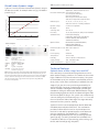
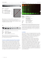
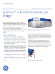
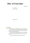
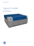
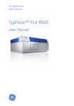
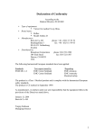
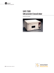
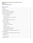
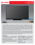
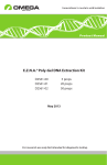
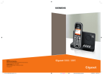
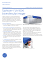
GE Healthcare
Life Sciences
Data file 29-0044-13 AB
Imaging systems, software, and accessories
Typhoon™ FLA 9500
biomolecular imager
Typhoon FLA 9500 (Fig 1) is a robust and versatile laser
scanner that is ideally suited to multiuser environments.
Biomolecular imaging applications include sensitive and
quantitative measurements of Western blots, multiplex
fluorescence, and radioisotopic labels by storage phosphor
as well as digitization of colorimetric stains.
Typhoon FLA 9500 delivers:
• Versatility: imaging of multifluorescent-, chemifluorescent-,
radioisotope-labeled, and colorimetric samples
• High resolution and quantitation: a pixel resolution of
up to 10 µm and a linear signal response over five orders
of magnitude provides precise quantitation in gels, blots,
tissue sections, and arrays
• High sample throughput: scanning area of 40 × 46 cm
enables simultaneous imaging of up to 20 gels or blots,
measuring 10 × 8 cm in size. This facilitates comparisons
among blots, and reduces workload and waiting time
• Flexibility: optimized performance for new applications
by adapting the system with stages, detectors, filters,
and lasers
Fig 1. Typhoon FLA 9500 is a high performance, versatile laser scanner for
sensitive and quantitative measurements in a multiuser environment.
The system provides several imaging modes such as
fluorescence, filmless autoradiography, and digitization
of colorimetrically stained gels. For chemiluminescence
detection of low abundance proteins the ImageQuant™
imager series is recommended.
The system is optimized for differential protein expression
studies and quantitative protein detection, e.g.
multifluorescent Amersham™ ECL Plex™ imaging for precise
quantitation of two or more proteins in the same Western blot.
• 2-D DIGE imaging: simultaneously image two 2-D DIGE
gels for differential expression studies
• Visible and infrared fluorescence imaging: optional near
infrared excitation for imaging IRDye™ and other infrared dyes
Typhoon FLA 9500 is a variable mode laser scanner with
modular access to the optical components (Fig 2) and
excitation sources, providing both versatile and flexible
imaging for precise quantitation of proteins, nucleic acids,
and other biomolecules (Table 1).
Fig 2. Filters are easily exchanged by the user.
Broad linear dynamic range
Table 1. Typhoon FLA 9500 specifications
Typhoon FLA 9500 provides a broad linear dynamic range in
all detection modes, for example when using Cy™5 labeled
proteins (Fig 3).
Detection modes:
log (integrated intensity)
8
7
6
5
4
3
1
2
3
4
5
6
log (pg carbonic anhydrase)
Carbonic anhydrase
18
6
2
0.7
0.2
0.08
0.025 (ng)
Fluorescence, phosphorimaging, digitization, and chemiluminescence
Excitation wavelengths: 473 nm (blue LD laser),
532 nm (green SHG laser),
635 nm (red LD laser),
685 nm (optional near IR LD laser), and 785 nm (optional near IR LD laser)
Radioisotopes:
3
H, 11C, 14C, 125I, 18F, 32P, 33P, 35S, 99mTc,
and other sources of ionizing radiation
Dynamic range:
5 orders of magnitude
Bit depth:
16-bit
Scanning area:
40 × 46 cm
Pixel sizes: 10, 25, 50, 100, 200 µm, and prescan 1000 µm
Standard filters:
IP (Phosphorimaging),
LPB (510LP), LPG (575LP), LPR (665LP),
LPR-Ch2 (665LP), BPB1 (530DF20),
and BPG1 (570DF20)
Optional filters:
BPFR700 (R715), BPFR800 (R810),
DBR1 (530DF20/665LP),
and DGR1 (570DF20/665LP)
Dimensions (W × H × D): 900 × 400 × 800 mm
Sample:
Gel:
Imaging:
Cy5:
LOD:
DR:
Linearity:
Carbonic anhydrase in
LMW marker
Amersham ECL™ Gel 4-20%
SDS-PAGE tris-glycine
Excitation Emission filter
635 nm
LPR (665 LP)
25 pg
4.3 orders of magnitude
R2=0.997 and slope k=0.96
(trendline in log-log plot)
Fig 3. Carbonic anhydrase was labeled with CyDye DIGE fluor Cy5 minimal
dye and separated using the Amersham ECL 1-D electrophoresis gel. The
gel was imaged with Typhoon FLA 9500. A selection of a three-fold dilution
series is shown in the image, arrow indicates the limit of detection (LOD).
The detection limit was 25 pg and the linear dynamic range (DR) was
4.3 orders of magnitude.
Weight:
97 kg
Line frequency:
50/60 Hz
Temperature:
15°C to 30°C
Humidity:
20% to 70% (no condensation)
Supply voltage:
100 - 240 VAC ± 10%
Power consumption:
Approx. 0.3 kVA
Technical features
Optimal choice of filter, stage, laser and PMT
Filters are easily accessed and exchanged without tools to
attain optimal imaging conditions. This makes the instrument
highly suitable for use in a multiuser environment. The system
accommodates up to four computer-controlled filter positions
at any time. Custom filters can be easily installed by the user.
Stages (Fig 4) give the correct positioning and stability for
optimal imaging of a range of sample types. Samples that
can be scanned include agarose and polyacrylamide gels,
membranes, DIGE gels, radioisotope-labeled samples using a
phosphorimaging plate, as well as microplates and glass slides
with the titer plate (TP) plug‑in. The system can simultaneously
scan two DIGE gels, each measuring up to 21.5 × 27.5 cm with
the Low Fluorescent Glass Plate stage. The stages are easily
removed from the system for cleaning.
Typhoon FLA 9500 comes equipped with both bi-alkali and
multialkali photomultiplier tubes (PMT). This combination
provides excellent detection over a very broad spectrum.
Each PMT is selected for optimal response to the detected
emission wavelength. The bialkali PMT 1 is suitable for
phosphorimaging and detection of dyes emitting blue and
green light (e.g. Cy2 and Cy3) whereas the multialkali PMT 2
is optimal for detection of dyes emitting yellow, red (e.g. Cy5),
far red, and infrared light (e.g. IRDye800).
2
29-0044-13 AB
A
C
B
D
Table 2. Emission filters
Filter type
Wavelength range (nm) Detection examples
IP
BP390 Phosphorimaging
LPB (510LP)
≥ 510
Cy2, SYBR™ Green,
FAM™, FITC,
Alexa Fluor™ 488, SYPRO™ Ruby,
SYPRO Orange, GFP
BPB1 (530DF20) 520 to 540 Scanning is rapid and detection is sensitive for laser-induced
fluorescence, radioisotopic imaging by storage phosphor, and
digitization. A fast 1000 µm prescan function gives a rapid
overview of the sample for selecting the correct settings.
At a pixel size of 200 µm, a 10 × 15 cm sample is scanned in
two minutes. The system provides a linear signal response
over five orders of magnitude. This, together with digitization
of the image with up to 16-bit resolution, provides a suitable
basis for the precise quantitation of proteins, DNA and other
labeled molecules.
Lasers can be exchanged in the field to accommodate new
applications and fluorophores. The system can house up
to four lasers simultaneously, from a choice of five laser
excitation wavelengths (473, 532, 635, 685, and 785 nm).
LPG (575LP)
≥ 575
Cy3, Deep Purple™, HEX,
Alexa Fluor 532 and 555,
SYPRO Red
BPG1 (570DF20) 560 to 580
Cy3 DIGE Fluor,
ECL Plex Cy3
LPR (665 LP)
≥ 665
Cy5, Alexa Fluor 633,
TOTO™ 3, DiD,
Cy5 DIGE Fluor,
ECL Plex Cy5
BPFR700 (R715) 713 to 726
Alexa Fluor 700,
IRDye680,
IRDye700
BPFR800 (R810) 814 to 826
Alexa Fluor 790,
IRDye800
Carbonic anhydrase
167
Sample:
Gel:
56
19
6
2
Carbonic anhydrase in LMW marker
Amersham ECL Gel 4-20% SDS-PAGE
tris-glycine
Excitation Emission filter
473 nm
BPB1 (530DF20)
170 pg
3.6 orders of magnitude
R2= 0.999 and slope k= 0.92
(trendline in log-log plot)
0.7
log (integrated intensity)
Fig 4. (A) The Phosphor Stage, (B) Multi Stage, (C) Low Fluorescent Glass Plate
Stage, and (D) Fluor Stage are designed to accommodate a variety of sample
formats and imaging modes.
Cy2 DIGE Fluor,
ECL Plex Cy2
0.3
0.2 (ng)
8
7
For the detection of radioactivity and fluorescence, emitted
light is collected and transformed to an electrical signal by
a photomultiplier tube (PMT). The electrical signal is then
converted into digital information by A/D conversion for
image display and analysis.
Imaging:
Cy2:
LOD:
DR:
Linearity:
Imaging applications
Fig 5. The LMW marker proteins were labeled with CyDye DIGE fluor minimal
dyes and separated using the Amersham ECL 1-D electrophoresis gel.
The gel was imaged with Typhoon FLA 9500. The images exhibit low and
even background in all three CyDye detection channels which allow for
multiplexing applications over a broad linear dynamic range. A selection of
the dilution series is shown in the image. The linear dynamic range of
Cy2-labeled carbonic anhydrase was 3.6 orders of magnitude.
Typhoon FLA 9500 covers all application modes, including
fluorescence, radioisotopes as well as colorimetric stains.
Flourescence detection
6
5
4
2
3
4
5
6
log (pg carbonic anhydrase)
Upon excitation, light is emitted from a fluorescently labeled
sample in proportion to the amount of labeled protein or
DNA in the sample. The high sensitivity and broad dynamic
range of Typhoon FLA 9500 makes it possible to measure low
and high abundant proteins in a single scan (Figs 5, 6, and 7).
Multiple fluorescent wavelengths can be detected with
minimal crosstalk for comparative expression experiments.
See Table 2 for emission filters.
29-0044-13 AB
3
Carbonic anhydrase
(ng)
409.6 204.8 102.4 51.2
25.6
12.8
6.4
Sample:
Gel:
1.6
0.8
0.4
0.2
Sample:
Gel:
Imaging:
Cy3:
Cy5:
LOD:
156
78
39
5
4.5
2
3
4
5
6
log (pg carbonic anhydrase)
0.8
0.4
0.2
0.1
Cy3 and Cy5 (data not shown) labeled 81mer oligonucleotide
TBE-Urea 1.0 mm
Excitation Emission filter
532 nm
BPG1 (570DF20)
635 nm
LPR (665 LP)
0.2 fmol for both Cy3- and Cy5-labeled 81mer
Sensitive multiplex detection of Western blots
Typhoon FLA 9500 is a versatile scanner that is ideal for
imaging of fluorescent Western Blot membranes. This
method is very sensitive and the signal is proportional to
protein quantity. Moreover, it is possible to detect more
than one protein at the same time by means of secondary
antibodies labeled with different fluorophores. Amersham
ECL Plex fluorescence detection systems provide high
sensitivity, as well as a broad linear dynamic range and are
well adapted to quantitative Western blotting (Fig 8).
29-0044-13 AB
313
6
Fig 7. A Cy3 labeled 81-mer oligonucleotide was run on a 1-D
electrophoresis gel. The gel was imaged with Typhoon FLA 9500.
The detection limit was 0.2 fmol, indicated by the arrow, for both the
Cy3- and Cy5-labeled oligonucleotides.
4
625
5.5
(fmol)
1.6
1250
7
DNA
3.2
(ng total protein)
2500
6.5
Fig 6. A dilution series of carbonic anhydrase in the LMW marker, labeled
with CyDye DIGE fluor Cy3 minimal dye, was separated using 1-D
electrophoresis.The gel was imaged with Typhoon FLA 9500. The detection
limit (LOD) was 0.2 ng carbonic anhydrase and the linear DR was 3.3 orders
of magnitude. Arrow indicates the LOD.
6.4
5000
7.5
4
12.8
HeLa cell lysate
8
log (integrated intensity)
Carbonic anhydrase
12% acrylamide
Tris-glycine
Imaging: Excitation Emission filter
Cy3:
532 nm
BPG1 (570DF20)
LOD:
0.2 ng carbonic anhydrase
DR:
3.3 orders of magnitude
Linearity: R2=0.998 and slope k=0.99
(trendline in log-log plot)
3.2
Sample:
Membrane:
Target proteins:
Detection:
Imaging:
Cy3:
Cy5:
LOD:
HeLa cell lysate mixed with transferrin
Hybond™ LFP
Transferrin and GAPDH
Primary antibodies:
Rabbit anti-human transferrin,
Mouse anti-GAPDH
Secondary antibodies:
ECL Plex Cy5 GAR and Cy3 GAM
Excitation Emission filter
532 nm
BPG1 (570DF20)
635 nm
LPR (665 LP)
GAPDH in 39 ng HeLa cell lysate
Fig 8. Multiplex detection of proteins by Amersham ECL Plex Western
Blotting. Transferrin and endogenous GAPDH were targeted in a dilution
series of HeLa cell lysate using ECL Plex anti-rabbit Cy5 (red) and antimouse Cy3 (green) secondary antibodies. Imaging was performed with
Typhoon FLA 9500 in separate detection channels. The arrow indicates
the limit of detection (LOD) for GAPDH. The minimal cross-talk and low
background enables reliable quantitation of specific signals relative to a
housekeeping protein.
2-D DIGE
The 2-D DIGE system is an integrated solution for accurate
quantitation of changes in protein expression. Typhoon FLA
9500 is a part of 2-D DIGE system that includes DeCyder™
2D Differential Analysis Software or ImageMaster 2D
Platinum Software (Fig 9, 10, and 11).
The strengths of Typhoon FLA 9500—high sensitivity
and broad dynamic range for measuring low and high
abundant proteins in one scan (Fig 3)—make it highly
suited for 2-D DIGE applications, enabling the user to
detect and accurately quantitate subtle changes in protein
expression. By generating overlaid, multichannel images
for each gel with minimal crosstalk, Typhoon FLA 9500
exploits the multiplexing potential of CyDye™ DIGE fluors to
remove experimental variation between gels. Images are
analyzed using DeCyder 2D or ImageMaster 2D Platinum to
accurately and confidently measure very small differences
in protein abundance.
Sample:
IPG strips:
Gel:
Imaging:
Cy2:
Cy3:
Cy5:
1 - Control:
Cell lysate of human cell line A431
2 - Treated:
Cell lysate of human cell line A431
treated with EGF
24 cm 3-11 NL
Precast low-fluorescent DIGE gel
Excitation Emission filter
473 nm
BPB1 (530DF20)
532 nm
BPG1 (570DF20)
635 nm
DBR1 (665LP)
Fig 9. Overlay image of a two-dimensional difference gel electrophoresis
(2-D DIGE) gel with control and treated samples, and internal standard. The
control and treated samples were labeled with Cy3 and Cy5 DIGE Fluors
(minimal dye labeling protocol). The internal standard sample was labeled
with Cy2 DIGE Fluor. The data sets were evaluated using the DeCyder 2D
Software (version 7.2), see Fig 10.
Fig 11. Example of a down-regulated protein, 2.3-fold decrease, upon
treatment in a DIGE experiment analyzed with ImageMaster 2D Platinum
(IMP) version 7 software. Both 2D and 3D images of the protein of interest,
Match ID 477, are shown in left side upper and lower, respectively. Eight
control samples (upper images) were compared to eight treated samples
(lower images) by IMP, and the p-value and fold-time change are presented
in the Class Analysis Table in the right upper panel highlighted with green
background color. A bar chart representative of the down regulation is
presented in right lower panel. In the DIGE experiment, the effect of an
activator of protein kinase C (PKC), Phorbol 12-myristate 13-acetate (PMA),
on the BALB/c 3T3 cell was examined. The control cells were treated with
DMSO. This experiment represents a DIGE application on intracellular signal
transduction pathway regulated by PKC.
Detection of radioactivity
Samples containing radioactive probes are exposed to a
storage phosphor screen. Light is emitted from the screen
in proportion to the amount of radioactivity in the sample
upon laser-induced stimulation (Fig 12). All GE Healthcare
provided storage phosphor screens are compatible with the
Typhoon FLA 9500.
(µCi/g)
200
70
20
7
2
log (integrated intensity)
14
C autoradiographic
standard (CFQ9702)
Imaging: Excitation Emission filter
FLA 9500 635 nm
IP (BP 390)
Exposure: 60 minutes exposure to
BAS-MS Imaging Plate
LOD:
0.2 µCi/g (S/N = 4)
Linearity: R2=0.999 and slope k=0.97
(trendline in log-log plot)
0.2
3
3.5 4 4.5
log (nCi/g)
0.07
8
Sample:
Fig 10. Example of a down-regulated protein upon treatment in a DIGE
experiment analyzed with DeCyder 2D Software. Four control samples
(left images) were compared to four treated samples (right images) in the
Biological Variation Analysis (BVA) module, and a t-test was then performed
in the BVA workspace. The number of detected spots was 2675 and 671 out
of them were found to have significant differences at p values < 0.001.
0.7
7.5
7
6.5
6
5.5
5
4.5
4
2
2.5
5
5.5
Fig 12. Scanned image of a 14C autoradiographic standard using the
Typhoon FLA 9500.
29-0044-13 AB
5
NIR detection
Digitization
Typhoon FLA 9500 has two PMT’s that are optimized for
sensitive detection over a broad spectrum from visible
light and up to the near infrared (NIR) region. Figure 13
shows how the multialkali PMT 2 gives a linear response
and sensitive detection of an IRDye 680 labeled secondary
antibody in a Western blot.
Excitation light passes through the sample and excites
a fluorescent plate. The emitted light from the plate
passes through the sample again and is collected and
converted to an electrical signal. The method is suitable for
documentation of colorimetrically stained gels.
Typhoon FLA 9500
Data are stored either in linear 16-bit grayscale TIFF (.TIF
file format) or in square root encoded 16-bit TIFF (.GEL file
format). The .GEL format encoding provides higher dynamic
resolution than .TIF at lower signal levels to exploit the
low signal detection capability of the phosphorimaging
technology.
1250
Transferrin
630
310
Sample:
Transferrin
Membrane: Hybond LFP
Detection: Primary antibody:
Rabbit anti-human
transferrin
Secondary antibody:
Anti-rabbit IRDye 680
Imaging:
Excitation Emission filter
Typhoon:
685 nm
BPFR700 (R715)
LOD:
19.5 pg transferrin
DR:
2.1 orders of magnitude
Linearity: R2=0.994 and slope k=0.94
(trendline in log-log plot)
160
log (integrated intensity)
2500
Data storage
78
39
19.5 (pg)
Image analysis
7
Designed for seamless data transfer and quantitative gel
and blot analysis, we provide image analysis software for
use with Typhoon FLA 9500 (Table 3).
6.5
6
5.5
5
4.5
4
Table 3. Image analysis software
1
2
3
log (pg transferrin)
4
Fig 13. A two-fold dilution series of transferrin starting at 2.5 ng was
subjected to Western blotting and detected with a rabbit anti-transferrin
primary antibody and anti-rabbit IRDye 680 secondary antibody. The arrow
indicates the LOD.
Software Analysis
ImageQuant TL 1-D gel electrophoresis, dot blots,
arrays, colony counting,
and user-defined gel analysis
DeCyder 2D
Differential high-resolution
2-D DIGE analysis including
Extended Data Analysis
ImageMaster 2D
Platinum
2-D gels, including
single stain and 2-D DIGE
Validation support
A comprehensive suite of life cycle validation services is
available for laboratory systems used in good practice
environments, such as GLP, GMP or GCP. The documentation
is developed and approved by validation experts.
Installation Qualification and Operation Qualification
(IQ/OQ) are performed on-site by trained service engineers.
Our engineers can also help with periodic re-qualification
(RQ) and evaluate, verify and, document system changes
and software upgrades with Change Control Protocols (CCP).
6
29-0044-13 AB
Ordering information
System
Quantity Typhoon FLA 9500 1
*
Code number
28-9969-43
* Includes 473 nm, 532 nm, and 635 nm lasers, filter tray, IP filter, LPB filter, LPG filter, LPR filter,
LPR filter for ch2, BPB1 filter, BPG1 filter, Fluor Stage, Membrane Weight, Phosphor Stage,
Low Fluorescent Glass Plate Stage, Multi Stage with TP plug-in. Fluorescent plate for
digitization, capture software, USB cable, mains cables (EU and USA), User Manual, Getting
Started Guide. One license of ImageQuant TL software is provided with Typhoon FLA 9500.
Upgrades and accessories* Quantity Code number
BAS-IP MS 2040 E
1
28-9564-74
1
28-9564-75
1
28-9564-76
1
28-9564-77
1
28-9564-78
1
28-9564-81
1
28-9564-82
1
29-0171-33
1
29-0171-39
1 63-0035-44
1 63-0035-45
1
28-9564-73
Phosphorimaging plate,
20 × 40 cm, multipurpose
BAS-IP MS 2025 E
Phosphorimaging plate,
20 × 25 cm, multipurpose
BAS-IP MS 3543 E
Phosphorimaging plate,
35 × 43 cm, multipurpose
BAS-IP SR 2040 E
Phosphorimaging plate,
20 × 40 cm, high resolution
BAS-IP SR 2025 E
Phosphorimaging plate,
20 × 25 cm, high resolution
BAS-IP TR 2040 E
Phosphorimaging plate, 20 × 40 cm,
for Tritium detection
BAS-IP TR 2025 E
Phosphorimaging plate, 20 × 25 cm,
for Tritium detection
BAS-IP ND 2040 E Phosphorimaging plate, 20 × 40 cm,
for Neutron detection
BAS-IP ND 2025 E Phosphorimaging plate, 20 × 25 cm,
for Neutron detection
Exposure Cassette
Unmounted Screen, 20 × 25 cm
Exposure Cassette
Unmounted Screen, 35 × 43 cm
FLA Image Eraser
*
BAS-IP phosphor screens are recommended for Typhoon FLA scanners. The different
screens are designed for general use (MS), high resolution suitable for morphological
work such as autoradiography (SR), detection of the weak energy of the Tritium signal (TR),
and detection of Neutron (ND). All mounted and unmounted GP phosphor screens, except
35x43 mounted GP phosphor screen, are compatible with Typhoon FLA 9500. These
products can be scanned with a Fluor stage included in the standard configuration.
Minimum computer requirement
OS
RAM
Processo
Hard disk
USB Ports
Optical drive
Monitor
Windows™ 7 Professional SP1 (32bit),
Windows XP™ SP3 (32-bit) or
Windows Vista™ Business SP2 (32-bit)
more than 1 GB
Intel™ Core™ 2 Duo processors
more than 80 GB
USB 2.0
DVD-ROM or Super Multi Drive
1280 × 1024 pixel resolution or higher
Please contact your local sales representative for the latest recommended computer
configuration.
29-0044-13 AB
7
GE, imagination at work, and GE monogram are trademarks of General Electric Company.
Amersham, Cy, CyDye, DeCyder, Deep Purple, ECL, ECL Plex, Hybond, ImageQuant, and Typhoon are trademarks of
GE Healthcare companies.
IRDye and LI-COR are trademarks of LI-COR Biosciences.
Alexa Fluor, SYBR, SYPRO, and TOTO are trademarks of Molecular Probes, Inc.
Windows, Windows XP, and Windows Vista are trademarks of Microsoft Corporation.
Intel and Core are trademarks of Intel Corporation.
FAM is a trademark of Applera Corporation.
2-D DIGE: 2-D Fluorescence Difference Gel Electrophoresis (2-D DIGE) technology is covered by US patent numbers
6,043,025, 6,127,134 and 6,426,190 and equivalent patents and patent applications in other countries and exclusively
licensed from Carnegie Mellon University.
DeCyder: This release of DeCyder version 2 (software) is provided by GE Healthcare to the customer under a nonexclusive license and is subject to terms and conditions set out in the 2-D Differential Gel Electrophoresis Technology
Access Agreement. Customer has no rights to copy or duplicate or amend the Software without the prior written
approval of GE Healthcare.
Deep Purple Total Protein Stain: Deep Purple Total Protein Stain is exclusively licensed to GE Healthcare from
Fluorotechnics Pty Ltd. Deep Purple Total Protein Stain may only be used for applications in life science research.
Deep Purple is covered under a granted patent in New Zealand entitled “Fluorescent Compounds”, patent number
522291 and equivalent patents and patent applications in other countries.
For local office contact information, visit
www.gelifesciences.com/contact
www.gelifesciences.com/quantitative_imaging
GE Healthcare Bio-Sciences AB
Björkgatan 30
751 84 Uppsala
CyDye: This product or portions thereof is manufactured under an exclusive license from Carnegie Mellon University
under US patent number 5,268,486 and equivalent patents in the US and other countries. Cy3-UTP or Cy5-UTP, Cy3.
5-dCTP or Cy5.5-dCTP, Cy3-CTP or Cy5-CTP: These products are manufactured for GE Healthcare UK Limited by Perkin
Elmer Life Sciences under US patent numbers 5047519 and 5151507. The cyanine dyes in the product are manufactured
under an exclusive license from Carnegie Mellon University under US patent numbers 5,268,486 and equivalent patents
in the US and other countries.
The purchase of CyDye products includes a limited license to use the CyDye products for internal research and
development but not for any commercial purposes.
A license to use the CyDye products for commercial purposes is subject to a separate license agreement with
GE Healthcare. Commercial use shall include:
1. Sale, lease, license or other transfer of the material or any material derived or produced from it.
2. Sale, lease, license or other grant of rights to use this material or any material derived or produced from it.
3. Use of this material to perform services for a fee for third parties, including contract research and drug screening.
If you require a commercial license to use this material and do not have one, return this material unopened to GE Healthcare
Bio-Sciences AB, Bjorkgatan 30, SE-751 84 Uppsala, Sweden and any money paid for the material will be refunded.
© 2011-2012 General Electric Company—All rights reserved.
First published Oct. 2011.
All goods and services are sold subject to the terms and conditions of sale of the company within GE Healthcare
which supplies them. A copy of these terms and conditions is available on request. Contact your local GE Healthcare
representative for the most current information.
GE Healthcare UK Limited
Amersham Place
Little Chalfont, Buckinghamshire, HP7 9NA
UK
GE Healthcare Europe, GmbH
Munzinger Strasse 5, D-79111 Freiburg
Germany
GE Healthcare Bio-Sciences Corp.
800 Centennial Avenue, P.O. Box 1327, Piscataway, NJ 08855-1327
USA
GE Healthcare Japan Corporation
Sanken Bldg., 3-25-1, Hyakunincho, Shinjuku-ku, Tokyo 169-0073
Japan
29-0044-13 AB
11/2012