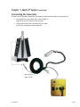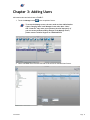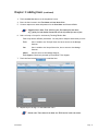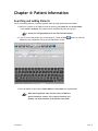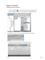
1
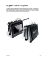
™
User Manual
™
Table of Contents
Mark X™ User Manual
Important Safety Information................................................................................ i
Specifications ..................................................................................................iii
Chapter 1: Mark X™ System ................................................................................. 1
The Mark X™ Color Panel Options..................................................................... 2
The Mark X™ Labels...................................................................................... 3
The Mark X™ Features................................................................................... 4
The Mark X™ Important Part Numbers............................................................... 5
The Mark X™ Key Connections Overview............................................................ 7
Connecting the Detector Cable f/Xmaru 1210 PCB................................................ 8
Connecting the Generator.............................................................................. 9
Powering Up the Mark X™ System.................................................................... 10
Chapter 2: Opening Mark X™ Software .................................................................. 11
Chapter 3: Adding Users.................................................................................... 13
Chapter 4: Patient Information............................................................................ 14
Searching and Adding Patients ....................................................................... 14
Editing an Existing Patient’s Information .......................................................... 16
Creating a New Study for an Existing Patient ..................................................... 16
Chapter 5: Creating Radiographs ......................................................................... 17
Choosing Anatomy and Positioning .................................................................. 17
Acquiring Radiographs ................................................................................. 19
Cropping a Radiograph................................................................................. 21
Chapter 6: Protocols.........................................................................................23
Using Protocols...........................................................................................23
Adding New Protocols................................................................................... 26
Editing Existing Protocols..............................................................................30
Renaming Protocols..................................................................................... 31
Deleting Protocols....................................................................................... 33
07202011
Sound-Eklin™
™
Mark X™ User Manual
Table of Contents
(continued)
Chapter 7: Managing Studies............................................................................... 35
Adding New Images to an Existing Study............................................................ 35
Reviewing a Study....................................................................................... 36
Chapter 7: Managing Studies............................................................................... 37
Chapter 8: Exporting Images to a Folder................................................................ 39
Chapter 9: Transferring Studies to the Storage Station............................................... 41
Transferring Individual Studies to the Storage Station........................................... 43
Transferring DICOM Batch to the Storage Station.................................................44
Confirm Storage of a Study............................................................................ 45
Chapter 10: Adding and Editing a DICOM Location.....................................................46
Appendix A: Digital Radiography Technique Charts................................................... 49
Portable 80/15........................................................................................... 49
Portable 100/30.......................................................................................... 52
Gantry.....................................................................................................54
Appendix B: Mark X™ System Field Replacement Guide..............................................56
Appendix C: Calibration Procedure.......................................................................60
Appendix D: Technical Support............................................................................ 62
Sound-Eklin™
Page 3
Important Safety Information
Note
Read all the instructions before connecting or operating the component
Keep this manual so you can refer to these safety instructions.
Warning
There are no user serviceable parts inside.
Refer all servicing to qualified service personnel.
To reduce the risk of fire or electric shock, do not expose the unit to
moisture or water. Do not allow foreign objects to get into the enclosure.
If the unit is exposed to moisture, or a foreign object gets into the
enclosure, immediately disconnect the power cord from the wall. Take the
unit to a qualified service person for inspection and necessary repairs.
Caution
Heed all warnings and safety information in these instructions
and on the product itself. Follow all operating instructions.
Clean the enclosure only with a dry cloth or a vacuum cleaner.
Clean the DR plate with a damp cloth. Use no chemicals or alcohol.
IPXO per IEC 60529
Type B per IEC 60601-1
You must allow 10 cm or 4 inches of unobstructed clearance around the
unit. Do not place the unit on a bed, sofa, rug, or similar surface that could
block the ventilation slots. If the component is placed in a bookcase or
cabinet, there must be ventilation of the cabinet to allow proper cooling.
Keep the component away from radiators, heat registers,
stoves, or any other appliance that produces heat.
Keep the component away from flammable materials.
The unit must be connected to a power supply only of the
type and voltage specified on the side panel of the unit
Sound-Eklin™
Page i
Important Safety Information (continued)
Connect the component to the power outlet only with the supplied
power supply cable or an exact equivalent. Do not modify the
supplied cable in any way. Do not attempt to defeat grounding
and/or polarization provisions. Do not use extension cords.
Do not route the power cord where it will be crushed, pinched, bent at severe
angles, exposed to heat, or damaged in any way. Pay particular attention
to the power cord at the plug and where it exits the back of the unit.
The power cord should be unplugged from the wall outlet if the unit is to be
left unused for a long period of time. Immediately stop using the component
and have it inspected and/or serviced by a qualified service agency if:
· The power supply cord or plug has been damaged.
· Objects have fallen or liquid has been spilled into the unit.
· The unit has been exposed to rain.
· The unit shows signs of improper operation
· The unit has been dropped or damaged in any way
Place the unit on a fixed, level surface strong enough to support its
weight. Do not place it on a moveable cart that could tip over.
System will become unresponsive when exposed to Electrostatic
Discharge (ESD) or Electrical Fast Transient (EFT) conditions.
This is the normal response to these environmental conditions.
Please restart the system to return to normal operation
USB ports are for keyboard and mouse only. User is
responsible for evaluating the system with any devices
connected to this port for compliance to IEC 60601-1-1.
Ethernet port is not to be used during acquisition. User
is responsible for evaluating the system with any devices
connected to this port for compliance to IEC60601-1-1.
Sound-Eklin™
Page ii
Specifications
Operation Environment: +15°C to 35°C,
30% to 75% RH, 70 to 106kPa (pressure)
Storage / Transport : –10° C to 55° C, 15% to 80% RH,
70 to 106kPa (pressure)
Power Requirements:
100 - 240 VAC 5.6A 50/60Hz
Weight (net) 47 lbs
CAUTION
Risk of electric shock.
Do not open.
Caution: To reduce the risk of electric shock, do
not remove cover. No user-serviceable parts inside.
Refer servicing to qualified service personnel.
This symbol is to alert the user to
important operating and maintenance
(service) instructions in this manual and
literature accompanying the product.
Sound-Eklin™
Applicable for USA, Canada or
where approved for the usage.
Caution: To prevent electric shock, match wide
blade of plug to wide slot. Insert fully.
This symbol is to alert the user to the
presence of uninsulated dangerous voltages
inside the product’s enclosure that may
constitute a risk of electric shock.
Page iii
Chapter 1: Mark X™ System
The Mark X™ system links seamlessly with the X-ray generator to provide the best image quality in a
small, lightweight, self-contained system. The Mark X™ portable Acquisition Stations are intended to
be used by Veterinarians for intermittent mobile diagnostic applications. They will connect the Mark
X Device to a Xmaru 1210PCB Amorphous Silicon Digital X-Ray Detector.
Sound-Eklin™
Page 1
Chapter 1: Mark X™ System
(continued)
The Mark X™ Color Panel Options
30-901 Red
30-900 Purple
30-904 Silver
30-902 Blue
30-899 Gun Metal
30-898 Green
Sound-Eklin™
Page 2
Chapter 1: Mark X™ System
(continued)
The Mark X™ Labels
Windows License
Key is located
on bottom of
Electronics Box
Mark X™ Electronics
Box Serial number
MarkX™ A–frame Serial
Number, CE Markings, and
Manufacture Date
MODEL: MARK X
INPUT POWER 120 VAC ~ 5.6A 60Hz
Sound-Eklin™
EN60601-1
EN60601-1-4
Page 3
Chapter 1: Mark X™ System
(continued)
The Mark X™ Features
Power Button
with LED Light
Hard Drive
Activity
Indicator
To Hold Receptor
Pigtail in Place
70–786 Neoprene
Cover for Xmaru
1210PCB Detector
Storage for 10–124
22’ Plate cable
f/Xmaru 1210PCB
Detector
Sound-Eklin™
Page 4
Chapter 1: Mark X™ System
(continued)
The Mark X™ Important Part Numbers
94-828 Mark X™
34-828 Mark X™,
Electronics Box Assembly
Sound-Eklin™
34-827 Mark X™,
A-Frame Assembly
Page 5
Chapter 1: Mark X™ System
The Mark X™ Important Part Numbers (continued)
10-121 Min-X-Ray Sync Cable for Mark X™ Syncs the generator’s prep and
expose cycle with the computer.
90-403 10’ Ethernet Patch Cable Connects the computer to the network to allow for easy
image transfers from the Mark X™ to PACS.
91-408 Xmaru 1210PCB Detector
Captures digital radiographs.
10-124 22’ Detector Cable f/Xmaru 1210PCB
Provides power to the plate
and syncs the plate and Mark X™ System.
10-171 Equine 20’ IEC-NEMA Power Extension Cord
Connects the Mark X™ to any standard U.S. 110V power
outlet (Nema 5-15).
Sound-Eklin™
Page 6
Chapter 1: Mark X™ System
(continued)
The Mark X™ Key Connections Overview
10–121 Min–X–ray Sync
Cable for MarkX™
10–124 22’ Detector
Cable f/Xmaru 1210PCB
10’ Ethernet Patch
Cable
24–122 Keyboard with
Touch–Pad
10–171 Equine 20’
IEC–NEMA Power
Extension Cord
Sound-Eklin™
Page 7
Chapter 1: Mark X™ System
(continued)
Connecting the Detector Cable f/Xmaru 1210 PCB
1. Remove the Plate and the Plate Cable.
2. Connect the 22’ Detector cable f/Xmaru 1210PCB (10–124) by aligning the red
dot and notch on the cable with the red dot and notch on the receptacle.
3. Connect plate cable to the plate if it is not already connected.
10–124 22’ Detector
Cable f/Xmaru 1210PCB
Sound-Eklin™
Page 8
Chapter 1: Mark X™ System
(continued)
Connecting the Generator
The sync cable should stay connected to the generator and should be stored in the generator box.
1. Plug the Min–X–ray Sync Cable (10–121) into the Mark X™.
Align by red dots on each end of the connector.
2. Plug the hand switch end of the Min–X–ray Sync Cable
(10–121) into the generator hand switch port.
Min–X–ray Sync
Cable (10–121)
Sound-Eklin™
Page 9
Chapter 1: Mark X™ System
(continued)
Powering Up the Mark X™ System
1. Plug the female end of the power cable into the power cable slot on the Mark X box.
Plug the other end into a grounded power outlet (see page ii Power Requirements).
2. With the cables connected, turn on the Mark X™ by pressing
the power button on the top of the unit.
Power Button
with LED Light
10-171 Equine 20’ IEC-NEMA
Power Extension Cord
Standard
US 110v
Power Outlet
(NEMA 5-15)
Cord
Sound-Eklin™
Page 10
Chapter 2: Opening Mark X™ Software
Open the Mark X™ software to begin the process of taking a radiograph.
1. Double-tap the TruDR™ icon on the desktop. This will load
the TruDR™ software for acquiring digital radiographs.
2. Enter the user name and password when the login screen appears.
The default login is:
User Name: admin
Password: admin
The software will load directly into the Patient Screen
if the “Remember Me” box is checked.
Sound-Eklin™
Page 11
Chapter 3: Adding Users
Add veterinarians and technicians to TruDR ™ .
1. Touch the Manage button in the Acquisition Screen.
To access the Manage screen, the user needs to have Administrative
rights. Changing items in the Manage Screen other than “Users”
and “Protocols” may cause your system to function improperly. If
you are concerned about making changes in the Manage Screen,
please contact Technical Support at 1.800.268.5354.
2. Select the Users menu from the left side of the screen to load the Users Screen.
Sound-Eklin™
Page 12
Chapter 3: Adding Users
(continued)
3. Touch the Add User button in the Acquisition Screen.
4. Enter the User’s name in the First Name and Last Name fields.
5. Create a unique user name and password in the User Name and Password fields.
Suggested user name: First initial of your first name plus last name
(e.g. jsmith for John Smith). Passwords can be left blank for ease of use.
6. Select a Group or Groups for each user by Touching Tech or Vet.
Each Group allows different permissions. You may select multiple check boxes per user:
Tech: User is added to the Tech pull down list has no access to the Manage
features.
Vet: User is added to the Vet pull down list, has no access to the Manage
features.
Admin: Has full access to the Manage features.
Tech Support: Should only be used by Sound-Eklin™ Tech Support
7. Touch the Save button to add the User.
8. Repeat steps 3 through 7 to add additional Users.
Please note: There must be at least one TECH active in the user menu.
Sound-Eklin™
Page 13
Chapter 4: Patient Information
Searching and Adding Patients
Search for existing patients or add new patients easily by using the filtered search fields.
1. Search for a patient in the Patient Screen by entering the Patient ID, the Patient Name,
or the Owner Last Name. The patient list will automatically filter as you type.
Patient list is displayed based on the User Selected Default.
2. If you do not see the patient you are searching for, Touch the Add
Enter key on the keyboard to bring up the Add Patient Screen.
button or press the
3. Enter the patient’s information. Patient Name and Last Name are required fields.
When entering patient name and last name, DO NOT use
special characters such as: % $ &. Special chracters and
symbols can cause problems in the patient name fields.
Sound-Eklin™
Page 14
Chapter 4: Patient Information
Searching and Adding Patients (continued)
4. Touch one of the Save buttons.
Save Saves the current patient and returns you to the Patient Screen.
Save+ Add Saves the current patient and returns to the Add Patient Screen.
Save+ Acquire Saves the current patient then opens the Anatomy/Protocol Screen.
Save+ Protocol Saves the patient and brings you to the Protocol Screen to select the
desired series.
Sound-Eklin™
Page 15
Chapter 4: Patient Information
(continued)
Editing an Existing Patient’s Information
Edit an existing patient’s information for future DICOM tagging.
Previous exam information cannot be changed
1. Select a patient to edit from the Patient Screen.
2. Touch the Edit button.
3. Edit the information as needed.
If the patient is from an owner previously entered, owner
can be selected from owner list on the left hand side.
4. Touch the appropriate Save button (see Page 17).
Creating a New Study for an Existing Patient
Create a new study for a patient previously entered in TruDR™ .
1. Search for a patient in the Patient Screen by entering the Patient ID, the Patient Name,
or the Owner Last Name. The Patient List will automatically filter as you type.
2. Select a patient by Touching on the name in the Patient List.
3. Touch the New button to create a new study.
4. Select the anatomy and the view from the Anatomy Screen (see Page 19).
Sound-Eklin™
Page 16
Chapter 5: Creating Radiographs
Choosing Anatomy and Positioning
Choose the anatomy and positioning to automatically enhance and position the radiograph.
1. Select the Tech and ordering Vet from the drop-down menus.
2. Touch the Mark X™ touchscreen and select the appropriate anatomy.
Sound-Eklin™
Page 17
Chapter 5: Creating Radiographs
Choosing Anatomy and Positioning (continued)
3. Touch on the appropriate view button for the exposure.
This will bring you to the acquisition screen.
Sound-Eklin™
Page 18
Chapter 5: Creating Radiographs (continued)
Acquiring Radiographs
1. Set the X-ray generator according to the Sound-Eklin™ Technique Chart using
the appropriate KVP and mAs (depending on the X-ray generator).
Due to variations between X-ray generators, optimal techniques may vary
somewhat from the values printed on the Sound-Eklin™ Technique Chart.
Users should be fully acquainted with State and local regulations
governing radiation protection and the use of diagnostic X-ray
equipment as well as the manual for the X-Ray Generator being used.
2. Align the generator 22 inches from the plate using the light beam
to fill the entire capture area defined on the plate.
If the generator is aligned perpendicular, the light will form
a rectangle with even sides filling the capture area.
The image capture area is highlighted by a white box
with the dotted cross-hairs in the middle.
3. From the Acquisition Screen press the hand switch halfway down to
prep and all the way down to expose the X-ray machine.
Proper Two-Stage Hand Switch Technique
1. To Prep, push the hand switch halfway down.
2. Wait until the Generator is ready to fire (i.e. prep
light on the X-ray machine is illuminated, etc.).
3. To expose, push the hand switch completely down .
Sound-Eklin™
Page 19
Chapter 5: Creating Radiographs
Acquiring Radiographs (continued)
4. The image will appear in less than 3 seconds. Use the Use Left, Right, Flip, Reverse,
ROI or Crop buttons to make orientation adjustments before shooting the next shot.
‘Window’ and level with finger in up and down, left or right motion.
Crop the images as needed (see Pages 23-24).
If you need to reshoot your X-ray, press the hand switch and fire again.
5. Touch the Anatomy button to change views.
6. Select the next view from the Views Screen. Touch the Anatomy
button from the Views Screen if another anatomy is needed.
7. Touch the Review button
to review. Touch the Patient
button to return to the Patient Screen.
Sound-Eklin™
Page 20
Chapter 5: Creating Radiographs (continued)
Cropping a Radiograph
Remove unwanted portions of the radiograph.
1. Make adjustments before shooting the next radiograph. To do so,
Touch the Action button.
Touch the Crop button in the Acquisition Screen.
The new image will appear lighter after Touching the crop button.
Based on the crop selection the image will be windowed and leveled again.
Sound-Eklin™
Page 21
Chapter 5: Creating Radiographs
Cropping a Radiograph (continued)
2. Frame the desired image dragging from upper left corner to lower
right hand corner of the touchscreen with finger.
3. The image will enhance once it is cropped. The image
will perform an ROI based on crop size.
4. Re-crop the image if necessary before taking the next radiograph by repeating the steps.
Sound-Eklin™
Page 22
Chapter 6: Protocols
Using Protocols
Shoot a series of X-rays in a preset order which automatically
advances to the next shot after each shot taken.
1. From the Patient Screen, select the Patient and Touch Protocol.
Or, from the Patient Screen, Touch New and Touch Protocol.
OR, from the Add Patient or Edit Patient screen, Touch Save + Protocol.
(to Add a Patient see page 16).
Sound-Eklin™
Page 23
Chapter 6: Protocols
Using Protocols (continued)
2. Select a Protocol button to begin the protocol series.
3. The Acquisition Screen now has a Protocol Information bar showing the Protocol shot
number. The current Shot Information is located in the upper left portion of the screen.
Sound-Eklin™
Page 24
Chapter 6: Protocols
Using Protocols (continued)
4. Shoot the first image. The software will automatically advance to the next shot in
the protocol series. The shot number in the Protocol Information Bar keeps track of
the shot sequence, while the current Shot Information appears in the upper left.
5. Use the Protocol Information bar to repeat a shot, skip a shot, restart the
protocol, move to the last shot in the protocol, or stop the protocol.
Move to the first shot in the protocol series
Repeat the previous shot
Skip to the next shot
Skip to the last shot in the protocol series
Stop the protocol
6. Touch the Review button to open the Review screen (see Page 25).
7. Touch the Patient button to return to the Patient Screen.
Sound-Eklin™
Page 25
Chapter 6: Protocols
(continued)
Adding New Protocols
Create new protocols for your individual studies.
1. Touch the Actions button to access the Manage button in the Patient Screen.
To access the Manage screen, the user needs to have Administrative
rights. Changing items in the Manage Screen other than “Users”
and “Protocols” may cause your system to function improperly. If
you are concerned about making changes in the Manage Screen,
please contact Technical Support at 1.800.268.5354.
Sound-Eklin™
Page 26
Chapter 6: Protocols
Adding New Protocols (continued)
2. Touch on the Plus Sign next to the Protocols List to show the existing protocols.
3. Select the Protocols menu from the left side of the screen to load the
Protocol Screen. Navigate to Species > Equine > 1210S CIE > Protocol.
4. Enter the name of your new protocol in the Protocol Name field.
5. Touch the Save button.
Sound-Eklin™
Page 27
Chapter 6: Protocols
Adding New Protocols (continued)
6. Filter the shots as necessary by selecting the Filter by Laterality drop down menu.
7. Select the first shot from the Shots for Species list.
8. Touch the Link button
Sound-Eklin™
to add the shot to the Shots Linked to Protocol box.
Page 28
Chapter 6: Protocols
Adding New Protocols (continued)
9. Repeat step 6 through 8 as necessary.
10. Touch the Unlink button to remove any shots
from the Shots Linked to Protocol box.
11. Select a shot in the Shots Linked to Protocol box and change the order
using the Move Up, Move Down, Move First, and Move Last buttons.
Moves your protocol shot up one shot in the series.
Moves your protocol shot down one shot in the series.
Moves your protocol shot from its location to the first shot in the series.
Moves your protocol shot from its location to the last shot in the series.
12. Touch the Save button to save your protocols.
13. Touch the Patient button to return to the Patient Screen.
If you have forgotten to save your protocols, a Save dialogue box will appear.
Touch the Yes button to save your changes and return to the Patient Screen.
Sound-Eklin™
Page 29
Chapter 6: Protocols
(continued)
Editing Existing Protocols
Edit current Protocols to customize them to your shot series.
1. Touch the Actions button to access the Manage button in the Patient Screen.
To access the Manage screen, the user needs to have Administrative
rights. Changing items in the Manage Screen other than “Users”
and “Protocols” may cause your system to function improperly. If
you are concerned about making changes in the Manage Screen,
please contact Technical Support at 1.800.268.5354.
2. Touch on the Plus Sign next to the Protocols list to show the existing protocols.
3. Select a Protocol to edit from the left side menu, make changes and Touch Save.
Sound-Eklin™
Page 30
Chapter 6: Protocols
Editing Existing Protocols (continued)
4. Continue with Step 8 on Page 34.
Closing the software and re-opening is required
to save protocol database changes.
Renaming Protocols
Rename current protocols.
1. Touch the Actions button to access the Manage button in the Patient Screen.
To access the Manage screen, the user needs to have Administrative
rights. Changing items in the Manage Screen other than “Users”
and “Protocols” may cause your system to function improperly. If
you are concerned about making changes in the Manage Screen,
please contact Technical Support at 1.800.268.5354.
Sound-Eklin™
Page 31
Chapter 6: Protocols
Renaming Protocols (continued)
2. Touch on the Plus Sign next to the Protocols List to show the existing protocols.
3. Select the Protocol to rename from the left side menu. Touch the Rename button.
4. Rename the protocol in the text box and Touch the OK button when finished.
5. Restart the software by touching Exit from the Main Patient Screen
Sound-Eklin™
Page 32
Chapter 6: Protocols
(continued)
Deleting Protocols
Delete current protocols.
1. Touch the Actions button to access the Manage button in the Patient Screen.
To access the Manage screen, the user needs to have Administrative
rights. Changing items in the Manage Screen other than “Users”
and “Protocols” may cause your system to function improperly. If
you are concerned about making changes in the Manage Screen,
please contact Technical Support at 1.800.268.5354.
2. Touch on the Plus Sign next to the Protocols List to show the existing protocols.
3. Select the Protocol to delete from the left side menu Touch the Delete button.
Sound-Eklin™
Page 33
Chapter 6: Protocols
Deleting Protocols (continued)
4. Touch the OK button to permanently remove the protocol.
5. Restart the software by Touching the close button in the upper right corner.
Sound-Eklin™
Page 34
Chapter 7: Managing Studies
Adding New Images to an Existing Study
Add new images to an existing patient’s study.
1. Search for a patient in the Patient Screen by entering the Patient ID, the Patient Name,
or the Owner Last Name. The Patient List will automatically filter as you type.
2. Touch the Acquire button.
This will take you to the Views Screen using the anatomy you selected
from the original study. This is shown in the description field.
Sound-Eklin™
Page 35
Chapter 7: Managing Studies
(continued)
Reviewing a Study
Change the layout or delete any unwanted images before
the study is archived on the Storage Station.
1. Select a patient by Touching on the name in the Patient List.
2. Touch on the Study from the list on the right.
3. Touch the Review button.
4. Select a thumbnail in the left-hand pane to load the image into the review
pane on the right. The selected image is highlighted by a yellow box.
Sound-Eklin™
Page 36
Chapter 7: Managing Studies
Reviewing a Study (continued)
5. Review the images by using the following techniques:
Zoom: Once the Zoom button is selected tap the screen on area you
would like to Zoom in on. To Zoom out tap revert button.
Pan: Tap the Pan button, Touch and drag the image with your finger.
Window Level: Tap Window Level button, Move up and down with finger on
screen to perform Level. Dragging finger Left to Right is Window.
6. Use the toolbar for advanced review features.
Patient: Returns to the Patient Screen.
New: Opens the Anatomy/Protocol Screen for creating a new study or protocol.
Acquire: Opens the Views Screen using the anatomy you
selected from the original study.
Anatomy: Opens the Anatomy/Protocol Screen to select a
new view to add into the same study.
Save: Saves the image with any changes that have altered the original image.
Warning message will appear to choose “Yes” to overwrite (replace)
the image or “No” to create a new (additional) image.
Delete: Removes any unwanted images.
Deleting an image permanently removes it from the computer.
Sound-Eklin™
Page 37
Chapter 7: Managing Studies
Reviewing a Study (continued)
Revert: Reverts to the original image.
Left: Rotates the image 90˚ counterclockwise.
Right: Rotates the image 90° clockwise.
Flip: Flips the image vertically.
Reverse: Flips the image horizontally.
Magnify: Activates the magnifier which will zoom on the selected area.
Actions: Shows a drop-down menu with additional review features.
Win Level: Selects the window level feature (also accessible by using the right
mouse button).
Invert: Inverts blacks and whites, replicating a fluoroscopy.
Pan: Pans the image (also accessible by using the left mouse button).
Crop: Trims an image without applying enhancements.
In the Review Screen, cropping does not re-enhance the image based on the
captured area. It only trims the image without re-enhancing the image.
Overlay: Opens menu for DICOM text display.
Hide: Hides all DICOM information.
Summary: Displays patient and shot information.
Detail: Displays patient, shot, and additional detailed information.
ROI: Draw a box from left to right to window and
level based on the Region of Interest.
7. Touch the Patient button to return to the Patient Screen.
Sound-Eklin™
Page 38
Chapter 8: Exporting Images to a Folder
Export images from a study to a folder to save on a computer, thumb drive, etc.
1. Search for a patient in the Patient Screen by entering the Patient ID, the Patient Name,
or the Owner Last Name. The Patient List will automatically filter as you type.
2. Select a patient and a study. The patient and study are highlighted in a blue box.
3. Touch the Folder button.
4. Select either the DICOM or JPEG image format from the Export As: drop-down menu.
5. When selecting the DICOM option from Step 4, Touch the Add DICOM viewer check
box only if the computer does NOT have a full DICOM viewer already installed.
Annotation Burning Imprints annotations made to saved images
Anonymize Images Strips all client and clinic information from the images
Repository Format
Places images in a repository folder format
Create DICOM DIR Adds a DICOM directory to quickly import
all images into a PACS system.
Sound-Eklin™
Page 39
Chapter 8: Exporting Images to a Folder
(continued)
6. When selecting the JPEG format from Step 4, select the
Overlay Burning options from the drop-down menu:
The default option is set to None:
None:
Does not show overlays on the JPEG image
Detail:
Shows a detailed DICOM overlay on the JPEG image (shows Image Number,
Manufacturers Model Number, Study ID, Image Anatomy, View Position,
Image Laterality Extended, Columns X Rows, Technique, Institution Name,
Physician of Record, Patients Name, Patient ID, Patient Breed Description,
Patients Birth Date, Patients Sex, Acquisition Number, Acquisition Date,
Acquisition Time.)
Summary: Shows a summary DICOM overlay on the JPEG image (shows Image Number,
Image Anatomy, View Position, Image Laterality Extended, Institution Name,
Physician of Record, Patients Name, Patient Breed Description, Patients
Birth Date, Patients Sex, Acquisition Date, Acquisition Time).
7. Touch the Select Button
8. Select a location to save the images.
Images are saved to a selected location in a folder with the
animals name and the clients last name (e.g. Trigger-Smith).
Sound-Eklin™
Page 40
Chapter 9:
Transferring Studies to the Storage Station
Transfer the studies from the Acquisition Station to the Storage/Review Station.
You must have your Mark X™ and your Storage/Review Station networked.
Please contact your local IT Person for any networking needs and
SoundEklin tech support for configuring DICOM Server for the Mark X.
Unit cannot be connected to a network while in use.
In addition you must remove network plug
1. Connect a Cat-5 Internet cable into the Internet port on the computer.
2. To bring up the Store Files to DICOM Server screen,
Touch the Server Button on the Patient Screen.
Sound-Eklin™
Page 41
Chapter 9: Transferring Studies to the Storage Station (continued)
3. Touch the Start Store button to begin the DICOM image transfer to the Storage Station.
This will store all the studies that were taken since your last
transfer and clear the queue. If any studies are missing, you
can transfer them individually (see the following section).
4. Status box displays transfer results. When the status box states “completed”,
your images have successfully transferred to the DICOM server. Touch the
Red close button in the upper right corner to close the screen.
Sound-Eklin™
Page 42
Chapter 9: Transferring Studies to the Storage Station (continued)
Transferring Individual Studies to the Storage Station
Transfer the studies from the Acquisition Station to the Storage/Review Station.
1. Search for a patient’s study by entering the Patient ID, the Patient Name, or the
Owner Last Name. The Patient List will automatically filter as you type.
2. Select a patient by Touching on the patient name in the Patient List field.
3. Touch the Server button.
4. Touch the Start Store button to begin DICOM image transfer.
Sound-Eklin™
Page 43
Chapter 9: Transferring Studies to the Storage Station
Transferring Individual Studies to the Storage Station (continued)
5. Status box displays transfer results. When the status box states “completed”,
your images have successfully transferred to the DICOM server. Touch the
Red close button in the upper right corner to close the screen.
Transferring DICOM Batch to the Storage Station
1. If you wish to send a group of selected studies from a specific timeframe, enter the
start and end date of these studies in the display at the top of the Batch Send Options
window.. The Dicom image transfer will send thes studies listed in the queue all at once.
2. Touch the Start Store button to begin DICOM image transfer.
Sound-Eklin™
Page 44
Chapter 9: Transferring Studies to the Storage Station (continued)
Confirm Storage of a Study
Confirm the study successfully sent to the Storage/Review Station.
1. On the Patient Screen, the Archive field will show a server name (e.g. SOUND-SERV1).
2. Hold the mouse over the server name to show a pop-up window with the transfer data.
The pop-up box displays how many images have been transferred and the
date and time of the transfer. The information is color coded for easy
reference. If only one study appears, it will be highlighted in blue and the
color coding will not appear:
Green Box— Image transfer complete
Red Box — Image transfer failed
Notice the Secondary Transfer field. Secondary Transfer
documents the number of studies sent to another PACS
system besides the main archive, ie Sound-Serv2.
Sound-Eklin™
Page 45
Chapter 10:
Adding and Editing a DICOM Location
Add DICOM server locations for sending images to a Storage Station, Radiologist, etc.
1. Touch the Manage button in the Acquisition Screen.
To access the Manage Screen, the user needs to have Admin rights.
Chang-ing items in the Manage Screen other than “Users” and
“Protocols” may cause your system to function improperly. If
you are concerned about making changes in the Manage Screen,
please contact Technical Support at 1.800.268.5354.
2. Select the Configuration menu from the left side of the screen to load the Users screen.
Sound-Eklin™
Page 46
Chapter 10: Adding and Editing a DICOM Location
(continued)
3. Select the DICOM button from the left to bring up the DICOM Server Screen.
4. Touch the Add button to add a new DICOM server. Or if editing select
the existing DICOM server from the DICOM Servers list on the Right.
5. Enter the AE Title, IP Address, and Port number for your DICOM server. OR if editing
enter the corrected AE Title, IP Address, and Port number for your DICOM server.
Contact Sound-Eklin™ Technical Support or the location you are sending
the DICOM images for the appropriate DICOM provider information.
6. Select additional information by choosing Yes from the drop-down menus.
Only select the following options if you are directed to
do so by a Technical Support representative:
Auto Send Server: Automatically sends images to the selected DICOM location.
Allow Worklist: Designates the DICOM location as a worklist modality.
Telemedicine Server: Designates the DICOM location as a telemedicine server.
Archive Server: Designates the DICOM location as an archive server.
7. Touch the Save button Sound-Eklin™
to save the current information.
Page 47
Chapter 10: Adding and Editing a DICOM Location
8. Touch the Verify button
9. A smiley face
(continued)
to verify that you can connect to the DICOM Server.
will appear when you have verified the connection to the DICOM Server.
If a frowning face appears, please contact Sound-Eklin™
Technical Support (1.800.268.5354).
10. Touch the Patient button to return to the Patient Screen.
Sound-Eklin™
Page 48
Appendix A:
Digital Radiography Technique Charts
Portable 80/15
Study
View
SID in/cm kVP
Time in Seconds
65 Degree Navicular
24/61
65
0.2
65 Degree P-3
24/61
80
0.05
DP
24/61
80
0.1
Lateral
24/61
80
0.1
Flexor Skyline
18/46
80
0.1
If Indicated
Nav. Obliques
24/61
80
0.1
If Indicated
65 P-3 Obl.
24/61
80
0.05
P-3
Lateral
24/61
80
0.1
65 Degree P-3
24/61
80
0.05
65 Degree Obliques
24/61
80
0.05
DP
24/61
80
0.1
Lateral
24/61
80
0.1
Medial Oblique
24/61
80
0.1
Lateral Oblique
25/64
80
0.1
DP
24/61
80
0.1
Medial Oblique
24/61
80
0.1
Lateral
24/61
80
0.1
Lateral Oblique
24/61
80
0.1
Flexed Lateral
24/61
80
0.1
Special Palmar
24/61
80
0.1
DP
24/61
80
0.07
Lateral
24/61
80
0.07
Medial Oblique
24/61
80
0.07
Lateral Oblique
24/61
80
0.07
Navicular
If Indicated
Pastern
Fetlock
If Indicated
Metacarpus/Metatarsus
Sound-Eklin™
Page 49
Appendix A: Digital Radiography Technique Charts
Portable 80/15 (continued)
Study
View
SID in/cm kVp
Time in Seconds
DP
24/61
80
0.1
Medial Oblique
24/61
80
0.1
Lateral
24/61
80
0.1
Lateral Oblique
24/61
80
0.1
Flexed Lateral
24/61
80
0.1
Distal Rad. Sky
18/46
80
0.1
Distal Row Sky
18/46
80
0.1
Prox. Row Sky
18/46
80
0.1
DP
24/61
80
0.15
Medial Oblique
24/61
80
0.1
Angle Down 5
Lateral
24/61
80
0.1
Angle Up 5
Lateral Oblique
24/61
80
0.1
If Indicated
Calcaneal Skyline
18/46
80
0.1
DV
24/61
80
0.25
Lateral
24/61
80
0.25
Right Oblique
24/61
80
0.25
Left Oblique
24/61
80
0.25
DV
24/61
80
0.2
Lateral
24/61
80
0.2
Right Oblique
24/61
80
0.2
Left Oblique
24/61
80
0.2
Cranial (Lateral)
24/61
80
0.2
Middle (Lateral
24/61
80
0.25
Caudal (Lateral)
24/61
80
0.3
Carpus
If Indicated
If Indicated
Tarsus
Skull (TMJ & Bullae)
Sinus/Dental
Cervical Spine
Sound-Eklin™
Page 50
Appendix A: Digital Radiography Technique Charts
Portable 80/15 (continued)
Study
View
SID in/cm kVp
Time in Seconds
Pharynx
Lateral
24/61
80
0.25
Shoulder
Lateral
24/61
80
0.4
Elbow
Cran->Caud
24/61
80
0.3
Medio-Lateral
24/61
80
0.2
Cran->Caud
24/61
80
0.2
Lateral
24/61
80
0.2
Caud->Cran
24/61
80
0.3
Lateral
24/61
80
0.2
Lateral Oblique
24/61
80
0.2
Caud->Cran
24/61
80
0.2
Lateral
24/61
80
0.2
Lateral
24/61
80
0.1
Radius
Stifle
Tibia
Withers
Sound-Eklin™
Page 51
Appendix A: Digital Radiography Technique Charts
(continued)
Portable 100/30
Study
View
SID in/cm kVp
mAs
65 Degree Navicular
30/76
80
3.2
65 Degree P-3
30/76
80
0.5
DP
30/76
80
1.7
Lateral
30/76
80
1.7
Flexor Skyline
18/46
80
1.7
If Indicated
Nav. Obliques
30/76
80
1.7
If Indicated
65 P-3 Obl.
30/76
80
0.5
Lateral
30/76
80
1.7
65 Degree P-3
30/76
80
0.5
65 Degree Obliques
30/76
80
0.5
DP
30/76
80
1.7
Lateral
30/76
80
1.7
Medial Oblique
30/76
80
1.7
Lateral Oblique
30/76
80
1.7
DP
30/76
80
1.7
Medial Oblique
30/76
80
1.7
Lateral
30/76
80
1.7
Lateral Oblique
30/76
80
1.7
Flexed Lateral
30/76
80
1.7
Special Palmar
30/76
90
1.7
DP
30/76
80
1.7
Lateral
30/76
80
1.7
Medial Oblique
30/76
80
1.7
Lateral Oblique
30/76
80
1.7
DP
30/76
80
1.7
Medial Oblique
30/76
80
1.7
Lateral
30/76
80
1.7
Lateral Oblique
30/76
80
1.7
Flexed Lateral
30/76
80
1.7
Distal Rad. Sky
18/46
80
1.7
Distal Row Sky
18/46
80
1.7
Prox. Row Sky
18/46
80
1.7
Navicular
P-3
If Indicated
Pastern
Fetlock
If Indicated
Metacarpus/Metatarsus
Carpus
If Indicated
If Indicated
Sound-Eklin™
Page 52
Appendix A: Digital Radiography Technique Charts
Portable 100/30 (continued)
Study
View
SID in/cm kVp
mAs
DP
30/76
80
1.7
Medial Oblique
30/76
80
1.7
Angle Down 5
Lateral
30/76
80
1.7
Angle Up 5
Lateral Oblique
30/76
80
1.7
If Indicated
Calcaneal Skyline
18/46
80
1.7
Flexed DP
30/76
100
1.7
DV
30/76
100
2.5
Lateral
30/76
100
1.4
Right Oblique
30/76
100
1.4
Left Oblique
30/76
100
1.4
DV
30/76
100
2.5
Lateral
30/76
80
1.7
Right Oblique
30/76
80
1.7
Left Oblique
30/76
80
1.7
Cranial (Lateral)
30/76
100
1.7
Middle (Lateral)
30/76
100
2
Caudal (Lateral)
30/76
100
2.5
Pharynx
Lateral
30/76
80
1.7
Shoulder
Lateral
30/76
100
3
Cran->Caud
30/76
100
1.4
Medio-Lateral
30/76
100
1.7
Cran->Caud
30/76
80
1.7
Lateral
30/76
80
1.7
Obliques
30/76
80
1.7
Caud->Cran
30/76
100
2
Lateral
30/76
80
1.7
Lateral Oblique
30/76
80
1.7
Patellar Skyline
18/46
80
1.7
Caud->Cran
30/76
100
1.4
Lateral
30/76
80
1.7
Obliques
30/76
80
1.7
Lateral
30/76
80
1.2
Tarsus
Skull (TMJ & Bullae)
Sinus/Dental
Cervical Spine
Elbow
Radius
Stifle
Tibia
Withers
Sound-Eklin™
Page 53
Appendix A: Digital Radiography Technique Charts (continued)
Gantry
Study
View
SID in/cm kVp
mAs
Navicular DP
40/102
65
5
DP P3
40/102
80
2
Foot DP
40/102
80
3.2
Lateral
40/102
80
3.2
Navicular
Navicular Skyline
40/102
80
3.2
If Indicated
Nav. Obliques
40/102
80
2
If Indicated
P3 Obliques
40/102
80
2
Lateral
40/102
80
3.2
DP P3
40/102
80
2
P3 Obliques
40/102
80
2
DP
40/102
80
3.2
Lateral
40/102
80
3.2
Dlpmo
40/102
80
3.2
Dmplo
40/102
80
3.2
DP
40/102
80
3.2
Dlpmo
40/102
80
3.2
Lateral
40/102
80
3.2
Dmplo
40/102
80
3.2
Flexed Lateral
40/102
80
3.2
Special Palmar
40/102
80
3.2
DP
40/102
80
3.2
Lateral
40/102
80
3.2
Medial Oblique
40/102
80
3.2
Lateral Oblique
40/102
80
3.2
DP
40/102
80
3.2
Dlpmo
40/102
80
3.2
Lateral
40/102
80
3.2
Dmplo
40/102
80
3.2
Flexed Lateral
40/102
80
3.2
Distal Rad. Sky
40/102
80
3.2
Distal Row Sky
40/102
80
3.2
Prox. Row Sky
40/102
90
3.2
P-3
If Indicated
Pastern
Fetlock
If Indicated
Metacarpus/Metatarsus
Carpus
If Indicated
If Indicated
Sound-Eklin™
Page 54
Appendix A: Digital Radiography Technique Charts
Gantry (continued)
Study
View
SID in/cm kVp
mAs
DP
40/102
95
3.2
Dlpmo
40/102
80
3.2
Angle Down 5
Lateral
40/102
80
3.2
Angle Up 5
Dmplo
40/102
80
3.2
If Indicated
Calcaneal Skyline
40/102
80
3.2
DV
40/102
100
4
Lateral
40/102
90
3.2
Right Oblique
40/102
90
3.2
Left Oblique
40/102
90
3.2
DV
40/102
100
3.2
Lateral
40/102
90
3.2
Right Oblique
40/102
90
3.2
Left Oblique
40/102
90
3.2
Cranial (Lateral)
40/102
90
3.2
Middle (Lateral
40/102
100
4
Tarsus
Skull (TMJ & Bullae)
Sinus/Dental
Cervical Spine
Caudal (Lateral)
40/102
110
15
Pharynx
Lateral
40/102
90
3.2
Shoulder
Lateral
40/102
110
15
Cran->Caud
40/102
110
4
Medio-Lateral
40/102
90
3.2
Cran->Caud
40/102
100
3.2
Lateral
40/102
90
3.2
Caud->Cran
40/102
110
12
Lateral
40/102
90
8
Pldmo
40/102
95
3.2
Caud->Cran
40/102
100
8
Lateral
40/102
90
6
Lateral
40/102
100
4
Elbow
Radius
Stifle
Tibia
Withers
Sound-Eklin™
Page 55
Appendix B:
Mark X™ System Field Replacement Guide
1
3
2
4
1. Using a Phillips screwdriver remove four (4) screws.
2. Box will remain on because of 3 Guide Pins. Gently slide electronics box
off Guide pins, By pulling back. There are no wires to disconnect.
1
3
2
Blind Mate Connector
Sound-Eklin™
Page 56
Appendix B: Mark X™ System Field Replacement Guide
(continued)
3. Flip electronics box over and pull out 16GB Compact Flash Card. The compact flash card
will have the most recent Database file on it with all settings required to restore.
You will need to place the old Compact Flash into
the new electronics box replacement.
X
To Slide out old 16GB CF via the cut out:
Gently pull left or right tab with thumb
while pulling card back towards “X”.
4. You will need to place the old Compact Flash
into the new electronics box replacement.
To Slide in new 16GB CF via the cut out:
Gently align card up with rails and push card
forwards towards “X”.
Sound-Eklin™
Page 57
Appendix B: Mark X™ System Field Replacement Guide
(continued)
5. After replacing CF card into new Electronics Box slide on using guide pins.
The guide pins will line up the blind mate connector for you.
1
3
2
Blind Mate Connector
6. Using a Phillips screwdriver to tighten four (4) screws.
1
3
2
4
Sound-Eklin™
Page 58
Appendix B: Mark X™ System Field Replacement Guide
(continued)
7. Obtain Internet Connection, Power On, then call Technical Support
at 1.800.819.5538 to restore settings. Restore procedure will take roughly 20 minutes.
Sound-Eklin™
Page 59
Appendix C: Calibration Procedure
1. Launch the TruDR application as Administrator (username: admin password: admin).
2. Select the Actions button located in the menu bar at the top of the screen.
3. Select the Manage button located at the top of the
dropdown that appears after selecting Actions.
4. Select Equipment from the menu on the left side of the screen.
5. The Equipment Configuration screen will appear in the window on the right.
At the bottom of the window select the Perform Calibration button.
You may need to minimize the On-Screen Keyboard in
order to see the button. To do this select the Keyboard
button located in the top left corner of the screen.)
6. The Calibration screen will load on top of the application. To begin the calibration,
select the Perform Gain button. Once you have done this, the application will begin to
acquire a series of 4 Dark images 30 seconds apart and average them to create the Offset
image. After each image is acquired, the Progress text box will read: Please Wait.
7. Next, you will be prompted to take a shot to check technique.
The target value for the gain calibration is ~11000 ADU.
A good starting technique at 50” SID is 80 kVp and .17 seconds
8. If the setting is correct the Calibration Progress box will display the Average
Exposure Count and allow you to continue the calibration. If the settings
are not correct, the Calibration Progress box will tell you if the settings are
too high or too low, reset, and then prompt you to adjust your settings and
take another image. (The window for acceptable values is +/ 20%.)
9. If your settings were correct, you will be prompted to
acquire a series of 4 images 30 seconds apart.
10. Once all 4 gain calibration images have been created, you will be prompted to acquire a
Flat Field image. Acquire this image at the same technique as your calibration images.
11. Once the Flat Field image has been acquired, the application will begin
calculating the statistics of the calibration. DO NOT CLICK ANY BUTTONS OR
EXIT THE SCREEN UNTIL THE STATISTICS HAVE BEEN GENERATED.
Sound-Eklin™
Page 60
Appendix C: Calibration Procedure
(continued)
12. Approximate Calibration Statistics:
Offset Median: 2129
Offset Std Dev: 258.1529
Gain Median: 9586
Gain Std Dev: 144.6174
13. Using the dropdown window next to the Get Image button at the bottom of the
Calibration Screen you can view the Offset, Gain, and Flat-Field images by selecting
them in the dropdown and selecting the Get Image button. Use the Brightness
and Contrast sliders at the bottom of the image window to adjust the image.
14. Select the Flat-Field image from the dropdown window and select Get Image.
Inspect the image for any defective lines, pixels, or any other artifact. If the
image is a mottled/uniform gray color then the calibration was successful.
Sound-Eklin™
Page 61
Appendix D:Technical Support
Shipping Address
Sound-Eklin™
6359 Paseo Del Lago
Carlsbad . California 92011
USA
Technical Support
Office hours: Weekdays 8:00 A.M. — 5:00 P.M. Pacific Time
Emergency 24-hour support is available.
Telephone: 800-268-5354 option 3
Website
www.soundeklin.com
Sound-Eklin™
Page 62
Sound-Eklin™ Technical Support
6359 Paseo Del Lago
Carlsbad . California 92011 USA
toll free: 800.268.5354 option 3
phone:760.918.9626
fax:76.918.9620
international:+1.760.918.9626
www.soundeklin.com
© 2011 All Rights Reserved Sound-Eklin™ — a VCA ANTECH company