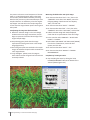
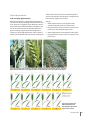
1
ISBN: 978-970-648-182-5 1 2 3 7 8 9 10 11 12 13 14 15 16 17 18 19 20 21 22 23 24 Osmotic adjustment Osmotic potential Photosynthetically active radiation Palm top computer Peedee Belemnite Pigment related index Photochemical reflectance index Photosystem (either I or II) Quadrat Ratio analysis of reflectance spectra chlorophyll a RARSb Ratio analysis of reflectance spectra chlorophyll b RARSc Ratio analysis of reflectance spectra carotenoid RGR Relative growth rate RH Relative humidity (%) RLD Root length density R-NDVI Red normalized difference vegetation index RUE Radiation use efficiency R:S Root to shoot ratio RW Root dry weight RWC Relative water content RWD Root weight density SC Stomatal conductance SIPI Structural independent pigment index SLA Specific leaf area SNO Spike number m-2 SPS Spikelets per spike SR Spectral reflectance SRa Simple ratio a SRI Spectral reflectance indices SRL Specific root length SS Sub-sample T Transpiration TDR Time-domain reflectrometry TE Transpiration efficiency TGW Thousand grain weight VI Vegetation index VPD Vapor pressure deficit WI Water index WP Water potential WSC Water soluble carbohydrates WU Water uptake WUE Water use efficiency 6 OA OP PAR PDA PBD PI PRI PS Q RARSa 5 Photosynthesis Chlorophyll concentration index (0-99.9) CGR Crop growth rate CHL Chlorophyll CID Carbon isotope discrimination CIMMYT International Maize and Wheat Improvement Center CT Canopy temperature DAE Days after emergence DAS Days after sowing DAA Days after anthesis DGC Digital ground cover DTM Days to maturity DW Dry weight ETR Electron transport rate F Light radiation intercepted FW Fresh weight GAI Green area index GB Grab-sample GLA Green leaf area G-NDVI Green normalized difference vegetation index GNO Grain number m-2 GPS Grains per spike GC Ground cover GS Growth stage (from Zadoks ‘decimal scale’) HI Harvest index IR Infrared IRGA Infrared gas analysis IRT Infrared thermometer K Canopy coefficient LAI Leaf area index LWP Leaf water potential NDVI Normalized difference vegetation index NIR Near infrared NIRS Near infrared reflectance spectroscopy NPQ Non-photochemical quenching NPQI Normalized pheophytinization index NWI-1 Normalized water index 1 NWI-2 Normalized water index 2 NWI-3 Normalized water index 3 NWI-4 Normalized water index 4 4 A CCI Centimeters Abbreviations 25 26 27 WHEAT Physiological Breeding II: A Field Guide to Wheat Phenotyping Alistair Pask, Julian Pietragalla, Debra Mullan and Matthew Reynolds (Eds.) Federal Ministry for Economic Cooperation and Development Acknowledgements The authors sincerely thank the following for their generous support to physiological breeding initiatives: • The Grains Research and Development Corporation (GRDC), Australia. • The United States Agency for International Development (USAID). • The Sustainable Modernization of Traditional Agriculture (MasAgro) Program, Mexico. • The Cereal Systems Initiative for South Asia (CSISA). • The Federal Ministry for Economic Cooperation and Development (BMZ), Germany. • The Generation Challenge Programme (GCP), Mexico. The International Maize and Wheat Improvement Center, known by its Spanish acronym, CIMMYT® (www.cimmyt.org), is a not-for-profit research and training organization with partners in over 100 countries. The center works to sustainably increase the productivity of maize and wheat systems and thus ensure global food security and reduce poverty. The center’s outputs and services include improved maize and wheat varieties and cropping systems, the conservation of maize and wheat genetic resources, and capacity building. CIMMYT belongs to and is funded by the Consultative Group on International Agricultural Research (CGIAR) (www.cgiar.org) and also receives support from national governments, foundations, development banks, and other public and private agencies. CIMMYT is particularly grateful for the generous, unrestricted funding that has kept the center strong and effective over many years. © International Maize and Wheat Improvement Center (CIMMYT) 2011. All rights reserved. The designations employed in the presentation of materials in this publication do not imply the expression of any opinion whatsoever on the part of CIMMYT or its contributory organizations concerning the legal status of any country, territory, city, or area, or of its authorities, or concerning the delimitation of its frontiers or boundaries. CIMMYT encourages fair use of this material. Proper citation is requested. Correct citation: Pask, AJD., Pietragalla, J., Mullan, DM. and Reynolds, MP. (Eds.) (2012) Physiological Breeding II: A Field Guide to Wheat Phenotyping. Mexico, D.F.: CIMMYT. AGROVOC descriptors: Wheat, Physiology, Drought stress, Genetic resources, Phenotypes, Canopy, Temperature, Crop improvement, Genetic markers, Physiological adaptation, Cultivation, Agriculture, Conservation AGRIS CATEGORY CODES: F01 Crop Husbandry F30 Plant Genetics and Breeding F63 Plant Physiology- Reproduction Dewey Decimal Classification: 631.531 PAS ISBN: 978-970-648-182-5 Design and layout: Marcelo Ortiz S., Eliot Sánchez P. and Miguel Mellado. Front cover photographs (in order from top left): Measuring canopy temperature with an infrared thermometer. Alistair Pask. Measuring stomatal conductance with a hand-held porometer. Mary Attaway. Measuring leaf chlorophyll content with a Minolta SPAD-502 chlorophyll meter. Julian Pietragalla. Taking light interception measurements with a hand-held ceptometer. Julian Pietragalla. Soil coring using a tractor mounted hydraulic corer. Alistair Pask. Breeder in a wheat field in NW Mexico. Petr Kosina. Back cover photograph: Measuring leaf chlorophyll content at the Ayub Agricultural Research Institute, Faisalabad, Pakistan. Muhammad Shahbaz Rafique. Index Introduction 2 Introduction Matthew Reynolds, Alistair Pask and Julian Pietragalla Canopy temperature, stomatal conductance and water relation traits 10 15 18 21 25 28 Chapter 1. Canopy temperature Julian Pietragalla Chapter 2. Stomatal conductance Julian Pietragalla and Alistair Pask Chapter 3. Leaf water potential Carolina Saint Pierre and José Luis Barrios González Chapter 4. Osmotic adjustment Carolina Saint Pierre and Vania Tellez Arce Chapter 5. Leaf relative water content Daniel Mullan and Julian Pietragalla Chapter 6. Carbon isotope discrimination Marta Lopes and Daniel Mullan Spectral reflectance indices and pigment measurement 32 37 41 Chapter 7. Spectral reflectance Julian Pietragalla, Daniel Mullan and Raymundo Sereno Mendoza Chapter 8. Normalized difference vegetation index Julian Pietragalla and Arturo Madrigal Vega Chapter 9. Chlorophyll content Debra Mullan and Daniel Mullan Photosynthesis and light interception 46 54 58 63 Chapter 10. Crop ground cover Daniel Mullan and Mayra Barcelo Garcia Chapter 11. Light interception Daniel Mullan and Julian Pietragalla Chapter 12. Leaf area, green crop area and senescence Alistair Pask and Julian Pietragalla Chapter 13. Gas exchange and chlorophyll fluorescence Gemma Molero and Marta Lopes Direct growth analysis 72 78 83 87 95 Chapter 14. Determining key developmental stages Alistair Pask Chapter 15. In-season biomass Julian Pietragalla, Debra Mullan and Eugenio Perez Dorame Chapter 16. Water soluble carbohydrate content Julian Pietragalla and Alistair Pask Chapter 17. Sampling soil for moisture, nutrient and root content Marta Lopes, J. Israel Peraza Olivas and Manuel López Arce Chapter 18. Grain yield and yield components Julian Pietragalla and Alistair Pask Index iii Crop observations 106 113 Chapter 19. Crop morphological traits Araceli Torres and Julian Pietragalla Chapter 20. Observations of in-season damage Alistair Pask and Julian Pietragalla General recommendations 120 126 130 iv Chapter 21. General recommendations for good field practice Alistair Pask and Julian Pietragalla Chapter 22. General recommendations for the use of instruments Julian Pietragalla and Alistair Pask Appendix: Glossary and abbreviations Physiological Breeding II: A Field Guide to Wheat Phenotyping Introduction Introduction 1 Introduction Matthew Reynolds, Alistair Pask and Julian Pietragalla than CT (being made on repeated individual leaves), and direct contact is required to take measurements which may affect the extremely sensitive stomata. Results do give repeatable, real-time measurements of stomatal performance in the field without the need for destructive or laboratory processing. Leaf water potential (LWP) takes even longer to measure than stomatal conductance and is therefore not a highthroughput technique; however, when measured during the day it provides a definitive measure of leaf water energy status, and when measured pre-dawn it Section 1: Canopy temperature, stomatal estimates the soil water potential of the active root zone conductance and water relations traits of a genotype. Therefore, LWP is a powerful and precise These traits are linked by the need of plants to transpire tool for estimating crop and soil water energy status and, water to fix carbon (see Cossani et al., Volume 1). Canopy although laborious, it can provide useful reference data temperature (CT) and carbon isotope discrimination (CID) on at least a few plots, such as check rows. Relative leaf have had widespread application in stress breeding, as water content is an alternative to estimating hydration they readily integrate the effects of many plants within status that does not require specialized instrumentation a crop canopy and hence reduce errors associated with nor involve significant sampling costs but tends to be plant-to-plant and leaf-to-leaf variation. Cooler CT is less precise, probably because several weighing steps are positively associated with yield under heat and drought involved. Osmotic adjustment (OA) is not straightforward stress and both physiological (Lopes and Reynolds, to measure because root zone water potential must 2010) and genetic (Pinto et al., 2010) evidence suggests be controlled for standardization purposes (which this to be associated with a root/vascular capacity. largely precludes field screens where root depth would However, CT is also sensitive to the environment and confound expression of OA). The value of the trait is in requires clear skies and low winds for a reliable reading. resisting dehydration of cells and tissues as it permits CID measured on non-stressed leaf tissue during early water retention in spite of otherwise unfavorable water development has been used to select for transpiration potential gradients. It has been implicated in maintaining efficiency (TE) in environments where a conservative use root growth under drought (Morgan and Condon, 1986). of water early in the cycle is necessary to compensate for extremely limited water availability during grainSection 2: Spectral reflectance indices and filling (Condon et al., 2004). In fact, the CID signal of any pigment measurement given tissue reflects the average internal carbon dioxide concentration during its growth. Therefore, when CID is Spectral reflectance (SR) techniques―using both visible measured on grain of different cultivars―especially that and infrared (IR) wavelengths―are quick and easy of water stressed crops―it is more likely to indicate their to apply in the field and do not require destructive relative access to water rather than water use efficiency sampling, so plots can be smaller (lowering costs) and per se. Hence the interpretation of CID data must measurements can be repeated many times on the always consider the growing environment and genetic same crop area. Many SR indices can be calculated for effects that influence the amount of water available a range of crop characteristics―vegetative, pigment to a cultivar and therefore the stomatal response. and water content which show genetic diversity (see However, CID is more expensive to measure than CT or Mullan, Volume 1). Those which have shown the most stomatal conductance, and requires access to a mass reliable association with crop performance are the spectrometer. water indices and to a lesser extent vegetative indices (Babar et al., 2006; Guttierrez-Rodriguez et al., 2010). Stomatal conductance has been proposed as a selection The advantage of the latter is that a dedicated sensor tool and, when measured on multiple plants in a exists―the ‘GreenSeeker’ (Hand Held Sensor Unit, 2002 canopy, is equally effective as CID or CT (Condon et al., Ntech Industries, Inc., Ukiah, CA, USA)―facilitating high2008). However, the instrumentation is less robust than throughput field screening; this is still in development infrared thermometry, measurements are much slower for the water indices (ML. Stone, Oklahoma State This manual describes the use of diverse phenotyping techniques for applied crop research, with an emphasis on the methods commonly used at the International Maize and Wheat Improvement Center (CIMMYT). The manual provides guidance on the accurate and reliable measurement of physiological traits throughout the wheat crop cycle, and follows on from the theory outlined in Volume 1 of Physiological Breeding (Interdisciplinary Approaches to Improve Crop Adaptation). 2 Physiological Breeding II: A Field Guide to Wheat Phenotyping University, personal communication). Nonetheless, a single measurement with a radiometer can provide information on many potentially useful traits, making it a useful investment. The main disadvantage of radiometry measurements is that they must be taken at high sun angles to avoid confounding effects. Leaf chlorophyll content can be measured directly by several dedicated devices, the most common of which is the simple-to-use hand-held SPAD meter (Spectrum Technologies Inc., Plainfield, IL, USA). The SPAD and GreenSeeker have built-in light sources (‘active’ sensors) and thus can be used under any conditions. Section 3: Photosynthesis and light interception Photosynthetic rate is the principal driver of yield in agronomically adapted crops. Direct measurement of gas exchange using infrared gas analysis (IRGA) can be performed in the field to quantify photosynthetic rate at the leaf level. However, the measurements are time consuming, require expensive instrumentation, and―because they are typically measured one leaf/ organ at a time―are not integrative (see Lopes, Molero and Nogues, Volume 1). Although expression of light saturated flag leaf photosynthetic rate has been associated with yield, other easier to measure traits like CT and stomatal conductance show equally good association (Fischer et al., 1998). Chlorophyll fluorescence is faster to measure than gas exchange and has been shown to explain genetic variation in crop performance (Araus et al., 1998); although it has not been adopted as a routine procedure in breeding programs because the protocol is not straightforward. In the absence of other constraints (i.e., in relatively high yielding environments), crop growth is light-limited. Therefore, measurement of light interception can be a proxy for the photosynthetic capacity of a plot at early developmental stages when ground cover is incomplete, and towards the end of the crop cycle as the leaves of the canopy senesce. However, the more recent spectral indices, such as the normalized difference vegetation index (NDVI), can provide possibly more reliable estimates of green area (Lopes and Reynolds, 2012). Leaf area index (LAI) or green area index (GAI) are precise ways of estimating the light-capturing capacity of a canopy and, although light interception tends to saturate at LAI >3, the distribution of leaves can effect radiation use efficiency (Parry et al., 2011). Early ground cover is also a valuable stress adaptive trait where, for example, it can reduce evaporative loss of soil moisture (Mullan and Reynolds, 2010). This can be measured using digital images captured by a camera, allowing rapid and low-cost screening of large populations for this trait. Section 4: Direct growth analysis Several growth-related traits can be estimated―and genetic differences effectively established―using some of the protocols described already: for example, in-season biomass can be estimated with SR indices, root capacity can be estimated with CT, and even yield can be quite well estimated under a range of environments with both methods. However, only direct measurement can provide absolute values, and sampling protocols for growth analysis are outlined in this section. This includes accurate determination of yield and its components, as these express the net effect of many of the traits described and demonstrate trait interactions with environmental conditions. Growth analysis should include estimation of water soluble carbohydrates in the stem, which are the principal source of reserve or stored carbohydrates and especially important for grain-filling under stress conditions. Also presented are root and soil sampling approaches, important for understanding plant water relations (see Herrera et al., Volume 1). This section also addresses the determination of key developmental stages, which is a pre-requisite for the correct interpretation of physiological data (see Slafer, Volume 1). Section 5: Crop observations Several anatomical and morphological traits have been associated with genetic gains in yield, including erect leaves under high-yield conditions (Fischer, 2007), wax and pubescence under abiotic stress (Reynolds et al., 2009), and long peduncles under drought (Acevedo et al., 1991). The advantage of all of these traits is that they can be measured rapidly or assessed by eye. In fact, visual scales can be used for almost any anatomical or morphological trait, for example, to assess the effects of lodging, frost or hail damage. Section 6: General recommendations This section is intended to provide selected tips to increase the precision of physiological trait measurement, while avoiding common mistakes that reduce data quality or waste resources. Also included are recommendations concerning instruments and their correct use, and a glossary of terms. Introduction 3 Three summary tables are included in this introduction to assist the reader: 1) An overview of wheat phenotyping techniques – detailing the physiological trait measured, the reason to measure it, and the advantages and disadvantages of each approach. 2) Resources required for each phenotyping technique – detailing the instrument and the resources (in cost and time) required, and the recommended experimental environment/s. 3) A timetable for phenotyping measurements – providing a guide to the most typical visual developmental stages to take individual measurements during crop growth, and those stages not recommended. References Acevedo, E., Craufurd, PQ., Austin, RB. and Pérez-Marco, P. (1991) Traits associated with high yield in barley in low-rainfall environments. Journal of Agricultural Science 116, 23–36. Araus, JL., Amaroa, T., Voltas, J., Nakkoulc, H. and Nachit, MM. (1998) Chlorophyll fluorescence as a selection criterion for grain yield in durum wheat under Mediterranean conditions. Field Crops Research 55, 209–223. Babar, MA., Reynolds, MP., van Ginkel, M., Klatt, AR., Raun, WR. and Stone, ML. (2006) Spectral reflectance to estimate genetic variation for in-season biomass, leaf chlorophyll and canopy temperature in wheat. Crop Science 46, 1046–1057. Condon, AG., Richards, RA., Rebetzke, GJ. and Farquhar, GD. (2004) Breeding for high water-use efficiency. Journal of Experimental Botany 55, 2447–2460. Condon, AG., Reynolds, MP., Rebetzke, GJ., van Ginkel, M., Richards, R. and Farquhar, G. (2008) Stomatal aperturerelated traits as early generation selection criteria for high yield potential in bread wheat. In: Reynolds, MP., Pietraglla, J. and Braun, H. (Eds.). International Symposium on Wheat Yield Potential: Challenges to International Wheat Breeding. Mexico, D.F.: CIMMYT. 4 Physiological Breeding II: A Field Guide to Wheat Phenotyping Fischer, RA. (2007) Understanding the physiological basis of yield potential in wheat. Journal of Agricultural Science 145, 99–113. Fischer, RA., Rees, D., Sayre, KD., Lu, Z-M., Condon, AG. and Saavedra, AL. (1998) Wheat yield progress associated with higher stomatal conductance and photosynthetic rate and cooler canopies. Crop Science 38, 1467–1475. Guttierrez-Rodriguez, M., Reynolds, MP. and Klatt, AR. (2010) Association of water spectral indices with plant and soil water relations in contrasting wheat genotypes. Journal of Experimental Botany 61, 3291–3303. Lopes, MS. and Reynolds, MP. (2010) Partitioning of assimilates to deeper roots is associated with cooler canopies and increased yield under drought in wheat. Functional Plant Biology 37(2), 147–156. Lopes, MS. and Reynolds, MP. (2012) Stay-green in spring wheat can be determined by spectral reflectance measurements (normalized difference vegetation index) independently from phenology. Journal of Experimental Botany (in review). Morgan, JM. and Condon, AC. (1986) Water use, grain yield, and osmoregulation in wheat. Australian Journal of Plant Physiology 13, 523–532. Mullan, DJ. and Reynolds, MP. (2010) Quantifying genetic effects of ground cover on soil water evaporation using digital imaging. Functional Plant Biology 37, 703–712. Parry, MAJ., Reynolds, MP., Salvucci, ME., Raines, C., Andralojc, PJ., Zhu, XG., Price, GD., Condon, AG. and Furbank, RT. (2011) Raising yield potential of wheat. II. Increasing photosynthetic capacity and efficiency. Journal of Experimental Botany 62(2), 453–467. Pinto, RS., Reynolds, MP., Mathews, KL., McIntyre, CL., OlivaresVillegas, JJ. and Chapman, SC. (2010) Heat and drought adaptive QTL in a wheat population designed to minimize confounding agronomic effects. Theoretical and Applied Genetics 121, 1001–1021. Reynolds, MP., Manes, Y., Izanloo, A. and Langridge, P. (2009) Phenotyping for physiological breeding and gene discovery in wheat. Annals of Applied Biology 155, 309–320. Table 1: An overview of wheat phenotyping techniques. Measurement Physiological trait/s Reason to measure trait Advantages of tool Disadvantages of tool 1. Canopy temperature Evaporative cooling from the canopy surface. Linked to many physiological factors: stomatal conductance, plant water status, roots and yield performance under a range of environments. Integrative; quick, easy and cheap to measure; non-destructive; remote. Sensitive to environmental fluxes; interaction with time of day and phenology. 2. Stomatal conductance Stomatal aperture. Gas exchange capacity; transpiration rate; photosynthetic potential; adaption to heat stress. Relatively quick Stomata are sensitive to measurement of stomatal manipulation; point activity; non-destructive. measurement only. 3. Leaf water potential Leaf water status. Adaptation to water stress; estimate of soil water potential in active root zone. Definitive measurement of leaf water energy. 4. Osmotic adjustment Cell solute concentration to maintain turgor/ hydration. Stomatal function is dependent on turgor, photosystem function and protection, and adaptation to water stress. Small number of samples Laboratory facilities required; relatively simple required; osmometer technique. required for measurement; requires control of soil water content. 5. Leaf relative water content Leaf hydration status. Adaptation to water stress. Simple and cheap to measure; low technology approach. 6. Carbon isotope Integrative discrimination measurement of stomatal aperture. Estimation of water uptake and transpiration efficiency (TE). Leaf samples give early Complicated data estimate of TE; grain interpretation; sample sampling is quick and easy, analysis outsourced. and gives integrative measurement. 7. Spectral reflectance Estimation of green biomass, leaf area index (LAI), photosynthetic potential, and plant water status. All indices available from Complicated data a single repeated interpretation; sensors measurement; integrative; can be relatively expensive. non-destructive. Vegetative, pigment and water indices. 8. Normalized Canopy size, Estimation of early cover, pre-anthesis difference vegetative greenness. biomass, nitrogen content, postvegetation index anthesis stay-green. Quick, easy and cheap to measure; integrative; non-destructive. 9. Chlorophyll content High pressures required; sequential (day and night) measurements required. Semi-analytical balance required (to 3 d.p.). Passive sensors are limited to good light conditions (resolved by ‘active’ sensors). Chlorophyll content of green tissues. Indicates photosynthetic potential; effects of stress; nutrient deficiency; stay-green. Quick, easy and cheap to Point measurement only – measure; non-destructive. data needs to be integrated across whole green canopy. 10. Crop ground Early vigor (green cover area and biomass). Early interception of radiation; early estimate of reduction in soil moisture evaporation. Quick, easy and cheap to measure; integrative; non-destructive. Digital ground cover requires software and image processing skills. 11. Light interception Allows calculation of green area index (GAI) and K (canopy extinction coefficient); links to canopy architecture. Quick to measure; non-destructive. Sensitive to environmental fluxes; interaction with time of day and phenology. Relates to light interception and photosynthetic performance, surfaces for transpiration, crop biomass. Easy to measure; absolute Typically measured by measurement. destructive sampling; leaf area meters can be slow. Light interception. by the canopy. 12. Leaf area Area of index, green photosynthetic area index leaf/canopy. and senescence Introduction 5 6 Measurement Physiological trait/s Reason to measure trait Advantages of tool Disadvantages of tool 13. Gas exchange (for photosynthesis) Leaf, plant and spike photosynthesis, and respiration. Measurements relate to respiration rate, photosynthetic potential, adaption to heat stress, and responses to environmental variables. Chamber allows precise control of environmental variables (such as CO2 and H2O concentrations, temperature, and light). Precision phenotyping only – not suitable for large screenings; operators need to be highly trained. 13. Chlorophyll fluorescence FV /FM, quantum yield of PSII (ФPSII), nonphotochemical quenching, light curve response, electron transport rate. Determination of the status of the photosynthetic apparatus. Easier and faster than gas Training is essential. exchange photosynthesis systems. Excellent for large screenings and plant response to stresses. 14. Determining key Crop development developmental stage. stages Essential for optimum timing of sampling; gives rate of development. Relatively quick and easy to observe. 15. In-season biomass Crop growth and crop growth rate. For radiation use efficiency (RUE) Integrative; absolute calculation; indicates photosynthetic measurement. efficiency; partitioning of plant between organs; morphology; nutrient/metabolite analysis. 16. Water soluble carbohydrates Accumulation of carbohydrates (sugars) in the stem. Allows estimation of carbohydrate storage capacity of stem (which contributes to grain-filling). 17. Soil coring for moisture content Soil moisture content, Crop water uptake; allows calculation water uptake. of water use efficiency (WUE), for biomass and yield. Direct measurement of soil water content and crop water uptake. 17. Soil coring for root content Root characteristics. Association of roots with crop water and nutrient uptake. Allows assessment of field As above; and, root grown crops. washing and preparation for scanning is laborious. 18. Yield Production of grain. Yield is ultimate expression of all physiological processes. Integrative; absolute measurement. Laborious, harvesting and threshing machinery required. 18. Yield components Crop fertility; determination of yield. Determination of yield by numerical components; source/sink relations. Relates yield to physiological processes through growth. Laborious. 19. Crop morphological traits Observable: wax, rolling, pubescence, thickness, angle, orientation, posture. Measurable: lengths (peduncle, leaves and awns) and plant height. Photo-protective adaptive traits to heat/drought stress; provides information on crop/canopy architecture; lodging risk. Quick, easy and cheap to Observations are measure; no instruments subjective so training is required; non-destructive. essential. 20. In-season damage Spike tipping, lodging, Useful information to explain crop damage by climate/ performance and assist disease. data interpretation. Physiological Breeding II: A Field Guide to Wheat Phenotyping Observations are subjective, training is essential. Time consuming and laborious; requires large capacity drying ovens. Sampling can be combined Stem carbohydrates are with biomass sampling; rapidly respired – samples simple approach. need to be processed quickly; sample analysis is typically outsourced. Observations only, no requirement for instruments. Hand coring is laborious; soils are heterogeneous and require multiple sampling. Observations are subjective so training is essential. Table 2: Resources required for each phenotyping technique. Measurement Instrument Canopy temperature Infrared thermometer Stomatal conductance Instrument unit cost (US$) Laboratory Data processing processing Most Field time time per time per representative per plot plot plot environments 150-500 + None + All Porometer 2,500-4,000 ++++ None + Irrigated/heat Leaf water potential Scholander pressure chamber 2,500-5,000 +++ None + Drought/heat Osmotic adjustment Vapor pressure osmometer 5,000-10,000 ++ +++ + Drought Leaf relative water content Semi-analytical balance (to 3 d.p.) 2,000-5,000 ++ +++ + Drought Carbon isotope discrimination of leaf tissue / grain Mass-spectrometer Outsourced, at >10 per sample ++ ++++ + All Spectral reflectance Spectral radiometer/ spectrometer 5,000-60,000 + None +++ All Normalized difference vegetation index (NDVI) ‘Greenseeker’ NDVI meter 2,500-5,000 + None ++ All Chlorophyll content Chlorophyll meter 200-3,000 +++ None + All Crop ground cover Digital camera 150-500 + None ++ All Light interception Ceptometer 1,500 ++ None ++ All Leaf area index and green area index Leaf area meter 4,000-9,000 ++ +++ + All Gas exchange Infrared gas analyzer 20,000-50,000 +++++ None +++ All Chlorophyll fluorescence Chlorophyll fluorometer 2,000-25,000 ++ None ++ All Determination of key developmental stages None None + None; ++ for microscopy ID + All In-season biomass None None +++ ++++ + All Water soluble carbohydrates Sample mill; near infrared reflectance spectroscope Outsourced, at 0.50 (NIRS) or 5 (Antherone) per sample ++ ++++ + All Soil coring for moisture content Hand corer; electric percussion hammer; hydraulic tractor corer +++++ (by hand); ++++ +++ (tractor) + All Soil coring for root content As above 500-2,000; 15,000; 15,000, respectively As above As above +++++ + All Yield Plot combine harvester 80,000-180,000 ++++ +++ + All Yield components Plot stationary thresher; small bundle thresher; seed counter: automatic/manual 20,000-30,000; 7,000-10,000; 5,000-7,000 /200 ++++ ++++ + All Crop morphological traits None None ++ None + All In-season damage None None ++ None + All Key: Time is divided into: + (<30 seconds); ++ (<2 mins); +++ (<5 mins); ++++ (<10 mins); +++++ (>10 mins); and, none (not applicable). ID = identification; NIRS = near infrared reflectance spectroscopy. Introduction 7 Table 3: A timetable for phenotyping measurements based on visible developmental stages. Seedling development Measurement Tillering Stem elongation Booting Heading Early Late Flowering grain-filling grain-filling Ripening Canopy temperature Stomatal conductance Leaf water potential Osmotic adjustment Leaf relative water content CID for potential TE (leaf tissue) CID for water uptake (grain) Spectral reflectance NDVI for growth analysis NDVI for pigments NDVI for senescence Chlorophyll content Crop ground cover Light Interception Green area index/Leaf area index Gas exchange and canopy fluorescence In-season biomass Water soluble carbohydrates Soil coring for root content Soil coring for moisture content Yield and yield components Crop morphological traits CID = carbon isotope discrimination; NDVI = Normalized difference vegetation index; TE = transpiration efficiency. Key: 8 Most typical time to take measurements Physiological Breeding II: A Field Guide to Wheat Phenotyping Measurements taken related to objectives Not recommended for measurements taken on the same day where phenology range >5 days, or during senescence Canopy temperature, stomatal conductance and water relation traits Canopy temperature, stomatal conductance and water relation traits 9 Chapter 1. Canopy temperature Julian Pietragalla The surface temperature of the canopy is related to the amount of transpiration resulting in evaporative cooling. A hand-held infrared thermometer (IRT) allows canopy temperature (CT) to be directly and easily measured remotely and without interfering with the crop. Studies have shown that CT is correlated with many physiological factors: stomatal conductance, transpiration rate, plant water status, water use, leaf area index and crop yield. Genotypes with ‘cooler’ canopy temperatures can be used to indicate a better hydration status. It is used routinely, particularly for stress diagnostic and breeding selection of stress adapted genotypes: (i) under drought conditions it is related to the capacity to extract water from deeper soil profiles and/or agronomic water use efficiency (WUE); (ii) under irrigated conditions it may indicate photosynthetic capacity, sink strength and/or vascular capacity –depending on the genetic background, environment and developmental stage; and (iii) under heat stress conditions is related to vascular capacity, cooling mechanism and heat adaptation. CT is an integrative measurement (i.e., scoring the entire canopy of many plants within a plot), and so has advantages over other methods used for stress detection, such as stomatal conductance and water potential, because it integrates a larger area of plant/ crop measurement, is non-destructive, does not interfere with stomata (which are sensitive), and is faster and not laborious. However, trait expression shows interaction with both developmental phase and time of day (e.g., pre-heading and/or morning readings are usually lower due to lower incident solar radiation and air temperature), which can be used to relate different canopy traits and stress tolerances. Site and environmental conditions Measurements must be taken when the sky is clear and there is little or no wind. It is important that the plant surfaces are dry and not wet from dew, irrigation or rain. Studies at CIMMYT have shown that CT is best expressed on warm, sunny, cloudless days with low relative humidity (RH < 60%) and warm air temperature (above 15°C) – i.e., conditions associated with high vapor pressure deficit. CT is sensitive to environmental fluxes: sites/days with low air temperature and/or high RH are not suitable for measurement as the low vapor pressure deficit reduces transpiration, decreasing the expression of CT. 10 Physiological Breeding II: A Field Guide to Wheat Phenotyping Time of day For irrigated/low water stress treatments take measurements from one hour before to two hours after solar noon; typically from 11:00h to 14:00h (when the plant is most water stressed). For severe stress treatments take measurements in the late morning, from two hours before solar noon to solar noon to detect drought adapted genotypes (under water scarcity, drought adapted genotypes have the ability to recover plant water status during night allowing higher level of transpiration and photosynthetic activity during the morning than non-adapted genotypes). Plant developmental stage Take measurements at least two times from full ground cover to the end of booting (‘pre-heading’), and at least two times from the end of anthesis to late grainfilling (‘grain-filling’) –with 5-7 days between each measurement– to give a reasonably heritable estimate of trait expression: (i) Pre-heading: CT measurements can be started when the crop ground cover is sufficient to maximize canopy interception, and should be stopped when the spike has become visible in 10% of the population (GS51). At early developmental stages particular care should be taken to avoid soil targeting when pointing the IRT (as the soil temperature is often much higher than that of the crop). Additional information and/or observations will assist data analysis, and may help to explain any anomalies observed. For instance, record any plots with plants that have visible spikes (e.g., by recording an ‘S’ for this plot). (ii) Grain-filling: CT measurements should be taken when plots have passed anthesis, and should be stopped when plants have reached late grain-filling (as senesced tissue will not provide relevant data, instead record an ‘M’ for this plot). Measurements should include the spike, peduncle and leaf temperature (recommended), rather than the spike and leaf temperatures separately (Figure 1.1). Additional information and/or observations will assist data analysis, and may help to explain any anomalies observed. For instance, record any plots with plants that do not have exposed spikes (e.g., by recording an ‘X’ for this plot). Number of samples per plot Take two measurements per plot. Procedure The following procedure describes taking measurements using the Sixth Sense LT300 IRT (Figure 1.2). Take the following equipment to the field: • Hand-held IRT • Temperature and humidity pen • Field form and clipboard Advice on taking measurements Always take measurements of the part of the plot which is most exposed to the sun, and ensure to avoid the shadow of the operator and/or shadows from the neighboring plots. Record from the same end of each plot with the sun behind you (e.g., stand at the southern end of north/south oriented plots in the northern hemisphere, and vice versa). Ensure to compare the two readings for each plot – these should be similar (i.e., within 1°C). If the two readings from a plot differ by more than 1°C then both should be repeated. However, if the readings still differ by >1°C and you cannot see any errors in the method of measurement or the IRT, then continue taking measurements. It is very important to hold the IRT at the same distance and angle from the crop for all measurements. The distance that you hold the IRT from the crop will determine the area of surface measured (i.e., the closer the IRT is to the object, the smaller the surface area measured; see Figure 1.3); and the angle that the IRT is held will dictate the part of the crop from which the temperature is taken. In particular, ensure that the IRT is held at an appropriate angle so that measurements are not taken from the soil (Figure 1.4). If the ground cover is low (i.e., leaf area index of less than 3) it is best to point the IRT at a low angle to the horizontal to minimize the likelihood of measuring soil. When taking measurements during grain-filling, it may be necessary to move the IRT closer to the plants to intercept the green area available. To take measurements, hold the trigger for 3-5 seconds – as the IRT averages the temperature readings during this time (Figure 1.2)– and move the IRT out and back over ‘AVG’ CT value Trigger Mode settings (A) (B) Figure 1.2. The ‘Sixth Sense LT300’ IRT, and main features: (A) front view; and, (B) side view. Whole canopy temperature (leaf, peduncle and spike) Figure 1.1. Readings during the grain-filling phase should include the whole canopy (spike, peduncle and leaf) temperature at the same time, rather than of the spike and leaf temperatures separately. During the grain-filling phase there is typically less green area (especially under stress) so it may be necessary to move the IRT closer to the plants and check the angle of the IRT to intercept the area available. D:S=1:10 S: 5 10 15 D: 50 100 150 Figure 1.3. The area of the measurement spot (S) is related to the distance (D) of the IRT from the crop by a ratio of 1:10 – so the distance and angle that the IRT is held from the crop will dictate the surface area from which the temperature is taken. Canopy temperature, stomatal conductance and water relation traits 11 the crop at a moderate speed, remembering to avoid the plot border (Figures 1.5 and 1.6). Ensure to record the average CT value for the sample (see Figure 1.2). Be careful when aiming the IRT at the crop – measurements taken when the trigger is held down but the IRT is not consistently pointed at the plot will be very inaccurate. It is necessary to control for phenology in populations with diverse anthesis dates as plants under different stages of development have different architectures and present differences in the source-sink relationships, and these may confound the analysis. This can be corrected by splitting the population into early and late lines, and therefore making different populations to be screened. A range of up to 10 days in anthesis date is quite reasonable. Plot border Sample area Area measured Shadow N Figure 1.5. Recommendations for CT field measurements: sown in (A) beds (readings taken along rows); or, (B) flat planting (readings taken diagonally across the plot). Figure 1.6. Shows the use of the IRT during in-field measurements at the preheading phase on bed plots with two rows. The dotted red lines illustrate the field of view of the IRT, and the arrow illustrates movement of IRT out and back over the crop (remembering to avoid the ends of the row which act as the border). (B) Direction of IRT movement along plot Direction of IRT movement along plot Figure 1.4. Ensure that the IRT is held at an appropriate angle so that measurements are taken from: (A) the crop canopy; and, (B) not from the soil (e.g., where there are establishment problems or low biomass). Before starting to take measurements it is recommended to first examine the entire trial –decide the angle and distance from the crop that maximizes the green area interception– and hold the IRT at this angle and distance for all plots within the trial. 12 Physiological Breeding II: A Field Guide to Wheat Phenotyping (B) IRT Advanced generations and some advanced line screening trials with 300 to 1,000 lines or families may be established in the field trial without replications. In this case, it is useful to include a known high and a low CT check-genotype every 1020 plots within the experimental design. CT values can be compared with these check values in the analysis to improve the ranking of lines. (A) (A) Scientist Specific advice for using the Sixth Sense LT300 Mode: press this to change the function between MAX, DIF, AVG, PRB – set this to AVG to give average of all temperature readings while the trigger is held; °C/°F: to change between °C and °F – set this to °C or °F, depending on units desired; EMIS: do not change – this should be 0.95; Lock: this is the function for permanent readings, and should be deactivated; Trigger + ↓: activates and deactivates the laser; Trigger + ↑: activates and deactivates the light on the screen. Preparations 1. After turning on the IRT, allow the instrument to equilibrate with the ambient temperature for around 10 minutes. Check that the average mode is selected (‘AVG’ is shown on the screen) and ensure that the lock function is deactivated (‘LOCK’ is not shown on the screen) (see Figure 1.2). 2. After turning on the temperature and humidity pen, allow the instrument to equilibrate with the ambient temperature for at least three minutes –or as much time required for the readings to stabilize– then record the air temperature and RH. During this time, ensure that the instrument is kept in the shade and not exposed directly to the sun. Trial measurements 3. Take two canopy temperature readings of each plot. Final measurements and completion 4. After measuring all plots, record the finish time, and re-record the air temperature and RH. Calibrations There is no need to calibrate the IRT. However, as readings are subject to in-field user judgment (i.e., to accept or reject the reading), it is sometimes useful to have an idea of the upper and lower CT thresholds – between which the crop CT readings should lie. This can be done by spraying: (i) a transpiration inhibitor, and (ii) water onto two different areas of a BORDER plot of the trial you are testing, waiting for three minutes, and then measuring their CT. The two readings serve as ‘reference readings’ for no transpiration (transpiration inhibitor; upper CT) and maximum transpiration (water; lower CT). Data and calculations CT readings depend on the environment in which the measurements were taken – there are as many responses in CT as there are environments! It is therefore a relative measurement. Generally, the ‘good’ genotypes are those which have relatively cooler canopies than genotypes with warmer canopies (typically by 1-2°C). Calculation of canopy temperature depression (CTD = air temperature – CT) is not recommended due to the errors associated with measuring air and canopy temperatures with different types of instruments: thermo-couple (air) and infrared (canopy), and the additional experimental error of measuring two values. Instead, it is recommended that CT values for genotypes are compared, and environmental variables accounted for within statistical analysis (e.g., using spatial analysis). CT measurements are comparable within a developmental phase (i.e., pre-heading or grainfilling) – as, in particular, the spike has a large effect on CT readings. Three measurements are taken at each developmental phase, approximately one week apart. This allows for a mean CT for each plot in each phase to be calculated. Box 1.1 Specifications to keep in mind when buying an IRT for CT • Sensor with an 8 to 14 μm spectral range. • Adjustable/adjusted emissivity between 0.95 and 0.98. • Measurement range between 0°C and 60°C with at least 0.1°C of resolution. • 10:1 to 50:1 of Distance:Spot (D:S) ratio. • An averaging mode – used to average multiple temperature readings over the sampling time. Canopy temperature, stomatal conductance and water relation traits 13 Troubleshooting Problem Solution The IRT is not giving an average reading; e.g., it is reading constantly or reading the MAX/MIN temperature. Ensure to select the ‘AVG’ option; check that ‘LOCK’ and ‘MAX’/‘MIN’ options have not been selected. The initial CT readings appear to be higher/lower than the readings from the rest of the trial (i.e., a ‘step-change’ due to automatic re-calibration). The IRT has not had sufficient time to adjust to ambient temperature before starting measurements. Ensure to allow at least 10 minutes for the IRT to equilibrate with the ambient temperature. The two readings differ by >1°C. Check that the IRT is being held correctly and consistently, and the correct part of the crop is being measured (i.e., avoiding borders, damaged/senesced leaves, bare soil, etc.). The plots are irregular, or the crop is small/is approaching mid grain-filling and is starting to senesce. Take a look through the plots/experiment before starting measurements. Decide on the most appropriate distance and angle at which to hold the IRT, and maintain this orientation throughout all plot measurements. Measurements take over an hour for a trial. The time taken to measure the trial is not important, unless the conditions become unsuitable for measurements. Gradients in environmental conditions (e.g., an increase in ambient temperature through the morning) will be accounted for using statistical analysis (such as lattice corrected adjusted means). Useful references Amani, I., Fischer, RA. and Reynolds, MP. (1996) Evaluation of canopy temperature as a screening tool for heat tolerance in spring wheat. Journal of Agronomy and Crop Science 176, 119–129. Ayeneh, A., van Ginkel, M., Reynolds, MP. and Ammar, K. (2002) Comparison of leaf, spike, peduncle and canopy temperature depression in wheat under heat stress. Field Crops Research 79(2-3), 173–184. Balota, M., Payne, WA., Evett, SR. and Peters, TR. (2008) Morphological and physiological traits associated with canopy temperature depression in three closely related wheat lines. Crop Science 48(5), 1897–1910. Eyal, Z. and Blum, A. (1989) Canopy temperature as a correlative measure for assessing host response to Septoria tritici blotch of wheat. Plant Disease 73(6), 468–471. 14 Physiological Breeding II: A Field Guide to Wheat Phenotyping Fuchs, M. (1990). Infrared measurement of canopy temperature and detection of plant water stress. Theoretical and Applied Climatology 42(4), 253–261. Olivares-Villegas, JJ., Reynolds, MP. and McDonald, GK. (2007) Drought-adaptive attributes in the Seri/Babax hexaploid population. Functional Plant Biology 34, 189–203. Rosyara, UR., Vromman, D. and Duveiller, E. (2008) Canopy temperature depression as an indication of correlative measure of spot blotch resistance and heat stress tolerance in spring wheat. Journal of Plant Pathology 90(1), 103–107. Saint Pierre, C., Crossa, J., Manes, Y. and Reynolds, MP. (2010) Gene action of canopy temperature in bread wheat under diverse environments. Theoretical and Applied Genetics 120(6), 1107–1117. Chapter 2: Stomatal conductance Julian Pietragalla and Alistair Pask Stomatal conductance estimates the rate of gas exchange (i.e., carbon dioxide uptake) and transpiration (i.e., water loss) through the leaf stomata as determined by the degree of stomatal aperture (and therefore the physical resistances to the movement of gases between the air and the interior of the leaf). Hence, it is a function of the density, size and degree of opening of the stomata; with more open stomata allowing greater conductance, and consequently indicating that photosynthesis and transpiration rates are potentially higher. The handheld porometer provides rapid measurement of leaf stomatal conductance in irrigated trials, though it is not a recommended measurement under water stress (unless very mild) as the stomata are generally closed. A relatively rapid drop in pressure, fast gas flow rate, or a rapidly changing relative humidity (RH) gradient through the instrument indicates that the resistance to gas conductance are relatively small and that the stomatal conductance is high. Results can be used as a proxy for measuring photosynthetic rate. The heritability of stomatal conductance is reasonably high, and gives high correlation with yield; greater leaf conductance under warmer temperatures has been associated with cooler canopy temperatures. Research at CIMMYT has shown that increased yield of CIMMYT wheat lines in favorable environments over a 30 year period reflects proportional increases in leaf conductance. Types of leaf porometers available: • Steady State (e.g., Decagon: SC-1, Figure 2.1; PPSystems: PMR-5) – an effectively open chamber is clamped to the leaf surface and water vapor released through the stomata sets up a RH gradient along the chamber. The instrument monitors RH at two points along the flux path and, once the flux gradient reaches a steady state, it calculates and displays the leaf diffusion conductance (the reciprocal of resistance). A leaf with a rapidly changing gradient indicates that the stomata are relatively open. • Dynamic Diffusion (e.g., Delta-T Devices: AP4) – measures the rate of RH increase in a chamber clamped to the leaf surface; as water vapor is released through the stomata, this causes the chamber RH to rise. A relatively rapid rise in RH indicates that the stomata are relatively open. • Viscous or Mass Flow (e.g., Thermoline) – measures the time (in 1/100th of a second) to force a fixed volume of pressurized air through the leaf. This gives a measure of resistance to mass flow, which is inversely proportional and linearly related to conductance. A relatively rapid drop in pressure or a fast flow rate means the resistances are relatively small. • Null Balance (e.g., LICOR: LI-1600) – measures the vapor flux and vapor gradient near the leaf surface by calculating the flow rate needed to keep stable RH inside the chamber (including air and leaf temperature). A leaf with low rate of gas exchange/ transpiration needs a relatively low dry flow rate to maintain a null balance. Site and environmental conditions Measurements should be taken when the sky is clear and there is not more than a slight wind. The operating environment for the porometer is 5–40°C and 10–70% RH. It is important that the leaf surfaces are dry and not wet from dew, irrigation or rain. Only take measurements in reasonably well watered trials, as porosity may be too low in drought trials to give a reliable reading. Time of day Take measurements close to solar noon; typically from 11:00h to 14:00h. Plant developmental stage Measurements can be taken at any developmental stage and/or at regular intervals from mid tillering to late grain-filling, depending on the experimental objectives/ timing of peak stress. To compare between genotypes, do not take measurements during heading and anthesis where differences in phenology may confound results. Typically, take one or two measurements between mid tillering and the end of booting, then one or two measurements during grain-filling. Number of samples per plot Take three readings on different, randomly chosen leaves from each plot. Canopy temperature, stomatal conductance and water relation traits 15 Procedure The following procedure describes taking measurements using the Decagon: SC-1 hand-held porometer (Figure 2.1). Take the following equipment to the field: • Hand-held porometer • Field form and clipboard Advice on taking measurements Remember that stomata are sensitive to physical manipulation, so avoid physical stress/contact with the leaf as much as possible. Make measurements as quickly and accurately as possible, as use of the porometer will alter the leaf surface and the boundary layer environment causing a drift in the conductance/ resistance value. Note that stomata are also sensitive to light, RH, carbon dioxide, water stress, pathogens and pollutants, and that agro-chemical products affect stomatal responses. (A) Measurements should be made on the youngest fully emerged leaf receiving sunlight; typically the flag leaf once fully expanded. Be sure to select leaves which are exposed to the sun, and not those in the shadow or shade as these will have very different readings to those leaves in the sun. The leaves must be clean, dry, intact, green, with no sign of disease or damage. Readings should be within 10% or approximately 50 mmol m-2 s-1 of each other, if not, then a further reading should be taken. Measurements are typically made on the upper (adaxial) surface of the leaf. In wheat, the ratio of stomatal frequency on the upper and lower leaf surface approaches 1.0, but the stomata on the upper surface show a greater degree of difference between genotypes in mid-day closure (when the temperature and radiation increases). Ensure that the leaf is consistently placed into the clamp in the same way, with the upper surface always facing upwards. When using the SC-1 porometer, it is of paramount importance that at no point do you touch the white porous Teflon filter disk, as this will cause inaccurate readings and the disk may need to be replaced. Do not breathe near the disk, leaf or chamber as this effects the humidity and carbon dioxide concentration gradient within the sensor head, do not take measurements when there is smoke in the air (e.g., from fires, cigarettes or pollution), and do not bring the sensor into contact with any sort of chemical vapor (e.g., glue, alcohol or gasoline). Preparations Check that the batteries are fully charged, and that the chamber seal and gaskets and sensor are free of dust, pollen, etc. (B) (C) Figure 2.1. Using the Decagon: SC-1: (A) top view showing chamber clamped at the mid-point of the sample leaf; (B) side view with the white Teflon disc clearly visible; and, (C) data output view showing the stomatal conductance reading of 471.5 mmol m-2 s-1. 16 Physiological Breeding II: A Field Guide to Wheat Phenotyping 1. After turning on the porometer, allow the instrument to equilibrate with the ambient temperature for around 10 minutes. Press the ‘MENU’ button, choose the ‘CONFIG MENU’ screen and use the arrows and ‘ENTER’ button to make necessary changes. 2. Check that the ‘MODE’ is set to ‘manual’ (not ‘automatic’), and that the ‘UNITS’ are set to ‘mmol m-2 s-1’ – this ensures that measurements are made in units of conductance, as the other two units (m2s mol-1 and s m-1) are of resistance. Return to the ‘MAIN MENU’. Trial measurements 3. Choose a flag leaf that is clean, dry, free of disease and receiving sunlight to the adaxial surface. 4. Place the leaf into the chamber at the mid-point of the leaf and ensure that the selected area of the leaf completely covers the aperture of the sensor. During the measurement take care to keep the white filter facing upwards and in full sun (do not allow other plants to shade the filter). 5. To start taking measurements press ‘ENTER’. Once the readings have equilibrated press ‘ENTER’ again to hold the reading. The reading can then either be recorded manually or saved to the instrument. It should take approximately 30–120 seconds to take the measurement. If the reading takes longer than 3 minutes to equilibrate then discard this sample. 6. There are three options on the screen: ‘SAVE’ to save the data; ‘DISCARD’ to discard this measurement; or, ‘ANNOTATE’ and press ‘ENTER’ to name this data file. After you have annotated and given your data a file name subsequent measurements can just be ‘SAVED’. 7. Between measurements, the porometer will request that the chamber is opened to ventilate any residual humidity. Data and calculations Depending on the instrument set-up, either take note of the values given during sampling, or save the data to be downloaded with the software supplied with the instrument. Data is typically downloaded as a ‘comma delimited’ text file and imported into MS Excel. Typical values for irrigated trials are: 300-700 mmol m-2s-1; and for mildly water stressed trials are: 80-300 mmol m-2s-1. Troubleshooting Problem Solution -2 -1 Values are low (<200 mmol m s ). Large error variance in data. Erratic values from porometer. Anomalous values (from steady state, dynamic diffusion or null balance porometers). The soil is too dry and stomata have closed. Only take measurements in reasonably well watered trials - irrigate and then repeat measurements. Ensure to minimize physical manipulation of leaves as stomata are sensitive. Uniform the leaf selection criteria. (e.g., same position, age, orientation etc.). Irregular soil moisture across the field – possibly due to patchy drying of soil – irrigate and then repeat measurements. Clouds passing in front of the sun – measurements are best taken with cloudless skies. Avoid exposing sensor head to solvent fumes (e.g., alcohol, acetone, gasoline). If this occurs, re-calibrate the sensor. Do not use solvents to clean sensor head. Useful references Decagon Devices. (2011) Available at: http://www.decagon.com/ (accessed 11 August 2011). Fischer, RA., Rees, D., Sayre, KD., Lu, Z-M., Condon, AG. and Saavedra, AL. (1998) Wheat yield progress associated with higher stomatal conductance and photosynthetic rate and cooler canopies. Crop Science 38, 1467–1475. Rebetzke, GJ., Read, JJ., Barbour, MM., Condon, AG. and Rawson, HM. (2000) A hand-held porometer for rapid assessment of leaf conductance in wheat. Crop Science 40, 277–280. Rebetzke, GJ., Condon, AG., Richards, RA. and Read, JJ. (2001) Phenotypic variation and sampling for leaf conductance in wheat (Triticum aestivum L.) breeding populations. Euphytica 121, 335–341. Rebetzke, GJ., Condon, AG., Richards, RA. and Farquhar, GD. (2003) Gene action for leaf conductance in three wheat crosses. Australian Journal of Agricultural Research 54, 381–387. Reynolds, MP., Balota, M., Delgado, MIB., Amani, I. and Fischer, RA. (1994) Physiological and morphological traits associated with spring wheat yield under hot, irrigated conditions. Australian Journal of Plant Physiology 21, 717–730. Reynolds, MP., Calderini, DF., Condon, AG. and Rajaram, S. (2001) Physiological basis of yield gains in wheat associated with the LR19 translocation from A. elongatum. Euphytica 119, 137–141. Canopy temperature, stomatal conductance and water relation traits 17 Chapter 3. Leaf water potential Carolina Saint Pierre and José Luis Barrios González Leaf water potential (LWP) is an estimate of the plant’s water energy status. Water in the plant is transported within the xylem system under tension (negative pressure), to allow movement upwards from the roots to the leaves. This tension is positively related to the amount of water stress, as lower water availability requires water to be drawn with a great pressure. Therefore, when a sample is cut for analysis, the water within the xylem system is rapidly pulled into the surrounding tissue and the amount of positive pressure required to return the water back into the xylem is an inverse measurement of the ability of the plant to maintain water status when water stressed (day measurement) and to recover when the water stress reduces (night measurement). • Second - before dawn (late night/very early morning when the plant is least water stressed and has had the opportunity to recover; the results will indicate the ability of the plant to rehydrate and reach an equilibrium with soil water potential). Plant developmental stage Samples can be taken at any developmental stage from mid stem elongation to late grain-filling, depending on the experimental objectives/timing of peak stress. For instance, in drought trials sampling is performed at early grain-filling as an assessment of the stress severity. Leaf water potential can be measured with the ‘Scholander pressure chamber’ (or ‘pressure bomb’) which exerts a positive pressure on sample material (e.g., leaf or stem) held within a sealed chamber. The chamber is gradually pressurized with compressed air until the distribution of water within the surrounding tissue and xylem vessels is returned to its initial, pre-excision state. The water can be observed returning to the cut ends of the xylem system, and at this point the ‘balance pressure’ can be recorded. Identifying genotypes which are able to maintain a lower balance pressure during stress conditions is an important means of identifying lines better adapted to water stress. Although this method does not take into account the tissue osmotic potential or effects of tissue respiration, for comparative phenotyping work these errors are of lesser importance compared with the large differences sought. Number of samples per plot Site and environmental conditions • 5 × cotton gauzes large enough to wrap the entire flag leaf Samples can be taken under most environmental conditions. However, it is important that the plant surfaces are not wet from dew, irrigation or rain. Take measurements in drought trials, or when root access to water and/or vascular capacity is limited in a high vapor pressure deficit (VPD) environment. In general, differences between LWP measurements in irrigated trials may be too small for genotypic discrimination. Time of day Two samples should be taken during a 24h period: • First - from one hour before to two hours after solar noon (when the plant is most water stressed; 18 the results will be commensurate with the level of stress); and, Physiological Breeding II: A Field Guide to Wheat Phenotyping Take 2-4 leaves per plot. Procedure Samples may be taken from plants either in the field or the greenhouse. Take the following equipment to the field: • Scholander pressure chamber • Cylinder of compressed air • Tools for connecting the pressure chamber to the cylinder • 2 × scissors • Magnifying glass • Water bottle with spray nozzle • Mobile work table and chairs • Field form and clipboard For the greenhouse, also take: • Large black plastic bags (large enough to cover both the plant and its pot) Advice on taking measurements It is very important to: ensure that the top of the pressure chamber is correctly and securely fitted in order to withstand the high pressures inside the chamber (>40 bar/4.0 MPa). Failure to do so may result in serious injury to the operator. It is very important to: release the pressure slowly and completely after taking measurements. Removing the top of the pressure chamber before all the pressure is released may result in serious injury to the operator. Care is needed to minimize the time between cutting the sample and its measurement. Typically, two people work together: one finds and cuts the samples, whilst the other operates the pressure chamber. To reduce the time per plot, it is possible to take and test two leaves together from the same plot (see Figure 3.2E) – although this requires additional care. Apply pressure slowly (at 1-2 bar per second). Values are typically lower for irrigated than for drought trials. In extreme water stress, large amounts of pressure will be needed to reach the balance pressure (>40 bar). The two leaves typically give slightly different values. If the values differ by more than 10% then two further leaves should be sampled. Each time that LWP is measured from a drought trial, it is useful to include some plots from irrigated trials as a reference/non-stressed comparison. Trial measurements 1. Choose clean, dry, healthy leaves receiving sunlight; typically the flag leaf after booting. 2. Wrap each leaf in turn in the wet gauze, and cut close to the leaf sheath (to avoid dehydration of the sample; Figures 3.2A and B). Quickly take the sample to the pressure chamber operator. 3. Place the sample/s within the rubber seal of the top of the pressure chamber with the cut end protruding slightly (Figure 3.2C). If the leaf is too wide for the aperture of the rubber seal, then the leaf lamina edge/s can be carefully pealed back. Pressure gauge Gas valve Control valve Compressed air cylinder Preparations Connect the pressure chamber and cylinder (Figure 3.1). Moisten the gauzes using the squirt water bottle (keep inside the chamber to ensure a moist atmosphere and avoid dehydration of the sample during pressurization). (A) (D) (B) Pressure chamber Figure 3.1: Equipment used to take measurements of leaf water potential, including the pressure chamber and compressed air cylinder. (C) (E) (F) Figure 3.2. Leaf sampling and leaf water potential measurement: (A) first wrap the selected flag leaf in a moist gauze; (B) and then cut the leaf at the base; (C) place the leaf sample within the rubber seal of the top of the pressure chamber; (D) use a magnifying glass to observe the point at which water appears at the cut ends; (E) water exuded from the vascular tissue; and, (F) the reading on the pressure dial (22 bar). Canopy temperature, stomatal conductance and water relation traits 19 If testing two leaves at the same time, arrange the leaves in the opposite orientation with adaxial sides facing each other (to facilitate observation). 4. Secure the top to the pressure chamber. Carefully check that it is located correctly. 5. Move the CONTROL valve from ‘off ’ into the ‘pressurize’ position. 6. Gently open the GAS valve to gradually allow compressed air into the chamber (at 1-2 bar per second) and increase the pressure (always keep one hand on this valve). 7. Carefully observe the cut end of the leaf/leaves using a magnifying glass whilst the chamber pressure slowly increases (Figure 3.2D). 8. At the point when water is observed arising at the cut end of the leaf (Figure 3.2E), close the GAS valve and record the pressure shown on the pressure gauge (Figure 3.2F). 9. If a second leaf is being tested at the same time, continue applying pressure until the same occurs on the second leaf, close the GAS valve and record the pressure shown on the pressure gauge. 10. Move the CONTROL valve into the ‘release/exhaust’ position, and slowly release the pressure. 11. Remove the top of the pressure chamber and remove the leaf/leaves. 12. Once all measurements have been taken, disconnect the pressure chamber from the cylinder. Greenhouse measurements Take measurements on greenhouse grown plants when they start to show signs of water stress. In the afternoon of the day before taking measurements, cover all the sample plants (including the pots) with individual black plastic bags – label each clearly (name of the genotype etc.) to allow measurements to be easily taken on the following day. Early morning the next day (5.00h-10.00h), sample two leaves from each pot and measure as described above. Data and calculations For irrigated trials, pre-dawn LWP values are typically between -5 bar (soil water at field capacity) and -10 bar (plants not water limited). During the day, values of <-10 bar indicate plant water stress (limiting physiological processes). For drought trials, LWP values are -20 to more than -40 bar. For samples where the balance pressure is greater than the maxima of the equipment, record as ‘<’ (e.g., < -40). Troubleshooting Problem Solution Top of the pressure chamber/control valve is difficult to open after pressurization. Put oil or grease on the thread of the chamber/valve connecter to ease opening. Pressure loss through the rubber seal. Ensure that the leaf samples are correctly inserted into the rubber seal. Check integrity of rubber seal; clean or change as necessary. Large difference in values between leaves. Ensure to choose healthy flag leaves from main culms. Ensure that the air inside the chamber is kept moist (using moistened gauzes). Cut surface of the leaf is not sufficiently clean or level. Ensure to cut samples carefully from the plant – do not re-cut leaf sample as this will affect values. Useful references PMS Instrument Company. (2011) Available at: http://www. pmsinstrument.com/ (accessed 12 August 2011). Soilmoisture Equipment Corp. (2011) Available at: http://www. soilmoisture.com/ (accessed 12 August 2011). 20 Physiological Breeding II: A Field Guide to Wheat Phenotyping Turner, NC. (1988) Measurement of plant water status by the pressure chamber technique. Irrigation Science 9, 289–308. Turner, NC. and Long, MJ. (1980) Errors arising from rapid water loss in the measurement of leaf water potential by the pressure chamber technique. Functional Plant Biology 7, 527–537. Chapter 4. Osmotic adjustment Carolina Saint Pierre and Vania Tellez Arce Osmotic adjustment (OA) refers to the net increase in cell solute concentration in order to maintain cell turgor (and hence hydration) as water deficit increases (i.e., lowering of water potential; WP). Cell solute concentration is increased by the accumulation of compatible solutes (e.g., amino acids, sugars, polyols, quaternary amines ions, and organic acids) rather than a lowering of cell volume or a reduction of cell water content under water deficit (note that protocols standardize for instantaneous hydration status). These solutes can stabilize and protect macromolecules, enzymes and membranes (e.g., sugars and alcohols can also act as scavengers of activated oxygen species reducing cell damage), allow turgor dependent processes (e.g., growth and stomatal activity) and overall can protect the photosystem complex during water deficit stress. OA has therefore been identified as a mechanism for maintaining physiological functions under drought stress conditions. Osmotic adjustment is calculated as the difference in osmotic potential (OP) at full turgor between stressed and non-stressed plants. It has been proposed as a screening tool for selecting lines with adaptation to severe drought stress. Measurement of OA requires only a small number of leaf samples, and furthermore, it is a relatively simple technique. The use of this method is supported by the genetic variability observed in OA for several crops; such as wheat, maize, rice, sorghum, barley, millet, sunflower, pea, chickpea and turfgrasses (e.g., Zhang et al., 1999). Values of OA differ with species, cultivars, and even between different organs of the same plant. They are also influenced by the water deficit level, the rate of water deficit development and environmental conditions. Future research is necessary to understand more precisely the nature and control of physiological processes associated with OA. The ideal method should quantify solute accumulation in response to water deficit independently of solute concentration due to water loss. This chapter details the ‘rehydration method’ (i.e., OP of plants that have been rehydrated) as the fastest and most economical method, with potential use for screening for performance under field conditions (Babu et al., 1999; Moinuddin et al., 2005). • OP of stressed plants extrapolated to the rehydrated state; and, • Sustained RWC at a given OP close to wilting. Site and environmental conditions Plants are grown in containers under controlled environment in a glasshouse. Evaluation of field grown plants can be performed but is not recommended as results may be affected by genotypic differences in root depth which confound the level of stress expressed by the plants (see recommended adaptations to procedure below). Time of day Two samples are taken over two days: the first on day 1 – leaf water potential (LWP) is taken before dawn (see this volume, Chapter 3); and, the second on day 2 is taken in the morning. Plant developmental stage Measurements can be taken at any developmental stage from the start of tillering to mid grain-filling, depending on the experimental objectives/timing of peak stress. For instance, in drought trials sampling is performed at early grain-filling as an assessment of adaptation to terminal drought stress. Number of samples per plot Cut one leaf sample from each plant within a pot. Or, in the field cut four leaves from different plants within a plot. Procedure This procedure describes the ‘rehydration method’ in greenhouse grown plants with notes for in-field measurements. The following equipment is required: • Large, clear plastic bags (that cover the plant and the pot) • 2 ml Eppendorf tubes Other methods for estimating OA are proposed (see Babu et al., 1999): • Latex gloves • Regressions of relative water content (RWC) with OP; • Paper towel • Scissors • Thermal vacuum flask Canopy temperature, stomatal conductance and water relation traits 21 • Ice (to conserve the samples) with a common ‘check’ genotype in each pot. • Freezer (-15°C) Grow each group of genotypes in the same pot to ensure common soil WP. It is important that measurements are taken on well-developed leaves. Ensure that the leaf is clean. Remove dust from surface of leaf samples using a moist paper towel and then dry well before placing the sample in the container. Always use latex gloves to avoid contamination of sample (with salts from sampler’s hands). • Vapor pressure osmometer • Calibration standard solutions • Paper sample discs • Glass rod • Pipette Advice on taking measurements Perform the measurements when the stressed plants show wilted leaves in the afternoon (approximately WP < -1.2 MPa (-12 bar) or RWC ≈ 60%). The rehydration method requires a control (well watered) and stressed (water withheld or droughted) treatment (Figure 4.1A). Use large pots (5-10L), each with a plant from four to six different genotypes. Arrange pots in either a lattice design (of four to six genotypes per sub-block) or in an unreplicated design (A) (D) Preparations Label the Eppendorf tubes with the name of the trial, genotype identification number and pot number. (B) (E) (C) (F) Figure 4.1. Taking osmotic adjustment readings: showing (A) droughted (left) versus fully irrigated plants (right); (B) overnight rehydration treatment; (C) cutting a leaf sample; (D) placing the leaf sample into the deep freeze; (E) crushing leaf tissue in the Eppendorf tube to extract a drop of cell sap; and, (F) taking a reading using a vapor pressure osmometer. 22 Physiological Breeding II: A Field Guide to Wheat Phenotyping Greenhouse measurements Laboratory measurements Day 1: Preparations To measure OP with the vapor pressure osmometer: Before dawn, measure the LWP of two or three fully expanded leaves from each pot using the ‘Scholander pressure chamber’ (see this volume, Chapter 3) to define the level of stress. 6. Check that the thermocouple of the osmometer is clean before assaying samples (according to the user manual). 7. In the afternoon, irrigate all pots to saturation and cover with a transparent, colorless plastic bag (Figure 4.1B). This allows plants to fully rehydrate overnight prior to leaf sampling. This rehydration is not expected to generate significant osmotic readjustment variations among 8. cultivars (Babu et al., 1999). 9. Day 2: Leaf sampling In the morning, collect fully expanded leaves of rehydrated plants: 1. Cut the leaf sample (Figure 4.1C). 2. Quickly dry the surface of the leaf using a paper towel. 3. Place leaf sample (rolled using forceps) into an Eppendorf tube and seal the lid. 4. Place sample tubes into the deep freeze (< -15°C; to rupture the cells) (Figure 4.1D). 5. Repeat procedure for each genotype in the pot. If you are unable to get the samples to the deep freeze immediately, then place the Eppendorf tubes into a plastic bag in a thermal vacuum flask with ice. Field measurements Calibrate the osmometer with known concentrations (e.g., sodium chloride solution of increasing concentration: 100, 290, and 1000 mmol kg-1; depending on model or brand of the instrument). Crush the tissue in the tube using a glass rod. Extract a drop of cell sap using a pipette (Figure 4.1E). Always change the pipette tip between samples. 10. Put the drop of cell sap onto a paper sample disc placed on the sampling cuvette of the osmometer. The optimum sample volume (10 l) should fully saturate the sample disc. 11. Read the value (Figure 4.1F). 12. Clean the cuvette of the osmometer using deionized water. Data and calculations The OP values obtained from the osmometer are in mmol kg-1, which need to be converted to MPa (pressure unit) according to the equation: OP (MPa) = (-R × T × osmometer reading) / 1000 Equation 4.1 Where: R is the gas constant (0.008314) and T is the Although not recommended, the protocol can be adapted laboratory temperature measured on the Kelvin scale to field measurement with the following considerations: (in this example T = 298K; i.e., 25°C). 1. Take samples before dawn. OA is calculated as the difference in OP between the 2. Cut four leaf samples, each leaf from a different plant non-stressed control (well watered) and stressed treatment (water withheld or droughted), both of within a plot. them at full hydration (turgor) status: 3. Place all samples in a labeled sample tube. 4. Add 1 cm of distilled water to each tube (for rehydration). OA = OPnon-stressed – OPstressed 5. Refrigerate the samples at 3-4°C for 4 hours in darkness. For example (using data from table 4.1): 6. Dry the leaf surface very carefully using a paper towel. 7. Place each sample in an individual sample tube. Equation 4.2 OA = (-0.409) – (-0.817) = 0.408 MPa Values of OA for wheat typically range from 0.1 to 1.2 MPa using the rehydration method. 8. Place sample tubes into the deep freeze. Canopy temperature, stomatal conductance and water relation traits 23 It is also possible to estimate turgor potential (Ψt) by the difference between water potential (Ψw) and osmotic potential (Ψs) when water potential is measured in each genotype before re-watering: Ψt = Ψw - Ψs Equation 4.3 Table 4. An example of osmometer reading conversion from mmol kg-1 to MPa. Osmometer reading (mmol kg-1) Osmometer reading/1,000 (mol kg-1) Osmotic potential (MPa) Osmotic potential +10% (MPa)* 150 300 0.15 0.30 -0.372 -0.743 -0.409 -0.817 Non stressed Stressed * The osmotic potential (OP) is corrected (OP + 10%) for the dilution of symplastic sap by apoplastic water, assuming 10% apoplastic water. Troubleshooting Problem Solution Large error variance in data due to differences in soil water potential. Grow genotypes in a statistical sub-block (lattice design) or in an unreplicated design with a common ‘check’ genotype in each pot. Ensure that sample leaves are clean and dry - use a paper tissue to clean and dry them. Difficulty calibrating the osmometer. The laboratory temperature must be stable. Check the expiration date of calibration standards. If this is due to contamination of the chamber or the thermocouple, then run a clean test. Clean the thermocouple when contamination level is >10. Erratic values from the osmometer. Ensure that samples are correctly loaded into the osmometer - samples greater than 11 I can contaminate the thermocouple. Remove air bubbles on the sample disc before proceeding - a bubble bursting inside the sample chamber will contaminate the thermocouple. Ensure that the sample holder is clean and undamaged (e.g., do not use metal forceps to remove wet sample discs). Use deionized water to clean equipment. 24 References Useful references Babu, RC., Pathan, MS., Blum, A. and Nguyen, HT. (1999) Comparison of measurement methods of osmotic adjustment in rice cultivars. Crop Science 39, 150–158. Moinuddin., Fischer, RA., Sayre, KD. and Reynolds, MP. (2005) Osmotic adjustment in wheat in relation to grain yield under water deficit environments. Agronomy Journal 97, 1062–1071. Zhang, J., Nguyen, HT. and Blum, A. (1999) Genetic analysis of osmotic adjustment in crop plants. Journal of Experimental Botany 50, 291–302. Morgan, J. (1983) Osmoregulation as a selection criterion for drought tolerance in wheat. Australian Journal of Agricultural Research 34, 607–614. Munns, R. (1988) Why measure osmotic adjustment? Australian Journal of Plant Physiology 15, 717–726. Physiological Breeding II: A Field Guide to Wheat Phenotyping Chapter 5. Leaf relative water content Daniel Mullan and Julian Pietragalla The relative water content (RWC; or ‘relative turgidity’) of a leaf is a measurement of its hydration status (actual water content) relative to its maximal water holding capacity at full turgidity. RWC provides a measurement of the ‘water deficit’ of the leaf, and may indicate a degree of stress expressed under drought and heat stress. RWC integrates leaf water potential (ψ; another useful estimate of plant water status) with the effect of osmotic adjustment (a powerful mechanism of conserving cellular hydration) as a measurement of plant water status. A genotype with the ability to minimize stress by maintaining turgid leaves in stressed environments will have physiological advantages (e.g., this allows turgor dependent processes such as growth and stomatal activity, and to protect and maintain the photosystem complex). Leaf RWC is easily and simply measured, without the need for expensive specialized instruments. Fresh leaf samples of field grown crops are first weighed then placed in water, chilled overnight, and re-weighed before being oven dried and weighed a final time. The relative difference in the water content of the leaf samples provides a quantitative measure of their infield hydration status. Trials can be rapidly screened for genotypes which maintain high leaf RWC values during water deficit stress, and vice-versa. Sources of error in the estimation of RWC can be summarized as: (i) change in dry weight (mainly due to respiratory losses), (ii) increases in water content in excess of full turgidity, and (iii) water accumulation in intercellular spaces (Barrs and Weatherley, 1962). heat trials sampling is performed at early grain-filling as an assessment of stress adaptation. Note that in severely stressed conditions plants will senesce quickly and measurements should be taken earlier. Sequential measurements throughout this period will allow assessment of changing leaf RWC. Number of samples per plot Take six leaf samples from different plants in each plot. Procedure The following procedure describes a whole leaf technique (modified from Stocker, 1929). Alternatively, a leaf disc technique may be used. Take the following equipment to the field: • Scissors • Labeled sample tubes (one per plot) • Cool box And, required in the laboratory: • • • • Semi-analytical balance (to 3 d.p.) Distilled water Blotting paper Oven Advice on taking measurements Samples can be taken under most environmental conditions. However, it is important that the plant surfaces are not wet from dew, irrigation or rain. Select the top-most fully expanded leaf receiving sunlight, typically the flag leaf, or select leaves down the canopy profile. Leaf sampling should be achieved as quickly and efficiently as possible, and use the shade of the sampler’s body when cutting and holding the samples. A field assistant is often useful. Time of day All weights should be recorded to the nearest milligram (3 d.p.). Site and environmental conditions The optimal time for sampling is at solar noon ±2 hours; as this is the most stable time of day with respect to irradiance and temperature and their effect on leaf water relations. A daily curve of leaf RWC can be obtained by taking measurements throughout the day. Plant developmental stage Samples can be taken at any developmental stage and/or at regular intervals from the start of tillering to late grainfilling, depending on the experimental objectives/timing of peak stress. For instance, in terminal drought and/or Preparations 1. Number and weigh empty sample tubes (tubeW; Figure 5.1A). Field measurements 2. Select and cut six fully expanded flag leaves from randomly chosen plants in each plot (Figure 5.1B). 3. Cut off the top and bottom of all the leaves together, and any dead or dying tissue (Figure 5.1C), Canopy temperature, stomatal conductance and water relation traits 25 to leave a 5 cm mid-section, and immediately place into the pre-weighed tubes and seal the lid (so that there is no moisture loss/gain from the system). 9. Take the leaf samples out of the tube, and quickly and carefully blot dry with paper towel (Figure 5.1F). 4. Immediately place the tube into a cooled, insulated container (at around 10°C–15°C; but not frozen). 11. Place the leaf samples in a labeled envelope and dry at 70°C for 24h, or until constant mass (Figure 5.1G). 5. Take the tubes to the laboratory as soon as possible. 12. Reweigh the leaf samples (DW; dry weight). Laboratory measurements Data and calculations 6. Weigh all sample tubes (tubeW+FW). First, obtain the fresh weight (FW) of the leaf samples: 7. Add 1 cm of distilled water to each tube (Figure 5.1D). FW = tubeW+FW - tubeW 8. Place the sample tubes in a refrigerator (at 4°C in darkness) for 24h (for leaves to reach full turgor) (Figure 5.1E). (A) (D) 10. Weigh the leaf sample (TW; turgid weight). Then calculate the leaf RWC: Leaf RWC (%) = ((FW-DW) / (TW-DW)) × 100 Equation 5.2 Where: FW = fresh weight; TW = turgid weight; DW = dry weight. (B) (E) Equation 5.1 (C) (F) (G) Figure 5.1. Measuring the leaf relative water content: (A) weighing empty tubes; (B) select and cut leaves in the field; (C) cut the top and bottom of all leaves together; (D) tube containing leaf samples filled with 1 cm of distilled water; (E) sample tubes in the refrigerator; (F) carefully blot dry the turgid leaf samples; and, (G) dried leaf samples. 26 Physiological Breeding II: A Field Guide to Wheat Phenotyping Worked example Table 5.1. Calculating the leaf relative water content of severely droughted flag leaves during early grain-filling. Plot 1 2 3 Tube weight (g) 12.065 12.111 12.022 Tube weight + fresh weight (g) 12.730 12.920 12.833 Fresh weight (g) 0.665 0.809 0.811 Turgid weight (g) 0.985 1.322 1.086 Dry weight (g) 0.292 0.350 0.345 Leaf RWC (%) 53.8 47.2 62.9 Typical values of RWC range between 98% in turgid and transpiring leaves to about 40% in severely desiccated and senescing leaves; leaf RWC at wilting is around 60–70%. Troubleshooting Problem Solution Lower than expected fresh weight values. The transfer of cut leaf samples to the sample tube is too slow, causing the leaves to dehydrate in the air. Leaf sampling should be achieved as quickly and efficiently as possible, and use the shade of the sampler’s body when cutting and holding the samples. Higher than expected turgid weight values. Blot drying of leaf samples after soaking is not sufficient: ensure to dry samples thoroughly of all surface moisture using dry absorbent tissue. Do not completely fill the tubes with water as this will overestimate the turgid weight by filling the inter-cellular spaces with water. References Useful references Barrs, HD. and Weatherley, PE. (1962) A re-examination of the relative turgidity technique for estimating water deficit in leaves. Australian Journal of Biological Sciences 15, 413–428. Stocker, O. (1929) Das Wasserdefizit von Gefässpflanzen in verschiedenen Klimazonen. Planta 7, 382–387. Hewlett, JD. and Kramer, PJ. (1963) The measurement of water deficits in broadleaf plants. Protoplasma 57, 381–391. Smart, RE. and Bingham, GE. (1974) Rapid estimates of relative water content. Plant Physiology 53, 258–260. Turner, NC. and Jones, MM. (1980) Turgor maintenance by osmotic adjustment: a review and evaluation In: Turner, NC. and Kramer, PJ. (Eds.) Adaptation of plants to water and high temperature stress, pp: 87–103. Weatherley, PE. (1950) Studies in the water relations of the cotton plant. I. The field measurement of water deficits in leaves. New Phytologist 49, 81–97. Canopy temperature, stomatal conductance and water relation traits 27 Chapter 6. Carbon isotope discrimination Marta Lopes and Daniel Mullan Carbon isotope discrimination (CID; Δ13C) provides an integrative measurement of stomatal conductance (Farquhar et al., 1989). Wheat, a C3 plant, discriminates (Δ) against the heavier stable carbon isotope (13C) in favor of the lighter (12C) and more abundant form (99%) during photosynthetic carbon dioxide fixation; due to a combination of diffusion effects through the stomata and enzymatic (Rubisco) preference. This discrimination is positively related to carbon dioxide levels in the intercellular air spaces of the leaf and, given a constant leaf-to-air vapor pressure difference, is also positively related to water uptake (WU; i.e., availability and xylem conductivity) and negatively related to transpiration efficiency (TE). Greater overall stomatal aperture allows increased rates of leaf gas exchange, allowing the plant to favor 12C but with higher water losses (Condon et al., 1990). When CID is measured in plant dry matter it provides an integrated indication of TE for the period of growth of the measured organ. CID has been used as a screening tool for identifying variations in water use efficiency (WUE) in wheat and the development of wheat varieties with improved WUE and drought tolerance. The organs sampled and their respective growing conditions determine how the results are interpreted; for example, CID can be measured either in the leaves during early canopy development in well irrigated trials – in which case cultivar effects will be related mainly to TE; or, on the grains at maturity from drought trials – in which case cultivar effects are most likely to be related to transpiration rate. Values may also be influenced by many different environmental factors other than water stress, including responses to pests and diseases, nutrient availability and soil constraints. In the early developmental stages, before the plant has experienced any water or other environmental stress, values indicate a measure of the plant’s potential TE, before effects such as rooting depth or phenology affect values. This approach has been successful in improving grain yields in Australian rain-fed wheat systems where selection favored low CID (Rebetzke et al., 2002). Values from the grain at maturity give an integrated, almost historical, measurement of WUE during the entire growth period and in this case, increased grain yields were associated with increased CID (i.e., low TE) in Mexico (Sayre et al., 1995). 28 Physiological Breeding II: A Field Guide to Wheat Phenotyping Site and environmental conditions Samples can be taken under any environmental conditions. For leaf sampling, it is important that all plots have been well irrigated from establishment to sampling (this ensures that the measurement of TE is not confounded by differences in genotypic response to a drying soil profile). Time of day Samples can be taken at any time of the day. Plant developmental stage Take leaf samples during seedling development, after the three leaf stage (GS13). Take grain samples after physiological maturity (GS87). Number of samples per plot For leaf sampling, randomly select 10-20 different plants per plot, avoiding borders. For grain sampling, take a single sample from a wellmixed dry bulk grain after plot harvest. Procedure Take the following equipment to the field for leaf sampling: • Pre-labeled bags • Scissors Advice on taking measurements Note that herbicide or pesticide applications potentially affect plant gas exchange, which may confound results. Careful records are therefore important for the interpretation of CID data. It is recommended to include several ‘double-up’ samples within the isotope analysis to check for consistency, typically repeat around 10% of samples. Preparations 1. Prepare labeled paper bags for oven drying. For leaf samples: use medium-sized bags with holes punched in them to increase oven drying efficiency (use a hole-punch, and ensure you have a similar hole pattern in each bag). For grain samples: use small bags or envelopes. Trial measurements Where: the ratio of heavy to light isotope (R = 13C/12C) for the sample is in comparison to Peedee Belemnite (PDB) carbonate (limestone) standard. Leaf sampling: 2. Using a pair of scissors, cut the newest fully expanded leaf from each of 10-20 plants. 3. Put the cut leaves into a pre-labeled paper bag. Grain sampling: Or, collect approximately 2-5 g of grain per plot. To provide a well-mixed sample, this should be taken from the bulk grain after plot harvest. For example, a δ13C value of -28 ‰ means that the 13C/12C ratio of the sample is 28 ppt lower than the PDB standard. Approximate δ13C values for C3 plants range from -35 to -20 ‰ and for C4 plants range from -17 to -9 ‰. Calculation of carbon isotope discrimination (Δ13C): Laboratory measurements Following Farquhar et al. (1989) – rather than using carbon isotope composition values (δ13C), positive values for CID (Δ13C) can be calculated for easier statistical analysis as: Preparation of samples for analysis: Δ13C = [(δa- δp)]/[1 + (δp/1000)] Equation 6.2 o 4. Oven dry leaf samples at 75 C for 48h as soon as Where δa and δp refer to the stable carbon isotope possible after the samples have been collected; grain composition of the atmosphere and plant sample, samples may already be sufficiently dry, but can be respectively. also oven dried. On the PDB scale, free atmospheric CO2 (δa) has a current 5. Grind the leaf/grain sample (e.g., using a sample composition of approximately -8 ‰, (Farquhar et al., mill with a 0.5 mm screen). Ensure to clean 1989) although this value may vary across different sites the mill carefully between samples using a (-9 to -7.5 ‰), and is becoming more negative each compressed air hose. year (ca. -0.02 to -0.03 ‰ per year) due to the effects of 6. Place ground sample into a labeled envelope. deforestation and use of fossil fuels. Therefore, in order to compare data across sites, environments (greenhouse 7. Store samples at room temperature in a dry place. or growth chamber; values of 10 to 13 ‰) and years, it is Carbon isotope analysis by mass spectrometry useful to measure the actual free air carbon isotope ratio of each experiment. Mass spectrometry analysis of samples is typically outsourced and performed by a specialist laboratory. In For example, if the δ13C value is -28: brief, a small, homogenized and accurately measured Δ13C = [-8 – (-28)]/[1 + (-28/1000)] quantity of the solid sample (1-5 mg) is heated to high o temperatures (1,400-1,800 C) to produce CO2 and N2 = 20/0.972 gases. The isotopic forms of carbon and nitrogen are = 20.58 measured by the isotope ratio mass spectrometer. Ensure to check the specific procedural requirements of the laboratory. Data and calculations Calculation of carbon isotope composition (δ13C): Mass spectrometers generate differential values of stable carbon isotope composition (δ13C), expressed negative values in parts per thousand (‰) (Farquhar et al., 1989): δ13C (‰) = [(Rsample/Rstandard)–1] × 1000 Leaves from plants with higher TE grown under well watered conditions show lower Δ13C (i.e., lower discrimination). Plants grown under water stress generally produce grain with lower Δ13C, which is negatively related to WU and positively related to TE. Equation 6.1 Canopy temperature, stomatal conductance and water relation traits 29 Troubleshooting Problem Solution Anomalous data due to sample contamination (which typically gives very high peaks of C detected, and can greatly change the isotope ratio in the sample). All materials in contact with samples (mortar and pestle, spatulas, Eppendorf tubes etc.) must be well cleaned with alcohol and free of dust. Additionally, if a mill is used for grinding, this should be carefully cleaned between samples with a compressed air hose/vacuum. Very low Δ13C in leaf samples. Plants were not well irrigated at the time of sampling. Make sure that your material is dried immediately after sampling as respiratory losses of carbohydrates (which occur even after cutting) may alter isotope ratios in the sample. 30 References Useful references Condon, AG., Farquhar, GD. and Richards RA. (1990) Genotypic variation in carbon isotope discrimination and transpiration efficiency in wheat. Leaf gas exchange and whole plant studies. Australian Journal of Plant Physiology 14, 9–22. Farquhar, D., Ehleringer, JR. and Hubick, KT. (1989) Carbon isotope discrimination and photosynthesis. Annual Review of Plant Physiology and Plant Molecular Biology 40, 503–537. Rebetzke, GJ., Condon, AG., Richards, RA. and Farquahr, GD. (2002) Selection for reduced carbon isotope discrimination increases aerial biomass and grain yield of rain fed bread wheat. Crop Science 42, 739–745. Sayre, KD., Acevedo, E. and Austin, RB. (1995) Carbon isotope discrimination and grain yield for three bread wheat germplasm groups grown at different levels of water stress. Field Crops Research 41, 45–54. Araus, JL., Slafer, GA., Reynolds, MP. and Royo, C. (2002) Plant breeding and drought in C3 cereals: what should we breed for? Annals of Botany 89, 925–940. Condon, AG., Richards, RA., Rebetzke, GJ. and Farquhar, GD. (2004) Breeding for high water-use efficiency. Journal of Experimental Botany 55, 2447–2460. Khazaie, H., Mohammady, S., Monneveux, P. and Stoddard, F. (2011) The determination of direct and indirect effects of carbon isotope discrimination (Δ), stomatal characteristics and water use efficiency on grain yield in wheat using sequential path analysis. Australian Journal of Crop Science 5(4), 466–472. Monneveux, P., Reynolds, MP., Trethowan, R., González-Santoyo, H., Peña, RJ. and Zapa, F. (2005) Relationship between grain yield and carbon isotope discrimination in bread wheat under four water regimes. European Journal of Agronomy 22, 231–242. Richards, RA. (2006) Physiological traits used in the breeding of new cultivars for water-scarce environments. Agricultural Water Management 80, 197–211. Physiological Breeding II: A Field Guide to Wheat Phenotyping Spectral reflectance indices and pigment measurement Canopy temperature, stomatal conductance and water relation traits 31 Chapter 7. Spectral reflectance Julian Pietragalla, Daniel Mullan and Raymundo Sereno Mendoza The reflectance of different wavelengths of light from the canopy is influenced by the optical properties of the plant, and gives a unique spectral signature of the constituent components of the crop canopy (e.g., proteins, lignin, cellulose, sugar, starch, water etc.). Field spectrometers (and spectro-radiometers) are used to measure spectral reflectance, and have a typical spectral range of 350–1100 nm, or with a more extended range of 350–2500 nm. This continuous range encompasses the visual and near-infrared regions of the electromagnetic spectrum, covering the wavelengths used for most canopy related indices. An understanding of the optical properties of plant canopies has allowed the development of an extremely useful series of measurements for physiological trait selection. Measurement and analysis of the reflected spectra can be used to capture a large amount of information on the physiological status of a crop canopy including calculation of vegetation, pigment and water indices (Table 7.1; Figure 7.1). These values allow estimation of the green biomass, photosynthetic area of the canopy, the amount of photosynthetically active radiation (PAR) absorbed by the canopy, its photosynthetic potential, varietal characteristics (e.g., glaucousness (wax) and canopy architecture), and grain yield has also been estimated using spectral reflectance indices during different developmental stages of the crop. Measurements can also be used to assess the effects of nutrient deficiencies and environmental stresses through estimations of chlorophyll and carotenoid concentrations, photosynthetic radiation use efficiency (RUE) and water content. Table 7.1. Commonly used spectral reflectance indices (SRI) for wheat canopy analysis where index types are: VI – vegetation index; PI – pigment related index; WI – water index. 32 Index NDVI Name Normalized difference vegetation index Physiological process Green area, photosynthetic capacity, N status Type VI Calculation [R900-R680]/[R900+R680] R-NDVI Red normalized difference vegetation index Green area, photosynthetic capacity, N status VI [R780-R670]/[R780+R670] G-NDVI Green normalized difference vegetation index Green area, photosynthetic capacity, N status VI [R780-R550]/[R780+R550] SRa RARSa Simple Ratio Ratio analysis of reflectance spectra chlorophyll a Green biomass Chlorophyll a content VI PI [R800/R680] and [R900/R680] [R675/R700] RARSb Ratio analysis of reflectance spectra chlorophyll b Chlorophyll b content PI R675/[R650×R700] RARSc Ratio analysis of reflectance spectra carotenoid Carotenoid content PI [R760/R500] NPQI Normalized pheophytinization index Normal chlorophyll degradation; can be used to estimate phenology, pest and diseases PI [R415-R435]/[R415+R435] SIPI Structural independent pigment index Senescence related to stress PI [R800-R435]/[R415+R435] PRI Photochemical reflectance index Dissipation of excess radiation PI [R531-R570]/[R531+R570] WI Water index Plant water status WI [R970/R900] NWI-1 NWI-2 NWI-3 NWI-4 Normalized water index 1 Normalized water index 2 Normalized water index 3 Normalized water index 4 Plant water status Plant water status Plant water status Plant water status WI WI WI WI [R970-R900]/[R970+R900] [R970-R850]/[R970+R850] [R970-R880]/[R970+R880] [R970-R920]/[R970+R920] Physiological Breeding II: A Field Guide to Wheat Phenotyping Figure 7.1. Measuring spectral reflectance from a crop canopy. Site and environmental conditions Number of samples per plot Measurements should be taken: Take 3–6 readings per plot at fixed positions, with at least 10 spectrum-averages per reading (to ensure that signal noise is reduced). • On a clear sunny day - as cloud cover or overcast conditions will increase the amount of diffuse (indirect) radiation incident on the canopy, increasing canopy light penetration and the amount of radiation absorbed by photosynthetic pigments. The estimation of vegetation indices will be overestimated under these conditions. • When there is negligible wind - as even a light wind can modify canopy structure and may distort the calculation of spectral indices. • When there is no dew or moisture on the surface of the leaves - as surface moisture will distort measurements due to changes in the reflection of light within the canopy. As a general rule, more frequent white reference panel measurements should be taken with increased environmental instability. Time of day Take the majority of measurements as close to solar noon as possible; typically from 11:00h to 14:00h. Plant developmental stage Measurements can be taken at any developmental stage and/or at regular intervals from the start of stem elongation to late grain-filling, depending on the experimental objectives/timing of peak stress. To compare between genotypes, do not take measurements during heading and anthesis where differences in phenology may confound results. Typically take two measurements between mid-tillering and the end of booting, then two measurements during grain-filling. Procedure Take the following equipment to the field: • Field spectrometer, equipped with an appropriate foreoptic lens (for the measurement of a wheat canopy). • White spectral reference panel, with the necessary support for maintaining a fixed and horizontal position in the field. Advice on taking measurements The field of view of the sensor should be taken into account when deciding its position above the crop, and distance between the foreoptic and the canopy. Generally, the sensor foreoptic must be centered above a crop row for most of the vegetation, pigment and water indices. However, for RUE determination, the field of view of the sensor must include both rows and the gaps between rows to take into account the radiation interception for the entire canopy area. Two ways to increase the measurement area are to: (i) open the field of view angle, and/or (ii) increase the distance to the target. The two main settings of the spectrometer are: (i) integration (exposure) time – i.e., the amount of time for which the sensor is open and registering radiation intensity. This setting may differ depending on the hardware configuration (e.g., foreoptic, filters, grating/ diffuser, correctors etc.) and quality of sunlight or light source used, and (ii) the number of readings averaged Spectral reflectance indices and pigment measurement 33 per data point – i.e., the number of spectrums read and Initial measurements averaged to produce a single data point. A higher number 3. Calibration is required before taking the first of readings per average reduces the data variation, but measurement: increases the processing time (to a maximum of 10). • Adjust the configuration of the device, including Important considerations which may influence the the foreoptic in use, the integration time and reflectance of electromagnetic radiation from a canopy: the number of readings averaged per data point. • Canopy structure and morphology: reflectance is Point the sensor foreoptic downwards in the nadir affected by canopy architecture (e.g., erectophile position (i.e., directly downwards) over the white and planophile types), glaucousness (wax), spikes reference panel at a set distance (usually 60-200 (presence, size and density), awns (density, cm), and set the integration time manually to keep length, and color). It is therefore important to the peak of white reference reflectance reading group genotypes by similar stage of phenological between 75% and 85% of the maximum so that development. reflectance value is not saturated at any wavelength • Amount and angle of incident radiation: this is when taking a white reference reading, yet is not continuously changing throughout the day due to the too low for data interpretation. During sampling, movement of the sun and the passing of clouds. These regularly re-take white reference measurements affect reflectance and calculated indexes to differing (every 15-30 plots) (see details below). degrees. It is therefore important to repeat the white and dark reference readings every 15-30 plots. Trial measurements Preparations Ensure the spectrometer is securely connected to the control computer, and that batteries for both devices are fully charged. 1. After turning on the spectrometer, allow the instrument to equilibrate with the ambient temperature for around 10 minutes. Always turn the spectrometer on first before turning the controlling computer on in order to reliably establish a secure connection (although this may be dependent on the specific device). 2. Open the data capture software on the computer. Set up the data capture file (including the date of measurement and trial information). (A) 34 (B) Physiological Breeding II: A Field Guide to Wheat Phenotyping (C) 4. Take a ‘dark reading’ to establish the lower reference point for the device (Figure 7.2A): • Completely cover the end of the fiber optic so that no light is captured by the spectrometer. • This reference line will then be a straight line at 0 (i.e., zero reflectance). The frequency of ‘dark readings’ will depend on the length of time that the instrument has been running. When the instrument is initially turned on the dark reading should be taken every 10 minutes, as the base line measurements will change as the instrument warms to operating temperature. Once this is reached a dark reading should be re-taken at the same time as the white reading. Figure 7.2. Taking readings with a field spectrometer: (A) dark reading; (B) white reading; and, (C) canopy reading. • Sample spectrum (intensity in counts). 5. Take a ‘white reading’ to establish the upper reference point for the device (Figure 7.2B): • Processed sample spectrum (reflectance; %). • Hold the probe vertically above the center of the white reference plate. Plant canopy reflectance (CR) is calculated using the equation: • Click on the appropriate white reading button in the software. • This reference line should be a straight line at 1 (i.e., 100% reflectance). However, there is often some signal noise in this reading due to atmospheric disturbance (e.g., if there is a high level of humidity in the air). Intensity (counts) This measurement will give the maximum amount of reflectance possible from the available incident radiation. Measurement of the reference panel provides a value for the spectra incident on the canopy, and is used to obtain a ratio with the spectrum reflected by the canopy. As the intensity of incident radiation is continuously changing with the zenith angle and other environmental variables, it is important to perform regular measurements of the white reference panel. One white reference measurement should be taken for every 15-30 plots, with the frequency of white reference readings increasing with the distance of the sun from the zenith angle. CR (%) = (Sample – Dark) / (White – Dark) × 100 Equation 7.1 Typical results for radiation reflected from a wheat canopy in comparison with white and dark reference readings, and canopy reflectance (%) in two environments (irrigated and droughted) in NW Mexico are shown in Figures 7.3 and 7.4, respectively. 60,000 White Reference 50,000 Sample Canopy Reflectance Dark Reference 40,000 30,000 20,000 10,000 0 400 437 473 509 545 580 615 650 684 717 751 783 816 848 879 910 940 970 Wavelength (nm) 6. Data may now be captured from the trial: Data and calculations Depending on the instrument set-up, data is either processed directly by the built-in software, or can be downloaded and imported into MS Excel. Data is usually presented in five columns, representing: • Wavelength (nm). • White reference (intensity in counts). • Dark reference (intensity in counts). Reflectance (%) • The foreoptic is held 60-200 cm above the crop canopy, either by hand or with Figure 7.3. Radiation reflected from wheat canopy with white and dark the assistance of a boom. The actual reference readings. distance will vary with differences in trial 100 designs and instruments, but should White Reference 90 take into account the canopy area, plant Sample Canopy Reflectance row spacing and field of view of the 80 foreoptic in use. Maintain a constant Dark Reference 70 vertical orientation of the foreoptic during 60 measurements (Figure 7.2C). 50 40 30 20 Irrigated 10 Droughted 0 400 437 473 509 545 580 615 650 684 717 751 783 816 848 879 910 940 970 Wavelength (nm) Figure 7.4. Canopy reflectance (%) of wheat in irrigated and droughted environments in NW Mexico. Spectral reflectance indices and pigment measurement 35 Troubleshooting Problem Solution After one hour of sampling, the readings (white/reflectance) are showing saturation. Ensure to set the integration time manually to keep the peak of white reference reflectance reading between 75% and 85% in the initial measurements. White reference panel of Spectralon is very expensive. This can be made from a mix of barium sulphate and white latex paint. There is inconsistency in readings across the trial. There are several important considerations to be made when performing remote sensing measurements and interpreting spectral results. The reflectance of electromagnetic radiation from a canopy may be influenced by numerous factors, including the following: - Canopy structure and morphology - Degree of canopy cover - Geometry of incident radiation - Degree of shading - Presence of clouds - Presence of nearby objects Useful references Aparicio, N., Villegas, D., Araus, JL., Casadesus, J. and Royo, C. (2002) Relationship between growth traits and spectral vegetation indices in durum wheat. Crop Science 42, 1547–1555. Babar, MA., van Ginkel, M., Reynolds, MP., Prasad, B. and Klatt, AR. (2007) Heritability, correlated response, and indirect selection involving spectral reflectance indices and grain yield in wheat. Australian Journal of Agricultural Research 58, 432–442. Blackburn, GA. (2006) Hyperspectral remote sensing of plant pigments. Journal of Experimental Botany 58, 855–867. Gutierrez, M., Reynolds, MP. and Klatt, AR. (2010) Association of water spectral indices with plant and soil water relations in contrasting wheat genotypes. Journal of Experimental Botany 61(12), 3291–3303. Osborne, SL., Schepers, JS., Francis, DD. and Schlemmer, MR. (2002) Use of spectral radiance to estimate in-season biomass and grain yield in nitrogen- and water-stressed corn. Crop Science 42, 165–171. 36 Physiological Breeding II: A Field Guide to Wheat Phenotyping Peñuelas, J., Filella, I., Biel, C., Serrano, L. and Savé, R. (1993) The reflectance at the 950-970 nm region as an indicator of plant water status. International Journal of Remote Sensing 14, 1887–1905. Peñuelas, J., Filella, I. and Gamon, JA. (1995) Assessment of photosynthetic radiation-use efficiency with spectral reflectance. New Phytologist 131(3), 291–296 . Prasad, B., Carver, BF., Stone, ML., Babar, MA., Raun, WR. and Klatt, AR. (2007) Potential use of spectral reflectance indices as a selection tool for grain yield in winter wheat under Great Plains conditions. Crop Science 47, 1426–1440. Wiltshire, J., Clark, WS., Riding, A., Steven, M., Holmes, G. and Moore, M. (2002) Spectral reflectance as a basis for in-field sensing of crop canopies for precision husbandry of winter wheat. HGCA Project Report No. 288. Home Grown Cereals Authority, Caledonia House, London, UK. Zhao, C., Wang, J., Huang, W. and Zhou, Q. (2009) Spectral indices sensitively discriminating wheat genotypes of different canopy architectures. Precision Agriculture 11, 557–567. Chapter 8. Normalized difference vegetation index Julian Pietragalla and Arturo Madrigal Vega The normalized difference vegetation index (NDVI) is widely used at ground level, and from low, high and satellite altitudes to measure vegetative greenness and canopy photosynthetic size. The field-portable NDVI sensor (Figure 8.1) provides rapid ground level measurement of crops at a resolution to characterize the canopy for: leaf area index (LAI) and green area index (GAI), biomass and nutrient content (e.g., nitrogen). Data can be used to estimate yield prediction, biomass accumulation and growth rate, ground cover and early vigor, senescence pattern estimations, and for biotic and abiotic stress detection. NDVI technology is also used for making decisions in precision agriculture: weed detection and herbicide spraying, and rate and timing of nitrogenous fertilizer applications. NDVI is calculated from measurements of light reflectance in the red and near infrared (NIR) regions of the spectrum. A healthy green canopy will absorb most of the red light and reflect most of the NIR light as chlorophyll absorbs mainly blue and red light and the mesophyll reflects NIR light: NDVI = (RNIR – RRed) / (RNIR + RRed) Equation 8.1 The majority of field-portable NDVI sensors are ‘active’ (i.e., they produce their own source of light) which allows measurements to be made under any light condition, and for data to be comparable across date and time of day. Site and environmental conditions For ‘active’ sensors, measurements can be taken under any light conditions (for NDVI sensors without a light source take measurements on a clear, sunny day). Take measurements when there is negligible wind as even a light wind can modify canopy structure. It is important that the plant surfaces are dry and not wet from dew, irrigation or rain. Time of day For active sensors, measurements can be taken at any time of day. For NDVI sensors without a light source, take the majority of measurements as close to solar noon as possible; typically from 11:00h to 14:00h. Plant developmental stage Measurements can be taken at any developmental stage and/or at regular intervals from the emergence to physiological maturity, depending on the experimental objectives/timing of peak stress. To compare between genotypes, do not take measurements during heading and anthesis where differences in phenology may confound results: • Early vigor: take three measurements, at 5, 10 and 15 days after emergence (DAE), to rank genotypes. It is recommended to use the same seed source for all genotypes, as seed from different environments may present variation in establishment which may confound analysis. (C) (A) (B) Figure 8.1. (A) Greenseeker NDVI portable sensor; (B) in-field use at GS31; and, (C) in-field use during grain-filling. Spectral reflectance indices and pigment measurement 37 • Biotic and abiotic stress detection: take measurements before, during and after the stress event/period. Effects on NDVI (e.g., for estimation of green biomass) will allow discrimination of sensitive and stress tolerant/resistant genotypes. • Biomass accumulation and crop growth rate: take measurements periodically from emergence to the end of anthesis to estimate biomass accumulation over time for the calculation of crop growth rate. • Senescence, stay-green and grain-filling duration: take measurements weekly from anthesis to physiological maturity. Genotypes which maintain canopy green area, greenness and duration are associated with higher yield. Number of samples per plot Take one measurement per plot of a fixed duration (depending on plot size); e.g., approximately 5 seconds for a 5 m plot. Procedure The following procedure describes taking in-field measurements using a hand-held Ntech ‘Greenseeker’ NDVI meter (an active sensor). Take the following equipment to the field: • Field portable NDVI sensor (A) Advice on taking measurements Whilst taking measurements, ensure to hold the sensor head: • Leveled horizontally so that the field of view is directly over the crop. • Consistently aligned over the plot, typically centered over the middle row. Ideally the field of view should cover two or more rows (Figures 8.1C and 8.2B). • At a distance of 60-120 cm above the crop - within the optimal distance range the readings are not affected by height variance (check manufacturer recommendations). Differences in plant canopy height between genotypes from emergence to the initiation of stem elongation are inconsequential, however, after heading, plant canopy height may differ between genotypes and it may be necessary to adjust the height of the sensor head between plots in order to maintain a constant distance between the sensor head and the crop canopy. A weighted string attached behind the sensor head helps the operator to maintain a constant distance between the sensor head and crop canopy (see Figure 8.1C). Walk at a steady speed (typically 1 m s-1). Most fieldportable NDVI sensors take a constant number of measurements per second while the trigger is held, and then provide an average these data. Walk up and down (B) Plot border Sample area Area measured Figure 8.2. (A) field map and direction of measurement (the order of sampled plot is: 1, 20, 21, 40, 41, 42, 39, 22…); and, (B) area sampled within each plot by passing the sensor above central crop rows and excluding plot borders. 38 Physiological Breeding II: A Field Guide to Wheat Phenotyping the rows, regardless of the experimental design as it is Trial measurements generally easier to rearrange the data in the office than to 2. Position the sensor at the start of the plot (see follow the plot number in the field (see Figure 8.2). advice on taking measurements). Press and hold the It is necessary to control for phenology in populations trigger whilst moving across the plot, release the with diverse anthesis dates as plants under different trigger at the end of the sample area. A continuous stages of development have different architectures and beep sound is produced while the trigger is held. present differences in the source-sink relationships, and 3. Walk up and down the rows, regardless of the these may confound the analysis. This can be corrected experimental design (see Figure 8.2). In case of an by splitting the population into early and late lines, and error during the sampling, take note of the ‘SAMPLE therefore making different populations to be screened. NO.’ and correct during data processing. A range of up to 10 days in anthesis date is quite reasonable. Final measurements and completion Preparations 4. After measuring the whole trial. Go to ‘FILE’ > ‘SAVE’ - assign a file name (e.g., trial name and date). Ensure that the sensor unit and palmtop computer (PDA) batteries are fully charged (this typically requires >6 hours). 5. Saved data can be downloaded with the software supplied with the instrument. Data is typically Check connections between the Greenseeker sensor downloaded as a ‘comma delimited’ text file and head, battery unit, telescoping tube and PDA. imported into MS Excel. Check the sensor head angle in relation to the ground Data and calculations (this should be horizontal), and check the distance from sensor head to the canopy by adjusting the sensor head First, it is necessary to re-order the downloaded angle mount and the telescoping pole. data where the sample number and plot ID are in a Adjust the shoulder straps to give a good balanced weight different order. Three individual text files are created for each trial: (i) a general file with NDVI and vegetation distribution of the instrument for comfortable working. index (VI) (Red/NIR) values for each measured point 1. After turning on the Greenseeker unit and PDA, (approximately 10 values per second); (ii) a file of the allow the instruments to equilibrate with the ‘AVG NDVI’ and VI data for each plot; and (iii) a file with ambient temperature for around 10 minutes. diagnostic information (indicated with the suffix ‘DIAG’). Go to ‘START’ > ‘PROGRAMS’ and run ‘NTECH Typically the AVG data file is used. NDVI values from CAPTURE’ software. Then go to ‘SENSOR’ > ‘START a crop canopy range from 0 to 1 (where: 0 represents GREENSEEKER’. Select ‘LOGGING PLOTS’ mode, the no green area, and 1 represents maximum greenness) display will show three cells: (Table 8.1). (i) SAMPLE NO.: shows the number of measured plots. (ii) NDVI: shows NDVI value of the last plot. (iii) AVG NDVI: shows the average NDVI value of all previously recorded plots. The sensor is now ready. Table 8.1. Sample of the AVG normalized difference vegetation index (NDVI) and vegetation index (VI) data. Time (ms) 173610 178610 184410 Plot Count NDVI 1 2 3 29 25 35 0.54283 0.45732 0.60763 VI_2 0.30748 0.38388 0.25526 Spectral reflectance indices and pigment measurement 39 Troubleshooting Problem Solution Large error variance in data. NDVI meter is not held centrally over plot, and/or small plots with a large border effect. Variable reflectance values and/or with high error variance. Low battery causes a reduction in the light source intensity affecting the reflectance value (i.e., the active sensor becomes a passive sensor). An unintentional value was recorded. Take a note of the plot number and mistake, and delete the unintentional value during data processing. No association between biomass and NDVI score. Confounding effects of plant height within the trial. This can be corrected by grouping phenotypically similar lines within the population. Useful references Araus, JL. (1996) Integrative physiological criteria associated with yield potential. In: Reynolds, MP., Rajaram, S. and McNab, A. (Eds.). Increasing yield potential in wheat: breaking the barriers. CIMMYT, Mexico, D.F. Gutierrez-Rodriguez, M., Reynolds, MP., Escalante-Estrada, JA. and Rodriguez-Gonzalez, MT. (2004) Association between canopy reflectance indices and yield and physiological traits in bread wheat under drought and well-irrigated conditions. Australian Journal of Agricultural Research 55(11), 1139–1147. N Tech Industries (2011) Greenseeker. Available at: http://www. ntechindustries.com/greenseeker-home.html (accessed 13 August 2011). 40 Physiological Breeding II: A Field Guide to Wheat Phenotyping Oaklahoma State Univeristy (2011) Nitrogen use efficiency. Available at: http://www.nue.okstate.edu/ (accessed 13 August 2011). Raun, WR., Solie, JB., Johnson, GV., Stone, ML., Lukina, EV., Thomason, WE. and Schepers, JS. (2001) In-season prediction of potential grain yield in winter wheat using canopy reflectance. Agronomy Journal 93(1), 131–138. Verhulst, N. and Govaerts, B. (2010a) The normalized difference vegetation index (NDVI) GreenSeekerTM handheld sensor: Toward the integrated evaluation of crop management. Part A: Concepts and case studies. CIMMYT, Mexico, D.F. Verhulst, N. and Govaerts, B. (2010b) The normalized difference vegetation index (NDVI) GreenSeekerTM handheld sensor: Toward the integrated evaluation of crop management. Part B: User guide. CIMMYT, Mexico, D.F. Chapter 9. Chlorophyll content Debra Mullan and Daniel Mullan Chlorophyll is the green photosynthetic pigment which absorbs sunlight (mainly in the blue and red portions of the electromagnetic spectrum) and transfers this energy to the reaction center of the photosystems. The chlorophyll content of leaves (and other green tissues) can be quickly, and non-destructively measured using a hand-held battery portable optical meter (e.g., Minolta SPAD-502 chlorophyll meter) which measures the chlorophyll content via light transmittance (absorbance of red light at 650 nm and infrared light at 940 nm) and compensates for differing leaf thicknesses. Measuring chlorophyll content, as a proxy for the entire photosynthetic complex, indicates photosynthetic potential. Loss of chlorophyll content, i.e., ‘chlorosis’, is indicative of stress induced by heat, drought, salinity, nutrient deficiency, ageing, etc., and reflects a loss of photosynthetic potential. However, it should be noted that such chlorophyll meters give only ‘point’ readings, and it is often advantageous to integrate the whole canopy chlorophyll content on an area basis either by integrating measurements within the canopy leaf area or by using instruments which measure whole canopy reflectance (e.g., NDVI sensors; see this volume, Chapter 8). Site and environmental conditions Measurements can be taken under any environmental conditions. It is important that the leaf surfaces are dry and not wet from dew, irrigation or rain. Time of day Measurements can be taken at any time of the day. Plant developmental stage Measurements can be taken at any developmental stage and/or at regular intervals from the start of stem elongation to mid grain-filling, depending on the experimental objectives/timing of peak stress: • For peak chlorophyll content: two measurements should be taken between the start of heading and mid grain-filling. For stressed treatments, the chlorophyll content is at a maximum earlier in the season; the severity of stress and experimental conditions will determine the optimum time for these measurements. Take measurements earlier in severely stressed conditions as plants will senesce quickly. • For the determination of stay-green or senescence patterns: measurements should start at mid grain-filling and continue at regular intervals (approximately every 4-7 days) until physiological maturity. Number of samples per plot Take three averages of five leaves per plot (i.e., 3 × 5 leaves). Procedure The following procedure describes taking measurements using the hand held Minolta SPAD-502 chlorophyll meter (Figure 9.1). Take the following equipment to the field: • Hand-held chlorophyll meter • Field form and clipboard Advice on taking measurements Measurements are typically made on the flag leaf (once fully expanded), although measurements of lower leaves may be taken to assess canopy chlorophyll profiles. The leaves must be clean, dry, intact, green, with no sign of disease or damage. Consistency is very important. Always place the adaxial (upper) surface facing upwards in the instrument. Avoid placing the midrib, major veins or particularly thick parts of the leaf in the chamber. Typically take measurements a third to half of the way along the leaf from the stem insertion (Figure 9.1B). Readings from the instrument are not absolute chlorophyll values, instead each reading is a ‘chlorophyll concentration index’ (CCI; ranging from 0 to 99.9). For this instrument, up to 30 measurements can be stored in the internal memory, although these are lost when the instrument is switched off (note that some models of instruments are available with a downloadable memory). For stay-green or senescence studies, where repeated measurements are to be taken on selected leaves, it is highly recommended to mark each culm with colored tape around the peduncle to facilitate their re-location. Spectral reflectance indices and pigment measurement 41 Preparations Ensure that the chamber is clean, and that the rubber seal surrounding the chamber is intact and clean (otherwise light may leak into the chamber causing incorrect readings). 1. After turning on the chlorophyll meter, allow the instrument to equilibrate with the ambient temperature for around 10 minutes. Initial measurements 2. Calibration is required before taking the first measurement (Figure 9.1C): • Hold the pinchers closed with nothing in the chamber. • Wait until you hear a beep and ‘N=0’ is displayed on the screen. During sampling, regularly check the accuracy of the readings by taking multiple readings from the same leaf and comparing the values. Calibration discs are provided with the SPAD-502 chlorophyll meter, and should be used regularly. Trial measurements 3. Randomly select five flag leaves (or youngest fully expanded leaf) from different plants within the plot, avoiding the buffer and outer rows. 4. Place the leaf in the sensor a third to half of the way from the base of the leaf (with the adaxial surface facing upwards, avoiding the midrib, major veins or particularly thick parts of the leaf). Use the ‘sensor location’ markers on the pinchers to align the sample and ensure it is correctly located (see Figure 9.1A). • The instrument is now calibrated. Sensor location Pinchers ON/OFF switch Controls for: saving, averaging and deleting data (A) Number of readings taken Average mode Reading as CCI (B) Calibration mode (C) 42 Physiological Breeding II: A Field Guide to Wheat Phenotyping Figure 9.1. Using the Minolta SPAD-502 chlorophyll meter: (A) the main parts of the instrument; (B) measuring a flag leaf at the mid-point, ensuring that the midrib or main vein is not in line with the indent on the instrument, showing the chlorophyll concentration index (CCI) reading; and, (C) the ‘average mode’, and ‘calibration mode’. 5. Hold the pinchers closed until the instrument beeps, then release. 9 Now delete all readings (otherwise they will be included in the next average reading). 6. A CCI reading will be displayed on the screen (Figure 9.1C). 10. Repeat, to provide 3 average readings per plot. 7. Once five measurements have been taken, ‘N=5’ will be displayed on the screen. At this point readings can be reviewed and outliers can be removed and measurements retaken where necessary. 8. Select ‘AVERAGE’, and record the average reading (Figure 9.1C). Data and calculations Data are recorded directly on the field form (unless the instrument has a data memory). Data are used to calculate a mean CCI for each plot (either of peak chlorophyll content, or of sequential sampling intervals). CCI values are typically 40-60 for a healthy green flag leaf at anthesis. Troubleshooting Problem Solution The SPAD chlorophyll meter is not giving a CCI reading, but is making a series of ‘beeps’ when the pinchers are closed. The chlorophyll meter is unable to make a reading – possibly the chamber and/or chamber seal is dirty or the leaf has not been inserted correctly into the chamber. Re-take this reading. There is a large variation in the CCI readings within a plot. It is important to maintain a consistent point of measurement along the leaf. Lower readings may be due to the fact that the leaf being measured is damaged, diseased or dirty, or that the tip of the leaf has been inserted. The SPAD samples only ‘point’ measurements of individual leaves so it is difficult to infer results to the whole canopy. For integrative measurements use the Field Scout CM 1000. There is damage to the surface of the crop (e.g., from frost, disease etc.) Do not take CCI measurements from dead material as the data will not be useful. Avoid damaged areas of the crop. Useful references Adamsen, FJ., Pinter, PJ., Barnes, EM., LaMorte, RL., Wall, GW., Leavitt, SW. and Kimball, BA. (1999) Measuring wheat senescence with a digital camera. Crop Science 39(3), 719–724. Babar, MA., Reynolds, MP., van Ginkel, M., Klatt, AR., Raun, WR. and Stone ML. (2006) Spectral reflectance to estimate genetic variation for in-season biomass, leaf chlorophyll, and canopy temperature in wheat. Crop Science 46, 1046–1057. Dwyer, LM., Tollenaar, M. and Houwing, L. (1991) A nondestructive method to monitor leaf greenness in corn. Canadian Journal of Plant Science 71, 505–509. Yadava, UL. (1986) A rapid and nondestructive method to determine chlorophyll in intact leaves. HortScience 21, 1449–1450. Spectral reflectance indices and pigment measurement 43 Photosynthesis and light interception Canopy temperature, stomatal conductance and water relation traits 45 Chapter 10. Crop ground cover Daniel Mullan and Mayra Barcelo Garcia Crop ground cover, or the percentage of soil surface covered by plant foliage, is an important measurement of crop establishment and early vigor (characterized by either fast development of leaf area and/or aboveground biomass). Genotypes with greater early cover are able to better intercept incident radiation, thereby increasing soil shading and decreasing soil evaporation which increases water use efficiency, and may have increased competitiveness with weeds and potentially decrease soil erosion. In particular, a rapid ground cover trait has potential benefits in Mediterranean-type environments (where water is available early in the season but rapidly declines as the crop approaches grainfill) or when planting has been delayed and may increase biomass and subsequent grain yields. Accurate phenotyping of ground cover and early vigor has typically been achieved by destructive sampling methods, but these are generally too time-consuming to perform within breeding programs. High throughput approaches to measuring ground cover are: visual assessment, digital analysis of photographs, or normalized difference vegetation index (NDVI; see this volume, Chapter 8). Visual assessment allows a rapid and low technology approach, but is subjective and may not have the resolution to distinguish between genotypes, whilst digital analysis of photographs enables a more quantitative and objective measurement. Site and environmental conditions Measurements can be taken under most environmental conditions. For photograph processing purposes it is best to take photographs when the light is diffuse (i.e., there is continuous cloud cover) and there is minimal shadow, and when the plant surfaces are dry and not wet from dew, irrigation or rain. Time of day Measurements can be taken during any hour of the day. Plant developmental stage Take measurements at regular intervals from emergence until full cover: at approximately 10, 20 and 30 days after emergence (depending on the environment in which the trial is sown), or take measurements when the average ground cover is approximately 20%, 50% and 80%. Number of samples per plot Take one assessment/photograph for small plots (e.g., with one raised bed, ≤2.0 m long), or if there is poor germination take two assessments/photographs per bed throughout the trial. Take two assessments/photographs for large plots (e.g., plots with two raised beds ≤3.5 m long), or if there is poor germination take three assessments/photographs per bed throughout the trial. In each case, ensure that the ground cover across the plot is accurately represented. Procedure Take the following equipment to the field: • Digital camera • Spare batteries • Field form and clipboard (for visual assessment) Advice on taking measurements Schedule measurements carefully: stop taking measurements when the first plot reaches maximum ground cover – even if three replications have not been taken – as cultivars are being compared relative to each other. Measurements taken after this time will lead to misinterpretation of results. In planning measurements, note that each visual assessment may take up to 10 seconds, and digital photographs may be taken at a rate of 1 per 5 seconds (meaning that a 500 plot trial can be screened in around 90 or 45 minutes, respectively) A. Visual assessment Trial measurements With experience, it is possible to estimate the crop cover of a plot from observation. As observations are subjective, it is important that ratings are consistent: • Ensure that the ratings of new observers are calibrated with those of an experienced observer (who is familiar with assessing ground cover) so that values are standardized. • If several people within the group will be making observations, it is recommended that all observers meet to calibrate their readings before starting, and regularly thereafter. • Ensure that only one person makes observations within a replicate. 46 Physiological Breeding II: A Field Guide to Wheat Phenotyping Scoring: i. Stand along the side of the plot so that the observer can look down directly over the crop. ii. Observe the crop. It is sometimes useful to look at the crop through a circle formed by the thumb and index finger held 10 cm from the eye. iii. Rate the crop cover using a scale from 0 (0%) to 10 (100%) – by estimating the percentage cover in increments of 10% (see Figure 10.1). B. Digital assessment Preparations 1. Before taking photographs, record the full details of the trial and include the name of the person taking the photographs (e.g., include this information on a photograph which is grouped with the sample photographs). • Ensure that the camera batteries are charged, and take spare batteries. • Check that the camera is set to take 640 × 480 resolution photographs. Higher resolution will increase file size and slow computer processing. • Do not use the zoom on the camera – maintain in the ‘nozoom’ position. • Do not include feet and shadows in photographs. When sampling from a large trial, separate each row by taking a photograph of the sky at the end of each row – this will facilitate orientation and minimize errors if plots are accidentally skipped or repeated. Once the whole trial has been photographed, take three photographs of the sky to indicate the end. Trial measurements 2. Stand along the side of the plot and take photographs looking down over the plot. Maintain a consistent height above the ground (typically 1 m) which captures the maximum amount of the plot without including any neighboring plots in the photograph. Take photographs in a vertical direction along the plot and ensure the camera is held centrally above the crop bed (Figure 10.2). • Download photographs onto a computer in preparation for digital processing. (A) (B) (C) (D) Figure 10.1. Visual ground cover ratings and corresponding percentage cover: (A) 1 (10% cover); (B) 3 (30% cover); (C) 5 (50% cover); and, (D) 9 (90% cover). Photosynthesis and light interception 47 Processing of digital ground cover photographs using ‘Adobe Photoshop’ 4. Select ‘WINDOW’ > ‘LAYERS’ to remove the ‘Layers’ palette, as it is not required. Photographs can now be analyzed to obtain a score for ‘digital ground cover’ (DGC). 5. Select ‘WINDOW’ > ‘MEASUREMENT LOG’ to activate the ‘Measurement Log’ palette. Software 6. The ‘Window’ menu should now be displayed as shown in Figure 10.3 (take note of those functions indicated with a tick). Use ‘Adobe Photoshop CS3 Extended’ software (Photoshop) or a later version, as this includes the functionality required to perform and export the automated DGC. A free trial copy is available at ‘www.adobe.com’, which will allow DGC photograph processing prior to purchasing - refer to the Adobe website and program instructions for minimum computer requirements. The speed of digital photograph processing will depend on these specifications – typical processing time of one photograph per second. 7. To enable a clear working environment, double click on the top of the ‘Measurement Log’ box to minimize, and move it to the bottom of the screen. The screen should now look similar to Figure 10.4. Interface set-up Photoshop is multi-functional, and consequently the software functions should be customized for DGC. The following description will allow a common software interface to be established: 1. Open Photoshop. 2 From the menu bar select ‘WINDOW’ > ‘WORKSPACE’ > ‘AUTOMATION’. 3. Select ‘YES’ to modify the menu and/or keyboard shortcut sets and to apply the workspace. Figure 10.3. Preferred options for the processing workspace. Consistent height (A) 48 Camera held centrally above plot (B) Physiological Breeding II: A Field Guide to Wheat Phenotyping Figure 10.2. Photographing plots for digital ground cover estimation. Maintain the camera at: (A) a consistent height; and, (B) constant orientation, centrally above the plot. Creating, recording and testing an ‘Action’ Selection of the green leaf area: The following instructions detail a series of actions for image manipulation which can be recorded by Photoshop, and then automatically repeated for the analysis of all DGC photographs: 6. In the ‘Navigator’ palette (Figure 10.4), zoom in on the sample image to 300%. 1. Open a file – either by dragging a photograph from a file into the Photoshop Workspace, or by selecting ‘FILE’ > ‘OPEN’. 2. Create a ‘New Action’ – click the ‘CREATE NEW ACTION’ button at the bottom of the ‘Actions’ palette (see Figure 10.4). Enter the Action name ‘Digital Ground Cover’ and leave all other options as default. 7. On the menu bar select, ‘SELECT’ > ‘COLOR RANGE’: adjust values to: ‘FUZZINESS’ = 0; and select ‘SELECTION’ to give a selection preview. and use this 8. Click on the ‘Plus Eye-dropper’ tool to select green pixels in the sample image. Sample as many of the green pixels on the leaves as possible to gain the full color range (Figure 10.6). 3. Click ‘RECORD’; the icon in the ‘Actions’ palette will turn red. Color adjustment of image to improve the resolution of green leaf area: 4. On the menu bar select ‘IMAGE’ > ‘ADJUSTMENTS’ > ‘HUE/SATURATION/LIGHTNESS’. 5. Adjust values to: Hue = 0; Saturation = +60; Lightness = –20; and click ‘OK’ (Figure 10.5). Figure 10.5. Hue/saturation settings. Figure 10.4. Workspace configuration indicating important features. Photosynthesis and light interception 49 This process is the most crucial component of the DGC action. It is critical that the green pixels in the image are accurately selected before automatic processing. Note that this process may take several attempts: if the sample selection is not satisfactory, select the ‘Regular Eye-dropper’ tool and click on the sample image once to reset the color range selection process. Start again to select pixels. Transforming the image into black and white: 13. On the menu bar select ‘EDIT’ > ‘FILL’; then in the ‘CONTENTS’ menu select ‘USE: WHITE’, ensure the following settings: ‘Mode: Normal’ and ‘Opacity: 100%’; and click ‘OK’. 14. On the menu bar select ‘SELECT’ > ‘INVERSE’. 15. On the menu bar select ‘EDIT’ > ‘FILL’; then in the ‘CONTENTS’ menu select ‘USE: BLACK’; and click ‘OK’. 9. Watch the ‘Selection Image’ in the ‘Color Range’ window as the color range accumulates, this will begin to represent a black and white image of the original sample image. 16. A black and white image will now be displayed, check that this is representative of the color image. 10. Stop selecting green pixels when the image represents the actual ground cover in the sample image (Figure 10.6). 18. Select ONLY ‘DOCUMENT’ and ‘GREY VALUE MEAN)’ from the list of Data Points, remove all other options; and click ‘OK’. 11. Check the green pixels now outlined in the sample image, this indicates the pixels which are within the color range selection. 19. On the menu bar select ‘SELECT’ > ‘ALL’. 12. In the ‘Navigator’ palette, resize the image to 100% and ensure that the green leaf area has been accurately selected. Figure 10.6. Example color range selection. 50 Measuring the black:white ratio of the image: Physiological Breeding II: A Field Guide to Wheat Phenotyping 17. In the menu bar select ‘ANALYSIS’ > ‘SELECT DATA POINTS’ > ‘CUSTOM’. 20. On the menu bar select ‘ANALYSIS’ > ‘RECORD MEASUREMENTS’. Completion of creating an action: 21. Stop recording the action by clicking the ‘STOP PLAYING/RECORDING’ button at the bottom of the ‘Actions’ palette (Figure 10.4). 29. In the ‘Color Range’ window it may be necessary to 22. The list of actions which have been performed to re-select color pixels as previously described (step 8); record DGC will now be able to be viewed from the click ‘OK’. drop down menu in the ‘Actions’ palette (Figure 10.7). Usually only very small changes in ‘Saturation’ are required when first setting up the program. When making Prior to the automated processing of sample adjustments try to maximize the differences between soil photographs, it is important to first test the accuracy of and leaf tissue. In particular, try to reduce the level of the DGC action on several representative photographs shine from the leaves as the same ‘white’ color is often and make adjustments. In particular, the ‘Hue/Saturation’ also present on shiny soil surfaces. To assess the accuracy color adjustment and the ‘Color Range’ selection of the discrimination between soil and leaf tissue, view process variables may need to be calibrated for different the amount of ‘spot’ selections in the soil – it is often not environments (this calibration becomes quicker and possible to completely eliminate soil selection entirely easier with experience). (depending on the soil type), but only a reasonably small amount should be tolerated (see Figure 10.8). 23. View the components of the DGC action in the ‘Actions’ palette – this is divided into 7 steps (Figure The DGC action has now been modified, with all 10.7). The ‘Hue/Saturation’ and ‘Color Range’ steps modifications automatically saved for the next analysis. are of primary interest. 30. Now open several additional images throughout the 24. Open a new sample image. trial (e.g., photographs of plots 1, 50, 100, 150 etc.) 25. In the ‘Actions’ palette, select ‘HUE/SATURATION’ to test for consistency of recorded parameters over - this will initiate only this component of the DGC sampling time. action. The ‘Hue/Saturation’ window will appear. Testing and adjusting the DGC action: 26. Now slightly adjust the levels of Hue, Saturation and Lightness (Figure 10.5; to reduce the effect of shades, and enhance the greenness of leaves); click ‘OK’. 27. In the ‘Navigator’ palette, zoom in on the sample image to 300%. 28. In the ‘Actions’ palette, select ‘COLOR RANGE’. The ‘Color Range’ window will appear. (A) (B) Figure 10.7. Components of the ‘Actions’ palette. Figure 10.8. Checking the color range selection: (A) accurate color selection; and, (B) inaccurate color selection. Photosynthesis and light interception 51 Running the DGC action Data processing To run the full DGC action on the sample images click on the ‘PLAY SELECTION’ button at the bottom of the ‘Actions’ palette. This will play through all components of the DGC action and present a black and white image of the sample photograph. The data recorded in the ‘Measurements log’ now need to be imported into a MS Excel spreadsheet so that it can be used to calculate the actual percentage ground cover. 1. Open the ‘Measurements log’ window. 2. Click on the ‘SELECT ALL MEASUREMENTS’ button. Automatic batch image processing Once the DGC action is established, all sample photographs for a ‘batch’ (i.e., site and/or trial) can be automatically processed. The processing of images may be interrupted by pressing ‘ESC’ at any time. 31. Create an empty folder in the Adobe Photoshop directory: C:\Program Files\Adobe\Adobe_ Photoshop_CS3. The program uses this folder to temporarily store images during processing, which are then removed by the program. 32. Ensure all data is removed from the ‘Measurement log’ (Figure 10.4): to do this click on the ‘SELECT ALL MEASUREMENTS’ button at the top of the ‘Measurements log’ palette, then select ‘DELETE SELECTED MEASUREMENTS’ . 33. Close any open images. 34. On the menu bar select ‘FILE’ > ‘AUTOMATE’ > ‘BATCH’. 35. In the ‘Batch’ window enter the options shown in Box 10.1; click ‘OK’. The program will now start to process all of the images in the selected folder and display data in the ‘Measurements log’ window. 3. Click on the ‘EXPORT SELECTED MEASUREMENTS’ button . 4. Use the ‘SAVE WINDOW’ to save the data as a TXT ‘Tab delimited’ file. 5. Open a MS EXCEL spreadsheet. 6. Import the TXT document using the ‘Text delimited’ option. The percentage ground cover is calculated as a proportion of the Mean Grey Value compared to Mean Grey Value if the image were completely white (255); given that the Mean Grey Value of a completely white image is 255 (100% cover), and the grey value for a completely black image is 0 (0% cover). 7. The spreadsheet should consist of two columns of data: Column 1 is the image name, and Column 2 contains the Mean Grey Value of the image. 8. In a third column, calculate the percentage ground cover (%GC) for the photograph using the equation: %GC = (Mean Grey Value / 255) × 100 BOX 10.1 Settings for automatic processing Play Set Action Default actions Digital Ground Cover Source Folder Choose Select this from the drop down menu Enter the location of the folder containing the digital ground cover images here Select ONLY the ‘Suppress File Open’ Options Dialogs’ and ‘Suppress Color Profile Warnings’ options Select Destination Folder Choose Select File Naming Starting serial Errors 52 Physiological Breeding II: A Field Guide to Wheat Phenotyping Select this from the drop down menu Enter the location of the empty folder (i.e., C:\Program Files\Adobe\Adobe_Photoshop_CS3) Select the ‘Override Action “Save As” Commands’ toggle Leave this as default (Document Name + extension) #1 Select ‘Stop For Errors’ from the drop down menu Equation 10.1 Worked example Figure 10.9, below, shows a photograph taken in the field for DGC and the corresponding processed image. Mean grey value = 24.9 % ground cover = (24.9 / 255) × 100 = 9.76% (A) (B) Figure 10.9. A worked example from: (A) digital photograph of a plot (.jpg); to, (B) a processed image for DGC. Troubleshooting Problem Solution There is variability in the scale of the pictures. The use of a calibrated support of known fixed height to standardize the photograph scale. The photographs are over-exposed (too much light). Take photos at early hours –during the morning or late in the afternoon– when the solar radiation is lower. Adjust the camera settings to account for the available light. Some leaves within the photograph are shaded and are difficult to select for green pixels. Care should be taken to avoid shadows. Useful references Mullan, DJ. and Reynolds, MP. (2010) Quantifying genetic effects of ground cover on soil water evaporation using digital imaging. Functional Plant Biology 37, 703–712. Photosynthesis and light interception 53 (C) Chapter 11. Light interception Daniel Mullan and Julian Pietragalla 54 Light (solar radiation) provides the energy to drive photosynthesis. Of the light spectrum, the range that can be used by plants for photosynthesis are wavelengths between 400 nm (blue) and 700 nm (red), and is termed ‘photosynthetically active radiation’ (PAR). The amount of light within the crop canopy can be measured with a ceptometer (a long thin probe with up to 80 PAR sensors along its length), from which the amount of PAR intercepted by the crop can be estimated. As light passes through the canopy it is absorbed or reflected, and the remaining light is transmitted to the lower leaves. Therefore, at a particular moment the fraction of incident light radiation intercepted (F) depends on the green area index (GAI; i.e., the area of the crop green surfaces per area of ground) and how the leaves are geometrically arranged in the canopy (K; canopy coefficient). For cereal crops, there is a diminishing increase in the proportion of radiation intercepted as the green area increases: for wheat crops with a GAI of 5 (typical of a wheat crop at heading), more than 95% of the incident PAR will usually be intercepted. Site and environmental conditions The most important attribute affecting the geometry of the canopy is leaf angle, but it is also affected by leaf surface properties such as thickness, size and shape, and the vertical stratification of the leaf area. There are substantial differences in the extent of light penetration into the canopy with leaf angle: canopies with more erect leaves will intercept less PAR per GAI, resulting in less saturation of the upper leaves and more PAR being available to the lower leaves. Over the season, the total amount of light intercepted by a crop canopy is a function of its size, longevity, optical properties and structure. As a physiological driver of yield, values for the amount of intercepted PAR can be used to calculate radiation use efficiency (RUE; i.e., efficiency of conversion of the intercepted light radiation into aboveground crop dry matter); and light interception (LI) can influence water use efficiency (WUE), and can indicate differences in canopy architecture and growth between genotypes. Ceptometer readings can also be used to estimate the GAI and leaf area index (LAI) by using the above- and below-canopy PAR readings combined with other variables, such as the zenith angle and the leaf distribution parameters. • For canopy light interception during growth: take measurements periodically from the start of stem elongation to full cover/anthesis to estimate change in light interception over time (e.g., for the calculation of RUE). Physiological Breeding II: A Field Guide to Wheat Phenotyping Measurements should be taken when the sky is clear and sunny, and there is negligible wind. Light conditions must remain constant throughout the trial sampling period – measurements may be taken when the sky is overcast (i.e., there is continuous cloud cover), although this is not recommended due to a disproportional increase in the amount of diffuse radiation. It is important that the plant surfaces are dry and not wet from dew, irrigation or rain. Time of day Take the majority of measurements as close to solar noon as possible; typically from 11:00h to 14:00h. Plant developmental stage Measurements can be taken at any developmental stage and/or at regular intervals from mid seedling development to mid anthesis: • For maximum canopy light interception: take measurements at anthesis +7days. For stressed treatments, the peak LI is at a maximum earlier in the season; the severity of stress and experimental conditions will determine the optimum time for these measurements. Take measurements earlier in severely stressed conditions as plants will senesce quickly. Number of samples per plot Take three readings at each sample level within the crop canopy. Procedure The following procedure describes taking measurements with the Decagon AccuPAR LP-80 ceptometer which allows simultaneous measurement of PAR above (using an external sensor) and below (using a probe) the canopy. Take the following equipment to the field: • Ceptometer and external above-canopy sensor • Weather station solarimeter (continuous PAR for RUE calculations) Advice on taking measurements Before taking any measurements, ensure that the time, date and location options are set correctly as this will determine zenith angle and consequently LAI. Once you have set these parameters for your location they will remain saved and these will only need to be reset for sampling at a different location. When placing the probe below the canopy take care that it does not become dirty. If it does, it is important to carefully clean the probe with an appropriate solution (e.g., recommended by the manufacturer) before taking more measurements. Readings may be taken at defined levels within the crop canopy (e.g., below the spike, below the flag leaf, at the soil level etc.). Hold the probe within the canopy – ensure that it is level and held in a representative orientation (e.g., in a two row plot hold the probe diagonally across both rows; see Figures 11.1 and 11.2). Hold/connect to the above-canopy sensor – ensure that that it is also level (use bubble spirit level) and not shaded. For instruments that are NOT capable of simultaneously measuring above and below the canopy, take three (B) (A) measurements of above-canopy PAR using the probe (by holding it above the canopy in the same place and orientation as the below-canopy measurements) (Figure 11.3, position A). For canopy reflectance, take three measurements using the probe (by holding it inverted above the canopy in the same place and orientation as the below-canopy measurements) (Figure 11.3, position B), then proceed to take the below-canopy measurements (Figure 11.3, position C). Note that reflectance is very small or negligible when the canopy has sufficiently high LAI or when the reflectance of the ground is similar to that of the canopy. (A) (B) Figure 11.2. Positioning the ceptometer correctly within the crop to take representative measurements: (A) in a system of two raised beds each with two rows of plants; and, (B) flat/ broadcast planting with seven rows of plants. Figure 11.1. Taking light interception measurements with a hand-held ceptometer: (A) measurements below the spike; (B) the Decagon AccuPAR LP-80 ceptometer; and, (C) measurements at GS31, also showing the external sensor being held for simultaneous above-canopy measurements. (C) Photosynthesis and light interception 55 Preparations Carefully clean the light probe before and after taking measurements using the recommended cleaning solution. 1. After turning on the ceptometer, allow the instrument to equilibrate with the ambient temperature for around 10 minutes. 2. Use the ‘MENU’ button to select the ‘PAR’ option. If you need to exit a function use the ‘ESC’ button. 3. Connect the ‘external sensor’ (Figure 11.1C). Trial measurements 4. To make above-canopy PAR measurements: press the up arrow ( ) key while in the PAR/LAI menu. 5. To make within-canopy measurements: press the down arrow ( ) or the green circular key in the upper right corner of the keypad ( ). When at least one or both of the above- and belowcanopy measurements have been taken, the other relevant data are displayed at the bottom of the screen (Figure 11.4). If the external sensor is attached, both the above- and below-canopy values will be taken each time the down arrow is pressed. Pressing ‘ENTER’ saves these values to memory, pressing ‘ESC’ deletes the values. Data and calculations Depending on the instrument set-up, either take note of the values calculated by the device during sampling, or save the data and download them with the software supplied with the instrument. Data are typically downloaded as a ‘comma delimited’ text file and imported into MS Excel. In its simplest form (when taking separate above- and below-canopy measurements): Light interception (%) = ((A-B) - C) / (A-B) × 100 Equation 11.1 A. B. Where: A = above-canopy PAR; B = reflected PAR; and, C = below canopy PAR (see Figure 11.3). Average above canopy reading Number of readings taken Average below canopy reading C. Figure 11.3. Taking measurements for calculation of light interception, of: (A) above-canopy PAR; (B) canopy reflectance (ceptometer turned upside down); and, (C) total canopy light interception. Tau Leaf area Beam index fraction Leaf distribution parameter Zenith angle Figure 11.4. Example AccuPAR LP-80 display. Adapted from Decagon Devices, 2010. Table 11.1: Typical output from a ceptometer positioned at the bottom of the canopy with simultaneous above-canopy photosynthetically active radiation measurement. Where: ‘interceptance’ is calculated as F=1-(transmitted/incident). Beam Leaf area Interceptance Time Plot Sample Transmitted Spread Incident fraction Zenith angle index (F) 11:30 1 1 49.9 0.44 1848.3 0.64 33.5 6.2 0.9730 11:30 1 2 42.6 1.56 1775.7 0.64 33.5 6.4 0.9760 11:30 1 3 81.6 1.87 1796.4 0.64 33.5 5.2 0.9546 11:32 2 1 18.5 0.90 1862.3 0.68 33.3 8.0 0.9901 11:33 2 2 25.6 1.15 1859.3 0.68 33.3 7.4 0.9862 11:33 2 3 26.8 2.62 1857.5 0.68 33.3 7.3 0.9856 56 Physiological Breeding II: A Field Guide to Wheat Phenotyping The sensors in ceptometers measure PAR, however, for sensors which measure total solar radiation (e.g., solarimeters), PAR is often taken to be 50% of the total solar radiation (Monteith, 1972), as an approximate average of a direct beam of light (45% PAR) and diffuse light within a canopy (60% PAR): PAR = Solar radiation / 2 Equation 11.2 The PAR intercepted by the crop on a daily basis can be calculated from the fractional interception at sampling multiplied by the total daily PAR (from weather station solarimeter). Over the growing season (e.g., GS31 to anthesis) the cumulative intercepted PAR can be calculated by multiplying the total daily radiation above the crop by the fraction of incident light intercepted by the canopy (assuming a linear rate of GAI increase with calendar time between sampling). For the calculation of RUE, where possible, light measurements should be taken in the quadrat sample before it is destructively sampled in order to increase the accuracy of calculating the canopy coefficient (K). RUE is calculated for each plot by dividing the cumulative biomass by the cumulative PAR intercepted (MJ m-2 d-1) over the same period: RUE (g MJ-1) = (MJt2 - MJt1) / (DWt2 - DWt1) Equation 11.3 Where: MJ = the cumulative PAR intercepted (MJ m-2); and, DW = the cumulative crop dry weight (g m-2) at the first (t1) or the second (t2) sampling. Troubleshooting Problem Solution What angle should the ceptometer probe be inserted into the crop? Ensure that the ceptometer is measuring a representative part of the crop – with the correct proportion of plant and gap (i.e., the space between the rows of plants). It is recommended that the ceptometer should be held at a diagonal angle for row planting. High error variance of data. This may be due to variable or sub-optimal environmental conditions (e.g., overcast or hazy skies); or variable canopy establishment. References Useful references Decagon Devices. (2010) Available at: http://www. decagon.com/ (accessed 7 January 2012). Monsi, M. and Saeki, T. (1953) Uber der lichtfator in den pflanzengesellschaften und seine bedeutung fur die stoffproduktion. Japanese Journal of Botany 14, 22–52. Monteith, JL. (1972) Solar radiation and productivity in tropical ecosystems. Journal of Applied Ecology 9, 747–766. Monteith, JL. (1994) Validity of the correlation between intercepted radiation and biomass. Agricultural and Forest Meteorology 68, 213–220. Reynolds, MP., van Ginkel, M. and Ribaut, JM. (2000) Avenues for genetic modification of radiation use efficiency in wheat. Journal of Experimental Botany 51, 459–473. Photosynthesis and light interception 57 Chapter 12: Leaf area, green crop area and senescence Alistair Pask and Julian Pietragalla The area of the leaf lamina or all green surfaces (leaf lamina, leaf sheath, stem and spike) of the crop relates to the light interception and photosynthetic potential, the surfaces for transpiration/water loss, and the aboveground biomass of the crop. The leaf area index (LAI) is the area of green leaf lamina surface per unit of ground area, and the green area index (GAI) is the area of total green plant surface area per unit of ground area. Crops with large canopies have the potential to intercept more light and be more productive, but may do so inefficiently in relation to the water and nutrients required to produce and maintain them. However, more rapid canopy closure during early developmental stages (i.e., up to booting) can significantly increase the total amount of light interception during this phase, and is strongly linked to increased biomass at anthesis and final grain yields in optimal conditions. The senescent phase of plant development is a highly organized and well regulated process. The stromal enzymes (such as Rubisco) are degraded early in senescence leading to a decline of photosynthetic capacity. Typically, the upper leaves of the canopy senesce from mid grain-filling onwards in favorable conditions. However, senescence in lower leaves can start before anthesis with the N being remobilized to the upper expanding leaves. In wheat, the oldest leaves senesce first, and the three uppermost leaves – in particular the flag leaf (which contributes the most assimilates to grain-filling) – remain active for the longest period. The roots are the last vegetative part to senesce and remain active during grain-filling. Prolonged green leaf area duration through delayed leaf senescence (‘stay-green’) allows photosynthetic activity to continue and enables the plant continue producing assimilates. Genotypes which maintain canopy green area and canopy greenness during the grain-filling phase are associated with higher yield. LAI and GAI can be measured: (A) directly by destructively sampling a known area of ground (usually at the same time as biomass measurements) and measuring the area of all plant parts with a planimeter (e.g., this level of detail is required for calculating the canopy coefficient; K from GAI); or, (B) indirectly and non-destructively using techniques based on light interception (e.g., Sunscan LAI-2000, although this also includes dead and dying plant parts), or photographs (see this volume, Chapter 10) or by visual assessment 58 Physiological Breeding II: A Field Guide to Wheat Phenotyping (e.g., these two methods can be used as rapid screening techniques for comparing genotypes). For assessment of crop senescence, regular assessment of the proportion of the canopy that is green and non-green (dead or dying) is important and can be determined by visual assessment of ‘% green leaf area’ (%GLA) remaining. This non-green plant tissue may intercept light, affecting light interception measurements, but does not contribute to crop photosynthesis and therefore must be excluded from measurements and calculations (e.g., for radiation use efficiency; RUE). Site and environmental conditions Samples for destructive sampling can be taken under most environmental conditions. However, it is important that the plant surfaces are not wet from dew, irrigation or rain. Measurements for non-destructive sampling can be taken under any environmental conditions. Time of day Samples for destructive sampling should be taken in the morning (to allow for same-day processing). Measurements for non-destructive sampling can be taken at any time of the day in irrigated crops, but in droughted treatments should be taken at the coolest part of the day (before leaf wilting affects leaf area). Plant developmental stage Measurements can be taken at any developmental stage and/or at regular intervals from emergence to mid grain-filling for LAI and GAI, and from mid grain-filling to physiological maturity for assessment of senescence and stay-green, depending on the experimental objectives/ timing of peak stress: • Early vigor: take three of non-destructive assessments (i.e., normalized difference vegetation index, photography or visual assessment), at 5, 10 and 15 days after emergence (DAE), to rank genotypes. It is recommended to use the same seed source for all genotypes, as seed from different environments may present variation in establishment which may confound analysis. • Canopy expansion: take non-destructive assessments every 7-10 days between the start of stem extension and the end of booting. • Maximum leaf/crop green area: take a destructive measurement (typically using the biomass sample) at anthesis +7days. • Senescence, stay-green and duration of grain-filling duration: take non-destructive assessments twice weekly between mid anthesis (for stressed crops)/ mid grain-filling (GS75; for favorable conditions) and physiological maturity. Number of samples per plot Take either one sample of 20 fertile stems, or one observation per plot (see individual measurements below). A. Destructive measurements with an automatic planimeter. The following procedure describes the determination of LAI and GAI from in-season biomass samples taken at anthesis +7days using an automatic planimeter. See Schematic 12.1. Procedure The following procedure describes the determination of leaf and green area using a sub-sample from the in-season biomass sample (as detailed in this volume, Chapter 15). The following equipment is required: • Secateurs/knife • Automatic planimeter • Calibration discs Advice on taking measurements When using the automatic planimeter, take care to ensure that the plant material passes through the sensors of the machine and is flat (i.e., not folded/ twisted). For non-flat surfaces (such as stems and spikes), take the planar area rather than the total area of green surface (this is better correlated with light interception). If an automatic planimeter is not available, a standard computer scanner and appropriate software can be used, or, the individual plant component can be measured for both length and width (which are strongly associated with area). If it is necessary to store samples before measurement, this can be done by keeping the plant material in a cool, moist atmosphere for up to four days (e.g., sealed in a plastic bag and between moistened tissue paper). Schematic 12.1. Determination of leaf and green area from a sub-sample from the in-season biomass sample using an automatic planimeter. Photosynthesis and light interception 59 Preparations B. Non-destructive measurements. 1. The following procedure describes the visual assessment of crop LAI and/or GAI and senescence. For measurements using techniques based onlight interception see this volume, Chapters 8 (NDVI) and 11 (Light interception). 2. After turning the automatic planimeter on, the instrument should be allowed to warm-up for around 10 minutes (during this time the ‘area count’ should remain at zero). Use the calibration discs provided or make paper shapes of known area (preferably resembling the shape of the material to be tested). Procedure Laboratory measurements Take the following equipment to the field: 3. • Scale for LAI/GAI (Figure 12.1) or leaf senescence scoring (Figure 12.2) • Field form and clipboard From the quadrat sample, randomly select a sub-sample of 20 fertile culms, ensuring that all culms have a well-formed spike. 4. Cut the spike from the stem at the spike collar. 5. Remove all the leaf lamina from each culm and either bulk together (all leaf lamina) or separate into leaf layers (i.e., flag leaf, leaf two, leaf three and below etc.). 6. Remove the yellow/dead material from the green tissue (do not discard this material). 7. Measure the green area of each component (i.e., all leaf lamina/leaf layers; stem with leaf sheath attached; and, spike) using the automatic planimeter. After area measurement, the material can be further processed (e.g., for dry weight, nutrient content etc.) as detailed in this volume, Chapter 15. Remember to return to the sample any yellow/dead material which has been removed. Advice on taking measurements As observations are subjective, so it is important that ratings are consistent: • Ensure that the ratings of new observers are calibrated with those of an experienced observer (who is familiar with assessing ground cover) so that values are standardized. • If several people within the group will be making observations, it is recommended that all observers meet to calibrate their readings before starting, and regularly thereafter. • Ensure that only one person makes observations within a replicate. Trial measurements for LAI and/or GAI With experience, it is possible to estimate the LAI/GAI of a plot from observation. Scoring: i. Place a quadrat in the plot (to define an area). (A) (B) (C) Figure 12.1. Visual estimation of green area index (GAI), showing GAIs of: (A) 0.9; (B) 2.0; and (C) 4.0. Images reproduced from Sylvester-Bradley et al., 2008. Credit: The Home-Grown Cereals Authority. 60 Physiological Breeding II: A Field Guide to Wheat Phenotyping ii. Stand along the side of the plot so that the observer can look down directly over the crop. iii. Observe this defined area of crop. iv. Rate the LAI/GAI – by estimating in increments of 0.1 (see Figure 12.1). Take repetitions/sequential measurements, approximately one week apart. For post-anthesis measurements it is also useful to take a close inspection of several individual culms to account for senescence in the lower canopy. Observe whole canopy senescence either by: • Making a general observation by standing at a 45° angle alongside the plot; or • Randomly selecting 10 main stems per plot (aim for 30 per treatment), and counting the number of green/partially green leaves from the flag leaf downwards (e.g., 3.5). Scoring: i. Observe senescence of individual leaves (typically the flag leaf), or on different leaf layers within the canopy (i.e., flag leaf, leaf two, etc.). ii. Rate using a scale from 0 (0% senescence) to 10 (100% senescence) using the guide, in increments of 10% (Figure 12.2). Trial measurements for assessment of senescence Senescence appears as yellowing, which turns brown with time. Canopy senescence starts with the lower leaves and progresses upwards to the flag leaf. Individual leaf senescence, in general, starts at the tip and progresses towards the base, finally reaching the leaf sheath. Repeated observations (e.g., every 10 days from mid grain-filling to physiological maturity) can be made to assess senescence rates. For stay-green or senescence studies, where repeated measurements are to be taken on selected leaves, it is highly recommended to mark each culm with colored tape around the peduncle to facilitate their re-location. 0 (0%) 1 (10%) 2 (20%) 3 (30%) 4 (40%) Data and calculations LAI = (total leaf lamina area for 20 stems) × (number of stems per m2/20) Equation 12.1 GAI = (total green area for 20 stems) × (number of stems per m2/20) Equation 12.2 SLA = Leaf dry weight /LAI Equation 12.3 Typical values for LAI and GAI at anthesis +7days in non-stressed conditions are 4.5 and 6.0 and in stressed conditions are 2.0 and 2.5, respectively. Specific leaf area (SLA; g m-2) is typically around 1 g of leaf DW per m2 of leaf green area. 5 (50%) 6 (60%) 7 (70%) 8 (80%) 9 (90%) 10 (100%) Figure 12.2. Flag leaf senescence scale (indicates approximate % senescence). Credit: The John Innes Centre and The University of Nottingham. Photosynthesis and light interception 61 Troubleshooting Problem Solution The automatic planimeter is ‘counting area’ before plant material is inserted. Ensure that the transparent belt is clean of dirt/marks, and that the instrument is correctly calibrated. How should 3D components (e.g., stems and spikes) be measured? Take the planar (flat) area of each component. Leaves are rolling making it difficult to measure the area. Cool and moisten samples (e.g., place leaves between moistened sheets of paper for 3-4 hours). Useful references Bréda, NJJ. (2003) Ground-based measurements of leaf area index: a review of methods, instruments and current controversies. Journal of Experimental Botany 54, 2403–2417. Scott, RK., Foulkes, MJ. and Sylvester-Bradley, R. (1994) Exploitation of varieties for UK cereal production: matching varieties to growing conditions. Chapter 3, pp.1-28. Home-Grown Cereals Authority, 1994 Conference on cereals R&D, HGCA, London, UK. 62 Physiological Breeding II: A Field Guide to Wheat Phenotyping Sylvester-Bradley, R., Berry, P., Blake, J., Kindred, D., Spink, J., Bingham, I., McVittie, J. and Foulkes, J. (2008) The Wheat Growth Guide. Pp. 30, Home-Grown Cereals Authority, 2nd Edition. HGCA, London. Available at: http://www.hgca.com/ (accessed 6 January 2011). Chapter 13: Gas exchange and chlorophyll fluorescence Gemma Molero and Marta Lopes With recent advances in the development of field portable instruments, measurements of gas exchange and chlorophyll fluorescence have become increasingly valuable in precision phenotyping studies. Direct measurements of photosynthesis from gas exchange are performed with an infrared gas analyzer (IRGA) which measures the carbon dioxide flux within a sealed chamber containing a leaf sample. Chlorophyll fluorescence measurements, using a fluorometer, provide an indirect estimation of the different functional levels of photosynthesis: processes at the pigment level, primary light reactions, thylakoid electron transport reactions, dark-enzymatic stroma reactions and slow regulatory processes (Fracheboud, 2006). Both measurements are made at the single-leaf level for precision phenotyping of small populations (i.e., <100 genotypes) when other measurements are not sufficiently precise to detect genetic differences (e.g., to detect the initial stages of stress on photosynthetic metabolism) or are not informative. Photosynthesis measurements have been successfully used to demonstrate genetic diversity in performance and to explain physiological responses to environmental effects (e.g., light, temperature, carbon dioxide concentration, relative humidity, ozone etc.) and crop inputs (e.g., herbicides). However, gas exchange measurements in the field are laborious and expensive, require detailed expertise, and provide complex data of only a limited number of plants. In comparison, chlorophyll fluorescence measurements can be taken much more quickly (<30 seconds per plant vs. at least 2 minutes per plant), and the instrument itself is lighter and cheaper. It is therefore a good option for measuring most types of plant stress and monitoring plant health. However, chlorophyll fluorescence is not a straightforward protocol –leaves must be dark adapted, the fluorescence signal shows highly dynamic kinetics– and relationships with performance have not proven to be especially dependable. The decision matrix shown in Figure 13.1 will help select the appropriate technique/s for individual target environments. Target environment Yield potential Stressed environment Drought Heat Large and small population Large population Small population Light and dark fluorescence Light and dark fluorescence Resources available* Large population Small population Light and dark fluorescence Resources available* Low Low High Light and dark fluorescence Gas exchange and light and dark fluorescence Light and dark fluorescence High Gas exchange and light and dark fluorescence Figure 13.1. Decision matrix to select either both gas exchange and chlorophyll fluorescence measurements, or only chlorophyll fluorescence measurements, for individual target environments. Note that variation in dark fluorescence has been found only under very severe stress, and should therefore not be used under moderate drought or heat stress. *‘Resources available’ refers in particular to time and money. Photosynthesis and light interception 63 However, gas exchange and chlorophyll fluorescence are not typically used to screen large numbers of genotypes in breeding programs. Instead, breeders use quicker and cheaper proxy measurements correlated with photosynthetic performance, such as, in this volume: canopy temperature and stomatal conductance (correlated with photosynthetic rate; Chapters 1 and 2); carbon isotope discrimination (to integrate stomatal aperture over the lifetime of the crop; Chapter 6), vegetative indices (which correlate with the size of the photosynthetic canopy; Chapter 7); chlorophyll content (for photosynthetic potential; Chapter 9); senescence/ stay green (loss/maintenance photosynthetic capacity; Chapter 12); biomass (for cumulative photosynthetic activity over the lifetime of the crop; Chapter 15); and, water soluble carbohydrates (for the accumulation of photosynthates; Chapter 16). Site and environmental conditions Take measurements when the sky is clear and the leaves are well illuminated. Measurements can still be taken under cloudy skies (with self-illuminating instruments), and when there is wind; however, photosynthetic parameters require more time to attain stability. It is important that the plant surfaces are dry and not wet from dew, irrigation or rain. Time of day Take the majority of measurements as close to solar noon as possible; typically from 11:00h to 14:00h. For dark measurements (dark chlorophyll fluorescence and dark respiration), take measurements at night or during the day with adapted leaves (see details of how to adapt leaves below). Plant development stage Measurements can be taken at any developmental stage from mid seedling development to mid grain-filling, depending on the experimental objectives/timing of peak stress: • Early evaluation of a population: take simultaneous measurements for all genotypes of gas exchange and/ or chlorophyll fluorescence at the 3-4 leaf stage. • Maximum photosynthetic capacity in a yield potential trial: take measurements of gas exchange and/or chlorophyll fluorescence at anthesis +7 to 14 days. 64 Physiological Breeding II: A Field Guide to Wheat Phenotyping • Stress tolerance: i. Heat stress tolerance - take measurements of gas exchange and/or chlorophyll fluorescence at, or shortly after, peak temperature. ii. Drought stress tolerance - take measurements of chlorophyll fluorescence only during the period of stress (gas exchange measurements are not recommended due to stomatal closure). Number of samples per plot For chlorophyll fluorescence: take measurements from 3-5 leaves per plot. For gas exchange: take measurements from at least 2-4 leaves per plot. Procedure General advice on taking measurements Note that these instruments are very sensitive and time should be taken to read the user manual carefully. The following procedures describe taking measurements separately; however, many gas exchange photosynthesis systems allow simultaneous measurements of leaf gas exchange and chlorophyll fluorescence (e.g., LI-COR 6400-XT, GFS-3000, CIRAS-2 and LCpro-SD) which is recommended to avoid spatial variation within a leaf. For both measurements: select the youngest, fully expanded leaf (typically the flag leaf once emerged) receiving sunlight to the upper surface. The leaves must be clean, dry, green, with no sign of disease or damage, and should be selected from plants that are representative of the plot. Ensure to select leaves of similar age, life history, position and orientation as photosynthesis parameters are sensitive to light intensity and temperature variation. Handle the leaf as little as possible, and avoid shading the leaves during measurement. It is necessary to control for phenology in populations with diverse anthesis dates as plants under different stages of development present physiological differences in photosynthesis (due to the stage of leaf development, plant and leaf architecture (e.g., leaf angle) and sourcesink relationships) which may confound the analysis. This is especially important in environments where the temperature is linearly increasing (e.g., during the grain-filling phase). This can be corrected by splitting the population into early and late lines, and therefore making different populations to be screened. A range of up to 10 days in anthesis date is quite reasonable. A. Chlorophyll fluorescence measurements Recommendations for dark adaptation: • Dark adaptation of a wheat plant takes at least 20 minutes during daytime. Alternatively, pre-dawn The following procedure describes measurements using a measurements can be taken (before sunrise) for F0, Fluorpen FP 100 chlorophyll fluorometer (Figure 13.2). Fm values which can be used to calculate other dark (A) The most used chlorophyll fluorescence parameters for: adaptation parameters. (a) light adaptation: ФPSII (quantum yield of photosystem • Use dark adaptation leaf clips either provided with (PS) II photochemistry – i.e., the number of fluorescent instrument or self-made (using aluminum foil, Figure events for each photon absorbed); Fo’(minimal 13.3; or laminated carton and a paper clip). fluorescence); Fm’ (maximal fluorescence); Fv’ (variable • Do not allow illumination of the dark adapted leaf fluorescence); Fv’/Fm’ (PS II maximum efficiency); and, (b) during measurement – if dark adapting with a selfdark adaptation: Fo (minimal fluorescence); Fm (maximal made dark adaptation leaf clip, then ensure to use fluorescence); Fv (variable fluorescence) and Fv/Fm a blackout cloth covering the plant, instrument and (maximum quantum efficiency of PS II photochemistry – operator during measurement. (B) i.e., the maximum efficiency at which light absorbed by PS II is used for reduction of Plastoquinone-A (QA)). For more details see Lopes, Molero and Nogues, Volume 1. Take the following equipment to the field: • Hand-held chlorophyll fluorometer • Dark adaptation leaf clips Advice on taking measurements (C) It is important that both the light and dark adapted measurements are taken on the same leaf. It is highly recommended to use fluorometers which are able to generate saturating pulse over 4,000 μmol m-2 s-1. Figure 13.3. A self-made dark adaptation leaf clip using aluminum foil. (ii) (i) (A) (B) Figure 13.2. Hand held chlorophyll fluorometer Fluorpen FP-100: (A) showing (i) PAR sensor, and (ii) sample leaf in sensor chamber; (B) field measurements in daylight conditions. Photosynthesis and light interception 65 If required, pre-program the instrument with measurement parameters, protocol and settings according to the user manual (e.g., for light adapted protocol mode: set the intensity, duration, frequency and gain of the measuring, actinic, saturating and far-red lights). Use the ‘Settings’ sub-menu to set the light color, light intensity, number and frequency of measurements, date, time and sound mode. 3. Place the leaf into the sensor head at the mid-point of the leaf and ensure that the selected area of the leaf completely covers the aperture of the sensor. 4. Press ‘SET’ to run the light fluorescence measurement. 5. Remove the leaf from the sensor head, and place a dark adaptation leaf clip onto the leaf at this point. 6. Repeat light fluorescence measurements for 3-5 leaves per plot. 7. Allow leaves at least 20 minutes for dark adaptation. 8. After which time, return to the previously measured leaves. 9. Press ‘MENU’ and select ‘FT’ or ‘OJIP’ for dark measurements. 10. Carefully perform the dark fluorescence measurement, ensuring to avoid illumination of the dark-adapted leaf. Trial measurements Final measurements and completion 1. Hold the ‘SET’ key for 1 second to turn the fluorometer on, then allow the instrument to equilibrate with the ambient temperature for around 10 minutes. 2. Select the ‘MEASURE’ menu and press the ‘SET’ key once. Press ‘MENU’ to scroll down in the main menu, and press the ‘SET’ key to select the option. For light measurements select the ‘QY’, ‘NPQ’, ‘LC1’ or ‘LC2’ mode according to the measurements being performed. 11. After measuring the whole trial. Go to ‘RETURN’, press the ‘SET’ key. Press the ‘MENU’ key to scroll down and select ‘TURN OFF DEVICE’ by pressing the ‘SET’ key. 12. Saved data can be downloaded with the software supplied with the instrument. Data is typically downloaded as a ‘comma delimited’ text file and imported into MS Excel. • It is highly recommended to use an instrument which provides far red pre-illumination for dark adaptation measurements (for a rapid transfer of electrons to PS I allowing the rapid re-oxidation of PS II). • Ensure that the measuring light is not actinic (i.e., not light which stimulates photosynthesis). Preparations Ensure that batteries are fully charged, and there is sufficient memory to record measurements. 66 Physiological Breeding II: A Field Guide to Wheat Phenotyping B. Gas exchange measurements The following procedure describes the measurements using a LICOR LI-6400 XT gas exchange photosynthesis system (Figures 13.4 and 13.5). The most used gas exchange parameters are: Anet (net CO2 assimilation rate); Amax (light-saturated net CO2 assimilation rate); gs (stomatal conductance); Ci (intercellular CO2 concentration) and E (transpiration rate). • Take the following equipment to the field: • Gas exchange photosynthesis system • Battery (use a car battery for long periods of field measurements) Advice on taking measurements • It is important to perform all measurements with the same setting parameters, and as close as possible to the actual crop environmental conditions and the target environment: ○ Relative humidity: set value to 50–80%. ○ Temperature: set the block temperature to equal air temperature. To measure the leaf temperature, do not change the leaf temperature settings as this will become constant across your measurements. For A/Ci curves, 25°C is preferred for the calculation of Rubisco kinetics. ○ CO2 concentration: set value to 350-400 ppm. ○ Air flux: set to 400 μmol s-1. (i) (ii) • • • ○ Light: Conduct a light curve before beginning any gas exchange measurements to determine the saturation point. Wheat is usually grown under high radiation environments and shows saturating photosynthesis rate below 1500 μmol m-2 s-1. ○ Leaf fan: set to fast. ○ Stomata ratio: Set to 1 (if the stomatal ratio is unknown), or determine the actual stomata ratio (although this is time-consuming). It is strongly recommended to use a compressed CO2 cylinder to reduce any problems associated with slight fluctuations in the concentration of incoming CO2. Avoid condensation inside the cuvette or tubes as humidity can damage the instrument. It is strongly recommended to perform an A/PAR curve before beginning gas exchange measurements to determine the photosynthetic active radiation (PAR) intensity inside the chamber in order to obtain the saturating photosynthetic rate (Amax). For A/Ci curves it is important to consider leaks in the leaf chamber within the sensor head (for details on how to minimize the error generated by such leaks, see Long and Bernacchi, 2003; Flexas et al., 2007; Rodegheiro et al., 2007). Preparations • Ensure that batteries are fully charged. • Ensure that the chamber and sensor are free of dust, pollen etc., and that the seals and gaskets are well placed and not damaged. (iii) (i) (iv) (A) (i) (B) (v) (vi) Figure 13.4. Console of a portable photosynthesis system LICOR LI-6400XT, showing: (A) (i) CO2 cartridge holder and regulator; (ii) screen and keyboard; (iii) fluorometer chamber connection; and (iv) tubes and connectors to the sensor head; and (B) (v) H2O desiccant tube; and (vi) CO2 scrub tube. Photosynthesis and light interception 67 • Check the connections between the chamber and the console, making sure that these are all well connected with no leaks. • Check that the instruments have sufficient memory to save all measurements. • Ensure that the ‘drierite’ (water desiccant) and soda lime (CO2 absorbant ‘scrubber’) are fresh and reactive. These chemicals typically have color indicators to show their condition: drierite turns from blue to pink, and soda lime turns from white to lilac when no longer useful. • Remove the previous carbon dioxide cylinder and attach a new one. Check the status of the ‘O’ rings – replace with new ones if these are swollen. Be careful to never remove a full cylinder from the console as the gas will be released at high pressure which can be dangerous. It is advisable to allow the cylinder to discharge slowly, 2. and once empty it can be safely removed. 1. After turning the gas exchange photosynthesis system on, the instrument should be allowed to warm-up for around 20 minutes. Check the following parameters: ○ Pressure: set to 100 kPa (exact pressure varies according to altitude – check user manual). ○ Light: check that this is working and that the LEDs are not damaged. ○ Thermocouple: check that this working by touching the sensor with a finger. Then disconnect thermocouple to check that the leaf temperature is equal to the block temperature (TªLeaf = Tªblock), if not adjust accordingly. (vii) ○ Flow rate: increase this to the maximum, turn the CO2 and H2O desiccants to full bypass, and check that the flow rate does not change; then turn the CO2 and H2O desiccant to full scrub, and check again that the flow does not change. If the flow rate does change more than 1-2 units then check that the air mufflers in the chemical tubes are not blocked or broken. Now set the flow rate to zero and switch off the leaf fan. If the flow value at this stage is not close to zero, go to the calibration menu and re-zero the flow meter. ○ Check that there are no leaks: breathe near the chamber, CO2 and H2O desiccant, connection tubes and console. Check that CO2 values do not increase more than 2 ppm. If so, try to localize the leak by breathing through a plastic straw. Calibrate the IRGA to zero: ○ The chamber must be empty and closed. ○ Fresh CO2 and H2O desiccants must be in ‘full bypass’ setting. ○ Wait until the reference CO2 is close to 5 μmol mol-1, and the reference H2O is close to 0.3 mmol mol-1. ○ If CO2_R or CO2_S are >5, or H2O_R or H2O_S are >0.3, go to ‘CALIBRATION MENU’ > ‘ZERO IRGA’ and follow the instructions. Wait until the values are stable: first zero H2O and wait for 1 minute to stabilize, and then zero CO2 and again wait for 1 minute to stabilize. ○ Return to ‘MAIN MENU’, and select ‘MATCH IRGA’, so that both IRGAs (sample and reference) are calibrated with the same values. (viii) (x) (ix) (xi) (A) (B) Figure 13.5. Sensor head of a portable photosynthesis system LICOR LI-6400XT, showing: (A) (vii) leaf chlorophyll fluorometer (LCF; optional extra); (viii) PAR (photosyntheticallly active radiation) sensor; (ix) tubes and connectors to the console; (x) leaf within the sensor head; and (xi) chamber fan; and (B) in-field use. 68 Physiological Breeding II: A Field Guide to Wheat Phenotyping Trial measurements Data and calculations 3. Open a new file: in the ‘New Measurements’ mode, press ‘1’ and then ‘F1’ (Open LogFile). Enter the trial name and press ‘ENTER’. 4. Define parameters: PAR (photosynthetically active radiation), FLOW, CO2, TEMP, RH (relative humidity), according to the experiment. Remember, to turn drierite tube to full bypass and check for the corresponding humidity readings. Adjust the humidity to the desired value by adjusting the drierite screw (observe the humidity changes while adjusting). If working with a compressed CO2 cylinder, then keep the CO2 screw in the full scrub position. 5. Match IRGAs once the humidity has stabilized. 6. Place the leaf in the sensor head and adjust: it is important that the leaf covers the whole area of the chamber or cuvette. If this is not possible (e.g., small leaves, drought stress, etc.) it is necessary to measure the area of leaf enclosed in the chamber and make adjustments to calculated values. 7. Wait until the values are stable (usually around 2 minutes) and record the value (press ‘1’ and then ‘F1’), or activate the saturating flash (press ‘0’ and the ‘F3’ or ‘F4’) to obtain simultaneous chlorophyll fluorescence measurement (recommended) and record the values. 8. Repeat gas exchange measurements for 2-4 leaves per plot. 9. Once all measurements have been taken, close the file. Press ‘ESCAPE’ to return to the ‘New Measurements’ mode, then press ‘1’ and select ‘CLOSE_FILE’ (F3). For most measurements, calculations are given directly by the instruments. Typical values for the most used gas exchange and chlorophyll fluorescence parameters for wheat in irrigated or stressed environments are shown in Table 13.1. Final measurements and completion 10. With the chamber empty and closed and the system still on, turn the drierite screw to the full scrub position and increase the flow to maximum, then wait until the relative humidity falls below 10%. 11. Turn off the system. Leave the CO2 cylinder attached, so that any remaining CO2 is released slowly. Ensure that the screws of the chamber and desiccants are loose when the system is not in use to avoid damaging the chemical tubes. 12. Saved data can be downloaded with the software supplied with the instrument. Data is typically downloaded as a ‘comma delimited’ text file and imported into MS Excel. Table 13.1. Typical data for gas exchange and chlorophyll fluorescence measurements in irrigated and stressed environments. Gas exchange: Irrigated Anet gs Stressed 15-30 μmol m-2 s-1 5-20 μmol m-2 s-1 300-700 mmol m-2 s-1 <300 mmol m-2 s-1 Chlorophyll fluorescence: Irrigated Stressed Fv/Fm PSII Close to 0.83 0.4-0.5 <0.75 <0.4 NPQ* 0.5-3.5 >3.5 * Non-photochemical quenching (NPQ) estimates the non-photochemical quenching from Fm to Fm’. To monitor the apparent rate constant for heat loss from PSII. References Fracheboud, Y. (2006) Using chlorophyll fluorescence to study photosynthesis. Institute of Plant Sciences ETH, Universitatstrass, CH8092 Zurich. Long, SP. and Bernacchi, CJ. (2003) Gas exchange measurements, what can they tell us about the underlying limitations to photosynthesis? Procedures and sources of error. Techniques 54(392), 2393–2401. Flexas, J., Díaz-Espejo., A, Berry, JA., Cifre, J., Galmés, J., Kaldenhoff, R., Medrano, H. and Ribas-Carbó, M. (2007) Analysis of leakage in IRGA’s leaf chambers of open gas exchange systems: quantification and its effects in photosynthesis parameterization. Journal of Experimental Botany 58(6), 1533–1543. Rodeghiero, M., Niinemets, U. and Cescatti, A. (2007) Major diffusion leaks of clamp-on leaf cuvettes still unaccounted: how erroneous are the estimates of Farquhar et al. model parameters? Plant, Cell and Environment 30(8), 1006–1022. Useful references Evans, JR. and Santiago, L. (CSIRO Publishing) A guide to measuring gas exchange and performing A/PAR and A/Ci curves with the LI-COR 6400. Available at: http://prometheuswiki.publish.csiro.au/ (accessed 30 August 2011). Sharkey, TD., Bernacchi, CJ., Farquhar, GD. and Singsaas, EL. (2007) In Practice: Fitting photosynthetic carbon dioxide response curves for C3 leaves. Plant, Cell and Environment 30(9), 1035–1040. Maxwell, K. and Johnson, GN. (2000) Chlorophyll fluorescence – a practical guide. Journal of Experimental Botany 51(345), 659–668. Photosynthesis and light interception 69 Troubleshooting Problem Chlorophyll fluorometer Solution Variable Fv’/Fm’ Make sure that the sample leaves are equally exposed to the light, and remember to measure the part of the leaf that is exposed to the light. Check that the saturating flash of light is sufficiently intense. Some instruments have very soft saturating flashes which do not permit good light measurements. Check that the fibre optics are working properly. Check that your PAR sensor is measuring correctly. If PAR readings are not correct, then there is no way to ensure that light fluorescence measurements are being performed at the same light intensity. Variable Fv/Fm Leaves were not completely dark adapted. Leaves should be in complete darkness for at least 20 minutes. If dark adapting with a self-made dark adaptation leaf clip, then ensure to use a blackout cloth covering the plant, instrument and operator when removing the clip to measure the leaf. Leaves are damaged and/or were handled excessively before measurements were taken. Infrared gas analyzer The equipment is making a ‘beep’ sound. Check the batteries. Flow values are not stable. Air mufflers in the chemical tubes are clogged or broken. Change or clean the tubes. Breathing into the chamber or console, causes the CO2 to increase more than 2 ppm. There is a leak - breathe through a plastic straw near the chamber, CO2 and H2O desiccant, connection tubes, and console to localize the leak. Values are not stable. Check for leaks. Is the IRGA warmed-up and ready? Wait for 20 minutes and check again. Anomalous values of photosynthesis. The instrument may not be calibrated correctly. Repeat the calibration process to zero and match the IRGAs. CO2 is not stable. Use a compressed CO2 cylinder. PAR is lower than defined. Check the LEDs are working, and that none are broken. gs values are not stable. Check that the sensor is working - touch it with a finger, if the leaf temperature does not change, then replace the sensor. The ambient humidity is too low and the relative humdity needs to be set to >50% when the drierite is in full bypass. Add 10 mL of water to the soda lime and wait 30 minutes for the H2O_S and H2O_R to become stable. CO2 of the reference and sample is too low. Change the compressed CO2 cylinder. 70 Physiological Breeding II: A Field Guide to Wheat Phenotyping Direct growth analysis Chapter 14: Determining key developmental stages Alistair Pask A sound understanding of wheat plant growth and development is essential for a successful experimental program. Many different scales exist for the evaluation of the developmental stage of a plant (‘growth stages’; GS), and the quick and non-destructive Zadoks scale ‘decimal code’ –based on ten major stages– (Table 14.1) is the most commonly used (Zadoks et al., 1974; Tottman and Broad, 1987). The precise determination of the crop development stage is important in physiological studies as key development stages (emergence, GS10; terminal spikelet, GS30 / first node at 1 cm above tillering node, GS31; heading, GS51; anthesis (flowering), GS61, and; maturity, GS87) mark important changes in the crop’s life cycle. Applications of fertilizer, irrigation, pesticides, insecticide and fungicide, and the impact of diseases, insects and stresses (e.g., frost, heat, drought) are also best related to crop GS rather than to calendar date. The optimum timing of sampling for physiological studies is best determined by crop GS, and data expressed in relation to thermal time (°Cd; = days × mean daily temperature) which (in addition to the day length and amount of vernalizing cold) drives the rate of growth and development. The response of development to heat units is approximately linear above a minimum base temperature (usually taken as 0°C) up to a maximum mean daily temperature of about 25°C. Typically, the thermal time taken to complete a given period of development is constant for a given genotype. On average, the thermal time to produce a mature crop is 1550°Cd (e.g., 15°C above base temperature for 103 days) for spring wheats, and 2200 °Cd for winter wheats. Site and environmental conditions Measurements can be taken under any environmental conditions. Time of day Measurements can be taken at any time of the day. Plant developmental stage Key development stages: emergence, terminal spikelet / first node at 1 cm above tillering node, heading, anthesis, mid grain-filling and maturity, are the most 72 Physiological Breeding II: A Field Guide to Wheat Phenotyping informative. Anthesis +7days sampling is considered strategically important for physiology studies as it is the moment where the structure of the spike reaches its maximum dry weight, the grain weight is insignificant, and the water soluble carbohydrate (WSC) reserves in stem are at their peak. Key sensitive stages: the date of heading is particularly useful under stress conditions, as it is clearly observed. Under extreme drought, anthesis may occur before spike emergence, and pollination can occur when the spike is still in the boot; and, under heat the spike will emerge but anther extrusion may not occur. In these cases, to determine the date of anthesis, either the flag leaf sheath can be opened to reveal the spike/floret which can be opened to reveal the anthers, or the date of anthesis can be determined retrospectively based on length of the developing grain, which takes 7-10 days after pollination to reach its full length depending on the environment. Number of samples per plot Take one observation and/or an assessment of 10 plants or 50 or 100 culms per plot (see individual measurements below). Procedure Take the following equipment to the field: • The Zadoks scale (Table 14.1) • Field form and clipboard Advice on taking measurements Continual assessment of crop development during the growth cycle is important. It is necessary to assess and record the developmental stage of individual plots every 2 or 3 days in the period leading up to the desired sampling stage of development. The rate of crop development is affected by genotype; therefore plots within a trial may reach key developmental stages at different dates. It may therefore be necessary to take samples over a period of several days to ensure comparability between genotypes. Breeders and scientists may wish to split populations into ‘early’ and ‘late’ lines to avoid confounding effects on data analysis (e.g., see Canopy temperature, this volume Chapter 1). A developmental stage is assigned when 50% of the main culms in a plot are at the stage up to and including GS31, and 50% of all culms thereafter. Data are usually presented as ‘days after sowing’ (DAS; 1 DAS is the day of sowing) for emergence, and ‘days after emergence’ (DAE; 1 DAE is the day of emergence) for following the developmental stages. Seedling emergence (GS10): This is when 50% of the seedlings have emerged – emergence being the appearance of the first leaf lamina breaking through the soil surface (the first leaf can be recognized by its round tip). A visual estimate is usually adequate as emergence is typically uniform. Daily counts of emerged plants can be made of plots until the number becomes constant, and the date of 50% emergence determined retrospectively. It takes approximately 105°Cd for a wheat plant to germinate and emerge from a depth of 3 cm. (Figure 14.1). (A) Terminal spikelet (GS30) / First node at 1 cm above tillering node (GS31): GS30 is estimated as the date at which the final spikelet can be observed on the forming spike within the stem of the main culm, typically measured on around 10 plants per genotype (e.g., 5 plants across 2 replicates). However, determination of GS30 can be laborious and typically requires a microscope for accuracy. Alternatively, GS31 is estimated in-field as the date at which the first node can be detected at approximately 1 cm above the tillering node, and is more easily seen with the naked eye. This is typically measured as per GS30. Most cultivars require approximately 80-100oCd to produce each tiller or leaf on the main shoot. (Figure 14.2). (B) Figure 14.1. Seedling emergence: (A) seedling emergence in-field (Credit: wheatbp.net); and, (B) complete emergence, showing GS12 (two leaves unfolded). Floret 2 terminal spikelet (A) (B) Floret n Floret 1 (C) Figure 14.2. Recognizing the start of stem growth: (A) plant at GS31; (B) developing spike with stem tissue removed showing GS30, GS31 and GS32 (Credit: wheatbp.net), and (C) magnified developing spikes at GS30 showing terminal spikelet (Magnification × 40; photograph: Ariel Ferrante, University of Lleida). Direct growth analysis 73 Heading (spike emergence) (GS55/59): This is when 50% of the spike is emerged (i.e., middle of the spike at the flag leaf ligule) on 50% of all stems (GS55); however, often it is recorded as when the base of 50% of the spikes have emerged from the flag leaf sheath (equivalent to ‘full heading’; GS59). Measurement is typically by visual assessment of the whole plot by the same observer judging all treatments within a trial. Alternatively assessment can be made by assessing 50 or 100 culms per plot. (Figure 14.3). Anthesis (GS61/65): This stage takes around 3-5 days from appearance of the first (A) (B) (C) (D) anther to completion for individual spikes, Figure 14.3. The sequence of spike emergence: the end of booting, (A) GS47 depending on ambient temperature. The and (B) GS49; and, the start of spike heading, (C) GS51 and (D) GS57 (Credit: start of anthesis (GS61) is defined as the wheatbp.net). date at which 50% of spikes have extruded recorded, which is defined as the date at which at least one anther; note that anthers first appear 50% of spikes have extruded 50% of their anthers. from florets in the middle of the spike and are then Anthers are initially yellow in color and turn white extruded both above and below the center (Figure with age. (Figure 14.4). 14.4C). Typically the date of mid-anthesis (GS65) is anthers stigmas carpel (A) (C) (B) 61 stamen filaments 65 1 mm 69 Figure 14.4. Anthesis: (A) mid-anthesis (GS65) showing both newly extruded yellow anthers, and older white anthers (Photograph: Xochiquetzal Fonseca, CIMMYT); (B) a carpel and associated anthers (Credit: wheatbp.net); and, (C) a schematic of anthesis showing GS61, GS65 and GS69. 74 Physiological Breeding II: A Field Guide to Wheat Phenotyping Grain-filling (GS71-85): Grain development passes through water, milk, soft and hard-dough stages. Grain growth for the 7-14 days after fertilization is mainly of the maternal pericarp – the ovary wall containing a watery fluid (GS71). Only then does starch deposition begin (GS73-GS77). Dough development begins when no liquid remains and the grain moisture content decreases (from 45% at GS83, 30% at GS85, to <20% at GS92). Hard-dough represents the attainment of maximum grain dry weight. developing endosperm. It starts as milky fluid that increases in solidity as the grain progresses through the milk and dough stage and becomes hard as the water content decreases. (Figure 14.5). Physiological maturity (GS87): This is when the grain reaches the maximum dry weight and the grain becomes viable. It is most easily determined in-field when 50% of the peduncles are ripe (i.e., yellow), and at this point the glumes (which are frequently the last part of the plant to senesce) will also be losing their color. Measurement is typically by visual assessment of the whole plot by the same observer judging all treatments within a trial; alternatively assessment can be made by assessing 50 or 100 culms per plot. (Figure 14.6). Typically measurements are taken at mid grain-filling (GS75), determined when 50% of the grain on 50% of the spikes have reached the ‘medium milk’ stage. Assessment is typically achieved by squeezing grains between the forefinger and thumb to exude the (A) (B) (C) (D) (E) Figure 14.5. Squashed grains during grain-filling at: (A) medium-milk (GS75); (B) late-milk (GS77); (C) early-dough (GS83); (D) soft-dough (GS85); and, (E) hard-dough – showing thumbnail impression - (GS87) developmental stages. (A) (C) (B) 83 87 92 Figure 14.6. Determination of physiological maturity: (A) a crop at physiological maturity (GS87); (B) a comparison of peduncles at GS83, GS87 and GS92; and, (C) a schematic of maturity showing GS83, GS87 and GS92. Direct growth analysis 75 Wheat growth stages The Zadoks scale ‘decimal code’ is based on 10 major stages, with each stage divided into 10 sub-stages (Table 14.1). Table 14.1. The Zadoks scale (Zadoks et al., 1974). GS Description Germination 00 Dry seed 01 Water uptake (imbibition) started 03 Imbibition complete 05 Radicle emerged from seed 07 Coleoptile emerged from seed 09 Leaf just at coleoptile tip Seedling development 10 First leaf emerged 11 First leaf unfolded 12 2 leaves unfolded 13 3 leaves unfolded 14 4 leaves unfolded 15 5 leaves unfolded 16 6 leaves unfolded 17 7 leaves unfolded 18 8 leaves unfolded 19 9 or more leaves unfolded Tillering 20 Main shoot only 21 Main shoot and 1 tiller 22 Main shoot and 2 tillers 23 Main shoot and 3 tillers 24 Main shoot and 4 tillers 25 Main shoot and 5 tillers 26 Main shoot and 6 tillers 27 Main shoot and 7 tillers 28 Main shoot and 8 tillers 29 Main shoot and 9 or more tillers Stem elongation or jointing 30 Pseudo stem erection 31 1st node detectable 32 2nd node detectable 33 3rd node detectable 34 4th node detectable 35 5th node detectable 36 6th node detectable 37 Flag leaf just visible 39 Flag leaf ligule/collar just visible GS Description Booting 41 Flag leaf sheath extending 43 Boot just visibly swollen 45 Boot swollen 47 Flag leaf sheath opening 49 First awns visible Heading 51 First spikelet of head visible 53 ¼ of head emerged 55 ½ of head emerged 57 ¾ of head emerged 59 Emergence of head complete Flowering or anthesis 61 Start of flowering 65 Flowering half complete 69 Flowering complete Kernel and milk development 71 Kernel watery ripe (clear liquid) 73 Early milk (liquid off-white) 75 Medium milk (milky liquid) 77 Late milk (more solids in milk) Dough development 81 Very early dough (slides when crushed) 83 Early dough (elastic, dry and shiny) 85 Soft dough (firm, thumbnail mark not held) 87 Hard dough (thumbnail impression held) 89 Late hard dough (difficult to dent) Ripening 91 Kernel hard (difficult to divide; 16% water) 92 Kernel hard (not dented by thumbnail) 93 Kernel loosening in daytime 94 Overripe, straw dead and collapsing 95 Seed dormant 96 50% of viable seed germinates 97 Seed not dormant 98 Secondary dormancy 99 Secondary dormancy lost 76 Physiological Breeding II: A Field Guide to Wheat Phenotyping Troubleshooting Problem Solution How should the main tiller be identified after tillering? The main tiller can be identified as the longest and most advanced (i.e., with most number of developed leaves). To do this, arrange all tillers from a single plant with their basal nodes together and select the culm which is the longest from the base of the stem to the tip of the newest fully expanded leaf. In-field identification of the terminal spikelet (for GS30 determination) is difficult and/or time consuming. For in-field identification of multiple plots, it is more useful to identify the first detectable node at 1 cm above tillering node (GS31). In drought stressed trials the spike has not emerged from the boot before anthesis. In order to identify the date of the anthesis for such trials, it may be necessary to open the flag leaf sheath to reveal the spike or to determine the date of anthesis retrospectively from the length of the developing grain. Comparison of developmental data across genotypes and sites does not show a clear relationship with time in days. It is more meaningful to use thermal time, as temperature drives the rate of development. References Useful references Tottman, DR. and Broad, H. (1987) The decimal code for the growth stages of cereals, with illustrations. Annals of Applied Biology 110, 441–454. University of Bristol. (2011) Wheat: The big picture. Bristol Wheat Genomics. Available at: http://www.wheatbp.net/ (accessed 11 January 2012). Zadoks, JC., Chang, TT. and Konzak, CF. (1974) A decimal code for growth stages of cereals. Weed Research 14, 415–421. Stapper, M. (2007) Crop Monitoring and Zadoks Growth Stages for Wheat. CSIRO Plant Industry, Canberra, ACT. Available at: http://www.biologicagfood.com.au/wheatmanagement/crop-monitoring-and-zadoks-growth-stages/ (accessed 10 January 2012). Sylvester-Bradley, R., Berry, P., Blake, J., Kindred, D., Spink, J., Bingham, I., McVittie, J. and Foulkes, J. (2008) The Wheat Growth Guide. Pp. 30, Home-Grown Cereals Authority, 2nd Edition. HGCA, London. Available at: http://www.hgca.com/ (accessed 6 January 2011). Direct growth analysis 77 Chapter 15: In-season biomass Julian Pietragalla, Debra Mullan and Eugenio Perez Dorame Biomass sampling provides information on crop growth and rate of growth, organ size, leaf area and dry mass partitioning between canopy components, for the calculation of radiation use efficiency, and is also a starting point for morphology measurements and nutrients or metabolite analysis (e.g., N, P, protein, water soluble carbohydrates (WSC), etc.). Adverse environmental conditions, such as drought and heat stress, can greatly reduce biomass production, which in turn reduces the ability of the crop to intercept solar radiation and consequently slows photosynthesis and/or radiation use efficiency. Decreased biomass production also decreases the amount of photosynthates (as WSC) available to be remobilized during grain-filling. Identifying genotypes which are able to maintain biomass production during stress conditions is an important means of identifying better adapted lines. Site and environmental conditions Samples can be taken under most environmental conditions. However, it is important that the plant surfaces are not wet from dew, irrigation or rain. Time of day Samples can be taken at any time of the day, although where possible samples should be taken in the morning to allow for same-day processing. Plant developmental stage Measurements can be taken at any developmental stage and/or at regular intervals from the start of tillering to physiological maturity, depending on the experimental objectives/timing of peak stress. Sampling is typically performed at sequential developmental stages/time intervals through crop growth. The most important stages are: start of stem elongation (GS30/31); start of booting (GS41); anthesis +7days (GS61+7d); mid grain-filling (GS75); and physiological maturity (GS87). For time interval sampling, use a defined number of days after emergence (e.g., 20, 40, 60 DAE) until the developmental stage becomes more apparent. At early developmental stages (up to the first node at 1 cm above tillering node) biomass is most easily sampled by uprooting plants and removing the roots (plants may also need to be carefully washed and well dried, as soil particles may adhere to the lower leaves). These data 78 Physiological Breeding II: A Field Guide to Wheat Phenotyping can also be used to calculate plant density. Between stem elongation and grain-filling stages (GS32-77) biomass is sampled as described in this chapter. At physiological maturity/ripening biomass is sampled as described in this volume, Chapter 18. Number of samples per plot Take one quadrat sample per plot; typically of >0.25 m2, from a representative part of the plot. Procedure The following procedure describes biomass sampling at anthesis +7days, including sub-sampling for the calculation of flag leaf or total leaf area, biomass partitioning, tiller density, spike index, and determination of WSC and/or nutrient content. See Schematic 15.1. Take the following equipment to the field: • Pre-labeled bags • Quadrat (open ended –‘U’ shaped– for ease of use) • Sickle or large cutting implement Advice on taking measurements When cutting samples for biomass measurement, it is important to cut the culms as close to the ground as possible, whilst avoiding including soil and roots. In drought conditions it may be difficult to cut plants as they are easily uprooted. In this case it is easier to cut the plants using scissors, ensuring to remove any roots before placing the sample in the bag. Biomass samples should be kept cool and in the shade until processing. Samples taken for WSC analysis should be kept cool and processed/dried rapidly (within 2 hours of cutting) to avoid respiratory losses of carbohydrates. Do not cut these stems into pieces, instead bend if necessary. (see this volume, Chapter 16). Detailed physiological studies often require partitioning of the canopy into individual organs (i.e., leaf lamina (all leaf lamina/individual leaf layers), leaf sheath, stem (internode lengths, and peduncle), and spike) for the measurement of biomass and/or nutrients content. Partitioning is typically based on a sample of >20 fertile culms. When sub-sampling/selecting culms, care is needed to ensure that all plant material associated with the culms is included. Note that nutrient analysis requires additional considerations. In most cases, determinations of dry mass are made on representative sub-samples to reduce oven space requirement, take additional measurements (e.g., fertile culm count) etc. Organize sampling to optimize use of the oven, in particular to avoid mixing fresh samples with dry samples. Preparations 1. Prepare field sample bags with clearly visible labels detailing the name of the trial, date of sampling and plot number (e.g., black plastic bags shown in Figure 15.1C). Use two labels per bag – one attached to the outside of the bag, and the other placed inside. 2. Prepare labeled paper bags for oven drying: large bags for 50 culm sub-sampling, medium bags for 20 culm/ spike sub-sampling, and small bags for partitioning. Punch holes in the bags to increase oven drying efficiency (e.g., using a hole-punch, and ensuring you have a similar hole pattern in every bag; Figure 15.1D). Field measurements 3. Select a representative length of the plot, avoiding borders (see Figures 15.1A and B). 4. Use the quadrat to cut the exact area of crop from the plot. 5. Immediately put the cut culms into the black plastic bag (check label with plot number) – ensure that all the biomass is carefully collected, and be careful not to include soil or roots in the sample. 6. Immediately place bagged samples in the shade, do not allow samples to ‘sweat’ in the sun (this may cause water to condense on the inside of the plastic bag, and plants may lose moisture unevenly). 7. Once you have finished sampling the plots, start laboratory processing as soon as possible. 50 cm (A) (B) (C) (D) Figure 15.1. Sampling for in-season biomass: (A) cutting a 50 cm quadrat sample at anthesis +7days; (B) using a quadrat to cut a sample at mid grain-filling in a drought treatment; (C) samples are immediately placed into the labeled black plastic bag; and, (D) paper bags with hole punches to increase oven drying efficiency. Direct growth analysis 79 Laboratory measurements 8. TARE the balance (by placing an empty large plastic bag with 2 × labels; see this volume, Chapter 22). 9. Immediately weigh the total quadrat sample fresh weight (FW_Q) of all samples. Ensure that the bags containing samples are accurately placed on the balance. Determination of number of fertile culms: 10. From the quadrat sample, randomly select a 50 culm sub-sample of green culms (i.e., the newest leaf is green), and weigh (FW_SS50). It is not important that these culms are fertile (i.e., with spikes), but it is important to have a representative mix of all culm classes. 11. Within the 50 culm sub-sample of green culms, count the number of culms either clearly booting or with visible spikes. 12. Place the FW_SS50 into the labeled large paper bag for oven drying and dry weight determination (check label with plot number). Partitioning: 13. From the quadrat sample, randomly select a 20 culm sub-sample of fertile culms, ensuring that all culms have a well-formed spike. 14. Cut the spike from the culm at the spike collar. 15. Re-count to ensure that there are 20 culms and 20 spikes. 16. From the 20 culms, remove leaf lamina (and separate into leaf layers: flag leaf, leaf two, etc.; or bulk all leaf lamina), cut the stem into internodes (as required) or bend stems for determination of WSC (to avoid losses of WSC from cut ends). Note that the leaf sheath is most easily removed from the stems once dried. Determination of flag leaf or total leaf area: 17. From the leaf lamina, separate the green tissue from the yellow/dead material and determine green area using an automatic planimeter (see this volume, Chapter 12). Ensure to return all the leaf material to the 20 culm sample/separate labeled bags for dry mass determination. Schematic 15.1. Measuring in-season biomass, with optional partitioning of culm into individual components. 80 Physiological Breeding II: A Field Guide to Wheat Phenotyping Determination of dry weight: 18. Place the stems, spikes, and other partitioned plant components, into separate, labeled small or medium paper bags (check label with plot number). It is not necessary to weigh the FW of these plant component sub-samples. 19. Place all sub-samples into a well ventilated/forceddraft oven at 60-75°C until they reach constant weight (typically for at least 48h). Include clean empty paper bags to use as ‘TARE’. 20. Remove sub-samples from the oven and allow to cool to ambient temperature (but do not allow time to absorb moisture from the air). Keep the samples in their bags (to avoid biomass losses). 21. Place the appropriate sized empty paper bag on the balance and re-zero (‘TARE’). 22. Record dry weights (DW_SS50; and/or DW_SS20_ stem, DW_SS20_spike, DW_SS20_leaf lamina, etc.). Determination of WSC: Remove leaf lamina and leaf sheath from the stem in the 20 culm sub-sample. Stems are weighed and ground (using either a plant tissue mill or grinder), and submitted for WSC analysis to give the soluble carbohydrate concentration (see this volume, Chapter 16). Determination of nutrient content: Nutrient analysis (e.g., for total N%) of the whole plant or of the individual plant organs (e.g., all leaf lamina/individual leaf layers, leaf sheath, stem, spike etc.) requires a sub-sample of 20 culms. The plant material is dried, milled to a fine powder, and sealed in an air-tight container (to prevent moisture re-absorption). Only a small sub-sample is tested (typically ≤1 g is required). This amount should be especially noted for small samples, as material losses occur during sample processing. Ensure to check the specific procedural requirements of the laboratory. Data and calculations Table 15.1. Formulas and a worked example for the calculation of biomass and its components from the in-season biomass sample. Component Formula per quadrat Calculation per quadrat* Per quadrat Per m-2 ** Biomass DW Number of tillers Number of fertile culms Spike index Leaf lamina DW Leaf lamina N content FW_Q × (DW_SS50 / FW_SS50) FW_Q / (FW_SS50 / 50) (#fertile culms in FW_SS50 / 50) × #tillers per Q DW_SS20_spike / (DW_SS20) (DW_SS20_leaf lamina / DW_SS20) × DW_Q DW_leaf lamina × N content 3000 × (120 / 500) 3000 / (500 / 50) (35 / 50) × 300 15 / (40 + 15) (10 / 55) × 720 131 × 3% 720 g 300 tillers 210 fertile culms 0.27 131 g 3.93 g N 900 g 375 tillers m-2 263 fertile culms m-2 0.27 164 g m-2 4.91 g N m-2 Where: FW = fresh weight; DW = dry weight; Q = quadrat; SS = sub-sample; 50 = number of green culms in sub-sample; 20 = number of fertile culms in partitioning subsample. * Assumptions: quadrat area = 0.80 m2; FW_Q = 3000 g; FW_SS50 = 500 g; DW_SS50 = 120 g; DW_SS20 = 40 g; and, DW_SS20_spike = 15 g; DW_SS20_leaf lamina = 10 g; %N leaf lamina = 3%. There are 35 fertile culms in the sub-sample of green culms. ** Data is typically expressed as per m2, calculated by multiplying the quadrat value by the fraction of area sampled by the quadrat (e.g., quadrat length (0.5 m) × width (1.6 m) = 0.80 m2; therefore 1/0.80 = 1.25 × per quadrat). Typical nitrogen concentrations at anthesis for field grown wheat are: leaf lamina, 2-4 %N; leaf sheath, 1-2 %N; stem, 1-2 %N; and spike, 1-3 %N. Nutrient remobilization studies require two or more points of sampling (e.g., anthesis +7 days and maturity). It is advantageous to mark culms that are uniform in morphology and phenology before the initial sampling to increase the comparability of data. Relative growth rate (RGR; g DW day-1) is the change in total crop dry weight per unit area per unit time. Determination of RGR requires sequential biomass measurements through the growth cycle. Logarithmic (ln) transformed values for DW can be used to increase the fit of the curve. RGR varies primarily with intercepted radiation (see Monteith, 1994): RGR = DW2 – DW1 / t2 – t1 Equation 15.1 Where: DW = the crop dry weight (g m-2); and t = the time (days) at the first (1), and the second (2) sampling. Direct growth analysis 81 Troubleshooting Problem Solution The surface of the leaves is wet with dew, irrigation and/or rain. Wait until the plant surfaces are dry (e.g., from dew in the late morning) as surface water will cause inaccuracy in biomass measurements due to sub-sampling. There is a variation in phenology between plots within the trial. It may be necessary to cut biomass samples over a period of days to allow comparability between data at a defined developmental stage. It is important to plan sampling schedule in advance to account for this. Which culms should be included in the 50 culm sub-sample? This sub-sample should reflect the consistency of the biomass sample. Culms should have a stem, but not necessarily a spike. When cutting and/or sub-sampling culms some material is lost. It is important that all the material associated with each culm is retained when cutting and/or sub-sampling. In the field ensure to check carefully the quadrat area and collect any fallen material after cutting. All the biomass samples cannot be processed in the laboratory in the same day. Cut material can be stored at 4°C for up to 4 days before processing (do not store samples for WSC analysis). When partitioning the fresh culms, it is difficult to remove the leaf sheath from the stem. It is much easier and quicker to remove the leaf sheath from the stem after drying. References Atwell, BJ., Kriedemann, PE. and Turnbull, CGN. (1999) Plant biomass. In: Plants in action: adaptation in nature, performance in cultivation. Macmillan Publishers, Australia. Available at: http:// plantsinaction.science.uq.edu.au/edition1/?q=content/6-1-2plant-biomass (accessed 20 December 2011). Monteith, JL. (1994) Validity of the correlation between intercepted radiation and biomass. Agricultural and Forest Meteorology 68, 213–220. 82 Physiological Breeding II: A Field Guide to Wheat Phenotyping Chapter 16: Water soluble carbohydrate content Julian Pietragalla and Alistair Pask Water soluble carbohydrates (WSC) are sugars such as fructans, sucrose, glucose and fructose which are accumulated in the stem as reserves. WSC accumulate up to and around anthesis and are partitioned to the stem, from where they are later available as a reservoir for remobilization to the developing grains. These reserves are an important source of carbon for grainfilling as grain demand frequently exceeds current assimilation, potentially contributing 10-20% of the grain yield under favorable conditions. In particular, this trait has been shown to be adaptive for drought, heat and/or disease stress tolerance when the supply of carbohydrates from photosynthesis during grain-filling is inhibited/limited and stored WSC may contribute up to 50% of the grain yield. For instance, under terminal drought stress (e.g., in Australian environments where deep soil water is not available), WSC have been shown to buffer biomass production, grain yield and harvest index (HI), associated with increased water uptake (WU) and water-use efficiency (WUE). Trait-based breeding for genotypes with greater stem storage and remobilization of WSC may result in improved grainfilling and increased yields. Accumulation of WSC is a function of genetic characteristics –specifically the stem’s storage capacityas well as environment which will influence the former as well as the subsequent availability of assimilates for storage. The total amount of WSC may be 40% or more of the total stem dry mass when WSC levels peak in early grain filling (Kiniry, 1993; Reynolds et al., 2009). WSC storage may show trade-off with investment in other sinks such as deeper root growth (Lopes and Reynolds, 2010), tiller survival or developing spikes. The major proportion of WSC are located in the peduncle and penultimate internode, so taller lines with long peduncles tend to have a larger capacity. WSC may be expressed as a concentration in dry mass (either as a percentage (%WSC) or as mg g-1) to demonstrate the potential stem storage capacity of the genotype; or as the content per stem (g stem-1) or per unit area (g m-2) to give an absolute measurement of the carbohydrates available to the grain. Site and environmental conditions Samples can be taken under most environmental conditions. However, it is important that the plant surfaces are not wet from dew, irrigation or rain. Time of day Samples should be taken in the morning –as this is coolest time of the day– to reduce carbohydrate losses from respiration, and allows time for same-day processing. Plant developmental stage Measurements can be taken at any developmental stage from the end of stem elongation, and/or at regular intervals from mid anthesis to physiological maturity, depending on the experimental objectives/ timing of peak stress: • For peak WSC: take samples at anthesis +7 (for drought) to 14 days (for favorable conditions). Note that in severely stressed conditions the peak WSC may occur before anthesis. • For measurement of changes in WSC accumulation and remobilization: take sequential samples from anthesis to physiological maturity; every 7-14 days. Number of samples per plot Take one sample of 20 culms per plot. Procedure The following procedure describes the determination of WSC concentration from randomly selected fertile main culms, alternatively culms can be selected from the inseason biomass samples taken at anthesis +7days (see this volume, Chapter 15). See Schematic 16.1. Take the following equipment to the field: • Pre-labeled paper bags • Secateurs/knife Advice on taking measurements Collect the stem samples in paper bags which have adequate ventilation to allow uniform drying (e.g., with holes punched in the bag). It is important that samples are kept cool and processed, and dried, as quickly as possible to reduce respiratory losses of carbohydrates – typically within 2 hours of cutting. Direct growth analysis 83 Sampling for WSC is often combined with in-season biomass sampling and partitioning (see this volume, Chapter 15). Ensure to plan sampling approach carefully to allow for maximal data collection/economy of sampling (e.g., data on partitioning weights can be collected on the same 20 culm sample). The leaf lamina and/or leaf sheath may also be analyzed for WSC separately, or not removed from the stem for ‘whole stem’ analysis. Laboratory measurements Preparations 1. Prepare labeled paper bags for oven drying: use medium-sized bags with holes punch in them to increase oven drying efficiency (use a hole-punch, and ensure you have a similar hole pattern in each bag). 4. Cut the spike from the stem at the spike collar. 5. Oven dry whole culm samples at 60-75°C until they reach constant weight (i.e., for at least 48h). 6. Remove the leaf lamina and leaf sheath from the stems (Figure 16.1B). 7. Weigh the dry stem sample (for calculation of WSC content per stem or unit area) (DW_20stems). 8. Grind the stem sample (e.g., using a mill with a 0.5 mm screen). Ensure to clean the mill carefully between samples (Figure 16.1C). 9. Place ground sample into a labeled envelope. Analysis Field measurements 2. Randomly select 20 fertile main culms from each plot, ensuring that all culms have a well formed spike (Figure 16.1A). 3. Place into a pre-labeled paper bag. Or, randomly select a 20 culm sub-sample from the in-season biomass sample (as detailed in this volume, Chapter 15). Analysis of prepared samples is typically outsourced to a specialist laboratory: (A) by the Anthrone method (cost US$ 5.00 per sample), or (B) or scanned by near infrared reflectance spectroscopy (NIRS) using a calibration curve (cost US$ 0.50 per sample). NIRS is an indirect method, but has the advantage of also giving %N values when using a %N calibration curve. (see Figure 16.2). Cut spikes from culms Take grab-sample 20 fertile culms Stem sample in bag Analyse WSC content Grind stem sample Remove leaf lamina and leaf sheath from stems Schematic 16.1. Determination of WSC concentration of wheat stems. 84 Physiological Breeding II: A Field Guide to Wheat Phenotyping Dry to constant weight Anthrone method for WSC concentration This is a quantitative colorimetric estimation for the carbohydrate content of a solution. A green color is produced when carbohydrates are heated with anthrone in acid solution (for details see Yemm and Willis, 1954). Near infrared reflectance spectroscopy using calibration curves Near infrared reflectance spectroscopy (NIRS) can be used to estimate WSC concentration using predictive (A) equations developed and cross-validated using the results of chemical analyses by the Anthrone method. Samples are scanned at 1585-1595 and 1900-2498 nm. A different calibration curve is required for different developmental stages and environments. Note that when NIRS is used, it is recommended to replicate 5% of samples analyzed by the Anthrone method to check the calibration (see Figure 16.2). (B) (C) Figure 16.1. Sampling for WSC content: (A) taking 20 stems in-field; (B) removing by hand the leaf lamina and leaf sheaves from dry stems; and, (C) grinding dry stem sample using a cyclone mill. 35 Data and calculations Data is typically given as %WSC in dry matter. This can be used to calculate the WSC content per stem (g stem-1) or per unit area (g m-2): WSC (g m-2) = WSC (g stem-1) × stems m-2 Equation 16.2 In optimal conditions, peak WSC concentration ranges between 10-25%; WSC content per 2 g stem is 0.2-0.5 g stem-1; and, WSC content per m-2 at a stem density of 300 m-2 is 60-100 g m-2. 25 %WSC NIRS WSC (g stem-1) = %WSC × ((DW_20stems) / 20) Equation 16.1 30 y = 0.97x - 4.26 20 R2 = 0.76 15 10 5 0 0 5 10 15 20 % WSC standard 25 30 35 Figure 16.2. Calibration curve to estimate WSC concentration (%) from near infrared reflectance spectroscopy values at anthesis (adapted from Pinto et al., 2006). Direct growth analysis 85 Troubleshooting Problem Large error variance in data. Solution Check that the mill is consistently grinding to 0.5 mm and sieve carefully to ensure good particle distribution within sample. When grinding samples, it is important that the mill is thoroughly cleaned between samples to avoid cross contamination. Ensure to re-dry samples before NIRS analysis to removed any reabsorbed moisture which may affect readings. References Useful references Kiniry, JR. (1993) Nonstructural carbohydrate utilisation by wheat shaded during grain growth. Agronomy Journal 85, 844–849. Lopes, MS. and Reynolds, MP. (2010) Partitioning of assimilates to deeper roots is associated with cooler canopies and increased yield under drought in wheat. Functional Plant Biology 37, 147-156. Pinto, S., González, H., Saint Pierre, C., Peña, J. and Reynolds, MP. (2006) Obtención de un modelo matemático para la estimación de carbohidratos solubles en paja de trigo (Triticum aestivum) mediante reflectancia espectral cercana al infrarojo (NIRS 6500). VI Congreso Nacional de la Asociación Nacional de Biotecnología Agropecuaria y Forestal (ANABAF A.C.), ITSON, Cd Obregón, Sonora, 22-25 Octubre 2006. Reynolds, MP., Manes, Y., Izanloo, A. and Langridge, P. (2009) Phenotyping approaches for physiological breeding and gene discovery in wheat. Annals of Applied Biology 155, 309–320. Yemm, EW. and Willis, AJ. (1954) The estimation of carbohydrates in plant extracts by anthrone. The Biochemical Journal 57, 508–514. Blum, A. (1998) Improving wheat grain filling under stress by stem reserve mobilization. Euphytica 100, 77–83. Dreccer, MF., van Herwaarden, AF. and Chapman, SC. (2009) Grain number and grain weight in wheat lines contrasting for stem water soluble carbohydrate concentration. Field Crops Research 112, 43–54. Pollock, CJ. (1986) Fructans and the metabolism of sucrose in vascular plants. New Phytologist 104, 1–24. Rebetzke, GJ., Van Herwaarden, AF., Jenkins, C., Weiss, M., Lewis, D., Ruuska, S., Tabe, L., Fettell, NA. and Richards, RA. (2008) Quantitative trait loci for water soluble carbohydrates and associations with agronomic traits in wheat. Australian Journal of Agricultural Research 59, 891–905. Ruuska, S., Rebetzke, GJ., Van Herwaarden, AF., Richards, RA., Fettell, N., Tabe, L. and Jenkins, C. (2006) Genotypic variation for water soluble carbohydrate accumulation in wheat. Functional Plant Biology 33, 799–809. Van Herwaarden, AF., Farquhar, GD., Angus, JF., Richards, RA. and Howe, GN. (1998) ‘Haying-off ’, the negative grain yield response of dryland wheat to nitrogen fertilizer. I. Biomass, grain yield, and water use. Australian Journal of Agricultural Research 49, 1067–1081. Xue, GP., McIntyre, CL., Jenkins, CLD., Glassop, D., Van Herwaarden, AF. and Shorter, R. (2008) Molecular dissection of variation in carbohydrate metabolism related to water-soluble carbohydrate accumulation in stems of wheat. Plant Physiology 146, 441–454. 86 Physiological Breeding II: A Field Guide to Wheat Phenotyping Chapter 17: Sampling soil for moisture, nutrient and root content Marta Lopes, J. Israel Peraza Olivas and Manuel López Arce Soil sampling provides information on the availability and use of resources (i.e., water and nutrients), and the interaction between the plant and the soil (i.e., roots). Data on the water and nutrient content of the soil allow estimation of the amount available to the plant and distribution in the soil profile; allowing calculation of the amount taken up by the crop, the uptake efficiency, and an estimation of the use efficiency for biomass and yield production. Root data give information on the specific characteristics of crop root systems: depth, rooting density and distribution. These are important considerations when breeding for heat and drought, and explaining data interactions with climate and environment variables. Root systems are known to be an important component of drought adaptation (Dreccer et al., 2007; Lopes and Reynolds, 2010). While there are many canopy-targeted instruments available for indirectly estimating water and nutrient uptake and root architecture, direct measurement via soil coring remains the most accurate approach for obtaining this information. Soil samples to a depth of 120 cm can be obtained either manually (with a hand corer) or hydraulically (with a tractor-mounted hydraulic soil corer) and either dried, analyzed and/or washed for the measurement of water, nutrient and/or root content, respectively. However, it should be noted that taking and processing field soil cores is a labor-intensive and timeconsuming process, especially in dry and/or compacted soils, and is therefore not a suitable rapid screening method for large trials. Site and environmental conditions Measurements can be taken in most environmental conditions. However, it is important that the soil is not extremely wet as this restricts and makes extremely difficult the movement of the equipment in the field. Time of day Measurements can be taken at any time of the day, although where possible samples should be taken in the morning to allow for same-day processing. Plant developmental stage Measurements can be taken at any developmental stage and/or at regular intervals from the start of tillering to physiological maturity, depending on the experimental objectives/timing of peak stress. Sampling is typically performed just after biomass sampling (which also avoids adverse effects of root damage on plant productivity, see this volume, Chapter 15). For total root biomass take samples from anthesis +7days to mid grain-filling. Number of samples per plot Take 4-6 soil cores per plot. However, as soils are extremely heterogeneous, soil moisture and root data can be very variable within a plot, and it is advisable to increase repetition where possible. Procedure Take the following equipment to the field: • Hand soil corer (e.g., 25 mm in diameter)/tractor with hydraulic soil corer (e.g., 42 mm in diameter; Figure 17.1) × 120 cm, and associated tools • Lubricant (e.g., used motor oil) • Pre-labeled plastic bags • Tape measure (to measure 30 cm sections of the core) • Spare plastic bags and marker pen And, required in the laboratory: • • • • Balance to 2 d.p. Numbered aluminum pots with lids (or aluminum foil) Tweezers Oven (to 105°C, not force draft-ventilated – so the soil is not blown away) Advice on taking measurements Typically, soil samples are taken after biomass sampling to avoid damage/disturbance to the crop, and to link data with crop growth; or choose locations at random (to avoid bias) and from all experimental rows of the plot. For the determination of crop water uptake, soil moisture samples must be taken after each irrigation event to measure the soil water content at t0 (i.e., time = zero). Where possible, avoid sampling soil in the vicinity of soil cracking (as this affects the soil dynamics), and any other obvious objects that may obstruct measurements (such as large stones). Taking and processing field soil cores is a time-consuming process, especially in dry and/or compacted soils. Approximate times for taking individual cores by hand are from 5 (irrigated soils) to 15 (dry soils) minutes and by hydraulic corer from 2 to 5 minutes. Laboratory Direct growth analysis 87 processing time is at least another 10 minutes. Allocate time carefully; it is important that samples are obtained from all plots within the same day or over two days in order to avoid confounding effects of environmental changes over time. Hand coring is advantageous for a small number of samples, to minimize disturbance to the plot (e.g., while plants are in the early stages of development), when field conditions make accessibility difficult, and is considerably cheaper. Hydraulic coring is advantageous for a large number of samples, for deeper soil profiles and taking wider samples for root content. However, using a tractor within the field during the crop cycle may cause damage to the plot. Ensure to incorporate these considerations into the experimental design/ sampling design, and (if possible) fit high access wheels to the tractor (see Figure 17.1A). Avoid applying too much pressure on the corer (e.g., do not allow it to lift the tractor). This may permanently damage the drill, cause soil compaction, and can be very dangerous to the operator if the corer was to break. In certain soil types, soil compaction when taking measurements can be a serious problem. If this occurs while obtaining a sample you will need to take the sample again. (A) When using the hydraulic corer for the deeper samples (typically >90 cm) the pin in the hydraulic ram will need to be adjusted during sampling in order to achieve these depths. Preparations Check tractor and hydraulic arm: hose connections, hydraulic oil, and grease the guiding bar and hydraulic ram. Ensure that the corer is level and drills in a vertical plane. 1. Weigh the clean, dry aluminum soil sub-sample pots with lids to 2 d.p. (‘empty pot weight’). 2. Prepare labeled plastic bags: with the number of the plot and soil depth (it is useful to abbreviate depths 0-30 cm, 30-60 cm, 60-90 cm and 90-120 cm to A, B, C and D, respectively). Field measurements 3. 4. Insert the corer manually or hydraulically into the soil to a depth of 120 cm (for spring wheats) or up to 200 cm (for winter wheats). Care is needed to avoid compaction of the soil sample. Carefully extract the corer containing the soil core. (B) Figure 17.1. Soil coring using: (A) a tractor with ‘Giddings’ hydraulic soil corer; and, (B) a hand-held soil corer. 88 Physiological Breeding II: A Field Guide to Wheat Phenotyping 5. Cut the core into a section of specific length (e.g., 30 cm), and place each into its respective plastic bag, and then tie tightly to avoid loss of moisture (Figure 17.2B). 6. The soil samples should be processed immediately, or kept refrigerated at 6-8oC. Laboratory measurements Determination of soil moisture content (see Schematic 17.1): 1. Complete a soil sampling form; including plot number, depth (e.g., 0-30 cm, 30-60 cm, etc.) and aluminum pot number, with spaces for sample fresh and dry weights. 2. Organize the field sample bags by plot number and depth. 3. BEFORE opening the plastic bag, break up and mix the soil sample as much as possible, and reincorporate any humidity/condensation. 4. Now open the plastic bag, and fill a numbered aluminum pot with a well-mixed sub-sample of the soil. 5. Carefully replace the lid of the pot, and clean the outside. 6. Weigh the pot, lid and soil sub-sample to 2 d.p. (‘pot + fresh soil’) 7. Dry the soil sub-samples in an oven at 105°C for 48h, with the lids ajar to allow for evaporation (Figure 17.2C). 8. Remove samples from the oven, and allow to cool to ambient temperature (but do not allow time to absorb moisture from the air). 9. Re-weigh the samples to 2 d.p. (‘pot + dry soil’). (A) (B) Determination of root content (see Schematic 17.1): 1. Root washing, cleaning and weighing: This method is laborious, and takes the most time (Figure 17.3). Carefully, use water to separate root tissue from soil and other debris within the soil core. Wash and clean each sample for the same duration, and in the same manner to make samples comparative. Each sample may take up to 1 hour to process and weigh. An ‘automated root washer’ can be used (e.g., RWCUM-2, Delta-T Devices Ltd, Cambridge, UK). i. Add water to the soil samples whilst still in their plastic bag, mix gently, tie and leave overnight. ii. Transfer the soil and water mix to a tray, stir gently by hand, wait a few minutes, and decant water through a 500 μm sieve to recover the roots. Remove large plant material and debris by hand. iii. Collect the roots into a heat-proof plastic tube. iv. Repeat the washing process at least three times as roots may remain in the soil at the bottom of the tray. v. Add a solution of 15% alcohol to tube (to preserve the root sample). vi. Store roots at 4–6°C. vii. Hand clean the root samples using forceps/ tweezers. Note that roots are fragile and must be cleaned very carefully. Remove all of the material that is not live roots, especially dead roots which can be identified from their darker color and their lack of elasticity and flexibility which is characteristic of living roots. viii. Dry root samples in tubes at 60-75°C for 24h. ix. Allow to cool and weigh to 3 d.p. (C) Figure 17.2. Soil coring: (A) coring in the biomass cut with multiple samples per plot; (B) soil core sample in labeled plastic bag; and, (C) soil sub-samples drying (lids slightly ajar to allow moisture to evaporate). Direct growth analysis 89 Schematic 17.1. Determination of soil moisture content and/or root content from a field soil core. 90 Physiological Breeding II: A Field Guide to Wheat Phenotyping (A) (B) (C) (D) (E) (F) Figure 17.3. Root washing and cleaning: (A) soil and water mixture is decanted through a sieve; (B) clean water is used to repeatedly clean the sample; (C) final mixture of roots and organic debris; (D) root sample cleaned by eye; (E) root sample ready for hand cleaning using forceps/tweezers; and, (F) a completed sample. 2. Rapid root analysis: This method is very quick and provides information on root content for genotype comparison. It is best utilized when it is expected that there will be large differences between cultivars and/or when measurement time is limited. The method uses visual observation of the soil cores with little processing required. This can be done either in the field or laboratory, however, ensure that soil moisture is not lost if samples are also to be assessed for moisture content. A sample can be processed every 5-10 minutes. i. Cut or break the soil core in half (in the horizontal plane) to expose a lateral profile. ii. Count/score (0-10) the number of roots that can be seen on each of the two sides (Figure 17.4), and take the average of these two values. (A) iii. Repeat the measurement 5 times per soil core section (e.g., 0-30 cm, 30-60 cm, etc.). Note that as observations are subjective, it is important that ratings are consistent: • Ensure that the ratings of new observers are calibrated with those of an experienced observer (who is familiar with assessing ground cover) so that values are standardized. • If several people within the group will be making observations, it is recommended that all observers meet to calibrate their readings before starting, and regularly thereafter. • Ensure that only one person makes observations within a replicate. (B) Figure 17.4. Exposed root content of soil cores for rapid root analysis (where brown circles represent the cut view of soil core, and yellow marks represent exposed roots): (A) count the number of exposed roots (e.g., 15 in example shown); or, (B) use a relative scale (0-10). Direct growth analysis 91 3. Root analysis using a digital scanner: Root preparation for scanner analysis requires more time than the rapid method, but yields more accurate results (Figure 17.5). Use computer software (e.g., ‘Delta-T SCAN’, Delta-T devices Ltd, Cambridge, UK; or, ‘WinRHIZO’, Regent Instruments Inc., Quebec, Canada) to analyze scans of root samples, and give data on the length, width and surface area of the roots. The preparation of root samples is not difficult, but care needs to be taken as there are a number of steps where mistakes can be made. i. Washing and cleaning Wash the roots –as previously described (steps 1.i and 1.ii)–, then (rather than mixing with alcohol) put them onto black paper (so that you can see the roots), moisten and keep in the refrigerator until cleaning. Hand clean the roots –as previously described (step 1.vii)– and put them on clearly labeled moist paper, wrap with plastic film, and store in the refrigerator or freezer until dyeing. ii. Dyeing and preparation for scanning Preparation of dye solution - (i) to make the concentrate solution: weigh 1 g of powder of ‘methyl violet’ (TOXIC!) and dilute this in 100 ml of 100% ethanol. Keep this in the dark glass bottle until use (as ‘methyl violet’ is light sensitive); (ii) dilute the concentrate solution before use by: diluting 1 ml of the concentrate solution in 9 ml of ethanol, then further dilute by adding this 10 ml solution to 90 ml of distilled water to give a 0.1% methyl violet solution. (A) To dye the root sample, the following is required: dye solution of 0.1% ‘methyl violet’; petri dishes; pipettes; colanders/strainers; absorbent paper; tweezers × 2; bleach; and, labels. a. Put the root sample in the center of a labeled petri dish. b. Cover the sample with the dilute dye solution and leave for at least an hour or overnight. c. Rinse the roots with water and drain twice. d. Put the roots onto a petri dish and separate the roots with the tweezers, use a little water to make separation easier. Standardize the time for each sample to 15 minutes. e. Dry carefully with absorbent paper to remove all the excess water, and ensure that there are no bubbles of air or water on the roots. iii. Scanning To scan the roots, the following is required: prepared root samples; scanner; and, ‘U lead Photo Express 3’ software. a. Create and name a new album (e.g., ‘Root trial 1’, ‘Root trial 2’, etc.). b. Click ‘GET’ > ‘SCANNER’ to open the scanner. c. Click ‘ACQUIRE’, and in the ‘SETTINGS’ window: select ‘LINE ART’ (the roots appear as lines), ‘AMPLIFICATION 100%’ (to show the actual size of roots), ‘600 dpi’ and ‘HIGH QUALITY’. d. Click ‘PREVIEW’ to see the scan; make adjustments as necessary (e.g., change the scan area). (B) Figure 17.5. Root scanning: (A) root scanner (WinRHIZO STD 1600+, Regent Instruments Inc., Quebec, Canada); and, (B) scan of barley roots (Photographs: Pedro Carvalho, The University of Nottingham). 92 Physiological Breeding II: A Field Guide to Wheat Phenotyping c. Open ‘SETUP’, and ensure that ‘IMAGE BACKGROUND’ is ‘WHITE’, and ‘MAGNIFICATION = 1’ (100%). d. Open ‘ANALYSIS’, and select ‘LENGTH SIN 0’. e. The software will analyze the files. f. Click ‘ENTER’ to see a results overview, and press <F6> to see the complete results. g. Software analysis for ‘Length Sin 0’ calculates the length, width, area and volume of the roots. This program can also be used for calculating the area of leaves and the size of soil particles. e. Click ‘SCAN’. f. Right click the mouse, and choose ‘RENAME’ (e.g., use plot name: 1B, 30–60 etc.). g. Click ‘SAVE’, as a ‘.TIFF’ file. iv. Analysis of the root scans To analyze the root scans, the following is required: ‘Delta-T SCAN’ software; and, .TIFF files of the root scans. a. Open the DT-SCAN ‘Application’. b. Open ‘FILE’, and ‘LOAD IMAGE FILE’. Data and calculations Soil moisture calculations Table 17.1. Example data for the determination of soil moisture content. Plot 1 1 1 1 Depth (cm) 0-30 30-60 60-90 90-120 Pot reference number 127 128 129 130 Depth Water Plot (cm) content (g) 1 0-30 15.0 1 30-60 17.6 1 60-90 16.8 1 90-120 19.5 Where: Water content (g) Gravimetric water content (%; GWC) Volumetric water content (%; VWC) Water content (mm) Total water uptake (mm) Daily water uptake (mm day-1) Empty pot weight (g) 27.62 27.77 26.79 27.41 Pot + fresh soil (g) 139.87 131.11 121.05 131.09 Pot + dry soil (g) 124.91 113.51 104.28 111.55 Fresh weight (FW) soil (g) 112.3 103.3 94.3 103.7 Dry weight (DW) soil (g) 97.3 85.7 77.5 84.1 Gravimetric water content (%) 15.4 20.5 21.6 23.2 Volumetric water content (%) 20.0 26.7 28.1 30.2 Water content (mm) 60.0 80.1 84.4 90.6 Total water uptake (mm) 53.5 33.4 29.1 22.9 Daily water uptake (mm day-1) 3.57 2.23 1.94 1.53 = FW soil – DW soil = (water content/DW soil) × 100 = Gravimetric water content × soil bulk density* = 10 ((GWC/100) × SBD × core section length§) = soil water content at t0† – soil water content at t1 = Total water uptake/number of days between irrigation and sampling‡ Assumptions - check experimental soils for correct values: * Soil bulk density (SBD); assumed to be 1.3 at all depths in this example. † Soil water content at t0; assumed to be 113.5 mm at all depths in this example, but should be measured after each irrigation event. ‡ Number of days between irrigation and sampling; 15 in this example. § Core length in cm. Soil root calculations Rooting depth: The maximum depth reached by the roots. It is an important trait as it determines the amount of soil profile that the plant can explore. It depends on the cultivar, soil type and below-ground resource availability. Typical rooting depths: spring wheats 80-120 cm and winter wheats 140-200 cm. Root to shoot ratio (R:S): This relates the biomass of the above-ground plant to that below-ground. Root dry weight (RW): This is the total RW and distribution through the profile. Total RW is observed to increase exponentially to anthesis when it reaches its maximum, with a small decrease to harvest due to a decrease in the RW in upper parts of the profile. Typical total RW values: spring wheats 75-110 g m-2. Root weight density (RWD): This describes the RW per unit of soil volume, and its distribution through the soil profile. Typical RWD values: spring wheats range from 2000 g m-3 in 0-30 cm, 300 g m-3 in 30-60 cm, 100 g m-3 in 60-90 cm and 30 g m-3 in 90-120 cm. RWD (g m-3) = RW/soil volume Equation 17.1 Direct growth analysis 93 Root length density (RLD): This is the root length (RL; cm) per unit of soil volume (cm3), and distribution through the profile. It is commonly used to describe root quality and soil exploration. It typically decreases exponentially with depth; theoretically RLD above 1 cm cm-3 will allow extraction of all available soil moisture. RLD (cm cm-3) = RL/soil volume Equation 17.2 Specific root length (SRL): describes the economy of root length production in relation to the ratio of root biomass investment. Theoretically a high SRL would be advantageous in resource-limited environments. Typical SRL values for spring wheats range from 100 to 200 m g-1. SRL (m g-1) = RL/RW Equation 17.3 References Dreccer, MF., Borgognone, MG., Ogbonnaya, FC., Trethowan, RM. and Winter, B. (2007) CIMMYT-selected derived synthetic bread wheats for rainfed environments: Yield evaluation in Mexico and Australia. Field Crops Research 100, 218-228. Irrometer (2011) Soil moisture measurement. Available at: http://www. irrometer.com/sensors.html/ (accessed 14 August 2011). Lopes, MS. and Reynolds, MP. (2010) Partitioning of assimilates to deeper roots is associated with cooler canopies and increased yield under drought in wheat. Functional Plant Biology 37, 147–156. Useful references Prior, SA., Runion, GB., Torbert, HA. and Erbach, DC. (2004) A hydraulic coring system for soil–root studies. Agronomy Journal 96, 1202–1205. Reynolds, MP., Dreccer, F. and Trethowan, R. (2007) Drought-adaptive traits derived from wheat wild relatives and landraces. Journal of Experimental Botany 58, 177–186. Troubleshooting Problem Solution Field measurements/moisture content Repeated measurements are Increase plot size in trials where destructive measurements are planned, or perform necessary during the crop cycle. measurements using sensors (e.g., time-domain reflectrometry (TDR), neutron probe, frequency domain sensors, capacitance probes, electrical resistivity, tomography, ground penetrating radar, among others) – however, this is limited to only a few plots, calibration for soil water content is required and deep sensing is very expensive. See Irrometer (2011). Soil compression during sampling. Motor oil can be used as lubrication to help soil penetration but ensure to avoid soil contamination with oil (especially when soil water content is being determined). If the operator feels too much resistance during soil penetration, then it is better to start sampling again in a different part of the plot. Unexpected readings. If loss of moisture between sampling and weighing occurs ensure that the bags are hermetically closed/double bagged. Where condensation occurs on the inside of the bag, ensure to break up and mix the soil thoroughly before opening the bag to avoid losing moisture. Incorrect oven drying temperature - check using an auxiliary thermometer. Do not use higher temperatures to reduce drying times as this may destroy some of the soil constituents and bias results. Re-absorption of moisture after drying - ensure to weigh dried samples once they have cooled sufficiently, whilst not allowing time for moisture re-absorption. Root content Contamination with previous crops Take test soil samples across the field where you plan to measure roots to check for the in the field. presence of roots at different depths. This can be done by visual observation of the cores. A lot of soil remains with roots Mix water, soil and roots by hand very gently, whilst destroying any existing soil aggregates. during sieving. Wait about 10 minutes without disturbing the mixture to allow the soil to drop to the bottom of the tray, then decant gently. Rapid root analysis Difficulties while cutting the Use a sharp instrument to cut the core (e.g., spatula, knife or guitar string, and use oil if core transversally. necessary to avoid the two parts sticking to each other after cutting). The soil is very dry and the core Cutting the core may be difficult, especially in very sandy soils. is crumbling. Root analysis using a digital scanner Roots dry up in the refrigerator. Ensure that the paper wrapping the roots is kept moist during processing. 94 Physiological Breeding II: A Field Guide to Wheat Phenotyping Chapter 18: Grain yield and yield components Julian Pietragalla and Alistair Pask The grain yield (‘yield’) is the ultimate expression of the many individual physiological processes which have interacted with the weather and environment during the crop’s growth cycle. Its accurate measurement is required to demonstrate (significant) association between physiological characteristics and productivity. The determination of grain yield and its components: spike number m-2 (SNO; plant number m-2 × fertile tillers per plant), grain number m-2 (GNO; spike number m-2 × grains per spike (spikelets per spike (SPS) × grains per spikelet)) and, thousand grain weight (TGW; g), is therefore essential for all breeding and physiology trials. Although determination is typically performed on destructively harvested samples, some in-field assessments are also possible; both approaches are discussed in this chapter. An understanding of yield components and yield compensation strategies of a wheat crop in a particular environment is the key for a successful breeding program. The three components of yield are developed sequentially during crop development: first SNO, then GNO, and finally grain weight. The number and potential weight of grains determines the sink size of the crop. Generally, a negative relationship is observed between GNO and TGW (e.g., Slafer et al., 1996), as additional grains are located in more distal florets and/or spikelets with lower grain weight potential. Wheat has the ability to mutually compensate yield through the sequential development of the components, and high yields are often attainable by diametrically opposite routes. For instance, should the plant number m-2 be low (e.g., due to poor establishment) then an increased survival of fertile tillers will maintain SNO. Site and environmental conditions Samples can be taken under most environmental conditions. It is important that the plant surfaces are dry and not wet from dew, irrigation or rain. Time of day Samples can be taken at any time of the day. Grain losses can be reduced by sampling in the morning when the spike moisture content is slightly higher. Plant developmental stage Take samples as soon after physiological maturity (GS87) as possible. Higher culm/spike moisture content (compared with the harvest ripe stage; GS92) will reduce losses of biomass (e.g., leaf lamina) or grain due to brittleness and shattering. Number of samples per plot Harvest either a large area of the plot (Methods A and B), or a smaller defined area (≥1 m2; Method C). Procedure Advice on taking measurements Remove buffer rows and the ends (50 cm) of the plot prior to harvesting (Methods A and B). Ensure to accurately measure the harvested area (length and width/count number of harvest rows) in order to accurately express data on a per area basis (typically m-2). It is often useful to mark the area to be harvested (e.g., using colored spray paint) to allow post-harvest measurement. Careful handling of fertile stems is important to avoid losses of grain from spike shattering, and other plant organs (especially the leaf lamina). The bag should cover the entire sample and culms should be placed inverted into the bag – with the spike at the bottom – so as to avoid grain losses. When cutting samples for biomass measurement, it is important to cut stems as close to the ground as possible, whilst avoiding inclusion of soil and roots. In drought conditions it may be difficult to cut plants as they are easily uprooted; in this case it is easier to cut the plants using clippers, ensuring to remove any roots before placing the sample in the bag. Detailed physiological studies often require partitioning of the canopy into individual organs (i.e., leaf lamina (all leaf lamina/individual leaf layers), leaf sheath, stem (internode lengths, and peduncle), and spike) for the measurement of biomass and/or nutrients content. Partitioning is typically based on a sample of ≥20 fertile culms. When sub-sampling/ selecting culms, care is needed to ensure that all plant material associated with the culms is included. Note that nutrient analysis requires additional considerations (see this volume, Chapter 15). Direct growth analysis 95 Dry samples in an oven at 60-75°C (lower temperatures are required for specific analyses) until constant weight (at least 48h). In the absence of a high capacity dryer, biomass, grain yield and harvest index (HI) can be based on ‘field-dry’ weights. In this case, harvest all samples and leave them for a few days to equilibrate their moisture content with ambient air humidity (in order to reduce variation between plots due to differences in maturity date), then weigh. Oven dry a few sub-samples to determine the overall moisture content. Note that in order to maintain germination potential, wheat seed must be kept below 12% moisture in a cool room. Drying seed at temperatures >40°C and/ or for long periods of time reduces their viability. It is important to avoid adverse treatment of seed which potentially may be used for future trials; in which case, dry a grain sub-sample to determine grain moisture and hence calculate total yield dry weight. Field measurements Three methods for harvesting are described: the choice of method depends on the availability of field time, machinery and labor (see Table 18.1; Figure 18.1). Sub-sampling and grab-sampling methodologies allow processing and weighing in laboratory with greater accuracy. Take the following equipment to the field: • Pre-labeled paper or textile bags • Quadrat (to give total sample area of ≥1 m2) (Method C only) • Small sickle, large knife (e.g., a bread knife) and/ or clippers • Field balance (as required) • Plot combine harvester/thresher (Methods A and B only) • Field form and clipboard (as required) Table 18.1. Samples to be measured when using the three alternative harvesting methods for estimating yield, biomass, and yield components from experimental yield plots. Samples to be measured Abbreviation A FW of harvested area biomass FW of sub-sample of harvested area biomass FW of harvested area grain FW of sub-sample of harvested area grain DW of sub-sample of harvested area grain DW of sub-sample / grab-sample of biomass DW of grain from sub-sample / grab-sample of biomass FW of 200 grains DW of 200 grains FW_HA FW_SS FW_HA_G* FW_HA_SS_G DW_HA_SS_G DW_SS / DW_GB DW_SS_G / DW_GB_G FW_200_G DW_200_G Method B C Where: FW = fresh weight; DW = dry weight; HA = harvested area; SS = sub-sample; GB = grab-sample; G = grain. * The grain from the SS / GB of biomass is separate from the FW of harvested area grain. 96 Physiological Breeding II: A Field Guide to Wheat Phenotyping Method A: Total biomass harvest. This method is recommended for high data accuracy, but requires more field time and labor than Methods B and C. The total biomass of the harvested area is cut, dried and threshed in-field to independently measure the fresh weight (FW) of biomass and yield. A subsample of fertile culms is taken and dried to allow calculation of biomass dry weight (DW) and HI, and to allow expression of data on a per culm or per spike basis. A sub-sample of grain is taken and dried to allow calculation of grain DW, and for TGW measurement. See Schematic 18.1. In the field: 1. Carefully measure the area to be harvested, excluding border rows and ends of the plot. 2. Cut all above-ground biomass within the harvested area, and weigh (FW_HA). 3. Grab sub-samples from the harvested area biomass (i.e., by grabbing biomass at randomly chosen and representative places in the biomass sample, and gaining a representative mix of culm classes), and count the number of fertile culms – continue until the sub-sample contains 50 or 100 fertile culms, and weigh (FW_SS). 4. Thresh all harvested area biomass when dry (using a large stationary thresher; Figure 18.1A), remove chaff, and weigh grain (FW_ HA_G). Remember that the grain from the sub-sample of harvested biomass is separate. 5. Take a sub-sample of harvested area grain, and weigh (approx. 50 g), and put in labeled paper envelope (FW_HA_SS_G). In the laboratory: 6. Dry the sub-sample of harvested area grain, and weigh (DW_HA_SS_G). 7. Dry the sub-sample from the harvested biomass, and weigh (DW_SS). 8. Thresh the sub-sample from the harvested biomass (using a small stationary thresher or by hand; Figures 18.1 C, D and E), remove chaff, and weigh grain (DW_SS_G). Schematic 18.1. Total biomass harvest. Direct growth analysis 97 Method B: Sub-sample harvest. This method is recommended when field time and/ or labor is limited; sampling is quicker than Method A (typically takes one person less than 5 minutes per plot), but care needs to be taken to ensure that representative grab-samples are taken. Several grab-samples are taken from the area to be harvested, until the sub-sample contains a specific number of fertile culms. This is then dried, weighed and threshed to allow calculation of HI, and to allow expression of data on a per culm or per spike basis. The harvested area is then machine harvested or cut and threshed to measure the FW of grain, and a sub-sample of grain is taken and dried to allow calculation of the DW of grain, and for TGW measurement. The yield and HI are measured independently, but the total biomass dry weight is calculated from yield/HI. See Schematic 18.2. In the field: 1. Carefully measure the area to be harvested, excluding border rows and ends of the plot. 2. Take grab-samples of biomass from the area to be harvested (i.e., by grabbing handfuls of culms Schematic 18.2. Sub-sample harvest. 98 Physiological Breeding II: A Field Guide to Wheat Phenotyping at randomly chosen and representative places within this area, including all harvested rows and gaining a representative mix of culm classes), and count the number of fertile culms - continue until the total grab-sample contains 50 or 100 fertile culms. 3. Put the total grab-sample into a labeled paper or textile bag (ensuring not to lose biomass). 4. Thresh all harvested area when dry (using a large stationary thresher or small combine harvester; Figures 18.1A and B), remove chaff, and weigh grain (FW_HA_G). Remember that the grain from the grab-sample of biomass is separate. 5. Take a sub-sample of harvested area grain, and weigh (approx. 50 g), and put in labeled paper envelope (FW_HA_SS_G). In the laboratory: 6. Dry the sub-sample of harvested area grain, and weigh (DW_HA_SS_G). 7. Dry the grab-sample of biomass, and weigh (DW_GB). 8. Thresh the grab-sample of biomass, remove chaff, and weigh grain (DW_GB_G). Method C: Reduced threshing harvest. This method is recommended when a large-scale threshing machine is not available, or when working with hard-to-thresh materials (e.g., wheat wild relatives or synthetic wheat) as only the sub-sample is threshed. Both sampling and processing are quicker than Methods A and B, but care needs to be taken to ensure that a representative sample is cut. The harvested area is smaller, typically with an area ≥1 m2, defined using a quadrat, or of a specific number and length of rows. A sample is taken from the plot, and a sub-sample of a specific number of fertile culms is taken and dried to allow calculation of biomass, and to allow expression of data on a per culm or per spike basis. The sub-sample is threshed and the grain weighed to allow calculation of HI, and for TGW measurement. The plot yield is calculated from biomass × HI. See Schematic 18.3. In the field: 1. Select and carefully measure a representative area to be harvested (with an area ≥1 m2), avoiding border rows and ends of the plot. 2. Cut all above-ground biomass within this defined area, and weigh (FW_HA). 3. Put the harvested area biomass into a labeled paper or textile bag (ensuring not to lose biomass). In the laboratory: 4. Grab sub-samples from the harvested area biomass (i.e., by grabbing biomass at randomly chosen and representative places in the biomass sample, and gaining a representative mix of culm classes), and count the number of fertile culms – continue until the sub-sample contains 50 or 100 fertile culms, and weigh (FW_SS). 5. Dry the sub-sample of biomass, and weigh (DW_SS). 6. Thresh the sub-sample of biomass, remove chaff, and weigh grain (DW_SS_G). Schematic 18.3. Reduced threshing harvest. Direct growth analysis 99 Worked examples for yield, biomass and HI calculation Assumptions: culm density = 300 per m2; FW per culm = 5.0 g; HI = 0.40; biomass/grain moisture content = 5.0% In these examples, each plot consists of two raised beds, each with two rows; there are border plots surrounding the trial. Each plot is 5.0 m in length and 1.6 m in width. After removing a 0.5 m buffer on each end, the total harvested length is 4.0 m: Harvested area: Methods A and B = 4.0 × 1.6 = 6.4 m2 Method C = 1.0 m2 Formulas and calculations are presented in Tables 18.2 and 18.3, respectively. Measuring individual yield components calculated from the yield, biomass and/or HI data obtained from the three harvesting methods (e.g., grains m-2; summarized in Table 18.4). Measurements prior to harvest Determination of the plant number m-2: A count of the number of plants m-2 should be made after the maximum number of plants has emerged and before tillering occurs (typically 5 days after the end of emergence). Occasionally the plant number m-2 may decrease through the season (e.g., winter kill) in which case a second count is advisable at GS31. Plant number m-2 typically varies between 50 and 300 plants m-2. It has a broad optimum which varies with variety, conditions and environment. In the field: 1. Randomly select two representative areas of the plot. 2. Place a 0.25 m2 quadrat in each area, and count the number of plants within the quadrat. The individual yield components are either measured directly in-field prior to harvesting (e.g., spikelets per spike), from harvest samples (e.g., TGW), or are Table 18.2. Formulas for calculating yield, biomass and harvest index using the three different harvesting methods. Method A: Total biomass harvest B: Sub-sample harvest C: Reduced threshing harvest (FW_HA_G × (DW_HA_SS_G / FW_HA_SS_G) + DW_GB_G) / HA yield / HI Biomass × HI Biomass (g m-2) (FW_HA_G × (DW_HA_SS_G / FW_HA_SS_G) + DW_SS_G) / HA FW_HA × (DW_SS / FW_SS) / HA Harvest index yield / biomass DW_GB_G / DW_GB -2 Yield (g m ) FW_HA × (DW_SS / FW_SS) / HA DW_SS_G / DW_SS -2 Where: FW = fresh weight; DW = dry weight; SS = sub-sample; GB = grab-sample; G = grain; HA = harvested area (m ). Formulas assume that grain is dried to 0% moisture. Table 18.3. Example data for the three presented methods for yield, biomass and harvest index determination. Method A: Total biomass harvest B: Sub-sample harvest FW_HA (g) FW_SS (g) FW_HA_G* (g) FW_HA_SS_G / FW_HA_GB_G (g) DW_HA_SS_G / DW_HA_GB_G (g) DW_SS / DW_GB (g) DW_SS_G / DW_GB_G (g) 9600 500.0 3640 50.00 47.50 475.0 190.0 Yield (g m-2) = (3640 × (47.50 / 50.00) + 190.0) / 6.4 = 570 C: Reduced threshing harvest 1500 500.0 3640 50.00 47.50 475.0 190.0 475.0 190.0 = 1425 × 0.40 = 570 Biomass (g m-2) = 9600 × (475.0 / 500.0) / 6.4 = 1425 = 570 / 0.40 = 1425 = 1500 × (475.0 / 500.0) / 1 = 1425 Harvest index = 570 / 1425 = 0.40 = 190.0 / 475.0 = 0.40 = 190.0 / 475.0 = 0.40 Where: FW = fresh weight; DW = dry weight; HA = harvested area; SS = sub-sample; GB = grab-sample; G = grain. In this example there are 100 fertile culms in the sub-sample / grab-sample of biomass. Formulas assume that grain is dried to 0% moisture. Grain yield at x% moisture (g m-2) = yield × (100 / 100-x). * The grain from the SS / GB of biomass is separate from the FW of harvested area grain. 100 Physiological Breeding II: A Field Guide to Wheat Phenotyping Determination of the spike number m-2: The number of spikes m-2 (i.e., fertile culm number m-2) is determined by events occurring between sowing to flowering and is dependent on variety, management and environment. When combined with the plant number m-2, it can be used to assess the number of fertile tillers per plant (typically 1-10). It can be easily and non-destructively measured during grain-filling (i.e., before physiological maturity) as this reduces yield losses due to shattering caused by movement through the plots. Under optimum conditions, 200-500 spikes m-2 could be expected to maximize yield potential. Table 18.4. Formulas for calculating individual yield components from harvest data for the three different harvesting methods. Yield component Formula Thousand grain weight (TGW; g) Grains m-2 (GNO) Fertile culm DW (g) Spikes m-2 (SNO) Grains per spike (GSP) DW_200_G × 5 yield (g m-2) / TGW × 1000 DW_SS / number of fertile culms biomass (g m-2) / DW_fertile culm (g) grains m-2 / spikes m-2 In the field: 1. Randomly select four representative areas of the plot. 2. Place a 0.10 m2 quadrat in each area, and count the number of spike bearing culms within the quadrat. See also ‘Measurements calculated from harvest data’ below. Determination of the number of spikelets per spike: Both the total number of spikelets and the number of fertile spikelets (i.e., those containing grain) per spike should be counted towards the end of grain-filling, but before physiological maturity (again to avoid losses due to movement through the plots). The total number of spikelets per spike is highly heritable, and varies little between environments; whilst the number of fertile spikelets per spike is greatly affected, with spikelets aborted from the base or tip of the spike. Values are typically around 10-25 spikelets per spike; of which >90% can be fertile in optimal conditions, or <50% in stressed conditions (e.g., drought, heat etc.). The plant reduces the number fertile spikelets as a stress escape mechanism (to ensure that at least some viable grain is Where: DW = dry weight; SS = sub-sample; G = grain. (A) (B) (E) (C) (D) (F) Figure 18.1. Harvesting for yield and yield components: (A) plot combine harvester; (B) large stationary thresher; (C) small stationary thresher; (D) hand-threshing a grab-sample; (E) a threshed grab-sample requiring final cleaning; and (F) a Contador seed counter (Pfeuffer GmbH, Kitzengen, Germany). Direct growth analysis 101 produced). However, as this reduction in spike fertility is irreversible, the plant is unable to restore lost fertility if the stress event should pass. In the field: 1. Randomly sample 6-10 spikes per plot, by selecting at the base of the culm (aim for 20-30 spikes per treatment). 2. Count the total number of spikelets (pair-by-pair, from the base to the tip). 3. Of these, count the number which are infertile (i.e., containing no grain). Measurements from harvest samples Determination of the thousand grain weight (TGW): Values are typically 20-50 g (i.e., 20-50 mg per grain) and tend to be characteristic of a variety – there are large differences between varieties even under good conditions. Reduction in TGW may be caused by weather (e.g., higher grain-filing temperature) or biological (e.g., pathogen) stress during grain-filling, or in-field effects (e.g., high plant population) due to the plasticity of yield components, and vice-versa. In the laboratory: 1. Take a random sample of whole grains - clean carefully to remove all broken and aborted grains and chaff, but do not discard small grains. 2. Count grain by hand or using a seed counting machine (see Figure 18.1F). 3. Either, count 200 grains, re-dry, and weigh (DW_200_G): TGW = DW_200_G × 5 Equation 18.1 Or, re-dry, weigh 10 g, and count number of grains (DW10g_#grains): TGW = (10 / DW10g_#grains) × 1000 Equation 18.2 In each case, two samples per plot should be taken. If the values differ by more than 10% then a third sample should be taken. Measurements calculated from harvest data Determination of the biomass and grain moisture content for use in calculation of total dry weights: Values of moisture content (MC) of green tissue biomass samples to mid grain-filling are typically 7080%; this decreases to <20% at harvest. Decreases in grain moisture content arise first through filling 102 Physiological Breeding II: A Field Guide to Wheat Phenotyping with dry matter (70% to 45%; GS73-77), grains stop accumulating dry matter when their moisture content falls below 45%, after which they continue to lose water to 20% at physiological maturity. Grain moisture content is typically between 5-15% at harvest, depending on the environment. In the field and laboratory: 1. Take a field sub-sample and weigh (FW_SS). 2. Dry sub-sample and weigh (DW_SS). MC (%) = (FW_SS - DW_SS) / (FW_SS) × 100 Equation 18.3 For example, the calculation of harvested area biomass FW to DW: DW_HA = (100 - %MC) × FW_HA Equation 18.4 Expression of yield or biomass per fertile culm: Detailed physiological studies often express data on a per fertile culm basis. To calculate values per culm it is important that the sub-sample is randomly selected to give representative mix of fertile culm classes, and that the number of fertile culms/spikes it contains is carefully counted. Alternatively, the pre-harvest fertile culm count data can be used. For example, the calculation of biomass DW per fertile culm: DW_fertile culm (g) = DW_SS / number of fertile culms Equation 18.5 Determination of the chaff dry weight: Values are around 0.5 g for a typical awnless spike; awns add around 20% to the chaff dry weight. Chaff dry weight is important in yield potential studies where it is related to the potential capacity of plants to set grains. An alternative method is to use the spike dry weight at anthesis as an approximation of the chaff weight at harvest. In the laboratory: 1. Cut spikes from dry culms at the spike collar in the sub-sample, count and weigh (DW_SS_S). 2. Thresh the sub-sample of spikes and weigh the grain (DW_SS_G). DW_chaff (g spike-1) = (DW_SS_S) - (DW_SS_G) / number of spikes Equation 18.6 Determination of the grain number m-2: The number of grains m-2 acts as a summary of all events up to and a little beyond anthesis – combining the effects of management and climate on plants m-2, spikes plant-1, spikelets spike-1, and grains spikelet-1 into a single term. The number of grains m-2 determines the sink size of the crop and under many conditions it is strongly correlated with yield. Under optimal conditions, 15,000-25,000 grains m-2 could be expected to maximize yield potential. Grains m-2 (GNO) = yield (g m-2) / TGW (g) × 1000 Equation 18.7 Determination of the spike number m-2: Spike number m-2 (as described above) can also be estimated from measured values. Determination of the number of grains per spike: Spike fertility is a function of fertile spikelets per spike and fertile florets per spikelet. Values typically range between 10-40 in stressed environments (i.e., heat and drought) and 40-100 in favorable conditions. It has a broad optimum which varies with variety, conditions and environment. Grains per spike (GSP) = grains m-2 / spikes m-2 Equation 18.9 Alternatively, the number of grains per spike can be measured independently by threshing a known number of randomly selected spikes per plot (at least 20 spikes per plot, and aim for 60-100 spikes per treatment). Spikes m-2 (SNO) = biomass (g m-2) / DW_fertile culm (g) Equation 18.8 Troubleshooting Problem Shattering of spikes during harvest causing losses of grain. Combine harvester or plot thresher loses grain through the chaff duct. Combine harvester or plot thresher loses unthreshed spikes in the chaff duct Solution Take samples when spike moisture content is higher such as soon after physiological maturity and/or in the morning. Adjust air flow through the thresher. Dry spikes more thoroughly to reduce moisture content to make them easier to thresh (either by harvesting at grain ripe stage, during a dry day and/or in the afternoon). Adjust rotor and/or cylinder speed of thresher. References Useful references Slafer, GA., Calderini, DF. and Miralles, DJ. (1996) Generation of yield components and compensation in wheat: Opportunities for further increasing yield potential. In: Reynolds, MP., Rajaram, S. and McNab, A. (Eds.) Increasing Yield Potential in Wheat: Breaking the Barriers, pp. 101–133. CIMMYT International Symposium. Mexico D.F.: CIMMYT. Slafer, GA. (2003) Genetic basis of yield as viewed from a crop physiologist’s perspective. Annals of Applied Biology 142, 117–128. Dolferus, R., Ji, X. and Richards, RA. (2011) Abiotic stress and control of grain number in cereals. Plant Science 181(4), 331-341. Direct growth analysis 103 Crop observations Chapter 19: Crop morphological traits Araceli Torres and Julian Pietragalla Crop morphological characteristics can be quickly, easily, cheaply and non-destructively observed or measured in the field to give quantitative trait data which can be related to yield, yield potential and stress tolerance. All these characteristics are highly heritable and typically demonstrate a large genetic variability with low environmental interaction. Measurable traits include: flag leaf length and width, peduncle and awn lengths, plant height and stem solidness. The area of light intercepting surfaces and canopy architecture provides information with respect to light distribution within the canopy, light penetration, light use efficiency and photosynthetic potential. Plant height and stem solidness both relate to harvest index (HI) and lodging risk, and to the storage capacity of the plant. They are therefore useful to breeders for rapid screening within large populations. Individual traits are discussed in more detail below. Easily observable traits include: leaf and/or spike pubescence (hairiness), leaf and/or spike glaucousness (waxiness), leaf rolling, leaf angle, leaf orientation and leaf posture. These adaptive traits confer advantages to plants under heat and/or water stress conditions by providing photo-protection and reducing transpiration from the canopy. All these traits reduce thermal load on the canopy by either increasing the amount of reflectance of incident radiation (pubescence and glaucousness) or by reducing the area of exposed lamina (leaf rolling, angle, orientation and posture). Pubescence also traps a border layer of air around the leaf, whilst leaf rolling traps air within the leaf, both processes function to reduce transpiration losses from the canopy. Leaf angle, orientation and posture have been related with optimization of radiation use in high yielding environments by affecting light penetration within the canopy. However, some of these canopy traits may not be desirable under favorable high yielding conditions due to the reduction in light intercepted by the photosynthetic tissues: e.g., leaf rolling is always associated with a reduction in yield potential in favorable conditions. Site and environmental conditions Measurements can be taken under any environmental conditions. However, it is easier to make observations when the plant surfaces are dry and not wet from dew, irrigation or rain. 106 Physiological Breeding II: A Field Guide to Wheat Phenotyping Time of day Measurements can be taken at any time of the day. Observe leaf rolling two times during the day: early morning (before 10:00h; when the plants are least stressed) and in the afternoon (between 13:00h and 16:00h; when plants are most stressed). Plant developmental stage Measurements should be taken at early grain-filling in favorable conditions. Observations should be taken from mid anthesis to mid grain-filling. Take both measurements and observations earlier in severely stressed conditions as plants will senesce more quickly. Number of samples per plot For precision phenotyping take measurements/ observations of 10 plants/culms per plot (aim for 30 per treatment); or, for rapid screening take three measurements/observations and record either all three or the median, or a general observation of the plot can be made. Procedure Take the following equipment to the field: • Ruler • Field form and clipboard Advice on taking measurements and observations Measurements and observations should be made on fully-emerged main culms. Culms should be clean, dry, intact, green with no sign of disease or damage (note that senescence causes some shrinkage of the tissue). Most measurements can also be made in the laboratory on biomass samples. For measurements, choose culms at random, by selecting from the base of the culm (to avoid bias). For observations, a general observation can be made by standing at a 45o angle alongside the plot, but a close inspection of the leaves, stems and spikes of several individual culms is recommended. Take two repetitions, approximately one week apart. As observations are subjective, it is important that ratings are consistent: Peduncle length • Ensure that the ratings of new observers are calibrated with those of an experienced observer (who is familiar with making crop observations) so that values are standardized. • If several people within the group will be making observations, it is recommended that all observers meet to calibrate their readings before starting, and regularly thereafter. • Ensure that only one person makes observations within a replicate. Measurement: Trait measurements Flag leaf length and width The flag leaf (uppermost leaf) is the major photosynthetic site from mid-booting until the end of the grain-filling period. The area of the flag leaf may constitute up to 75% of the light interception surface of the plant, is maintained the longest, and consequently contributes the most assimilates during grain-filling. It can therefore be related to the potential grain weight and total yield. The length and width of the flag leaf are genetically controlled, and are strongly correlated to the surface area of the leaf. Typical ranges of length are 100 to 300 mm and width are 10 to 25 mm. (Figure 19.1A). Measurement: • Measure the length from the base to the tip of the flag leaf, record to the nearest millimeter. • Measure the width at the widest part of the flag leaf, record to the nearest mm. • Note that the flag leaf is fully expanded from mid booting. (A) (B) The peduncle (uppermost internode of the stem) consists of a lower unexposed part (covered by flag leaf sheath) and an upper exposed part. It may account for up to half the total shoot height, and is a location for significant soluble carbohydrate and nutrient storage for mobilization to the grain. The upper part of the peduncle also intercepts significant amounts of light, and contributes to assimilate production during grain-filling. A long peduncle can make combine harvesting easier, although it may also increase lodging and reduce HI. Typical range is 25 to 60 cm. (Figure 19.1B). (C) • Measure from the uppermost (last) node on the stem to the spike collar, record to the nearest centimeter. • Note that the peduncle continues to lengthen until the end of anthesis. Awn length The awn is a long slender extension of the lemma in wheat. It is an important photosynthetic and transpiratory organ on the spike, and also provides some protection for the grain. Awns increase the total surface area of the spike, and are located at the top of the canopy giving high light exposure. Awns can significantly contribute to spike photosynthesis, are maintained well into the later stages of grain-filling, and with high water use efficiency. Typical range is 0 to 75 mm. (Figure 19.1C). Measurement: • Measure from the tip of the spike to the tip of the longest awn, record to the nearest millimeter. • Awn color (green to brown) and ‘awnedness’ (scale 0-10) can also be recorded. (D) Figure 19.1. Crop morphological measurements: (A) peduncle length; (B) flag leaf length; (C) awn length; and, (D) plant height. Crop observations 107 Plant height Stem solidness Plant height is typically 70-120 cm, with current CIMMYT elite varieties 80-100 cm, although some dwarf varieties can be <50 cm. Plant height is strongly controlled by genes, in particular the Rht genes (height reducing genes), and it is therefore highly heritable. Plant height shows a strong correlation with peduncle length, carbohydrate storage capacity and HI. A taller stem can make combine harvesting easier, although may also increase lodging and reduce HI; whilst a shorter stem may reduce carbohydrate storage capacity and make combine harvesting difficult. Heights typically range from <50 cm (dwarf), 50-70 cm (short), 70-120 cm (semi-dwarf), and >120 cm (tall). (Figure 19.1D). Most wheat varieties have hollow stems (no internal pith), whilst some have stems which are partly or entirely filled with pith (‘solidness’; the pith consists of undifferentiated parenchymatous cells). This pith has been shown to be a store of soluble carbohydrates as stem reserves for grain-filling (see this volume, Chapter 16). Stem solidness is known to confer resistance to wheat stem sawfly (Cephus cinctus Norton) (Eckroth and McNeal, 1953). The level of expression of stem solidness is highly heritable, but is affected by the environment, and stems tend to be more solid when plants are exposed to high temperature or drought during stem elongation. (Figures 19.2 and 19.3). Measurement: Measurement: • Measure the length of individual culms from the soil surface to the tip of the spike, record to the nearest centimeter. • Do not to include the awns in your measurement. • Ensure that the ruler is flat on the soil surface, avoiding any mounds or cracks in the soil. • Take measurements at 7-14 days after anthesis. • Measure at the mid-point of the internode (upper internodes for remobilization studies, or lower internodes for lodging/stem sawfly studies). • Rate stem solidness using a scale from 0 (hollow) to 10 (solid) using the guide (Figure 19.2). • Specific stem weight (dry weight per unit length) may also be calculated. Figure 19.2. Stem solidness scale, from hollow (0) to solid (10). In the diagram, dark green signifies stem wall and light green signifies pith. (A) (B) Figure 19.3. Examples of stem solidness: (A) hollow (score of 0); and, thick pith (score of 8). 108 Physiological Breeding II: A Field Guide to Wheat Phenotyping adaxial surface of the leaf; (ii) for the peduncle/spike it starts on the peduncle, then the spike - moving from the base upwards. (Figures 19.4 and 19.5). Trait observations Leaf and spike glaucousness Glaucousness appears as a grayish/white substance on the surface of the plant (although transparent waxes also occur which are not apparent to the naked eye). Surface waxes can be easily rubbed off between forefingers, and this can be used to estimate the quantity/thickness of glaucous covering. In general, waxiness progresses in sequence: (i) for the flag leaf/sheath it starts on the leaf sheath, then the abaxial surface of the leaf, and finally the (A) (B) Scoring: i. Observe glaucousness on the flag leaf sheath, adaxial and abaxial surface of the leaf lamina. • Rate glaucousness using a scale from 0 (none) to 10 (total cover) using the guide (Figure 19.6). ii. Observe glaucousness on the peduncle and/or spike. • Rate glaucousness using a scale from 0 (none) to 10 (total cover). (C) Figure 19.4. Glaucous and non-glaucous genotypes: (A) glaucousness on the flag leaf, peduncle, and spike; (B) the peduncle and spike of a non-glacuous plant; and, (C) in-field glaucous and non-glaucous genotypes. Figure 19.5. Flag leaf and leaf sheath glaucousness scale (indicates approximate % glaucousness cover). Crop observations 109 Leaf and spike pubescence Leaf rolling Pubescence appears as silvery hairs on the surface of the plant, typically no more than 1 mm in length. The density and location of hair varies. In addition to a visual assessment, it is often also useful to feel the amount of pubescence on the leaf or spike. This can be done by running your finger along the organ: in a backwards direction - hairier organs will feel more ‘resistant’; in a forwards direction - hairier organs will feel ‘softer’. (Figure 19.6). Leaf rolling is most apparent on the flag leaves but can occur on lower leaves in the canopy. It can be either a mechanism to reduce canopy light interception and/or a response to plant water stress. In general, leaves start rolling from the tip of the leaf. Make observations during grain-filling for adaptation to terminal drought and/or heat stress. (Figure 19.7). Scoring: i. Observe pubescence on adaxial (upper) and/or abaxial (lower) surface of the flag leaf lamina. ii. Observe pubescence on the glumes and raquis of the spike. • Rate pubescence using a scale from 0 (no hair: ‘glabrous’), 5 (some hair), to 10 (very hairy). Scoring: • Make two observations of leaf rolling (as trait expression is sensitive to environmental conditions). • Make observations at two times during the day: early morning (before 10:00h) and afternoon (between 13:00h and 16:00h) (depending on severity of stress, differences between genotypes will appear on morning or afternoon scores). • Observe the most recent fully expanded/flag leaf (recommended), or all green leaves. i. Rate the proportion of the leaves within the plot which are affected by rolling, using a scale from 0 (0%) to 10 (100%), in increments of 10%. ii. Rate the leaf rolling using a scale from 0 to 3 (Table 19.1). The extent of leaf rolling (%) of the most recent fully expanded leaf can be estimated by: % of leaf rolled = (1–rolled leaf width/unrolled leaf width) × 100 Equation 19.1 Figure 19.7. A tightly rolled flag leaf (score of 3). Figure 19.6. Pubescence on the glumes of the spike (score of 8). Table 19.1. Leaf rolling scale. 110 Physiological Breeding II: A Field Guide to Wheat Phenotyping Score Description of rolling 0 1 2 3 None Leaf loosely rolled from the tip Leaf moderately rolled Leaf tightly rolled Percentage of leaf rolled None <33% 34-66% >67% Leaf angle and orientation The angle at which the leaves are held relative to the vertical axis (rather than to the stem) is most apparent on the flag leaves. This can lead to the appearance of either an ‘open’ canopy (through which light penetrates to the lower leaves; for erect or pendant leaves) or a ‘closed canopy’ (where the upper leaves capture the majority of the incident light; for horizontal leaves or erect leaves which flop mid-way). The degree of canopy ‘closure’ is sometimes scored separately. (Figures 19.8 and 19.9). Scoring: • Score the flag leaf angle at heading and at early grain-filling stages. • Score by dividing the vertical plane into three sectors of approximately 60°. • Rate the leaf angle using a scale of either 1 (erect leaves; 0-60°), 2 (intermediate or horizontal leaves; 60-120°), or 3 (pendant leaves; 120-180°) (see Figure 19.8). 1 1 2 sĞƌƟĐĂůĂdžŝƐ 2 sĞƌƟĐĂůĂdžŝƐ 3 (A) 3 (B) Figure 19.8. Scoring of leaf angle should measure the angle at which the leaves are held: (A) relative to the vertical axis; (B) rather than to the stem axis. (A) (B) (C) Figure 19.9. Flag leaf angle scoring: (A) 1 for erect leaves (0-60°); (B) 2 for horizontal leaves (60-120°); and, (C) 3 for pendant leaves (120-180°). Crop observations 111 Troubleshooting Problem Solution Large variation of morphology within a plot. Check seed origin – i.e., confirm the seed is not mixed with other genotypes. Ensure that the sowing and crop husbandry is uniform across each plot (e.g., even sowing depth for all rows). Observe a larger area of plot, or take more samples per plot. Separate into groups within a plot (and ensure to make a comment on the field form; e.g., values for short/tall). Large variation in data within a plot. References Eckroth EG. and McNeal FH. (1953) Association of plant characters in spring wheat with resistance to the wheat stem sawfly. Agronomy Journal 45, 400–404. Useful references Duncan, WG. (1971) Leaf angles, leaf area, and canopy photosynthesis. Crop Science 11, 482–485. Holmes, MG. and Keiller, DR. (2002) Effects of pubescence and waxes on the reflectance of leaves in the ultraviolet and photosynthetic wavebands: a comparison of a range of species. Plant, Cell & Environment 25, 85–93. Innes, P. and Blackwell, RD. (1983) Some effects of leaf posture on the yield and water economy of winter wheat. The Journal of Agricultural Science 101, 367–376. 112 Physiological Breeding II: A Field Guide to Wheat Phenotyping Izanloo, A., Condon, AG., Langridge, P., Tester, M. and Schnurbusch, T. (2008) Different mechanisms of adaptation to cyclic water stress in two South Australian bread wheat cultivars. Journal of Experimental Botany 59, 3327–3346. Kadioglu, A. and Terzi, R. (2007) A dehydration avoidance mechanism: Leaf rolling. The Botanical Review 73, 290–302. Maes, B., Trethowan, RM., Reynolds, MP., Ginkel, MV. and Skovmand, B. (2001) The influence of glume pubescence on spikelet temperature of wheat under freezing conditions. Australian Journal of Plant Physiology 28, 141–148. Richards, RA., Rawson, HM. and Johnson, DA. (1986) Glaucousness in wheat: Its development and effect on water-use efficiency, gas exchange and photosynthetic tissue temperatures. Functional Plant Biology 13, 465–473. Saint Pierre, C., Trethowan, R. and Reynolds, MP. (2010) Stem solidness and its relationship to water-soluble carbohydrates: association with wheat yield under water deficit. Functional Plant Biology 37, 166–174. Chapter 20: Observations of in-season damage Alistair Pask and Julian Pietragalla In-season damage to the crop may occur as a consequence of adverse weather, environmental conditions, pest and/or disease effects. In each case it is important to maintain a concise record of damage to the crop in order help explain potentially confounding effects on data. Negative consequences on yield depend on the timing of the event and/or the organ/s affected – with effects to the spike typically causing the largest reduction in yield. For instance, severe and/or unusual weather events can cause injury to the plant: early frosts on spring wheat may damage only the lower leaves, giving little resultant effect on the yield; whilst, late frosts to the same crop between the onset of stem extension and flowering may damage the spike – either the florets (causing sterility) or the grains (causing shriveling) – causing a reduction in yield. Three types of in-season damage are discussed. (i) Spike ‘tipping’ appears as the premature senescence of the upper half of the spike, typically occurring at around spike emergence in stressed environments, or after adverse weather conditions (e.g., frosts). It is a common feature in drought environments where it may act as an escape mechanism by reducing the grain number and therefore the spike demand during grain-filling. However, should the drought pass, then the permanent tipping effect reduces the yield potential. (ii) Lodging is the permanent displacement of plant stems from the vertical, resulting in stems leaning or lying horizontal on the ground. It is typically caused by strong winds and/ or excess water causing a very wet soil (either from precipitation or irrigation) in combination with tall and thin stems and/or root or stem rots which weaken the plant base. Lodging is an undesired trait and it is usually expressed under high yielding or favorable conditions, during late grain-filling. (iii) Vegetative damage caused by adverse weather conditions, pests and/or diseases, which may damage all of the above-ground parts of the plant throughout the growth cycle. It is important to record proportion of the canopy that is green or dying (e.g., by fungal disease or insects), or the proportion of the spike that is damaged (e.g., by birds or rodents). Perhaps the most prominent diseases are rusts (although this is a large topic, and is discussed more comprehensively elsewhere; e.g., Roelfs et al., 1992). Site and environmental conditions Measurements can be taken under any environmental conditions. Time of day Measurements can be taken at any time of the day. Plant developmental stage Observations should be made as soon as possible after the damage has occurred. Number of samples per plot Take one observation and/or assessment of 10 plants/ culms (aim for 30 per treatment) per plot. Procedure Take the following equipment to the field: • Scale for spike tipping (Figures 20.2), leaf lamina senescence (Figure 12.2) and/or disease scoring (Figure 20.5) • Camera (as required) • Field form and clipboard Advice on taking measurements Take two assessments, as damage often becomes more pronounced with time (as the affected tissue dies and turns brown). It is recommended to take an assessment immediately after the event and the second after 7-10 days. In each case, both the proportion of the plot affected and severity of damage within each plot is recorded. Record the date, days after anthesis (DAE), developmental stage of the crop and the probable cause of the damage. A general observation can be made by standing at a 45° angle alongside the plot, but a close inspection of several individual culms is recommended. As observations are subjective, it is important that ratings are consistent: • Ensure that the ratings of new observers are calibrated with those of an experienced observer (who is familiar with making assessments) so that values are standardized. Crop observations 113 • If several people within the group will be making observations, it is recommended that all observers meet to calibrate their readings before starting, and regularly thereafter. • Ensure that only one person makes observations within a replicate. It is often useful to take a photographic record of the damage for later reference and calibration purposes. Trial measurements Spike tipping Spike tipping appears as a premature senescence of the spike, caused by induced sterility of spikelets, evident by a desiccated/yellowish tip. The effect typically starts at the tip and progresses towards the base of the spike. (Figure 20.1). Scoring: • Observations should be made around mid grainfilling for stress effects, or started within a few days of an exceptional event (e.g., frost). i. Rate the proportion of spikes within the plot which are affected by tipping, using a scale from 0 (0%) to 10 (100%), in increments of 10%. ii. Rate the proportion of each of the spikes which are affected, using a scale from 0 (0%) to 10 (100%) using the guide, in increments of 10% (Figure 20.2). Figure 20.1. Spike tipping due to: (A) drought (score of 4, 40% of spike is damaged); and, (B) frost (score of 1, 10% of spike is damaged) with a bleached white appearance (‘chlorosis’) 3 days after the event. (A) (B) Figure 20.2. Spike tipping scale. 0 (0%) 2 (20%) 4 (40%) 6 (60%) 114 Physiological Breeding II: A Field Guide to Wheat Phenotyping 8 (80%) 10 (100%) Lodging Vegetative damage Two forms of lodging are recognized: (i) stem lodging – where the roots are held firmly in the soil and the wind force causes failure at the lower internodes of the stem, and (ii) root lodging – where the root anchorage strength is reduced by a weak soil and/or poorly developed root anchorage and failure occurs at the root–soil connection. Lodging is most likely in the post-anthesis period, influenced by the increasing weight of the spike. Lodging typically reduces crop yield (-1% per day that a crop is lodged after anthesis) and quality, and causes the crop to dry slowly. (Figure 20.3). Damage to the vegetative parts of the plant can be caused by adverse weather conditions (e.g., frosts), or pests and/or diseases. Vegetative damage may affect physiological processes (e.g., light interception) thereby reducing growth, biomass and ultimately yield, with effects to the spike typically causing the largest reduction in yield. It is important to record the plant part/s affected, the extent of the damage and the probable cause of the damage. (Figure 20.4). Scoring: • Observations should be taken as soon as possible after the lodging event (since the angle of the crop may change with time). • Continue to re-assess the lodged crop (at least every 7-10 days) as it is now more susceptible to diseases. • Record the type of lodging (i.e., stem or root) i. Rate the proportion of culms within the plot which are affected by lodging, using a scale from 0 (0%) to 10 (100%), in increments of 10%. ii. Rate the average angle of the stems in relation to the vertical. For this, use a scale of 0 (no lodging), 1 (stems leaning to 45° from the vertical), to 2 (stems between 45° and 90° from the vertical). Scoring: • Observations should be made as soon after the damage event as possible, and repeated after 7-10 days (as effects often become pronounced with time). i. Rate the proportion of culms within the plot which are damaged, using a scale from 0 (0%) to 10 (100%), in increments of 10%. ii. Rate the proportion of each plant part/s or total culm which is affected, using a scale from 0 (0%) to 10 (100%) using the guides, in increments of 10% (Figures 12.2, 20.2 and 20.5). A ‘lodging score’ (LS) can be calculated by: Lodging score = proportion of the plot affected × degree of lodging Equation 20.1 e.g., if 50% of the plot is affected with a 30° lodging; LS = (0.50 × 30) LS = 15 (A) (B) (C) Figure 20.3. Lodging of wheat crops: (A) a lodged crop during grain-filling; (B) stem lodging; and, (C) root lodging (Photographs: Pete Berry, ADAS Ltd., U.K.). Crop observations 115 (A) (B) (C) Figure 20.4. Vegetative damage of wheat crops, by: (A) frost of leaf lamina giving a bleached white appearance; (B) leaf lamina and leaf sheath damage from rust; and, (C) bird damage to the spike during early grain-filling. Figure 20.5. A rust scoring scale (adapted from Roelfs et al., 1992): (A) actual percentage occupied by rust uredinia; and, (B) rust severities of the modified Cobb scale, after Peterson et al. (1947). 116 Physiological Breeding II: A Field Guide to Wheat Phenotyping Troubleshooting: Problem Solution The crop appears to be unaffected by a frost event. It will take several days for the true effect of the frost event to become apparent. Take a second observation after one week when the damaged tissue has started to die and turn brown. Spike tipping is becoming progressively worse (either in number of spikes affected and/or severity of effect). Once damage becomes apparent, it is important to take repeated assessments to account for any worsening of crop condition (either a fixed number of days or at a developmental stage, e.g., heading). References Peterson, RF., Campbell, AB. and Hannah, AE. (1948) A diagrammatic scale for estimating rust intensity of leaves and stem of cereals. Canadian Journal of Research Section C, 496–500. Roelfs, AP., Singh, RP. and Saari, EE. (1992) Rust diseases of wheat: concepts and methods of disease management. CIMMYT, Mexico, D.F. 81 pp. Useful references Berry, PM., Sterling, M., Baker, CJ., Spink, J. and Sparks, D. (2003) A calibrated model of wheat lodging compared with field measurements. Agricultural and Forest Meteorology 119, 167–180. Berry, PM., Sylvester-Bradley, R. and Berry, S. (2007) Ideotype for lodging resistant wheat. Euphytica 154, 165–179. Kansas State University (1995) Spring freeze injury in wheat, modified from the Kansas State University Cooperative Extension Service publication C-646 revised March 1995. Available at: http://www.oznet.ksu.edu/library/crpsl2/C646. PDF (accessed 14 August 2011). Texas Agricultural Extension Service (2011) Freeze injury on wheat. Available at: http://varietytesting.tamu.edu/wheat/docs/mime4.pdf (accessed 14 August 2011). Tripathi, SC., Sayre, KD., Kaul, JN. and Narang, RS. (2003) Growth and morphology of spring wheat (Triticum aestivum L.) culms and their association with lodging: effects of genotypes, N levels and ethephon. Field Crops Research 84, 271–290. Tripathi, SC., Sayre, KD., Kaul, JN. and Narang, RS. (2004) Lodging behavior and yield potential of spring wheat (Triticum aestivum L.): effects of ethephon and genotypes. Field Crops Research 87, 207–220. Warrick, BE. and Travis, DM. (1999) Freeze injury on wheat. Texas Agricultural Extension Service. Zuber, U., Winzeler, H., Messmer, MM., Keller, M., Keller, B., Schmid, JE. and Stamp, P. (1999) Morphological traits associated with lodging resistance of spring wheat (Triticum aestivum L.). Journal of Agronomy and Crop Science 182, 17–24. Crop observations 117 General recommendations Chapter 21: General recommendations for good field practice Alistair Pask and Julian Pietragalla It is important for researchers to have clearly defined experimental objectives in order to correctly select the most appropriate experimental design, sampling method and choice of measurements. Ensure to plan carefully, taking into account the time and resources available for accurate and repeatable field measurements. 1. Experimental design for the physiological characterization of germplasm Choice of target environment: that is appropriate to the objectives of the experiment (i.e., temperature profile, daily radiation, rainfall, latitude, soil type, etc.) and gives appropriate treatments (i.e., sowing dates, crop management, etc.). It is advisable to replicate trials across a number of locations within the target environment. 1000 100 10 Choice of germplasm: considerations when selecting material should include: (i) general adaptation to the target environment; (ii) acceptable range of phenology; (iii) acceptable agronomic type; (iv) pest and disease resistance; (v) genetic and trait diversity; (vi) not contrasting in genes for height reduction (Rht), photoperiod (Ppd) or growth habit (Vrn) unless under study; and, (vii) low variation in factors which may confound analysis (e.g., height). Number of lines: start with a broad range of genetic diversity for the trait in preliminary observations. In subsequent cycles, numbers can be reduced drastically to lines that encompass the full range of genetic diversity for detailed observations. Number and type of plots: is set by the number of genotypes, treatments and replicates being tested, according to the objectives of the experiment. Replicated statistical designs are used for detailed phenotyping (e.g., lattice design), or unreplicated designs with repeated checks (e.g., local checks) are used for rapid-screening of large populations. Also include buffer plots around the trial to absorb external effects. Experimental establishment: it is important to have consistent establishment across a field experiment to reduce inter-plot variation. This includes: consistent agronomy (e.g., depth of sowing, seed quality, water availability, pest and disease control); avoiding neighboreffects (e.g., shade from trees and buildings); considering gradients (e.g., block treatments along slopes); row orientation (i.e., typically in a N/S direction to minimize inter-plot shading especially when the sun angle is low); and, minimizing soil heterogeneity (e.g., use the best and most consistent part of the field for stress treatments as these experiments are most susceptible to site variation). Plot size: each plot should contain sufficient crop material to provide the maximum degree of accuracy of data (by reducing the variation due to uncontrolled variables and border effects) so that it can be treated independently of its neighbors (e.g., water, fertilizer, and/or pesticide applications, and harvesting techniques). Too small plots will increase inter-plot variation – however, the optimum plot size requires field experience and scientific judgment. Analysis and interpretation: data are assessed for: (i) significant and consistent expression of the trait of interest, and (ii) an association of the trait with performance among genotypes. Interpretation of association between traits and performance may be confounded by other genetic factors, such as differences in phenology, plant type, etc. in non-homozygous populations. 120 Physiological Breeding II: A Field Guide to Wheat Phenotyping 2. Sampling and sample selection For an unbiased and representative selection of culms, plants and/or areas within a crop, it is important to maintain a uniform selection criterion for plant materials throughout the sampling process. The following points should be taken into consideration: Do: Do not: Do choose a sample size which provides the maximum degree of accuracy of data. Consider the number of replicates, variable studied, variability between plots, degree of accuracy desired, experimental design and resources available. Do not sample from the borders of plot (i.e., the outer row/s and ends (typically ≥50 cm) of the plot) as these will show unrepresentative growth. When sampling repeatedly though the season do ensure to leave suitable buffers between samplings. (Figures 21.1 and 21.2). Do select samples randomly: (i) select culms or plants from the base and not from the top or spike to avoid selection bias (e.g., for chlorophyll content); or (ii) select areas by placing quadrats or choosing rows at random (e.g., for in-season biomass). Do not sample from unrepresentative parts of the plot (e.g., areas of poor establishment and/or distinctly poor/good growth). These areas should be marked during early growth to aid identification during later developmental stages of the crop. Do select samples systematically: (i) select culms or plants by counting to a given predetermined position (e.g., every 10th stem); or (ii) select areas at a predetermined distance into the field or plot. Do not choose culms, plants or areas for sampling which are unrepresentative of the field or plot. In general, avoid visual selection of samples (unless the sample is very obviously not representative). Do use sub-sampling and grab-sampling where it is not possible to measure a whole quadrat sample (e.g., due to constraints in time or labor, and to reduce space/resource requirements) (Figure 21.3). Do not restrict sampling to one part of the plot. Distribute samples around the plot to include as much of the plot as possible (e.g., in a two-row raised bed design it would be advisable to sample equally from all rows/both beds). At least 50 cm (A) (B) (C) Plot border Sample area Selected sample stems/plants Figure 21.1. Random stem/plant sampling within different planting systems: (A) one raised bed with two rows of plants; (B) two raised beds each with two rows of plants; and, (C) flat/broadcast planting with eight rows of plants. General recommendations 121 At least 50 cm (A) (B) (C) Plot border Sample area Selected sample area Figure 21.2. Random and systematic quadrat sampling within different planting systems: (A) one raised bed with two rows of plants; (B) two raised beds each with two rows of plants; and, (C) flat/broadcast planting with eight rows of plants. (A) (B) (C) Figure 21.3. Sampling approaches: (A) quadrat sampling; (B) grab sampling; and, (C) area sampling. 122 Physiological Breeding II: A Field Guide to Wheat Phenotyping 3. Taking measurements and observations The following points should be taken into consideration for throughout the measurement process. When using accurate and representative measurements, observations instruments, also see the general recommendations for the correct use of instruments (this volume, Chapter 22). and results. It is important to maintain uniform approach Do: Do not: Do take samples/measurements as accurately and consistently as possible to reduce experimental error, increase comparability between data, and reduce the differences between varietal means to increase the success of statistical analysis. Do not change observer/instrument operator during sampling. It is important that the same person takes all measurements within a sampling event or experimental unit (e.g., repetition or block). Do follow standard procedures and comprehensively train observers/operators (especially for subjective measurements/observations). Remember to record the name of the observer/operator on the field form (see example, Figure 21.4). Do not take single measurements. Two or more values should be taken per plot and compared to ensure incorrect values, errors and instrument malfunction are quickly spotted and the values discarded. Repeat measurements as necessary (e.g., where readings differ by >10%). Do plan for possible inaccurate readings when measuring large trials. Partition a large trial into small areas (i.e., replications, blocks, rows or columns) to reduce errors and operator fatigue. An assistant is useful, and can help spot errors. Do not forget to take a field trial map, and individually label each plot to help orientate the observer/scientist. Ensure to complete a field form for each sampling event. Do be familiar with expected values for observations/ measurements, and readings for instruments typical for each treatment/environment (examples given in each chapter). Remember to check the label with the plot number. Do not start without being familiar with the methodology, pre-preparing equipment, allowing sufficient in-field time, and organizing the laboratory in advance to facilitate uninterrupted processing (e.g., leaf water potential). 4. Field form and field map observers, start and end times, environmental observations (e.g., air temperature, relative humidity etc.), any relevant observations (e.g., wind, crop condition etc.) (Figure 21.4). Each field form should contain: name of trial, date of sampling, environment (e.g., either ‘irrigated’, ‘drought’ or ‘heat’ etc.) and/or treatment, plant developmental stage, names of scientists/operators/ Canopy Temperature Field Form Trial name: Physiology Elite Start Date: 20 March 2011 Time: 11:30 Environment: Irrigated Air temperature: 32.8 Phenological stage: Vegetative Relative humidity: 34 Scientists: J.P. and M.R. Observations: Very slight wind, IRT needs new batteries. 41. 42. 43. 44. 45. 46. 47. 48. End 12:00 33.4 33 49. 50. 40. 39. 38. 37. 36. 35. 34. 33. 32. 31. 21. 22. 23. 24. 25. 26. 27. 28. 29. 30. 20. 19. 18. 17. 16. 15. 14. 13. 12. 11. 1. 25.8 25.6 2. 25.4 25.3 3. 26.1 26.0 4. 5. 6. 7. 8. 9. 10. Figure 21.4. Sample field form for canopy temperature measurement. Form enables effective and manageable recording of data and important crop, site and environmental information is also recorded. General recommendations 123 5. Recording crop, site and environmental information Data and observations on the crop, site and environment throughout the experimental cycle and during measurements/observations are important to assist the analysis and interpretation of physiological data, and may help identify and explain data anomalies (Figures 21.5). Crop: • Health: a healthy experimental crop is essential to ensure quality data which represent the yield potential of the genotypes under trial in that particular environment. Record: incidence/s of disease, pests, weeds (including identification, date and severity). • Development: sowing and establishment dates, and periodic recording of developmental stages, especially leading up to heading, anthesis and physiological maturity. • Effect of stress: resulting from imposed stresses in experimental conditions: drought, heat and their interaction. • Damage: caused by the weather (e.g., frost), environment (e.g., drought tipping, lodging), pests (e.g., aphids, birds) or diseases (e.g., rust). • Husbandry: applications of fertilizer, herbicides, pesticides and fungicides may affect crop physiology (e.g., plant gas exchange) and so records are essential for the planning of sampling/measurement. 124 Physiological Breeding II: A Field Guide to Wheat Phenotyping Site: • Location: name of site and physical location: latitude and longitude coordinates. • Information: soil depth, texture, toxicities, organic matter content, moisture distribution, nutrient content, and physical root barriers should be made before/at planting; gradients, such as slope of the land etc. • Previous use: cropping and/or land use for the last 3 years. • Water availability: existing at sowing, inputs from precipitation and irrigation. Environment: • Meteorology: this should be taken as close as possible to the trial location for at least the duration of the crop cycle, and on a daily basis: i. Temperature: minimum, maximum and mean (often estimated as an average of the minimum and maximum). ii. Rainfall/precipitation. iii. Sun hours/solar radiation. iv. Relative humidity. • Current conditions during measurements/ observations: record conditions which may affect crop physiology and/or measurements: observations of wind (e.g., was the wind light or moderate) or clouds (e.g., some cloudiness) etc. Figure 21.5. Sample form for yield trial notes. General recommendations 125 Chapter 22: General recommendations for the use of instruments Julian Pietragalla and Alistair Pask 1. Correct use of instruments Instructions may vary according to the make and model of your instrument. Refer the instrument user manual for specific information (modes, measurement, data download etc.), further details and clarification. Do: Do not: Do ensure that the operator is familiar with each instrument, functionality, correct approach to take data, and expected readings before going into the field – it is worth receiving advice and training from an experienced user and reading the user guide. Do not use an instrument before it has equilibrated with ambient temperature and relative humidity (RH) as this may affect the calibration and data. Take the instrument out of its protective case and turn on at least 10 minutes before starting use. Do take measurements consistently – this is very important. In particular, ensure that the instrument is calibrated correctly before (and sometimes again during) use. Keep a careful eye on the data during measurement to guard against erroneous data, and large variations within a plot. Do not leave an instrument in direct sunlight/heat before use as this can affect calibration and may cause incorrect readings (especially for instruments with black cases). When reading air temperature and RH, stand with your back to the sun so as not to expose the instrument to direct sun during measurements. Do maintain batteries of correct type, size and polarity. Recharge batteries fully before use (note that this may require overnight charging). Take spare batteries to the field to ensure that measurements are not interrupted. Do not operate an instrument outside specified temperature and RH range for the instrument, as measurements taken may be incorrect (check the user guide for specifications). Excess heat and moisture/RH may cause permanent damage (note that instruments are typically not water resistant). Do ensure to take the whole repetition with the same instrument. If more than one instrument is available, cross-compare between instruments to check that they are giving similar data. Do not discard instruments after use. Remember to clean the instrument, return it to its protective case, and to the equipment store room. It is important that each instrument is stored clean, dry, dust-free and in the correct protective case. Do always make data easy to interpret/process at a later date. For example, when taking readings with a data logger which records only basic information, at the end of each section take two blank readings without a sample in the sensor chamber as an ‘end marker’. Do not discard malfunctioning instruments when anomalies or problems have been noted during equipment use. Repairs and/or recalibrations to instruments may be required which may involve returning the instrument to the factory/specialist. This could take weeks or months. 2. Drying of samples It is important that samples are dried to absolute dry weight (DW), i.e., 0% moisture. The DW refers to the sample weight reached after drying in a well ventilated/forced-draft oven (Figure 22.1A) at 60-75°C until constant weight (typically for at least 48h) (see Table 22.1). When drying samples: • Do not mix fresh samples with dry samples. • Organize sampling to optimize use of the oven, and of oven space. • Use a non-draft oven for drying open container samples (e.g., soil moisture samples; Figure 22.1B). 126 Physiological Breeding II: A Field Guide to Wheat Phenotyping Set dryer temperature and time depending on the type of sample, estimated moisture content and capacity of the dryer: Table 22.1. General drying temperatures and times for dry weight determination. Note that drying time may differ according oven drying capacity. Material Temperature (°C) Time (hours) Relative leaf water content 60-75 24 Grain moisture* 60-75 24-48 Biomass (maturity) 60-75 48 Root biomass 60-75 48 Biomass (emergence to grain-filling) 60-75 48-72 Soil moisture (gravimetric) 105 48 * Note that seed which potentially may be used for future trials should not be oven dried, as drying seed at temperatures >40°C and/or for long periods of time reduces their viability. Notes for drying samples for nutrient and/or metabolite analysis: • • Dry biomass samples at 60-75°C for N, P, K and water soluble carbohydrate determination. High drying temperatures >90°C for long periods may affect the nutrient content. Some specific metabolites analyses (e.g., enzymes, proteins etc.) require freeze drying of samples or heat drying at a precise temperature and duration. Ensure to check the specific procedural requirements of the laboratory. 3. Accurate weighing of samples It is essential that accurate weights are recorded for sampled material. Poor weighing technique and/or incorrect use of the balance will cause significant data errors: either consistent (e.g., due to not removing the bag ‘TARE’ weight) or random (e.g., due to irregularly cooled oven dried samples). Note that all balances are sensitive to changes in the environment, and that laboratory balances (both precision and analytical) are more sensitive than field scales (i.e., battery powered bench balance or spring mechanical scale). Follow manufacturer’s instructions for installation, and: • Keep level (use inbuilt spirit level). • Keep on a stable, non-vibrating surface (e.g., a concrete plinth). • Avoid areas near heaters, ovens or air conditioners. • Avoid direct sun and air flows. • Avoid sharing power circuits with high consumption items (e.g., a microwave oven). It is essential to select the type of balance according to the capacity and resolution demanded (Table 22.2; Figure 22.2). It is often observed that samples are weighed on inappropriate balances (e.g., weighing stems from the partitioning of 20 culms on a large precision balance rather than a small precision balance). (A) When weighing samples: • Do not weigh hot samples direct from the oven –allow time for sample to cool to room temperature before weighing– to avoid incorrect readings and/or causing damage to the balance. Table 22.2. Recommendations for the type of balance and minimum resolution required for the determination of sample weight of various sample types. Sample (B) Figure 22.1. Drying ovens: (A) large capacity forced-draft oven; and, (B) small capacity non-draft oven (suitable for drying open container samples; e.g., soil moisture samples). 2 m2 plot grain weight FW 2 m2 plot biomass FW 100 culm sub-sample FW 100 culm sub-sample DW Sub-sample grain weight Soil moisture (of 100 g) 20 culm stem biomass DW 200 grain FW and DW Leaf samples for RWC Root biomass (of 100g) Typical weight (g) Type of balance >1000 >1000 500 200 50 30 20 10 <2 <2 Industrial/retail bench Industrial/retail bench Large precision Medium precision Small precision Small precision Small precision Small precision Semi-analytical Semi-analytical Minimum resolution (g) 5 5 1 1 0.1 0.1 0.01 0.01 0.001 0.001 Where: FW = fresh weight; DW = dry weight; RWC = relative water content. General recommendations 127 • Do not allow time for samples to absorb moisture after oven drying. Once dried, samples tend towards ambient humidity over time (this may be from hours to days depending on the RH and type of sample). • Samples for precision weighing may be kept in a desiccator after drying (only appropriate for small quantities). • Distribute the weight of the sample evenly across the balance plate. • For small samples (<20 g), carefully empty the sample from the container (i.e., the bag, envelope etc.) into a specific weighing container (and remember to subtract the container weight from the total weight). • For samples >20 g, keep the sample in its container to avoid losses (and remember to TARE the container weight). • Note that individual container weights may vary slightly. Ensure to select a good, representative TARE container. An alternative to using a TARE container is to: (i) subtract the average DW weight of the containers (use 10+ empty containers to do this); or (ii) to weigh individual containers (as for the aluminum pots in the determination of soil moisture content, this volume, Chapter 17). Removing the container weight by using a ‘TARE’: When weighing samples in containers (e.g., a bag, envelope, tube etc.), remember to first ‘TARE’ this weight so that the weight of this container is deducted from the gross weight to give the sample weight. This is typically appropriate for samples >20 g. Multiple Area / length Weight 1,000,000 10,000 1,000 1 0.01 0.001 Hectare (ha) Meter (m / m2) Centimeter (cm) Millimeter (mm) Ton (t) Kilogram (kg) Gram (g) Milligram (mg) To do this: • Select an empty container which is otherwise identical to that of the samples (i.e., from the same box/packet, with the same ventilation holes/staples if any, etc.). • Dry the empty container in the oven next to samples (for the same drying time). • Before weighing samples, place this empty container on the balance and press ‘RE-ZERO’/‘TARE’. • The balance should show zero with the empty container on the balance plate, or a negative value when the empty container is removed (and the plate is empty). 0.00 0.000 (A) (B) 4. Typical ranges and units It is recommended to keep all measurements in the same unit system; typically on the decimal scale (Tables 22.3 and 22.4). Table 22.3. Useful units of measurement. Table 22.4. Typical units of data expression. Sample Measured as Expressed as -1 Grain yield and biomass weights g plot g m-2 or t ha-1 Dry weight of culms and crop components (e.g. leaf lamina, leaf sheath, stem) g per 20 culm sub-sample g m-2 or g culm-1 Root biomass g g soil-1 g cm3 soil-1 0 0.0 (C) (D) Figure 22.2. Types of balances for physiological measurements, showing: (A) semi-analytical (3 d.p.); (B) small precision (2 d.p.); (C) medium/large precision (1 d.p.); and, (D) Industrial/retail bench balances (0 d.p.). 128 Physiological Breeding II: A Field Guide to Wheat Phenotyping 5. Suggestions on models of instruments Reference to specific instruments is made in most chapters. The mention of trade names and commercial products are for information purposes only, and do not imply endorsement by CIMMYT. Prices quoted serve as a guideline – and will vary according to accessories, functionalities, taxes and customs fees. Table 22.5 provides details of suggested models of instruments. Table 22.5. Suggested models of instruments (websites accessed August 2011). Instrument Ceptometer Brand Delta-T Devices Decagon Devices Chlorophyll Opti-Sciences fluorometer Qubit Systems WALZ Hansatech Intruments Chlorophyll meter Minolta Field Scout Opti-Sciences Hansatech Intruments Apogee FT Green, LLC Qubit Systems Infrared Sixth Sense thermometer Mikron Extech Leaf area meter Licor CID Bio-Science Delta-T Devices Leaf porometer Delta-T Devices Decagon Devices Normalized difference NTech Industries vegetation index Holland Scientific (NDVI) Sensor Field Scout Qubit Systems Photosynthesis LI-COR system PP Systems CID Bio-Science WALZ ADC Plot combine Wintersteiger Almaco Sample mill (Grinder) UDY Corporation IKA FOSS Thomas Wiley Scholander pressure Soil moisture chamber Equipment Corp. Skye PMS Instrument Company Seed counter Seedburo (automatic) Pfeuffer Seed counter (manual) Seedburo Soil corer set (electric Eijkelkamp Agrisearch percussion hammer) Equipment Soil corer (tractor mounted) Spectrometer Spectroradiometer Thresher Vapor pressure osmometer Giddings Soil Sampling Co Spectral Evolution Ocean Optics PP-Systems CID Bio-Science ASD Inc Spectral Evolution Almaco EliTech Group - Wescor Model/s SunScan System, and SS1 AccuPAR LP-80 OS1-FL, and OS-30p Z990 FluorPen PAM-2500, MINI-PAM FMS 2, Pocket-PEA SPAD 502 Plus CM 1000 CCM-200 CL-01 CCM-200 At Leaf Z955 Nitrogen Pen LT300 MI-N14 42540 LI-3100C, and LI-3000C CI-202, and CI-203 WinDIAS 3 AP4 SC-1 GreenSeeker Hand Held Crop Circle Handheld CM 1000 NDVI Z950 NDVI 6400-XT CIRAS-2 CI-340 GFS-3000 LCpro-SD Classic PMC 20, SPC 20 Cyclone MF 10.1 Cyclotec 1093 Model 4, and Mini 3000 Series, and 3005 Series Measurement level Canopy Canopy Leaf Leaf Leaf Leaf Leaf Canopy Leaf Leaf Leaf Leaf Leaf Canopy Canopy Canopy Leaf Leaf Leaf Leaf Leaf Canopy Canopy Canopy Leaf Leaf/plant Leaf/plant Leaf Leaf Leaf Plot Plot Grain/biomass Grain/biomass Grain/biomass Grain/biomass Leaf Website http://www.delta-t.co.uk/ http://www.decagon.com/ http://www.optisci.com/ http://www.qubitsystems.com/ http://www.walz.com/ http://www.hansatech-instruments.com/ http://www.specmeters.com/ http://www.specmeters.com/ http://www.optisci.com/ http://www.hansatech-instruments.com/ http://www.apogeeinstruments.com/ http://www.atleaf.com/ http://www.qubitsystems.com/ http://www.instrumart.com/ http://www.mikroninfrared.com/ http://www.extech.com/instruments/ http://www.licor.com/ http://www.cid-inc.com/ http://www.delta-t.co.uk/ http://www.delta-t.co.uk/ http://www.decagon.com/ http://www.greenseeker.com/ http://www.hollandscientific.com/ http://www.specmeters.com/ http://www.qubitsystems.com/ http://www.licor.com/ http://www.ppsystems.com/ http://www.cid-inc.com/ http://www.walz.com/ http://www.adc.co.uk/ http://www.wintersteiger.com/ http://www.almaco.com/ http://www.udyone.com/ http://www.ika.net/ http://www.foss.dk/ http://www.thomassci.com/ http://www.soilmoisture.com/ SKPM 1405/50 Model 600 801 Count-A-Pak CONTADOR Placement Trays Percussion drilling set with light electrical percussion hammer #15 Leaf Leaf Grain Grain Grain sample Soil/root http://www.skyeinstruments.com/ http://www.pmsinstrument.com/ http://www.seedburo.com/ http://www.pfeuffer.com/ http://www.seedburo.com/ http://www.eijkelkamp.com/ Soil/root http://www.soilsample.com/ Canopy/leaf Canopy/leaf Canopy/leaf Leaf Canopy/leaf http://www.spectralevolution.com/ http://www.oceanoptics.com/ http://www.ppsystems.com/ http://www.cid-inc.com/ http://www.asdi.com/ Canopy/leaf Plot/bundle sample Tissue sap http://www.spectralevolution.com/ http://www.almaco.com/ http://www.wescor.com/ PSR-2500 JAZ UniSpec SC, and UniSpec DC CI-700 (leaf clip ready) FieldSpec 3, AgriSpec, and HandHeld 2 PSR-2500, and PSR-1100 SBT and LPT VAPRO 5600 General recommendations 129 Appendix: Glossary and abbreviations Anthesis: or flowering; is the period when the plant produces pollen and sets grains. Each floret’s lemma and palea are forced apart by swelling of their lodicules, which allows the anthers to protrude. Cultivar: is a type of wheat with desirable characteristics, which has been commercially released and is grown and cultivated. Early generation selection (EGS): for traits expressing good association with performance and moderate to high heritability, allows the elimination of poor material from a breeding program. EGS allows testing of large amounts of material in early generations, whilst saving time and resources to select that with the most potential. Conventional tillage: inverting the soil surface layer, incorporating crop residues and vegetation, and breaking up the surface to a fine tilth. Developmental phase: the development of the wheat plant is divided into three key phases: (i) vegetative (from germination to the appearance of the terminal spikelet); (ii) reproductive (from the appearance of the terminal spikelet to the end of anthesis); and (iii) grain-filling (from the end of anthesis to physiological maturity). Developmental stage: or ‘growth’ stage; the development of the wheat plant is divided into ten key stages which mark important changes in the crop’s life cycle (see The Zadoks scale, this volume, Chapter 14). Dry weight: refers to the constant weight reached after drying; for plant material typically at 60-75°C for 48h in a well ventilated / forced-draft oven. Fertile culms: those culms expected to produce spikes (during the period GS30-50), or bearing a spike (after GS50). Genotype: is a specific genetic identity of a wheat plant/ crop, usually with reference to a specific character under consideration and/or parentage. Grab-sample: is taken in the field by grabbing sample material at random from within a plot, taking into account all harvested rows, until a defined number of culms/plants or weight is reached. This method reduces the in-field sample volume. 130 Physiological Breeding II: A Field Guide to Wheat Phenotyping Harvest index: is the ratio of grain yield to aboveground biomass. Minimum tillage: with a limited number of passes of machinery, it aims to achieve some soil disturbance and physical weed control but to leave much of the crop residues on the surface of the soil or in the surface layers. Phenology: is the occurrence of events during the life cycle of the plant (e.g., the date of the initiation of flowering). Phenotype: is the sum of the observable characteristics of a wheat plant/crop; such as its morphology, development, biochemical and physiological properties. It is an expression of both the genotype and environment. Photosynthetically active radiation: is the proportion of the light spectrum that can be used by plants for photosynthesis, it has wavelengths between 400 (blue) and 700 nm (red). Plant water status: is a description of the water content of a plant/leaf in relation to that required for optimal growth. Population: is a collection of wheats for breeding or experimental purposes, usually from common parentage (e.g., F1 population). Senescence: is the loss of greenness in photosynthetic tissues, normally brought about by aging but also by disease or stress. Sink potential: is the capacity of the grains to use assimilates from photosynthesis. Solar noon: is the moment when the sun appears at the highest point in the sky during the day. The angle of the sun with respect to the horizon (90° ) is termed the ‘zenith angle’ (required for calculation of certain canopy structure parameters, e.g., leaf area index; it is also important to record the longitude, latitude, date, and time of day). Source potential: is the capacity of the plant/crop to produce photosynthetic assimilates. Stem elongation stage: is the period when the stem elongates by extending the regions between the stem nodes. The first nodes (joints) become visible and progressively larger after the terminal spikelet has formed on the microscopic spike. Stomata: are pores (openings) on the surface of the leaf and stem which are used for gas exchange (i.e., carbon dioxide and oxygen). Stress: is a negative pressure on the yield of a crop (e.g., drought, heat). Stress adaptation: is the ability of a plant/crop to reduce and/or resist the negative effects of a particular stress. Sub-sample: is a proportion of a field-sample taken in the laboratory. This method allows processing and weighing in laboratory with greater accuracy. Tiller: is a side shoot, thus the tillers of a plant do not include the main culm. Trait: is a specific characteristic of a plant/crop (e.g., deep rooting). Transpiration: is the loss of water from the surface of a plant, typically through the stomata. Transpiration efficiency: is the amount of water transpired per gram of carbon dioxide fixed (calculated as: photosynthesis/transpiration (i.e., A/T)), it can be considered as equivalent to water use efficiency at leaf level. Vapor pressure deficit: is the difference between the saturated vapor and actual pressure of the air. Vigor: is the term used to describe the capacity of a seed, plant or organ to grow. Water potential: is a parameter which describes the energy status of the water within a plant; as the sum of several components: gravitational, matric, osmotic, and pressure potentials. Water uptake: is the amount of water extracted/ consumed by a plant/crop during a defined period of time. Water use efficiency: is the amount of water taken up per gram of carbon fixed by the plant (in terms of physiological processes), or per gram of grain yield produced (as an agronomic definition). Yield potential: is the yield of an adapted genotype grown under optimal management and in the absence of biotic stresses. Parts of the plant and plant organs The plant can be partitioned between tillers (i.e., the shoots originating from the base of the plant) to identify the main culm (i.e., the primary shoot that emerges first from the soil and from which tillers originate), and the second and third culms from the remaining tillers (typically between 3-10 in total, depending on cultivar and environment). Each culm (i.e., the above-ground stem of the wheat plant) can be partitioned into its component organs, shown in Figure 23.1. Where: A. Awns Long, slender extension of the lemma creating course, hair-like protrusions of the spike. B. Spike Which forms at the top of the culm, contains the florets/seeds within the spikelets, see details of spike partitioning (also called ‘ear’ or ‘head’). C. Peduncle Uppermost internode of the stem (between upper internode and spike collar). D. Flag leaf Uppermost leaf lamina of a spikebearing culm (the flattened portion of a leaf above the sheath) with the upper (adaxial) and lower (abaxial) surfaces. E. Leaf sheath The lower part of a leaf wraps around and encloses the stem. A small auricle exists at the point where the leaf sheath meets the leaf lamina. F. Node A region on the stem where leaves are attached (also called a ‘joint’). G. Internode The part of a stem between two nodes. H. Stem The pseudo-stem of the culm (also called the ‘true stem’). I. Lower leaves Produced during late seedling development. J. Crown From where the tillers originate. K. Roots Consisting of seminal and nodal (or crown) roots. The seminal roots form from the seed and typically grow to depths of up to 120 (spring) to 200 (winter) cm. The nodal roots form from the lower nodes are associated with tillers in the upper (<60 cm) soil layers. Appendix 131 The spike (inset) can be further partitioned: Glumes: The pair of bracts located at the base of a spikelet in the head. Rachis: The main axis of the spike. Spikelet: The flower (of a grass) consisting of a pair of glumes and one or more enclosed florets. At anthesis: Anther: The part of the flower that produces the pollen. Carpel: The part of the flower containing the ovule (which develops into the seed). An individual flower within the spike Floret: (enclosed by the lemma and palea). At harvest: Chaff: All the spike structures except grain. Grain: The seed (also called kernels). A rachis B spikelet 6 spikelet 4 C spikelet 2 spikelet 5 spikelet 3 spikelet 1 D collar peduncle E F G H I 10 cm. J Figure 23.1. Parts of the wheat plant showing the main culm and its component organs. 132 Physiological Breeding II: A Field Guide to Wheat Phenotyping K 1 2 3 7 8 9 10 11 12 13 14 15 16 17 18 19 20 21 22 23 24 Osmotic adjustment Osmotic potential Photosynthetically active radiation Palm top computer Peedee Belemnite Pigment related index Photochemical reflectance index Photosystem (either I or II) Quadrat Ratio analysis of reflectance spectra chlorophyll a RARSb Ratio analysis of reflectance spectra chlorophyll b RARSc Ratio analysis of reflectance spectra carotenoid RGR Relative growth rate RH Relative humidity (%) RLD Root length density R-NDVI Red normalized difference vegetation index RUE Radiation use efficiency R:S Root to shoot ratio RW Root dry weight RWC Relative water content RWD Root weight density SC Stomatal conductance SIPI Structural independent pigment index SLA Specific leaf area SNO Spike number m-2 SPS Spikelets per spike SR Spectral reflectance SRa Simple ratio a SRI Spectral reflectance indices SRL Specific root length SS Sub-sample T Transpiration TDR Time-domain reflectrometry TE Transpiration efficiency TGW Thousand grain weight VI Vegetation index VPD Vapor pressure deficit WI Water index WP Water potential WSC Water soluble carbohydrates WU Water uptake WUE Water use efficiency 6 OA OP PAR PDA PBD PI PRI PS Q RARSa 5 Photosynthesis Chlorophyll concentration index (0-99.9) CGR Crop growth rate CHL Chlorophyll CID Carbon isotope discrimination CIMMYT International Maize and Wheat Improvement Center CT Canopy temperature DAE Days after emergence DAS Days after sowing DAA Days after anthesis DGC Digital ground cover DTM Days to maturity DW Dry weight ETR Electron transport rate F Light radiation intercepted FW Fresh weight GAI Green area index GB Grab-sample GLA Green leaf area G-NDVI Green normalized difference vegetation index GNO Grain number m-2 GPS Grains per spike GC Ground cover GS Growth stage (from Zadoks ‘decimal scale’) HI Harvest index IR Infrared IRGA Infrared gas analysis IRT Infrared thermometer K Canopy coefficient LAI Leaf area index LWP Leaf water potential NDVI Normalized difference vegetation index NIR Near infrared NIRS Near infrared reflectance spectroscopy NPQ Non-photochemical quenching NPQI Normalized pheophytinization index NWI-1 Normalized water index 1 NWI-2 Normalized water index 2 NWI-3 Normalized water index 3 NWI-4 Normalized water index 4 4 A CCI Centimeters Abbreviations 25 26 27 ISBN: 978-970-648-182-5