1
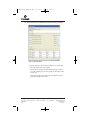
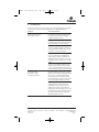
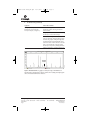
tbd021.0810:EIVD_TM.qxd
2/11/2011
8:02 AM
Page a
Technical Bulletin
PowerPlex® Matrix
Standards, 310
INSTRUCTIONS FOR USE OF PRODUCT DG4640.
PRINTED IN USA.
Revised 8/10
Part# TBD021
tbd021.0810:EIVD_TM.qxd
2/11/2011
8:02 AM
Page 1
PowerPlex® Matrix Standards, 310
All technical literature is available on the Internet at: www.promega.com/tbs/
Please visit the web site to verify that you are using the most current version of this
Technical Bulletin. Please contact Promega Technical Services if you have questions on use
of this system. E-mail [email protected].
1. Description..........................................................................................................1
2. Product Components and Storage Conditions ............................................2
3. Detection of Matrix Fragments Using the ABI PRISM® 310
Genetic Analyzer and GeneMapper® ID Software.....................................3
A.
B.
C.
D.
Instrument Preparation .......................................................................................3
Matrix Sample Preparation.................................................................................4
Capillary Electrophoresis and Detection ..........................................................4
Matrix Generation for the ABI PRISM® 310 Genetic Analyzer.....................5
4. Troubleshooting.................................................................................................7
5. Appendix ...........................................................................................................10
A. Detection of Matrix Fragments Using the
ABI PRISM® 377 DNA Sequencer....................................................................10
B. Detection of Matrix Fragments Using the
ABI PRISM® 310 Genetic Analyzer and GeneScan® Software ....................14
C. Composition of Buffers and Solutions ............................................................17
6. Related Products ..............................................................................................17
1.
Description
Proper generation of a matrix file is critical to evaluate multicolor systems with
the ABI PRISM® 310 Genetic Analyzer and ABI PRISM® 377 DNA Sequencer. To
prepare a matrix, four standards are analyzed using the same electrophoresis
conditions as those for samples and allelic ladders. The PowerPlex® Matrix
Standards, 310, consists of DNA fragments labeled with four fluorescent dyes:
one tube contains DNA fragments labeled with fluorescein, one tube contains
DNA fragments labeled with carboxy-tetramethylrhodamine (TMR), one tube
contains DNA fragments labeled with 6-carboxy-4´,5´-dichloro-2´,7´dimethoxyfluorescein (JOE), and one tube contains DNA fragments labeled
with carboxy-X-rhodamine (CXR).
Promega Corporation · 2800 Woods Hollow Road · Madison, WI 53711-5399 USA
Toll Free in USA 800-356-9526 · Phone 608-274-4330 · Fax 608-277-2516 · www.promega.com
Printed in USA.
Revised 8/10
Part# TBD021
Page 1
tbd021.0810:EIVD_TM.qxd
1.
2/11/2011
8:02 AM
Page 2
Description (continued)
Use the fluorescein Matrix, JOE Matrix, TMR Matrix and CXR Matrix for the
blue, green, yellow and red standards, respectively. The PowerPlex® Matrix
Standards, 310, can be used with the PowerPlex® 16, PowerPlex® 16 HS,
PowerPlex® Y, PowerPlex® ES and PowerPlex® S5 Systems. It also can be used
with the PowerPlex® 1.2 System (fluorescein- and TMR-labeled) or any of the
fluorescent STR systems (fluorescein-labeled). A matrix should be generated for
each individual instrument. Protocols to operate the fluorescence-detection
instrumentation should be obtained from the manufacturer.
For information on other Promega fluorescent STR systems, refer to the
PowerPlex® 16 System Technical Manual #TMD012, PowerPlex® 16 HS System
Technical Manual #TMD022, PowerPlex® Y System Technical Manual #TMD018,
PowerPlex® ES System Technical Manual #TMD017, PowerPlex® S5 System
Technical Manual #TMD021 and GenePrint ® Fluorescent STR Systems Technical
Manual #TMD006. These Technical Manuals and additional product
information are available at: www.promega.com
2.
Product Components and Storage Conditions
Product
PowerPlex® Matrix Standards, 310
Size
50µl (each dye)
Cat.#
DG4640
Not for Medical Diagnostic Use. Includes:
•
•
•
•
•
50µl
50µl
50µl
50µl
1ml
Fluorescein Matrix
JOE Matrix
TMR Matrix
CXR Matrix
Blue Dextran Loading Solution
Storage Conditions: Store all components at –20°C in a nonfrost-free freezer.
Do not store reagents in the freezer door, where the temperature can fluctuate.
The fragments in the matrix standards are light-sensitive and must be stored in
the dark. We strongly recommend that the matrix standards be stored with
post-amplification reagents (away from pre-amplification materials) and used
separately with different pipettes, tube racks, etc.
Additional product information and ordering information for accessory
components and related products are available upon request from Promega or
at: www.promega.com
Promega Corporation · 2800 Woods Hollow Road · Madison, WI 53711-5399 USA
Toll Free in USA 800-356-9526 · Phone 608-274-4330 · Fax 608-277-2516 · www.promega.com
Part# TBD021
Page 2
Printed in USA.
Revised 8/10
tbd021.0810:EIVD_TM.qxd
3.
2/11/2011
8:02 AM
Page 3
Detection of Matrix Fragments Using the ABI PRISM® 310 Genetic
Analyzer and GeneMapper ® ID Software
Materials to Be Supplied by the User
• dry heating block, water bath or thermal cycler
• crushed ice or ice-water bath
• 310 capillaries, 47cm × 50µm
• performance optimized polymer 4 (POP-4™ polymer)
• 10X genetic analyzer buffer
• sample tubes and septa
• aerosol-resistant pipette tips
• Hi-Di™ formamide (Applied Biosystems Cat.# 4311320)
!
The quality of formamide is critical. Use Hi-Di™ formamide. Freeze formamide in
aliquots at –20°C. Multiple freeze-thaw cycles or long-term storage at 4°C may cause
breakdown of formamide. Poor-quality formamide may contain ions that compete
with DNA during injection, which results in lower peak heights and reduced
sensitivity. A longer injection time may not increase the signal.
!
Formamide is an irritant and a teratogen; avoid inhalation and contact with skin.
Read the warning label, and take the necessary precautions when handling this
substance. Always wear gloves and safety glasses when working with formamide.
3.A. Instrument Preparation
Refer to the ABI PRISM® 310 Genetic Analyzer User’s Manual for instructions on
cleaning the pump block, installing the capillary, calibrating the autosampler
and adding polymer to the syringe.
1. Open the ABI PRISM® 310 Data Collection Software, Version 3.1.0.
2. To preheat the ABI PRISM® 310 Genetic Analyzer to 60°C, select “Manual
Control” in the Window menu. In the Function menu, select “Temperature
Set”. Set Value to “60.0”, then select “Execute”. Close the Manual Control
screen.
3. In the File menu, select “New” to open the Create New menu. Open a
GeneScan® sample sheet (either “48-Tube” or “96-Tube”).
4. In the upper right corner of the sample sheet, “4 Dyes” should be selected.
Enter the appropriate sample information in the Sample Name field. Matrix
sample names should be descriptive; for example, add the color to the
sample name. Label tubes with the corresponding sample names.
5. To save the sample sheet, select “Save As" in the File menu. Assign a name
to the file, and save to the Sample Sheet folder. Close the file.
6. In the File menu, select “New” to open the Create New menu.
7. Open the GeneScan® injection list.
Promega Corporation · 2800 Woods Hollow Road · Madison, WI 53711-5399 USA
Toll Free in USA 800-356-9526 · Phone 608-274-4330 · Fax 608-277-2516 · www.promega.com
Printed in USA.
Revised 8/10
Part# TBD021
Page 3
tbd021.0810:EIVD_TM.qxd
2/11/2011
8:02 AM
Page 4
3.A. Instrument Preparation (continued)
8. Select the sample sheet (i.e., the .gss file) that was created in Step 5.
9. The recommended module is GS STR POP4 (1mL) F.md4; choose this
module using the pull-down menu.
The settings should be:
Inj. Secs:
Inj. kV:
Run kV:
Run °C:
Run Time (minutes):
3
15.0
15.0
60
30
Note: The injection time may need to be increased or decreased, depending
on instrument sensitivity. Peak heights of 1,000–4,000RFU are optimal for
matrix generation.
10. Select “none” for the matrix file.
3.B. Matrix Sample Preparation
1. Thaw the matrix standards. For each matrix standard, vortex the tube for
5–10 seconds to mix, then add 2µl of matrix standard to 25µl of Hi-Di™
formamide.
2. Denature each sample for 3 minutes at 95°C, and immediately chill on
crushed ice or in an ice-water bath for 3 minutes. Denature samples just
prior to loading.
3. Place tubes in the appropriate autosampler tray (48-tube or 96-tube).
4. Place the autosampler tray in the instrument, and close the instrument
doors.
3.C. Capillary Electrophoresis and Detection
1. After loading the sample tray and closing the doors, select “Run” to start
the capillary electrophoresis system.
2. Monitor the electrophoresis by observing the raw data and status windows.
Each sample will take approximately 40 minutes for syringe pumping,
sample injection and electrophoresis.
Note: The matrix files that are created will be .fsa files. After the run is
finished, save or transfer the .fsa files to a secure location where they can
be opened in a GeneMapper ® project.
Promega Corporation · 2800 Woods Hollow Road · Madison, WI 53711-5399 USA
Toll Free in USA 800-356-9526 · Phone 608-274-4330 · Fax 608-277-2516 · www.promega.com
Part# TBD021
Page 4
Printed in USA.
Revised 8/10
tbd021.0810:EIVD_TM.qxd
2/11/2011
8:02 AM
Page 5
3.D. Matrix Generation for the ABI PRISM® 310 Genetic Analyzer
1. Open a new GeneMapper ® project. To add matrix sample files to the new
project, select "Add Samples to Project" in the File menu. Choose the
appropriate run folder containing the .fsa files from Section 3.C. Highlight
the run folder, select “Add To List”, then “Add”.
2. To open the raw data for a specific matrix sample file, locate “Project” in
the upper left corner of the screen, and double-click on the run folder to
reveal the .fsa files
3. Choose a single .fsa file to observe the raw data. While viewing the raw
data, move the cursor to the region that is to the right of the primer peak
and to the left of at least five peaks. Choose a region in a flat part of the
baseline.
4. Record the data point value found at the lower left portion of the screen for
use in Step 6. Repeat this step for each matrix standard.
Dye Color
Blue
Green
Yellow
Red
Corresponding Matrix
Fluorescein Matrix
JOE Matrix
TMR Matrix
CXR Matrix
“Start At” Value
5. To create a new matrix, select “GeneMapper Manager” in the Tools menu.
Select the Matrices tab, then “New”.
6. Define the new matrix in the Matrix Editor (Figure 1).
Note: The Matrix Name, “Start At” values and Matrix Result values shown
in Figure 1 will change, depending on your instrument.
a. Assign a matrix name in the Matrix Name field.
b. Set Number of Dyes to “4”.
c. To select each matrix standard sample file, click on the dye color for
each matrix (B for fluorescein, G for JOE, Y for TMR and R for CXR).
Navigate to the .fsa sample file that corresponds to that dye, and
double-click on it to add the sample file. Repeat this step for each
matrix standard.
Note: To find the .fsa files in the default location, go to: “My Computer”,
“AB SW8DATA (D:)“, “Applied Bio”, “310”, then “Runs”, and locate the
correct run folder.
Promega Corporation · 2800 Woods Hollow Road · Madison, WI 53711-5399 USA
Toll Free in USA 800-356-9526 · Phone 608-274-4330 · Fax 608-277-2516 · www.promega.com
Printed in USA.
Revised 8/10
Part# TBD021
Page 5
tbd021.0810:EIVD_TM.qxd
2/11/2011
8:02 AM
Page 6
9106TA
3.D. Matrix Generation for the ABI PRISM® 310 Genetic Analyzer (continued)
Figure 1. The Matrix Editor.
d. Enter the data point value recorded from Step 4 in the “Start at” field.
Repeat this step for each matrix standard.
e. Click on the Create button. The Matrix Result should give a value of
1.000 when comparing a dye to itself. Typically, all other values will be
less than 1.000.
Select “OK”, and the matrix will be created in the Matrices tab of the
GeneMapper Manager. Select “Done”.
Promega Corporation · 2800 Woods Hollow Road · Madison, WI 53711-5399 USA
Toll Free in USA 800-356-9526 · Phone 608-274-4330 · Fax 608-277-2516 · www.promega.com
Part# TBD021
Page 6
Printed in USA.
Revised 8/10
tbd021.0810:EIVD_TM.qxd
4.
2/11/2011
8:02 AM
Page 7
Troubleshooting
For questions not addressed here, please contact your local Promega Branch Office or Distributor.
Contact information available at: www.promega.com. E-mail: [email protected]
Symptoms
Causes and Comments
Unable to generate a matrix
due to faint or no peaks
Poor capillary electrophoresis (CE) injection.
Re-inject the sample. Check the syringe for
leakage. Check the laser power.
Poor-quality formamide was used. Use only
fresh Hi-Di™ formamide when running samples
on the ABI PRISM® 310 Genetic Analyzer.
Samples were degraded due to improper
storage. Store matrix standards in the dark at
–20°C protected from light. Do not store in the
freezer door or in a frost-free freezer.
Peak heights were too low. Peak heights should
be 1,000–4,000RFU for the ABI PRISM® 310
Genetic Analyzer and 800–2,000RFU for the
ABI PRISM® 377 DNA Sequencer. To increase
peak heights, increase injection time or loading
volume.
Samples were not denatured. Heat-denature
samples, and immediately chill on crushed ice
or in an ice-water bath before loading the gel or
capillary. Denature samples just prior to loading.
Poor-quality matrix
(extra peaks visible in
one or all color channels)
CE-related artifacts (“spikes”). Minor voltage
changes or urea crystals passing by the laser can
cause “spikes” or unexpected peaks. Spikes
sometimes appear in one color but often are
easily identified by their presence in more than
one color. Re-inject the samples to confirm.
CE-related artifacts (contaminants).
Contaminants in the water used with the
ABI PRISM® 310 Genetic Analyzer and for
diluting the 10X genetic analyzer buffer can
generate peaks in the blue and green dye colors.
Use autoclaved water to clean the pump block
and prepare sample dilutions. Change vials,
and wash the buffer reservoir.
Promega Corporation · 2800 Woods Hollow Road · Madison, WI 53711-5399 USA
Toll Free in USA 800-356-9526 · Phone 608-274-4330 · Fax 608-277-2516 · www.promega.com
Printed in USA.
Revised 8/10
Part# TBD021
Page 7
tbd021.0810:EIVD_TM.qxd
4.
2/11/2011
8:02 AM
Page 8
Troubleshooting (continued)
Symptoms
Causes and Comments
Poor-quality matrix (elevated
baseline and/or inverted peaks
in analyzed samples; see Figure 2)
Matrix used was generated on another
instrument. A matrix must be generated for
each instrument.
Wrong dye was used. Generate the matrix using
the same dyes as those in the samples.
Oversubtraction of signal occurred because
signal was saturated. When generating a matrix,
avoid choosing samples with peak heights that
are higher than the recommended RFU values,
as this can result in a matrix that causes inverted
peaks or elevated baseline. Analyzed sample
results may be improved by diluting the matrix
samples in water before preparing them for use.
2883TA03_0A
Elevated
baseline
Figure 2. Elevated baseline. A sample was analyzed using an ABI PRISM® 310
Genetic Analyzer and GeneScan® analysis software. The resulting electropherogram
shows an elevated baseline below 270 bases.
Promega Corporation · 2800 Woods Hollow Road · Madison, WI 53711-5399 USA
Toll Free in USA 800-356-9526 · Phone 608-274-4330 · Fax 608-277-2516 · www.promega.com
Part# TBD021
Page 8
Printed in USA.
Revised 8/10
tbd021.0810:EIVD_TM.qxd
2/11/2011
8:02 AM
Page 9
Symptoms
Causes and Comments
Inverted peaks in the matrix
baseline (Figure 3)
Incorrect or no “Start At” value was entered. The
“Start At” value chosen in Section 3.D, 5.A or
5.B should have a flat baseline.
Wrong colors were assigned to the dyes.
Confirm the dye and color selection:
Fluorescein:
Blue
JOE:
Green
TMR:
Yellow
CXR:
Red
Changes to or aging of instrument components.
Instrument sensitivity can change if the
instrument was moved or recently serviced
(replacement or realignment of the laser, CCD
camera, power supply or mirrors). The
sensitivity also can change over time due to
aging of the instrument. These changes can
result in poor matrix performance. Generate a
new matrix.
2884TA03_0A
Previously generated matrix
no longer performs optimally
Figure 3. Inverted baseline. The four matrix samples from the PowerPlex® Matrix
Standards, 310, were analyzed using an ABI PRISM® 310 Genetic Analyzer. A matrix
was made using the GeneScan® analysis software, but no “Start At” point was
entered for the matrix samples. The resulting matrix was applied to the JOE Matrix
sample file, and analysis was performed using all four colors. The result shows
inverted peaks in the blue, yellow and red channels.
Promega Corporation · 2800 Woods Hollow Road · Madison, WI 53711-5399 USA
Toll Free in USA 800-356-9526 · Phone 608-274-4330 · Fax 608-277-2516 · www.promega.com
Printed in USA.
Revised 8/10
Part# TBD021
Page 9
tbd021.0810:EIVD_TM.qxd
5.
2/11/2011
8:02 AM
Page 10
Appendix
5.A. Detection of Matrix Fragments Using the ABI PRISM® 377 DNA Sequencer
Materials to Be Supplied by the User
(Solution compositions are provided in Section 5.C.)
•
dry heating block, water bath or thermal cycler
•
crushed ice or ice-water bath
•
Long Ranger® gel solution (Lonza Cat.# 50611) or Long Ranger Singel®
pack for ABI sequencers 377-36cm (Lonza Cat.# 50691)
•
10% ammonium persulfate
•
TEMED
•
Urea (Cat.# V3171)
•
TBE 10X buffer
•
Nalgene® tissue culture filter (0.2 micron)
•
aerosol-resistant pipette tips
•
gel-loading pipette tips
•
36cm front and rear glass plates
•
36cm gel spacers (0.2mm thick)
•
36-well sharkstooth comb or 34-well squaretooth comb (0.2mm thick)
•
clamps (e.g., large office binder clamps)
•
Liqui-Nox® or other detergent
•
60cc syringe
•
30cc syringe
•
18-gauge needles
!
Acrylamide (Long Ranger® gel solution) is a neurotoxin and suspected
carcinogen; avoid inhalation and contact with skin. Read the warning label, and
take the necessary precautions when handling this substance. Always wear
gloves and safety glasses when working with acrylamide solutions.
Polyacrylamide Gel Preparation
Hazardous reagents are used in the preparation and use of gels for the
ABI PRISM® 377 DNA Sequencer. The reagents and their hazards are listed in
Table 1.
Table 1. Hazardous Reagents.
Reagents for ABI PRISM® 377 DNA Sequencer
acrylamide
(Long Ranger® gel solution)
ammonium persulfate
TEMED
urea
formamide
(contained in the Blue Dextran Loading Solution)
Hazard
suspected carcinogen,
toxic
oxidizer, corrosive
corrosive, flammable
irritant
irritant, teratogen
Promega Corporation · 2800 Woods Hollow Road · Madison, WI 53711-5399 USA
Toll Free in USA 800-356-9526 · Phone 608-274-4330 · Fax 608-277-2516 · www.promega.com
Part# TBD021
Page 10
Printed in USA.
Revised 8/10
tbd021.0810:EIVD_TM.qxd
2/11/2011
8:02 AM
Page 11
The following protocol is for preparing a 36cm denaturing polyacrylamide gel
for use with the ABI PRISM® 377 DNA Sequencer. Low-fluorescence glass plates
are recommended and can be obtained from the instrument manufacturer.
1. Thoroughly clean glass plates with hot water and a 1% Liqui-Nox® solution
or another dilute laboratory detergent solution. Rinse extremely well using
deionized water. Allow glass plates to air-dry in a dust-free environment.
2. Assemble glass plates by placing 0.2mm side gel spacers between the front
and rear glass plates. Hold plates together using binder clamps (four
clamps on each side). Place the assembly horizontally on a test tube rack or
similar support.
3. Prepare a 5% Long Ranger® acrylamide gel (total of 50ml) by combining
the reagents listed in Table 2. Stir the solution until the urea has dissolved.
Table 2. Preparation of a 5% Long Ranger® Polyacrylamide Gel.
Component
urea
deionized water
10X TBE
50% Long Ranger® gel solution
total volume
5% Gel
18g
26ml
5ml
5ml
50ml
Final Concentration
6M
–
1X
5%
Note: Long Ranger Singel® Packs can be used.
4. Filter the acrylamide solution through a 0.2 micron filter (e.g., Nalgene®
tissue culture filter), and degas for 5 minutes.
5. Add 35µl of TEMED and 250µl of fresh 10% ammonium persulfate to the
50ml of acrylamide solution, and mix gently.
6. Using a disposable 60cc syringe, pour the gel by starting at the well end of
the plates and carefully injecting the acrylamide between the horizontal
glass plates. Allow the solution to fill the top width of the plates. While
maintaining a constant flow of solution, gently tap the glass plates to assist
the movement of solution to the bottom of the plates.
7. Insert a 36-well sharkstooth comb or 34-well squaretooth comb between the
glass plates. Sharkstooth combs with 64 or 96 wells also can be used.
Note: The gel can be stored overnight by placing a paper towel saturated
with deionized water around the top and bottom and covering with plastic
wrap to prevent the gel from drying out. Crystallization of the urea will
destroy the gel.
8. Secure the comb with three evenly spaced clamps.
9. Keep the remaining acrylamide solution as a polymerization control.
10. Allow polymerization to proceed for >2 hours. Check the polymerization
control to be sure that polymerization has occurred.
Promega Corporation · 2800 Woods Hollow Road · Madison, WI 53711-5399 USA
Toll Free in USA 800-356-9526 · Phone 608-274-4330 · Fax 608-277-2516 · www.promega.com
Printed in USA.
Revised 8/10
Part# TBD021
Page 11
tbd021.0810:EIVD_TM.qxd
2/11/2011
8:02 AM
Page 12
5.A. Detection of Matrix Fragments Using the ABI PRISM® 377 DNA Sequencer
(continued)
Instrument Preparation
1. Open the ABI PRISM® 377 Data Collection Software.
2. Prepare a sample sheet as described in the GeneScan® Analysis Software
User’s Manual. Enter the appropriate sample information in the Sample Info
column.
3. Create a new GeneScan® run, and use the following settings:
Plate Check Module:
PreRun Module:
Run Module:
Collect Time:
Well-to-Read Distance:
Plate Check A
PR GS 36A-2400
GS 36A-2400
3 hours
36cm
4. Select the appropriate sample sheet and comb selection using the pull-down
menus.
5. Select “none” for the gel matrix file.
Gel Prerun
1. Remove the clamps from the polymerized acrylamide gel. If necessary,
clean any excess acrylamide from the glass plates with paper towels
saturated with deionized water.
2. Shave any excess polyacrylamide away from the comb, and remove the
comb. If using a sharkstooth comb, carefully insert the sharkstooth comb
teeth into the gel approximately 1–2mm.
3. Position the gel/glass plate unit in the 377 cassette.
4. Secure the cassette in the instrument, and perform a plate check as
recommended in the ABI PRISM® 377 DNA Sequencer User’s Manual. If the
horizontal line graph is not flat, remove the cassette, clean the plate surface
and repeat plate check.
5. Add 1X TBE buffer to the top and bottom buffer chambers of the instrument.
6. Using a 30cc syringe filled with buffer, remove any air bubbles and
unpolymerized acrylamide from the well area of the gel, and place the lid
on the upper buffer chamber. Using a syringe with a bent 18-gauge needle,
remove any air bubbles from the bottom of the gel.
7. Attach the heating plate, connect the water tubing, attach all electrodes,
close the instrument door, and click on the PreRun button. Allow the gel to
prerun for 15–20 minutes or until the gel temperature is at least 40°C. Open
the status window to monitor the gel temperature.
8. Prepare the matrix standards during the gel prerun.
Promega Corporation · 2800 Woods Hollow Road · Madison, WI 53711-5399 USA
Toll Free in USA 800-356-9526 · Phone 608-274-4330 · Fax 608-277-2516 · www.promega.com
Part# TBD021
Page 12
Printed in USA.
Revised 8/10
tbd021.0810:EIVD_TM.qxd
2/11/2011
8:02 AM
Page 13
Sample Preparation and Loading
1. Combine 1.5µl of each matrix standard with 1.5µl of Blue Dextran Loading
Solution.
2. Denature each sample for 3 minutes at 95°C, and immediately chill on
crushed ice or in an ice-water bath for 3 minutes. Denature samples just
prior to loading the gel.
Note: Instrument detection limits vary; therefore, the amount of product
mixed with loading cocktail may need to be increased or decreased.
3. After the 15- to 20-minute prerun, pause the instrument by selecting
“Pause”. When the prerun is paused, the water will continue to circulate to
keep the gel warm during the sample loading.
4. Use a 30cc syringe filled with buffer to flush the urea from the well area.
5. Load 1.5µl of each denatured sample into the respective wells.
6. Place the lid on the upper buffer chamber, and close the instrument door.
Gel Electrophoresis and Detection
1. After loading, select “Cancel” to stop the prerun. Make sure that the run
time is set at 3 hours, then select “Run” to begin electrophoresis.
2. Monitor electrophoresis by observing the gel image and status windows.
3. Allow electrophoresis to proceed for 3 hours. The largest fragment will
have migrated past the laser.
4. Track and extract the gel lanes.
Matrix Generation for the ABI PRISM® 377 DNA Sequencer
1. Open the GeneScan® project.
2. Review the raw data from the individual matrix samples. Highlight the
sample file name, then go to the Sample menu and select “raw data”. Move
the cursor beyond the primer peak so the crosshair is on a flat portion of
the baseline. Record the X value number shown at the bottom of the
window. Select an area for matrix generation. For optimal results, use as
many peaks as possible. See Figure 4.
3. In the File menu, select “New”, then click on the matrix icon. The Points
field should have the default value of 100,000. Click on the dye color for
each matrix, and indicate the sample file that corresponds to that dye. Enter
the recorded X value from Step 2 in the Start At field.
Dye Color
Blue
Green
Yellow
Red
Corresponding Matrix
Fluorescein Matrix
JOE Matrix
TMR Matrix
CXR Matrix
“Start At” Value
Promega Corporation · 2800 Woods Hollow Road · Madison, WI 53711-5399 USA
Toll Free in USA 800-356-9526 · Phone 608-274-4330 · Fax 608-277-2516 · www.promega.com
Printed in USA.
Revised 8/10
Part# TBD021
Page 13
tbd021.0810:EIVD_TM.qxd
2/11/2011
8:02 AM
Page 14
5.A. Detection of Matrix Fragments Using the ABI PRISM® 377 DNA Sequencer
(continued)
4. Select “OK”, and the matrix file will be generated.
5. Save the matrix file in the Matrix Standards folder located in the
GeneScan® folder. A copy of the matrix file should be stored in the ABI
folder located in the system folder.
6. A new matrix can be applied to previously run samples by highlighting the
sample in the GeneScan® project. In the Sample menu, select “Install new
matrix”, highlight the new matrix and select “Open”. The new matrix will
be applied to the sample file, and the samples can be analyzed using the
new matrix.
Reuse of Glass Plates
Separate the glass plates, and discard the gel. Clean glass plates with hot water
and a detergent such as 1% Liqui-Nox® detergent. Rinse extremely well with
deionized water, and allow plates to air-dry. Do not scrape plates with
abrasive materials during this process.
Gel extrusion (i.e., gel expands into the comb during a run) can occur due to a
buildup of residue. If this occurs, soak the plates in 2N HCl for 15 minutes,
then rinse thoroughly.
5.B. Detection of Matrix Fragments Using the ABI PRISM® 310 Genetic Analyzer
and GeneScan® Software
Materials to Be Supplied by the User
•
dry heating block, water bath or thermal cycler
•
crushed ice or ice-water bath
•
310 capillaries, 47cm × 50µm
•
performance optimized polymer 4 (POP-4™ polymer)
•
glass syringe (1ml)
•
sample tubes and septa
•
aerosol-resistant pipette tips
•
10X genetic analyzer buffer
•
Hi-Di™ formamide (Applied Biosystems Cat.# 4311320)
!
The quality of formamide is critical. Use Hi-Di™ formamide. Freeze
formamide in aliquots at –20°C. Multiple freeze-thaw cycles or long-term
storage at 4°C may cause breakdown of formamide. Poor-quality formamide
may contain ions that compete with DNA during injection, which results in
lower peak heights and reduced sensitivity. A longer injection time may not
increase the signal.
!
Formamide is an irritant and a teratogen; avoid inhalation and contact with
skin. Read the warning label, and take the necessary precautions when
handling this substance. Always wear gloves and safety glasses when working
with formamide.
Promega Corporation · 2800 Woods Hollow Road · Madison, WI 53711-5399 USA
Toll Free in USA 800-356-9526 · Phone 608-274-4330 · Fax 608-277-2516 · www.promega.com
Part# TBD021
Page 14
Printed in USA.
Revised 8/10
tbd021.0810:EIVD_TM.qxd
2/11/2011
8:02 AM
Page 15
Instrument Preparation
1. Refer to the ABI PRISM® 310 Genetic Analyzer User’s Manual for instructions
on cleaning the pump block, installing the capillary, calibrating the
autosampler and adding polymer to the syringe.
2. Open the ABI PRISM® 310 Data Collection Software.
3. Prepare a GeneScan® sample sheet as described in the ABI PRISM® 310
Genetic Analyzer User’s Manual. Enter the appropriate sample information in
the Sample Info column. Create a new GeneScan® injection list. Select the
appropriate sample sheet using the pull-down menu.
4. Select the “GS STR POP4 (1ml) A” Module using the pull-down menu.
Change the run time to 30 minutes, and keep the settings for the remaining
parameters as shown below:
Inj. Secs:
Inj. kV:
Run kV:
Run °C:
Run Time (minutes):
5
15.0
15.0
60
30
Note: The injection time may need to be optimized for individual
instruments.
5. Select “none” for the matrix file.
Sample Preparation
1. Thaw the matrix standards. For each matrix standard, vortex the tube for
5–10 seconds to mix, then add 2µl of matrix standard to 25µl of Hi-Di™
formamide or water.
2. Denature each sample for 3 minutes at 95°C, and immediately chill on
crushed ice or in an ice-water bath for 3 minutes. Denature samples just
prior to loading.
3. Place tubes in the appropriate autosampler tray (48-tube or 96-tube).
4. Place the autosampler tray in the instrument, and close the instrument doors.
Capillary Electrophoresis and Detection
1. After loading the sample tray and closing the doors, select “Run” to start
the capillary electrophoresis system.
2. Monitor the electrophoresis by observing the raw data and status windows.
Each sample will take approximately 40 minutes for syringe pumping,
sample injection and electrophoresis.
Promega Corporation · 2800 Woods Hollow Road · Madison, WI 53711-5399 USA
Toll Free in USA 800-356-9526 · Phone 608-274-4330 · Fax 608-277-2516 · www.promega.com
Printed in USA.
Revised 8/10
Part# TBD021
Page 15
tbd021.0810:EIVD_TM.qxd
2/11/2011
8:02 AM
Page 16
5.B. Detection of Matrix Fragments Using the ABI PRISM® 310 Genetic Analyzer
and GeneScan® Software (continued)
Matrix Generation for the ABI PRISM® 310 Genetic Analyzer
1. Open the GeneScan® project.
2. Review the raw data from the individual matrix standards. Highlight the
sample file name, then go to the Sample menu and select “raw data”. Move
the cursor beyond the primer peak, so the crosshair is on a flat portion of
the baseline. Record the X value number shown at the bottom of the
window. See Figure 4. Select an area for matrix generation. For optimal
results, use as many peaks as possible.
3. In the File menu, select “New,” then click on the Matrix icon. The “points”
field should have the default value of 100,000. Click on the dye color for
each matrix, and indicate the sample file that corresponds to that dye. Enter
the X value recorded from Step 2 in the “Start At” field.
Dye Color
Blue
Green
Yellow
Red
Corresponding Matrix
Fluorescein Matrix
JOE Matrix
TMR Matrix
CXR Matrix
“Start At” Value
4. Select “OK”, and the matrix file will be generated.
5. Save the matrix file in the Matrix Standards folder located in the
GeneScan® folder. For the Macintosh® version of the software, a copy of the
matrix file is automatically saved in the GS Matrix folder. For the Windows
NT® version of the software, store a copy of the matrix file in the Matrix
folder at: C:\appliedbio\shared\analysis\sizecaller\matrix.
“Start At”
2882TA03_0A
+
Figure 4. TMR matrix raw data. The TMR matrix standard was analyzed using an
ABI PRISM® 310 Genetic Analyzer. GeneScan® analysis software was used to view
the “raw data” (in the Sample menu). The cursor was placed on the baseline, and the
“Start At” value of 3529 was determined using the readout in the lower left corner of
the window.
Promega Corporation · 2800 Woods Hollow Road · Madison, WI 53711-5399 USA
Toll Free in USA 800-356-9526 · Phone 608-274-4330 · Fax 608-277-2516 · www.promega.com
Part# TBD021
Page 16
Printed in USA.
Revised 8/10
tbd021.0810:EIVD_TM.qxd
2/11/2011
8:02 AM
Page 17
6. A new matrix can be applied to previously run samples by highlighting the
sample in the GeneScan® project. In the Sample menu, select “Install new
matrix”, highlight the new matrix and select “Open”. The new matrix will
be applied to the sample file, and the samples can be analyzed using the
new matrix.
5.C. Composition of Buffers and Solutions
10% ammonium persulfate
Add 0.05g of ammonium persulfate
(Cat.# V3131) to 500µl of deionized
water. Use 250µl of 10% ammonium
persulfate for each 50ml of
acrylamide gel solution.
Blue Dextran Loading Solution
88.25%
15mg/ml
4.1mM
6.
formamide
blue dextran
EDTA (pH 8.0)
TBE 10X buffer
107.8g Tris base
7.44g EDTA
(Na2EDTA • 2H2O)
~55.0g boric acid
Dissolve Tris base and EDTA in
800ml of deionized water. Slowly
add boric acid, and monitor the pH
until the desired pH of 8.3 is
obtained. Bring the volume to 1 liter
with deionized water.
Related Products
Product
PowerPlex® Matrix Standards, 3100/3130
PowerPlex® 16 System
PowerPlex® 16 HS System
PowerPlex® Y System
PowerPlex® S5 System
PowerPlex® 1.2 System
PowerPlex® ES System
Size
25µl (each dye)
100 reactions
400 reactions
100 reactions
400 reactions
50 reactions
200 reactions
100 reactions
400 reactions
100 reactions
100 reactions
400 reactions
Cat.#
DG4650
DC6531
DC6530
DC2101
DC2100
DC6761
DC6760
DC6951
DC6950
DC6101
DC6731
DC6730
Not for Medical Diagnostic Use.
Promega Corporation · 2800 Woods Hollow Road · Madison, WI 53711-5399 USA
Toll Free in USA 800-356-9526 · Phone 608-274-4330 · Fax 608-277-2516 · www.promega.com
Printed in USA.
Revised 8/10
Part# TBD021
Page 17
tbd021.0810:EIVD_TM.qxd
6.
2/11/2011
8:02 AM
Page 18
Related Products (continued)
Accessory Components
Product
Internal Lane Standard 600*
Gold ST★R 10X Buffer
Nuclease-Free Water*
Size
150µl
1.2ml
50ml
Cat.#
DG1071
DM2411
P1193
*For Laboratory Use.
© 2006, 2010 Promega Corporation. All Rights Reserved.
GenePrint and PowerPlex are registered trademarks of Promega Corporation.
ABI PRISM, GeneMapper and GeneScan are registered trademarks of Applera Corporation. Hi-Di and POP-4 are trademarks
of Applera Corporation. Liqui-Nox is a registered trademark of Alconox, Inc. Long Ranger and Long Ranger Singel are
registered trademarks of Lonza BioProducts. Macintosh is a registered trademark of Apple Computer, Inc. Nalgene is a
registered trademark of Nalge Nunc International. Windows NT is a registered trademark of Microsoft Corporation.
Products may be covered by pending or issued patents or may have certain limitations. Please visit our Web site for more
information.
All prices and specifications are subject to change without prior notice.
Product claims are subject to change. Please contact Promega Technical Services or access the Promega online catalog for the
most up-to-date information on Promega products.
Promega Corporation · 2800 Woods Hollow Road · Madison, WI 53711-5399 USA
Toll Free in USA 800-356-9526 · Phone 608-274-4330 · Fax 608-277-2516 · www.promega.com
Part# TBD021
Page 18
Printed in USA.
Revised 8/10