1
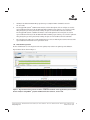
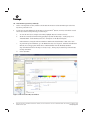
TECHNICAL MANUAL
PowerPlex® 6C Matrix
Standard
InstrucƟons for Use of Product
DG4900
Revised 10/15
TMD046
PowerPlex® 6C Matrix Standard
All technical literature is available at: www.promega.com/protocols/
Visit the web site to verify that you are using the most current version of this Technical Manual.
E-mail Promega Technical Services if you have questions on use of this system: [email protected]
1. Description......................................................................................................................................... 1
2. Product Components and Storage Conditions ........................................................................................ 2
3. Instrument Preparation and Spectral Calibration Using the
Applied Biosystems® 3500 and 3500xL Genetic Analyzers ...................................................................... 2
3.A. Matrix Sample Preparation ......................................................................................................... 2
3.B. Instrument Preparation .............................................................................................................. 3
4. Instrument Preparation and Spectral Calibration Using the Applied Biosystems® 3130 and 3130xl
Genetic Analyzers with Data Collection Software Version 4.0 with the DC v4 6-Dye Module v1 License .........5
4.A. Matrix Sample Preparation ......................................................................................................... 5
4.B. Instrument Preparation .............................................................................................................. 6
5. Troubleshooting.................................................................................................................................. 8
5.A. Spectral Calibration .................................................................................................................... 8
5.B. Spectral Calibration on the Applied Biosystems® 3130 and 3130xl Genetic Analyzers ...................... 9
6. Summary of Changes ......................................................................................................................... 10
1.
Description
Proper generation of a spectral calibration file is critical to evaluate multicolor systems with the Applied Biosystems®
3130, 3130xl, 3500 and 3500xL Genetic Analyzers. The PowerPlex® 6C Matrix Standard(a,b) consists of DNA fragments
labeled with six different fluorescent dyes (FL-6C, JOE-6C, TMR-6C, CXR-6C, TOM-6C and WEN) in one tube. The
spectral calibration is performed using the J6 dye set. Once generated, the spectral calibration file is applied during
sample detection to calculate the spectral overlap and separate the raw fluorescent signals into individual color signals.
The PowerPlex® 6C Matrix Standard was developed for use with the Promega 6-color PowerPlex® Systems and is
compatible with the Applied Biosystems® 3500 and\ 3500xL Genetic Analyzers as well as Applied Biosystems® 3130
and 3130xl Genetic Analyzers with Data Collection Software Version 4.0 with the DC v4 6-Dye Module v1 License (Life
Technologies). A protocol to operate the Applied Biosystems® 3130, 3130xl, 3500 or 3500xL Genetic Analyzer should
be obtained from the manufacturer.
A spectral calibration must be generated for each individual instrument. A new matrix should be run after major
maintenance on the system, such as changing the laser, calibrating or replacing the CCD camera or changing the
polymer type or capillary array. We also recommend that you generate a new matrix after the instrument is moved to a
new location. In some instances, a software upgrade may necessitate generation of a new matrix. Individual labs should
determine the frequency of matrix generation.
Promega CorporaƟon · 2800 Woods Hollow Road · Madison, WI 53711-5399 USA · Toll Free in USA 800-356-9526 · 608-274-4330 · Fax 608-277-2516
www.promega.com
TMD046 · Revised 10/15
1
2.
Product Components and Storage Conditions
PRODUCT
PowerPlex® 6C Matrix Standard
SIZE
C A T. #
5 preps
DG4900
Not For Medical Diagnostic Use. Includes:
•
150µl
• 5 × 200µl
6C Matrix Mix
Matrix Dilution Buffer
Storage Conditions: Upon receipt, store all components at –30°C to –10°C in a nonfrost-free freezer. After the first
use, store the PowerPlex® 6C Matrix Standard components at 2–10°C. We strongly recommend that the PowerPlex® 6C
Matrix Standard be stored with post-amplification reagents. The PowerPlex® 6C Matrix Standard is light-sensitive;
dilute the 6C Matrix Mix in the Matrix Dilution Buffer in the provided amber tube. Store the diluted 6C Matrix Mix at
2–10°C for up to 1 week.
3.
Instrument Preparation and Spectral Calibration Using the Applied Biosystems® 3500 and 3500xL
Genetic Analyzers
Materials to Be Supplied by the User
•
centrifuge compatible with 96-well plates
•
aerosol-resistant pipette tips
•
3500/3500xL capillary array, 36cm
•
POP-4® polymer for the 3500 or 3500xL
•
•
•
anode buffer container with 1X buffer
cathode buffer container with 1X buffer
MicroAmp® optical 96-well plate and septa
•
Hi-Di™ formamide (Applied Biosystems Cat.# 4311320)
For additional information on performing spectral calibration, refer to the Applied Biosystems 3500/3500xL Genetic
Analyzer User Guide.
3.A. Matrix Sample Preparation
1.
At the first use, thaw the 6C Matrix Mix and Matrix Dilution Buffer completely. After the first use, store the
reagents at 2–10°C.
2.
Vortex the 6C Matrix Mix for 10–15 seconds prior to use. Add 10µl of the 6C Matrix Mix to one tube of Matrix
Dilution Buffer. Vortex for 10–15 seconds. Label the tube with the date of dilution. The diluted 6C Matrix Mix
can be stored for up to 1 week at 2–10°C.
2
Promega CorporaƟon · 2800 Woods Hollow Road · Madison, WI 53711-5399 USA · Toll Free in USA 800-356-9526 · 608-274-4330 · Fax 608-277-2516
TMD046 · Revised 10/15
www.promega.com
3.
Add 10µl of the diluted 6C Matrix Mix prepared in Step 2 to 500µl of Hi-Di™ formamide. Vortex for
10–15 seconds.
4.
For the Applied Biosystems® 3500xL Genetic Analyzer, wells A1 through H3 of the 96-well plate are used for
spectral calibration. Add 15μl of the 6C Matrix Mix with formamide prepared in Step 3 to each of the 24 wells.
After placing the septa on the plate, briefly centrifuge the plate to remove bubbles. Do not heat denature.
For the Applied Biosystems® 3500 Genetic Analyzer, wells A1 through H1 of the 96-well plate are used for
spectral calibration. Add 15μl of the 6C Matrix Mix with formamide prepared in Step 3 to each of the eight wells.
After placing the septa on the plate, briefly centrifuge the plate to remove bubbles. Do not heat denature.
5.
Place the plate in the 3500 series 96-well standard plate base, and cover with the plate retainer. Do not start the
spectral calibration run until the oven is preheated to 60°C.
3.B. Instrument Preparation
We have found that the use of fresh polymer and a new capillary array results in an optimal spectral calibration.
12793TA
Representative data are shown in Figure 1.
Figure 1. Representative data for the PowerPlex® 6C Matrix Standard on the Applied Biosystems 3500xL
Genetic Analyzer using POP-4® polymer and Data Collection Software, Version 2.0.
Promega CorporaƟon · 2800 Woods Hollow Road · Madison, WI 53711-5399 USA · Toll Free in USA 800-356-9526 · 608-274-4330 · Fax 608-277-2516
www.promega.com
TMD046 · Revised 10/15
3
3.B. Instrument Preparation (continued)
1.
Set the oven temperature to 60°C, and then select the Start Pre-Heat icon at least 30 minutes prior to the first
injection to preheat the oven.
2.
To perform a spectral calibration for the Promega 6-color PowerPlex® Systems, a new dye set should be created.
If a new dye set was created previously, proceed to Step 2c.
a.
To create the new dye set, navigate to the Library, highlight “Dye Sets” and select “Create”.
b.
The Create a New Dye Set window will appear (Figure 2). Name the Dye Set (i.e., Promega J6), select
“Matrix Standard” for the Chemistry and select “J6 Template” for the Dye Set Template.
Under Parameters, change the After Scan number to 800 from the default number of 500. Select “Save”.
To perform the spectral calibration, go to the Maintenance tab, select “Spectral”, and under the Calibration
Run tab, choose the appropriate fields: Choose “Matrix Standard” from the Chemistry Standard
drop-down menu and the new Promega 6-color dye set (i.e., Promega J6) created in Step 2b from the Dye
Set drop-down menu.
d.
Select “Start Run”.
12794TA
c.
Figure 2. The Create New Dye Set window.
4
Promega CorporaƟon · 2800 Woods Hollow Road · Madison, WI 53711-5399 USA · Toll Free in USA 800-356-9526 · 608-274-4330 · Fax 608-277-2516
TMD046 · Revised 10/15
www.promega.com
3.
If fewer than the recommended number of capillaries pass, the spectral calibration run will be repeated automatically
up to three times. Upon completion of the spectral calibration, check the quality of the spectral in the Capillary
Run Data display and choose either “Accept” or “Reject”.
Note: Refer to the 3500 Series Data Collection Software User Manual for the criteria recommended when
accepting or rejecting a spectral calibration.
4.
Instrument Preparation and Spectral Calibration Using the Applied Biosystems® 3130 and 3130xl
Genetic Analyzers with Data Collection Software Version 4.0 with the DC v4 6-Dye Module v1 License
Materials to Be Supplied by the User
•
centrifuge compatible with 96-well plates
•
aerosol-resistant pipette tips
•
3130/3130xl capillary array, 36cm
•
POP-4® polymer for the 3130 or 3130xl
•
•
•
10X genetic analyzer buffer with EDTA
MicroAmp optical 96-well plate and septa
Hi-Di™ formamide (Applied Biosystems Cat.# 4311320)
For additional information on performing spectral calibration, refer to the Applied Biosystems 3130/3130xl Genetic
Analyzer User Guide.
4.A. Matrix Sample Preparation
1.
At the first use, thaw the 6C Matrix Mix and Matrix Dilution Buffer completely. After the first use, store the
reagents at 2–10°C.
2.
Vortex the 6C Matrix Mix for 10–15 seconds prior to use. Add 10μl of the 6C Matrix Mix to one tube of Matrix
Dilution Buffer. Vortex for 10–15 seconds. Note the date of dilution on the tube. The diluted 6C Matrix Mix can
be stored for up to 1 week at 2–10°C.
3.
Add 10μl of the diluted 6C Matrix Mix prepared in Step 2 to 500μl of Hi-Di™ formamide. Vortex for
10–15 seconds.
4.
For the Applied Biosystems 3130xl Genetic Analyzer, 16 wells are used for spectral calibration on 16 capillaries
(wells A1 through H2 of a 96-well plate). Add 15µl of the 6C Matrix Mix with formamide prepared in Step 3 to
each of the 16 wells. After placing the septa on the plate, briefly centrifuge the plate to remove bubbles. Do not
heat denature.
For the Applied Biosystems 3130 Genetic Analyzers, four wells are used for spectral calibration on four capillaries
(wells A1 through D1 in a 96-well plate). Add 15µl of the 6C Matrix Mix with formamide prepared in Step 3 to
each of the four wells. After placing the septa on the plate, briefly centrifuge the plate to remove bubbles. Do not
heat denature.
5.
Place the plate in the 3130 series 96-well standard plate base, and cover with the plate retainer. Do not start the
spectral calibration run until the oven is preheated to 60°C.
Promega CorporaƟon · 2800 Woods Hollow Road · Madison, WI 53711-5399 USA · Toll Free in USA 800-356-9526 · 608-274-4330 · Fax 608-277-2516
www.promega.com
TMD046 · Revised 10/15
5
4.B. Instrument Preparation
We have found that the use of fresh polymer and a new capillary array results in an optimal spectral calibration.
12795TA
Representative data are shown in Figure 3.
Figure 3. Representative data for the PowerPlex® 6C Matrix Standard on the Applied Biosystems 3130
Genetic Analyzer with Data Collection Software Version 4.0 with the DC v4 6-Dye Module v1 License
using POP-4® polymer.
1.
Set the oven temperature to 60°C, and preheat the oven for at least 15 minutes prior to the first injection.
2.
To perform a spectral calibration for Promega 6-color PowerPlex® Systems, a new Run Module and Protocol
should be created. If a new Run Module and Protocol were created previously, proceed to Step 2.d.
a.
6
In the Run Module Editor, select “New”. Select “SPECTRAL” in the Type drop-down list, and select
“Spect36_POP4” in the Template drop-down list. Change the Data Delay Time to 400 and the Run Time to
800. Change the Injection Time to 6 seconds. See Figure 4.
Promega CorporaƟon · 2800 Woods Hollow Road · Madison, WI 53711-5399 USA · Toll Free in USA 800-356-9526 · 608-274-4330 · Fax 608-277-2516
TMD046 · Revised 10/15
www.promega.com
12796TA
Figure 4. Run Module Editor settings.
b.
Name the Run Module (e.g., Promega J6), and select “OK”.
c.
In the Protocol Manager, under Instrument Protocols, select “New”. Type a name for your protocol
(e.g., Promega J6).
d.
Make the following selections in the Protocol Editor:
•
“Spectral” in the Type drop-down list
•
“J6” in the DyeSet drop-down list
•
“POP4” for the polymer
•
“36” in the Array Length drop-down list
•
“Matrix Standard” for the chemistry
•
Select the spectral module you created in the previous step in the Run Module drop-down list.
•
Select “Edit Parameters”, and change the Minimum Quality Score (“Q value”) to 0.95.
•
Select “OK” in the “Edit Parameters” window, and select “OK” in the Protocol Editor.
3.
In the Plate Manager, create a new plate record as described in the instrument user’s manual. In the dialog box
that appears, select “Spectral Calibration” in the Application drop-down list, and select “96-well” as the plate
type. Add entries in the owner and operator windows, name the plate and select “OK”.
4.
In the spectral calibration plate editor dialog box, place sample names in the appropriate cells.
5.
In the Instrument Protocol column, select the protocol you created in Step 2. Ensure that this information is
present for each sample. Select “OK”.
Promega CorporaƟon · 2800 Woods Hollow Road · Madison, WI 53711-5399 USA · Toll Free in USA 800-356-9526 · 608-274-4330 · Fax 608-277-2516
www.promega.com
TMD046 · Revised 10/15
7
4.B. Instrument Preparation (continued)
6.
Run your plate as described in the instrument user’s manual.
7.
Upon completion of the run, check the status of the spectral calibration in the Event Log window. For the Applied
Biosystems® 3130xl Genetic Analyzers, we recommend that a minimum of 12 of the 16 capillaries pass calibration.
For the Applied Biosystems® 3130 Genetic Analyzers, we recommend that a minimum of three of four capillaries
pass calibration. If fewer than the recommended numbers of capillaries pass, repeat the spectral calibration.
5.
Troubleshooting
5.A. Spectral Calibration
This section provides general information about spectral calibration. For information specific to the Applied Biosystems®
3130 and 3130xl Genetic Analyzers, see Section 5.B.
For questions not addressed here, please contact your local Promega Branch Office or Distributor. Contact information
available at: www.promega.com. E-mail: [email protected]
Symptoms
Fewer than the recommended number of
capillaries passed the spectral calibration
Causes and Comments
Poor-quality formamide was used. The quality of formamide is
critical. Use Hi-Di™ formamide. Freeze formamide in aliquots
at –20°C. Multiple freeze-thaw cycles or storage at 4°C may
cause breakdown of formamide. Poor-quality formamide may
contain ions that compete with DNA during injection, which
results in lower peak heights and reduced sensitivity.
Matrix standard was too dilute. Matrix standard that is too
dilute will result in low spectral calibration peak heights, which
may result in spectral calibration failure. Increase the volume of
diluted 6C Matrix Mix added to the Hi-Di™ formamide during
sample preparation.
Matrix standard was too concentrated. Matrix standard that is
too concentrated may result in spectral calibration failure due to
saturated peaks, bleedthrough or oversubtraction in other dye
colors. Decrease the volume of diluted 6C Matrix Mix added to
the Hi-Di™ formamide during matrix sample preparation.
Reboot the CE instrument and the instrument’s computer.
Repeat the spectral calibration.
Ensure that the oven is preheated to 60°C prior to spectral
calibration.
All capillaries failed spectral calibration
8
For best spectral calibration results, use fresh polymer, fresh
buffer and water, and a capillary array with fewer than
100 injections.
Promega CorporaƟon · 2800 Woods Hollow Road · Madison, WI 53711-5399 USA · Toll Free in USA 800-356-9526 · 608-274-4330 · Fax 608-277-2516
TMD046 · Revised 10/15
www.promega.com
5.B. Spectral Calibration on the Applied Biosystems® 3130 and 3130xl Genetic Analyzers
The following information is specific to spectral calibration on the Applied Biosystems® 3130 and 3130xl Genetic
Analyzers with Data Collection Software Version 4.0 with the DC v4 6-Dye Module v1 License. For general troubleshooting information about spectral calibration, see Section 5.A.
Symptoms
Fewer than the recommended number of
capillaries passed the spectral calibration
Causes and Comments
Peak heights for the matrix standard were too low. Matrix peak
heights must be a minimum of 750RFU to pass spectral
calibration. Increase the injection time.
Peak heights for the matrix standard were too high
(>5,000RFU). Decrease the injection time.
Check the Event Log in the Instrument Status screen for a
reason for the failure (e.g., bad dye order or too many spectral
calibration candidates).
Check the raw data view of the failed capillaries in the Spectral
Viewer (failed capillaries are shaded tan in the plate diagram).
Look for signs of low signal, high signal or baseline noise before
the matrix peaks. Adjust the run conditions as described below,
and re-inject the matrix standard.
If the cause for failure is unclear after viewing the Event Log and
Spectral Viewer, monitoring fragment migration in the Capillaries
Viewer during spectral calibration run can provide information
that will be useful for troubleshooting purposes. Re-inject the
matrix standard, and monitor the Capillaries Viewer during the
run. Note any unusual peak formations or extremely low or high
peak heights. Based on the information obtained while watching
the Capillaries Viewer, you may need to adjust the run conditions.
Peaks in multiple dye channels were detected prior to the orange
peak. Increase the data delay time to exclude those peaks, and
re-inject the matrix standard.
The data delay time specified for the Applied Biosystems® 3130
or 3130xl Genetic Analyzer was too long, and the orange peak
was not captured during data collection. Decrease the data delay
time, and re-inject the matrix standard.
Promega CorporaƟon · 2800 Woods Hollow Road · Madison, WI 53711-5399 USA · Toll Free in USA 800-356-9526 · 608-274-4330 · Fax 608-277-2516
www.promega.com
TMD046 · Revised 10/15
9
6.
Summary of Changes
The following changes were made to the 10/15 revision of this document:
1.
Expired legal disclaimers were removed.
2.
The WEN dye name was changed to “WEN” from “WEN-6C”.
(a)
Patent Pending.
(b)
TMR-6C, CXR-6C, TOM-6C and WEN dyes are proprietary.
© 2015 Promega CorporaƟon. All Rights Reserved.
PowerPlex is a registered trademark of Promega CorporaƟon.
Applied Biosystems and MicroAmp are registered trademarks of Applied Biosystems. Hi-Di is a trademark of Applera CorporaƟon. POP-4 is a registered
trademark of Life Technologies CorporaƟon.
Products may be covered by pending or issued patents or may have certain limitaƟons. Please visit our Web site for more informaƟon.
All prices and specificaƟons are subject to change without prior noƟce.
Product claims are subject to change. Please contact Promega Technical Services or access the Promega online catalog for the most up-to-date
informaƟon on Promega products.
10
Promega CorporaƟon · 2800 Woods Hollow Road · Madison, WI 53711-5399 USA · Toll Free in USA 800-356-9526 · 608-274-4330 · Fax 608-277-2516
TMD046 · Revised 10/15
www.promega.com