1
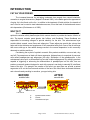
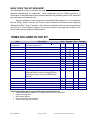
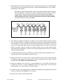
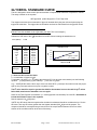
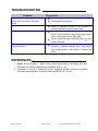
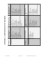
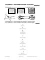
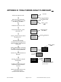
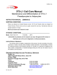
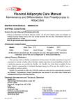
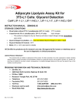
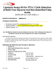
Wrinkle Treatment Screening Kit Human Adipocyte Differentiation Assay Cat# DIF-10 INSTRUCTION MANUAL ZBM0020.01 STORAGE CONDITIONS Frozen human preadipocytes Store in liquid nitrogen IMMEDIATELY upon receipt. No expiration date is applicable; however, the cells must be plated within 1 week of receiving the kit to account for the expiration of the kit components. Media, Reagents A & B, Buffers: Store at 2 - 8 C. See kit label for expiration date [NOTE: expiration is 4 weeks from date of manufacture.] Glycerol Standard -20°C For Research Use Only Not For Use In Diagnostic Procedures LIMITED PRODUCT WARRANTY This warranty limits our liability to replacement of this product. No other warranties of any kind, expressed or implied, including without limitation, implied warranties of merchantability or fitness for a particular purpose, are provided by Zen-Bio, Inc. Zen-Bio, Inc. shall have no liability for any direct, indirect, consequential, or incidental damages arising out of the use, the results of use, or the inability to use this product. ORDERING INFORMATION AND TECHNICAL SERVICES Zen-Bio, Inc. 3200 Chapel Hill-Nelson Blvd., Suite 104 PO Box 13888 Research Triangle Park, NC 27709 Telephone (919) 547-0692 Facsimile (FAX) (919) 547-0693 Toll Free Electronic mail (e-mail) 1-866-ADIPOSE (866)-234-7673 [email protected] World Wide Web http://www.zenbio.com Rev 8/18/2008 Page 1 of 12 PATENT PROTECTED (6,153,432) INTRODUCTION FAT AS YOUR FRIEND The increased demand for anti-aging treatments has ranged from natural botanicals, cosmetics to surgical intervention ( Majeed & Prakash 2002). In the battle against the appearance of aging, fat is the friend, not the foe. In addition to the treatment of facial wrinkles, the transfer of one’s own fat can be used to treat indented acne scars, fill out the back of the hands and correct skin depressions (Christensen et al. 2005). WHY FAT? Skin is made up of 3 layers: the epidermis, the dermis and the subcutaneous layer. The epidermis contains mainly keratinocytes which secrete keratin to provide the barrier function of skin. The dermis contains sweat glands, hair follicles, and fibroblasts. These fibroblasts are responsible for secreting collagen to provide elasticity for the skin. The subcutaneous layer contains blood vessels, nerve fibers and adipocytes. These adipocytes provide the volume that helps add to the thickness and suppleness of skin associated with youth. Loss of the fat cell layer that occurs with age or free radical damage results in the uneven depression in skin commonly observed as wrinkles and lines. Considering the myriad of synthetic cosmetic filler products available, one must ask, why use fat? Fat presents as an ideal soft tissue augmenter. Adipose tissue is comprised of precursor cells called preadipocytes and adipocytes (fat cells). Stimulation of the preadipocytes, in the subcutaneous skin layer, to differentiate into fat cells is called adipogenesis. By studying products capable of triggering or enhancing the differentiation of preadipocytes into fat cells, one can produce a cosmetic product designed to work with a person’s own body to naturally fill in deficient areas of the skin. For example, the creation of fat cells in an aged face can provide a natural augmenter to fill out the fine lines and wrinkles of the face. Skin would have increased thickness and volume thereby resulting in smoother, younger looking skin. BEFORE AFTER wrinkled skin smooth skin A A DERMIS DERMIS B B C C ADIPOGENESIS PREADIPOCYTES FAT CELLS FAT CELLS A B C Rev 8/18/2008 Epidermis/skin surface Dermis Subcutaneous layer Page 2 of 12 PATENT PROTECTED (6,153,432) WHAT DOES THIS KIT MEASURE? The differentiation assay kit provides the tools to study the compounds that stimulate human adipocyte differentiation or lipogenesis. Such compounds may be PPAR agonists or a combination of thiazolidinediones and glucocorticoids that are potentially useful in the stimulation and maintenance of human fat cells. This kit is designed to test compounds as potential PPAR agonists. It is our experience that the PPAR agonist used as the Positive Control sufficiently stimulates human adipocyte differentiation after 7 days of treatment. The protocol is designed so that the test compounds are also used in a 7-day treatment regimen. The end point assay is total triglyceride content of the cells 2 weeks after the initiation of differentiation. ITEMS INCLUDED IN THE KIT Item Description Human SQ Preadipocytes PM-1 NC Human subcutaneous preadipocytes, > 2.0 X10 cells/vial, cryopreserved Preadipocyte medium (See Appendix A) Negative control: (See Appendix A) PC Positive control: (See Appendix A) VC Vehicle control (See Appendix A) IM MM Wash buffer Lysis buffer Reagent A Reagent B Glycerol standard Standards diluent Tray Data sheet Plate A Assay Plate, Plate 6 Unit Quantity Item Storage VIAL 1 1 BOTTLE 50 ml VIAL 500 l 2 2 Liquid nitrogen 4°C 4°C VIAL 500 l 2 4°C VIAL 2 4°C 3 2 2 2 2 2 2 4°C 4°C 4°C 4°C 4°C 4°C -20°C Reconstitute w/ 2.5 ml deionized water prior to use 4°C 500 l 300 ml 100 ml 50 ml 25 ml 11 ml 2.5 ml Glycerol @ 1mM [Reconstitute with 200 l Standards Diluent to make the 200 M glycerol standard; see page 5 for recommended dilution scheme] VIAL 50 l BOTTLE 2 ml 3 1 1 2 BOTTLE Initiation medium (See Appendix A) Maintenance medium (See Appendix A) BOTTLE BOTTLE BOTTLE Reconstitute w/ 11.0 ml deionized water prior to use Clear polyvinyl tray for multi-channel pipetters Certificate of Analysis and protocol 96-well plate for plating and differentiating 96-well plate, blank (for samples & standards) BOTTLE EACH EACH PLATE PLATE Other equipment/reagents required but not provided with the kit: Additional Maintenance Medium, if necessary (see background information) Single-channel pipetter Multi-channel pipetter Plate reader with a filter of 540 nm Tubes to dilute glycerol standards Rev 8/18/2008 Page 3 of 12 PATENT PROTECTED (6,153,432) ------------- 4°C ------------- ASSAY PROCEDURE A. Differentiation Procedure On each day of the procedure, the appropriate medium must be warmed to 37 C prior to use. Note: This protocol is designed to accommodate a weekday work schedule if started on a MondayThursday. Any deviation may require weekend work. We strongly recommend testing all compounds in triplicate. Day 1: This is the day the cells are plated. 1. Remove cells from liquid nitrogen and place immediately into a 37 C water bath and agitate while in bath. Be careful not to submerge the cap of the vial into water. Do not leave the vials in water bath after most of the content has thawed. Rinse the vials with 70% ethanol before taking them to the culture hood. 2. Upon thawing, transfer the cells to a sterile conical bottom centrifuge tube containing 10 ml of Preadipocyte medium (cat # PM-1). 3. Centrifuge: 1,200 rpm (282Xg) / 20 C / 5 minutes. Aspirate the supernatant. TAKE CARE TO NOT ASPIRATE ANY OF THE CELL PELLET. 6 4. The cell vial contains a minimum of 2.0 x 10 viable cells; however, we recommend performing a cell count to determine a more exact number of cells. Resuspend the cell pellet in 2 ml Preadipocyte medium; dilute an aliquot in 0.4% trypan blue solution. We suggest withdrawing an aliquot of 50 l of cells and mixing with 100 l of the trypan blue solution, resulting in a dilution factor of 3. Count live (unstained) cells on a hemacytometer. The cell concentration required for 2 6 approximately 40,000 cells / cm in the 96 well format with 150 l /well is 1.3 x 10 cells in 15 ml Preadipocyte medium. 5. Plate cells in one of the 96 well plates provided in the kit. Do not agitate the plate, as cells will not plate evenly. 6. Place plate in 37 C incubator, 5% CO2, 97% humidity. The cells will be maintained in the incubator after each manipulation until Day 14. NOTE: This kit contains a sufficient volume of Initiation medium (IM) to use 10 ml of medium per compound dilution for a maximum of 29 compounds tested in triplicate (87 wells) on the 96-well plate, leaving 9 wells for controls. If a compound stock is too concentrated to accomplish the desired dilution, use an appropriate solution (not supplied) to prepare an intermediate concentration that would allow for a final volume of 10 ml. Also the positive control in this kit, the PPAR agonist, has a final solvent concentration of 0.1% DMSO. This is included in the vehicle control (VC). If the concentration of any solvent for the compounds used is high enough to potentially alter differentiation, please include that solvent concentration as an additional treatment. We do not recommend treating the cells with solutions exceeding 1% of any solvent, as higher concentrations may be toxic to the cells. Rev 8/18/2008 Page 4 of 12 PATENT PROTECTED (6,153,432) Day 2: 7. Twenty-four hours later, check the cells for confluence. 8. Using the Initiation Medium (IM), prepare treatments. Plan to do all treatments in triplicate. A blank plate map is included in these instructions to record the well treatments. 9. When all treatments are prepared, remove Preadipocyte medium from control wells. We recommend doing the treatments in small groups so the cells do not dry out. 10. Pipet 150 l Positive Control, (PC), 150 l Negative Control (NC), and 150 l Vehicle Control (VC) into appropriate wells. 11. Remove media from experimental wells and pipet 150 l each treatment media into appropriate wells. Incubate the plate for 7 days. Day 8: 12. Using a multichannel pipetter remove 100 l media from all wells. Gently feed all wells with 100 l of the Maintenance Medium (MM) that is provided with this kit. Incubate the plate for 7 days. Day 15: 13. Cells are now mature. Proceed to part B. 14. The positive control wells should exhibit significantly greater lipid accumulation than the negative control wells or the vehicle control wells. Refer to page 11 for a picture of a typical positive control when the adipocytes are mature. B. Triglyceride Assay o 1. Warm the Wash Buffer and Lysis Buffer in a 37 C water bath. Bring Reagent B to room temperature. Prepare the Reagent B by adding 2.5ml deionized water per bottle and gently invert. DO NOT VORTEX! Use a pipet to ensure that the powder is completely dissolved. Keep at room temperature. Store in a light protected bottle. Reconstituted Reagent B is stable for 60 days refrigerated (2-8 C); store any remaining solution refrigerated (2-8 C). Bring Reagent B to room temperature. 2. Remove all media. Using about 15 ml of the Wash Buffer, wash the cells one time with 150 µl wash buffer. Label the disposable tray “wash buffer” and retain for later use. o o 3. Remove all Wash Buffer. Using a new tray, add 15 l Lysis buffer. Incubate at 37 C – 50 C for 20 minutes. 4. After the incubation is complete, visually confirm cell lysis by checking the wells under a microscope. If cells are not fully lysed, incubate for another 10 minutes. 5. Add 135 l warm Wash Buffer to each well. 6. Add 20 l Reagent B to each well. It is not necessary to mix at this time, however, gently tap plate o to help mix reagents. Incubate the plate at 37 C for 2 hours. Rev 8/18/2008 Page 5 of 12 PATENT PROTECTED (6,153,432) 7. Bring Reagent A and the glycerol standards to room temperature during this time. The Wash Buffer can also be kept at room temperature at this point. Warm the Standards Diluent to 37 C. Prepare the standard curve as follows: Pipette 200 l of the Standards Diluent into the 1 mM glycerol standard tube provided and mix well by vortexing. This produces a diluted stock glycerol standard of 200 M. Pipette 125 l of diluent into 6 tubes (not provided). Using the newly diluted stock glycerol solution, prepare a dilution series as depicted below. Mix each new dilution thoroughly before proceeding to the next. The 200 M stock dilution serves as the highest standard, and the diluent serves as the zero standard. 200 l Standards Diluent 125 l 125 l 125 l 125 l 125 l 125 l Std 200 M 100 M 50 M 25 M 12.5 M 6.25 M 3.125 M 8. Also at this time prepare the Reagent A by adding 11.0 ml deionized water per bottle and gently invert. DO NOT VORTEX! Use a pipet to ensure that the powder is completely dissolved. Keep at room temperature. If using a Reagent A solution previously prepared and stored at 2-8 C, also bring to room temperature. Make sure there is enough Reagent A from one solution to treat all the points in the assay. It may be necessary to combine solutions. Store in a light protected bottle. Reconstituted Glycerol Reagent A is stable for 60 days refrigerated (2-8 C); store any remaining solution refrigerated (2-8 C). 9. To a blank 96 well plate, add 80 l wash buffer to each well needed for the assay (NOTE: do not add Wash Buffer to the wells used for the standard curve). 10. Working with one row or column at a time, mix the lysates very well using a multi-channel pipet. Immediately transfer 20 l per well of the lysates to the corresponding well of the plate containing the wash buffer. This results in a Dilution Factor of 5. 11. Prepare the standard curve. Pipet 100 l of each standard into a well. (NOTE: Eight wells are necessary for the curve. If there are remaining wells on the assay plate, you can utilize the remaining wells. If not, a second plate is included in this kit). 12. Using the third tray, add 100 l Reagent A to samples and standards. Mix by pipetting up and down one time. Incubate at room temperature for 15 minutes. 13. Read at 540 nm using a microtiter plate reader. Rev 8/18/2008 Page 6 of 12 PATENT PROTECTED (6,153,432) GLYCEROL STANDARD CURVE This kit is designed to show relative lipid accumulation of experimental treatments compared to controls. The assay is based on the equation 1 M Triglyceride yields 1M glycerol + Free Fatty Acids The reagent measures the concentration of glycerol released after lysing the cells and hydrolyzing the triglyceride molecules. The triglyceride concentration can then be determined from the glycerol values. Generate standard curve: see example below [DO NOT use this standard curve to generate your data. This is an example.] Subtract the OD value of the 0 M standard from all OD values including the standard curve. M Glycerol 3.125 6.25 12.5 25 50 100 200 slope = intercept= 2 r= = .040 OD 0.054 0.066 0.082 0.138 0.214 0.402 0.711 OD blank 0.014 0.026 0.042 0.098 0.174 0.362 0.671 Standard Curve 0.800 0.600 OD Zero (blank) 0.400 y = 0.0034x + 0.0075 R2 = 0.9985 0.200 0.000 0 50 100 150 200 250 Glycerol in uM 0.0034 0.0075 0.9985 y = observed O.D. minus the blank x = concentration of glycerol in M To calculate x for each y, (i.e. to change the observed O.D. into glycerol concentration) use the following equation: y=(slope) times (x) plus intercept y=mx+b so x=(y-b)/m x=(y – 0.0075)/0.003 where 0.003= slope of the line and 0.0075= y intercept. Be careful to enter the proper sign for the y intercept value as it may be a negative number. 2 2 The R value should be equal or greater then 0.98 for the standard curve to be valid. Any R values below 0.98, must have the standard curve run again. Solve for the Total Glycerol concentration (i.e. total triglyceride concentration) for each OD. Remember to include the Dilution Factor in the equation. Data is expressed as M Glycerol. NOTE: Any OD values that are negative after the blank is subtracted should be considered to be 0 for the OD value. Also any OD values greater than the highest standard (200 M) should be suspect. The compound should be re-assayed using a lower dose of the compound at treatment OR a more dilute solution of the condition medium at the time of the assay. Rev 8/18/2008 Page 7 of 12 PATENT PROTECTED (6,153,432) TROUBLESHOOTING Suggestions Problem High background or the triglyceride reagent turns a darker color before the assay begins. Use clean tray and tips Change pipet tips frequently Edge effects Ensure a saturated humidity in the incubator to prevent evaporation from the outside wells Inconsistent OD reading Be careful when pipetting to avoid bubbles. If bubbles persist, burst the bubbles using a large gauge needle prior to reading and read the plate again. Mix the lysates well before transferring the 20 l to the Wash buffer plate. Cells appear dead after 7 days treatment with my compound An acute treatment for 3 days in Initiation Medium followed by additional feedings each 7 days should yield suitable positive control signal to complete the assay. REFERENCES 1. Majeed M and Prakash L. (2002) Novel natural approaches to anti-aging skin care. Cosmetics and Toiletries Manufacture Worldwide, 2002: 11-15. 2. Christensen L, Freiting V, Janssen M, Vuust J, Hogdall E. Adverse reactions to injectable soft tissue permanent fillers. Aesthetic Plast Surg (2005) 29(1): 34-48. Rev 8/18/2008 Page 8 of 12 PATENT PROTECTED (6,153,432) Rev 8/18/2008 Page 9 of 12 PATENT PROTECTED (6,153,432) Components Reagents Components Reagents DMEM / Ham's F-12 medium HEPES Fetal bovine serum Penicillin Streptomycin Amphotericin B Preadipocyte Medium DMEM / Ham's F-12 medium HEPES Fetal bovine serum Biotin Pantothenate Human insulin Dexamethasone Penicillin Streptomycin Amphotericin B Isobutylmethylxanthine (IBMX) DMSO Vehicle Control (VC) IBMX, 0.1% DMSO DMEM / Ham's F-12 medium HEPES Fetal bovine serum Biotin Pantothenate Human insulin Dexamethasone Penicillin Streptomycin Amphotericin B Maintenance Medium (MM) DMEM / Ham's F-12 medium HEPES Fetal bovine serum Biotin Pantothenate Human insulin Dexamethasone Penicillin Streptomycin Amphotericin Isobutylmethylxanthine (IBMX) PPAR agonist TNF Negative Control (NC) IBMX, PPAR agonist, TNF APPENDIX A: COMPOSITIONS OF KIT REAGENTS DMEM / Ham's F-12 medium HEPES Fetal bovine serum Biotin Pantothenate Human insulin Dexamethasone Penicillin Streptomycin Amphotericin B Isobutylmethylxanthine (IBMX) Initiation Medium (IM) DMEM / Ham's F-12 medium HEPES Fetal bovine serum Biotin Pantothenate Human insulin Dexamethasone Penicillin Streptomycin Amphotericin B Isobutylmethylxanthine (IBMX) PPAR agonist Positive Control (PC) IBMX, PPAR agonist APPENDIX B: PLATE LAYOUT A B C D E F G H 1 2 3 4 5 6 7 8 9 10 11 12 Rev 8/18/2008 Page 10 of 12 PATENT PROTECTED (6,153,432) APPENDIX C: DIFFERENTIATION PICTURES PREADIPOCYTE MATURE ADIPOCYTE nucleus Lipid droplets nucleus APPENDIX D: DIFFERENTIATION FLOWCHART DAY 1 PLATE CELLS. INCUBATE 24 HOURS 37 C DAY 2 APPLY TREATMENTS IN INITIATION MEDIUM INCUBATE 7 DAYS 37 C DAY 8 FEED CELLS MAINTENANCE MEDIUM INCUBATE 7 DAYS 37 C DAY 15 CELLS READY FOR ASSAY MOVE ON TO TRIGLYCERIDE ASSAY PROTOCOL Rev 8/18/2008 Page 11 of 12 PATENT PROTECTED (6,153,432) APPENDIX E: TRIGLYCERIDE ASSAY FLOWCHART All media OOOOOOOOOOOO OOOOOOOOOOOO OOOOOOOOOOOO OOOOOOOOOOOO OOOOOOOOOOOO Remove all media from wells. 150 l Wash Buffer Wash with 150 l Wash Buffer Remove all Wash Buffer from wells and add 15 l Lysis Buffer. 150 l Wash Buffer OOOOOOOOOOOO OOOOOOOOOOOO OOOOOOOOOOOO OOOOOOOOOOOO OOOOOOOOOOOO Add 15 l Lysis Buffer o Incubate 20 minutes at 37 – 50 C Verify lysis and add 135 l warm Wash Buffer. Add 135 l warm Wash Buffer OOOOOOOOOOOO OOOOOOOOOOOO OOOOOOOOOOOO OOOOOOOOOOOO OOOOOOOOOOOO Add 20 l Reagent B OOOOOOOOOOOO OOOOOOOOOOOO OOOOOOOOOOOO OOOOOOOOOOOO OOOOOOOOOOOO Add 20 l Reagent B and incubate 2 o hours at 37 C. One hour prior to assay, reconstitute Glycerol Reagent A and prepare standards. Keep all at room temperature. Add 80 l Wash Buffer to a new plate. Mix lysates and transfer 20 l lysates to the wells containing Wash Buffer. Transfer 100 l of each standard to a new plate. Add 80 l Wash Buffer OOOOOOOOOOOO OOOOOOOOOOOO OOOOOOOOOOOO OOOOOOOOOOOO OOOOOOOOOOOO Standards OOOOOOOOOOOO OOOOOOOOOOOO OOOOOOOOOOOO OOOOOOOOOOOO OOOOOOOOOOOO Add 100 l Reagent A to samples and standards. 20 l OOOOOOOOOOOO OOOOOOOOOOOO OOOOOOOOOOOO OOOOOOOOOOOO OOOOOOOOOOOO O O O O O GLYCEROL REAGENT A OOO OOO OOO OOO OOO An additional blank assay plate may be necessary for the assay of glycerol standards. o Incubate 15 minutes at 25 C (room temperature). Pop the bubbles in each well. Measure the optical density of each well at 540 nm using a spectrophotometer plate reader. Rev 8/18/2008 Page 12 of 12 PATENT PROTECTED (6,153,432)