1
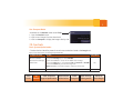
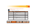
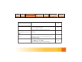
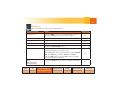
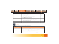
2D System Overview - Release 3.0
ACUSON S2000 Ultrasound System
Quick Reference Guide
www.siemens.com/S2000
2
Introduction
This Quick Reference Guide contains brief descriptions and instructions for the functions most
commonly used to perform 2D and Doppler exams. This supplement contains both standard
and optional features but is not a substitute for the system User Manual.
Image Disclaimer
Images used in this guide are for educational purposes only. They have been modified or
compressed, and may not reflect the actual image quality of the system.
Note: This guide shows multiple workflow options (keyboard and mouse) for Release 3.0.
UI refers to the Control Panel and is noted UI.
Contents
▪ Exam Basics
▪ 2D Controls
▪ Color & Spectral Doppler Optimization
▪ Measurements & Calculations
▪ Text & Pictograms
▪ Data & Image Management
▪ Siemens Exclusive Technologies
3
Exam Basics
Press the Power On/Off Switch on the upper left side of the UI to turn the system on and off. A
green blinking light indicates the power is on. The Standby option will quickly turn on, or shut
down the system, within 30 seconds.
6. Click OK or press the Store button
on the UI to start the exam
Patient Registration
Button
Patient Registration
Icon
Begin an Exam
1. Press the Patient Registration
button on the UI or click the Patient
Registration icon on the toolbar
2. Enter the patient name, ID and other exam
information
3. Press the TAB key or roll the trackball to
move to the next data field
4. Select the proper Transducer and Study
type from the drop-down menus
5. Select a Workflow Protocol from the
Protocol list (optional)
Exam
Basics
Optional if Using the Patient Scheduler
(Modality Work List)
1. Press Patient Browser (F2)
to open the Patient Browser
2. Double-click on the Patient Scheduler
icon to view an updated Work List
3. Highlight the patient name
(press the first letter of the last name to
quickly scroll the list)
4. Click on the Patient drop-down menu and
select Register
5. Select the appropriate transducer and
Study Type on the Patient Demographic
page
Measurements
2D
Text &
Color & Spectral
Controls Doppler Optimization & Calculations Pictograms
Data & Image
Management
Exclusive
Technologies
Exam
Basics
Measurements
2D
Text &
Color & Spectral
Controls Doppler Optimization & Calculations Pictograms
6. Click OK or press the Store button on the
UI to start the exam
Note: If the patient name does not appear in
the Work List:
a. Click on View at the top of the page
b. Select Patient based work list query
c. Enter the patient last name
d. Click on Get Work List
Store and Review Images
1. Press the Store button to save a
static image on the local database
2. Press Clip Store to acquire and
save a clip to the local database
3. The saved data displays in the thumbnail
panel at the bottom of the screen
Print an Image
▪ Press the Print button to print the
onscreen image
Data & Image
Management
Exclusive
Technologies
Note: Use the system configuration menu to
assign print and/or store functions to the Print
button.
Review an Image
▪ Press the Review button during an
active exam to view and delete images
Note: The system must be preset to transfer
images at end of exam, in order to delete
images from Review mode.
▪ Press the Review button or double-click a
thumbnail image to activate Review mode
during an active exam
Activate Review on a Previous Exam
1. Press Patient Browser (F2) to open the
Patient Browser
2. Click on the Patient Folder
3. Double-click the Image icon to load the
patient exam into Review
4
5
Use Compare Mode
▪ Available on the Review screen under Tools
1. Press the Review button
2. Click on two images from the thumbnails
3. Click on Compare to display the images side-by-side
2D Controls
Basic System Optimization
▪ The chart below identifies presets that can be customized and saved to the Image knob.
Items in the table below are arranged alphabetically.
To Adjust / Control
Do This
2D Sector Width
▪ Press the Priority key to select 2D FOV
▪ Roll the trackball to sector in the width of the image
▪ Press the Next key to go between Size and Position of 2D FOV
▪ Press the Select key to lock the image size
Increase line density/
frame rate
Image Knob
No
Note: Manually re-size the image to return to full screen.
Depth
Exam
Basics
▪ Rotate the Depth control
Measurements
2D
Text &
Color & Spectral
Controls Doppler Optimization & Calculations Pictograms
No
Data & Image
Management
Exclusive
Technologies
Exam
Basics
Measurements
2D
Text &
Color & Spectral
Controls Doppler Optimization & Calculations Pictograms
Data & Image
Management
Dynamic Range
▪ Toggle the Dynamic Range key up or down to add or remove
Adjust grayscale
levels within an
image
the amount of grayscale levels
Note: Can be used on live or frozen images.
Dynamic TCE
▪ Rotate the second LED control on the left side of the UI
Note: Settings include Off, Low, Medium and High. As the
Provides speckle
reduction and contrast level is increased, both Dynamic TCE speckle reduction and
enhancement
contrast enhancement are increased.
Edge
▪ Rotate the third LED control on the left side of the UI
Note: Range is 0 – 4; 4 applies the most edge enhancement.
Focal Zones
Control position and
number of focal zones
▪ Rotate the Focus control for position
▪ Press the Focus control to select the number of focal zones
Frequency
▪ Toggle the MultiHz key up or down on the UI to adjust the
frequency
HD RES (Write) Zoom
▪ Press the Zoom control to initialize HD RES
▪ Rotate the Zoom control to adjust the size of ROI box
▪ Press Zoom again to activate RES
Exclusive
Technologies
Yes
Yes
Yes
No
Yes
No
Note: When RES is activated, HDZ appears on the lower right
corner of the screen.
6
7
Image Presets
▪ Rotate the Image control on the UI to change the image preset
▪ Press the Image control to see a list of currently available
Yes
image presets
Overall Gain
▪ Press the TEQ button
▪ Rotate the 2D control
▪ Rotate the Gain Freeze / Cine wheel
▪ Adjust the DGCs
No
Note: Adjust according to user preference.
Persistence
Weighted average of
frames over time
Post Processing Maps
Selects a processing
curve that assigns
echo amplitudes to
grayscale levels
Exam
Basics
▪ Select the Persistence option located on the 2D tab of the
onscreen Image Menu
▪ Rotate the Menu control to change the level (0-4)
Note: Can only be adjusted when using SieClear 2, and not
when using Advanced SieClear Spatial Compounding (ASSC).
Increased persistence averages more frames over time, creating
a smoother image. Decreased persistence, averages fewer
frames over time, creating a grainier image.
Yes
▪ Rotate the second LED control on the right side of the UI to
select and apply
Note: Maps range from A-F and can be adjusted on live or
frozen images and in M-mode and PW Doppler.
Measurements
2D
Text &
Color & Spectral
Controls Doppler Optimization & Calculations Pictograms
Data & Image
Management
Yes
Exclusive
Technologies
Exam
Basics
Measurements
2D
Text &
Color & Spectral
Controls Doppler Optimization & Calculations Pictograms
SieClear Spatial
Compounding and
Advanced SieClear
Spatial Compounding
Data & Image
Management
Exclusive
Technologies
▪ Rotate the first LED on the left side of the UI to change the
compounding level
Note: The selected transducer determines how many levels of
compounding are available.
Utilizes multiple lines
of sight to provide
improvements in contrast
resolution and border
detection
Yes
Space/Time
Spatial resolution vs.
temporal resolution
▪ Rotate the first LED control on the right side of the UI
Note: Increase for better spatial resolution and decrease for
better temporal resolution.
Yes
Tints
Colorizes the grayscale
image
▪ Rotate the third LED control on the right side of the UI
Note: Tints range from 0-15 and can be adjusted on live or
frozen images and in M-mode and PW Doppler.
Yes
Tissue Harmonic
Imaging (THI)
Reduces noise clutter
with better contrast
enhancement and
improved axial resolution
over fundamental imaging
▪ Press the soft key on the home base to turn on or off
▪ Toggle the MultiHz key up or down
Yes
8
9
▪ Rotate the Zoom control to increase or decrease image
Zoom
magnification
▪ Roll the trackball to pan the image
No
Note: Zoom magnifies from the center of the onscreen image;
use pan to bring in the area of interest. Available on live or
frozen images and in M-mode and PW Doppler.
2D Imaging Features
Items in the table below are arranged alphabetically.
Feature
To Use Feature
ASC Dual
▪ Click on the ASC Dual option in the 2D tab of the onscreen Image menu to
Display two live
images side-by-side
with and without
compounding
Dual
Side-by-side imaging
display
Note: The left side displays the image without spatial compounding and the
right displays with spatial compounding. Can only be used when a level of
ASSC 3 or higher is selected.
▪ Press the Dual key to activate dual screen display
▪ Toggle the Update key to go between left and right images
Note: Color must be activated before entering Dual mode.
Exam
Basics
Measurements
2D
Text &
Color & Spectral
Controls Doppler Optimization & Calculations Pictograms
Data & Image
Management
Exclusive
Technologies
Exam
Basics
Measurements
2D
Text &
Color & Spectral
Controls Doppler Optimization & Calculations Pictograms
Live Dual
Data & Image
Management
Exclusive
Technologies
▪ Select the “Live Dual” option in the 2D tab of the onscreen Image menu
Display two live images Note: Can be used with color at any time.
side-by-side
SieScape Panoramic
Imaging
▪ Select SieScape from the Feature menu on the left side of the screen
▪ Press the Update key to start acquiring image
▪ Scan through area of interest in a long axis of the transducer
▪ Press Gain Freeze/Cine to stop the acquisition
Note: Once an image is frozen, roll the trackball to rotate the image. Use the
Scroll wheel or Zoom control to zoom. Use Restore to bring the image back
to original size. Press the Calcs key to measure on a SieScape image. Use the
Gain Freeze/Cine wheel to Cine through the SieScape image.
Steer Image
▪ Press the Priority key to activate Steer
▪ Roll the trackball to steer the FOV left or right
Linear transducers only
Improve the angle of
Note: ASC must be off or set to SC2.
insonance between
anatomy and the
ultrasound beam
Trapezoid
Linear transducers only
Allows more
visualization
of tissue on the screen
▪ Press the soft key to activate Trapezoid image format
▪ Press the Next key to go between position and size of the image
10
11
Color Doppler & Spectral Doppler Optimization
Color Doppler Imaging
Items in the table below are arranged alphabetically.
To Adjust / Control
Do This
Baseline
▪ Toggle the Baseline key up or down
▪ Press the soft key to cycle between (CDE) and velocity
Color Doppler Energy and
Color Doppler Velocity
Color Gain
Engage Color Doppler
Exit Color Doppler
Image Knob
color (CDV)
▪ Rotate the C control
▪ Press the C control
▪ Press the PW control or press the 2D control to return to
imaging
Frequency
Image Presets
▪ Toggle the MultiHz key up or down
▪ Rotate the Image knob to change image presets
▪ Press the Image knob to view a list of all available image
Yes
No
No
No
No
Yes
Yes
presets
Exam
Basics
Measurements
2D
Text &
Color & Spectral
Controls Doppler Optimization & Calculations Pictograms
Data & Image
Management
Exclusive
Technologies
Exam
Basics
Measurements
2D
Text &
Color & Spectral
Controls Doppler Optimization & Calculations Pictograms
Invert
Data & Image
Management
▪ Press the Invert button
Yes
Change the orientation of the
Color Doppler velocity display
ROI Size and Position
▪ With color active, press the Next key to alternate
Scale
▪ Roll the trackball to adjust size and position
▪ Toggle the Scale key up or down to increase or
between Size and Position
Pulsed Repetition Frequency
(PRF)
Exclusive
Technologies
decrease
No
Yes
Spatial Resolution and
Speed
▪ Rotate the first LCD control on the left side of the UI
Note: Spatial resolution is inversely proportional to
temporal resolution.
Yes
Steer
▪ Press the Priority key to activate C ROI
▪ Press Next and select Steer
▪ Roll the trackball to get to the proper angle
No
12
13
PW Doppler
Items in the table below are arranged alphabetically.
To Adjust / Control
Do This
Angle Correct
▪ Press the 0/60° control to cycle through angle correction
▪ Rotate the 0/60° control to fine tune the angle
▪ Toggle the Baseline key up or down
▪ Toggle the MultiHz key up or down
▪ Roll the Scroll wheel up or down
▪ Rotate the Doppler (PW) control
▪ Press the PW control to engage PW Doppler
▪ Roll the trackball to position the cursor
▪ Press PW again to start spectral waveform or press Update
Baseline
Frequency
Gate Size
Overall Doppler Gain
PW Doppler
Image Knob
Note: Press Next to go between Position and Steer.
▪ Press the PW control or press the 2D control to exit PW
Doppler and return to imaging
Scale
Pulsed Repetition
Frequency (PRF)
Exam
Basics
▪ Toggle the Scale key up or down to increase or decrease
Measurements
2D
Text &
Color & Spectral
Controls Doppler Optimization & Calculations Pictograms
Data & Image
Management
Yes
Yes
Yes
Yes
No
No
Yes
Exclusive
Technologies
Exam
Basics
Measurements
2D
Text &
Color & Spectral
Controls Doppler Optimization & Calculations Pictograms
Steer
Sweep Speed
TEQ
Data & Image
Management
▪ Press the Priority key
▪ Select the PW gate
▪ Press Next to steer
▪ Roll the trackball to adjust the steer angle
▪ Press Select to lock in the angle
▪ Rotate the LCD control on the right side of the UI
Exclusive
Technologies
Yes
Note: Sweep speed can be adjusted during real-time or on
a frozen M-mode Doppler strip.
Yes
▪ Press the TEQ button
No
M-mode
Items in the table below are arranged alphabetically.
To Adjust / Control
Do This
M-mode
▪ Press the M control to engage M-mode
▪ Roll the trackball to adjust the position of the M-mode cursor
▪ Press the M control again to activate spectral waveform
▪ Press the M control to exit, or
▪ Press the 2D control to exit and return to 2D imaging
14
15
M-mode Cursor
▪ Press Next to select Size or Position
▪ Roll the trackball to make adjustments
Note: The active function is displayed in green. Sweep speed can be
adjusted during real-time or on a frozen M-mode Doppler strip.
Overall Gain
▪ Rotate the M control
Measurements
& Calculations
General Measurement Tools
Activate Calculations
(one of two methods):
▪ Press the Calcs button to activate
the measurement function on
real-time or frozen images
or
▪ Press the Gain Freeze/Cine button
to automatically go to Calcs
(must be preset in the Setup menu)
Exam
Basics
Perform an Unlabeled Measurement
1. Roll the trackball to place the first caliper in
the area of interest
2. Press Next for the second distance caliper
or ellipse control
3. Press Select to anchor a caliper or enter a
measurement
4. Press Next to go between the active
calipers
5. Press Priority to reactivate locked caliper
sets or scroll through different caliper sets
Tip: To delete the caliper set, press the Delete
soft key or Cine back one position.
Measurements
2D
Text &
Color & Spectral
Controls Doppler Optimization & Calculations Pictograms
Data & Image
Management
Exclusive
Technologies
Exam
Basics
Measurements
2D
Text &
Color & Spectral
Controls Doppler Optimization & Calculations Pictograms
Data & Image
Management
Exclusive
Technologies
Perform a Labeled Measurement and
Add it to the Report Package
6. Press Priority to reactivate locked caliper
sets or scroll through different caliper sets
Note: Application-specific measurements are
particular to one study type.
▪ The measurement tools and labels are
organized by the active imaging mode and
application (e.g., abdomen)
▪ Only labeled measurements are copied to a
patient report
1. Press the Gain Freeze/Cine button
2. Click on a measurement label (e.g., Liver)
Note: A measurement label can also be
selected after calipers are placed.
3. Roll the trackball to place the first caliper in
the area of interest
4. Press Next to activate the second caliper
5. Press Select or Store to complete a
measurement and send the value(s) to the
Report
Additional Measurements
1. Activate the Report page
2. Click on the Page 1 drop-down menu on
the imaging screen
3. Scroll down to the desired page
4. Press Select to activate page
Perform Advanced Measurements,
e.g, Volume Measurement
1. Select the Compound drop-down menu
from the top left-hand side of the screen
when Calcs are active
2. Select the appropriate measurement from
the drop down list: Stenosis, Volume,
Area Ratio, Distance Ratio or Volume
Flow
16
17
Calculation Reports
▪ Calculation worksheets and reports are
offered for each preset when labeled
measurements are entered
1. Press Report (F1) to activate Reports
2. Scroll to view the Report pages
3. Click on Store Report to capture the entire
formalized worksheet
4. Click End Exam to close the patient study
Note: Transfer Report will send DICOM
measurements to structured reporting in PACS
(customers must have structured reporting
capabilities).
3. Roll the trackball to drag the image into
appropriate square
4. Press Select to anchor the image
Note: Click on the Format icon to display up
to 16 images on the Report page.
Import Images into the Report
1. If necessary, press Report (F1) to activate
Reports
2. Scroll down to the bottom of the Report
screen and click on an image in the
thumbnails
Exam
Basics
Measurements
2D
Text &
Color & Spectral
Controls Doppler Optimization & Calculations Pictograms
Data & Image
Management
Exclusive
Technologies
Exam
Basics
Measurements
2D
Text &
Color & Spectral
Controls Doppler Optimization & Calculations Pictograms
Text & Pictograms
Enter Text
1. Press the Text key to activate
the cursor
2. Roll the trackball to place the cursor in the
appropriate place in the image
3. Type the text and press Select, or, if
configured, press the appropriate Text
button
Note: Text A, B, C, and D can be customized
and reordered for each exam preset.
▪ The system can be preset to automatically
enter the Text Menu by pressing the Gain
Freeze/Cine button
Tip: When typing, press Tab to auto-complete
the annotation.
▪ The annotation can be configured to be on
or off
Data & Image
Management
Exclusive
Technologies
Move Text
1. If necessary, press the Text key to activate
the cursor
2. Roll the trackball to place the cursor within
the text
3. Double-click Select to highlight
(text is highlighted in green)
4. Roll the trackball to the new location and
press Select
Drag and Drop Text
1. If necessary, press the Text key to activate
the cursor
2. Position the cursor in the text to move
3. Double-click Select to highlight
(text is highlighted in green)
4. Roll the trackball to move the selection
over the text to be replaced and press
Select
18
19
Delete Text
1. If Calcs is active, press the soft key, and
select either Clear Screen, Delete Word or
Delete Line
Tip: If Calcs is not active, press and hold the
Text key until the screen is cleared.
Place an Arrow(s)
1. Press the Arrow Up button on the
bottom right side of the keyboard
2. If necessary, press Next to rotate the
direction of the arrow
3. Roll the trackball to position the arrow and
press Select
Note: Use the steps above to place additional
arrows.
Note: The Delete Arrow soft key deletes one
arrow at a time, starting with the last arrow
placed onscreen.
Add a Pictogram(s)
1. Press the Body Marker key on the
bottom right side of the keyboard
2. Rotate the Scroll wheel to cycle through
pictograms
3. Press Next to go between Position and
Rotation of the marker
4. Roll the trackball to position the marker on
the pictogram and press Select
Delete a Pictogram
1. Press the Delete Body Marker or Clear
Screen soft key
Delete an Arrow(s)
1. Press the Delete Arrow or the Clear
Screen soft key
Exam
Basics
Measurements
2D
Text &
Color & Spectral
Controls Doppler Optimization & Calculations Pictograms
Data & Image
Management
Exclusive
Technologies
Exam
Basics
Measurements
2D
Text &
Color & Spectral
Controls Doppler Optimization & Calculations Pictograms
Data & Image Management
Restart an Exam
1. Press Patient Browser (F2) to open the
Patient Browser
2. Select the patient name and click on the
middle yellow folder
3. Click on the Patient drop-down menu and
select Register
4. Select the transducer and operator initials
5. Click OK
Note: Restart is available at all times. New
images can be added to the existing patient
exam.
End an Exam
1. Press the Review button
2. Click on End Exam
Note: Starting a new patient also ends a
current exam.
Data & Image
Management
Exclusive
Technologies
Send Patient Exam to PACS
1. Press Patient Browser (F2) to open the
Patient Browser
2. Select the Archive To icon
3. Select Destination
4. Select Archive To
Note: This can be done before closing the
study.
Tip: Closing a study will automatically send to
PACS if the system is preset to “Send on study
close”.
Delete Images from an Active Case
1. Click on an image from the thumbnail
2. Click on the Delete icon located
on the lower right of the screen
Note: The system must be preset to “Send
at the End of Exam” or the image will not be
deleted.
20
21
Delete Images from
Review During an Active Exam
1. Press Review
2. Click and highlight the image to delete
3. Press the Delete soft key
Transfer an Exam from
the Hard Drive to CD-R/DVD
1. Insert the CD-R/DVD
2. Press Patient Browser (F2) to open the
Patient Browser and Local Database
3. Select the patient name/s
Tip: Hold CTRL to select more than one
patient name.
4. Use one of the following transfer methods
below
Transfer in DICOM Format
1. With patient/s selected, select the Transfer
drop-down menu
Exam
Basics
2. Select Archive to CD-R
Note: syngo fastView and Showcase DICOM
viewers will be burned onto the CD-R/ or DVD.
Transfer in PC Format (AVI or JPEG)
1. With the patient(s) selected, select the
Transfer drop-down menu
2. Select Export to CD in PC Format
Transfer to a USB Drive in PC Format
1. With the patient(s) selected, select the
Transfer drop-down menu
2. Select Export to USB in PC format
Note: The USB must have an export
folder on it to accept images.
Import All Images from
a CD to the Local Database
1. Insert the CD-R/DVD
2. Press Patient Browser (F2) to open the
Patient Browser
Measurements
2D
Text &
Color & Spectral
Controls Doppler Optimization & Calculations Pictograms
Data & Image
Management
Exclusive
Technologies
Exam
Basics
Measurements
2D
Text &
Color & Spectral
Controls Doppler Optimization & Calculations Pictograms
3. Click on CD
4. Click on the Transfer drop-down menu
5. Click on Import from Off-line
Note: Image(s) will be removed when the
exam is closed.
Resend an Exam to PACS
1. Press Patient Browser (F2) to open the
Patient Browser
2. Click on Local Database
3. Click on the patient name
4. Click the Archive to icon or select Archive
to from the drop-down menu
5. Select the destination
6. Select Archive to
Data & Image
Management
Exclusive
Technologies
De-Identify a Patient
(Teaching Files, Name Changes)
1. Press Patient Browser (F2) to open the
Patient Browser
2. Click on Local Database
3. Click the patient name
4. Click on the image folder
5. Click on Patient drop-down menu and
select De-Identify
6. Rename the exam
7. Click OK
Note: The new case will be added to the Local
Database within minutes. 3D/4D volumes will
not be transferred for re-manipulation.
22
23
Siemens
Exclusive Technologies
Clarify Vascular Enhancement
Technology (purchasable option)
1. During 2D imaging, press the Clarify soft
key
2. Roll the trackball to place the ROI box
3. Press Next to go between Size and
Position
4. Roll the Scroll wheel to adjust the level of
sensitivity:
1 = least sensitive, 7 = most sensitive
highlighted in green.
3. Click on the desired measurement
(semi-automated measurements are live
onscreen)
4. Press Next to go between the cursors
(active and available)
5. Press Select to lock in the measurement
and transfer it to the Report page
Tip: Press Store to transfer the measurement.
syngo Auto OB (purchasable option)
1. Ensure the OB exam type is selected
2. Press the Calcs button
Note: Available Auto OB measurements are
Exam
Basics
Measurements
2D
Text &
Color & Spectral
Controls Doppler Optimization & Calculations Pictograms
Data & Image
Management
Exclusive
Technologies
Exam
Basics
Measurements
2D
Text &
Color & Spectral
Controls Doppler Optimization & Calculations Pictograms
syngo eSieCalcs Native Tracing
Software (purchasable option)
1. During 2D imaging, press the Gain Freeze/
Cine button
2. Press the Calcs button
3. Select the eSieCalcs menu option
4. Roll the trackball to move the cursor into
the image
5. Press the Next key to activate ROI box
Note: Next toggles between the two active
calipers allowing movement of the ROI box
and a change in size.
6. Press Select to calculate and display the
measurement
Data & Image
Management
Exclusive
Technologies
Adjust an eSieCalc Measurement
1. Press Next to reactivate calipers
2. Roll the trackball to trace a new area of
interest
3. Press Select to recalculate measurement
Tip: When placing the ROI box over the area
of interest, do not make the box too big.
24
ACUSON, S2000, Clarify, Dynamic
TCE, eSieCalcs, Advanced SieClear,
SieClear and SieScape are
trademarks of Siemens Medical
Solutions USA, Inc. and syngo is a
trademark of Siemens AG.
Order No. A91US-187-1C-4A00 | Printed in the USA
WS 0711 | © 07.2011, Siemens Medical Solutions USA, Inc.
Global Siemens Headquarters
Siemens AG
Wittelsbacherplatz 2
80333 Muenchen
Germany
Global Siemens
Healthcare Headquarters
Siemens AG
Healthcare Sector
Henkestr. 127
91052 Erlangen, Germany
Phone: +49 9131 84-0
www.siemens.com/healthcare
Legal Manufacturer
Siemens Medical Solutions USA, Inc.
Ultrasound
1230 Shorebird Way
Mountain View, CA 94043 USA
Phone: +1-888-826-9702
www.siemens.com/healthcare