1
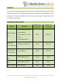
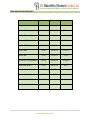
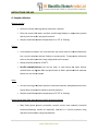
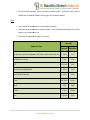
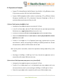
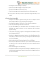
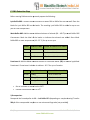
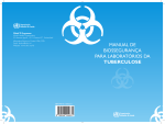
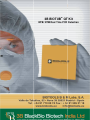
3B BIOTUB® QT Kit
MTB/ NTM Real Time PCR Detection
0|Page
3B BIOTUB QT Kit
www.3bblackbio.com
3B BIOTUB® QT Kit
MTB/ NTM Real Time PCR Detection
For detection and differentiation of Mycobacterium tuberculosis complex (MTBC) and
nontuberculous mycobacteria (NTM) from human specimens
For use with
ABI Prism 7500 SDS, 7500 Fast SDS, ViiA™ 7 and QuantStudio™ 12K Flex (Applied Biosystems)
Rotor
otor-Gene Q5/6 plex Platform (Qiagen)
CFX384 Touch™, CFX96 Touch™ (BioRad)
PicoReal 24, PicoReal 96 (Thermo)
LightCycler® 96 (Roche)
LineGene K, LineGene 9600 (Bioer)
FOR IN VITRO DIAGNOSTIC USE
Product No.: 111111
48 tests
Temperature limitation
April 2013
3B BlackBio Biotech India Ltd.
1|Page
3B BIOTUB QT Kit
www.3bblackbio.com
CONTENT
PAGE NO.
INTENDED USE
3
PRINCIPLE
3
REAGENTS
4
3B MTB/NTM REAL TIME KIT
4
MTB DNA EXTRACTION KIT
5
INSTRUCTIONS FOR USE
6
Sample collection
6
Sputum samples
6
Tissues
6
Urine
6
Aseptic fluids (body fluid, bone marrow, blood), Bronchial washing
6
Stool
7
Preparation of Samples
8
Viscous Fluids (sputum, body fluids, Pus)
8
Non-viscous
-viscous Fluids (aspiration, body fluids, urine, pleural fluid)
8
Fresh tissue (Biopsy, Endometrium tissue)
9
Paraffin-- embedded tissue
10
Stool
10
Blood, Bone marrow aspiration, Menstrual blood
10
Broncho Alveolar La
Lavage (BAL)
11
DNA Extraction Step
12
REAL TIME PCR PROTOCOL FOR MTB/NTM DETECTION
15
RESULT ANALYSIS
16
SPECIFICATIONS
18
TROUBLESHOOTING
21
STORAGE AND HANDLING & ADDITIONAL REQUIREMENTS
23
GENERAL PRECAUTIONS
24
REFERENCES
25
2|Page
3B BIOTUB QT Kit
www.3bblackbio.com
INTENDED USE
3B MTB/NTM KIT accurately differentiates Mycobacterium tuberculosis (MTB) from nonnon
tuberculosis Mycobacterium species (NTM) in a qualitative/quantitative
quantitative form from various
sources of clinical samples
es using Real time PCR.
PRINCIPLE
3B MTB/NTM detection is a Real
Real-Time Amplification test for the qualitative/quantitative
qualitative
detection of Mycobacterium tuberculosis complex (MTC) and Non-tuberculosis
tuberculosis mycobacteria
(NTM) in clinical samples.. Mycobacterium tuber
tuberculosis/ Non-tuberculosis
tuberculosis DNA is extracted from
samples, amplified using Real Time Amplification and detected using fluorescent reporter dye
probes specific for M. tuberculosis complex and all mycobacterium genus.. In real-time
real
PCR, the
fluorescent signal is generated from the presence of an oligonucleotide probe specific for target
DNA sequence. The probe contains a fluorescent dye molecule on its 5’ end and a quencher
molecule on its 3’ end. The probe hybridizes with one of the chains of the amplified fragment.
fra
During synthesis of a complementary chain, Taq DNA polymerase which possesses 5' - 3'
exonuclease activity cleaves the probe. As a result, the fluorescent dye and quencher dye are
separated, and the total fluorescence of reaction volume increases in direct proportion to the
number of amplicon copies synthesized during PCR. The fluorescent signal is measured in each
cycle of reaction, and the threshold cycle value is determined from the obtained curve. The
threshold cycle is proportional to the initia
initiall number of DNA copies in a sample and its value
allows qualitative/quantitative
quantitative comparisons of analyzed and control samples.
In 3B MTB/NTM detection kit is based on amplification of region upstream of the 65 kDa heat
shock protein (65kDa hsp) gene11, 2, 3 by primer and probes specific for M. tuberculosis complex
and mycobacterium genus4. In this kit there are three independent reactions running in parallel
in each tube: the first detects Mycobacterium tuberculosis complex (M. tuberculosis, M.
africanum, M. bovis, M. bovis BCG, M. microti) ((HEX channel), second detects all
mycobacterium by genus specific probe (FAM channel) and the third detects internal control
(IC) DNA (Tex Red channel) which allows excluding unreliable results.
3|Page
3B BIOTUB QT Kit
www.3bblackbio.com
REAGENTS
The Kit contains amplification
mplification reagents for performance of 48/96 amplification
n reactions. Thaw
and handle reagents on ice. Do not freeze/thaw Kit vials repeatedly. In case of frequent use, we
recommend to aliquot the contents of the vials into 10 reactions each. This will also
a rule-out
kit/ reagent contamination.
3B MTB/NTM REAL TIME KIT
Volume in µL
Volume in µL
48 reactions
96 reactions
500 µL
500 µL X 2
100 µL
100 µL X 2
100 µL
100 µL X 2
• MTB Positive
ositive Control
50 µL
100 µL
• NTM Positive
ositive Control
50 µL
100 µL
• Sterilized water
50 µL
100 µL
Reagent
Description
• Hot-start
start DNA polymerase
Multiplex Master
Mix
• Reaction Buffer
• dNTPs (dATP, dCTP, dGTP,
dTTP)
MgCl2 and stabilizers
MTB/ NTM
Primer probe mix
Internal control
Primer probe mix
• Primer and probe mix for
MTB and NTM detection
• Primer, probe and DNA mix
for IC detection
Positive Control
MTB
Positive Control
NTM
Negative Control
4|Page
3B BIOTUB QT Kit
www.3bblackbio.com
MTB DNA EXTRACTION KIT
MTB DNA Extraction Kit
Reagent
48 Reactions 96 Reactions 240 Reactions
Volume
Volume
Volume
Buffer BT1
20 ml
20 ml X 2
100
0 ml
Buffer BB1
12 ml
12 ml X 2
60 ml
Reagent BB2
3 ml
3 ml X 2
15 ml
7 ml X 2
7 ml X 4
40 ml X 2
Buffer BBW
30 ml
30 ml X 2
75 ml x 2
Buffer BBE
15 ml
15 ml X 2
75
5 ml
Proteinase K (Lyophilized)
30 mg
30 mg X 2
75 mg x 2
Proteinase Buffer
1.8 ml
1.8 ml X 2
8 ml
MTB DNA Spin Columns
50
100
250
50
2 ml Collection Tubes
100
200
500
00
Label for Buffer BB3
1
1
1
User Manual
1
1
1
Buffer BB5 (Concentrate)
5|Page
3B BIOTUB QT Kit
www.3bblackbio.com
INSTRUCTIONS FOR USE
A. Sample collection
Sputum samples
•
Collection of early morning sputum specimen is optimal.
•
Rinse the mouth with water and then should cough deeply to expectorate sputum
directly into the sterile, leak
leak-proof container.
•
Samples should bee stored at temperatures of 22-8 ⁰C or freezing.
•
Fresh biopsies of up to 5 m
mm
m must be used. Any tissue must be collected aseptically
Tissues
into a sterile container without fixatives or preservatives. To keep moist add sterile
saline to the dried specimen
specimen. Keep refrigerated until transport.
•
Biopsy should be stored at -15±8 ⁰C.
•
Paraffin-embedded
embedded tissues can also be used, in cases where the tissue fixation
method does not degrade DNA and purification of DNA is performed with methods
specific for this kind of sample.
Urine
•
An early morning midstream specimen should be collected. Multiple specimens over
several days are optimal to obtain a positive specimen.
•
Samples should be stored at temperatures of 22-8 ⁰C or freezing.
Aseptic fluids (body fluid, bone marrow, blood
blood), Bronchial washing
•
Body fluids (spinal, pleural, pericardial, synovial, asc
ascitic, bone marrow), bronchial
secretions/washings
washings should be aseptically collected in a sterile container using
aspiration
tion techniques or surgical procedures.
6|Page
3B BIOTUB QT Kit
www.3bblackbio.com
•
For fluids that may clot, sterile potassium oxalate (0.01 ~ 0.02 ml of 10% neutral
oxalate per ml fluid) or heparin (0.2 mg per ml) should be added.
Stool
•
Stool should be passed into a clean and dry container.
•
Carefully
lly open the specimen vial and collect a small sample with the help of a sterile
spoon in to a new sterile vial
vial.
•
Close the vial tightly and shake to mix well.
Quantity
Specimen Type
Optimal
Minimum
5–10
10 mL
1 mL
5 mL
1 mL
CSF
5–15
15 mL
1 mL
Abscess (wound)aspirates
5–15
15 mL
1 mL
Body fluids/ aspirates
5–15
15 mL
1 mL
Urine
30–50mL
50mL
10 mL
Stool
2-5 g
1 gm
Tissue
5 mm
Visible
1-10
10 mL
1 mL
Respiratory specimens (sputum, BAL, BW, tracheal aspi
aspirate, etc)
Blood/bone marrow
Pus
7|Page
3B BIOTUB QT Kit
www.3bblackbio.com
B. Preparation of Samples
•
Prepare 2% N-Acetyl
Acetyl Cystein
Cystein/ NaOH [Dissolve 2 g of NaOH, 1.45 g of Sodium citrate,
0.5 g N-acetyl
acetyl cystein in 100 ml of sterile distilled water].
•
Prepare 0.067M phosphate buffer solution by dissolving 2.37 g of Dibasic Sodium
Phosphate (Na2HPO4) and 2.27 g Monobasic Potassium
otassium Phosphate in 500 ml of
sterile distilled water
water. Adjust the final pH to 6.8.
i) Viscous Fluids (sputum,
sputum, body fluids
fluids, Pus):
•
Homogenize the sample by vigorous vortexing for 55-10 min.
•
In the 15 ml falcon tube, add the 2% NALC
NALC-NaOH with the specimen (sputum/body
fluids/urine) to an equal volume (1:1) and vortex for 1 min.
•
Incubate the mixture for 15 min at room temperature with occasional shaking.
•
Adjust the volume to 15 ml with sterile water or sterile phosphate buffer solution
(pH 6.8) and mix well
well.
•
Transfer 1.5 ml sample to a 2 ml EEppendorf tube using a pipette with a tip with
aerosol barrier, mark it, and centrifuge at 10,000 g or 15,000 rpm for 10 min. Discard
the supernatant, add 1 ml of PBS solution and mix well
well.
Note: (if the pellet is not visible, remove the supernatant leaving about 100
10 μl of the
sample)
•
Centrifuge at 10,000 g or 15,000 rpm for 10 min, discard the supernatant and rere
suspend the pellet as per DNA extraction step.
ii) Non‐viscous Fluids (aspiration,
aspiration, body fluids, urine
urine, pleural fluid):
•
Homogenize the sample by vigorous vor
vortexing for 5-10 min.
•
Transfer 1.5 ml of the sample to a 2 ml Eppendorf tube using a pipette with a tip
with aerosol barrier, mark it, and centrifuge at 10,000 g or 15,000 rpm for 10 min.
min
•
Discard the supernatant and re-suspend
suspend the pellet as per DNA extraction
extracti step.
8|Page
3B BIOTUB QT Kit
www.3bblackbio.com
Note: (if the pellet is not visible, remove the supernatant leaving about 100 μl of the
sample)
iii) Fresh tissue (Biopsy, Endometrium tissue)
•
If RBCs are present then give PBS wash to remove RBC’s totally.
•
If tissues are soft then add NALC
NALC-NaOH solution and start processing as described
below. If tissues are tough, then grind and homogenize it with PBS in sterile mortarmortar
pestle and process as described below.
•
Process sample in original sample container. If sample is in syringe or small container
then transfer
fer and process the sample in 50 ml Falcon tube (preferred for more
surface area) or 15 ml falcon tube.
•
To specimen add 2% NALC
NALC-NaOH
NaOH solution in 1:1 ratio. Incubate for 15-20
15
min
depending upon sample consistency at room temperature with intermittent
vigorous vortexing.
•
If the sample is very viscous or appears to be tough/thick then incubate it in water
bath of temperature 40
40-50⁰C.
•
Neutralize the digested
digested-decontaminated
decontaminated specimens by adding Phosphate buffer
saline (PBS) up to 15 ml and mix by inverting. Ce
Centrifuge
ntrifuge 1.5 ml neutralized sample
for 15 min at 13000 rpm.
•
Wash the pellet with 1ml PBS and centrifuged for 15 min at 13000 rpm.
•
Check the pH of supernatant and discard it. If pH is neutral then wash pellet with
1ml water and if it is still alkali then gi
give
ve one more (or two till it become neutral)
wash with PBS.
•
After neutralization give 1 more PBS washing.
•
Now add 1ml sterile water to the pellet, vortex and centrifuge at 13000 rpm for 10
min. and carefully discard the supernatant. Repeat this step once and
an proceed as per
DNA Extraction step.
9|Page
3B BIOTUB QT Kit
www.3bblackbio.com
iv) Paraffin‐ embedded tissue
•
Add 1 ml of n-Octane
Octane or Xylene to the tube containing several pieces of paraffin
fragment.
•
Vortex vigorously and incubate at room temperature for about 30 min. Vortex
occasionally. Centrifuge
ifuge at 13,000 rpm for 3 min. Pipette off supernatant. (If paraffin
remains, repeat step 1 and 2)
•
Add 1 ml of ethanol (96
(96-100%) and then vortex it.
•
Centrifuge for 3 min at 13,000 rpm. Pipette o
out supernatant.
•
Repeat step 3 and 4.. Pipette off as much of th
the ethanol as possible.
•
Incubate the open tube at 37 ⁰C
C until the ethanol has evaporated (15 min).
•
Discard the supernatant and re
re-suspend
suspend the pellet as per DNA extraction step.
Note: (if the pellet is not visible, remove the supernatant leaving about 100 μl of the
sample)
v) Stool
•
Homogenize the sample by vigorous vortexing for 55-10 min.
•
Mix the stool (at least 1 g) with 10 ml of 2% NALC-NaOH,, vortex vigorously for 30 sec
and incubate
ncubate for 15 min at room temperature.
•
Centrifuge for 10 min at 1,500 rpm, trans
transfer
fer 1.5 ml of the supernatant to 2 ml
Eppendorf tube using a pipette with a tip with aerosol barrier, mark it, and
centrifuge at 10,000 g or 15,000 rpm for 10 min
min.
•
Discard the supernatant and re
re-suspend
suspend the pellet as per DNA extraction step.
Note: (if the pellet is not visible, remove the supernatant leaving about 100 μl of the
sample)
vi) Blood, Bone marrow aspiration
aspiration, Menstrual blood
•
Take 0.5 ml of whole blood (or Bone marrow aspiration) and mix well with 1 ml of
sterile water.
10 | P a g e
3B BIOTUB QT Kit
www.3bblackbio.com
•
Centrifuge for 10 min at 110,000 g or 15,000 rpm.
•
Discard the supernatant and add 1 ml sterile water to pellet.
•
Mix well and centrifuge at 15,000 rpm for 10 min.
•
Discard
iscard the supernatant and re
re-suspend
suspend the pellet as per DNA extraction step.
Note: (if the pellet is not visible, remove the supernatant leaving about 100 μl of the
sample)
vii) Broncho Alveolar Lavage (BAL)
•
Homogenize the sample by vigorous vortexing for 55-10
10 min. If sample is viscous
dilute sample with water in 1:0.5 ratio and vortex to homogenize.
•
Centrifuge whole sample at 13,000 rpm for 15 min. discard the supernatant.
•
If RBCs are present then give PBS wash to remove RBCs.
•
To pellet add 500-600
600 µl 2% NALC
NALC-NaOH
NaOH solution and incubate for 15-20
15
min
depending upon sample consistency at room temperature with intermittent
vigorous vortexing.
•
If the sample is very viscous or appears to be tough/thick then incubate it in water
bath of temperature 40
40-50 ⁰C.
•
Neutralize the digested
digested-decontaminated
decontaminated specimens by adding Phosphate buffer
saline (PBS) up to 15 ml and mix by inverting. Centrifuge whole neutralized sample
for 15 min at 13000 rpm.
•
Wash the pellet with 1ml PBS and centrifuged for 15 min at 13000 rpm.
rp
•
Check the pH of supernatant and discard it. If pH is neutral then wash pellet with
1ml water and if it is still alkali then give one more (or two till it become neutral)
wash with PBS.
•
After neutralization give 1 more PBS washing.
•
Now add 1ml sterile wa
water,
ter, vortex pellet and centrifuge at 13000rpm for 10 min. and
carefully discard the supernatant. Repeat this step once and re-suspend
suspend the pellet as
per DNA Extraction step
step.
11 | P a g e
3B BIOTUB QT Kit
www.3bblackbio.com
C. DNA Extraction Step
Before starting DNA extraction protocol prepare the followi
following:
Lysis Buffer BB3: Transfer the total contents of Buffer BB1 to Buffer BB2
B2 and mix well. Place the
labels for Lysis Buffer BB3
B3 on the bottle. The resulting Lysis Buffer BB3
B3 is stable for up to one
year at room temperature.
Wash Buffer BB5: Add the below indicated volume of ethanol (96 – 100 %) to Wash
W
Buffer BB5
Concentrate. Mark the label of the bottle to indicate that ethanol was added. Store Wash
Buffer BB5
B5 at room temperature (18 – 25° C) for up to one year.
Format
Volume of BB5 Volume of Ethanol to be added
48 rxns
7 ml x 2
28 ml to each bottle
96 rxns
7 ml x 4
28 ml to each bottle
240 rxns 2 x 40 ml
160 ml to each bottle
Proteinase K: Add the below indicated volume of Proteinase Buffer (PB) to dissolve lyophilized
Proteinase K. Proteinase K solution
tion is stable at - 20 °C for up to 6 months.
Format
Qty. of Proteinase K Volume of PB to be added
48 rxns
30 mg
1.35 ml to each vial
96 rxns
30 mg X 2
1.35 ml to each vial
240 rxns 75 mg X 2
3.35 ml to each vial
•
Set on incubator or water bath to 50
50⁰C
•
Preheat Elution Buffer BBE to 70
70⁰C
i) Resuspension:
Resuspend the formed pellet in 0.2 – 1 ml Buffer BT1 (depending on sample viscosity).Transfer
200 μl of the resuspended sample to a new microcentrifuge tube (not provided).
ovided).
12 | P a g e
3B BIOTUB QT Kit
www.3bblackbio.com
ii) Pre‐lyse sample:
Add 180 μl Buffer BT1 and 25 μl Proteinase K solution. Vortex to mix. Be sure that the samples
are completely covered with lysis solution.
Note: If processing several samples, Proteinase K and Buffer BT1 may be premixed directly
dire
before use. Do not mix Buffer B
BT1 and Proteinase K more than 10 – 15 min before addition to
the sample.
Incubate at 56 °C until complete lysis is obtained (at least 1 – 3 h). Vortex occasionally during
incubation or use a shaking incubator.
Note: Samples
les can be incubated overnight as well. If RNA
RNA-free
free DNA is crucial for downstream
downs
applications, an RNase digest may be performed: Add 20 μl RNase A (20 mg/ml) solution (not
included
d and incubate for an additional 5 min at room temperature.
iii) Lyse sample:
Vortex the samples. Add 200 μl Buffer BB3
BB3, vortex vigorously and incubate at 70 °C for 10 min.
Vortex briefly.
Note: If insoluble particles are visible in Buffer BB3,, centrifuge for 5 min at high speed (e.g.,
10,000 rpm)) and transfer the supernatant to a new micro
micro-centrifuge
centrifuge tube (not provided)
iv) Adjust DNA binding condition
conditions:
Add 210 μl ethanol (96 – 100 %) to the sample and vortex vigorously
vigorously.
v) Bind DNA:
For each sample, place one MTB DNA Spin Column into a Collection Tube. Apply the sample to
the column. Centrifuge for 1 min at 10,000 rpm. Discard the flow-through
through and place the column
back into the Collection Tube.
Note: If the sample is not drawn completely through the matrix, repeat the centrifugation step
at 10,000 rpm.. Discard flowthrough.
13 | P a g e
3B BIOTUB QT Kit
www.3bblackbio.com
vi) Wash silica membrane
1st wash
Add 500 μl Buffer BBW.. Centrifuge for 1 min at 10,000 rpm. Discard flow-through
through and place the
column back into the Collection Tube.
2nd wash
Add 600 μl Buffer BB5 to the column and centrifuge for 1 min at 10,000 rpm.
rpm Discard flowthrough and place the column back into the Collection Tube.
vii) Dry silica membrane
Centrifuge the column for 1 min at 10,000 rpm.. Residual ethanol is removed during this step.
viii) Elute highly pure DNA
Place the MTB DNA Spin Column into a 1.5 ml micro-centrifuge
centrifuge tube (not provided) and add
100 μl pre-warmed Buffer BBE
BE (70° C
C).. Incubate at room temperature for 1 min. Centrifuge 1
min at 10,000 rpm.
xi) 50 -100 ng of the eluted DNA should be used for the amplification.
14 | P a g e
3B BIOTUB QT Kit
www.3bblackbio.com
REAL TIME PCR PROTOCOL FOR MTB/NTM DETECTION
1. REACTION PREPARATION
a) Prepare the PCR Mix as follows:
Name of the Reagent
For “1” rxn.
Multiplex Master Mix
10 µl
MTB /NTM Primer probe mix
2 µl
Internal control Primer probe mix
2 µl
b) Mix well by inverting or quick vortexing and centrifuge briefly
briefly.
c) Transfer 14 μl of the above prepared PCR Mastermix in 0.2 ml PCR tubes and close the
tubes.
d) For 14 μl of above reaction mix, add 50-100 ng of DNA samples and make up the final
volume 20 μl with nuclease free water.
Name of the Reagent
DNA
For “1” rxn.
50-100 ng
Nuclease free Water
Make up to 20 µl
2. PROGRAM SET UP
Define
fine the following setting for Temperature Profile and Dye Acquisition
Step Temperature, °С Time Dye Acquisition Cycles
1
94
15 min
1
94
15 sec
2
60
30 sec
35
72
20 sec
Yes
3. CHANNEL SELECTION
Define
fine the following setting for channel selection
Detection
MTB specific DNA
NTM specific DNA
Internal Control
Detector Name
MTB
NTM
IC
Reporter
HEX
FAM
Tex Red
15 | P a g e
Quencher
None
None
None
Gain Setup
43
50
50
3B BIOTUB QT Kit
www.3bblackbio.com
RESULT ANALYSIS
Amplification
Amplification Amplification
Signals in
Signals in
Case
Signals in
Interpretation
Texas Red
HEX Channel
FAM Channel
Channel
Mycobacterium tuberculosis (MTB) is
present.
Present/
1
Present
Present
Absent#
Test sample is positive for MTB or coinfection* of MTB and NTM
Non-tuberculosis
tuberculosis Mycobacterium
species (NTM) is present.
Present/
2
Absent
Present
Absent#
Test sample is negative for MTB but
positive for NTM
Test sample is negative for MTB and
3
Absent
Absent
Present
NTM
4
Absent
Absent
Absent
PCR inhibition, retest the sample
infection but must be confirmed by further tests, e.g. genotyping
* Co-infection
# Detection of the Internal Control is not required for positive results in the FAM
or HEX detection channels. High MTB or NTM-load
load in the sample can lead
lea to
reduced or absent Internal Control signals.
16 | P a g e
3B BIOTUB QT Kit
www.3bblackbio.com
Case 1
Case 2
Case 3
17 | P a g e
3B BIOTUB QT Kit
www.3bblackbio.com
SPECIFICATIONS
A. Sensitivity:
1) M. tuberculosis
The analytical sensitivity for M. tuberculosis complex of the 3B MTB/NTM KIT was determined
by analyzing dilution seriess of known ccopy number of plasmid containing MTB target gene.
gene The
3B MTB/NTM KIT test demonstrated the ability to reproducibly detect the presence of M.
tuberculosis at the level of ≥ 10 copies/µ
copies/µl.
Target Concentration (copies/µl) Average (Ct)
5 X 10⁶
17.30
5 X 10⁵
21.03
5 X 10⁴
24.02
5 X 10³
27.03
2
30.19
5 X 101
33.25
10
34.89
5 X 10
Table 1 Analytical
nalytical sensitivity of the 3B MTB/NTM KIT for MTB detection
2) NTM [Mycobacterium avium subsp.avium (MTCC1723)]
The analytical sensitivity for NTM of the 3B MTB/NTM KIT was determined by analyzing dilution
series of known copy number of plasmid containing NTM target gene. The 3B MTB/NTM KIT
tests demonstrated the ability to reproducibly detect the prese
presence
nce of NTM at the level of ≥ 50
copies/µl.
18 | P a g e
3B BIOTUB QT Kit
www.3bblackbio.com
Target Concentration (copies/µl) Average (Ct)
5 X 10⁶
17.97
5 X 10⁵
21.06
4
24.66
5 X 10³
28.37
5 X 10²
31.61
5 X 101
34.76
5 X 10
Table 2 Analytical
nalytical sensitivity of the 3B MTB/NTM KIT for NTM detection
B. Clinical Performance
The clinical performance of the 3B MTB/NTM KIT is evaluated regularly by analysing reference
samples and diagnostic samples previously tested with a reference method (mycobacteria
culture). 181 specimens derived from Sputum, Aseptic fluids,, Broncho Alveolar Lavage,
Lavage Aseptic
tissues and Pus collected in different laboratories and hospitals were tested for determining the
diagnostic sensitivity and specificity of the 3B MTB/NTM KIT. Results were achieved by
comparing the results obtained
ined with the 3B MTB/NTM KIT against results obtained by
mycobacteria culture for the individual specimen.
Mycobacteria Culture
Positive
Negative Total
MTB
NTM
MTB
102
0
2
104
NTM
0
7
0
7
Negative
5
2
63
70
Total
107
9
65
181
Positive
3B MTB/NTM KIT
Table 3 Clinical performance of 3B MTB/NTM KIT
19 | P a g e
3B BIOTUB QT Kit
www.3bblackbio.com
C. Clinical performance analysis
Percentage
Results
(%)
MTB
102/107
95.32
NTM
7/9
77.77
63/65
96.92
MTB
102/104
98.07
NTM
7/7
100
63/70
90
Sensitivity
Specificity
Positive Predictive Value
Negative Predictive Value
Table 4 Analysis of cclinical performance of 3B MTB/NTM KIT
D. Specificity.
Specificity of 3B MTB/NTM KIT is assured by selection of specific primers and probes as well as
the selection of stringent reaction conditions. TThe
he primers and probes were checked for
possible homologies and to ensure that all relevant MTB and NTM are detected, to all gene
banks published sequences by sequence comparison analysis. Specificity of 3B MTB/NTM KIT
was confirmed in laboratory clinical ttrials
rials too. It has been tested for cross-reactivity
cross
with
several common human pathogenic bacteria other than MTB
MTB/NTM and as a result there was no
case of real time PCR amplification was observed.
20 | P a g e
3B BIOTUB QT Kit
www.3bblackbio.com
E. Reproducibility
The Reproducibility of specificity,
pecificity, sens
sensitivity and accuracy of 3B MTB/NTM KIT were evaluated
by reproducibility tests carried out at different points of time in the course of two months by
different experimenters. The results turned out to be the same, confirming the reproducibility
of the kit.
TROUBLESHOOTING REAL TIME PCR
No.
Observation
Probable causes
1
Amplification signal in
Cross
negative control
handling
component
Incorrec
Incorrect PCR mixture
Check whether all components are
added.
Missing control sample during DNA
mixing
Be careful when pipetting
2
No amplification signal
with positive controls
contamination
Comments
during
Changing DNA during DNA mixing
Leaving
reagents
at
room
temperature for a long time or
incorrect storage condition
3
Weak or no signal of the
Internal Control in Tex
The PCR conditions do not comply
with the protocol
Reagent has been thawed and
frozen too often or exposed to
inappropriate storage conditions
Red channel
The PCR was inh
inhibited
21 | P a g e
Check
for contamination of kit’s
Write down sample number on the 1.5
ml micro centrifuge tube and the PCR
tube
Please check the storage condition and
the expiration date(see the kit label) of
the reagents and use a new kit, if
necessary
Repeat the
e PCR with corrected settings
Please mind the storage conditions
given in manual
DNA of Poor quality may interfere with
the PCR reaction, use a recommended
isolation method
3B BIOTUB QT Kit
www.3bblackbio.com
TROUBLESHOOTING DNA EXTRACTION
No.
Observation
Probable causes
Comments
Incomplete lysis
Sample not thoroughly homogenized and mixed
mi
with Buffer
BT1/ Proteinase K. The mixture has to be vortexed vigorously
immediately after the addition of Buffer BT1.
Decreased Proteinase K activity: Store dissolved Proteinase K at
-20°C for 6 months.
1
Reagents not applied
properly
No or poor
DNA yield
Suboptimal elution of
DNA from the column
2
Incomplete lysis
Poor
quality
DNA
Reagents not applied
properly
RNA in sample
3
Too much sample
material used
Clogged
columns
Incomplete lysis
Reagents not applied
properly
Prepare Buffer BB3, Buffer BB5, and Proteinase K solution
according to instructions. Add ethanol to the lysates before
loading them onto the columns.
Preheat Buffer BBE to 70°C before elution. Apply Buffer BBE
directly onto the
e center of the silica membrane.
Elution efficiencies decrease dramatically, if elution is done
with buffers with a pH < 7.0. Use slightly alkaline elution buffers
like Buffer BBE (pH 8.5).
Especially when expecting high yields from large amounts of
material,
al, we recommend elution with 200 µl Buffer BBE and
incubation of the closed columns in an incubator at 70°C for 5
min before centrifugation.
Sample not thoroughly homogenized and mixed with Buffer
BT1/ Proteinase K. The
he mixture has to be vortexed vigorously
immediately after the addition of Buffer BT1.
Decreased Proteinase K activity: Store dissolved Proteinase K at
-20°C for 6 months.
Prepare Buffer BB3, Buffer BB5, and Proteinase K solution
s
according to instructions. Add ethanol to the lysates before
loading them on the columns.
If RNA-free
free DNA is desired, add 10 µl of RNase A solution (5
mg/ml; not supplied with the kit) before addition of Buffer BB3
and incubate at 37°C for 5 min.
Do not use more sample material than recommended (25 mg
for most tissue types). If insoluble material like bones or hair
remains in the lysate, spin down the debris and transfer the
clear supernatant to a new microcentrifuge tube before
proceeding with addition of Buffer BB3 and ethanol.
Sample not thoroughly homogenized and mixed with Buffer
BT1/ Proteinase K. The mixture has to be vortexed vigorously
immediately after the addition of Buffer BT1.
Decreased Proteinase K activity: Store dissolved Proteinase K at
-20 °C for 6 months.
Prepare Buffer BB3, Buffer BB5, and Proteinase K solution
according to instructions. Add ethanol to the lysates
lys
before
loading them on the columns.
22 | P a g e
3B BIOTUB QT Kit
www.3bblackbio.com
STORAGE AND HANDLING
All the components of 3B MTB/NTM KIT should be stored at -20˚C
˚C and stable until the date of
expiry stated. The reagents can be aliquoted and stored at -20˚C in-order
order to maintain the
stability and sensitivity.
All the components of MTB DNA EXTRACTION KIT sshould
hould be stored at room temperature and
stable until the date of expiry stated.
Note: Proteinase K should be stored at -20⁰C, once dissolved.
MATERIAL AND DEVICES REQUIRED BUT NOT PROVIDED
•
Adjustable pipettes with sterile filter or positive displacement ttips
•
2% NALC-NaOH
•
Phosphate Buffer Saline (PBS)
•
Disposable powder-free
free gloves
•
Sterile bidistilled water
•
Sterile 1.5 ml and 2 ml microcentrifuge tubes
•
50 ml conical tubes
•
Vortex mixer
•
Heating-block
block for incubation at 70
70⁰C
•
Water Bath
•
3B Termi-DNA-Tor
Tor or equiva
equivalent,
lent, in order to remove DNA from working surfaces
•
Desktop centrifuge
•
Real time PCR
•
Laminar airflow cabinet
•
PCR vials (0.2 ml, thin
thin-walled)
•
96 – 100% ethanol
•
Personal protection equipment (lab coat, gloves, goggles)
23 | P a g e
3B BIOTUB QT Kit
www.3bblackbio.com
GENERAL PRECAUTIONS
The user should always
ays pay attention to the following:
•
Use sterile pipette tips with aerosol barriers and use new tip for every procedure.
•
Thaw all components thoroughly at room temperature before starting detection.
•
When thawed, mix the components and centrifuge briefly.
•
Usee disposable gloves, laboratory coats, and protect eyes while samples and reagents
handling. Thoroughly wash hands afterwards.
•
Do not eat, drink, smoke, apply cosmetics, or handle contact lenses in laboratory work
areas.
•
Do not use a kit after its expirati
expiration date.
•
Dispose of all samples and unused reagents in compliance with local authorities’
requirements.
•
Samples should be considered potentially infectious and handled in a biological cabinet
in accordance with appropriate biosafety practices. Infected mat
material
erial and disposable
plasticware that was in contact with infected material must be treated with chlorinechlorine
containing solutions.
•
Clean and disinfect all sample or reagent spills using a disinfectant, such as 0.5 % sodium
hypochlorite or other suitable disin
disinfectant.
•
Avoid contact with the skin, eyes and mucosa. If skin, eyes and mucosa contact,
immediately flush with water, seek medical attention.
•
Material Safety Data Sheets (MSDS) are available on request.
•
Use of this product should be limited to personnel ttrained
rained in the techniques of DNA
amplification.
•
The laboratory process must be uni
uni-directional;
directional; it should begin in the Extraction Area
and then move to the Amplification and Detection Areas. Do not return samples,
equipment and reagents to the area in which the previous step was performed.
24 | P a g e
3B BIOTUB QT Kit
www.3bblackbio.com
REFERENCES
1. ICMR Bulletin. (2002). What is new in the diagnosis of Tuberculosis? Part I: Techniques
for the diagnosis of Tuberculosis. Vol.32, No.8.
2. Kapur V, Li L-L,
L, Hamrick MR, Plikaytis BB, Shinnick TM, Telenti A, et al. (1995). Rapid
Mycobacterium species assignment and unambiguous identification of mutations
associated with antimicrobial resistance in Mycobacterium tuberculosis by automated
DNA sequencing. Arch Pathol Lab Med 119:131
119:131–8.
3. Kim H, Kim SH, Shim TS, Kim MN
MN,, Bai GH, et al. (2005). Differentiation of Mycobacterium
species by analysis of the heat
heat-shock
shock protein 65 gene (hsp65). Int J Syst Evol Microbiol
55: 1649–1656
4. Tobler, N. E., M. Pfunder, K. Herzog, J. E. Frey, and M. Altwegg. (2006). Rapid detection
and species
pecies identification of Mycobacterium spp. using real
real-time
time PCR and DNADNA
microarray. J. Microbiol. Methods 66
66:116-124.
NOTICE
The user should always pay attention to the following:
This test is for use with Sputum, Aseptic fluids (body fluid, blood, bone marrow
ma
aspiration,
menstrual blood), Bronchial washing, Aseptic tissues (fresh tissues, paraffinparaffin embedded
tissue, endometrium tissue), Pus, Urine and Stool
Stool.
Store DNA samples at -20°C
20°C until ready for use and keep on ice during use.
Avoid microbial contamination
tion of reagents when removing aliquots from reagent tubes. The
use of sterile disposable pipette tips is recommended.
Specimens should be handled as if infectious using safe laboratory procedures. Thoroughly
clean and disinfect all work surfaces with 0.5% Sodium Hypochlorite in de-ionized
ionized or distilled
water.
25 | P a g e
3B BIOTUB QT Kit
www.3bblackbio.com