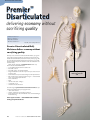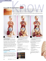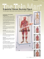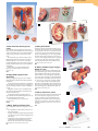
1
Sharing t he knowledge Digital Catalogue DGi Welcome Sharing the Knowledge The most complete, top-quality line of models in the industry. Now, more than ever you’ll find it easy to shop DGi. We know you’ll like our “ No Surprises” way of doing business. Here’s why: Frozen Prices. All prices published in your Sharing the Knowl- edge catalogue are F-R-O-Z-E-N through December 2011. No interim increases. NO SURPRISES! FREE DELIVERY — from our shelves to your door. You won’t pay a penny extra for shipping or handling. The price you see is the price you pay. No add-ons. NO SURPRISES! FREE GIFTS with your order. Not worthless “premiums”, but use- ful educational products selected from this catalogue to make lab sessions more effective. No bunk. No junk. NO SURPRISES! Skeletons 01 Human Anatomy 55 Contents Skeletons.............................................................................. Pages 1-54 Featuring Premier Skeletons — the only skeletons GUARANTEED FOR LIFE! Human Anatomy Models......................................................Pages 55-107 Featuring Autographed Anatomy. Models — 100% American made — the only models personally autographed and dated by their hand-crafter — and the only models with a LIFETIME GUARANTEE of satisfaction. Biochemistry................................................................... Pages 108-109 Nursing Trainers, Patient Simulators.................................. Pages 110-121 Charts & Multimedia.........................................................Pages 122-129 Biochemistry 108 Allied Health 110 Charts & Multimedia 128 Front Cover Art — “Globules rouges, globules blancs et plaquettes sanguines” or “Erythrocytes, white blood cells and platelets” original artwork by Francis LeRoy, Biocosmos Centre, Belgium. Used with permission. premier skeletons Premier™ Skeletons combine the best of the old and new worlds. Premier Medical Grade Skeletons are repli-cast in Germany from the bones of a select adult male natural-bone specimen, ensuring unparalleled fidelity to nature. Final assembly, hand-painting, numbercoding and labeling is carried out here at our DGi U.S. Factory. Delivery is from stock. And should you ever have the need, replacement parts, repairs and refurbishing of your Premier Skeleton are just a toll-free phone call away. And every Premier skeleton is backed by our Lifetime Guarantee of satisfaction — we will repair or replace any component which proves defective due to our workmanship, for as long as you own the skeleton — no charge, of course. Premier™ Skeletons M o u n t i n g O p t i o n s — w h i ch i s b e s t ? You’ll enjoy the following standard features with every Premier Skeleton: • 100% non-breakable plastic bones with stainless-steel fittings • Detachable 4-part skull with individual extractable/reinsertable teeth — complete with roots — each tooth in its own socket • Detachable spring-articulated mandible • Hinged mandibular flap which opens to expose roots of teeth in their sockets and impacted wisdom tooth • Detachable calvaria for access to neurocranium • Detachable upper and lower extremities • Rolling 5-spoked swivel-caster safety-stand • Choice of mounting options — sacral mount or suspension mount (see explanation at right) Our wide range of options and instructional enhancements let you tailor your skeleton selection to suit your program needs. Sacral Mount Skeletal support rod passes upward through the sacrum into spinal column. Nearly effortless single-handed freewheeling around the lab or from classroom to classroom because of inherent stability of this mounting style. Suspension Mount Skeleton is suspended from a support ring above the skull. Skeleton quickly detaches from suspension hook to accommodate manipulation and demonstrations in the prone and supine positions in addition to those in the erect posture while skeleton is suspended. ▼▼▼ Choose from three Premier Series: Premier Kinesiology Series; pp. 2–3 Premier Flexible Series; pp. 4–5 Premier Academic Series; pp. 6–7 G o o n l i n e at w w w. d e n o y e r. c o m 1 kinesiology skeletons Suspension Mount Premie Kinesi Premier™ Kinesiology Series Skeletons a a Sacral Mount b Faithfully reproduce life-like positioning and motions of the head, spine, limbs, hand and foot, with natural flexibility. Ideally suited for manipulation and demonstration in kinesiology, orthopedics, physical therapy, and chiropractic. Thanks to our exclusive Ultra-Flex™ flexiblyarticulated vertebral column with compressible intravertebral discs, completely life-like bending, twisting, lateral flexion and stretching movements of the cervical, thoracic and lumbar regions, including the pivoting of the skull on the atlas and axis, can be duplicated. Flexible hip and shoulder articulations allow a broad range of normal motions — shoulder blades actually rotate on their axes when arms are abducted above shoulder height. Other demonstratable movements include: • flexion and extension of the elbows and knees • hyperextension of the shoulders • medial and lateral rotation of the arms and thighs • pronation and supination of the forearms One hand and one foot are loosely-articulated with high-tensile nylon filament, permitting manipulation with full visibility of the articulating surfaces. Additional features included are: • cervical vertebral arteries • spinal cord with medulla • paired spinal nerve roots • detachable sacral flap which exposes the ramifications of the cauda equina Choose the options and mounting method best suited to your program requirements. All include 5-spoked swivel-caster safety-stand — sacral or suspension-type — appropriate to the mounting method you select (please refer to page 1 for help deciding which mounting method is best for you). A. Unpainted, Suspension-Mount Kinesiology Skeleton b S59 B. Unpainted, Sacral-Mount Kinesiology Skeleton b 2 F r e e G i f t s w h e n y o u o r d e r — s e e b a c k c o v e r. No S u r p r i s e s ! S58 kinesiology skeletons ™ er iology C. Hand-painted and labeled muscle attachments A wise choice for introductory musculo skeletal studies All the bones on the right half of the skeleton have been enhanced with hand-painted muscle origins (red) and insertions (navy) which have their names hand lettered in place. Where space limitations prevent spelling out muscle names, alphanumeric coding is employed. Additional colors differentiate the components of the erector spinal muscle complex from other muscles of the neck and back regions — iliocostalis: origins (orange), insertions (yellow) — longissimus: origins (dark green), insertions (light green). On the left half of the skeleton, all bones and osteological features of anatomical and clinical significance have been number coded for identification. A comprehensive key to all coded structures is furnished. c c S58L Sacral-Mount Painted and Labeled S59L Suspension Mounted Painted and Labeled D. Hand-painted and number coded muscle attachments The ideal choice for quizzes, self-testing and review Identical in all respects to S58L and S59L above, except that all hand-painted muscle origins and insertions have been number coded rather than labeled by name. A comprehensive key to all coded features is included. S58PN Sacral Mount, Painted and Number-Coded S59PN Suspension Mount, Painted and Number-Coded d U. S . t o l l f r e e 8 0 0 - 6 2 1-1014 3 fle xible skeletons Now with choice of male or Premier™ Flexible Series Skeletons Replicate the natural twisting and bend ing movements of the spine; illuminate the intimate relationship of the central nervous system to the axial skeleton. He knew the anguish of the marrow The ague of the skeleton; No contact possible to flesh Allayed the fever of the bone. — Whispers of Immortality, stanza 4; T. S. Eliot Our Flexible Series Skeletons incorporate our detachable 4-part skull with individual extractable/ reinsertable teeth, and all the standard Premier Skeletal features as cataloged on page 1, PLUS: • A flexibly articulated spinal column which permits natural twisting and bending motions of the spine and thorax • Resilient spinal cord with medulla • Paired spinal nerve roots • Cervical vertebral arteries • Herniated lumbar disc between L4 and L5 • Detachable sacral flap which exposes the branching of the cauda equina • Detachable upper and lower extremities Select the options and mounting style best-suited to your program requirements. Twelve ways to choose; all include Skeletal Lab Investigations Bonus Package (described on page 1), and 5-spoked swivel-caster safety stand — sacral or suspensiontype, appropriate to mounting method you select (please refer to page 1 for help deciding which mounting method to choose). a Premier Flexible Series Skeletons with Hand-painted Muscle Attachments — with your choice of number-coded muscles, or handlabeled muscles. Both employ additional colors to enhance comprehension and reduce confusion. A. Hand-painted and Labeled Muscle Attachments A wise choice for introducing musculoskeletal studies All the bones on the right half of the skeleton have been enhanced with hand-painted muscle origins (red) and insertions (navy) which have their names hand-lettered in place. Where space limitations prevent spelling out muscle names, alphanumeric-coding is employed. Additional colors differentiate the components of the erector spinae muscle complex from other muscle origins and insertions of the neck and back regions — ilicostalis: origins (orange), insertions (yellow) — longissimus: origins (dark green), insertions (light green). On the left half of the skeleton, all bones and osteological features of anatomical and clinical significance have been number-coded for identification. A comprehensive key to all coded structures is furnished. S68L Sacral Mount, Painted and Labeled Flexible Skeleton S68LF Sacral Mount, Painted and Labeled Flexible Skeleton with Female Pelvis S67L Suspension Mount, Painted and Labeled Flexible Skeleton S67LF Suspension Mount, Painted and Labeled Flexible Skeleton with Female Pelvis 4 P r e m i e r ™ S k e l e t o n s G UARANTEE D F OR LI F E fle xible skeletons female pelvis B B. Hand-painted and Number-Coded Muscle Attachments The ideal choice for quizzes, self-testing and review Just like S68L and S67L at left, except that all the hand-painted muscle origins and insertions have been number-coded rather than labeled by name. A comprehensive key to all coded features is included. S68 S68F S67 S67F Sacral-Mount, Painted, Number-Coded as above, with Female Pelvis Suspension-Mount, Painted, Number-Coded as above, with Female Pelvis C. Premier Flexible Series Unpainted Skeletons S66 S66F S65 S65F Sacral Mount c Sacral Mount Sacral Mount, Female Pelvis Suspension Mount Suspension Mount, Female Pelvis Suspension Mount c detail of painted spine a b A l l p r i c e s f r o z e n — No S u r p r i s e s ! 5 Prem ac a d e m i c s k e l e t o n s ™ Premier Academic Series Skeletons Combine durability, affordability and fidelity to nature Characterized by a rigidly-articulated vertebral column which exhibits normal spinal curvatures and intervertebral discs, our Academic Series skeletons incorporate all standard Premier features: • 100% non-breakable plastic bones repli-cast from those of a select male specimen • Stainless-steel fittings • Detachable 4-part skull • Individual extractable/reinsertable teeth — complete with roots — each tooth in its own socket • Detachable spring-articulated mandible • Hinged mandibular flap which opens to expose roots of teeth in their sockets • Detachable calvaria for access to the neurocranium • Detachable arms and legs A A A B. Hand-Painted and Labeled Muscle Attachments A wise choice for beginners All bones of the right half of the skeleton have been enhanced with hand-painted muscle origins (red), Choose the options and mounting method best suited to your program requirements. and insertions (navy). Muscle names are hand-lettered in place. Where space limitations prevent All include Skeletal Lab Investigations Bonus Packspelling out muscle names, alphanumeric coding is age (described on page 1), and 5-spoked swivel-caster employed. safety-stand — sacral or suspension-type — appropriate to the mounting method you select (please refer Additional colors differentiate the components of to page 1 for help deciding which mounting method is the erector spinae muscle complex from other muscles of the neck and back regions — ileocostalis: oribest for you). gins (orange), insertions (yellow) — longissimus: oriA. Special Edition Academic Skeleton looks gins (dark green), insertions (light green). just like real bone Bones and osteological features of anatomical Each bone of our Academic Special Edition Skeleton and clinical significance are number-coded on the left half ofthe skeleton. A key to identify all coded has been hand-rubbed with a custom formulated features is provided. stain, highlighting every fissure, fossa and pore, creating a convincing natural bone appearance. A S64L Sacral Mount magnificent specimen for study and display. S64LF Sacral Mount with Female Pelvis S62SP Sacral Mount S61SP Suspension Mount 6 S63L Suspension Mount S63LF Suspension Mount with Female Pelvis A ut o g r a p h e d A n at o m y ™ Mo d e l s G UARANTEE D F OR LI F E b C. Hand-Painted and Number-Coded Muscle Attachments The right choice for quizzes, self-testing and review Exactly like S64L and S63L, except that all handpainted muscle origins and insertions have been number-coded rather than labeled by name. A key identifying all coded features is furnished. S64 S64F S63 S63F Sacral Mount Sacral Mount with Female Pelvis Suspension Mount Suspension Mount with Female Pelvis D. Premier Unpainted Academic Series Skeletons S62 S62F S61 S61F Sacral Mount Sacral Mount with Female Pelvis Suspension Mount Suspension Mount with Female Pelvis mier Academic ACADEMIC s k e l e t o n s ™ Sacral Mount d Suspension Mount d E. Premier Academic Series Skeletons now offer 18-piece Take-Apart, Snap-together Skulls in choice of natural tone or colorcoded editions. Choice of male or female pelvis too! Snap plugs let your students quickly disassemble the Premier Take-Apart skull into 18 individual elements — then snap them back together again — a hands on learning experience guaranteed to reinforce understanding of the interrelationship of the cranial and facial bones which together comprise the human skull. Additional true-to-life details which will enhance the learning experience include a two-part temporal bone which divides to expose the inner ear within, and teeth with full roots which can be extracted from the maxillae and mandible, and reinserted into e their sockets. e e Color-Coded Skull Female Pelvis e Premier Academic Skeletons with natural tone 18-piece Take-Apart Skull S54 Sacral Mount S55 Suspension Mount S54F Sacral Mount with Female Pelvis S55F Suspension Mount with Female Pelvis Premier Academic Skeletons with Color-Coded 18-piece Take-Apart Skull S54P Sacral Mount S55P Suspension Mount S54PF Sacral Mount with Female Pelvis S55PF Suspension Mount with Female Pelvis G o o n l i n e at w w w. d e n o y e r. c o m 7 u lt r a f l e x s k e l e t o n s ™ UltraFlex Skeletons the marriage of form and function UltraFlex Skeletons meld the bony components and hardware of our Premier Flexible skeleton series with the classroom-proven synthetic ligaments of our UltraFlex full function major joint reproductions. This winning combination of two superior product ranges guarantees a skeletal specimen of uncompromising quality and versatility second to none. You’ll enjoy the following features with your UltraFlex Skeleton: • Choice of two series: Quadra™ Series with UltraFlex synthetic ligaments at four major joints of the right side, or Ultima ™ Series with UltraFlex ligaments at all six major joints of the right side. • Hand-painted muscle origins (red) and insertions (navy) on the left side, with choice of hand lettered identification labeling or number coding. Or choose the unpainted version. Painted versions also include number coding of bones and osteological features of the right side. A comprehensive identification key is supplied • Non-breakable plastic bones with stainless steel hardware and fittings. • Detachable 4-part skull with detachable calvaria and with individual extractable/reinsertable teeth, complete with roots. • Detachable spring-articulated mandible with hinged flap which opens to expose roots of teeth in their sockets, as well as an impacted wisdom tooth. • Flexibly-articulated spine which permits natural twisting and bending. • Resilient synthetic spinal cord with medulla and paired spinal nerve roots. • Cervical vertebral arteries • Herniated lumbar disc between L4 and L5 • Detachable sacral flap which exposes the branching cauda equina. • Detachable upper and lower extremities of the left side. • Choice of sacral or suspension mount. (Please refer to page 1 to see which mounting option is best for you.) • Rolling 5-spoked swivel-caster stand and dust cover. • Free Bonus Lab Investigation Package. S82 S84 Sacral Mount Suspension Mount A. UltraFlex Quadra™ Series Skeletons with UltraFlex ligaments on the shoulder, elbow, hip and knee joints of the right side S82Sacral mount with painted and number-coded muscles S81Suspension mount with painted and number-coded muscles S82L Sacral mount with painted and labeled muscles S81L Suspension mount with painted and labeled muscles S86 Sacral mount, unpainted version S85 Suspension mount, unpainted version B. UltraFlex Ultima™ Series Skeletons with UltraFlex ligaments at the shoulder, elbow, wrist, hip, knee and ankle of the right side S84Sacral mount with painted and number-coded muscles S83 Suspension mount with painted and number-coded muscles S84L Sacral mount with painted and labeled muscles S83L Suspension mount with painted and labeled muscles S88 Sacral mount, unpainted version S87 Suspension mount, unpainted version 8 P r e m i e r ™ S k e l e t o n s G UARANTEE D F OR LI F E a B u lt r a f l e x s k e l e t o n s a b a b b a b a b b U. S . t o l l f r e e 8 0 0 - 6 2 1-1014 9 deluxe skeletons Delux Deluxe Male Skeletons A. Deluxe Human Skeleton Reproduction Classic Craftsmanship Cast from the bones of a nearly-perfect adult male specimen, each bone is individually cast by hand, then assembled and articulated by hand — no short cuts or hurry-up techniques. Enduring Performance Corrosion-resistance is maximized by the exclusive use of stainless-steel wire and custom stainless hardware components for articulation. Built-in extras — most not even available on competitive skeletons — are standard with our Deluxe Skeleton Reproductions. These include: • X-ray opacity — just like real bone — due to a special additive introduced during the casting process • a “window” cut into the frontal sinus • a breakaway maxilla giving access to the paranasal sinuses • one hand and one foot (on opposite sides) articulated with springs, permitting natural flexion • detachable calvaria • spring-mounted detachable mandible • detachable major components — disarticulates into 12 parts • Full One-Year Warranty (manufacturer’s replacement parts and repair service always available.) Anatomy manual and five legged swivel-caster stand are included. A S100 B. Deluxe Skeleton With Painted and Labeled Muscle Attachments In addition to all the features of our S100 Deluxe Skeleton, the bones of the right side of this skeleton are hand-painted to show muscle origins in red and insertions in blue. Muscle names are hand-lettered at each muscle origin and insertion location. Complete with anatomy manual and deluxe chrome full-frame rolling stand. S105 b c 10 C. Muscle Skeleton Cast from the bones of a 6-foot adult male, this ingeniously executed skeletal reproduction is fitted with life-like synthetic muscles, nerves and ligaments of the cervical, thoracic and lumbar regions including the cervical plexus, brachial plexus, right shoulder joint and rotator cuff. Cervical muscles include: scalenus anterior, medius and posterior • longus capitus • longus colli. Thoracic muscles include: transversospinalis • rotatores. The right shoulder is fully articulated and includes all ligaments • capsule of the shoulder joint • levator scapulae • teres major and muscles of the rotator cuff: supraspinatus • infraspinatus • teres minor • subscapularis. Lumbar muscles include: psoas major • iliacus • piriformis • superior and inferior gemellus • quadratus lumborum • lumbar section of multifidus • erector spinae • gluteus minimus and medius • and obturator internus. Other features include: detachable calvaria and mandible • solid-cast carpal tunnel right hand and wrist • and right knee ligament. Complete with five-legged swivel caster stand. Added bonus: Muscular System Flash Card Set — 303 double-sided cards complete with hard plastic file box. S52 P r e m i e r ™ S k e l e t o n s G UARANTEE D F OR LI F E deluxe skeletons xe D. Orthopedic Skeleton Preferred by medical professionals for orthopedic instruction of students and patients, because it incorporates all the soft tissues applicable to orthopedics — nerves, arteries, muscles and ligaments, including: • Deluxe Flexible Vertebral Column with soft discs, vertebral arteries, cervical laminectomy, lumbar herniated disc, complete dura, and sacral plexus • Deluxe Brachial Plexus which includes all 14 major shoulder muscles, axillary vein and artery, major nerves, plus anterior, medius and posterior scalenus, and serratus muscles • Deluxe Elbow with major muscles, arteries and nerves • Deluxe Hand and Wrist with intraosseus muscles, median, ulnar and radial nerves, tendons, ligaments, arteries and transverse ligament • Deluxe Hip which disarticulates at ball and socket joint capsule, and includes pelvic ligaments, sacral plexus with sciatic nerve, femoral nerve, veins and arteries, and major muscles • Deluxe Knee with “bucket handle” tear in medial meniscus, cruciate and collateral ligaments, patellar tendon, and individual muscles • Deluxe Foot and Ankle which includes muscles, nerves, tendons and ligaments. Covered by a full-year warranty, the Orthopedic Skeleton includes an anatomy manual and chrome full-frame deluxe rolling stand. Deluxe Brachial Plexus includes all 14 major shoulder muscles – just one of the special details of this skeleton S320 E. Deluxe Biology Skeleton Removable internal organs and special fea tures add versatility and educational impact To our Deluxe Skeleton with Painted and Labeled Muscle Attachments S105 (described on page 10), we have added removable internal thoracic, abdominal and pelvic organs, plus special enhancements for increased versatility. Highlights include: Removable 2-part brain with hand-painted and lettered functional centers • Rib cage with sternum split vertically and hinged for easy access to lungs and heart • Color-coded lobar lungs reproduction • 2-part sagittally-sectioned heart • Individual abdominal organs — liver, stomach (divides into halves), pancreas, spleen, colon with mesentery, small intestine, right and left kidney reproductions with suprarenals (right kidney divides in half) • Deluxe male pelvis which is sagit tally sectional and hinged to permit the right hip to swing out through 45° • Both hips incorporate special joint articulation with capsules, to demonstrate normal ball and socket joint function. Covered by a full one-year warranty, the Deluxe Biology Skeleton is furnished with an anatomy manual, and chrome full-frame deluxe rolling stand. S335 d e A l l p r i c e s f r o z e n u nt i l D e c e m b e r 20 0 9 — No S u r p r i s e s ! 11 s k e l e -t o r s o Skele-Torso™ — The Ultimate Human Anatomy Specimen First choice of instructors in the medical professions, Skele-Torso™ stands alone in providing: Removeable Skull cap reveals two part dissectable Brain Authenticity Bones are cast from those of a near-perfect adult male specimen. Enhancing this skeletal framework are synthetic soft tissues and viscera cast from an expertly-prepared cadaver. The bones are even x-ray opaque — just like real bone! Synthetic muscles, nerves, ligaments and arteries Left arm is removable for closer study Versatility Separating into a total of 41 parts, and identifying some 755 anatomical features, Skele-Torso is the ultimate instructional tool for anatomy, physiology, orthopedics, physical therapy, sports medicine, chiropractic and patient education. Special enhancements Study Guide included • Synthetic muscles, nerves, ligaments and arteries of the right shoulder, elbow, hand and wrist, and left hip, knee, foot and ankle • Skull with muscles of mastication on the left side — perfect for TMJ studies • Nerves, including the cranial nerves — even the olfactory bulb and optic chiasm • Arteries — including the complete Circle of Willis • “Window” into the frontal sinus • “Breakaway” maxilla to access the paranasal sinuses • Spine which exhibits a cervical laminectomy, a herniated lumbar disc and lumbar hemilaminectomy • Male pelvis, sagittally sectioned and hinged, permitting the right hip to swing open through 45°, exposing a medial view through the pelvic bones, muscles, rectum, anus, bladder, prostate and penis Detachable major skeletal components — ideal for close-up study — including: • skull • left arm (detaches at the shoulder) • both legs (at the hip — left hip disarticulates at ball and socket joint capsule); and right lower leg (at the knee) Individual internal organs which can be removed and replaced at will: • two-part brain with color-coded functional centers • lobal lungs — lobes are color-coded and held together elastically • normal heart which divides in two to permit simultaneous viewing of all four chambers • liver with gall bladder and ducts • stomach (divides into halves) • pancreas • spleen • kidneys with suprarenals (right kidney divides sagittally) • small intestine with “windows” exposing the duodenal junction and ileocecal junction • colon and mesentery with “window” into transverse colon • male genitalia bisected to show erectile tissue of penis and path of urethra. Right testicle divides sagittally. Your Skele-Torso comes complete with a 30 page illustrated Study Guide, chrome, full-frame deluxe rolling stand, and dust cover. S250 12 F r e e G i f t s w h e n y o u o r d e r — s e e b a c k c o v e r. No S u r p r i s e s ! h u m a n a n at o m y r e s o u r c e l a b Save $300 When You Purchase the Human Anatomy Resource Lab Exclusively from DGi Multifaceted and comprehensive, each of the Lab’s 5 major components conveys a unique and distinct perspective to the learning process. Together they reinforce one another … creating a synergy which stimulates insightful perception, accelerating the effective grasp and ultimate mastery of the complexities of human anatomy. Your Anatomy Resource Lab includes all this: A. Skele-Torso™ The synthetic cadaver which gives you authentic detail without the inconvenience — the ultimate human anatomy specimen. Dissectible into 41 parts and identifying some 755 structures, SkeleTorso™ is featured on page 12. S250 C. 4-part Male Pelvic Anatomy b c Life size and “dissecting” into four parts, the model encompasses the right half of the pelvic region along with the entire male urogenital system. Divided along the median plane, the left half of the genitalia and surrounding structures including the pubic symphasis, prostrate with ejaculatory duct, urinary bladder, and rectum detach as a unit. The scrotal wall on the left side has been partially cutaway to expose the testis, spermatic card and vascular network. Sectioned medially and transversely, the left half of the penis detaches to reveal the urethra, corpus spongiosum and the erectile chambers of the corpus cavernosum. Supported on a rectangular base, the pelvic model measures 9¾×7×10½ inches overall (25×18×27 cm) A618-4 B. 3-part Female Pelvic Anatomy This life size counterpart to our male pelvic anatomy model A618-4, incorporates the entire female urogenital system and our open dissection of the right half of the pelvic region. Divided along the median, the left half of the female reproductive organs along with surrounding structures including the ovary, fallopian tube with fimbria, uterus, vagina, urinary bladder, urethra, rectum and pubic symphasis remove as a unit. The uterus further divides providing medial and transverse sectional views. Mounted on a rectangular base, the female pelvis model measures 9¾×7×10½ inches overall (25×18×27 cm). A618-3 D. The Thin Man Sequential Human Anatomy Figure Lets you explore body organs and systems in-depth by peeling away layer-after-layer of tissue, identifying more than 200 structural details during the course of the “dissection.” The Thin Man is featured on page 77. TM70 E. Anatomica: Digital Atlas of the Human Body CD-ROM a D B o n u s sav i n g s Your purchase of the Human Anatomy Resource Lab entitles you to 3 FREE GIFTS Easy-to-use, Anatomica provides a detailed look at the human body — a layer at a time! Unique “cutaway” digital atlas lets your students view over 2,000 parts of twelve major body systems. Twelve detailed, graphical layers can be removed one at a time by first selecting a body area and perspective — “front,” “back,” or “side.” Then use the cursor-directed location and magnification tools to access a detailed view of the anatomical region. Text and images can be printed for later study. Systems Requirements: Win 95 or higher. Mac 7.5 or higher. 16MB RAM. Described fully on page 128. CD160 Order the complete lab S273 Human Anatomy Resource Lab E G o o n l i n e at w w w. d e n o y e r. c o m 13 budget skeletons Budget-stretching Skeletons emphasize affordability and value Cast from molds prepared from the bones of an adult male, all feature: • 3-part skull with spring-articulated mandible and detachable calvaria • rigidly-articulated spine with normal curvatures • dust cover • rolling swivel-caster stand Your choice of three versions: 1. Basic, Unpainted Version 2. With Hand-painted Muscle Attachments 3. For an extra touch of functional anatomy — with Flexibly-Ligamented Joints at the shoulder, elbow, hip and knee Each version gives you the added option of a sacral or suspension mounting method. b A. Unpainted Version Includes 5-spoked swivel-caster stand. 221 Sacral Mount 221-10/1 Suspension Mount Suspension mount Sacral mount B. Number-Coded Muscle Attachment Version Muscle origins (red) and insertions (blue) have been hand-painted and number-coded on the left half of the skeleton. Bones and anatomically significant features have been hand-numbered on the right half. A key to coded-structures is furnished. Includes 5-spoked swivel caster stand. 224 Sacral Mount 224-11/1 Suspension Mount C. Flexibly-Ligamented Version Resilient synthetic ligaments flexibly-articulate the right shoulder, elbow, hip and knee, providing realistic anatomical functionality and convincing detail. Includes 5-spoked swivel-caster stand. 201-12 Sacral Mount a 14 A ut o g r a p h e d A n at o m y ™ Mo d e l s G UARANTEE D F OR LI F E c b u d g e t a l l- i n - o n e s k e l e t o n s D. Versatile “All-in-One” Skeletons pack a host of features into a single attractivelypriced package. Full-featured and understandably popular, “All-inOne” Skeletons combine the attributes of paintedmuscle skeletons, ligamented skeletons, and flexiblespine skeletons “all-in-one” specimen. Features include: • 3-part skull with spring-articulated mandible and detachable calvaria • hand-painted muscle attachments on the left half — origins red, insertions blue • resilient simulated joint ligaments articulate the right shoulder, elbow, hip and knee • number-coded bones and anatomicallysignificant structures of the right half • flexibly-articulated spinal column capable of natural bending and twisting postures • lumbar disc prolapse • simulated spinal cord and paired spinal nerves • cervical vertebral arteries • multilingual identification key to coded structures • dust cover Your choice of sacral or suspension mounting options. Both include 5-spoked swivel caster stand. 207-13 Sacral Mount 207-13/1 Suspension Mount d Budget U. S . t o l l f r e e 8 0 0 - 6 2 1-1014 15 female skeletons S41-25 Female Suspension, Painted S40-25 Female Suspension a b Female Skeletons A. Deluxe Female Suspension Skeleton Molded of special non-breakable plastic which closely approximates the appearance, feel and weight of real bone, even fine surface detail is faithfully rendered. Except for the skull, bones are individually articulated. Vertebral discs are flexible — one lumbar disc is herniated. Unobtrusive articulation of the limbs permits realistic movement while allowing easy removal. Non-corrosive fittings are used throughout. The highly accurate three-part skull has a sectioned calvaria, and the articulated detachable mandible incorporates the full adult dentition. Standing 162cm tall, the female skeleton is complete with five-leg swivel-caster stand. B. Female Suspension Skeleton with Ligaments, Painted Muscle Attachments, and Color-Coded Skull Bones Our S40-25 Female Skeleton, but with the following added features: • Synthetic pelvic ligaments have been incorporated on the right side • Muscle attachments — origins red, insertions blue have been hand-painted onto the bones of the left side • Bones in the right half of the 3-part skull have been painted in contrasting colors for easy identification Complete with key card and five leg swivel-caster stand. S41-25 S40-25 16 A ut o g r a p h e d A n at o m y ™ Mo d e l s G UARANTEE D F OR LI F E mini skeletons When space limitations and portability are primary consider ations, our Mini-Skeletons could be just the ticket Proving that good things come in small packages, our remarkablydetailed scaled down replicas of the adult human skeleton are large enough to incorporate essential anatomical details, yet are lightweight and compact enough to be easily manipulated and transported — from lecture hall to laboratory, or from building to building. Nearly all bones, except those of the hands and feet, are individually molded and articulated, reproducing natural movement at joints you would only expect to find on full-size preparations. Molded of specially formulated plastic compound which has the look and feel of natural bone, our mini-skeletons are virtually non breakable in normal use. A. 28 inch Mini-Skeleton Both arms, both legs and the skull are detachable, as are the calvaria and spring-articulated mandible. Complete with sacral support stand with hardwood base. b S28 Full size skull shown for size comparison B. 28 inch Mini-Skeleton with flexible spine Incorporating all the features of our S28 Mini-Skeleton, this scientifically accurate replica is further enhanced with a spine which is flexibly articulated, permitting natural bending and twisting motions of the torso. Complete with sacral support stand with hardwood base. S28X a e C. 32 inch Mini-Skeleton with spinal nerves Both arms, both legs, and the three-part skull are detachable. The legs can be further separated at the knee and ankle joints. In addition, this mini displays the spinal nerves. Complete with sacral support stand with hardwood base. S33 D. Deluxe 32 inch Mini-Skeleton with flexible spine In addition to all the features of our S33 Mini-Skeleton, including spinal nerves, our deluxe edition incorporates a flexibly-articulated spinal column which replicates natural bending and twisting motions of the neck and torso. Complete with sacral support stand with hardwood base. S34 E. Deluxe 32 inch Mini-Skeleton with muscle outlines This rendition of our S34 deluxe mini delineates color-outlined muscle origins (red), and insertions (blue), which are numbered for identification in the accompanying key. Complete with sacral support stand with hardwood base. S34P F. 31 inch Mini-Skeleton offers choice of mounting options. c d Perfectly scaled down from a full size adult skeleton using computerized engineering techniques. This half scale replica features a detachable three-part skull, detachable upper and lower extremities and a flexible vertebral column with soft intervertebral discs. Available with choice of sacral or suspension mounting options. S180 Sacral mount with support stand S181 Suspension mount with suspension stand S181P Suspension mount, Painted and Numbered with stand f A l l p r i c e s f r o z e n — No S u r p r i s e s ! 17 d i s a r t i c u l at e d s k e l e t o n s ™ Premier Disarticulated Six ways to choose — every one a winner! Unsurpassed quality — Best selection Medical-grade specimens comprising exactly the same bones and skull used to fashion our fully-articulated Premier Skeletons. With every Premier Disarticulated Skeleton you get the following standard features: • 4-part skull with individual extractable teeth • detachable calvaria • detachable spring-articulated mandible • hinged mandibular section which opens to expose roots of teeth in their sockets • one hand and one foot loosely articulated on high tensile nylon filament • vertebrae loosely strung in sequence on (and detachable from) high tensile nylon filament • intervertebral discs • hyoid • sternum with costal cartilages • sacrum with coccyx • all other bones individual and disarticulated • available with or without locking compartmented case Tailor your skeleton to suit your program needs with this wide range of optional enhancements: • 22-part totally disarticulated skull in addition to standard 4-part skull • color-coded vertebrae • hand-numbered bones and osteological features • hand-painted and numbered muscle attachments • hand-painted and labeled muscle attachments. I forced the last stone into its position; I plastered it up. Against this new masonry I re-erected the old rampart of bones. For the half of a century no mortal has disturbed them. In pace requiescat! — The Cask of Amontillado, Edgar Allan Poe A. Premier Standard Disarticulated Skeleton Incorporates all our standard features as delineated at left. S72C with case (SC63) S72 skeleton only B. Premier Disarticulated Skeleton with Color-Coded Vertebrae To aid identification and comprehension, the vertebrae of this enhanced version have been color-coded as follows: cervical (C1-C7) yellow. Thoracic (T1-T12) orange. Lumbar (L1-L5) dark brown. S74C with case (SC63) S74 skeleton only C. Premier Double-Header Because two heads are better than one — our Double-Header disarticulated skeleton includes our SK29 take-apart skulll in addition to our 4-part skull and all the standard features. S70C with case (SC63) S70 skeleton only D. Premier Numbered Disarticulated Skeleton Bones and significant osteological features of the left half have been hand-numbered for identification. Supplied with key to coded structures. S71C with case (SC63) S71 skeleton only Six ways to choose — every one a winner. C b 18 F r e e G i f t s w h e n y o u o r d e r — s e e b a c k c o v e r. No S u r p r i s e s ! F d i s a r t i c u l at e d s k e l e t o n s Full Premier Disarticulated Skeletons with Handpainted Muscle Attachments — your choice of labeling or number-coding. E. Labeled Muscle Attachments ▼ ▼ A wise choice for introductory musculoskeletal studies Muscle origins (red) and insertions (navy blue) hand-painted on all the bones of the right half, have their names hand-lettered in place. Where space doesn’t permit name-lettering, muscles are number-coded. To prevent confusion, additional colors differentiate the origins and insertions of the erector spinae complex — iliocostalis: orange and yellow; longissimus: light and dark green. Bones and osteological features of the left side have been hand-numbered for identification. Complete with key to coded structures. S73LC with case (SC63) S73L skeleton only F. Number-Coded Muscle Attachments The ideal choice for quizzes, self-testing and review Exactly like S73LC & S73L above, except that all hand-painted muscle origins and insertions have been number-coded rather than labeled. Identification key is included. S73C with case (SC63) S73 skeleton only G. Compartmented Disarticulated Skeleton Case Color-coded spine replaces white spine in items S74 See page 23 for a full description and photo. SC63 E a b G o o n l i n e at w w w. d e n o y e r. c o m 19 d i s a r t i c u l at e d s k e l e t o n s ™ Premier Disarticulated delivering economy without sacrificing quality No Rack can torture me — My Soul — at Liberty — Behind this mortal Bone There knits a bolder One — No. 384, stanza 1, Emily Dickinson Premier Disarticulated HalfSkeletons deliver economy without sacrificing quality. When you select a Premier Disarticulated Half-Skeleton, you get the same medical-quality osteological components incorporated in our full disarticulated skeletons, featured on the two previous pages. But since the right and left sides of the body are mirror images, our halfskeletons include only the ribs and bones of the extremities from one side of the body. You’ll enjoy the full range of standard features with every Premier Disarticulated Half Skeleton: • 4-part skull with individual extractable teeth • detachable calvaria • detachable spring- articulated mandible • hinged mandibular section which opens to expose roots of teeth in their sockets • hand and foot loosely articulated on high-tensile nylon filament • full complement of vertebrae loosely strung on — and detachable from — high tensile nylon filament • intervertebral discs • hyoid • sternum with costal cartilages • sacrum with coccyx • all other bones disarticulated Our wide range of optional instructional enhancements lets you match your half-skeleton to your program needs: • color-coded vertebrae • number-coded bones and osteological features • hand-painted muscle attachments with choice of hand-labeling or hand-numbering Choice of five versions — all available with or without locking compartmented case. 20 P r e m i e r ™ S k e l e t o n s G UARANTEE D F OR LI F E Color-coded spine replaces white spine in items S78 d i s a r t i c u l at e d s k e l e t o n s Half ▼ A. Premier Standard Disarticulated Half-Skeleton b Encompasses all our standard features as outlined on page 20. S76C with case (SC63) S76 half-skeleton only B. Premier Number-Coded Disarticulated Half-Skeleton Bones and significant osteological features have been hand-numbered for identification. Supplied with key to coded structures. S76NC with case (SC63) S76N half-skeleton only ▼ C. Premier Disarticulated Half-Skeleton with ColorCoded Vertebrae To speed identification and aid comprehension, the vertebrae of this enhanced edition have been color-coded in the following manner: cervical (C1-C7) yellow. Thoracic (T1-T12) orange. Lumbar (L1-L5) dark brown. S78C with case (SC63) S78 half-skeleton only Case available with skeleton or separately (item F) e Premier Disarticulated HalfSkeletons with Hand-painted Muscle Attachments — your choice of labeling or number-coding. D. Labeled Muscle Attachments The best choice for introductory musculo-skeletal studies. Hand-painted muscle origins (red) and insertions (navy blue) have their names hand-lettered in place. Where space doesn’t permit namelettering, muscles are number-coded. To avoid confusion, additional colors differentiate the origins and insertions of the erector spinae muscle complex — iliocostalis: orange and yellow; longissimus: light and dark green. Supplied with key to coded structures. S77LC with case (SC63) S77L half-skeleton only E. Number-Coded Muscle Attachments The right choice for quizzes, self-testing and review Identical to S77LC & S77L above, except that painted muscle origins and insertions have all been number-coded rather than labeled. Identi fication key to numbered structures is included. S77C with case (SC63) S77 half-skeleton only d f F. Compartmented Case safeguards your disarticulated skeleton under lock and key. Handsome yet rugged case protects your disarticulated skeleton or half-skeleton during storage and transport. Compartments provide organized storage of skeletal components. Constructed of heavy-gauge plywood clad with tough, leather-grain plastic. Metal-reinforced corners maintain rigidity while absorbing bumps and dings. Complete with comfort-grip handle, twin locking latches and keys. SC63 U. S . t o l l f r e e 8 0 0 - 6 2 1-1014 21 d i s a r t i c u l at e d s k e l e t o n s Disarticulated Skeletons A. Deluxe Medical Grade Disarticulated Skeleton features x-ray opaque bones Complete with compartmented case for transport and storage Individually cast from a near-perfect adult male skeleton, the bones of this professional medical reproduction replicate the features, heft, color — even the x-ray opacity — of real bone. The complete complement of skeletal components is included: skull with detachable calvaria, hinged mandible, both styloid processes, sutures, and faithfully-reproduced internal and external anatomy – including full adult dentition • vertebrae strung in sequence on nylon filament • both hands and both feet articulated on nylon filament • complete set of ribs and sternum • all other bones • carrying case and anatomy manual. S303 $1,069.00 B. Female Disarticulated HALF Skeleton Medical Quality This superior preparation comprises the same meticulously detailed osteological components utilized in the assembly of our fully articulated female skeletons. Closely approximating the appearance, feel and mass of real bone, all the bones, with the exception of the skull, are disarticulated. Includes: 3-part skull with detachable calvaria and mandible • complete set of vertebrae (strung on nylon) • ribs of one side • sternum with costal cartilages • bones of one arm, complete with scapula and clavicle • bones of one leg, complete with half pelvis. The bones of the hand and foot are loosely strung on nylon filament. a b SA49 Order with compartmented locking case (SC63) and SAVE! SA49C with case C. Female Disarticulated FULL Skeleton Medical Quality Like SA49 Half Skeleton (item B) but with all bones included. The 2-part skull is uncut. Vertebrae, hands and feet are loosely strung on nylon filament. Case not included. SA48 Order with compartmented locking case (SC63) and SAVE! SA48C with case c D. Female Disarticulated HALF Skeleton with Painted Muscle attachments Medical Quality Bony components are identical with those of Half Skeleton SA49 described above, but with the addition of hand-painted muscle attachments — origins red, insertions blue. A truly outstanding teaching preparation. Supplied with an identification key. Without case. SA49P Order with compartmented locking case (SC63) and SAVE! SA49PC with case E. Budget-stretching Disarticulated FULL Skeleton features a premium-quality 4-piece skull Offering unsurpassed quality where it counts, this economicallypriced skeleton includes our Premier Medical Quality 4-part skull SK80 as described on page 26, which features realistic teeth and a highlydetailed cranial vault, detachable calvaria and hinged mandible flap. All other bones are disarticulated except those of one hand and foot which are loosely strung on nylon filament. The hyoid and sternum with costal cartilages are included as well. d 218-80 Order with compartmented locking case (SC63) and SAVE! 218-80C with case F. Disarticulated HALF Skeleton With 4-Piece Premium Skull Cast from the same molds as our 218-80 disarticulated skeleton, but including only the bones from one side of the skeleton. All vertebrae and sternum with costal cartilages are included as well. e 22 f A ut o g r a p h e d A n at o m y ™ Mo d e l s G UARANTEE D F OR LI F E 219-80 219-80C with case s t o r ag e & d i s p l ay Give your valuable bones the protection they deserve a b A. Five-Leg Sacral Support Stand fits most sacral-mounted skeletons Replace damaged and broken stands with this affordable unit. As furnished with our Premier™ Sacral mounted skeletons, this rugged swivel caster stand is compatible with most sacral mounted skeletons. 22 inch diameter ABS plastic base with tubular steel upright. S26 B. Deluxe Full-Frame Suspension Stand features mobility, stability, and complete accessibility — front and back Our deluxe stand provides outstanding support for suspension skeletons — real bone or plastic. Compare these stand out design features: wide stance for tip-proof stability • easy maneuverability, thanks to smooth-rolling swivel casters • fullframe design for unhindered access, front and back • and all-metal, heavily chrome-plated tubular construction to stand the test of time. Utility — stability — mobility — accessibility — durability — and affordability. When it comes to skeleton stands — this stand stands alone. Overall dimensions: 23×25×71 inches (58×63×180 cm). S20 C. Affordable Five-Leg Suspension Stand positions skeleton directly over center of gravity to prevent tipping As furnished with our Premier™ suspension skeletons, this swivel-caster stand will support any real bone or plastic suspension type skeleton. Consisting of a five-leg base molded of spaceage ABS plastic, with swivel casters for full mobility, the satin-finished tubular steel upright has been formed into a vertically-elongated question mark shape, thus suspending the full mass of the skeleton directly over the center of the base to maximize stability. Overall dimensions: 22 dia.x74 inches (56×188 cm). c d e D. Display Stand for Spines One-piece heavy-gauge nickel-plated steel construction with hook to support any suspensiontype spinal column. This same stand is included with all Premier™ spines. Overall height: 34 inches (86 cm). SP61 E. Zippered Shroud won’t let the dust settle on your skeleton Heavy-guage duraflex cover with full-length zipper will provide years of dust-free protection for your articulated skeleton. Featuring a transparent window, and measuring 70×18 inches (178×46 cm), the shroud will accommodate either suspension-mounted or pelvicmounted skeletons. F (Bones not included) SC28 F. Locking Skull Case One size fits all — real bone or plastic Constructed of solid plywood clad with leathergrain plastic, and lined with high-density urethane foam to cushion and protect fragile specimens, deluxe features include hinged lid, key-locking latch, and protective feet. SC70 zG. Compartmented Case safeguards your disarticulated skeleton under lock and key G Handsome yet rugged case protects your disarticulated skeleton or half-skeleton during storage and transport. Compartments provide organized stowage of skeletal components. Constructed of heavy-gauge plywood clad with tough, leather-grain plastic. Metal-reinforced corners maintain rigidity while absorbing bumps and dings. Complete with comfort-grip handle, twin locking latches and keys. SC63 S25 A l l p r i c e s f r o z e n — No S u r p r i s e s ! 23 premier components Rejuvenate your bone-tired bone boxes; resto * Indi Disarticulated Skull Sternum With Costal Cartilages Atlas & Axis Skull Sacrum & Coccyx Vertebrae Cervical Thoracic Lumbar Clavicle Hyoid Scapula Ribs Of One Side Ulna Humerus Premier™ Unmounted Skeletal Components Radius Four-Part Skull with Individual Teeth Thoracic Vertebra Fully described on page 26. Single vertebra typifying this region of the spine. SK80 SB14 Take-apart Skull, 22-Parts Thoracic Vertebrae Set (T1 through T12) SK29 Loosely strung with detachable high-test line. Includes: Atlas, Axis, and typical cervical (colored yellow) • Typical thoracic (colored orange) • Typical lumbar (colored brown). Loosely strung with detachable high-test line. SB14-1 SB19 SB17 Thoracic Vertebrae Set, Color-Coded Orange Cervical Vertebra T1 through T12 loosely strung with detachable hightest line. Representative Vertebrae, Set of Five, Natural Tone Single vertebra other than atlas and axis. SB16 Atlas and Axis SB13 Cervical Vertebrae Set (C1 through C7) Lumbar Vertebra Single vertebra typifying this region of the spine. Representative Vertebrae, Set of Five, ColorCoded SB19-1 Complete Vertebral Column Includes: Atlas, Axis, Sacrum, Coccyx. Loosely strung with detachable high-test line. Natural Tone. SB15 SB10 Lumbar Vertebrae Set (L1 through L5) Ribs of One Side, Set of 12, Unmounted Loosely strung with detachable high-test line. SB15-1 SB20-SLeft SB20-DRight SB12 Lumbar Vertebrae Set, Color-Coded Brown Sternum and Costal Cartilages SB21 Hyoid L1 through L5 loosely strung with detachable hightest line. SB11 SB18 Sacrum and Coccyx Loosely strung with detachable high-test line. SB13-1 Cervical Vertebrae Set, Color-Coded Yellow C1 through C7 loosely strung with detachable hightest line. SB23 24 A ut o g r a p h e d A n at o m y ™ Mo d e l s G UARANTEE D F OR LI F E premier components ore those decrepit disarticulated skeletons ividual bones Replace lost and damaged bones with Premier™ medical-quality specimens — the cost-effective way to breathe new life into broken-down bone sets. Your choice of left or right appendicular components and ribs — so you can replenish the missing pieces of your sets with corresponding parts. You can even upgrade your conventional bone collections with color-coded vertebrae to speed recogni tion, enhance comprehension. Male Pelvis ...there was a noise, and behold a shaking, and the bones came together, bone to his bone. – Ezekiel 37:7 KJV Innominate Fibula Femur Bones Of One Foot Tibia Patella Bones Of One Hand Innominate Ulna Bones of One Foot with Distal Tibia and Fibula SB22-S Left SB22-DRight SB34-SLeft SB34-DRight Loosely strung on high-test line. Male Pelvis with 4th and 5th Lumbar Vertebrae Bones of One Hand with Distal Radius and Ulna SV60 Loosely strung on high-test line. Female Pelvis with 4th and 5th Lumbar Vertebrae SV61 SB48-SLeft SB48-DRight SB38-SLeft SB38-DRight Femur Clavicle SB42-S Left SB42-DRight SB36-SLeft SB36-DRight Patella Scapula SB41-S Left SB41-DRight SB35-S Left SB35-DRight Tibia Humerus SB43-SLeft SB43-DRight SB32-S Left SB32-DRight Fibula Radius SB44-SLeft SB44-DRight SB33-S Left SB33-DRight G o o n l i n e at w w w. d e n o y e r. c o m 25 premier skulls Premier™ Skulls More than just anoth Premier™ Medical-Quality 4-Part Skulls stress fidelity to nature. Repli-cast from a select natural bone specimen for unparalleled realism, every Premier Skull features individual teeth with complete roots — each in its own socket — not just a set of fused dentures like other synthetic skulls. An exclusive hinged flap in the right mandible swings open to expose the roots of the two premolars and first two molars in their sockets, plus an impacted third molar (wisdom tooth.) These five teeth are permanently anchored. All the remaining teeth in the upper and lower jaws are secured in their sockets with a permanently-flexible cement which allows you to extract teeth individually for thorough examination, and then reinsert them! Once replaced in their sockets, teeth will not fall out. This unprecedented extraction procedure can be repeated time and again. Detachable springs permit natural articulation of the mandible as well as jaw removable. The sectioned calvaria detaches to access the neurocranium. a Webster was much possessed by death And saw the skull beneath the skin; ... — Whispers of Immortality, Stanza 1 T.S. Eliot b d A. Standard Premier 4-Part Skull This medical quality replica incorporates all the Premier features described above. SK80 skull only SK80B skull plus hardwood display base SK80C skull plus locking case B. Special Edition Premier Skull looks just like real bone b This Special Edition Premier Skull has been hand-rubbed with a custom formulated stain, highlighting every suture, fissure, fossa and pore, and creating a convincing natural bone appearance. A magnificent specimen for study and display. SK80SP skull only SK80SPB skull plus hardwood display base SK80SPC skull plus locking case C. Special Edition Numbered Premier Skull b We’ve taken our Special Edition skull with natural bone finish and hand-numbered each of the cranial and facial bones and other significant features, including fissures, protuberances, fossae and foramina. An identification key is supplied. SK81 skull only SK81B skull plus hardwood display base SK81C skull plus locking case D. Locking Skull Case One Size fits all — real bone or plastic Constructed of solid plywood clad with leather-grain plastic, and lined with high-density urethane foam to cushion and protect fragile specimens, deluxe features include hinged lid, key-locking latch, and protective feet. SC70 c 26 F r e e G i f t s w h e n y o u o r d e r — s e e b a c k c o v e r. No S u r p r i s e s ! premier skulls er pretty face Alas! Poor Yorick. I knew him, Horatio; a fellow of infinite jest, of most excellent fancy. — Hamlet, Prince of Denmark, Act V, Scene I William Shakespeare E. Premier™ Teaching Skull To simplify identification of the individual cranial and facial bones which comprise the skull mosaic, the bones of our Teaching Skull have been handpainted with contrasting colors to reinforce learning their shapes and relationships. To avoid confusion, left and right members of paired bones are painted alike. Bones have been numbered to correspond with the printed identification key which accompanies the skull. e SK80P Skull only SK80PB with hardwood base SK80PC with locking case F. Premier Skull with Painted and Labeled Muscle Attachments Perfect for introductory musculoskeletal studies Muscle origins (red) and insertions (blue) have been hand-painted on the right half of this Premier skull, and have their names hand-lettered in place. Bones and significant skeletal features have been number-coded on the left half to correspond with the identification key included with the skull. SK80PL Skull only SK80PLB with hardwood base SK80PLC with locking case G. Premier Skull with Painted and Numbered Muscle Attachments — great for quizzes, self-testing and review Hand-painted muscle origins (red) and insertions (blue) which have been number-coded for identification enhance the right side of this Premier Skull. Hand-numbering on the left side identifies skull bones and prominent features. Complete with key to coded features. f G SK80PN Skull only SK80PNB with hardwood base SK80PNC with locking case H. Premier Medical Demonstration Skull This version of our 4-Part Premier Skull includes the following enhancements — individual bones and significant features of the left side have been hand-numbered for identification • hand-painting within the neurocranium traces the course of the venous sinus of the dura mater (blue) and the cranium arteries (red) • color-coded nerves and blood vessels supplying the roots of the teeth are exposed within the dissected right mandible. An identification key is furnished. SK82 Skull only SK82Bwith hardwood base SK82C with locking case h U. S . t o l l f r e e 8 0 0 - 6 2 1-1014 27 premier skulls Premier™ Skulls b a Featuring detailed cranial vault A. Premier Medical Demonstration Skull with Painted and Labeled Muscle Attachments Incorporating all the special enhancements of our Medical Demonstration Skull SK82, this skull has been further enhanced with hand-painted muscle origins (red) and insertions (blue) on the right side, which have their names hand-lettered in place. Supplied with an identification key. SK83 Skull only SK83B with hardwood base SK83C with locking case B. Premier Color-Coded Teaching Skull on Cervical Spine Featuring bones hand-painted in contrasting colors, this rendition of out Teaching Skull is supported in its natural position atop a flexibly-articulated cervical spine, complete with color-coded atlas and axis, vertebral arteries, paired spinal nerves, medulla and spinal cord, and is mounted on a hardwood platform. Skull bones are numbered for identification in the key provided. SK85 C. Premier Skull on Cervical Spine — especially useful for demonstrating the role of the atlas and axis. This edition of our Premier Skull is flexiblyarticulated in its natural position atop the cervical spine which is mounted on a stable hardwood platform. All seven cervical vertebrae, paired spinal nerves, vertebral arteries, medulla and spinal cord are present. SK84 D. Premier Skull on Cervical Spine with Painted and Labeled Muscle Attachments We have enhanced this version of our Premier Skull on Cervical Spine with hand-painted muscle origins (red) and insertions (blue) on the right side, while on the left side bones and significant features are number-coded for identification. Larger muscles have their names hand-lettered in place — smaller muscles are numbered. For the sake of clarity, additional colors are employed to distinguish between the components of the deeplying erector spinae complex — iliocostalis: origins (orange) insertions (yellow), longissimus: origins (light green) insertions (dark green). Mounted on a hardwood platform. A printed identification key is furnished. SK84PL 28 c P r e m i e r ™ S k e l e t o n s G UARANTEE D F OR LI F E d cl assic skulls Budget-stretching Classic Skulls e f g h E. Classic See-Thru Skull With 5-Part Brain H. Classic Didactic Skull on Cervical Spine, 4-Parts When mated with our Classic 5-Part Brain (described on page 83), you can actually observe the brain in situ within the transparent skull. Brain nests perfectly within the skull, and lifts out for hands on study. SK262 F. Classic Three-Part Skull with Numbering This rendition of our Classic Skull has been enhanced with hand-numbering of the bones and major osteological features on one side. In addition, all the suture lines have been highlighted in yellow to make it easier to discern the interlocking bony plates which together comprise the human skull. A key to all numbered structures is furnished. 257-21 i j G. 7-Part Deluxe Didactic Skull — a true split personality Divided along the medical sagittal plane, the right half of our Deluxe Skull is transparent, while the left half is natural bone tone. Visible on the transparent half are the paranasal sinuses and cranial sinuses which have been highlighted in different colors. The carotid, vertebral and facial arteries have also been depicted in red for maximum visibility. The right brain hemisphere is visible in situ through the transparent cranium. The periodontal sockets can be viewed through the transparent right maxilla and mandible half. Flexibly articulated, the mandible demonstrates opening and closing of the jaws. Resting upright on the cervical spine, the following components are detachable: both left and right halves of the calvaria, the left half of the base of the skull, the nasal septum, the mandible in its entirety, and a brain half. Mounted on a support stand and base. Employing 19 colors to distinguish the various bony plates which make up the human skull, this special didactic edition of our Classic Skull has been flexibly mounted atop the cervical spine whose atlas, axis and seventh vertebra have been color-coded as well. Also present are the spinal cord, cervical spinal nerves, vertebral arteries, and after lifting off the calvaria, the medulla, basilar artery and posterior cerebral arteries will be located within the cranial chamber. A stand and base supports the model in its natural position. SK202 I. Classic Three-Part Plastic Skull Cast from a specially-selected natural bone specimen, all fissures, foramina, sutures and fossae are faithfully reproduced. The calvaria detaches for neurocranial studies. The spring-articulated mandible also detaches. Special attention has been paid to the teeth, which have a totally natural appearance. 253 J. Classic Numbered 3-Part Skull with Dissected Mandible This 3-part skull incorporates the following special enhancements: A dissection of the mandible which exposes the roots of the teeth, and the nerves and blood vessels supplying them • color highlighted skull sutures • and within the cranial vault, the paths of significant blood vessels and sinuses of the meninges are traced in color and numbered. Some 104 internal and external number - coded features are identified in the accompanying key. 255 SK283 A l l p r i c e s f r o z e n — No S u r p r i s e s ! 29 s p e c i a l f e at u r e s k u l l s Special Feature Features hinged mandible and 32 removeable teeth Sectioned mandible and maxilla A. 10-Section Medical Demonstration Skull Dissectable into five parts Color highlights cranial nerves and meningeal sinuses a More detail than ever before achieved in a plastic skull replica. Cast from a specially prepared natural specimen, this incomparable teaching tool incorporates the following features: the calvaria detaches, then the remainder of the skull divides into halves along the mid-sagittal plane. The perpendicular plate of the ethmoid and vomer is hinged to expose the sphenoid sinus. Highlights of the left half: temporal bone detaches • maxilla is sectioned open revealing the alveolar blood vessels and nerves • frontal bone has a hinged flap into the frontal sinus. Highlights of the right half: temporal bone is partially chiseled away baring the facial nerve canal and the semicircular canals of the inner ear • a hinged flap into the frontal sinus. The detachable mandible is sectioned open on the left side exposing the alveolar blood vessels and nerves. A hinged flap on the right side discloses the roots of the molars and premolars. All 32 teeth are individually removable. Color is used to highlight the sinuses and semicular canals, and to trace cranial nerve tracts and the arteries and meningeal sinuses within the cranium. 251 B. Deluxe Bisected Skull reveals the hid den complexities of the sinuses and nasal passages With the look and feel of real bone, this highlydetailed plastic reproduction divides mid-sagittally. Especially suited for examination of the cranial vault, sinuses, nasal passages and palate, one half includes the nasal septum and complete mandible. The other half exposes the nasal conchae and nasal passages. 281 C. Hydrostone Male and Female Skull Replica Set Not plastic. These exquisite one-piece replicas are cast in hydrostone, a gypsum compound which, while more fragile than plastic, is noted for its ability to reproduce even the most subtle details of form and texture. Reproduced from natural bone originals, these museum-quality replicas make welcome additions to classes in anatomy, art, medicine and forensics. SK222 Male/Female Skull Set SK20 Male Skull Only SK21 Female Skull Only b c 30 F r e e G i f t s w h e n y o u o r d e r — s e e b a c k c o v e r. No S u r p r i s e s ! The ideal illustration of sexual dimorphism in humans s p e c i a l f e at u r e s k u l l s Skulls D. Premier Muscled Skull brings full dimensionality to the study of head and facial musculature Superior to charts and textbook illustrations which lack depth, this fully three dimensional presentation incorporates life like reproductions of the muscles of facial expression and mastication. Muscles have been molded from a specially formulated material which is flexible, yet tough enough to heavy hands on study. Among the muscles represented are: Frontalis • Temporalis • Orbicularis oculi • Nasalis • Levator anguli oris • Levator labii superioris • Zygomaticus major • Zygomaticus minor • Masseter • Orbicularis orbis • Mentalis • Depressor labii inferioris • Depressor anguli oris • Pterigoid • Mylohyoid • Left and right digastrics. Ideally suited for anatomy studies, patient education, allied health training, medical-legal documentation, and forensic science. Complete with illustrated key. D SK86 E. Functional synthetic muscles of masti cation ideally suit this Classic Skull for TMJ demonstrations Perfect for patient education, allied health training and medical-legal documentation, as well as anatomy studies, this version of our Classic Skull has been enhanced to graphically demonstrate the functioning of the temporomandibular joint and the associated TMJ Syndrome. Functional synthetic jaw muscles, consisting of resilient colored plastic and elastic bands, reproduce the temporalis and masseter muscles on the right side, the lateral and medial pterygoids on the left. Hand-lettered labeling identifies each muscle. E 289-24 F. 14-Section Medical Demonstration Skull unveils the secrets of the sinuses and nasal passages A specially selected and prosected natural skull was employed as the pattern from which this highly detailed specimen was cast. Distinctive features include: • Horizontally sectioned calvaria which detaches, exposing the cranial cavity in which the network of meningeal blood vessels, venous sinuses and internal carotid have been traced in contrasting colors. • A sagittal section through the base of the skull reveals the intricacies of the nasal passages. • The frontal sinus is exposed through a resection of the frontal bone on one side and by means of a hinged bony flap on the other. • The temporal on one side of the bone detaches and divides in two exposing the semicircular canals and inner ear. • A hinged bony flap through the ethmoid plate and vomer on one side opens to reveal the chamber of the sphenoid sinus. • Both the mandible and maxilla of one side have been sectioned open to bare the alveolar blood vessels and nerves which supply the roots of the teeth. • The detachable mandible has a hinged bony on one side which opens to expose the roots of the teeth. • The teeth are individually extractable and reinsertable. F SK14 G o o n l i n e at w w w. d e n o y e r. c o m 31 ta k e - a pa r t s k u l l s Life-Size Take-Apart Skulls A. Premier 18-piece Modular Skull Snap plugs let your students quickly separate our modular skull into its 18 individual elements — then just as quickly snap them back together again — a hands-on-learning experience guaranteed to reinforce mastery of the interrelationships of the 8 cranial and 14 facial bones which together comprise the human skull. The vomer and palatine bones detach as a single element, as do the paired nasal bones. SK18 b B. Color-Coded 22-piece Take-Apart Skull Kit This version of our 22-piece Skull Kit has been molded in nine contrasting colors to facilitate learning the identities, shapes and relationships of the individual skull components. Paired bones are molded in the same color. a SK29P C. Color-Coded 14-Piece Take-Apart Skull Medical Quality c This version of the Take-Apart Skull is further enhanced through the use of hand-painted contrasting colors to distinguish the individual bones which together form the skull. In all other respects it is identical to the Take-Apart Skull SK52 below. Furnished with identification key. SK52P D. Color-Coded Premier 18-piece Modular Skull To expedite student mastery of the shapes, identities and relationships of the bones of the skull, this edition of our Premier Modular Skull is molded in nine distinctive colors. Paired bones share matching colors. A powerful hands on learning tool. SK18P d E. 22-piece Take-Apart Skull Kit The mosaic of 22 bones which comprise the human skull are molded individually in this life-size reproduction. Held together with flexible connectors, the bones can be readily disjoined and rejoined for a thorough understanding of their interrelationships. Sutures have been simplified to facilitate assembly and disassembly. SK29 F. 14-Piece Take-Apart Plastic Skull Medical Quality An invaluable exercise in mastering the orientation and complex relationships of the bones comprising the adult human skull. Consisting of 14 separate elements, the skull can be quickly dismantled and then reassembled by means of flexible snap-together plugs. Sutures have been simplified to facilitate assembly and disassembly. e G SK52 G. Auditory Ossicles Medical Grade Life-Size replicas of the three tiny bones of the middle ear, the malleus (hammer), incus (anvil) and stapes (stirrup) cast from actual human specimens. Ossicles are labeled and embedded in plastic for protection. A32 f 32 P r e m i e r ™ S k e l e t o n s G UARANTEE D F OR LI F E individual skull bones U. S . t o l l f r e e 8 0 0 - 6 2 1-1014 33 female skulls Ladies First — quality fema A. Two-Piece Female Skull is perfect for Forensic Studies Cast from a near-perfect female specimen using a specially-formulated plastic which mimics the look, feel and heft of natural bone, even such fine details as complete styloid processes and the delicate nasal conchae have been beautifully reproduced. Mandible and maxillae bear the full complement of adult dentition. The articulating mandible readily detaches and reattaches. With its calvaria uncut and cranium intact, this skull is ideally suited for comparison with male specimens for studies in sexual dimorphism and forensic science. SK33 B. 3-Piece Female Skull In addition to exhibiting all of the true-to-life detail of our Two-Piece Skull SK33 described on page 35, this three-piece counterpart has a sectioned detachable calvaria to facilitate studies of the interior of the cranial vault as well. a b SK32 C. Hydrostone Female Skull Replica Not plastic, this exquisite one-piece full-size replica is cast in hydrostone, a gypsum compound which, while more fragile than plastic, is noted for its ability to faithfully reproduce even the most subtle details of form and texture. Reproduced from a natural bone original, this museum-quality replica will make a welcome addition to classes in anatomy, art, medicine and forensic science. SK21 D. Female Medical Skull with Major Bones Color-Coded for Easy Identification The same highly detailed casting as used in our Female Numbered Skull SK57 described on page 35, but the bones of this 3-part specimen have been hand painted in contrasting colors. This greatly simplifies the process of identifying cranial and facial bones, while it reinforces learning their shapes and relationships. Calvaria and mandible are detachable. Skull exhibits the full 32 tooth adult dentition. Identification key is supplied. c d SK56 E. Deluxe Female Neurovascular Skull with 8-Part Soft Brain Trace cranial nerves and arteries WITHOUT confusion Because they lack depth, charts and textbook illustrations — even computer simulations — can frustrate attempts at tracing cranial nerve branches and blood vessels. Not subject to these visual limitations, our fully dissectible, 3-dimensional Neurovascular Skull delivers more meaning to neuro-cranial studies. Distributed over the exterior and cranial floor of our medical-quality skull replica are the principal arteries and nerves — including all 12 cranial nerves. Nerves are color-coded yellow; arteries red. Nested within the cranium is our life-size 8-part brain, cast from a very soft, resilient material which deforms and feels like real tissue. SK58 34 e A ut o g r a p h e d A n at o m y ™ Mo d e l s G UARANTEE D F OR LI F E m i n i & F e ta l s k u l l s ale skulls Mini Skulls & Fetal Skulls A. Three-Part Mini Skull As furnished with our 28 inch Mini-Skeleton, S28 (described on page 18), this anatomical miniature includes a detachable calvaria and spring-articulated detachable mandible. f SK28 B. Mini-Skull — 3 Part This precisely scaled replica is the same as included with our 31 inch Mini-Skeletons S180 and S181 featured on page 18. Both the calvaria and mandible are detachable. a SK185 C. Fetal Skull — Unpainted Medical Quality Reproduced from a natural specimen, this plastic replica exhibits the fontanels and the developing bones of the infant. SK38 D. Color-Coded Fetal Skull b g To simplify identification of cranial and facial components, our technicians have hand-painted the individual bones of this fetal skull replica with contrasting colors. An extraordinary teaching tool. F. Female Neurovascular Skull with Cervical Spine, on Stand Because it doesn’t suffer from the visual limitations of 2-dimensional diagrams — allowing study from all aspects — our Neurovascular Skull eliminates much of the confusion associated with neuro-cranial studies. Our adult female medical skull and all seven cervical vertebrae are situated on a stand. Exhibited externally and internally on the left side are the arteries (colored red) supplying the head and neck, while on the right, the 12 cranial nerves (colored yellow) and the distribution of their branches are depicted. SK38P E. Color-Coded Fetal Skull with Display Base c This version of our SK38P color-coded fetal skull comes with its own custom-fitted hardwood display base. As beautiful as it is practical, the skull simply lifts off for close up scrutiny. SK38PB F. Fetal Half Skull with Dura Mater Medical Quality SK59 G. Female Medical Skull with ColorCoded Numbers Cast from a unique custom-formulated plastic with the look, feel and heft of real bone, this adult female specimen reproduces even such fine details as the delicate nasal conchae, internal carotid canal, and full-size styloid processes. The sectioned calvaria detaches to permit study of cranial vault, and the articulated, detachable mandible bears the full adult complement of teeth. Skull sutures are color accented. Bones and major features are identified by number — using four different colors for easy reference: bones and teeth, numbered black • sutures, numbered green • processes, protuberances, chondyles, numbered brown • foramina and fissures, numbered red. Complete with identification key. d The perfect complement to our full Fetal Skull SK38, this medical quality plastic reproduction of an actual specimen is bisected and beautifully illustrates the dural membrane, fontanels and developing bones. SK39 e f SK57 A l l p r i c e s f r o z e n — No S u r p r i s e s ! 35 premier spines Premier™ Spines are more than flexible — You’ll find a Premier Spine perfectly suited to your appli cation in anatomy • physical education • allied health • physical therapy • chiropractic • sports medicine • or patient education. A B A. Premier Flexible Spine with Movable Femur Heads Includes suspension stand Natural cast from a real bone specimen this full-size replica provides lifelike accuracy in color, texture, and special details including: completely flexible articulation to reproduce natural vertebral movements and alignments • occipital bone of skull • spinal cord with medulla • vertebral arteries • paired spinal nerves • detachable sacral flap which exposes the branching cauda equina • proximal femur segments articulated to duplicate movement at the ball and socket joint of the hip. SP62 b B. Premier Flexible Spine with Disc Prolapse and Movable Femur Heads Includes suspension stand This version of our Premier flexible spine not only incorporates all of the life-like details of our SP62 spine, but in addition includes a herniated lumbar disc between L4 and L5. SP65 a c C. Color-Coded Premier Flexible Spine with Disc Prolapse and Movable Femur Heads Includes suspension stand Without peer for patient education as well as introductory classes in anatomy and physical therapy, this special edition of our Premier spine series includes all the exceptional attributes of our SP62 and SP65 spines, plus distinctive colors to differentiate the cervical (yellow), thoracic (orange) and lumbar (brown) vertebrae. SP63 D. Premier Flexible Spine, without femur heads Includes suspension stand Except that it is lacking femur heads, this rendition of our Flexible Premier Spine is identical in every respect with our SP62 Flexible Spine listed above. SP58 E. Premier Flexible Spine with Female Pelvis, without femur heads Includes suspension stand The female counterpart to our number SP58 Flexible Spine, this rendition encompasses the very same features, but has a female pelvis rather than a male pelvis. SP58F F. Premier Flexible Spine with Painted Muscle Attachments, Disc Prolapse and Movable Femur Heads Includes suspension stand and identification key d f e 36 A ut o g r a p h e d A n at o m y ™ Mo d e l s G UARANTEE D F OR LI F E Beginning as our Flexible Premier spine SP65, we have enhanced this version with hand-painted muscle attachments on the right side — origins red, insertions blue. Larger muscles have their names hand lettered in place. Smaller muscles are numbered. For the sake of clarity, additional colors are used to distinguish between the components of the deep-lying erector spinae complex — iliocostalis: orange and yellow, longissimus: light and dark green. SP65PL premier spines they’re versatile, too! G. Premier Flexible Spine with Female Pelvis and movable femur heads Includes suspension stand The female counterpart to our SP62 Flexible Spine with movable femur heads detailed on the previous page, it is identical in all respects with SP62 except that the pelvis is female rather than male. SP62F H. Premier™ Flexible Spine with Thorax and Movable Femur Heads Perfect for respiratory studies This full-size natural cast replica of an adult male specimen makes an ideal subject for meaningful study of respiratory mechanics. Because it is provided with the entire rib cage in addition to a flexibly-articulated vertebral column, you can reproduce all the natural movements of the spine and thorax. Additional true-to-life features included are: occipital bone of the skull • spinal cord with medulla • cervical vertebral arteries • paired spinal nerves • detachable sacral flap which exposes the branching cauda equina • proximal femur segments articulated to duplicate movement at the ball and socket joint of the hip. Complete with suspension stand. g h SP64 I. Premier™ Flexible Spine with Thorax and Painted and Labeled Muscle Attachments, and Movable Femur Heads Hand-painted muscle attachments — origins red, insertions blue — enhance this special version of our SP64 flexible spine with thorax. Larger muscles have their names hand lettered in place. Smaller muscles are number-coded. For the sake of clarity, additional colors are used to distinguish between the components of the deep-lying erector spinae complex — ilicostalis: orange and yellow — longissimus: light and dark green. Supplied complete with printed key and suspension stand. SP64PL J. Ultraflex™ Spine with Movable Femur Heads This special edition is fitted with discs of ultra soft resilient plastic permitting the ultimate in manipulative flexibility. In addition, the Ultraflex™ Spine incorporates the occipital bone of the skull • spinal cord with medulla • vertebral arteries • paired spinal nerves • detachable sacral flap which exposes the branching of the cauda equina • complete pelvic girdle • and movable femur heads. SP69 K. Ultraflex Spine without femur heads Except that it lacks femur heads, this edition of our Ultraflex spine incorporates all of the other special Ultraflex features found in our SP69 spine described above. i j k l SP70 L. Ultraflex Spine without Pelvis Incorporation the ultrasoft discs and special features of our other Ultraflex spines listed on this page, this rendition does not include the pelvic bones. It does include the sacrum and coccyx. SP71 G o o n l i n e at w w w. d e n o y e r. c o m 37 spine models ™ Premier and BON A. Premier Flexible Thoracic Spine Medical quality Encompassing the twelve thoracic vertebrae, t1 – t12, and their intervertebral discs, this medical quality reproduction has been flexibly articulated to faithfully demonstrate natural bending and twisting of the thoracic spine. Complete with resilient spinal cord and paired spinal nerves, the spine is mounted on a hardwood base. SP45 B. Premier Flexible Cervical Spine with Nerves and Arteries Medical quality A Flexibly-articulated, this natural cast replica encompasses the seven cervical vertebrae • intervertebral discs • occipital bone • spinal cord with portion of medulla and pons • paired spinal nerves • and vertebral arteries. Mounted on a hardwood base, the spine demonstrates the natural twisting and bending of the head and neck. SP66 C. Premier Flexible Lumbar Spine with Sacrum and Spinal Cord Medical quality Natural cast and functional, this flexibly-articulated spine consists of the five lumbar vertebrae and the sacrum along with the spinal cord and paired spinal nerves which are molded of resilient plastic. A section of the sacrum is detachable to show the branching cauda equina. The invertebral disc between L4 and L5 is herniated. Mounted on a hardwood base. SP67 D. Switch Hitter — Premier™ Lumbar Vertebrae Set exhibits common disc pathologies Medical Quality Faithfully reproducing the human 4th and 5th lumbar vertebrae, this natural cast replica includes the spinal cord and paired spinal nerves. In addition to the normal intervertebral disc, the set is furnished with two interchangeable herniated discs which illustrate different disc pathologies — one exhibits a central prolapse which would result in compression of the dura and cauda equina, while the second displays a dorsolateral prolapse whose bulging would press on the nerves of one side. L4 is detachable to allow you to switch from one disc to another. Mounted on a hardwood base. Overall dimensions: 6×6×5 inches (15×15×13 cm). SP68 E. Premier™ Cervical Vertebrae Sets* B All seven cervical vertebrae (C1 through C7) loosely strung in sequence on high-test line. Vertebrae can be easily unstrung and restrung. Choice of natural color or yellow color-coding. SB12 Color-Coded (Yellow) Cervical Set SB13-1 Natural Bone Color Cervical Set F. Premier Thoracic Vertebrae Sets* All twelve thoracic vertebrae (T1 through T12) loosely strung in sequence on high-test line. Vertebrae can be easily unstrung and restrung. Choice of natural color or orange color-coding. E SB16 Color-Coded (Orange) Thoracic Set SB14-1 Natural Bone Color Thoracic Set G. Premier Lumbar Vertebrae Sets* F All five lumbar vertebrae (L1 through L5) loosely strung in sequence on high-test line. Vertebrae can be easily unstrung and restrung. Choice of natural color or brown color-coding. C SB18 Color-Coded (Brown) Lumbar Set SB15-1 Natural Bone Color Lumbar Set H. Representative Vertebrae, Set of Five, Color-Coded* G D 38 H The ideal collection for demonstrating and studying the structural similarities, differences and specializations of vertebrae from different regions of the spine. Included in the set are: • atlas (C1), axis (C2), and typical cervical vertebra (all colored yellow) • typical thoracic vertebra (colored orange) • and typical lumbar vertebra (colored brown) loosely strung on high-test line. Vertebrae are readily unstrung and restrung. SB19 Color-Coded, Set of Five *Set colors may vary. SB19-1 Natural Bone Color, Set of Five F r e e G i f t s w h e n y o u o r d e r — s e e b a c k c o v e r. No S u r p r i s e s ! spine models ™ ONElike Spines The ultimate in manipulative flexibility ™ BONElike I. BONElike Spine with pelvis and occipital bone Is this real bone? No! It’s BONElike Consisting of the full complement of 24 human vertebrae complete with intervertebral discs, plus occipital bone of the skull and male pelvis, all reproduced in BONElike synthetic resin which maximizes true-to-life detail, natural feel and heft. The vertebral column is flexibly articulated to replicate natural bending and twisting motions of the axial skeleton. The occipital bone and atlas are individually detachable. Standing 33½ inches tall (85 cm) the spinal column is supplied complete with display stand. Revolutionary new plastic material reproduces even the finest bone details while exhibiting the natural feel and heft of real bone. Even medical experts have been fooled upon initial examination of BONElike reproductions. Non-breakable under normal usage, BONElike reproductions make ideal substitutes for natural bone specimens in medical teaching situations, allied health training, and for patient education. SP79-4 BONElike Vertebral sets J. BONElike Cervical Vertebrae, Set of 7 Encompassing the full complement of neck vertebra, including the atlas and axis, each vertebra is identified by number (C1 – C7). The vertebrae share a display base in which each rests in its own form-fitting depression. Each reproduction quickly lifts out for detailed examination from all aspects. Overall dimensions: 12×8¼×2½ inches (30×21×6 cm). j SB79 K. BONElike Lumbar Vertebrae, Set of 5 Each BONElike lumbar vertebral reproduction is identified by number (L1 – L5), and nests in its own form-fitting niche within the shared display base. Individual vertebrae lift out for hands-on scrutiny. 12×8¼×2½ inches (30×21×6 cm) overall. SB79-2 k i L. BONElike Human Vertebrae, Set of 24 Our complete set of BONElike vertebrae, cervical, thoracic, and lumbar – each labeled for identification (C1 –C7, T1 – T12, L1 – L5) is supplied in a fully padded compartmented storage/carrying case. Overall dimensions: 16×15¾×5¾ inches (41×40×12 cm). SB79-3 Reproduces the natural surface detail and weight of real bone, while retaining the durabilty of plastic l U. S . t o l l f r e e 8 0 0 - 6 2 1-1014 39 p r ecm l ai esrs i sckseplienteosn s Budgetstretching Classic Spines A. Flexible Classic Spine with Occipital Bone and Pelvis Flexibly articulated to simulate natural twisting and bending motions, this budget-priced vertebral column also includes the following features: occipital bone of skull • spinal nerve roots • cervical vertebral artery • a lumbar disc prolapse between L3 and L4 • complete pelvis. Stand included. 264-58/1 B. Classic Flexible Spine with Female Pelvis The female version of our Classic Flexible Spine 0264-58/1 described above. It has exactly the same features, but has a female pelvis in place of the male. Complete with suspension stand. SP584 C. Classic Flexible Spine with Femur heads and painted muscle attachments a b c Encompassing all the features of our 0264-58/1 Classic spine, this version also incorporates movable femur heads and has painted muscle origins (red), and insertions (blue) on one side. Complete with suspension stand. SP583 D. Deluxe Flexible Spine with femur heads and painted muscle attachments Exactly like our painted Classic spine SP583, but with the addition of an exposed brain stem and a detachable portion of the sacrum which reveals the cauda equina within. Complete with suspension stand. SP587 E. Lifetime Flexible Spine Incorporating all the features of our best selling 0264-58/1 Classic flexible spine, but with a special lifetime articulation conduit which will withstand repeated bending and twisting without fatigue and breakage. Ideally suited to classroom and chiropractic demonstrations. Complete with suspension stand. SP591 F. Lifetime Flexible Spine with femur heads g Identical to our SP591 Lifetime spine described above, but with the addition of movable femur heads. Complete with suspension stand. SP592 G. Color-Coded Flexible Spine includes Brain Stem, Cerebellum, Brachial Plexus, Herniated Disc and Movable Femur Heads d e f With cervical and lumbar vertebrae molded in brown plastic, this 35-inch instructional spine also incorporates the following special features: brainstem • cerebellum • brachial plexus • paired spinal nerves • and sciatic nerve complex (all colored green) • herniated disc between L4 and L5 • and movable femur heads. Complete with suspension stand. SP55 40 A ut o g r a p h e d A n at o m y ™ Mo d e l s G UARANTEE D F OR LI F E p rm em u si ecrl esdk eslpei n to es ns h H. Cervical Spine with Musculature and Nerves PLUS Skeletal Muscles Flashcard set i Flexibly-articulated, this natural cast specimen incorporates synthetic muscles of resilient plastic to reproduce natural movements of the neck. Muscles included are: rectus capitis posterior major and minor • obliquus capitis superior • interspinales • scalenus medius • scalenus posterior • longus capitis • longus colli • and multifidis (cut). Other features include: brain stem • cerebellum • spinal nerves • cervical vertebral artery • cervical and brachial plexi • greater occipital nerve • display stand. j k l Each of these spines is furnished with our Muscular System Flashcard Set. More than 300 colorillustrated cards, complete with hard plastic file box. SP53 L. Deluxe Muscle Spine I. Lumbar Spine with Musculature, Nerves, PLUS Skeletal Muscles Flashcard set On top of all the attributes of the Advanced Muscle Pelvis, and Femur Heads PLUS Skeletal Muscles Flashcard set Spine SP56, our Deluxe Muscle Spine is further enhanced with additional musculature and anatomNatural cast from the bones of an adult male, this flexibly articulated specimen has been fitted with syn- ical features. thetic muscles of resilient plastic attached with elastic Additional muscles included are: longissimus thoracis and spinalis thoracis of the erector spinae bungee cord. complex • sacral and lumbar regions of the mul The following muscles are included: quadratus lumborum • piriformis • psoas major • iliacus • lumbar tifidis • gemellus superior and inferior • gluteus medius and minimus • obturator internus (cut). and sacral sections of multifidis • gemellus superior and inferior • gluteus medius and minimus • and obtu- Additional features included are: cervical and lumbar vertebrae color-coded blue • L4 and L5 show rator internus. Other features included are: paired spinal nerves • bone degeneration • sacrospinal ligament. sciatic nerve complex • sacrospinal ligament • and a her- SP57 niated disc between L4 and L5. Complete with suspension stand. M. Flexible Torso Skeleton incorporates SP52 J. Flexible Muscle Spine PLUS Skeletal Muscles Flashcard set lifelike muscles, ligaments and nerves of the cervical, thoracic and lumbar regions PLUS Skeletal Muscles Flashcard set Replicating the bones of a six-foot adult male, this skillfully-rendered skeletal specimen is fitted with This 35-inch flexibly-articulated vertebral column is fitted with the following synthetic muscles of resilient resilient synthetic muscles and ligaments of the polyurethane: rectus capitis posterior major and minor neck, shoulder, spine and pelvis attached with bungee cord. Coupled with a flexibly-articulated ver• obliquus capitis superior • transversospinalis • rotatores • iliacus • psoas major • and quadratus lumborum. tebral column, this arrangement permits dynamic bending and stretching demonstrations. Other key features included are: occipital bone • brain stem • cerebellum • paired spinal nerves • cervi- Other features included are: lifelike brain stem cal vertebral arteries • sciatic nerve complex • and her- • cerebellum • paired spinal nerves • cervical plexus niated lumbar disc between L4 and L5. • greater occipital nerve • occipital triangle • brachial plexus • sciatic nerve complex • movable femur SP54 heads • cervical and lumbar vertebrae color-coded blue. Complete with suspension stand. K. Advanced Muscle Spine PLUS Skeletal Muscles Flashcard set S56 Encompassing all of the muscles and features of our SP54 flexible muscle spine, our advanced edition spine incorporates additional muscles and enhancements. Additional muscles included are: longus capitis • longus colli • scalenus medius • scalenus posterior • and piriformis. Additional features included are: 1st and 2nd ribs • brachial plexus • and facet syndrome between L5 and sacrum. Three times life-size, this lumbar vertebra with intervertebral discs, spinal cord and nerve roots shows both anterior longitudinal and supraspinal ligaments. Also illustrated are removable replicas of the psoas muscle and the five main back muscles. Dissects into three parts and displayed on stand with key card. 17×14×15cm. SP56 m N. Lumbar Vertebrae with Muscles SA25 n A l l p r i c e s f r o z e n — No S u r p r i s e s ! 41 s p i n e s w i t h i n n e r vat i o n Cervical Thoracic Lumbar Articulated Vertebral Sets with Nerves Closely mimicking the appearance, weight and feel of real bone, these full-size vertebral replicas are fitted with flexible synthetic nerves in their correct anatomical relationships. A. Cervical Vertebrae Set with Cervical Plexus Includes the occipital bone, the seven cervical vertebrae with their discs, vertebral arteries, cervical spinal cord, and the nerves of the cervical plexus. SA47 B. Thoracic Vertebrae Set a b c All twelve thoracic vertebrae, their discs, the thoracic spinal cord and paired spinal nerves are beautifully rendered. SA45 C. Lumbar Vertebrae and Sacrum Set The five lumbar vertebrae, their discs (one is herniated); sacrum, coccyx, lumbar spinal cord, and the nerves of the lumbar plexus are faithfully reproduced. SA46 D. Seven-Piece Vertebrae Collection Includes Display Stand Representing all vertebral types, this collection includes: the first and second cervical vertebrae (atlas and axis) • typical cervical vertebra • two thoracic vertebrae with intervertebral disc • and a lumbar vertebra. Vertically mounted on a display stand, all components are removable for detailed examination. Dimensions: 5×5×9 inches (12×12×22 cm). d 211-75 E. Cervical Vertebra with Spinal Cord This single cervical vertebra is accompanied by a section of the spinal cord. Clearly shown are white and grey matters, anterior and posterior horns, and associated nerves. Displayed on base. SA60 F. Lumbar Vertebrae with Pelvis showing Lumbar and Sacral Plexi e Showing five lumbar vertebrae, together with the pelvis, this spine model illustrates a herniated disc, as well as lumbar and sacral plexi. f SA67 G. Premier™ Flexible Mini-Spine with Pelvis Perfect for hands-on study by individuals and small groups Faithful in every detail except size, this 12-inch replica of the human spine features: individual vertebrae and discs • spinal cord • paired spinal nerves • cervical vertebral arteries • fully flexible articulation • bones of the pelvis articulated to allow slight movement at the sacroiliac joint • suspension ring for hanging • display stand with hardwood base. Skull shown for size reference only; not included with spine SP60 H. Flexible Miniature Vertebral Column g 42 h A ut o g r a p h e d A n at o m y ™ Mo d e l s G UARANTEE D F OR LI F E Complete with occipital bone of the skull, sacrum, coccyx, pelvis and intervertebral discs, this onehalf scale mini spine has been flexibly articulated to duplicate natural twisting and bending motions as well as demonstrating abnormal spinal curvatures. Resting upright on a display stand, the spine quickly lifts off for hands on manipulation. SP182 s p i n a l pat h o l o g i e s Separately, or together in our budget-saving Lab – Insight into Lumbar Spine Pathologies Demonstrate lifting technique: the right way… and the wrong way! a A. Lifting Manikin distinguishes right from wrong Ingeniously engineered and executed, this functional figure dramatically contrasts correct and incorrect lifting techniques, and exhibits the stress and distortion of the lumbar spine resulting from improper lifting posture. The manikin will raise a weight from either of two clearly marked positions on its base. Illustrations on the base detail the effects of right and wrong technique on lumbar vertebral alignment. No assembly required. Complete with booklet providing background information. b c SA50 B. Deluxe Osteoporosis Series illustrates loss of bone mass Demonstrating the progressive reduction in bone density and resulting atrophy, which typify this condition associated most often with postmenopausal women, this series consists of three medially sectioned lumbar vertebrae and their intervertebral discs. The topmost vertebra in the series exhibits normal healthy bone structure. The middle vertebra reflects the increased porosity and loss of bone mass characterizing the osteoporotic condition. Typifying advanced osteoporosis, the bottom vertebra manifests deterioration, deformation, flattened plates and greatly reduced bone density. Detachably mounted on a support base. d SP67 268-78 C. Switch Hitter – Premier™ Lumbar Vertebrae Set exhibits common disc pathologies Medical Quality Faithfully reproducing the human 4th and 5th lumbar vertebrae, this natural cast replica includes the spinal cord and paired spinal nerves. In addition to the normal intervertebral disc, the set is furnished with two interchangeable herniated discs which illustrate different disc pathologies — one exhibits a central prolapse which would result in compression of the dura and cauda equina, while the second displays a dorsolateral prolapse whose bulging would press on the nerves of one side. L4 is detachable to allow you to switch from one disc to another. Mounted on a hardwood base. Overall dimensions: 6×6×5 inches (15×15×13 cm). SP68 D. Degeneration of the Lumbar Spine, 4-Stage Series Mounted together on a hardwood base are four sets of 4th and 5th lumbar vertebrae (L4 and L5) with their intervertebral disc. The first set typifies the normal healthy condition of bone and disc. The three remaining pairs exhibit progressive stages in pathological degeneration of the lower spine — herniated disc and facet syndrome • thinning disc and arthritic bone spurring • advanced bone degeneration with fusion (spondylosis). Overall dimensions: 16×3×4 inches (41×8×10 cm). SP59 E. Vertebra showing Arthritis and Osteoporosis Representing a single adult vertebra and nerve, this model is divided lengthwise and is hinged. One half is normal, while the other half shows outgrowths from arthritis and wearing away from osteoporosis. Displayed on stand. SA24 e CD985 f F. Lumbar Spine Pathologies Instructional Lab Features all the items pictured on this page! By working with the concrete examples provided in this versatile hands-on lab, your students of physical therapy, sports medicine, orthopedics, radiology, geriatrics and gerontology will gain perspective and insight into lumbar spine pathologies resulting from injury and degenerative diseases. Lab contents: • Premier™ Flexible Lumbar Spine with Sacrum and Spinal Cord (SP67 as featured on page 38) • Exploring The Spine CD-ROM (CD985 as featured on page 128). Plus featured products from this page: • Premier Lumbar Vertebrae Set with Common Disc Pathologies, (SP68) • Degeneration of the Lumbar Spine, 4-Stage Series, (SP59) • Deluxe Osteoporosis Series, (268-78) • Lifting Manikin, (SA50). • Vertebra showing arthritis and osteoporosis (SA24). SLi-96 G o o n l i n e at w w w. d e n o y e r. c o m 43 p e lv i c m o d e l s Life-Size Pelvic Reproductions A A. Deluxe 6-part Female Pelvic Replica with ligaments, blood vessels, nerves, pelvic organs and pelvic floor b B. Childbirth Demonstration Pelvis For demonstrating the progress of the fetus through the maternal pelvis during childbirth, this In addition to all the features of our SV203 reproduc- functional reproduction consists of an actual cast of the female pelvic skeleton with movable pubic tion, our deluxe rendition incorporates the entire symphysis, complete with sacrum, coccyx and two pelvic floor. Mid-sagittally sectioned, a portion lumbar vertebrae, articulated to accommodate pasof the external anal sphincter, external urethral sage of a full-term fetal skull replica mounted on an sphincter, deep and superficial transverse perineal muscles are removable. Also removable as a unit are omnipositioning flexible gooseneck support. the rectum, uterus with fallopian tubes and ovaries, The pelvis is mounted on a rectangular base. Overall dimensions: 13×10×7 inches (33×26×18cm) and vagina. These will further divide in half along the mid-sagittal plane. SV30 The right half of the pelvis includes: • the common iliac artery and its internal and external C. Female Pelvic Skeleton with genitalia, branches • the common iliac vein and eternal vein • 3 parts the right sacral plexus • right sciatic nerves • and the This natural casting of the female bony pelvis, right pudendal nerve. complete with the 4th and 5th lumbar vertebrae, sacrum and coccyx, has been fitted with external SV204 and internal genitalia, perineum, rectum, anus and bladder to illustrate their relationship to the underlying bony framework. All soft tissue structures have been molded of resilient soft plastic. For complete visibility, the bladder and a portion of the uterus with one fallopian tube and ovary can be detached. Mounted on a rectangular base, the three-part model measures 13×10×7 inches (33×26×18cm) SV31 c d D. Composite Pelvis & Pelvic Floor, 5Parts Separating into five parts, this life-size female pelvis has the look of real bone. Its dissectible pelvic floor is comprised of distinctively colored, flexible genitalia and associated muscles. SA27 44 A ut o g r a p h e d A n at o m y ™ Mo d e l s G UARANTEE D F OR LI F E p e lv i c m o d e l s E. Premier Male and Female Bony Pelves Medical Quality Cast from molds prepared from natural bone specimens, these plastic reproductions illustrate the morphological distinctions between male and female pelvic structures. Each pelvis includes the left and right innominates with pubic symphysis, 4th and 5th lumbar vertebrae with intervertebral discs, the sacrum and coccyx. Typical of the majority of humans, each pelvis exhibits four pairs of sacral foramina. SV60Male Pelvis SV61Female Pelvis SV610 Pelvis Set — includes SV60 and SV61 E Male F Female H Adult Male Female E F. Premier Male and Female Pelves with femur heads Medical Quality Identical with Premier Pelves SV60 and SV61 detailed above, except that these reproductions also include movable right and left femur heads. SV63Male Pelvis with femur heads SV62Female Pelvis with femur heads SV623 Pelves Set — includes SV62 and SV63 G. Classic Female Pelvic Skeleton Consisting of the hip bones with pubic symphysis, two lumbar vertebrae with discs, sacrum and coccyx, this pelvis differs from our Premier Female pelvis in that it possesses five pairs of sacral foramina as is found in some individuals — a nice example of variable morphology among humans. G 215-61 Distinguish between male, female and juvenile pelvic structure H. 4-Piece Take-Apart Pelves Adolescent Cast from our unique bone-like plastic and fully articulated by means of wing nuts, these pelves quickly disarticulate into four pieces — left and right innominates, sacrum and pubic symphysis — ideally suiting them to detailed measurements of size, shape, angle, inlets and outlets. Useful for studies in anatomy, OB/GYN, midwifery, and forensics. The two adult pelves include the 5th lumbar vertebra. SA44 Adult Male Take-Apart Pelvis SA40 Adult Female Take-Apart Pelvis SA42 Adolescent Take-Apart Pelvis SAVE when you purchase all three Take-Apart Pelves as a set. Set includes one each Adult Male (SA44), Adult Female (SA40), and Adolescent Take-Apart Pelvis (SA42). SA444 Adult Female H H Take-Apart H Front I. Adult Female Ligamented Pelvis with Nerves, and Pelvic Floor In addition to all the pelvic ligaments included in our SA41 above, this pelvis has been further enhanced with the principal nerves and a flexible, detachable 2-part muscular pelvic floor. SA51 J. Adult Female Ligamented Pelvis J I Cast from the same molds as our Adult Female Take-Apart Pelvis, these invaluable teaching preparations have been fitted with lifelike synthetic pelvic ligaments which in life hold the bones of the pelvic girdle together. These pelves cannot be disarticulated. Back SA41 K. Flexible Adult Female Pelvis is perfect for demon strating pelvic malformities A specially formulated resilient plastic is used in the molding of this life-size replica. It will readily deform to demonstrate a variety of pelvic structural abnormalities often associated with difficulty during childbirth. The model will resume its original shape on its own. I K SA43 U. S . t o l l f r e e 8 0 0 - 6 2 1-1014 45 upper e x tremit y b c d A. Six-Part Muscular Anatomy of the Arm and Shoulder Scaled to 3/4 life-size, this plastic replica presents the muscles, nerves, tendons, blood vessels and bones of the left hand, wrist, arm and shoulder. Five superficial muscles can be individually detached to expose the more deeply lying muscles. Model stands 24 inches (60 cm) high on a pedestal base, and lifts off for hands-on scrutiny. Includes key card. a 343-10 B. 6-piece Full-Size Muscle Anatomy of the Upper Extremity Complete with detachable support base, this deluxe life-size rendition includes the muscles, tendons and nerves of the shoulder, arm, wrist and hand. To expose deep-lying structures, you can detach the following muscles: (1) deltoid, (2) biceps, (3) triceps, (4) palmaris longus with flexor carpi radialis, (5) brachio radialis with extensor carpi radialis brevis. Overall dimensions: 16×19×19 inches (41×48×48 cm). 350-11 C. Trace the entire vascular network of the arm and hand — in three dimensions — with our Deluxe Vascular Arm! Superior in every way to two-dimensional charts or diagrams, this full-scale plastic rendering of the left arm and hand includes, in addition to the skeletal components, the brachial, radial, and ulnar arteries and their tributaries, plus all the accompanying veins. In addition to accurately reproducing their comparative diameters, blood vessels have been colorcoded — arteries red, veins blue — and tagged for identification in the accompanying key. Supported on a stand in the semi-flexed position, the arm lifts off for unobstructed observation from all aspects. Overall dimensions: 30×7×11 inches (66×17×28 cm). e SA53 D. Budget-priced musculature of the arm, shoulder, & hand Life-size, 7-part f g Encompassing superficial and deep lying muscles, major vessels, nerves and ligaments of the shoulder, arm, wrist and hand, this hand-painted model is “dissectible” into seven parts for in-depth study. The following parts are detachable: • deltoid muscle • biceps brachii muscle • triceps brachii muscle • palmaris longus muscle with flexor carpi radialis muscle • brachioradialis muscle with extensor carpi radialis brevis muscle • palmar aponeurosis. Overall dimensions: 29×8×4¾ inches (74×20×12 cm). A631 46 A ut o g r a p h e d A n at o m y ™ Mo d e l s G UARANTEE D F OR LI F E E. Three-part model fleshes out the Structural Anatomy of the Hand and Wrist Complete with fingerprints, this exceptionallydetailed full-scale model of the left hand presents a superficial dissection of its dorsal surface which exposes the veins, nerves, tendons, and the extensor retinaculum, while the palmar surface features three dissections at progressively deeper levels: 1st layer — palmar aponeurosis • 2nd layer — exposes the flexor retinaculum, superficial palmar arch, and tendons of the flexor digitorum and lumbricales muscles • 3rd layer — reveals the deep palmar arch, and deep layer of muscles, nerves, tendons and ligaments. 48 anatomical structures are coded for identification in the accompanying key. 244-18 F. Budget 3-part hand anatomy Scaled to twice life-size to maximize visibility, this detailed rendition of the human hand depicts, muscles, nerves, ligaments, blood vessels and the underlying bony framework. Dividing into three sections, the model exposes deep lying structures as well as depicting some in cross section. Overall dimensions: 10½×2×16¾ inches (27×5×40 cm). A637 G. Budget 4-part hand musculature Dissections at several levels are featured on the palmar aspect of this life-size replica, which also includes a superficial dissection of the dorsal aspect. To reveal the underlying network of muscles, tendons, blood vessels and nerves, the palmar aponeurosis and palmaris brevis muscle are detachable. Also detachable is a segment of the thenar muscles which exposes the superficial palmar branch of the radial artery. The hand is supported on a detachable pivoting pedestal and measures 11¾×4⅜×6 inches (30×11×15 cm). A680 d e l u x e d e sk t o p reference t h e ra p y ha n d See p.71 for full details. upper e x tremit y H. Premier™ Arm Skeleton with Painted and Labeled Muscle Attachments L. Elastic-mounted Hand Skeleton with Distal Radius and Ulna Muscle attachments — origins red, insertions blue — have been hand-painted on the bones of this fullyarticulated right arm, shoulder, wrist and hand. Muscles are identified by name with hand-lettering. A teaching aid which is as beautiful as it is useful. All bones have been individually cast and articulated with elastic cord, to facilitate observation of their orientation, relationships and articular surfaces. Especially useful for studying the carpal bones of the wrist. Medical Quality Plastic Replica SA86PL I. Premier Arm Skeleton with Shoulder Girdle and Hand Medical Quality Plastic Replica Articulated to permit the normal range of motions, this natural cast reproduction includes all the bones of the arm, shoulder, wrist and hand — humerus, radius, ulna, clavicle, scapula, carpals, metacarpals and phalanges — all individually articulated. SA86 J. Premier Numbered Arm Skeleton with Shoulder Girdle and Hand Medical Quality Plastic Replica We’ve hand-numbered all the bones of the arm, shoulder, wrist and hand, to identify each bone along with its prominent features, including condyles, fossae, tuberosities, processes, grooves, and tubercles. Supplied with printed identification key. SA86N K. Premier Mini-Arm Skeleton with Shoulder Girdle and Hand Perfect for hands-on study by individuals and lab teams. Lilliputian in scale but without sacrificing detail, this 12-inch replica of the human arm skeleton is flexibly articulated to reproduce natural motions of the upper extremity. Convenient and easily manipulated, the Mini-Arm makes a superlative specimen for desktop study, quizzes and lab practicals. SA86Mi Medical Quality Plastic Replica SB37 SB37PL w/Painted & Labeled Muscle Attachments M. Functionally-Articulated Carpal Tunnel Hand Perfect for patient education, this functional replica features: elastically-articulated scaphoid and lunate carpal bones which are movable. • flexor retinaculum which has been cut to allow it to be removed • median nerve and its palmar branches • all the bones of the hand and wrist • distal radius and ulna. Mounted on a hardwood base. Overall dimensions: 15×8×5 inches (38×20×13 cm). SA54 N. UltraFlex™ Ligamented Hand and Wrist Reproduction executes life-like wrist flexion This enhanced nonbreakable cast hand skeleton with distal radius and ulna, features custom-formulated UltraFlex ligaments which emulate the natural movements of the human wrist including ulnar and radial flexion. Hardwood display base is included. Overall dimensions: 5×5×13 inches (13×13×33 cm). SJ66M H O. UltraFlex™ Ligamented Hand, Arm and Elbow Reproduction demonstrates flexion, pronation and supination This special edition functional replica encompasses the distal half of the humerus, the radius, ulna and the complete hand and wrist skeleton. The elbow and wrist have been articulated with our speciallyformulated UltraFlex resilient ligaments which are not only anatomically-correct, but also reproduce flexion and extension of the wrist and elbow joints — including radial and ulnar flexion of the wrist — plus pronation and supination of the forearm as well. Overall dimensions 25×4×2 inches (63×10×5 cm). Unmounted. SJ66ME P. Life-Size Human Index Finger Model Mounted on a wire support stand, this full-scale plastic replica faithfully reproduces the bones, muscles and tendons of the index finger. Structures are numbered for identification in the accompanying key card. Overall dimensions: 8×5×7 inches (20×13×19 cm). K I J 244-19 P L M N O A l l p r i c e s f r o z e n — No S u r p r i s e s ! 47 Lower E lower e x tremit y Premier™ Lower Extremity Skeleton offerings feature plain, number-coding, muscle painting or color-coding — the choice is yours. A. Premier Leg Skeleton with Hip, Ankle and Foot Medical Quality Plastic Replica Flexibly mounted to simulate the normal range of motion at each joint, this natural cast reproduction includes all the bones of the upper and lower leg, hip, knee, ankle and foot — innominate, femur, patella, tibia, fibula, tarsals, metatarsals, and phalanges — all individually articulated. a SA87 B. Premier Numbered Leg Skeleton with Hip, Ankle and Foot Medical Quality Plastic Replica We’ve hand-numbered all the bones of the leg, thigh, hip, knee, ankle and foot, to identify each bone along with its prominent features, including condyles, fossae, tuberosities, processes, grooves, and tubercles. Supplied with printed identification key. b SA87N C. Premier Loosely Strung Foot Skeleton with Distal Tibia and Fibula Medical Quality Plastic Replica All the bones of the foot and ankle, strung on high- test line with sufficient slack to facilitate study of individual bones and articular surfaces without losing their place in the whole. Choice of left or right foot. SB48-S Left Foot SB48-D Right Foot D. Premier Leg Skeleton with Painted and Labeled Muscle Attachments Medical Quality Plastic Replica Muscle attachments —origins red, insertions blue — have been handpainted on the bones of this flexibly-articulated leg, thigh, hip, knee, ankle and foot. Muscle names are hand-lettered in black or white for maximum readability. A teaching aid which is as useful as it is beautiful. c SA87PL d E. Lightweight and portable, Premier Mini-Leg Skeleton is just the right size for hands-on desktop study, quizzes and lab practicals Convenient and easily manipulated, yet sparing no detail, this 13-inch reproduction of the human lower extremity encompasses the innominate, femur, patella, tibia, fibula and bones of the ankle and foot. Flexibly articulated to replicate the natural motions of the hip, knee and ankle joints, the mini-leg is unsurpassed for independent study and review, and for cooperative learning amount lab team members. e SA87Mi f F. Premier™ Elastic-mounted Foot Skeleton with Distal Tibia and Fibula Medical Quality Plastic Replica All bones have been individually cast, and articulated with elastic cord, to facilitate observation of their orientation, relationships and articular surfaces. Especially useful for studying the tarsal bones of the ankle. f 48 A ut o g r a p h e d A n at o m y ™ Mo d e l s G UARANTEE D F OR LI F E SB47 SB47PL w/Painted & Labeled Muscle Attachments Extremity p rleow m i eerr sekxetlreetm oint sy G. UltraFlex™ Ligamented Foot and Ankle Reproduction replicates both form and function of the human ankle This specially enhanced non-breakable cast foot skeleton with distal tibia and fibula features custom-formulated UltraFlex ligaments which permit dorsiflexion, plantarflexion, inversion and eversion of the ankle joint. Hardwood display base is included. Overall dimensions: 11×6×10 inches (26×15×25 cm). SJ66P H. Nine-Part Muscular Anatomy of the Leg and Hip Dissectible into a total of nine parts, this three-quarter scale reproduction depicts the muscles, nerves, bones, tendons and blood vessels of the left leg, foot and hip. Eight superficial muscles detach individually to expose the deeper muscle layer. Key features are numbered for identification in the accompanying key card. Mounted vertically in an inverted position on a pedestal base the leg stands 30 inches (77 cm) tall. G 342-20 H I. 7-piece Full Size Muscle Anatomy of the Lower Extremity Mounted upright on a detachable support base, our deluxe life-size lower extremity reproduction incorporates the muscles, tendons and nerves of the hip, thigh, leg, ankle and foot. You can disassemble the model into a total of seven component parts. First, detach the thigh from the lower leg (2) just below the knee, then you can proceed to detach the following muscles and muscle groups: (3) biceps femoris (long head) along with the semitendinosus, (4) sartorius, (5) gluteus maximus, (6) rectus femoris, (7) gastrocnemius. Overall dimensions: 51×13×16 inches (130×33×41 cm). 351-21 J. Life-Size Human Foot Model depicts internal and external anatomy Faithfully portraying the bones, muscles, ligaments, tendons and nerves of the adult right foot and ankle, this full-size handcrafted replica reproduces the external morphology as well. Structural features have been hand-numbered to correspond with the identification key furnished with the model. 248-30 K. Budget-priced Muscular Anatomy Of The Human Leg & Hip I 13 parts, life-size Complete with major blood vessels, ligaments and nerves, this lifesize reproduction encompasses the musculature of the hip, leg, ankle and foot. For detailed study, and to expose the underlying deep muscles, the following muscles and structures are detachable: • sartorius • biceps femoris (long head) • gluteus maximus • solaris • gastrocnemius • gluteus medius • gracilis • semitendinosus with semimembranosus • rectus femoris • extensor digitorum longus • tensor fasciae latae • sole of the foot. The hand-painted plastic model measures 43×7½×5¾ inches overall (110×19×14.5 cm). A636 J L. Budget 3-part Foot Anatomy Incorporating a superficial dorsal dissection, as well as a deep plantar dissection, this life size rendition of the human right foot depicts its muscles, nerves, tendons, ligaments and blood vessels. For maximum visibility of deep lying structures, the plantar aponeurosis detaches, as does the flexor digitorum brevis muscle. Overall dimensions: 8¾×4¾×3 inches (22×12×8 cm). A690 L K G o o n l i n e at w w w. d e n o y e r. c o m 49 shoulder joints a b c A. Flexible Ligamented Shoulder e Beginning with natural cast bones of the shoulder girdle — clavicle, scapula and proximal half of the humerus — we’ve affixed synthetic ligaments and tendons of resilient plastic. These include: coracoacromial ligament • trapezoid ligament • conoid ligament • glenohumeral ligament • acromioclavicular ligament • tendon of subscapularis muscle • infraspinatus muscle. Provided with a hardwood display base, the model lifts off for hands-on manipulation. SA55 B. Flexible Ligamented Shoulder with Muscles of Rotator Cuff Plus Skeletal Muscles Flash Card Set This functional shoulder model incorporates all the synthetic ligaments of our SA55 Ligamented Shoulder described above, plus we’ve added all the muscles of the rotator cuff. Molded of life-like resilient plastic the rotator cuff muscles include: supraspinatus • infraspinatus • teres minor • and subscapularis. These four muscles, collectively known as the rotator cuff, prevent the humerus from being dislocated by the action of the larger muscles of the shoulder girdle. The shoulder rests on a hardwood base, and lifts off for hands-on manipulation. Special Bonus f We’ve included our Muscular System Flash Card Set, more than 300 color-illustrated cards, complete with hard plastic file box, with this shoulder. SA56 C. Budget functional ligamented shoulder Fitted with flexible synthetic ligaments for functionality, this life-size plastic replica depicts the framework of the human shoulder girdle including humerus, scapula and clavicle. The model is supported in an upright position on a pedestal base. Overall dimensions: 11½×4¾× 4¾ inches (29x12x12 cm). AJ413 g 50 d D. UltraFlex™ Full-Function Shoulder Reproduction reliably replicates both form and function of the human shoulder joint Our specially formulated resilient ligaments articulate the bones of the shoulder girdle — clavicle, scapula and proximal half of the humerus — to faithfully reproduce both the form and functionality of the human shoulder joint. Mounted on a hardwood display base, this lifesize replica lifts off to accommodate manipulation and examination from all aspects. Overall dimensions: 8×8×9 inches (20×20×23 cm). SJ66S E. Classic Functional Shoulder Joint Model is articulated with flexible ligaments This cast replica includes the proximal half of the humerus, clavicle and scapula, fitted with flexible synthetic ligaments, which demonstrate motions of the shoulder joint. The model is mounted on a display stand. 6×5×8 inches overall (16×12×20 cm). 235-80 F. Five-Part Shoulder Joint with rotator cuff Encompassing the bony framework of the shoulder joint — clavicle, scapula, and proximal humerus, on which origin and insertion sites of the shoulder muscles have been highlighted in color (origins red, insertions blue), the model has been further enhanced with the addition of synthetic, yet anatomically accurate muscles of the rotator cuff. Each of the four rotator cuff muscles — subscapularis, supraspinatus, infraspinatus, and teres minor — can be detached, and once this has been accomplished, all the motions of the shoulder joint can be performed including: abduction, adduction, inward and outward rotation, anteversion, retroversion, elevation and circumduction. The model is supported on a rectangular base. SJ88 G. Advanced Shoulder Joint Model Suitable for applications in Sports Medicine, patient education, and rehab instruction, as well as classes in anatomy, physiology and kinesiology, our Advanced Series Joint Models represent a step up from the Classic Joint Series, offering improved feel and better range of motion. Consisting of the scapula, clavicle, and proximal humerus, with articular surfaces colored blue, the synthetic flexible ligaments duplicate much of the natural range of motion of the shoulder joint. The life-size model is mounted on a base. SJ801 F r e e G i f t s w h e n y o u o r d e r — s e e b a c k c o v e r. No S u r p r i s e s ! e l b ow & H i p j o i n t s a b A. Hip Joint with detachable muscles, 7-Part This detailed life-size replica presents the right hip and proximal femur of an adult male which depicts muscle origins and insertions in color-coded raised relief – origins red, insertions blue. The hip is fitted with life-like synthetic muscles which are individually detachable to facilitate understanding of their attachments and interrelationships. Mounted upright on a square plastic pedestal, the model measures 7x7x12 ½ inches (18x18x32 cm) overall. SJ881 B. Elbow Joint with detachable muscles, 8-Part Highly realistic, this life-size replica embodies the elbow joint complete with distal humerus and proximal radius and ulna on which muscle origins and insertions are presented in color-coded raised relief – origins red, insertions blue. The model has been provided with individual life-like synthetic muscles. Each muscle can be detached to speed understanding of its origin and insertion attachments as well as its relationship to the other muscles. Mounted on a square plastic pedestal support, the model’s overall dimensions are: 10x10x16 inches (25x25x41 cm) SJ883 C. UltraFlex™ Elbow Reproduction replicates both form and function of the human elbow joint Mounted in an inverted position on a hardwood display base, this natural-cast replica incorporates the radius, ulna and distal half of the humerus which have been articulated with specially formulated resilient ligaments which permit natural flexion of the elbow joint as well as pronation and supination of the forearm. Overall dimensions: 5×5×15 inches (13×13×38cm) SJ66E D. UltraFlex™ Ligamented Hand, Arm and Elbow Reproduction demonstrates flexion, pronation and supination This special edition functional replica encompasses the distal half of the humerus, the radius, ulna and the complete hand and wrist skeleton. The elbow and wrist have been articulated with our speciallyformulated UltraFlex resilient ligaments which are not only anatomically-correct, but also reproduce flexion and extension of the wrist and elbow joints — including radial and ulnar flexion of the wrist — plus pronation and supination of the forearm as well. Overall dimensions 25×4×2 inches (63×10×5cm) Unmounted. SJ66ME d c E. Advanced Functional Elbow and Hip Joint Models Suited to applications in Sports Medicine, patient education, and rehab instruction as well as classes in Anatomy, physiology and kinesiology, our Advanced Joint Series represents a step up from our popular Classic Joint Series, providing improved feel and range of motion. Advanced Elbow Joint Comprised of the distal humerus with proximal radius and ulna, whose articular surfaces have been colored blue, and synthetic flexible joint ligaments which allow most movements of the elbow to be demonstrated. SJ831 e Advanced Hip Joint Encompassing the innominate and proximal femur whose articular surfaces are colored blue, the joint is articulated by means of faithfully reproduced synthetic ligaments whose flexibility permit a range of natural motions about the hip. SJ811 F. Flexible synthetic ligaments articulate this Classic Functional Elbow Joint This functional replica encompasses the distal half of the humerus, radius and ulna, articulated with flexible ligaments molded from resilient plastic which reproduce the natural movement of the elbow joint. The model is mounted in an inverted position on a display stand. Overall dimensions: 5×5×15 inches (12×12×39 cm). f g 234-83 G. Classic Functional Hip Joint Model articulates with life-like flexible ligaments Faithfully replicating the bones and ligaments of the human hip, this natural cast reproduction encompasses the innominate and proximal half of the femur articulated with resilient synthetic ligaments which simulate the natural function of the ball-and-socket joint of the hip. Mounted upright on a display base, the model measures 7×5×13 inches (17×12×33 cm) overall. 233-81 H. Ligamented Hip Joint This natural-size, flexible model has the floor of the acetabulum cut away to show the ligaments. Mounted on stand with corresponding key card. SJ79 i h I. UltraFlex™ Full-Function Hip Repro duction exhibits both form and function of the human hip joint Our finest functioning hip reproduction incorporates the ilium and ischium of the pelvic girdle, and the proximal half of the femur articulating with realistic resilient ligaments which faithfully replicate both the form and function of the ball-and-socket joint of the human hip. Mounted upright on a hardwood display base, the life-size replica readily lifts off to permit hands-on study and manipulation. Overall dimension: 8×8×13 inches (20×20×33cm). SJ66H U. S . t o l l f r e e 8 0 0 - 6 2 1-1014 51 knee joints Knee Joints A. UltraFlex™ Full-Function Knee Reproduction embodies both form and function of the human knee joint Mounted upright on a hardwood display base, our UltraFlex Full-Function Knee is a life-size nonbreakable replica encompassing the distal half of the femur, the patella and proximal halves of the tibia and fibula which have been articulated with specially-formulated resilient ligaments and menisci. This anatomically correct articulation faithfully executes both the form and functionality of the human knee joint. Overall dimensions: 5×5× 13 inches (13×13×33 cm). A B SJ66K B. Budget functional ligamented knee C Encompassing the distal femur, proximal tibia and fibula fitted with resilient meniscus, and synthetic lateral, patellar and cruciate ligaments, this lifesize functional reproduction of the human knee joint is budget priced and mounted upright on a square base. Overall dimensions: 4¾×4¾×12¼ inches (12×12× 31 cm). AJ418 C. Lifelike synthetic ligaments articulate this Classic Functional Knee Joint Realistically duplicating the functionality of the human knee joint, this natural cast replica includes the distal half of the femur, patella, and proximal tibia and fibula articulated with ligaments of resilient plastic. Mounted upright on a display base, the model measures 5×5×13 inches (12×12×34 cm) overall. 232-82 D. Cutaway model typifies structure of Synovial Joint Color-coded to simplify identification of key components, this rigid life-size cutaway through the bone and joint capsule of a typical diarthrotic synovial joint clearly displays: the articular disc • articular cartilage • synovial membrane • and capsular ligament, as well as the structure of the articulating bones. Hand-painted plastic construction. SJ31 E. 3-piece Full Size Lower Leg Musculature with Sectioned Knee With a detachable knee cross section which exposes the articular surfaces of this important synovial joint, this life-size replica of the lower leg, ankle and foot also features a detachable gastrocnemius muscle. Supported in an upright posture, the model lifts off its base for hands on scrutiny. Overall dimensions: 24×10×11 inches (60×26×27 cm). 352-22 d F. Advanced Functional Knee Joint Well suited for applications in Sports Medicine, rehab instruction, and patient education as well as classes in anatomy, physiology and kinesiology, our Advanced Joint Series represents a step up from our popular Classic Joint Series, providing improved feel and superior range of motion. Depicting the distal femur, patella, and proximal tibia and fibula, articular cartilage (colored blue), and faithfully rendered ligaments whose flexibility permit much of the natural range of motion of the knee joint to be duplicated. The lifesize model is mounted upright on a base. SJ821 G. 3-Piece Sectional Knee Joint Standing upright on a display base, this full size reproduction will prove invaluable for patient education, sports medicine and imaging technology as well as for classes in anatomy, physiology and physical education. Depicting a healthy right knee joint, the model incorporates bony components (distal femur, patella, proximal tibia and fibula), ligaments, and articular menisci (colored blue). The patella with its attached femoral tendon detaches to expose the interior of the joint, the anterior half of the knee, which has been frontally sectioned also detaches. SJ89 52 e A ut o g r a p h e d A n at o m y ™ Mo d e l s G U A R A N TEE D F O R L I F E F G joint sets U lt r a f l e x ™ q u a r t e t A. Pick our UltraFlex Joint Quartet and SAVE $58 With this collection, you get UltraFlex Full-Function reproductions of the four primary joints of the extremities: • (1) UltraFlex Shoulder SJ66S, as featured on p. 50. • (2) UltraFlex Elbow SJ66E, as featured on p. 51. • (3) UltraFlex Hip SJ66H, as featured on p. 51. • (4) UltraFlex Knee SJ66K, as featured on p. 52. SJ664 B. SAVE $95 when you choose our UltraFlex™ Full-Function Joint Quintet a U lt r a f l e x ™ q u i n t e t Representing all the major joints of the upper and lower extremities, this collection includes the following five UltraFlex Full-Function reproductions: • (1) Shoulder SJ66S, featured on p. 50 • (2) Hand, Arm and Elbow SJ66ME, featured on p. 51 • (3) Hip SJ66H, featured on p. 51 • (4) Knee SJ66K, featured on p. 52 • (5) Foot and Ankle SJ66P, featured on p. 49 SJ66Q C. Select our UltraFlex™ Joint Sextet and SAVE $106 This series encompasses individual functional models of every major joint of the upper and lower limbs. The following six UltraFlex Full-Function reproductions are included: • (1) Shoulder SJ66S, featured on p. 50 • (2) Elbow SJ66E, featured on p. 51 • (3) Hand and Wrist SJ66M, featured on p. 47 • (4) Hip SJ66H, featured on p. 51 • (5) Knee SJ66K, featured on p. 52 • (6) Foot and Ankle SJ66P, featured on p. 49 b SJ66 U lt r a f l e x ™ s e x t e t D. SAVE when you purchase our Classic Functional Joint Set — shoulder, elbow, hip & knee Economical as well as educational, this hands-on collection includes our Classic Shoulder Joint (23580, featured on p. 50), Classic Elbow Joint (234-83, featured on p. 51), Classic Hip Joint (233-81, featured on this p. 51), and our Classic Knee Joint (23282, also featured on p. 52). Best of all, you’ll pay less for the Classic Joint Set than you’d pay for all four models purchased individually. 236-84 E. SAVE when you opt for our Advanced Functional Joint Collection A step up from our popular Classic Joint Series, and providing improved feel and superior range of motion, our Advanced Set includes: • (1) Advanced Shoulder SJ801, featured on p. 50. • (2) Advanced Elbow SJ831, featured on p. 51. • (3) Advanced Hip SJ811, featured on p. 51. • (4) Advanced Knee SJ821, featured on p. 52. c SJ800 d e A l l p r i c e s f r o z e n — No S u r p r i s e s ! 53 Joint Sets joint sets A. Set of Five Joints encompasses all three functional joint types Immovable (synarthroses) • slightly movable (amphiarthroses) • freely movable (diarthroses) — and all three structural types — fibrous • cartilaginous • synovial. Natural cast, this series of fullscale plastic reproductions includes the following examples: Fibrous Joints • cranium — skull sutures: synarthrotic (immovable) • teeth in sockets — gomphoses: synarthrotic (immovable), one tooth extracts (tethered to prevent loss) • tibia-fibula joint — syndesmosis: amphiarthrotic (slightly movable) Cartilaginous Joints • vertebral joint — symphysis: amphiarthrotic (slightly movable) Synovial Joint • hip joint — ball-and-socket: diarthrotic (freely movable) a SJ30 B. Our Mini-Joint Set is just right for desktop study and review Articulated to simulate natural joint movements, and scaled to onehalf life-size, this set of four plastic replicas represents the major joints of the upper and lower extremities — shoulder, elbow, hip, and knee. Blue coloration has been employed to delineate the articular surfaces of each bone. Sharing a hardwood base, each of the four joints lifts off for individual manipulation and hands-on scrutiny from all aspects. Compact, portable, and mini-priced too — you’ll want to strategically station several sets around your lab. B 236-Mi Half-Size Functional Mini-Joint Models feature colorcoded articular surfaces. Ideal for desktop study and review, these half-scale plastic replicas approximate natural joint motions and delineate their articular surfaces with blue color-coding. Each mini-joint is accompanied by a companion cross sectional model. C. Functional Mini-Shoulder Joint The bony components of the shoulder girdle — clavicle, scapula and proximal half of the humerus — articulated in their natural relationship, comprise this half-scale model. 235-Mi D. Functional Mini-Elbow Joint Included are the distal half of the humerus articulated with the proximal halves of the radius and ulna. 234-Mi C D E. Functional Mini-Hip Joint Encompassing the bony components of the ball-and-socket joint of the hip — proximal femur and innominate — articulated in their natural relationship to approximate natural movement, the mini-hip is mounted on a display base. 233-Mi F. Functional Mini-Knee Joint Articulated to demonstrate normal bending at the knee, this half-size reproduction includes the distal half of the femur, patella, and proximal tibia and fibula. 232-Mi E 54 F F r e e G i f t s w h e n y o u o r d e r — s e e b a c k c o v e r. No S u r p r i s e s ! autogr aphed anatomy model s Autographed Anatomy™ Models 100% American made Lifetime Guarantee Creating and manufacturing anatomical models for nearly a century, Denoyer-Geppert proudly offers the following benefits with every Autographed Anatomy model purchased . . . • 100% American made Manufactured exclusively by Denoyer-Geppert, every step in the production of an Autographed Anatomy model . . . • molded in non-breakable plastic • hand-sanded to prepare the model for painting • hand-painted using specially formulated lacquers that permanently bond to the plastic substrate, and will not chip, rack, peel or transfer from one part to another • hand-numbered and labeled . . . takes place here in our Illinois, USA manufacturing facility, under our stringent quality control. • Pride in Craftsmanship Utilizing no assembly lines or “cookie cutter” shortcuts, every Autographed Anatomy model is painstakingly handcrafted from premium raw materials. Upon completion, every Autographed Anatomy model is proudly autographed and dated by the skilled artisan who hand painted it — thus “Autographed Anatomy”. • Enduring Quality Best of all, every Autographed Anatomy model carries our Lifetime Guarantee of satisfaction. We will repair or replace any Autographed Anatomy model which proves defective due to our workmanship, for as long as you own the model. No charge, of course. • Service and Support Providing the utmost customer assistance, Autographed Anatomy models are shipped directly from our Illinois production facility to your door. And should the need ever arise, replacement parts, repairs and refurbishing of your Autographed Anatomy models are just a toll-free phone call or email away. So remember, to get the very best anatomy models, with the very best guarantee, watch for our symbol and insist on Autographed Anatomy™. G o o n l i n e at w w w. d e n o y e r. c o m 55 m u lt i -t o r s o s My fate cries out, And makes each petty artery in this body As hardy as the Nemean lion’s nerve. — Hamlet, Prince of Denmark Act I, Scene IV William Shakespeare The FULL-SIZE Multi-Torso is… Brain Multi-Torso Eye Heart Trachea • Multipurpose… you get complete musculature on one side, and skin and surface topography on the other. • Multifaceted… you get 11 LIFE-SIZE organ systems: Skin • Skeletal • Muscular • Nervous and Sense Organs • Respiratory • Cardiovascular • Lymphatic • Digestive • Urinary • Endocrine • Reproductive. • Multidimensional… with 5 interchangeable pelvic inserts — genderless, male, non pregnant female, or pregnant female in the 1st or 3rd trimesters — you get 5 different torso models rolled into one. • Multifarious… you can design your own configuration to meet your own specific requirements. • Multidisciplinary… should your needs change after your initial purchase, you can purchase additional inserts at a later date — our competition does not allow that. Autographed Anatomy™ With hand-painted detail proudly autographed and dated by its artisan. The Multi-Torso — only by Denoyer-Geppert. Pectoral Muscle Kidney Lungs Mammary Gland Stomach Liver Breastplate Small Intestine Colon and Large Intestine Removeable Vertebrae 56 P r e m i e r ™ S k e l e t o n s G U A R A N TEE D F O R L I F E m u lt i -t o r s o s Au t o g r a p h e d A n at o m y ™ M o d e l s The only models in the world which are personally autographed and dated by their hand-crafter! 100% American-made, every step in the production of every Autographed Anatomy model — molding, hand sanding, hand painting, number-coding, labeling and autographing — is carried out here in our U.S. factory. Quality is unsurpassed. Delivery is from stock. And should you ever have the need, replacement parts, repairs and refurbishing of your Autographed Anatomy model are just a toll-free phone call away. And every Autographed Anatomy model carries our Lifetime Guarantee of Satisfaction — We will repair or replace any component of any Autographed Anatomy model which proves defective due to our workmanship, for as long as you own the model — no charge, of course. How to select your Multi-Torso Because of the interchangeable pelvic inserts, you can configure your Multi-Torso to fit your specific requirements. You can get your MultiTorso with the full complement of interchangeable parts now … or you can start with just the sexless version and at a later date, add additional capability as needed, with convenient interchangeable pelvic inserts. Sexless Configuration Male Multi-Torso • 39 dissected or dissectible structures • 23 detachable parts • 43 dissected or dissectible structures • 25 detachable parts Includes all the standard Multi-Torso features, and is furnished with two-part sexless insert. Can be converted to male, female or pregnancy at any time with appropriate interchangeable pelvic inserts — available separately. Furnished with the four-part male pelvic insert in addition to all the standard MultiTorso features. Can be converted to female, sexless, or pregnancy by adding the appropriate interchangeable pelvic inserts which can be purchased separately. T12 Female Multi-Torso Male and Female Configuration • 42 dissected or dissectible structures • 26 removable parts • 48 dissected or dissectible structures • 30 detachable parts Includes the four-part female pelvic insert and removable mammary gland plus all the standard Multi-Torso features. Easily converts to male, sexless or pregnancy with the corresponding pelvic inserts. Complete with two interchangeable sets of reproductive organs. Includes the four-part male pelvic insert as well as the four-part female insert plus mammary gland in addition to all the standard Multi-Torso features. Additional inserts can be added at any time. T14 T15 T13 Male, Female and Sexless Configuration • 50 dissected of dissectible structures • 32 detachable parts This extremely versatile option comes complete with three sets of interchangeable pelvic inserts plus all the standard Multi-Torso features. Includes two-part sexless, four part male, and four-part female inserts, and attachable mammary gland. Additional inserts — available separately — can be added at any time. T16 U. S . t o l l f r e e 8 0 0 - 6 2 1-1014 57 m u lt i -t o r s o s Multi-Torsos with Pregnancy 3 Three-In-One Male, Non-pregnant Female, and 1st Trimester Pregnancy Configuration • 55 dissected structures • 33 detachable parts Furnished with three sets of interchangeable reproductive inserts, this comprehensive configuration includes: • Male — 4 parts • Non-pregnant Female with Mammary Gland — 5 parts • 1st Trimester Pregnancy — 3 parts T15E 4 Four-In-One Male, Female, Genderless and 3rd Trimester Pregnancy Configuration • 52 dissected or dissectible structures • 36 removable parts This is the all-in-one version of the Multi-Torso. It is supplied with four complete sets of interchangeable pelvic inserts which suit it for multiple applications in a wide range of grade levels. • Genderless — 2 parts • Male — 4 parts • Non-pregnant Female with Mammary Gland — 5 parts • 3rd Trimester Pregnancy — 4 parts T18 58 A ut o g r a p h e d A n at o m y ™ Mo d e l s G U A R A N TEE D F O R L I F E m u lt i -t o r s o s T72 Genderless Inserts T73 Male T74 Female T77 1st Trimester T78 3rd Trimester C A. Design-Your-Own Multi-Torso Configuration Supplied and priced without pelvic inserts. To design your own custom Multi-Torso configuration, simply order this option along with your choice of one or more of the pelvic inserts listed below. T19 B. Safely Transport Your Torso with our Heavy-Duty Carrying Bag Fits ANY full-size torso regardless of brand. Fully padded — competitors’ bags offer no protection. Perfect for storing your torso and for transporting it. This heavyduty, fully-padded carrying bag with two handles and full-length zipper provides protection and portability for your model. See photo on page 61. T70 C. Multi-Torso Interchangeable Pelvic Inserts 5-way convertibility for multi-uses … in multi-subjects … at multi-levels. Add features as you need them — next year, or ten years from now! No one but Denoyer-Geppert lets you add features after your initial purchase. Here’s everything you need to convert from one Multi-Torso configuration to another. Each pelvic insert stands on its own hardwood display base when not installed in your Multi-Torso. Genderless Insert — 2 parts Includes bisected urinary bladder and urethra but without reproductive structures. T72 Male Insert — 4 parts Includes bisected male genitalia, urinary bladder and prostate. T73 5 Five-In-One Male, Female, Genderless, 1st Trimester Pregnancy, 3rd Trimester Pregnancy Configuration • 59 dissected structures • 39 removable parts This torso model, the most versatile available anywhere, is supplied with the full complement of five sets of interchangeable pelvic inserts: • Genderless — 2 parts • Male — 4 parts • Non-pregnant Female with Mammary Gland — 5 parts • 1st Trimester Pregnancy — 3 parts • 3rd Trimester Pregnancy — 4 parts Non-Pregnant Female Insert with Mammary Gland — 5 parts Includes medially sectioned internal reproductive structures, urinary bladder, external genitalia, and mammary gland which attaches to pectoral muscle of torso. T74 1st Trimester Pregnancy Insert with Removable 12-Week Embryo — 3 parts Includes bisected reproductive structures with external genitalia, and features a removable 12-week embryo. T77 3rd Trimester Pregnancy Insert — 4 parts Includes 2-part uterus and reproductive structures, removable 7-month fetus, and detached placenta. T78 T18E A l l p r i c e s f r o z e n — No S u r p r i s e s ! 59 k n ow b o dy ™ t o r s o s KnowB Autographed Anatomy™ Models The only models in the world which are personally autographed and dated by their handcrafter! 100% American-made, every step in the production of every Autographed Anatomy model — molding, hand sanding, hand painting, number-coding, labeling and autographing — is carried out here in our U.S. factory. Quality is unsurpassed. Delivery is from stock. And should you ever have the need, replacement parts, repairs and refurbishing of your Autographed Anatomy model are just a toll-free phone call away. And every Autographed Anatomy model carries our Lifetime Guarantee of Satisfaction — We will repair or replace any component of any Autographed Anatomy model which proves defective due to our workmanship, for as long as you own the model — no charge, of course. a c A. Hands-On Take Apart Torso — the perfect starter torso Molded in unbreakable plastic in full-scale proportion, this simplified reproduction presents ten body systems: Nervous • Circulatory • Respiratory • Digestive • Urinary • Endocrine • Lymphoid • Integumentary • Muscular • and Skeletal. 10 detachable parts and contrasting colors capture student interest and reinforce the learning process through hands-on activity. Complete with a 19 page study guide that not only identifies 122 numbered features but explains their physiological functions as well. Overall dimensions: 35×16×12 inches (89×41×30 cm). U80 B. An Overview of Human Anatomy: The Head and Torso Dr. Tom Quinn, Creighton University School of Medicine C. Basic KnowBody Teaching Torso • 19 dissected or dissectible structures • 10 detachable parts • 150 hand-numbered anatomical features With basic anatomical details for introducing the functional systems of the human body, the life size Basic KnowBody divides into the following parts: • Eyeball • One-piece brain half • Left and right lung • Heart (2 halves) • Liver and gallbladder unit with segment of diaphragm • One-piece stomach • One-piece small and large intestine unit • One-piece torso and head unit Rigidly mounted on a hardwood base, it stands 37 inches (94 cm) tall. T28 This powerful 40-minute presentation takes a regional approach to the human body, employing the Denoyer-Geppert Deluxe Knowbody Torso to provide a “tour” of major organs emphasizing their function as part of an integrated whole. Complete script and review questions are provided. V710 60 $49.00 d D. Advanced Knowbody Teaching Torso • 20 dissected or dissectible structures • 14 detachable parts • 200 hand-numbered anatomical features More detailed than the Basic KnowBody, our advanced KnowBody separates into the following components: • Eyeball with extraocular muscles • 4-part brain half • Left lung with ribs • Right lung with ribs (with blood vessels and bronchioles) • Heart (2 halves) • Liver with gallbladder and diaphragm segment • Stomach (2 halves) •One-piece small and large intestine unit • One-piece torso and head unit Rigidly mounted on a hardwood base, it stands 37 inches (94 cm) tall. T29 b F r e e G i f t s w h e n y o u o r d e r — s e e b a c k c o v e r. No S u r p r i s e s ! Body k n ow b o dy ™ t o r s o s ™ e E. Deluxe KnowBody Teaching Torso with Detachable Head and Hardwood Turntable Base • 23 dissected or dissectible structures • 16 detachable parts • 300 hand-numbered anatomical features Much more internally detailed than the Basic and Advanced KnowBody torsos, with emphasis placed on the brain, cardiopulmonary and digestive organs, the Deluxe KnowBody has a detachable head which aids in studies of the brain, head and neck. Other detachable parts include: • Eyeball with extraocular muscles • 4-part brain half with arteries • Left lung with ribs (with blood vessels and bronchioles sectioned open) • Right lung with ribs (with blood vessels and bronchioles intact) • Heart (2 halves) • Liver and gallbladder with color-coded blood vessels and ducts (includes diaphragm section) • Stomach (2 halves) • Large intestine • Small intestine • Torso unit Stands 38 inches (97 cm) tall on rotating hardwood turntable base. T10 F. Deluxe Black KnowBody Teaching Torso with Detachable Head and Hardwood Turntable Base f • 23 dissected or dissectible structures • 16 detachable parts • 300 hand-numbered anatomical features Internally identical to the Deluxe KnowBody Torso T10, the Deluxe Black KnowBody Teaching Torso externally reflects the African heritage. Stands 38 inches (97 cm) tall on rotating hardwood turntable base. T11 G. Safely transport your torso with our Heavy-Duty Carrying Bag Fits ANY full-size torso, regardless of brand This heavy-duty, fully-padded carrying bag with two handles and full-length zipper provides both protection and portability for your model. T70 g G o o n l i n e at w w w. d e n o y e r. c o m 61 cl assic torsos a C B D Classic Torsos! All classic torsos are hand-painted on high quality plastic to show true detail. Developed and modeled by skilled craftsmen, each comes with a Torso Guide. 34¼×15×9¾ inches (87×38×25 cm). A. Classic Unisex Torso, 12-Part This economical manikin includes a two-part head, with the left half exposed to view the structures of the skull. The skull is also exposed in the cerebrum to show the brain. The eyeball, with optic nerve, can be removed. The model’s other removable parts include: two lungs, a two-part heart, a one-part stomach, a liver with gall bladder, a two-part intestine, and a kidney half. T309 B. Classic Unisex Torso, 14-Part Containing a three-part head, the nose and mouth cavities are exposed to the pharynx, and the skull cap is exposed to show the inner structures of the skull. The removable brain half includes arteries, and the removable eyeball contains muscles, ligaments, and the optic nerve. The model’s other removable parts consist of two lungs, a two-part heart, a one-part stomach, a liver with gall bladder, a two-part intestine, a kidney half, and a bladder half. E T313 D. Classic Deluxe Dual-Sex Torso with open back, 28-Part Perfect for advanced demonstrations, this detailed torso contains an opened neck and back section to facilitate study of the vertebrae, invertebral discs, spinal cord, spinal nerves, vertebral arteries, and more. Featuring a six-part head, the occiput and skull cap are exposed to show the skull’s inner structures. The four-part brain with arteries is dissectible into frontal with parietal lobe, temporal with occipital lobe, and half of brain stem and half of cerebellum. The nose and mouth cavities are exposed at the pharynx, and the eyeball with muscles, ligaments, and optic nerve can be removed. Other removable parts include: two-part heart • two lungs • two-part stomach • four-part intestinal tract • front half of kidney • liver with gall bladder • 7th thoracic vertebra • female chest wall • three-part female genital insert with embryo • four-part male genital insert. T335 E. Life-Size Axial Torso depicts human anatomy in cross section The advent of modern diagnostic imaging procedures including CT scans, ultrasound, and Magnetic Resonance Imaging (MRI) has necessitated mastery of the human internal anatomy as viewed in axial planes. In response to the demands of these growing fields, this plastic reproduction presents a series of 15 axial sections in full-color, each faithfully replicating the topographical features of its particular section plane. Individual sections are cleverly mounted on pivoting spokes which allow them to swing out horizontally, rotate, or detach from the stand for ease of study. Overall dimensions: 51×16×14 inches (130×40×35cm). C. Classic Deluxe Dual-Sex Torso, 24-Part 320 This comprehensive torso includes removable: three-part head • two-part stomach • four-part intestine system • female breast covering • two lungs • two-part heart • liver with gall bladder • front kidney half • four-part male genital insert • three-part female genital insert with embryo. F T330 F. Classic Mini-Torso, 12-Part Half life-size but still retaining all quality and detail, this mini-torso is ideal for hands-on study in small groups. Its removable parts include: two head halves • brain half • two lungs • two-part heart • stomach • liver with gall bladder • two-part intestinal tract. All parts are numbered and identified in a corresponding manual. 21¼×9½×7 inches (54×24×18 cm). T322 62 P r e m i e r ™ S k e l e t o n s G U A R A N TEE D F O R L I F E budget torsos G BudgetStretching Torsos! G. Budget Sexless Torso “dissects” into 16 component parts Includes Torso Guide on CD-Rom Without genitalia but representing all other major organ systems, this faithfully-detailed yet budgetpriced life-size model is well suited for any study of the human body. Half of the head has been dissected open, as has the anterior aspect of the neck, and the thoracic and abdominal cavities. Vital organs are removable, some of which further “dissect” to expose internal features. Important anatomical structures have been numbered for identification in the key which accompanies the model. Also included with the torso is a digital torso guide on CD-Rom which is organized by body system. Designed for hands-on study, the following organs and structures are detachable: • half of the brain sectioned medially showing blood vessels and internal structural details • eye with optic nerve and extraocular muscles • descending thoracic aorta with esophagus • right and left lung halves, exposing the bronchial tree and vasculature • heart which further divides in two to reveal atria, ventricles and valves • anterior half of one kidney, sectioned frontally • remainder of urinary system (kidneys, ureters, bladder, prostate, renal artery and vein, abdominal aorta and vena cava, iliac arteries and veins) as a unit • stomach • pancreas with duodenum and spleen, showing ducts and vessels • liver • small and large intestine unit with detachable portion of cecum. Mounted on a rectangular base in an upright posture, the torso’s overall dimensions are 35×13×9 inches (90×33×23 cm) T651 H I H. Budget Torso “dissects” into 25 component parts and features interchangeable male and female urogenital organs Includes Torso Guide on CD-Rom Life-size, hand-painted plastic fully three dimensional reproduction of the human torso and head on which the thoracic/abdominal wall has been completely cut away, exposing the internal organs in situ. Significant anatomical structures are numbered and referenced in the accompanying key. Additionally, a CD-ROM Guide in PDF format and organized by body system is included with the mode. Its highresolution images highlight anatomical details of all body systems from a variety of angles. The following organs and structures are detachable for hands-on study and to expose underlying features: • Eye with extra ocular muscles and optic nerve • Brain hemisphere, divided along the longitudinal fissure, showing blood vessels and intern structural details • Trachea • Descending thoracic aorta with esophagus • Right and left lungs — each further divides into anterior and posterior halves — exposing the bronchial tree and vasculature • Liver with gallbladder • Kidney half sectioned along the frontal plane • Heart which divides in two to reveal its pumping chambers and valves • Stomach • Pancreas, duodenum and spleen as a unit with cutaways to show the pancreatic duct and its entrance into the duodenum • Small and large intestine unit with dissectible cecum • Male urogenital organs, sectioned medially, in four parts • Interchangeable four pieces medially sectioned female urogenital system showing both external and internal anatomy. The torso stands on a platform base and measures 35×13×9 inches (90×33×23 cm) overall. T600 I. Budget 27-part Torso features opened back with exposed spinal column Includes Torso Guide on CD-Rom Incorporating all the features of our 25 part Budget Torso number T600, but with the addition of a dissection of the back from the base of the head to the sacrum, this life-size torso exposes the entire spinal column and spinal cord, meninges, nerve branches and cauda equina. The following components are detachable from the model: • Eye with extra ocular muscles and optic nerve • Brain half sagittally sectioned along the median including the brain stem and blood vessels • Descending thoracic aorta and esophagus • Both lungs — each of which divides into anterior and posterior halves exposing vessels and bronchi • Liver with gallbladder • Kidney half sectioned frontally • Heart which divides in two to reveal its chambers and valves • Stomach which dissects into two parts • Pancreas with duodenum and spleen, dissections expose pancreatic dust and its entrance into the duodenum • Small and large intestine unit with detachable cecum cover • Interchangeable male and female urogenital organs — four parts each • Twelfth thoracic vertebra which shows the spinal cord in cross section Complete with key to numbered structures and Torso Guide on CD-Rom, the model is mounted upright on a pedestal base and measures 35×13×9 inches (90×33×23 cm) overall. T650 U. S . t o l l f r e e 8 0 0 - 6 2 1-1014 63 m u s c u l a r a n at o m y We’ve got a Muscular Anatomy Model to fit your instructional requirements — AND budget. C D Half-Size A. 1/2 Scale Muscular Anatomy Figure with Internal Organs and Genitalia 33 removable parts; nearly 400 numbered features Standing half as tall as a 5 foot 6 inch adult, this scaled-down replica is furnished with interchangeable genitalia of both sexes, and can be disassembled into 33 components: One-piece body unit consisting of head, torso, and both legs • Calvaria • 2-piece brain • Thoracic and abdominal cover • Right mammary gland • Right and left lungs (2 parts) • 2-piece heart • Stomach •Liver • Large and small intestine unit with detachable cecum • Female genital organs (2 halves) • Male genitals (2 halves) • 8 muscles of the left leg • Right arm • Left arm with 5 detachable muscles (6 parts). The 33 inch (84 cm) figure stands on a platform base. Overall height, 35 inches (88 cm). 1/2-size A B 346-55 B. 1/2 Scale Muscular Figure without Internal Organs 20 component parts Lacking male genitalia and internal organs, but otherwise identical to 346-55, this figure disassembles into 20 components: Body unit consisting of head, trunk, and legs • Calvaria •R ight arm • Left arm with 5 detachable muscles ( 6 parts) • 8 muscles of the left leg • Thoracic/abdominal cover • Female genitals (2 parts). 347-56 Three-Quarter Size 3/4-size C. Muscular Anatomy figure with Internal Organs and Interchangeable Genitalia Separates into 45 component parts; nearly 700 number-coded features Scaled to three-quarters life size, this plastic replica depicts the surface musculature of the human body as well as the more deeply lying muscles, nerves and blood vessels. Complete with interchangeable genitalia of both sexes and a full complement of internal organs, the figure disassembles onto 45 components: Head with neck • Calvaria • Brain (3 parts) • Torso with right leg and shoulder • Thoracic and abdominal wall • Mammary gland • Lungs (2 parts) • Thymus • Heart (2 halves) • Diaphragm • Liver with gallbladder • Stomach (2 parts) • Duodenum with pancreas • Intestine (2 parts) • Half of a kidney • Male genitalia (4 parts) • Female genitalia (2 parts) • Right arm • Left arm with 5 removable muscles (6 parts) • Left leg with 8 removable muscles (9 parts). Mounted on a stand with castors, the figure stands 54 inches (138 cm) tall. Full Size — your BEST VALUE E. Life Size Muscular Anatomy Set — A TOTAL anatomy package you can afford Featuring the Male and Female Multi-Torso with Musculature , with 45 detachable parts; nearly 800 hand-numbered features This replica features full-size internal organs depicting 11 body systems hand-painted in more than 50 colors, exposed vertebral column, interchangeable genitalia, plus additional models portraying the complete musculature of one arm and one leg. Among the total 63 anatomical structures which are dissectible or already dissected are the following: Head • Left hemisphere of the brain (4 parts) 348-50 • Eyeball with muscles • Half of larynx • Pectoral muscle • Mammary gland • Thoracic and abdomiD. 3/4 Scale Muscular Figure without nal wall • Lung halves (2 parts) • Heart (2 halves) • Internal Organs Liver with gallbladder • Stomach (2 halves) • Small Separates in 22 pieces and large intestines • Kidney half • 2 lumbar verteSuperficially identical to 348-50, but without any brae • Female genitalia (4 parts) • Male genitalia (4 internal organs or male genitalia, the figure disassembles into 22 components: Head and neck with re- parts) • Torso • 3/4 scale left arm and shoulder with movable calvaria • Torso with right leg and shoulder 5 detachable muscles • 3/4 scale left leg and hip with 8 detachable muscles. • Right arm with 5 removable muscles (6 parts) • Left leg with 8 removable muscles (9 parts). Overall Securely mounted on a hardwood Lazy-Susan style turntable base, the Multi-Torso stands 38 height on caster base, 54 inches (38 cm). inches (97 cm) tall. 349-51 T25 64 A ut o g r a p h e d A n at o m y ™ Mo d e l s G U A R A N TEE D F O R L I F E F. Full Size Human Torso manifests superf icial and deep musculature 27 parts While emphasizing musculature, this versatile, highly detailed male model, sans genitalia, exhibits internal viscera as well. Many organs are detachable to lay bare underlying structures, and to facilitate their detailed examination from all aspects. Several further dissect to expose internal features. Detachable structures include: • Calvaria • Brain – dissectible into six parts • Eyeball with optic nerve • Left and right sternocleidomastoid muscles • Pectoralis minor muscles • Thoracic/abdominal wall • Larynx – dissectible into two parts • Both lungs • Heart – dissectible into two parts • Diaphragm • Stomach – dissectible into two parts • Liver with gallbladder • Complete intestinal tract with appendix • Anterior half of one kidney • Half of urinary bladder • Gluteus maximus muscle Supported on a rectangular base, the torso measures 37½×23½×14 inches (95×60×35 cm) overall. T160 m u s c u l a r a n at o m y Full-size Highly Enlarged Striated Muscle Fiber Magnified approximately 40,000 diameters, and sectioned at various levels this model depicts the ultrastructure of skeletal muscle fibers including features of the sarcomere with sarcaplasmic reticulum, myosin and actin micro fibrils, and a neuromuscular end plate with synaptic vesicles carrying neurotransmitters. The model is mounted on a rectangular base. Overall dimensions: 13×9×12 inches (33×22.5×30 cm). e H632 $175.00 Microanatomy Muscle Fiber Fully three-dimensional, and magnified some 10,000 times, this model reproduces a single striated muscle fiber — the basic unit of all skeletal muscles — along with its neuromuscular junction or mot0r end plate. Mounted on a base. Overall dimensions: 9¼×10¼×7¼ inches. (23.5×26×18.5 cm). H360 $244.00 G. Budget Full-body, Half-size Muscle Figure “dissects” into 30 component parts Representing the entire human body minus integument and genitalia, this half life size replica includes internal organs, and depicts superficial and deep musculature, tendons, ligaments, nerves and blood vessels. The following components are detachable for close examination, and to expose underlying structures: • upper portion of cranium • brain which divides medially into hemispheres • masseter muscle • thoracic/abdominal wall • left and right lung halves •heart • stomach • liver • pancreas with duodenum and spleen – duodenum and pancreas are dissected open to show pancreatic duct • nine muscles of the lower limb • right and left arms • five muscles of the arm. Standing upright on a base the figure measures 34x13x12 inches overall (87x33x30 cm). T656 F $1,329.00 L i f e -S i z e Hu m a n A n at o m y F i g u r e See p.66 for full details. g A l l p r i c e s f r o z e n — No S u r p r i s e s ! 65 m u s c u l a r a n at o m y A. Life-Size “Dissectible” Human Anatomy Figure separates into 41 parts − including interchangeable male and female genitalia Standing fully five foot eight inches tall on a wooden roller base, this versatile life-like figure has the skin peeled away on its left half exposing the underlying musculature of the head & neck, thorax, abdomen, upper and lower limbs, while the integument remains intact on the right. Simply lift off the thoracic/abdominal wall to access the highly detailed removable viscera. The following component parts can be detached from the figure for detailed study: • Two-part head • Brain hemisphere • Mammary Glands • Thoracic/abdominal wall • Both lungs • Heart (divides in two) • Liver • Stomach (divides in two) • Kidney half • Intestines (4-parts) • Interchangeable female genitalia with embryo (3 parts) • Interchangeable male genitalia (4 parts) • Sternocleidomastoid muscle • Deltoid muscle • Biceps brachii • Triceps brachii • Palmaris longus with flexor carpi radialis muscle • Brachioradialis with extensor carpi radialis muscle • Skin of the left arm • Upper left leg • Lower left leg • Sartarius muscle • Gluteus maximus muscle • Rectus femoris muscle • Gastrocnemius muscle • Biceps femoris long head with semitendinosus muscle Special Bonus Software – worth over $120.00 Included at no extra cost with the anatomy figure are three CD ROMs: 1) Illustrated key to the human figure with detailed illustrations and descriptions of individual structures in seven languages. 2) Anatomy Trainer (CD101) 3) Muscle Trainer (CD 102) A T530 Life Size Human Anatomy Figure B. One Quarter Scale Desktop Anatomy Figure sets a new standard for anatomical accuracy A boon to serious artists, medical professionals, sport, fitness, health and wellness trainers, as well as students of anatomy, this meticulously-detailed perfectly-scaled anatomy figure has been sculpted referencing cadaver dissections and callipered line models. Cast in durable polyurethane, its convenient desktop size, portability and ease of handling ideally suit it to small lab team study. Superficial muscles, ligaments, tendons and major blood vessels are carefully delineated. Flayed sections reveal deeper muscles and expose key skeletal features. All are painstakingly hand-painted in like-like colors. Magnetic attachments permit removal of arms, head and genitalia for individual study. Posed in a neutral resting posture, the figure 66 b has one forearm pronated and the other supinated for comparison. The model stands on a detachable weighted base and is supplied with the following accessories: • a black and white 2-sided quick-reference muscle guide 10½w × 23½ h inches (27×60 cm), printed on heavy card stock • a 2-sided full color self-test anatomical wall chart printed to the same scale as the figure itself — each side shows front and back views of the figure — labeled references on one side-unlabeled on the other. Chart measures 40w ×26h inches (102×66 cm) • a protective medical gown/dust jacket. Desktop anatomy figure measures 24×5×10 inches overall (61×13×25 cm). DT99 A ut o g r a p h e d A n at o m y ™ Mo d e l s G U A R A N TEE D F O R L I F E c C. Female Mini-Muscular Figure Two Parts Standing just 22 inches tall and representing the surface musculature of the human body, this minimodel contains a detachable thoracic/abdominal cover that exposes the underlying viscera. Internal organs cannot be removed. Molded entirely in plastic, it is furnished with a key to its number-coded structures. Overall dimensions: 22×10×7 inches (57×25×18 cm). 345-59 m u s c u l a r a n at o m y d D. Right Half of Head and Neck Musculature This sagittally sectioned, unbreakable replica represents the human head and neck down to the level of the clavicle. Surface muscles and blood vessels are depicted on one side, while the other shows the median section through the interior. 119 features are numbered and identified in the corresponding key. Overall dimensions: 13×8×7 inches (33×20×18 cm). A74 E. External/Internal Head Displaying superficial muscles, nerves and blood vessels of the right half of the human head, the model also exhibits internal structures of the head and neck as seen in medial sagittal section. Life-size, the model is mounted on a detachable square pedestal base. Overall: 8¾×7×18 inches (22×18×46 cm). d A140 F. 3/4 – Scale Head and Neck with musculature, 5 parts Somewhat less than life-size, this highly detailed model replicates the entire human head and neck. Surface musculature and some of the deeper muscles are sculpted in full relief, as are major blood vessels, nerves, larynx and trachea. The calvaria detaches exposing the brain which lifts out and divides into three sections. Complete with key to the 186 coded features. Overall dimensions: 7×7×14 inches (18×18×36 cm). A500 G. Premier Muscled Skull brings full dimensionality to the study of head and facial musculature f Superior to charts and textbook illustrations which lack depth, this fully three dimensional presentation incorporates life like reproductions of the muscles of facial expression and mastication. Muscles have been molded from a specially formulated material which is flexible, yet tough enough to heavy hands on study. Among the muscles represented are: Frontalis • Temporalis • Orbicularis oculi • Nasalis • Levator anguli oris • Levator labii superioris • Zygomaticus major • Zygomaticus minor • Masseter • Orbicularis orbis • Mentalis • Depressor labii inferioris • Depressor anguli oris • Pterigoid • Mylohyoid • Left and right digastrics. Ideally suited for anatomy studies, patient education, allied health training, medical-legal documentation, and forensic science. Complete with illustrated key. e SK86 H. Budget Musculature of the head, neck and upper thorax Depicting surface musculature on the left side and the deeper layer on the right side, this life-size rendition also clearly exhibits the pathways of the carotid and sub clavian arteries. Mounted bust-like on a rectangular base, the one piece model measures 14½×9×18 inches overall (37×23×46 cm). A603-1 g h G o o n l i n e at w w w. d e n o y e r. c o m 67 muscled arms A. Six-Part Muscular Anatomy of the Arm and Shoulder Muscled Arms & Shoulders Scaled to 3/4 life-size, this plastic replica presents the muscles, nerves, tendons, blood vessels and bones of the left hand, wrist, arm and shoulder. Five superficial muscles can be individually detached to expose the more deeply lying muscles. Model stands 24 inches (60 cm) high on a pedestal base, and lifts off for hands-on scrutiny. Includes key card. 343-10 B. 6-piece Full-Size Muscle Anatomy of the Upper Extremity Complete with detachable support base, this deluxe life-size rendition includes the muscles, tendons and nerves of the shoulder, arm, wrist and hand. To expose deep-lying structures, you can detach the following muscles: (1) deltoid, (2) biceps, (3) triceps, (4) palmaris longus with flexor carpi radialis, (5) brachio radialis with extensor carpi radialis brevis. Overall dimensions: 16×19×19 inches (41×48×48 cm) 350-11 a C. Budget-priced life-size model clearly depicts shoulder musculature b Priced so attractively you can station several around your anatomy lab, this one-price model is sectioned so as to plainly illustrate superficial and deep muscles of the shoulder with their attachments, as well as ligaments and bones of the shoulder girdle. A621 D. Budget-priced musculature of the arm, shoulder, & hand Life-size, 7-part Encompassing superficial and deep lying muscles, major vessels, nerves and ligaments of the shoulder, arm, wrist and hand, this hand-painted model is “dissectible” into seven parts for in-depth study. The following parts are detachable: • deltoid muscle • biceps brachii muscle • triceps brachii muscle • palmaris longus muscle with flexor carpi radialis muscle • brachioradialis muscle with extensor carpi radialis brevis muscle • palmar aponeurosis. Overall dimensions: 29×8×4¾ inches (74×20×12 cm). A631 d c Arm dissects into 7 parts Laminated Clinical Anatomy and Physical Therapy Posters Heat-sealed on both sides with clear plastic, they are durable, tear proof, and can be written on/wiped off using dry-erase markers! These posters are bound with sturdy rods on the top and bottom, and a hook is included for easy hanging. Poster Size: 20 x 28 inches (50 x70 cm). 3004P-08 Male Muscular System Poster 3036P-08Musc System — Deep Layers Front View Poster 3037P-08Musc System — Deep Layers Rear View Poster 3050P-08Musculature Of The Shoulder And Back Poster 68 See our entire line of anatomy charts, featured on pages 122-127 A ut o g r a p h e d A n at o m y ™ Mo d e l s G U A R A N TEE D F O R L I F E muscled legs Muscled Legs & Hips E. Nine-Part Muscular Anatomy of the Leg and Hip Dissectible into a total of nine parts, this 3/4 scale reproduction depicts the muscles, nerves, bones, tendons and blood vessels of the left leg, foot and hip. Eight superficial muscles detach individually to expose the deeper muscle layer. Key features are numbered for identification in the accompanying key card. Mounted vertically in an inverted position on a pedestal base the leg stands 30 inches (77 cm) tall. 342-20 F. 7-piece Full Size Muscle Anatomy of the Lower Extremity Mounted upright on a detachable support base, our deluxe life-size lower extremity reproduction incorporates the muscles, tendons and nerves of the hip, thigh, leg, ankle and foot. You can disassemble the model into a total of seven component parts. First, detach the thigh from the lower leg (2) just below the knee, then you can proceed to detach the following muscles and muscle groups: (3) biceps femoris (long head) along with the semitendinosus, (4) sartorius, (5) gluteus maximus, (6) rectus femoris, (7) gastrocnemius. Overall dimensions: 51×13×16 inches (130×33×41 cm). 351-21 e G. 3-piece Full Size Lower Leg Musculature with Sectioned Knee f New With a detachable knee cross section which exposes the articular surfaces of this important synovial joint, this life-size replica of the lower leg, ankle and foot also features a detachable gastrocnemius muscle. Supported in an upright posture, the model lifts off its base for hands on scrutiny. Overall dimensions: 24×10×11 inches (60×26×27 cm). ! 352-22 H. Budget-priced Muscular Anatomy Of The Human Leg & Hip 13 parts, life-size Complete with major blood vessels, ligaments and nerves, this life-size reproduction encompasses the musculature of the hip, leg, ankle and foot. For detailed study, and to expose the underlying deep muscles, the following muscles and structures are detachable: • sartorius • biceps femoris (long head) • gluteus maximus • solaris •gastrocnemius • gluteus medius • gracilis • semitendinosus with semimembranosus • rectus femoris • extensor digitorum longus • tensor fasciae latae • sole of the foot. The hand-painted plastic model measures 43×7½×5¾ inches overall (110×19×14.5 cm). g h A636 Leg dissects into 13 parts complete sarcomere model See p.70 for full details. h U. S . t o l l f r e e 8 0 0 - 6 2 1-1014 69 sarcomere model Complete Sarcomere Model Why depend on textbook diagrams to attempt to explain physiology and mechanics of skeletal muscle contraction when you can utilize an actual 3-dimensional working model? ▲ ▲ Contraction FREE Muscular System chart with every model purchase To further enhance teaching . . . Elongation Complete Sarcomere Model ▲ ▲ Nerve Synapse Model sold separately Representing a complete sarcomere — the smallest functional unit of a myofibril of striated muscle — with Z lines or disks at either end, and scaled to tens of thousands of times actual size, this first-ever working model graphically illustrates the sliding filament theory of skeletal muscle contraction. Reflecting the most recent advances in electron microscopic ultrastructure, the model unequivocally demonstrates that individual myofilaments neither change shape nor shorten, rather the proteins actin and myosin slide past one another, resulting in the shortening of the sarcomere itself. Clearly depicted are the arrangement, configuration, and function of thin filaments (actin, troponin, tropomyosin), and thick filaments (myosin, myosin heads), the M-line and Z-line. 70 Invaluable not only for physiology classes, but also for athletic trainers, sports medicine specialists, physical and massage therapists, the working sarcomere model can also plainly demonstrate muscle tone, muscle cramps, knots and tears. Included with the sarcomere model is a translucent sheath (shown at right) representing the sarcoplasmic reticulum which can be wrapped around the sarcomere to elicit discussion and elucidate understanding of the role calcium ion transport plays in muscle contraction. Measuring 22L×6W×8H inches overall, the working sarcomere model comes complete with sarcoplasmic reticulum and user manual. AP79 Original Sarcomere concept by Mark Bigelow, LMT, Licensed Masssage Therapist. Patent Pending. A ut o g r a p h e d A n at o m y ™ Mo d e l s G U A R A N TEE D F O R L I F E SAVE $75 with the purchase of our 318 Nerve Synapse (shown above, and featured on p. 79) and our complete Sarcomere Model! SAVE $100 with the purchase of our A67 Motor Neuron model (shown here, and featured on page 79) and our complete Sarcomere Model. reference hand & muscle threader A. Deluxe Desktop Reference Therapy Hand Complete with quick-reference booklet covering hand anatomy relevant to hand injury and therapy, this colorful visually-oriented desktop model illustrates key features of structural anatomy that are essential to understanding and treating hand injuries. Cast from an actual human hand and wrist, both the volar and dorsal aspects of this life size replica have been schematically painted to aid the clinician or therapist in the visualization of the following significant anatomical structures and injury zones within the hand: • Bony anatomy • Primary tendon and ligament structures and how they relate to bony anatomy • Palpable superficial tendons, nerves and blood vessels of the volar wrist • Schematic representations of the thenar and hypothenar muscles of the palm • Five primary types of fracture injuries common to the hand • Tendon injury zones on both the dorsal and volar aspects of the hand Freestanding in an upright position, the desktop therapy hand model measures 13H×6W inches overall. DH99 Original concept by Sheri Roberts, Occupational Therapist/Certified Hand Therapist. B. Forearm MuscleThreader ™ A learn-by-doing tool for teaching muscle origins and insertions of the forearm Consisting of a vacuum-formed relief template of the human forearm, this interactive model challenges students to “thread” color-coded strings — each representing a specific muscle of the forearm — through their appropriate osseofibrous tunnels and onto their correct origin and insertion locations. Content is divided into anterior and posterior compartments. Muscle threads can be detached and restrung repeatedly until mastery is achieved. Featuring a quick-reference key, useful for self-study, review and testing, printed on the model, the Forearm Muscle Threader measures 24L×14W×1H inches overall. a FMT 100 Original concept by Beverly Anderson and Sheri Roberts, Occupational Therapist/Certified Hand Therapist. b A l l p r i c e s f r o z e n — No S u r p r i s e s ! 71 biomechanical models Master the Musculoskeletal System Using these revolutionary Biomechanics Model Kits Biomechanics kits offer a complete lab-tested system for teaching and learning how bones, joints and muscles work together to produce support and movement. Finally, all the anatomy makes sense as students discover the function of the musculoskeletal system and relate it to their own bodies. Through guided exploration, students discover the basic principles of biomechanics and move from basic knowledge of parts to real understanding of function. Tests have shown that students really enjoy this laboratory and are strongly motivated to understand these concepts that are so connected to their own actions. Working ideally in teams of four, students practice observation, measurement, data gathering and analysis, prediction, plus calculations and problem solving. Setup and care is explained in a short, illustrated manual. A teaching and learning guide helps with lesson planning and implementation. A laboratory guide takes students through the learning activities in a step-by step manner, then keeps them studying out of class with problem sets. A test bank provides assessment items in various formats that you may use directly or adapt for your special needs and objectives. Using these features in combination with the models themselves, you will always have reliable, repeatable, hands-onresults. Comes complete with cirriculum A. Complete Biomechanics Module Your Best Buy Includes: Biomechanical Arm Model • Biomechanical Leg Model • Support Base and Rod • Support Clamps • Force Cord Assembly (Cable, handle, connectors, fine adjustment, force gauge) • Brass Weights — 1-50g, 1-100g, 2-200g • Instructor’s Curriculum Guide — containing illustrated instructions for set up and care of models, teaching and learning guide, and test bank • A reproducible Student Laboratory Guide. Note: Because this module includes only one Support Base and one Force Cord Tensioner, Arm Model and Leg Model cannot be used simultaneously. a b Perfect for lab group studies ... students will: • practice observation • measurements • data gathering • predictions • problem solving AP15 B. Biomechanical Arm Kit Includes: Biomechanical Arm Model • Support Base and Rod • Support Clamps • Force Cord Tensioner (Cable, handle, connectors, fine adjustment, force gauge) • Brass Weights — 1-50 g, 1-100 g • Instructor’s Curriculum Guide — containing illustrated instructions for setup and care of models, teaching and learning guide, and test bank • A reproducible Student Laboratory Guide. AP11 C. Biomechanical Leg Kit Includes: Biomechanical Leg Model • Support Base and Rod • Support Clamps • Force Cord Tensioner (Cable, handle, connectors, fine adjustment, force gauge) • Brass Weights — 2-200 g • Instructor’s Curriculum Guide — containing illustrated instructions for setup and care of models, teaching and learning guide, and test bank • A reproducible Student Laboratory Guide. AP12 a c 72 F r e e G i f t s w h e n y o u o r d e r — s e e b a c k c o v e r. No S u r p r i s e s ! heart pump The Heart as a Pump explore and learn the fundamental mechanical features of the heart Hands-on Cardiac Function Simulator Designed for hands on collaborative learning by teams of 3-4 students, this working model simulates all of the key functions of the human heart. Through a series of open-ended investigations which emphasize problem solving, students will learn: • How valves function and how they produce one way blood flow. • To measure cardiac output and the factors and conditions which affect cardiac output. • To collect quantitative, experimental data and use it to explain heart function. • To solve problems and predict how different conditions influence and affect cardiac function. • To relate the mechanical function of the heart, to heart structural anatomy. Equally well suited to programs in community colleges, 4-year institutions and professional schools, the complete learning package includes the pumping heart model with support base, and illustrated manual which incorporates a 10-page instructor guide, set-up instructions, and a 3-4 hour hands on inquiry exercise, directly insertable into any anatomy and physiology curriculum. Hands-on lab activities reinforce learning and maxi mize retention AP44 FREE Heart chart with purchase of model Students will learn: • how valves function and how they produce oneway blood flow • to calculate cardiac output and other cardiac parameters • to collect and analyze data to solve problems • how the mechanical function of the heart relates to its anatomical structure Incorporating the latest technology into our Biomechanics Kits (featured on p. 72), your class will take data collection to the next level . . . Replacing our current scale with this digital Dual-Range Force Sensor . . . Collecting data has never been quite this easy . . . or fun! For more information on the benefits your students will receive, please vist www.denoyer.com/BioMechanics you will be able to capture data: • direct to your computer using our GoLink adapter or • using our hanhheld stand-alone LabQuest unit, shown at left AP70 Dual Force Range Sensor AP71 Go!Link Interface AP72 LabQuest G o o n l i n e at w w w. d e n o y e r. c o m 73 func tional e ye model Seeing is believing… Normal focus and inverse projection on the retina Using Rubin’s Eye and Vision Lab, your students won’t have to rely on visualization — they’ll actually see how the human eye functions! Rubin’s Eye and Vision Lab Through a series of engaging hands-on, eyes-on experimental activities, your students will not only witness how the human visual organ functions, they will also recreate common visual disorders and discover how to correct them. Using the Rubin’s eye lab they will first demonstrate how crisp, inverted images are projected onto the retina during normal vision (emmetropia.) Next they’ll discover how the internal lens of the eye must adjust its focal length in order to bring near objects into sharp focus (accommodation.) They will go on to reproduce each of the following visual disorders: • Myopia (nearsightedness) • Hyperopia (farsightedness) • Presbyopia (age-related near-vision failure to accommodate) • Astigmatism (blurred or distorted vision at any distance due to irregular corneal curvature) For each disorder, your students will in turn have to implement appropriate corrective steps — reinforcing the learning experience as they do so. Rubin’s Eye and Vision Lab makes the inner workings of the human eye clear. A multitude of easily-interchangeable lenses are included to demonstrate visual disorders Complete Learning Package Astigmatic lens Caddy with Plano lens Convex lens Concave lens The self-contained Rubin’s Eye Lab package incorporates: • An optical bench with track and indicator markings for experimental repeatability. • Object slide holder. • Anterior eye hemisphere with integral corneal lens holder and patented trigger-focusable internal lens. • Posterior eye hemisphere with retina. • Long life halogen light source (20W, 115V, 50/60 Hz) • Four adjustable supports for positioning components on bench. • Set of five interchangeable color-coded corneal lenses made of impact-resistant optical plastic: (1) plano lens (clear) (2) concave lens (pink) (3) convex lens (gray) (4 & 5) two astigmatic (toric) lenses (yellow) • Handy lens caddy for stowing corneal lenses. • Step-by-step illustrated instructional guide to experiments and additional suggested activities. Overall: 18Lx4½Wx8H inches (46×11×20 cm). AP66 Rubin’s Eye and Vision Lab Original design concept patented by Bruce Rubin, licensed optician. Developed, engineered and manufactured exclusively by Denoyer-Geppert. 74 A ut o g r a p h e d A n at o m y ™ Mo d e l s G U A R A N TEE D F O R L I F E func tional e ye, ear, heart A. Functioning Ear Model Instead of studying textbook diagrams, let your students participate in a dynamic hands-on demonstration that really works. With its life-like vibrating eardrum, movable ossicles, and liquid-filled cochlea with basilar membrane, the functioning ear model mimics your own ear — it lets you and your students actually demonstrate how the structure of the ear makes hearing possible. How the model works The model is a greatly enlarge representation of the human middle and inner ear. (Since the model does not deal with the sense of balance, the semi circular canals are not included.) By turning a crank, you initiate a reciprocating flutter in the highly flexible tympanic membrane simulating the action of sound waves striking the eardrum. These vibrations are transmitted directly through the chain of three movable middle ear ossicles – the malleus, incus and stapes – to the oval window of the liquid-filled cochlea. The pulsations of the oval window generate a series of compression waves within the liquid, causing the basilar membrane and its sensory hair cells to vibrate. For the sake of visibility, the cochlea is not coiled, but is represented by a straight, transparent plexiglass tube. Just as with your own ears, high and low frequencies stimulate different groups of sensory hair cells. C. Functioning Pumping Heart and Circulatory Schematic Rhythmic pumping action initiates pulsing “blood circulation” Ingeniously engineered, this fully-functional schematic replica portrays a frontally-sectioned human heart showing all four chambers, valves, and major vessels of the cardiovascular system. More convincing than a computer simulation, the liquid-filled working model provides positive visual evidence of the double pump nature of the heart, the isolation of the pulmonary and systemic circulatory systems, and the directional flow of the blood stream. Transparent plastic tubing comprising the pulmonary arteries and veins connects the heart to the lungs, completing the pulmonary circuit. Additional tubing representing the aorta and vena cava connects the heart to a schematic capil- lary bed, illustrating the systemic circuit. Vessels which transport “deoxygenated blood” are tinted bluish lending a darker hue to “blood” flowing through them than the brilliant red of the “oxygenated blood” vessels. Grasping a rubber bulb in each hand and alternately squeezing them compresses the walls of the atria, then those of the ventricles, emptying the upper and lower chambers in turn. This pumping action results in the rhythmic pulsing circulation of the “blood” (colored water.) Supplied complete with powdered food dye and plastic syringe for filling the system. Intentionally allowing some air bubbles to remain in the vessels greatly aids the observation of “blood” flow. The model is mounted on a 14x15 inch (35x37 cm) formica-laminated base board with support legs. AP43 No extras to buy Featuring a dual-position drive pulley for changing frequency (“pitch”), and twin-position eccentric cam for altering the strength of vibrations (“amplitude or volume”), the model is furnished complete with the following accessories: • Stand-up mirror which lets you readily observe all motions simultaneously • syringe for filling cochlea • full-color poster of internal ear anatomy • set of life-size auditory ear ossicles, plastic embedded and labeled • operating instructions. AP40 B. Functioning Eye Model features flexible lens which changes its shape to provide true-to-life performance Eyeball lengthens or shortens to demonstrate nearand far sightedness, as well as normal vision. Your own eye focuses on near or distant objects by altering the thickness of its internal lens. This has been next to impossible to duplicate in a model – until now. Simulating the visual performance of the human eye, you can control the shape of the flexible silicone elastomer lens within this ingenious model, changing the focus of images projected onto its retina. You can also lengthen or shorten the eyeball itself to demonstrate nearsightedness (myopia), farsightedness (hyperopia), and normal vision (emmetropia). Vividly demonstrate: • accommodation (focusing on near objects by increasing lens curvature) • near-point of vision • yellow spot and blind-spot • presbyopia (age-related lack of accommodation due to loss of lens elasticity) • myopia • hyperopia (hypermetropia) •and use of corrective lenses. Complete with detailed instruction manual, external corrective lenses – one convex and one concave, lens holder, and projection object on stand. a Jumbo Standard Two Sizes – Identical Functions – Your Choice Jumbo Functioning Eye For maximum visibility. 18x12 inches (450x300 mm) AP41 Standard Functioning Eye Ideal for small hands-on group study. 12x7 inches (320x180mm) overall AP42 b c U. S . t o l l f r e e 8 0 0 - 6 2 1-1014 75 p r e m i e r pslkaenlee tj o ans e Plane Jane ™ in your class and say goodbye Enroll to student confusion over anatomical planes SAGITTAL Transverse FRONTAL Sagittal plane with arm showing flexion/extension/ hyperextension Transverse plane with arm showing transverse flexion (horizontal adduction) Frontal plane with arm showing abduction/ adduction Components stow in handy storage base Original Plane Jane concept © Jennifer Bridges, Ph.D. Professor & Kinesiology Department Chair Human Performance Lab Coordinator, ACSM Certified Saginaw State University, University Center, Michigan 48710 DON'T MISS our Kinesiology Skeletons featured on pages 2-3 76 A ut o g r a p h e d A n at o m y ™ Mo d e l s G U A R A N TEE D F O R L I F E The concept of Cardinal Planes, which divide the human body into equal halves in three different directions, is used to define how the body moves through space. It also provides the basis for movement terminology such as “flexion and extension” at the body’s joints. Frequently taught in kinesiology, anatomy, biomechanics or related courses, the Cardinal Planes concept is difficult for many students to grasp. Textbook diagrams, severely limited by their twodimensionality, often add to the confusion. A three-dimensional working model is needed for the visual, kinesthetic/tactile learner. Developed by a college professor, Plane Jane, in prototype form, has been successfully used in the classroom to teach the concept of Cardinal Planes to college-level students for more than 15 years. Students working with the Plane Jane model have found it to be extremely helpful to the learning process, and lots of fun as well. Plane Jane is a fully three-dimensional onequarter size human figure, standing in the “Anatomical Position,” and exhibiting the three Cardinal Planes: • Sagittal Plane — dividing the body into left and right halves • Frontal or Coronal Plane — dividing the body into front and rear portions • Transverse Plane — dividing the body into top (superior) and bottom (inferior) portions Color-coded and detachable, each plane can be demonstrated independently, or in any combination with the other planes. Jane also comes with a set of three interchangeable arms — each is color-coded to match one of the planes, and drilled to accommodate a plastic rod which represents the axis of rotation. With the axis rod inserted through the arm and into the model, the arm will move parallel to its plane, defining movements within that plane. A handy foam-lined storage compartment built into Jane’s base — complete with hinged lid and magnetic closure — conveniently stows arms and axis rods. Plane Jane’s overall dimensions: 12Lx9Wx22H inches (30×23×56cm). An added dimension — at no extra cost! Accompanying each Plane Jane model is an easyto-use companion CD-ROM. Its brief live-action video lectures, presented by Plane Jane’s developer, employ Plane Jane in demonstrating the link between movement terminology and cardinal anatomy planes. Learning is further reinforced through interactive quizzes, ready-to-print activity handouts (in PDF format), and a list of terms and related websites. Plane Jane Complete Learning Package Includes Plane Jane Model and Plane Jane CD-ROM. PJ99 PJ99-CD Plane Jane CD only the thin man The Thin Man Sequential Human Anatomy Figure Lets you explore body regions layer-by-layer for in-depth understanding. The Thin Man™ Sequential Human Anatomy Figure ™ Posterior 1st Layer Detailing human anatomy in a way no chart or torso can duplicate, the Thin Man is a near life-size fullcolor rendition of the human body which by means of a sequence of transparent mylar overlays, allows the peeling away of layer after layer of tissue, progressing ever more deeply into the body. This provides extremely valuable insight into the 3-dimensional spatial relationships of principal structures, major vessels and organ systems. Displayed on the back side of the Thin Man is a full-figure view of the skeletal and nervous systems. Highlighted at each level of the sequential “dissection” are the following features: 2nd 1st layer Musculature of the head, neck, thorax and abdomen, (superficial muscles on the right side of the figure; deeper muscles and ribs on the left.) 30 structures keyed and labeled. 2nd layer Brain, thyroid and salivary glands, eye, tongue, teeth, heart and major vessels, lungs, stomach, liver and intestines. 44 structures keyed and labeled. 3rd 3rd layer Sinuses, nasal septum, tongue, trachea, heart chambers and vessels, pancreas, spleen, large intestine. 58 structures keyed and labeled. 4th layer Esophagus, pleura, aorta, inferior vena cava, intercostal vessels and nerves, autonomic nerve trunk, kidneys and adrenal glands. 27 structures keyed and labeled. 4th 5th layer (full figure) Brain, pharynx, vertebral column, rib cage, muscles of upper and lower extremities, pelvic organs and muscles. 77 structures keyed and labeled. In all, well over 200 anatomical features are named, indexed, and keyed. Standing 63 inches (160 cm) tall, the Thin Man is mounted on a warp-proof composition board, and is supported by a tubular steel frame on a tip-proof four-legged stand. TM70 5th A l l p r i c e s f r o z e n — No S u r p r i s e s ! 77 he ad & neck models Headliners A. Five-Part Bisected Head This life-size reproduction of the head and neck has been sectioned into halves near the median to display interior details of the brain, nose, mouth and throat, as well as exterior features of the head and skull. The skin and muscles of the right half have been removed to expose the facial bones of the skull, sinuses, air cells of the ethmoid bone, parotid gland, and the eyeball in its bony orbit. To facilitate further study, the eyeball, tongue, and facial bone detach. 104 numbered features are identified in the corresponding key. Overall dimensions: 15×14×10 inches (38×36×25 cm). A76 a $711.00 B. Right Half of Head and Neck Musculature This sagittally sectioned, unbreakable replica represents the human head and neck down to the level of the clavicle. Surface muscles and blood vessels are depicted on one side, while the other shows the median section through the interior. 119 features are numbered and identified in the corresponding key. Overall dimensions: 13×8×7 inches (33×20×18 cm). A74 $482.00 C. External/Internal Head Displaying superficial muscles, nerves and blood vessels of the right half of the human head, the model also exhibits internal structures of the head and neck as seen in medial sagittal section. Life size, the model is mounted on a detachable square pedestal base. Overall dimensions: 8¾×7×18 inches (22×18×46 cm) A140 D. Median Section through the Human Head b b c 78 $358.00 Depicting a midsagittal section through the human head and neck, this life size plastic replica is accented with contrasting colors to enhance perspective of the relationship of the brain and spinal cord to other key structures. The model is mounted on a plaque. Overall dimensions: 10¼×13×2 inches (26×33× 5 cm) A412 d P r e m i e r ™ S k e l e t o n s G U A R A N TEE D F O R L I F E $110.00 nervous System models A. Motor Neuron Diorama Magnified more than 2500 times and fully threedimensional, a neuron model is depicted in its natural setting. With the membranous envelope cut away, the cytological ultrastructure, organelles and inclusions within the cell body are depicted in contrasting colors. A section of the axon lifts off to expose the enveloping myelin sheath and neuro-lemma, as well as the Schwann cell that formed them. Dendrites of the neuron extend into the background, and synaptic vesicles carrying neurotransmitters can be seen via a cutaway view. 54 features are identified in an illustrated key. Mounted on a hardwood base, its overall dimensions are: 17×18×11 inches overall (43×20×28 cm). A67 $619.00 B. Deluxe Spinal Cord 6 times life-size, this plastic model replicates a segment of the upper thoracic spinal cord. Sectioned both longitudinally and in cross section, a portion of white matter of the right half has been cut away to expose the anterior and posterior nerve roots. Color-coding differentiates them and a color-keyed circuitry schematic is mounted along with the model on a hardwood base. 30 numbered features are identified in a corresponding key. Overall dimensions: 12×8×5 inches (30×20×13 cm). A65 a $298.00 C. Spinal Cord with Nerve Endings Scaled to 5 times life size, this reproduction accurately depicts the anatomy of the human spinal cord including central channel, gray matter, surrounding white matter, spinal ganglia, spinal nerve roots with branches and the lagers of the spinal column. Accompanying the model on the base are four colorcoded cross sectional illustrations of the spinal cord from the cervical, thoracic, lumbar and sacral regions. Complete with illustrated manual. Overall dimensions: 10¼×7½×5 inches (26×19×13 cm). H41 $193.00 D. Cervical Vertebra with Spinal Cord This single cervical vertebra is accompanied by a section of the spinal cord. Clearly shown are white and grey matters, anterior and posterior horns, and associated nerves. Displayed on base. SA60 b $114.00 c E. Rat Brain Ideal for comparative studies in psychology, neuroanatomy and comparative anatomy, this plastic reproduction depicts the brain of a rat scaled to six times natural size. A midsagittal section divides the brain, which can be disassembled into right and left halves. The right half has been color coded as follows: cerebrum – pink, cerebellum – blue, and brain stem – yellow. The largely transparent left half provides a view into the lateral ventricle and hippocampus, both of which have been highlighted in contrasting colors. The brain is supported in its natural position on a base, which also incorporates a life size cast of an actual rat brain, and a scaled down illustration of a midsagittal section through a human brain. Both employ the same color coding scheme utilized on the main model. Overall dimensions: 5½×4×6¼ inches (14×10×16 cm). A429 d $192.00 F. Nerve Synapse — Highly Magnified Magnified many thousands of times, this depiction of a synapse is based upon electron microscopic interpretation. Sectioned open, the synaptic knob reveals its neurotubules, neurofilaments, synaptic vesicles, and the membranous ultrastructure of the postsynaptic apparatus. Overall dimensions: 8×9×9 inches (20×22×22 cm). 318 $499.00 f e G o o n l i n e at w w w. d e n o y e r. c o m 79 Giant giant br ain models All beauty comes from beautiful blood and a beautiful brain. — Preface to Leaves of Grass, Walt Whitman a To facilitate ease in observation of fine detail, Denoyer-Geppert Giant Brain models are scaled to twice normal life-size. Yet despite their size, Denoyer-Geppert Giant Brains are safe for pass around — they are molded of non-breakable plastic, and because their detachable segments are secured with pins, they won’t disassemble until pulled apart. Giant Brains A70, A72 and A73 nest on special display bases which afford simultaneous observation of their superior, inferior and lateral aspects. Simply lift them off their bases for disassembly and hands-on study. a a A. Giant 4-Part Brain with reproducible copy masters* Depicted in full relief and enhanced with handpainted colors, this giant human brain replica initially divides into hemispheres. 73 neuroanatomical structures are hand coded on the left hemisphere, while 22 are coded on the right, which further divides in two. The right half of the brain stem and cerebellum then detach as a unit, revealing an additional 35 coded features. In all, 130 features coded on the model are identified in the accompanying key, with all b twelve cranial nerves itemized in standard Roman numeral notation. Overall dimensions: 8×8×10 inches (20×20×25cm). A70 B. Giant Regional Brain with reproducible copy masters* Besides exhibiting some 120 numbered neurostructures, this giant brain replica incorporates nine vibrant colors to differentiate the frontal, parietal, occipital and temporal lobes, as well as the cerebellum, brain stem and the motor, somato sensory and limbic cortices. In addition, each of the lobes, the limbic cortex, pre- and postb central gyri is identified with hand lettering. Complete with key to numbered structures and reproducible copy masters, the model measures 8×8×10 inches (2×20×25cm) overall. A73 b 80 A ut o g r a p h e d A n at o m y ™ Mo d e l s G U A R A N TEE D F O R L I F E Brains giant br ain models C. Giant Functional-Center Brain with reproducible copy masters* Our most popular brain model reproduces the brain of a right-handed person, employing contrasting colors and hand-lettered captions to locate and identify motor and sensory functional centers. In addition to focusing on the intellectual role of the dominant left brain, and the creative role of the right, the model highlights the emotional, sexual, memory and learning functions of the limbic system. Sensory regions and centers receptive to 20 specific body regions are distinguished, as are the motor centers controlling 19 body regions. More than 120 features, hand-numbered on the model, are identified in the corresponding key. Overall dimensions: 8×8×10 inches (20×20×25cm). A72 D. SAVE $99 when you purchase our Giant Brain Set You get one each of Giant 4-Part Brain (A70), Giant Regional Brain (A73), and Giant Functional-Center Brain (A72), PLUS set of reproducible copy masters. A70GBS *Reproducible copy masters of the brain, meninges and ventricles are included with Giant Brain models A70, A72, A73 and Giant Brain Set A70GBS, allowing you to print illustrated study/quiz sheets. Answer keys are printed on the reverse. c Get a FREE full-color Denoyer-Geppert Brain Anatomy Chart with any Giant Brain model featured on pages 80 & 81. c Available in two convenient sizes: • Poster Size — 22×28 inches (56×71 cm). With metal grommets for hanging. Laminated — write on – wipe off with dry erase markers (not included) • Notebook Size — 8½×11 inches (21.6.x28 cm). Three-hole punched for handy binder accessibility. The perfect companions to reinforce learning with our Giant Brain models, since Denoyer-Geppert Giant Brain models were employed to create the charts. A FREE notebook version chart will automatically be included with your order for any Giant Brain model featured on these two pages. Of course, you can purchase charts as well. Here’s how: C7170 Poster Size Brain Anatomy Chart C7170NB Notebook Size pack/10 d U. S . t o l l f r e e 8 0 0 - 6 2 1-1014 81 br ain models A. Giant Brain, Student Edition Cast from non-breakable plastic in the same molds as our other giant brains, this student rendition incorporates simplified numbering and a budgetstretching monochrome painting scheme. Component parts are locked together with pins, and will stay together during pass-around until pulled apart. Fifty principal features of the cerebral lobes, cerebellum, brain stem and ventricles are number-coded for identification in the accompanying 3 page key — perfect for introductory study. Supplied without a base. Overall dimensions: 8×8×9 inches (20×20×23 cm). U85 B. SAVE $98 when you purchase a class pack of 4 Student Edition Giant Brains U85CP C. Deluxe 8-Part Life-Size Brain with Arteries includes reproducible illustrated copy masters Cast in non-breakable plastic, our deluxe Human Brain model incorporates the arterial blood supply complete with termini of the internal carotids, basilar artery, and Circle of Willis. Cranial nerves and a dissectible brain stem are featured as well. The right hemisphere divides in two, while the left splits into four segments, including half of the brain stem and half of the cerebellum. Also detachable are the pituitary and the basilar artery. Safe for pass-around — because all detachable components are secured with pins — the brain won’t come apart until pulled apart. Resting on its molded display base which permits simultaneous observation of its superior, inferior and lateral aspects, the brain simply lifts off its base for disassembly and hands-on study. 115 features, including the twelve cranial nerves, are numbered on the model and are identified in the accompany key. Overall dimensions: 6×5×7 inches (14×13×18 cm). a b Extra Value Feature Supplied with this model is a set of reproducible copy masters for producing illustrated study/quiz sheets with multiple views of the brain, its ventricles, meninges and sinuses. Drawings are numbered to correspond with the numbers on this model. Answer keys are printed on the reverse. d A77 D. Hands-On Pass-Around Brain shows arterial blood supply and divides into 8 parts d c Steel pins keep this model’s 8 parts together during pass-around, yet allow quick disassembly as well facilitating hands-on study. Components include: the large basilar artery, the pituitary, the right hemisphere which divides in two, and the left which splits in four, including half the cerebellum and half of the brain stem. Resting on a molded display base which permits simultaneous viewing of the brain’s superior, inferior and lateral aspects, the non-breakable model introduces 100 hand numbered features which are identified in the accompanying key. Overall dimensions: 6×5×7 inches (14×13×18 cm). U91 c 82 F r e e G i f t s w h e n y o u o r d e r — s e e b a c k c o v e r. No S u r p r i s e s ! br ain models New Like the wheel of a cart is the mind of a fool; his thoughts revolve in circles. — Sirach (Ecclesiasticus) 33:5 NAB ! New ! E. New! Life-size 2-part Brain Dividing into hemispheres along the midsagittal plane, these life-size Autographed Anatomy ™ models embody all salient structures of the human cerebral cortex, cerebellum and brain stem, including the origins of the twelve cranial nerves. Some 50 features are numbered for identification in the accompanying key. E f AB70 & AB73 fit perfectly in our Premier Skulls, featured on pages 26-28 AB70 F. New! Life-size 2-part Regional Brain Incorporating all the features above, plus: vibrant contrasting colors differentiate, and with hand lettering, identify the various lobes of the cerebral cortex, as well as the motar somatosensary and limbic cortices, cerebellum and brain stem. AB73 G. Deluxe Classic Brain with Arteries, 9-part Medially divided, the brain is represented with arteries, and the basilar artery is removable. Both halves can be disassembled into: frontal with parietal lobes • temporal with occipital lobes • half of brain stem • half of cerebellum. Sits on removable base. 5½×6¼×5½ inches (14×16×14 cm). A420 g H. Classic Brain, 5-part Cast from an actual human brain and midsagittally sectioned, the left half can be disassembled into the frontal and parietal lobe, the temporal and occipital lobe, the encephalic trunk, and the cerebellum. Sits on removable base. 5×5½×7 inches (13×14×17.5 cm). A418 I. Human Brain Study Set Cast from an actual human brain, this soft plastic rendition has the look, feel and weight of the real thing. Dividing into hemispheres, the model is studied in conjunction with a set of blackline copy masters that illustrate the brain as observed from the medial, lateral and inferior aspects. 84 structures are numbercoded in the copy masters and are identified in a separate key, making them ideal for quiz purposes or individual study. h i A71 New ! J. New! Budget two-part color-coded Regional Brain Distinctive color coding distinguishes the following regions of this human brain replica: • frontal lobe • parietal lobes • occipital lobe • temporal lobes • motor cortex • somatosensory cortex • limbic cortex • cere bellum • and brain stem. Nesting upright in a formfitting molded base, the brain lifts out and divides in two along the median sagittal plane. 6¼×7¾×5½ inches (16×20×14 cm) overall. A616-1 j New ! K. New! Budget two-part Brain with Arteries Divided into hemispheres along the median sagittal plane, this life-size replica depicts the left and right cerebrum, cerebellum, brain stem, medulla and blood vessels. Affordable too. You’ll want to place several around the lab for hands on study by small groups. The brain rests in a molded base. Overall dimensions: 6¼×7¾×5½ inches (16×20×14 cm). k A616 A l l p r i c e s f r o z e n — No S u r p r i s e s ! 83 Neuroanato in depth br ain models A. Base of Head with 7-Part Brain Portraying the base of the human head, this lifesize non-breakable replica features a detailed brain, complete with arterial blood vessels, including the basilar artery and Circle of Willis, cranial nerves, and dissectible brain stem. The entire brain can be lifted from the cranial vault and separated in seven parts. The right cerebral hemisphere divides along its length following the border of the corpus callosum, while the left can be split to expose the posterior horn of the lateral ventricle and hippocampus. Dissections of the cranial floor expose the eyeball, its muscles, lacrimal gland, optic nerve, and the middle and inner ear. 107 hand numbered features are identified in the accompanying key. Overall dimensions: 8×9×6 inches (17×22×15cm). a b A75 B. Classic Neuro-Anatomical Brain, 8-part Divided into hemispheres along the medial sagittal plane, this life-size reproduction displays color-coded cerebral lobes on the right half, while on the left it highlights motor and sensory centers including: • pre-and post central gyri • Broca and Wernicke speech centers • Heschl’s gyrus • cranial nerves • ventricles. Each half can be divided into 4 pieces as follows: • frontal and parietal lobe • temporal and occipital lobe • half of brain stem • half of cerebellum. A molded plastic base supports the model in an upright position. 5½×5½×6¾ inches (14×14×17.5 cm) overall. A422 C. Human Brain with arteries resting in base of head Horizontally sectioned with cranium removed, this life-size model depicts the base of the head in which the brain rests in situ, to facilitate study of the brain’s orientation within the skull. Complete with arterial blood supply including a detachable basilar artery, the brain lifts out and divides along the medial sagittal plane. Each half further divides into the following segments: • frontal and parietal lobe • temporal and occipital lobe • brain stem • cerebellum. The model measures 6×6×9 inches (15×15×23cm) overall. c A525 D. Giant Brain, 14-Part Our largest brain model, this giant replica is 2.5 times life-size, and ideal for hands-on study in large groups. Highly detailed, all the brain’s structures and the ventricles are visible through the median, frontal and horizontal sections. Sits on removable base. A409 d 84 P r e m i e r ™ S k e l e t o n s G U A R A N TEE D F O R L I F E omy br ain models E. 15-Part Brain with Ventricles Ideal for studying the complex three-dimensional structures of the cerebral cortex. brain stem and ventricles, this life-size unbreakable model of the human brain can be disassembled into 15 component parts working from the outside inward. During reassembly, the large subcortical nuclei of the brain stem (caudate nucleus, lenticular nucleus, and thalamus) and the internal capsule can be grouped around the ventricles to form the complete brain stem. Next, the fornix and hippocampal formation can be added, along with the adjacent portions of the temporal and occipital lobes, as well as the cerebellum. Finally, the corpus callosum and cerebral cortex can be attached to complete the brain, which then rests in its natural position on the base of the skull. A66 F. Brain Ventricles — life-size This life-size model clearly represents both lateral ventricles, the 3rd and 4th ventricle, and the sylvian aqueduct. Displayed on stand. 5½×4¼×5½ inches (14×11×14 cm). A410 H. Sagittal Brain Section E Sectioned through the right side of the brain and base of the skull, this enlarged section is also double-sided. One surface conforms to the median plane and includes a section of the falx cerebri. The reverse surface depicts a sagittal cut which exposes the lateral ventricle. 49 color-coded features of the brain and meninges are identified in the accompanying key. The model rests on a detachable stand. Overall dimensions: 10×7×5 inches (25×18×12 cm). A79 I. Cerebro-spinal Fluid Circulation Model Sectioned through the right side of the brain and base of the skull, this enlarged double-sided section shows the cut meningeal edges. Color-coding illustrates the regions within and surrounding the brain that contain cerebrospinal fluid, and arrows indicate the direction in which the fluid flows. Hand-painted in contrasting colors, the model sits on a detachable stand and includes key card. Overall dimensions: 10×7×5 inches (25×18×12 cm). F A78 Save 10% When You Purchase Both Brain Sections As a Set! Purchase our Sagittal Brain Section and our Cerebrospinal Fluid Circulation Model at the same time and SAVE 10%. Includes A78 and A79. Brain Section Set A78-9 H I G o o n l i n e at w w w. d e n o y e r. c o m 85 Ey giant e ye models Get a FREE full-color Denoyer-Geppert Eye Anatomy Chart with either Giant Eye Model featured on this page Available in two convenient sizes: • Poster Size — 22×28 inches (56×71 cm), with metal grommets for hanging. Laminated — write on-wipe off with dry erase markers (not included). • Notebook Size — 8½×11 inches (21.5×28 cm). Three-hole punched for handy binder accessibility. Perfectly suited to reinforce learning with our Giant Eye models, since Denoyer-Geppert Giant Eye models were utilized in creating the charts. A FREE Notebook size chart will automatically be included with your purchase of either Giant Eye models on this page. Of course, you can purchase charts separately as well. Here’s how: C7102 Poster size Eye Anatomy Chart C7102NB Notebook, pack/10 It’s easy to see the value in our Autographed Anatomy™ Eye Models A. Giant Five-Part Eyeball features Lucite lens which inverts images 6 times life-size, this unbreakable plastic replica is packed with useful teaching features. On the exterior of the eyeball is the cornea, through which the iris and pupil are visible, the large lacrimal (tear) gland, attachments for all six muscles that move the eyeball, the optic nerve, and surrounding blood vessels. The eyeball divides in half to study the interior. The iris/cornea can be removed as can the functional Lucite lens, which magnifies and forms inverted images. The transparent vitreous body exposes the choroid coat and retina. Rods, cones, and other retinal microstructures are detailed in a highly magnified diagrammatic cross section. 42 hand-coded features are identified in the corresponding key. Mounted on a hardwood base, the model measures 6×6×6 inches (15×15×15 cm). a b A22 B. Giant Eye in Bony Orbit 6 times life-size (twice the size of competitors’ models) and dissectible into eight parts, this unbreakable eyeball sits in its bony socket to provide a better understanding of its relationship with major blood vessels, muscles and nerves. All six muscles of movement can be seen and the upper half of the eyeball with its lacrimal (tear) gland lifts off. Present and extractable are the superior and lateral rectus muscles, the iris and cornea, which can be removed as a unit, the Lucite lens, which actually magnifies and inverts images, and the transparent vitreous body, which exposes the fovea centralis on the retina. The choroid coat, retina, and optic disk can also be seen, as well as a schematic cross section through retinal layers. 77 hand-numbered features are identified in the accompanying key. Also included is a reproducible black line copy master with numbered parts that correspond with the model, making it easy to print illustrated study sheets. Answer key is on reverse. Overall dimensions: 12×10×9 inches (30×25×23 cm). b A28 86 A ut o g r a p h e d A n at o m y ™ Mo d e l s G U A R A N TEE D F O R L I F E yes C. Budget Eye in orbit separates into eleven parts — 5 times life size E ye models c Resting in situ in its bony orbit, this magnified replica of the human eyeball is complete with extraocular muscles, lacrimal gland, and optic nerves. For in-depth study, the model “dissects” into eleven parts, including both halves of the eyeball, cornea, iris, lens, vitreous body, and extraocular muscles. The model is mounted on a rectangular base, and measures 16½× 11¾×12½ inches overall (42×30×31 cm). A603 D. Classic Eye, 3 times life-size, 6-Part This model is dissectible into the following parts: both halves of sclera with cornea and eye muscle attachments • both halves of choroids with iris and retina • lens • vitreous humour. Displayed on stand. 3½×3 ½×6 inches (9×9×15cm). A150 E. Classic Eye in Orbit, 3 times life-size, 7-Part Similar to the model above (A150), this replica also features the optic nerve in its natural position in the eye’s bony orbit (floor and medial wall). Displayed on base. 7×10¼×7½ inches (18×26×19 cm). A130 F. Giant Classic Eye, 5 times life-size, 6-Part d Ideal for hands-on study, the removable parts of this large-scale model include: upper half of the sclera with cornea and eye muscle attachments • both halves of choroids with iris and retina • lens • vitreous humour. Displayed on stand. 5×5½×8¼ inches (13×14×21 cm) A100 G. Giant Classic Eye with Eyelid and Lacrimal System, 8-Part Five times life-size, this model includes all the features of the Giant Eye (A100) above, plus the eyelid and lacrimal system. Displayed on base of bony orbit. 7¾×7×8¼ inches (20×18×21 cm) A120 e f g U. S . t o l l f r e e 8 0 0 - 6 2 1-1014 87 Ea giant e ar models Get a FREE full-color Denoyer-Geppert Ear Anatomy Chart with either Giant Ear Model featured on this page Available in two convenient sizes: • Poster Size — 22×28 inches (56×71 cm), with metal grommets for hanging. Laminated — write on-wipe off with dry erase markers (not included). • Notebook Size — 8½×11 inches (21.5×28 cm). Three-hole punched for handy binder accessibility. Perfectly suited to reinforce learning with our Giant Ear models, since Denoyer-Geppert Giant Ear models were employed in creating the charts. A FREE Notebook size chart will automatically be included with your purchase of either Giant Ear models on this page. Of course, you can purchase charts separately as well. Here’s how: C7133 Poster size Ear Anatomy Chart C7133NB Notebook size, pack/10 A. Giant Three-Part Ear dwarfs ordinary ear models — and demonstrates equilibrium with liquid-filled canals 5 times life-size, this unbreakable polymer reproduction depicts the human organ of hearing and balance. Sagittally sectioned along the length of the auditory canal and Eustachian tube, it is easy to trace the path of sound waves entering the ear, and it becomes clear how pressure is equalized on both sides of the eardrum. The entire middle ear chamber and the labyrinth is exposed to view. The malleus (hammer) and incus (anvil), and the attached see-through tympanic membrane lift out, as well as the entire labyrinth unit, for hands-on study. Two of the three semicircular canals are made of transparent tubing filled with colored fluid representing the endolymph. An air bubble in each will shift position when tilted, demonstrating the function of the canals in maintaining equilibrium. A spiral cutaway of the cochlea shows its three canals in contrasting colors to teach the concept of pitch. 36 hand-numbered features are identified in the accompanying key. Overall dimensions: 16×11×7 inches (41×28×18 cm). a A33 B. Deluxe Eight-Part Ear 5 times life-size in unbreakable plastic, our most detailed ear model provides a more complete perspective of the middle and inner ear structures within the temporal bone. All the skin has been peeled off the detachable auricle to expose its cartilaginous structure, muscles, auricular arteries and nerves, as well as the entrance to the auditory canal. Sections of the petrous and mastoid portions of the temporal bone can be lifted off to expose the middle and inner ear chambers. The tympanic ring with its see-through tympanic membrane and attached ossicles can also be lifted out. The extractable labyrinth is dissectible into three portions. Half of it, consisting of the semicircular canals and vestibule, demonstrates the sense of balance. Transparent tubing with colored liquid represents two of the semicircular canals. An air bubble in each will move when tipped, demonstrating the canals’ function in equilibrium. The other half of the labyrinth, upon removal, shows the vestibular and cochlear nerves, which join to form the auditory nerve. The cochlea itself separates into halves to reveal cochlear, vestibular and tympanic ducts and the branching of the cochlear nerve. 95 hand-numbered features are identified in the corresponding key. Overall dimensions: 17× 11×8 inches (43×28×20 cm). b b A34 88 F r e e G i f t s w h e n y o u o r d e r — s e e b a c k c o v e r. No S u r p r i s e s ! ars e ar models C. Classic Ear, 4-Part Approximately 3 times life-size, this model replicates the outer, middle, and inner ear. The eardrum with hammer, anvil and stirrup, as well as the two-part labyrinth with cochlea and auditory/ balance nerve are all removable. Mounted on base. 13¼×6¼×7½ inches (34×16×19 cm). A310 D. Advanced Classic Ear, 6-Part Three times life-size, this model contains all the features of the Classic Ear (A310) above, with the addition of two removable bone sections to close the middle and inner ear. Mounted on base. 13¼×6¼×7½ inches (34×16×19 cm). A311 c E. Budget 3-part Ear 3 times life size Sectioned so as to reveal the interior of the external ear canal, middle ear chamber, inner ear and Eustachian tube, the model has the following detachable parts: • eardrum with malleus, incus and stapes (hammer, anvil and stirrup) • cohlea and labyrinth with vestibular and cochlear nerves. Mounted on a rectangular base, the model measures 13×9×8¼ inches overall (33×23×21 cm). A622 F. Classic Desktop Ear, 1.5 times life-size This economical but quality model illustrates the outer, middle and inner ear. Mounted on base. 5½×4×5¾ inches (14×10×14.7 cm). A312 D Our desktop model proves the adage that good things come in small packages. f e A l l p r i c e s f r o z e n — No S u r p r i s e s ! 89 He giant he art models Discover why the Heart of America™is the most widely used heart model in the world! • Twice the size of a human adult heart to show detail and facilitate study • Three hinged portals allow full access to heart chambers and valves • Attached portions of trachea and esophagus demonstrate their relationship to major vessels • Unbreakable model fully rotates on wooden pedestal, or lifts off for passaround viewing • 100% American Made — authentic hand-painted detail is proudly signed and dated by its crafter — Autographed Anatomy™ exclusive to Denoyer-Geppert A. The Original Heart of America Boasting no less than 63 cardiac structures, all number-coded and identified in a corresponding key, this lifelike model is renowned for its anatomical detail. Overall dimensions on base: 7×7×15 inches (18×18×38 cm). A40 B. The Heart of America PLUS Possessing all the exclusive features of the Original Heart of America … PLUS an accurately scaled brachiocephalic trunk, left common carotid and left subclavian artery, complete with right and left internal thoracic (internal mammary) arteries, PLUS attachable/detachable bypass vessels of flexible tubing in a variety of lengths. Simply attach tubing with build-in quick connects to model to configure single, double, triple, LIMA or bilateral IMA bypass grafts. Unlike our competition who require a separate model for each configuration, the Heart of America PLUS does it all … at one low price! Overall dimensions on base: 7×7×17 inches (18×18×43 cm) engraved base A49 Get a FREE full-color Denoyer-Geppert Heart Anatomy Chart with either Heart of America model featured on this page a Available in two convenient sizes: Poster Size: 22×28 inches (56×71 cm) with metal grommets for hanging. Laminated write on – wipe off with dry erase markers (not included) Notebook Size: 8½×11 inches (21.5×28 cm) three-hole punched for handy binder accessibility. Perfectly suited to reinforce learning with our Heart of America models, since Denoyer-Geppert Heart of America models were utilized in creating the charts. A FREE Notebook size chart will automatically be included with your purchase of either Heart of America model featured on this page. Of course you can purchase charts separately as well. Here’s how: C7140 Poster Size Heart Anatomy Chart C7140NB Notebook Size, pack/10 b 90 A ut o g r a p h e d A n at o m y ™ Mo d e l s G U A R A N TEE D F O R L I F E earts pgri ae n mti ehre sakretl m e toodnesl s “Take thy beak from out my heart, and take thy form from off my door!” Quoth the Raven “Nevermore.” — The Raven Edgar Allan Poe C. The Heart of America — Student Version Derived from the same mold as our Original Heart of America, this more simplified reproduction is perfect for introductory study. A 7-page study guide explains the physiological function of each of the 39 features number-coded on the model. Overall dimensions on base: 7×7×15 inches (18×18×38 cm). U81 D. The Heart of Texas™ — it’s Texas-size! Poised in its correct anatomical position, this realistic hand-painted model is prepared from an original sculpture by Texan medical artist D.J. Saunders. Three times life-size, with four hinged doors providing access to the heart chambers, it stands 17 inches tall on a heavy pedestal where it can be fully rotated or removed for pass around viewing. Blood flow to and from heart is clearly indicated with colorcoded arrows. 108 features are numbered and identified in a corresponding 24-page manual. Overall dimensions: 11 1⁄2×9×161⁄2 inches (29×23×42 cm). A46 E. MICROanatomy™ of Artery and Vein Illustrating the reciprocal anatomical relationship between the artery and vein, as well as the basic functions of the venous valves, this model, magnified 14 times, contains a medium-size muscular artery with two adjacent veins from the antebrachial area, along with adjoining fat tissue and muscle. The left vein and middle artery lie in the upper anterior segment to show the layers of the wall structure from cross and longitudinal sections, as well as from a top view. The right vein is opened in the anterior segment to reveal the orifice of the feeder vein and two venous valves. The back of the model has a relief of two veins, and depicts the venous valves’ functions. 10 ¼×7 ½×7 ¼ inches (26×19×18.5 cm). C H420 F. Atherosclerosis Model with Cross Section of Artery This model provides an excellent visual explanation of the changes in blood vessels due to arteriosclerosis. It contains a horizontally dissected artery fork with arteriosclerotic changes in four different stages, ranging from slightly sedimented to a completely clogged vessel. D H400 E F G o o n l i n e at w w w. d e n o y e r. c o m 91 he art models Heart m A. E.P. Heart for practicing catheter and lead placement Four hinged doors lead to the heart chambers of this twice life-size model, complete with a display stand. Additional openings which allow for catheter and lead placement, include: brachiocephalic vein, left subclavian vein, superior vena cava, aorta, aortic valve, inferior vena cava, orifice of the coronary sinus, and atrial septal defect. Actual pacing leads can be positioned in the right atrial appendage, right ventricle orifice of the coronary sinus, through the ASD, and in the left ventricle. 60 coded structures are identified in the 8 page instructional guide. Overall dimensions: 8×7×12½ inches (20×18×32 cm). Catheter is not supplied. A445 B. The Helicalheart™ a b For over 500 years, the heart was thought to have a helical structure. However, it was not until Dr. F. Torrent-Guasp used hand dissection to successfully unwind the rope-like “Gordian Knot” that defining the structure in this configuration has been possible. Scrolling it back into this helical arrangement illustrates a transverse basal loop and an oblique apical loop with a “figure-8” shape that forms a helix with a conical apex. The helical shape enables the twisting and untwisting of the heart muscles, which allows for blood ejection, and suction for cardiac filling for the next heart beat. The power and strength of the heart lies in its simplicity of shape. This simplicity is disrupted when the conical heart becomes spherical, and thus can no longer effectively wind and unwind. This disruption causes the enlarged heart of patients with congestive heart failure, the world’s leading cause of death. This revolutionary discovery in cardiac anatomy is illustrated through the HelicalHeart model. Life-size, it can be unfolded and refolded in order to demonstrate the origin and end of the human heart muscle. A448 Helical Heart DVD (not pictured) The history and evolution of this finding is explored in detail. 2002. 36 min. DVD448 C. Heart with Major Blood Vessels Vividly colored, this enlarged model of the heart and major blood vessels is made from a flexible material with openings to represent the ventricles and atria. The anterior wall is removable to expose the pumping chambers. In addition to the aortic arch, the brachiocephalic trunk, superior vena cava, pulmonary valve, subclavian, and common carotid can be seen. Displayed on stand. 10×4¼×5 inches (25×11×13 cm) overall c 92 A642 P r e m i e r ™ S k e l e t o n s G U A R A N TEE D F O R L I F E models he art models — the Lord god formed man out of the clay of the ground and blew into his nostrils the breath of life, and so man became a living being. Genesis 2:7 NAB D. Giant Heart with Pericardium and Diaphragm d Its large size making it ideal for group study, this heart separates into two parts, making it easy to trace blood flow through the chambers, valves and vessels. 59 features are number coded and identified in an accompanying key including: the coronary arteries, circumflex artery, coronary veins and coronary sinus, segments of the esophagus and trachea, lower portion of the pericardium, diaphragm section, flexible tricuspid valve, pulmonary valve, mitral valve and aortic valve. Overall dimensions: 7×5×10 inches (17×13×25 cm). A41 E. Heart with Esophagus and Trachea, 2 times life-size, 5-Part This giant heart model has exceptional handdetailing and is perfect for study in large groups. The ventricles, atria, valves, veins, and aorta are precisely shown, and the front heart wall is removable to reveal the inner valves and chambers. In addition, the upper section of the esophagus is represented, along with the upper bronchus and the ascending aorta. Includes removable base and product manual. 12½×7×7 inches (32×18×18 cm). A313 e F. Non-breakable Life-Size Heart Available with or without detachable/reattachable coronary bypass grafts, this resilient heart divides into anterior and posterior sections to access its pumping chambers and valves. Vibrant hand-painted colors permanently bond with the model, and are guaranteed not to chip, crack, fade, peel or transfer. Vessels are color-coded red or blue to indicate whether they carry oxygenated or deoxygenated blood. Significant cardiac features, 33 in all, are handnumbered on the model for identification in the accompanying key. Nesting on a form-fitting display base, the heart lifts off for hands-on observation and study. Overall dimensions: 4×4×6 inches (11×11×15cm). A45 G. Coronary Bypass Edition Like A45 Standard Edition, but fitted with detachable/re-attachable bypass vessels for demonstrating single, double, and triple coronary bypass grafts, as well as normal heart without bypasses. f g A47 U. S . t o l l f r e e 8 0 0 - 6 2 1-1014 93 Cardio he art models A. Classic Heart, Student Size, threequarters life-size, 2-Part Economically priced, this heart replica shows the ventricles, atria, veins and aorta in precise detail. The front heart wall is detachable to reveal its inner valves and chambers. Displayed on stand. 7½×4¾×4¾ inches (19×12×12 cm). A380 B. Classic Heart with Bypass, 2-Part Ideal for explaining the treatment of coronary heart disease, this model contains all the features of the Classic Heart (A380) above, plus color representations of venal bypasses to the right coronary artery and ramus interventricularis anterior, as well as the ramus circumflexus of the left coronary artery. Displayed on removable stand. 7½×4¾×4¾ inches (19×12×12 cm). a A385 C. Classic Heart with Conducting System, 2-Part Transparent to illustrate the vessels of the heart, the coronary arteries and veins are presented in colored relief, and the complete conducting system is also represented in color. The front heart wall is removable, and the entire heart can be taken off its stand. 7½×4¾×4¾ inches (19×12×12 cm). A383 D. Classic Heart Set on Single Wood Base Includes A380, A383 and A385. A388 E. Cardiopulmonary System (Heart and Respiratory Organs) b c Fully three-dimensional and mounted on a hardwood base, this life-size unbreakable plastic model includes lungs, trachea, heart, esophagus, and complete larynx with vocal cords, all in their natural position. The right half of the larynx can be removed, as well as the heart and half of the left lung, which exposes the bifurcation off the trachea and bronchial tree, the pulmonary arteries and veins. The heart itself splits in halves to show its four chambers and valves, thus providing understanding of the directional blood flow there. Pulmonary circulation can also be easily traced, and with major vessels of the systemic circulation depicted, blood flow through the entire body can be demonstrated. 58 numbered structures are identified in the corresponding key. Overall dimensions: 17×11×6 inches (43×28×15 cm). A42 Hearts d e 94 A ut o g r a p h e d A n at o m y ™ Mo d e l s G U A R A N TEE D F O R L I F E opulmonary cardiopulmonary F. Respiratory Organs This unbreakable life-size model is distinctively colored to illustrate anatomical detail. Clearly shown are: the diaphragm, the aorta with the origins of five abdominal arteries, three thoracic and lumbar vertebrae, a sectioned left lung, a sectioned larynx, and a heart that can be bisected. On stand with key card. 16×9×7½ inches (41×23×19 cm). A649 G. Lung model with Larynx, 7-Part This colorful replica includes the following removable parts: two-part larynx • trachea with bronchial tree • two-part heart • subclavian artery and vein • vena cava • aorta • pulmonary artery • esophagus • two-part lung (front halves removable) • diaphragm. 13½×16¼×4¾ inches (34×41×12 cm). A315 f g H. Human Circulatory model facilitates understanding of the Blood Vascular System Scaled to one-half life size, this colorful semi-schematic relief model lets you easily trace the circulation of blood from the right side of the heart to the lungs and back to the left side and thence to the head, limbs and vital organs. Vessels carrying deoxygenated blood are colorcoded blue; those carrying oxygenated blood are red. Mounted on a rectangular molded plastic plaque, the model measures 31½×12×2½ inches (80×30×6 cm). i A300 I. Human blood cell types Depicted at a magnification of 2000X, this colorful presentation shows the double concave erythrocytes, platelets and the various types of leucocytes (lymphocytes, monocytes, neutrophils, eosinophils and basophils) — all in full relief. Mounted on a rectangular plaque-type base, the model measures 21×15×2½ inches (53×38×6.5 cm) k A607-4 J. Life Size Human Lungs Faithfully reproducing all external features of the human left and right lung in life like detail, each lung is mounted on its own pedestal and shares a rectangular base with its counterpart. Overall dimensions: 13x9x12¼ inches (33x23x31 cm). h j L. Budget-priced human thoracic organs, 7-parts Life size, this model reproduces the human respiratory system including the larynx, trachea, lungs, and diaphragm, plus the heart – all in their natural positions as they would be found in the thoracic K. Human Segmental Lungs cavity. In this life size replication of the left and right lungs, The larynx detaches and splits in two along the the segmental nature of the lungs is highlighted median plane exposing the vocal cords within. The through the use of color coding to distinguish lung right half of the larynx includes muscles and the lobes in contrasting colors. Sharing a rectangular base, each lung is mounted thyroid. on its own pedestal support. Overall dimensions: Both lungs have detachable anterior halves to 13x9x12¼ inches (33x23x31 cm). reveal the ramifications of bronchi and blood vessels. A612-5 Removing the heart, uncovers the bifurcation of the trachea. The heart itself dissects in two to disSave when you purchase both models close its pumping chambers and valves. Human lungs Set Includes A612-7 Human lungs and A612-5 Segmental Mounted on a rectangular plaque, the model mealungs. sures 10¾×15¾×4¾ inches overall (26×40×12 cm). A612-7 A612-75 A612-15 l A l l p r i c e s f r o z e n — No S u r p r i s e s ! 95 l arynx & bronchial models A. Giant Functional Larynx 4 times life-size and uniquely American-made, this unbreakable replica is complete with hyoid bone and epiglottis. The right side depicts only the cartilaginous structure, while the left shows the musculature as well. Movable vocal cords, arytenoid cartilages and epiglottis all accurately demonstrate their functions. Perfect for speech correction and music, as well as anatomy and physiology, 55 hand-coded features are identified in the accompanying key. Overall dimensions: 15×7×6 inches (38×18×15 cm). AP48 B. “Dissectible” Human Larynx divides into 7 parts, twice life-size Depicting the human voice box and associated structures, the model includes: larynx • hyoid • upper portion of the trachea • ligaments • muscles • blood vessels • nerves • thyroid. Dividing medially, the model can be further “dissected” for detailed study by detaching the thyroid cartilage, two muscles, and both halves of the thyroid gland. Features 7 detachable parts for detail study b A210 C. Budget Human Larynx, 2 times life size, 5-parts Scaled to twice life size for ease of observation, this model reproduces the human larynx complete with upper portion of trachea, hyoid bone, cartilaginous structure, thyroid, blood vessels and nerves. Separating into a total of five parts including a mid sagittal section which divides the larynx into right and left halves, key features are numbered and referenced in the accompanying identification key. Mounted upright on a circular pedestal support, the model measures 7×4½×3 inches overall (18×11×8 cm). a A612-11 D. Human Larynx, functional model 2½ times life size Scaled to 2½ times life size, this model features movable arytenoids cartilages, vocal cords and epiglottis to demonstrate the functionality of the human larynx. Also depicted are the hyoid bone • cricoid cartilage • thyroid cartilage • one half of the thyroid gland with parathyroid. The model is mounted upright on a square plastic base, and measures 5½×5½×11 inches (14×14×28 cm). e AP200 d E. Deluxe Bronchial Tree with Larynx and Transparent Lungs Produced using molds created on the basis of computer tomographic (CT) scans of a living, healthy, 40 year old man, this remarkable replica exhibits all the features of our model A423 below,including flexible epiglottis, hyoid, medially-sectioned detachable larynx, trachea, and bronchi of the various lung segments molded in transparent contrasting colors, then incorporates even more realism through the addition of detachable transparent lungs. This vividly elucidates the three dimensional spatial relationships of respiratory components. Complete with detachable pedestal base. Overall dimensions: 9×7×15 inches (22×18×37 cm). A423-1 F. Life-size budget Bronchial Tree Mounted on pedestal display base, this one-piece plastic model replicates the human bronchial tree, including the trachea, its bifurcation and subsequent branching. Bronchopulmonary segments are color-coded in contrasting colors. Overall dimensions: 4½×4½×10¾ inches (11.5×11.5×27.5 cm). A612-10 96 c G. Color-coded Bronchial Tree with larynx Molded from computer tomographic (CT) scans of a living 40 year-old male, this unique replica incorporates the larynx, trachea and the ramifications of the bronchial tree. The larynx with flexible epiglottis detaches at the level of the second tracheal cartilage, and then bisects along the medical plane. The larynx, trachea, primary and lobar bronchi are depicted in mono color, while the bronchi of the various lung segments are molded of flexible plastic in contrasting transparent colors. Complete with detachable support base. Overall dimensions: 9×7×15 inches (22×18×37 cm) A423 A ut o g r a p h e d A n at o m y ™ Mo d e l s G U A R A N TEE D F O R L I F E f g digestive system models F. Life-Size 5-Part Digestive System This plastic model replicates the entire alimentary tract from mouth to anus, including: • Detachable lower jaw half, complete with teeth, muscles of mastication and salivary glands. • Dissected pancreas exposing the pancreatic and common bile ducts. • Duodenum sectioned open to reveal the hepatopancreatic ampulla and the entrance of the accessory pancreatic duct. • Dissection of the terminal portion of the ileum and cecum showing the ileocecal valve and the orifice of the vermiform appendix. • Section through the rectum, anal canal and anus to reveal the anal columns and anal sphincter. The liver and 2-part stomach also detach to ease in observation. Features are number-coded for identification in the accompanying key, and the model is mounted on a hardwood base. Overall dimensions: 36×11×6 inches (91×28×15 cm). g A93 G. Classic Digestive System, 3-Part This life-size model demonstrates the path that food takes through the body during the digestive process. The entire digestive system is featured in graphic relief, including: the nose, the mouth cavity and pharynx, the esophagus, the GI tract, the liver with gall bladder, the pancreas, and the spleen. The duodenum, cecum, and rectum are opened, and the liver, stomach, and transverse colon are removable. Mounted on baseboard. 32×13×4 inches (81×33×10 cm). A321 H. Intestinal Villi Magnified hundreds of times, three of the more than 5 million villi that line the small intestine are replicated in this model depicting their essential role in digestion and nutrient absorption. One villus is shown intact. Another is sectioned to reveal its network of arterioles and venules. The remaining is sectioned to show its lymphatic vessels. The corresponding key identifies 23 numbered structures. Overall dimensions: 10×16×4 inches (25×41×10 cm). f A52 H I. Internal Organs of The Upper Abdomen Three times life-size, this plastic model replicates the pancreas, duodenum, gallbladder, spleen, and kidneys with adrenals. Dissections of the pancreas, duodenum and gallbladder let you trace the pancreatic, cystic and bile ducts. Major blood vessels including the portal vein are depicted. 59 handnumbered structures are defined in the accompanying key. Model pivots on hardwood stand. Overall dimensions: 12×15×5 inches (30×38×13 cm). A56 J. Budget Life Size Pancreas with Duodenum Spleen i j Resting on a pedestal support on which it can pivot, this one-piece replica exhibits the relationship of important abdominal organs. Included is the pancreas — dissected open to expose the pancreatic duct, vascular supply, spleen, and duodenum — also dissected open to show the entrance of the pancreatic and bile ducts. Overall dimensions: 6¼x6x9 inches (16x15x23 cm) A609-11 G o o n l i n e at w w w. d e n o y e r. c o m 97 urinary system a b A. Deluxe Kidney’s giant size, contrasting colors enhance comprehension Three times life-size, this vinyl-plastic model of the left kidney is parasagitally sectioned over threefourths of its length. Illustrated in deep relief are: the renal artery and vein, a portion of the ureter, cortex, medulla, pyramids, pelvis, major and minor calyces, internal blood supply and drainage. Nephrons are detailed in contrasting colors with the distal and proximal convoluted tubules somewhat simplified for clarity. Several loops of Henle with glomeruli are also detailed in relief. 34 hand-numbered features are identified in the accompanying key. On base. Measurements: 14×8×5 inches (36×20×13 cm). A57 B. Free-Standing Urinary System This three-dimensional model of the male urinary system and its blood supply is freestanding, enabling study from all aspects. Made of unbreakable vinyl polymer, the larger than life replica illustrates: 1) the kidneys, adrenals and blood vessels • 2) a section of the pelvic bones with pubic symphysis, lower portion of the bladder and prostate • 3) the upper portion of the bladder and prostate. Flexible plastic tubing represents the ureters. 50 hand-numbered features correspond with those on the accompanying reproducible illustrated black line copy master. Identification key is printed on the flip side — lets you produce study/quiz sheets with or without answers. Overall dimensions: 12×19×8 inches (30×48×20 cm). d A55 c D. MICROanatomy™ Kidney This highly detailed model is greatly magnified to show the morphologic/functional units of the kidney. Containing six model zones, the following fine tissue structures, which serve in urine production, are featured: longitudinal section of a kidney • section of renal cortex and renal medulla • wedgeshaped kidney lobe section containing three nephrons with Henle’s loops of different lengths and vascular supply • nephron with a short Henle’s loop, along with vascular supply • opened renal corpuscle with nephron, along with transverse sections of the proximal, attenuated, and distal segments of a renal tubule • opened renal corpuscle. Mounted on base. 10×7½×9¼ inches (25.5×19×23.5 cm). H130 E. Budget Priced Enlarged Kidney model includes adrenal gland Scaled to three times life-size, this hand-painted model depicts a frontally-sectioned human kidney exposing its structural details. Clearly visible are the cortex, medulla, renal pyramids with papillae – (one pyramid includes a schematic representation of its renal corpuscles and collecting tubules with Henle’s loop), renal pelvis, calyces, ureter, renal artery and vein. Positioned in situ atop the kidney is a suprarenal (adrenal) gland, which has also been frontally sectioned to reveal its cortex and medulla. The model is supported on a circular pedestal and measures 15¾×6×3 inches (40×15×8 cm) A614-6 C. Right Kidney and Adrenal Gland opens F. Budget Student Version Kidney with like a book The vinyl-plastic reproduction, complete with adre- adrenal gland is perfect for hands on nal gland and proximal portion of the ureter, swings investigations open to expose its internal anatomy, including: the cortex, medulla, major and minor calyces, renal columns, pelvis and the ramifications of the renal artery and vein. Features are hand-numbered and identified in a corresponding key. Hinged kidney fully rotates on hardwood base or can be removed for hands-on study. Overall dimensions: 5×5×11 inches (13×13×28 cm). e A58 f 100 P r e m i e r ™ S k e l e t o n s G U A R A N T E E D F O R L I F E Two-parts, 1½ times life-size Ideally suited for students working in small teams, this larger than life replica of the human kidney, complete with attached adrenal, “dissects” in two to display key internal structural details including cortex, medulla, calyces, renal pelvis, ureter and blood vessels. And it’s so attractively priced, you’ll want to place one at every lab station. Mounted on a square pedestal support, the model measures 9¾×4×4 inches (25×10×10 cm) A614-11 urinary system h g i G. Male and Female Urinary System, 6-Part Provided with interchangeable male and female pelvic inserts, this model shows the urinary system and its surrounding structures including adrenals, major vessels, pelvic bones and muscles, in full relief. The detachable 2-part male insert depicts the bladder and prostate which have been sectioned frontally, while the 2-piece female counterpart includes the bladder, ovaries and uterus sectioned laterally. Also detachable is the anterior half of the right kidney. Mounted on a rectangular molded plastic plaque, the model measures 16×12×6 inches (41×31×15 cm). A532 H. Budget Kidney, Nephron And Glomerulus Sharing a single plaque-type base, three models illustrate kidney anatomy and detail its filtration mechanism. The first model shows a frontally-sectioned kidney and adrenal 3 times life-size to clearly depict its cortex, medulla, pyramids, renal pelvis and blood vessels. Magnified 120 times, the second model in the series shows a nephron with its network of renal tubules and Henles’ loop. A single glomerulus magnified 700 times is depicted in the third model showing Bowman’s capsule, afferent and efferent vessels. Overall dimensions: 29×11½×4¾ inches (70×29×12 cm) A614-14 I. Kidney, Nephron and Glomerulus Three vinyl-plastic models on a single base consist of: • The right kidney scaled to three times life-size and sectioned open. • A wedge of the kidney magnified approximately 120 times to show two nephrons with glomeruli and tubules. • A single glomerulus magnified approximately 700 diameters showing Bowman’s capsule, afferent and efferent arterioles and capillaries. Dimensions: 20×11×4 inches (52×29×9 cm). J. Kidney Stone Model Designed to educate patients about kidney stones and urinary stones, this life-size, opened right kidney includes the renal calices, renal pelvis, and ureter, which are also opened to facilitate viewing of stones in the following areas: the renal pyramids • the origin of the upper calyx group • the renal cortex • the connecting tubule of the lower calyx group • and the ureter. The base of the model contains four color illustrations of various kidney stones. 5½×4×6½ inches (14×10×16.5 cm). H290 K. Kidneys with Rear Organs of Upper Abdomen, 3-Part j This life-size, comprehensive model displays the upper abdominal organs in their natural positions and removable from the kidneys. Presenting the kidneys with the suprarenal glands, outgoing ureters, renal vessels, and large vessels, the right kidney’s front half is removable to show the renal pelvis, renal calices, renal cortex, and renal medulla. The rear organs include: the partially opened duodenum, the gall bladder and opened bile ducts, the pancreas revealing large ducts, the spleen, and the surrounding vessels. Displayed on stand. 9½× 7×11½ inches (24×18×29 cm). A223 k L. Kidneys with Vessels, 2-Part This life-size model incorporates the kidneys with the suprarenal glands, outgoing ureters, renal vessels, and large vessels. The front half of the right kidney is removable to allow viewing of the renal pelvis, renal calices, renal cortex, and renal medulla. Displayed on stand. 8¼×7×11 inches (21× 18×28 cm). A221 l A53 A l l p r i c e s fro z en — No S u r p r i s e s ! 101 p r e mbi e orn essk e&l etteoe n th s A. Bone Structure Model Depicting a fragment of compact bone about the size of a pinhead magnified approximately 500 times, this hand-painted model shows one complete haversian system and a portion of two others in cross-section. Lamellae are cut away at various depths in step-wise fashion. 6×6×7 inches overall (15×15×18 cm). Mounted on a hardwood base and supplied with an identification key. A92 B. Bone Structure Model Magnified 80 times, this three-dimensional section of the lamellar bone shows the typical structure of a tubular bone. The model illustrates various planes in cross and longitudinal sections through all levels of the bone, and also contains a two-plane section through the inner structure of the bone marrow. The typical elements of the lamellar bones are easy to identify and aid in understanding the structure and function of the Haversion systems. The individual bone components, including spongy and compact substance, endosteum, cortical substance, osteocytes, Volkmann, and Haversian canals, are all displayed. Supplied with base. 10¼×7½×5¾ inches (26×19×14.5 cm). a H379 C. Giant Three-Root Molar b c Our largest tooth model, molded of unbreakable plastic, portrays a molar from the upper jaw 15 times normal size for ease of study. Divide the molar longitudinally to expose its interior from crown to root. Next, remove the pulp from one half of the tooth. Then extract a section of the crown from the other half. Finally, insert any of the three additional interchangeable sections exhibiting advancing stages of caries. Principal structures, hand-numbered on the model, are identified in the accompanying key. The tooth pivots on a hardwood base, and measure 7×7×3 inches (18×18×33 cm). A87 D. Teeth and Jaw Development 4-Part Series Four life-size unbreakable plastic models, mounted on a single hardwood base, trace the development of human jaws and teeth from birth through adulthood. Each model depicts the right half of the maxilla and mandible cut open to expose the roots of the teeth and developing teeth within the jaws. Color-coded to identify deciduous, successional and superadded permanent teeth, this series includes jaws of a newborn, a 5-year old, a 9-year old and an adult. Overall dimensions: 9×18×4 inches (23×46×10 cm). d A88 E. Dentition Development Cast from natural specimens, this model includes four upper and lower jaw halves representing four different stages of development, including a newborn, a 5-year old child, a 9-year old child, and a young adult. 13×4×8 inches (33×10×20cm). A220 e 102 F ree G ift s w h e n y o u o r d e r — s e e b a c k c o v e r. No S u r p r i s e s ! tpereetmhi e&r jsaw k e ls e t o n s F. Dental Morphology 7-Part Series Ten times life-size, six teeth representative of every human tooth type are included in this series: 1) Lateral Incisor, 2) Canine (cuspid); 3) First Premolar (bicuspid) with advanced caries; 4) Second Premolar; 5) Detachable bridge, consisting of first Molar with gold crown; 6) Second Molar. Since it is nearly identical morphologically to the lateral incisor, the central incisor is not included, nor is the third molar, which is so similar to the second. Each tooth has its name hand-lettered on its surface, and is removable from the transparent base. 22 hand-coded features are identified in the accompanying key. 22×10×8 inches (56×25×20 cm) overall. A84 F Lateral Incisor Only The lateral incisor, which separates into halves to show internal features, may also be purchased individually with its own key. 10×31⁄2×21⁄2 inches (25×9×65 cm). Adults's Jaw Chiild's Jaw A85 G. Giant Hands-On Child’s Jaw and Adult’s Jaw These two models, offered as a set for the first time, trace tooth development from childhood through adulthood. Magnified 3½ times natural size, the Child’s Jaw dramatizes progressive stages in tooth succession in the lower jaw of an eleven-year old. Four extractable teeth, each typifying a distinct tooth type, are portrayed. A cutaway of the jawbone exposes the roots of the teeth, as well as the nerves and blood vessels that supply them. Developing permanent teeth are shown below the gum line, and one molar is sectioned from crown to root, allowing the enamel, dentin and pulp to be viewed. 21 hand-numbered features are identified in its corresponding guide. The Adult Jaw, approximately the same size and scale as the Child’s Jaw, shows the full adult complement of teeth. Three teeth are split open from crown to root: the first a healthy tooth, the second with advancing decay, and the third with extensive decay and disease. 31 features are numbered and identified in its accompanying key. Each jaw model measures approximately 14×5×9 inches overall (36×13×23 cm). G Four extractable teeth G H U90 H. Lower Jaw of Preadolescent with Removeable Teeth The lower left jaw of an eleven-year-old depicts deciduous teeth being replaced by permanent teeth. Made of unbreakable plastic, the model has been scaled to 3½ times life-size to enhance difficult-toobserve details. The outer portion of the jaw bone has been cut away, exposing the roots of the teeth, their nerves, and blood vessels. Four teeth can be extracted from the jaw; an incisor, a premolar, a molar, and a developing canine. One molar has caries. Another is sectioned longitudinally, revealing its enamel, dentine, cementum and pulp. Thirty-one hand-numbered structures are identified in the accompanying key. Overall dimensions: 14×9×4 inches (36×23×10 cm). A86 I. Mandible of Young Adult The left mandible of a postadolescent with its full complement of eight teeth, approximately three times life-size, is faithfully rendered in unbreakable plastic. The bony outer layer of the jaw is cut away, exposing the mandibular blood vessels, roots of the teeth, and their nerves. Sectioned longitudinally from crown to root, one molar shows the destructive action of caries, while another exhibits advanced inflammation (pulpitis). The inner surface of the jaw shows the submaxillary and sublingual salivary glands. Forty-eight numbered features are defined in the accompanying key. Overall dimensions: 13×14×9 inches (33×36×23 cm). A83 I G o o n l i n e at w w w. d e n o y e r. c o m 103 peryeem , ieearrsakn e lde n t oons s e a Special Senses Models A. Student Version Giant Eyeball at a budget price 6 times life-size with five detachable parts including the upper hemisphere, the iris/cornea unit, the Lucite Lens that magnifies and inverts images, the transparent vitreous body, and the retina. The eyeball disassembles and reassembles with ease, so students can effectively learn through hands-on activity. A 2-page study guide highlighting its 19 principal features is included. Overall dimensions: 6×6×6 inches (15×15×15 cm). U82 B. Giant Student Version Ear encourages hands-on investigation Without sacrificing size or key detail this three-part replica exposes the entire length of the ear canal and Eustachian tube so that the sound waves entering the ear can be easily traced and to demonstrate how air pressure on both sides of the eardrum is equalized. The hammer, anvil, transparent ear drum, and the labyrinth all lift out for hands-on study. Functions of the 18 hand-numbered primary features are explained in depth in the accompanying study guide. Overall dimensions: 16×11×7 inches (41×28 x18 cm). b U83 C. MICROanatomy™ Eye — the Lord god formed man out of the clay of the ground and blew into his nostrils the breath of life, and so man became a living being. — Genesis 2:7 NAB c This two-part model illustrates the microscopic structure of the retina with choroids and sclera. The left side shows the retina’s complete structure, including the supplying vascular layer and parts of the sclera from a light microscopic view. The model’s right side is even more enlarged, and contains a section of the microscopic structure of the photoreceptors and the cells of the pigmented layer. 10×9×7¼ inches (25×23×18.5 cm) overall. H160 D. Nose and Olfactory Organ Set Two unbreakable plastic models on one hardwood base depict the nose and nasal passages enlarged to 4 times their normal size. The model on the right shows the nasal septum, its arteries and veins, the inner group of olfactory nerve filaments, nasopalatine nerve, soft and hard palate, uvula and the cranial floor. The model on the left depicts the left nostril, olfactory nerve, bulb and filaments, sphenopalatine ganglion, communicating sinuses, arteries and veins, the pharyngeal orifice of the Eustachian tube and the nasal conchae. The turbinate bone and nasolacrimal canal are also presented. 32 numbered features are identified in the accompanying key. Overall dimensions: 18×4×14 inches (48×10×36 cm). A95 d 104 A ut o g r a p h e d A n at o m y ™ Mo d e l s G U A R A N T E E D F O R L I F E Senses o p rl e fac mie to r rsyk e&l etac ton t isl e E. Human Skin Series — Plus Burn Pathologies Three-dimensional and magnified 75 times, this series is really six models in one. The front face of the model compares and contrasts normal healthy skin from three different body regions: • the palm or sole (totally hairless) • the axilla or armpit (sparsely endowed with hair) • the scalp (completely hirsute) The epidermis, dermis, and hypodermis, as well as the glands, hair follicles, nerves and receptors are all sculpted in full relief with contrasting colors. 46 features are identified in the accompanying key. The back face shows the progressive severity of injury caused by burns — from the reddening and transitory damage of the first degree burn, to the blistering, often permanent damage of the second degree burn, to the deep charring and permanent tissue destruction of the third degree burn. Burn segments are fully labeled on model. Measuring 16×3×10 inches LWH (41×8×25 cm), the model is mounted on a 6×18 inch (15×46 cm) hardwood base. FREE Chart Bonus Our Skin Chart is yours free when you purchase the A942 Human Skin Series — Plus Burn Pathologies Model. Measuring 20×26 inches (51×66 cm), it provides illustrations and descriptions, acting as the perfect mate to our Human Skin Series Model. A942 F. Special Senses Model with Audio Cassette The five senses and their related nerves are illustrated within a life-size sectioned skull, along with a flexible trigeminal nerve that permits exposure to the underlying petrosal nerve. The tongue is also sectioned, and the eye is removable. All component nerves are identified and a key card is included, as well as an 18-minute lecture on audio cassette. 19×15×15cm. Free Skin Chart with this model e AS400 G. Human Sinuses Model 4 times life-size, in hand-painted vinyl, this is the only model that exposes all the human sinuses. Perfect as a surgical trainer, key anatomical landmarks such as the internal carotid artery and optic nerve are portrayed. The middle turbinate is detachable, exposing the partially dissected unciate, infundibulum, and ethmoid bulla. The bulla has been perforated to demonstrate the endoscopic pathway to the ethmoid cells, and for patient education, the physician can quickly demonstrate the surgical procedure, thus alleviating preoperative patient anxiety. The model pivots on a hardwood base and measures: 14×12×4 inches (36×30×11 cm). A96 f H. Mini Skin, Hair & Nail This model represents the skin’s microscopic structure in precise detail, and also includes supplementary models of the hair and nail. Containing a section of both the hairless and hairy skin, the cell layers, embedded sweat glands, touch receptors, blood vessels, nerves, and hair with root are all depicted. A separate hair root with all its cell layers is also included. Lastly, a nail section shows the nail plate, nail bed, and nail root. 4×5×5½ inches (10×12.5×14 cm). H314 g h U. S . t o l l f r e e 8 0 0 - 6 2 1-1014 105 reproduc tive system Reproductive A. New! 3-part female pelvic anatomy New New ! ! This life-size counterpart to our male pelvic anatomy model A318-4, incorporates the entire female urogenital system and our open dissection of the right half of the pelvic region. Divided along the median, the left half of the female reproductive organs along with surrounding structures including the ovary, fallopian tube with fimbria, uterus, vagina, urinary bladder, urethra, rectum and pubic symphysis remove as a unit. The uterus further divides providing medial and transverse sectional views. Mounted on a rectangular base, the female pelvis model measures 9¾×7×10½ inches overall (25×18×27 cm). A618-3 B. 4-part male pelvic anatomy b a T73 Male T74 Female Life-size and “dissecting” into four parts, the model encompasses the right half of the pelvic region along with the entire male urogenital system. Divided along the median plane, the left half of the genitalia and surrounding structures including the pubic symphysis, prostate with ejaculatory duct, urinary bladder, and rectum detach as a unit. The scrotal wall on the left side has been partially cutaway to expose the testis, spermatic card and vascular network. Sectioned medially and transversely, the left half of the penis detaches to reveal the urethra, corpus spongiosum and the erectile chambers of the corpus cavernosum. Supported on a rectangular base, the pelvic model measures 9¾×7×10½ inches overall (25×18×27 cm). A618-4 C. Male Reproductive Organs – 4 Parts c d Includes bisected male genitalia, urinary bladder and prostate. Model rests on wire support stand with hardwood base. 5×3 ½×4 ½ inches (12.75×9×11.5cm) T73 D. Female Reproductive Organs – 5 Parts Includes medially sectioned internal reproductive structures, urinary bladder, and external genitalia. A mammary gland is also included. Model rests on wire support stand with hardwood base. 3 ½×5 ½×4 inches (9×14×10.15cm) T74 E. Prostate Model, 1/2 life-size Painted in great detail, this cross section of the male genital organs presents a healthy prostate with bladder, testicle, urethra, symphysis, and rectum. The base of the model contains an additional four cross sectional depictions of the narrowing of the urethra due to a change of the prostate. 5¼×4×5½ inches (13.5×10×14 cm). H410 F Male and Female Pelvic Sections e f Each of these life-size replicas presents a median section through the adult pelvic region depicting the pubic symphysis, vertebrae, spinal cord, colon and rectum. Included in their entirety, half the urinary bladder and reproductive organs detach as a unit permitting detailed study of their internal anatomy. Each model measures 12×16×8 inches (31×41×19 cm) overall. 308-11 Male Pelvic Section 302-10 Female Pelvic Section 106 P r e m i e r ™ S k e l e t o n s G U A R A N T E E D F O R L I F E reproduc tive system New ! G. New! Human Embryonic Development Series 12 early stages from fertilization through the 2nd month Recreating the critical early cleavage, tissue differentiation and embryo-forming steps in human ontogeny, this highly magnified 12-part series illuminates the following key developmental stages: • Zygote immediately following fertilization • Bicellular stage • 4-cell stage • 7-cell stage • Marula • Blastocyst with trophoblast and embryoblast (7th day) • Blastocyst at the onset of the formation of the 3 germ layers • Blastocyst at the onset of uterine implantation (beginning of 2nd week) • Embryo at the 12th day • Embryo at the 20th day • Embryo at the 28th day • Embryo in the 2nd month (amniotic sac very pronounced) The first 8 models are scaled to approximately 4000 times life size, share a common display base, and detach individually for close scrutiny. Each of the four later stages is approximately 4 to 5 times life size and has its own base. Overall measurements 5x23x16 inches (12x59x41 cm) G T77 1st Trimester T78 3rd Trimester HE39 I. Female Reproductive System in 1st Trimester of Pregnancy with Removable 12-Week Embryo — 3 Parts Includes bisected reproductive structures with external genitalia. Model rests on wire support stand on hardwood base. 4¾×5½×5½ inches (12×14×14 cm). T77 J. Female Reproductive System in 3rd Trimester of Pregnancy — 4 Parts Includes a two-part uterus and reproductive structures, along with a removable seven-month fetus, and detached placenta. Model rests on detachable hardwood base. 5×6½×12 inches (12.75×16.5× 30.5 cm). I J Life-size models bring embryo and fetal development to life T78 K. 9-Stage Human Pregnancy Series details human development from 4th week through 7th month Encompassing a four-week embryo depicted 25 times life-size, plus eight life-size models of the pregnant uterus illustrating key developmental stages and fetal positions, including twinning — each of the five later stages permits the fetus to be lifted from the uterus for detailed examination. Mounted on individual pedestal bases, the models range in size from 5×5×7 inches (12×12 x19 cm) to 6×13×11 inches (15×32×27 cm). K 304-11 L. 5-Stage Human Pregnancy Series Representative developmental stages selected from our 9-Stage Series 304-11, share a single base. Stages included are: 1st month • 2nd month • 3rd month • 5th month • 7th month. The 5th and 7th month stages have detachable fetuses. Overall dimensions: 5×16×13 inches (13×41×33 cm). 304-11/9 k l A l l p r i c e s fro z en — No S u r p r i s e s ! 107 Human Gen u im a ns kgeelneettoi n cs p r ehm er A. Cell Division Models These models contain cell organelles in three-dimensional, color-coded relief. They have magnetic backs to facilitate their arrangement on a magnetic board in a classroom, or on the included storage board (15¾×23½ inches/40×60 cm) which can be attached to a wall. Description and lesson handouts are also included. Mitosis Model (Magnetic) Magnified approximately 10,000 times, the following nine phases of the mitosis of a typical mammalian cell are included: interphase ∙ prophase ∙ early prometaphase ∙ later prometaphase ∙ metaphase ∙ early anaphase ∙ later anaphase ∙ telophase ∙ cytokinesis. 23½×15¾×2 ¼ inches (60×40×6cm) HM10 Meiosis Model (Magnetic) Magnified approximately 10,000 times, the 10 stages in the reduction division of the meiosis of a typical mammalian germ cell are included: interphase (stage of G1-phase) ∙ prophase I (leptotene) ∙ prophase I (zygotene and pachytene) ∙ prophase I (diplotene) ∙ prophase I (diakinesis) ∙ metaphase I ∙ anaphase I ∙ telophase I, cytokinesis I, interkinesis, prophase II, and metaphase II ∙ anaphase II ∙ telephase II and cytokinesis II. 23½×15¾×2¼ inches (60×40×6 cm) HM20 B. Heredity of Human Traits — Lab Investigation Challenge your students to determine the phenotype, genotype and frequency of easily observed human traits and identify the dominant and recessive genes for each trait. With the class as a sample population, students will: • Use a variety of taste test papers to determine the percentage of individuals who can detect a unique taste • apply the Hardy-Weinberg Principle to predict the allele frequency of this trait’s gene and compare the estimate with their sample result • construct and analyze a family pedigree for a selected trait • learn to use Punnet squares to predict ways genes can combine during fertilization L27 Class Size — 40 Students C. Modeling DNA Structure & The Process of Replication — Lab Investigation Now your students can accurately model the structure of DNA using color-coded, plastic parts. The model even twists to form a double helix! Additionally, it can be unzipped so that DNA replication can be represented. Your class will also construct models of four DNA bases and learn how complementary base pairs bond. The investigation includes comprehensive teacher and student guides. Completely reusable, providing for years of study! L19 Class Size — 40 Students a dents with the ability to test their knowledge through use of the included assessment function. Systems Requirements: Can be used with either Windows or Mac. Windows 95 or higher. Power Mac 7.5 or higher. 16MB RAM. CD86 Single-user CD86LAB 5-CD Lab Pack CD86LIB 20-CD Library Pack CD86NET Network Version b E. A DNA Murder Mystery — Lab Investigation Teach your class about the latest innovations in DNA technology and their application to forensic science through the simulation of DNA fingerprinting and its application to a mystery. Given the completely reusable kit, the class is challenged to solve a murder by matching the “pop bead” DNA fingerprints of suspects versus samples collected at the scene of the crime. In the process, the class becomes familiar with DNA structure and extraction gel electrophoresis and autoradiography, and why DNA fingerprinting is so highly accurate. L32 Class Size — 40 Student c F. Genes and Family Relations — Lab Investigation Utilize the latest technology in gene identification — DNA fingerprinting —to identify and trace a genetic disorder within the family of famed Russian czar, Nicholas II. This completely reusable kit provides the class with “pop beads” to simulate the DNA fingerprints of various members of the Romanoff family. With this information, your students trace the hereditary patterns which resulted in traits such as hemophilia. The activity provides a fun and engaging means to study such topical concepts as the Human Genome Project, DNA structure and extraction, and a real world application of gel electrophoresis. L34 Class Size — 40 Students d D. DNA: Molecule of Life CD-ROM A comprehensive look at DNA from cellular structure to the human genome project. An engaging, multimedia look at: cells • DNA structure and replication • genes • transcription • translation • gene mutation and disease Clear, concise text is complemented by stimulating 2-D and 3-D graphics and animations. Each section is reinforced by a range of activities which provides stu- 108 e F ree G ift s w h e n y o u o r d e r — s e e b a c k c o v e r. No S u r p r i s e s ! f nome p ruemmain h e rgsekneeltei c to s ns G. As your students unzip this Giant DNA Molecule, they’ll unlock the secrets of the genetic code. Towering nearly 3 feet tall, this highly visible DNA molecule is ideally suited to lecture hall presentations. But your students will find it is even more effective for hands-on investigations into the functional unit of heredity. Magnetic connectors, representing hydrogen bonds, permit the two strands of the double helix to unzip completely, dramatically evoking the mechanism of DNA replication, and RNA synthesis (genetic transcription). A removable nucleotide lets your students experience first hand why a larger purine base must always pair with a smaller pyrimidine base, and that base pairs are complementary — adenine must pair with thymine, forming two hydrogen bonds, while guanine must mate with cytosine, forming three hydrogen bonds. Non-separable atoms form the sugar-phosphate backbone of the molecule. Molded of high density polystyrene, and encompassing six base pairs, the double helix is supported on a hardwood stand which can be rotated for viewing from all angles. Complete with instructional guide, the model measures 34×16 inches (86×41 cm). AP61 Manipulatives g H. Your students will enjoy learning how cells divide using this Mitosis Manipulatives Kit Educator developed — classroom proven Totally reusable — no consumable components Following the same hands on approach as our DNA and Protein Synthesis Manipulatives, this instructional kit consists of a large demo model with “how to” CD for the teacher, plus sufficient smaller scale materials for a class of 30 students to construct their own cell division models. Since none of the components is consumed during the hands on exercise, the kit can be reused again and again. AP77 I. rDNA Manipulatives Kit lets your students master the principles of recombinant DNA technology without costly lab apparatus Educator-developed — classroom proven Totally reusable — no consumable components Encompassing both teacher demonstration models and student manipulatives sets, this innovative kit will enable your students to comprehend recombinant DNA techniques. Using specially designed plastic manipulatives, your students will grasp how restriction enzymes are employed to cut plasmid DNA segments to form recombinant plasmids. Laminated-boards for you and your students, provide a gel electrophoresis simulation. No extras to buy. Everything is included. Sufficient for a class of 30 students, the totally reusable kit includes 78 specially designed plastic manipulatives, electrophoresis simulation boards, and “how-to” manual, plus an instructional CD which features an inuse classroom demonstration conducted by an experienced teacher. Overall dimensions: 22×17×4 inches (56×43×10cm) AP75 h J. This ingenious Manipulatives Kit makes protein synthesis easy to teach … and fun to learn Educator-developed — classroom proven Totally reusable — no consumable components Complete with “how to” CD, and developed by the same educator who designed our rDNA manipulatives, this reusable package incorporates a set of colorful largescale chalkboard manipulatives for you. Comprising DNA, messenger RNA, ribosome, transfer RNA, and amino acid molecules, they are visible even from the back of the class. Smaller-scale manipulatives — enough for a class of 30 — are supplied for your students to work with. As you introduce and simulate a process, your students follow along, working through the process with their models, asking questions and internalizing concepts as they do so. Quick and easy student assessment. Since every student receives a unique DNA sequence, they must identify the resulting amino acid sequence. You can easily assess each student’s mastery by verifying their results utilizing the teacher key provided. Containing no consumable components, the kit is reusable indefinitely. Overall dimensions: 29×14×4 inches (74×36×10cm) AP76 i i j G o o n l i n e at w w w. d e n o y e r. c o m 10 9 e m e r gp ern em c yi elri fsek se u l eptpoonrst Crisis Manikins help prepare for those times when proper training really counts A. Life/form® Child CRiSis™ Manikin This operative CPR simulator of an 8-year old child supplies complete PALS training capabilities, all in one manikin. It contains a Child Airway Management Trainer which enables auscultation, intubation and suctioning. An IV arm includes replaceable skins and veins, as well as authentic flashback. A Blood Pressure Arm has adjustable heart rate, sound volume, auscultatory gap, systolic and diastolic levels. A Defibrillation Chest Skin includes 4 ECG and 2 defibrillation sites, as well as manual, automatic, and semi-automatic defibrillation, arrhythmia recognition and pacing. Finally, an Intraosseous Infusion/Femoral Access Leg contains palpable landmarks and pulse, and replaceable bones, skin and injection pad. 46¾ inches long. 20 lbs. Patient arrhythmia simulator not included. 3-year warranty. N3616 Em B. Deluxe Child CRiSis™ Manikin with ECG Simulator The same as above, plus a hand-held Arrhythmia Patient Simulator which replicates 17 pediatric and 17 adult heart rhythms. An external pacer allows simulated electronic capture, and cardio version with manual, semiautomatic, and automatic defibrillation can also be simulated. N3617 a C. Life/form® Infant CRiSis™ Manikin This complete resuscitation system provides PALS training with 4-lead ECG monitoring sites, and practicable IV therapy, umbilical catheterization, airway management, intraosseous infusion, and CPR. The IV Arm and Leg is ideal for venipuncture practice, and has replaceable skins and veins, and authentic flashback from a pressurized system. The ECG/Umbilical Cannulation Skin permits practice of umbilical catheterization, cannulation, cutting, and clamping. The Airway Management Head allows oral and digital intubation and suctioning. The Intraosseous Infusion Leg has palpable landmarks, replaceable bones and skin, and has a pressurized system that allows fluid aspiration. Finally, an articulated head, neck and jaw allow CPR to be practiced. Includes hard carrying case. 3-year warranty. N3709 D. Life/form® Deluxe Infant CRiSis™ Manikin The same as above plus the hand-held Interactive ECG Simulator, which replicates 17 pediatric and 17 adult heart rhythms. An external pacer allows simulated electronic capture, and cardio version with manual, semiautomatic, and automatic defibrillation can also be simulated. N3718 b Replacements and Supplies N3624 N3625 N1109 N3683 N3639 N3641 N3642 N3643 N3644 Leg Skin Replacement Kit Pk/4 Intraosseus Leg Skin Replacement, Pk/4, and Bones, Pk/10 Bone Replacement Kit, Pk/10 Intraosseus Infusion Needle IV Leg Replacement Skin/Veins IV Arm Replacement Skin/Veins Umbilical Cannulation Replacement Cords, Pk/10 Umbilicus Clamps Pk/6 Pump Spray Lubricant REN Cleaner, 12 oz. Removes stains from simulators. N9919 110 A ut o g r a p h e d A n at o m y ™ Mo d e l s G U A R A N T E E D F O R L I F E c d permeemrigeernsckye l iefteo snus p p o r t mergency e f g E. Life/form® Complete CRiSis™ Manikin The Adult CRiSis™ Manikin is a Complete Resuscitation System that includes: a full body CPARLENE® CPR Manikin with palpable landmarks, the Airway Larry Airway Management Trainer for practicing the Sellick maneuver, auscultation, suctioning, and intubation, the IV arm with replaceable skins and veins and authentic flashback from a pressurized system, the Blood Pressure Arm with adjustable systolic and diastolic levels, heart rate, sound volume, and auscultatory gap, and the Defibrillation Chest Skin containing 4 ECG and 2 defibrillation sites, and allowing manual, semi-automatic, and automatic defibrillation. Hard storage case sold separately. 3-year warranty. N3953 F. Deluxe CRiSis™ Manikin This has all of the features of the Complete CRiSis™ Manikin plus a handheld arrhythmia patient simulator, which replicates 17 adult and 17 pediatric heart rhythms. Electronic capture can be simulated, as well as cardio version with manual, semiautomatic, or automatic defibrillation. Hard storage case sold separately. N3955 G. Deluxe Plus CRiSis™ Manikin This has all the features of the Deluxe CRiSis™ Manikin, plus an Interactive ECG Simulator, a printer for CPR evaluation, and a hard carrying case. Includes: electronic monitoring, memory, and printer unit light controller with adult and child settings to provide running averages and hard copy printout. N3956 H. CRiSis™ Auscultation Manikin Ideal for ACLS, paramedic, EMT, and nursing training, this simulator has the same features as the Complete CRiSis™ Manikin, plus auscultation sites. The instructor can select a heart condition via remote control. Students can then make a diagnosis of the instructor’s selected condition using the SmartScope™ to hear various heart and lung sounds. Lung sounds are detectable at five anterior and two midaxillary locations, and auscultation can be performed at six anterior heart sites. Includes manikin with auscultation sites, remote control with LCD display, SmartScope™ with single and dual-user headpieces, and hard storage case. Uses two “AA” and two “AAA” batteries (included). 3-year warranty. N3965 h i j I. CRiSis™ Auscultation Manikin with ECG Simulator This trainer is identical to the CRiSis™ Auscultation Manikin, with the addition of an ECG Simulator that replicates 17 pediatric and adult rhythms and contains an external pacer to simulate electronic capture. Cardioversion can also be simulated with manual, semiautomatic, or automatic defibrillation. N3966 J. Deluxe Plus CRiSis™ Manikin Auscultation Manikin This has all the features of the Deluxe CRiSis™ Auscultation Manikin, plus an electronic monitoring, memory, and printer unit light controller that provides running averages and hard copy printout. N3968 N3465 Hard-Sided Manikin Case U. S . t o l l f r e e 8 0 0 - 6 2 1-1014 111 venipunc ture & injec tion c d a b f A. Life/form® Venatech IV Trainer This cost-effective IV trainer attaches to a person’s arm, giving students experience with a live model. It contains the three main veins in phlebotomy: the cephalic, basilic, and median cubital, all in the correct anatomical position, as well as an arterial vessel for arterial blood gas simulation. Students can practice positioning a patient’s arm, starting IVs in a moving ambulance, as well as patient communication skills. N1162 N1163 Life/form® Venatech IV Trainer, Black N1175 Life/form® Venatech IV Trainer, Clear Replacements and Supplies N1165 N1169 N1166 N1178 White Skin, Vein & Artery Replacement for N1162 Clear Skin, Vein & Artery Replacement for N1175 Black Skin, Vein & Artery Replacement for N1163 Replacement Blood Kit for N1162, N1163, & N1175 C. Two-Vein Venipuncture Training Aid Venipuncture techniques and the mechanics of “no stick” IV access devices can be practiced on this unique model. So economical, each student can have his or her own trainer, making group study ideal. With two different sized veins positioned at the same depth, the user can progress from the larger and easier-to-access vein, to one that is smaller, and more difficult to access. The skin rolls when the vein is palpated, and a discernable “pop” can be felt when the needle enters the vein. Drawing back on the syringe confirms correct placement. The model is both lightweight and non-staining. 27/16×5⅝ inches. MVT352 B. Advanced Venipuncture Training Aid™ Twice the size of the original Venipuncture Training Aid, and at less than half the cost of an IV arm, this model is ideal for hospitals and health agencies to effectively practice accessing veins of multiple sizes. The blue veins can be seen through an overlaying semi-transparent, skin-like material, and a distinct “pop” can be felt when the needle enters them. The realistic flashback of “blood” confirms correct needle placement. The model includes four veins in three sizes: two large (3/16"), one medium (1/8"), and one small (1/16"). One of the large veins is deeply placed. Includes four blue veins in a semi-transparent tissue-like material, simulated blood, tubing, and backing plate. 5×10 inches. MVT365 Replacements and Supplies Optional Carrying Case (soft-sided) with storage pouch for supplies Transport and store the Advanced Venipuncture Training Aid™ in this sturdy, padded fabric bag with zipper. MVT375 MVT366 Blood Reservoir Bag for MVT365 Simulated Blood For use with (MVT800)Chester Chest (MVT400P) and Advanced Venipuncture Training Aid (MVT365). MVT491 1 quart MVT494 1 gallon 112 e Two-Vein Venipuncture Training Aids MVT352-5 Package of 5 MVT352-10 Package of 10 MVT352-20 Package of 20 D. Four-Vein Venipuncture Training Aid The same as above, but with four veins in three sizes: two large, one medium, and one small. One of the large veins is deeply placed. 4⅝×5⅝ inches. MVT354 g F. Life/form® Intramuscular Injection Simulator This model of an adult lower torso contains simulated bony structures, as well as realistic skin and muscle textures. The representations of the superior end of the femur, the posterior superior and anterior superior iliac spines, as well as the sacrum serve as palpable landmarks for students to identify injection sites. Dorsogluteal, ventrogluteal, and vastus lateralis injections can be taught on this simulator, and a portion of the upper, outer quadrant of the left gluteal area has been cut away to help students visualize the gluteus medius and gluteus maximus muscles, sciatic nerve, and vascular structures. Comes with teaching guide, syringes, and hard carrying case. 3-year warranty. 22×16×10 inches. N961 N963 Replacement Muscle for N961 G. Intramuscular Injection Simulator Designed for teaching proper intramuscular and subcutaneous injection procedures, this simulator straps on to the upper right arm of a live model and contains all necessary landmarks, including the acromion and humerus. Its built-in control box provides audio and visual feedback on such things as accuracy of injection location, and inadvertent bone contact. The instructor has the E. Life/form® Venatech IM & SubQ Simulator option of programming the simulator to give immediate This economical simulator fits on the region of the deltoid muscle, the vastus lateralis, rectus femoris, ventrogluteal, results during the procedure, or to store the informaand dorsogluteal areas. With fluid deep within the simu- tion for a post-procedure evaluation. The life-like silicone “skin” allows multiple punctures, but can be easily lated tissue, there is a possibility of seeing “blood” in the replaced after heavy use. hub of the needle on aspiration. 3-year warranty. Operates on two “AA” batteries (included), and N1184 includes instruction manual, replacement skin, 5 ml N1185 Life/form® Venatech IM & SubQ Simulator, syringe, 21G/0.8 needles. 12½×5½×4¾ inches Black (32×14×12 cm). Replacements and Supplies MM550 N1186 White Skin & Vein Replacement for N1184 MM550-RS Replacement Skin for MM550 N1187 Black Skin & Vein Replacement for N1185 Four-Vein Venipuncture Training Aids MVT354-5 Package of 5 MVT354-10 Package of 10 MVT354-20 Package of 20 P r e m i e r ™ S k e l e t o n s G U A R A N T E E D F O R L I F E Venipuncture & Injection venipunc ture & injec tion I. Complete Multi-Venous IV Arm Kit H. IV Injection Arm Made from SKINlike™ silicone that is both easy to clean and stain-resistant, this simulator offers practical training for medical personnel. Ideal for practicing intravenous injections, puncturing peripheral veins for blood sampling, and positioning a venous catheter, the training arm permits access to the basilic, cephalic, and median cubital veins, as well as the dorsal venous rete of the hand. The “skin” can be replaced quickly and easily. Includes stand, synthetic blood, two replacement tubing systems, plastic bottle, syringe, and deluxe storage carton. 29×7½×5½ inches (74×19×14 cm). MV500 Replacements and Supplies MV500-RS Replacement Skin for MV500 (includes fingers) MV500-IB Infusion Bottle for MV500 MV500-BC Artificial Blood Powder and Infusion System (10 Bags) MV500-RV 3 Tubing Systems for MV500 h Featuring replaceable skin and veins, plus a unique rotating deltoid for easy anterior and posterior vein access, this full-size adult arm and hand offers multiple venipuncture sites along the arm and hand, plus it permits intramuscular and subcutaneous injection practice as well. Venipuncture can be executed at the following locations: Dorsal veins of the hand (3 sites) • Cephalic vein • Median vein • Basilic vein Intravenous blood drawing (phlebotomy) and intravenous infusion is possible at all sites. Needing no extras, you get: Adult IV arm with hardware • Five syringes • IV bag with tubing and clamp • Replacement skin • Replacement multi-vein set • Bottle of synthetic blood concentrate • Silicone spray lubricant kit • Illustrated instruction guide • Storage/carry case. MA50 MA51 Multi-Venous IV Training Arm Only MA35-BC Replacement Blood Concentrate, 4oz. Replacement Skin and Multi-Veins Set for MA51 or MA50 MA50-RSV J. Complete IV Training Kit This durable vinyl full-arm trainer provides venous access at the antecubital fossa and the dorsum of the hand. Blood drawing (phlebotomy) or infusion is possible at both sites. Complete with Extended Life IV Arm MA31R and extra set of replacement veins and skin, this popular self-contained unit comes with all the right stuff to successfully train your students in venipuncture technique. You get: MA31R Extended Life IV Arm • Spare set of veins and skin • Five syringes • Blood bag • Synthetic blood concentrate • Tubing with connector • Clamp and hook • Silicone spray lubricant kit • Illustrated instruction guide • Rigid vinyl-clad storage/carry case. MA35 MA31R Extended Life IV Arm only MA35-BC 4oz. Replacement Blood Concentrate Replacement Skin and Veins Set for MA31R or MA35 MA31R-RSV i K. Complete Arterial-Stick Arm Kit Including life-like details such as an arterial pulse and flexible articulated wrist — for proper positioning to perform the radial artery stick — this kit has everything you need for instruction in drawing arterial blood samples, necessary for monitoring blood gases. Kit includes: Full-size adult arterial-stick arm • Spare skin • Spare set of replacement arteries • Pulse bulb for manually generating arterial pulse • Arterial access at the radial and brachial sites • Drain plug at deltoid of arm • Flexible wrist for proper procedure • Synthetic blood concentrate • Five syringes • Silicone spray lubricant kit • Illustrated instruction guide • Vinyl-clad storage/carry case. MA30 MA29 Arterial-Stick Arm only MA35-BC 4oz. Replacement Blood Concentrate j g Replacement Skin and Arteries Set for MA29 and MA30 MA30-RSA L. Our Extended Life IV Hand has replaceable skin and veins k l Incorporating an adult-size hand and wrist, with venous access at the dorsum of the hand, replaceable skin and veins insure longevity of the trainer. Complete with silicone spray lubricant kit. MA11 MA11-RSV Replacements Skin and Veins Set A l l p r i c e s fro z en — No S u r p r i s e s ! 113 venipunc ture & injec tion Chester B. Chester Chest™ A Optional Case Optional Port Arm ▲ This realistic model of a human torso and detachable right arm is ideal for gaining proficiency with long term Vascular Access Devices (VADs), such as Implanted Ports, Central Venous Catheters (CVCs), and Peripherally Inserted Central Catheters (PICCs). Capable of being used in an upright or supine position, it enables trainees to practice assessment, dressing care, securement, and maintenance of these VADs. Made with a unique material that has the look and feel of real human tissue, a life-like sensation can even be felt when palpating a port location. The simulator contains inserts to practice a deeply placed port, a tipping port, and a wandering port. Further adding to the model’s realism, the Vascular Access Devices used with the model are actual clinical instruments. A simulated blood reservoir bag attached to the distal ends of each device allows “blood” withdrawal and fluid infusion. In addition, external jugular and subclavian catheters can be attached to the trainer’s existing openings. Includes: life-size torso with neck and chin, detachable right arm, Outer Tissue Flap, an actual implanted port, CVC, PICC, openings for attachment of a subclavian or external jugular catheter, three Difficult Accessing Inserts — one that simulates a Wandering Access Device, one that simulates a Tipping Access Device, and one that simulates a Deeply Placed Device, “blood” reservoir bags, tubing, talc baby powder, and user’s information. MVT400P Located in the detachable right arm, the cephalic and basilic veins are easily discernable with the PICC exiting from the basilic vein. The distal end of the catheter is attached to a “blood” reservoir bag to allow fluid infusion and “blood” withdrawal when attached or separated from the chest. Ideal for practicing dressing care, securements, and the maintenance of the PICC, as well as infusion sets. MVT408 MVT409 Dual Lumen Catheter MVT410 Triple Lumen Catheter MVT415 Chester Chest Standard Arm Replacements and Supplies Optional Carrying Case (Soft-Sided) Transport and store simulator in this sturdy, padded fabric bag with zipper. MVT501 A. Peter PICC Line™ ▲ ▲ This human tissue-like material overlays the port, and the skin actually moves during palpation! Realistic blood aspiration confirms correct needle placement. Can be used to apply dressings for Huber needles and infusion sets. MVT405 Generic Port (IVAD) ▲ An actual implanted port under the tissue flap on the simulator’s left side. MVT406 F ree G ift s w h e n y o u o r d e r — s e e b a c k c o v e r. No S u r p r i s e s ! ▲ Central Venous Catheter (CVC) Located in the right chest area, the distal end of the catheter is attached to a “blood” reservoir bag to allow fluid infusion and “blood” withdrawal. Ideal for practicing dressing care, securements, and the maintenance of the CVC, as well as infusion sets. MVT407 114 ▲ ▲ Arm Vein Tubing Set for MVT600 Body Vein Tubing Set for MVT600 Arm Skin Replacement for MVT600 Body Skin Replacement for MVT600 Rib Replacement for MVT600 Muscle Tissue for MVT600 SVC Viewing Vial Replacement Large Blood Reservoir Bag Difficult Accessing Insert Simulates Wandering Access Device Simulates the feel of a wandering access device when accessing the port. MVT420 Difficult Accessing Insert Simulates Tipping Access Device Simulates a tipping port when placed on insert’s lower portion, and simulates a properly placed port when placed on insert’s upper portion. MVT430 Difficult Accessing Insert Simulates Deeply Placed Access Device Simulates a deeply placed device when used in conjunction with the MVT420 insert, and placed over the port. MVT440 MVT450 Triple Y-Set, Connecting Tubing MVT451 Blood Reservoir Bag—for Torso Body MVT452 Blood Reservoir Bag—for Standard Arm MVT453 Blood Reservoir Bag—for Optional port arm MVT455 Arm-Body Attachment Hardware MVT456 Velcro Coins, 1 3/8”—One Pair MVT457 Velcro Coins, 3/4”, —One Pair MVT458 Cornstarch Talc (for maintenance of outer tissue flap) Outer Tissue Flap Replacement for MVT400P Replacements and Supplies MVT601 MVT602 MVT603 MVT604 MVT606 MVT607 MVT608 MVT609 Chester Chest™ Optional Port Arm Permits the suturing of a peripheral port (not included) to a recessed tissue-like material. The distal end of the port’s catheter attaches to a “blood” reservoir bag for fluid infusion and blood withdrawal. A human tissuelike material goes over the port for realistic palpation and accessing. Optional Port Arm — when ordered with Chester Chest™ Substitute standard arm with Optional Port Arm on your new Chester Chest™. MVT386 Optional Port Arm — when ordered separately Optional Port Arm replacement for your existing Chester Chest™. MVT386R ▲ Intended for practicing Peripherally Inserted Central Catheter (PICC) insertion, this trainer features anatomically correct superior vena cava, subclavian, jugular, median basilic, basilic, and cephalic veins. The movable chin simulates the obstruction of the jugular vein, which can keep the PICC from taking this path. The ribs are palpable to facilitate measuring correct catheter length from the insertion site to the second or third intercostal space, as well as assuring proper placement of the catheter’s distal tip in the viewable superior vena cava. Standard IV placement is also possible in the major veins. This portable, lightweight model includes upper torso with neck, chin, right arm, ribs, muscle tissue, arm skin, body skin, arm vein set, body vein set with viewable superior vena cava, fluid bag with tubing, and a sturdy soft-sided cloth carrying case with supply pouch. MVT600 PICC Line Simulated Blood For use with Nita Newborn™ (MVT800), Chester Chest (MVT400P), and Advanced Venipuncture Training Aid (MVT365). MVT491 1 quart MVT494 1 gallon c a r d i ac & b l o o d p r e s s u r e c a b f D e A. Life/form® Auscultation Trainer and SmartScope™ E. Life/form® Dlx Blood Pressure Simultor w/ Speaker System A wireless remote control capable of operating one or multiple manikins allows the instructor to select from a menu of heart and lung conditions. Students must identify correct auscultation sites, as they move the SmartScope™ to different areas of the body, and thus determine a diagnosis as selected by the instructor. Lung sounds are detectable at 5 anterior, 10 posterior, and 2 midaxillary sites. Auscultation can be practiced at 6 anterior heart sites. Sounds can be heard with the scope’s headpiece, or by connecting to an amplifier/speaker (not included). Remote control has 100ft. range. Includes manikin, remote control with LCD display, and one SmartScope™ with single and dual-user headpieces. Uses two “AA” and two “AAA” batteries (included). 3-year warranty. N1142 Ideal for practicing listening to and distinguishing blood g h pressure sounds (with five replicated Korotkoff phases), the instructor can select systolic and diastolic settings, adjust pulse rate and volume, and control auscultatory gap on this realistic simulator. Students can then find the preset results, allowing the instructor to know invariably whether the procedure was performed correctly. The handy speaker system is perfect for amplifying the digitally recorded blood pressure sounds to the entire classroom. Includes: arm replica with embedded speaker, sphygi momanometer, electronic control unit (6 “AA” batteries included), instruction booklet, hard carrying case, and amplifier/speaker system, complete with speaker and wire H. VitalSim™ Sounds Trainer connections. Stethoscope not included. 3-year warranty. This trainer teaches multiple students auscultation N1129 and recognition of over 40 normal and abnormal heart, breath, and bowel sounds, including: lung sounds with variable breathing rate 0-60 bpm • and heart sounds F. Blood Pressure Training Arm with variable heart rate. This realistic arm with LCD operation includes palpable Includes four auscultation speakers, connection brachial and radial pulse, as well as programmable syscable, and soft carrying case. tolic, diastolic heart rate, and auscultatory gap. A gauge reading is indicated as pressure is increased or decreased, Must be used with VitalSim™ Vital Signs Simulator (sold separately), located on page 117. and volume is adjustable. MVS150 Includes adult arm, blood pressure trainer, cuff, 9volt battery, trainer controller, and carrying case. M3754 I. VitalSim™ Blood Pressure Trainer Intended for practicing noninvasive blood pressure (NIBP) measurement, this trainer offers auscultated G. VitalSim™ Arrhythmia Trainer Displaying 1,400+ rhythm variations for 3-4 lead rhythm and palpated blood pressure simulation and includes: Korotkoff sounds synchronized with pulses, and with recognition, this standard clinical monitor includes: variable rate, rhythm, abnormalities, and duration • pro- volume control • systolic and diastolic pressure that grammable waiting rhythms • 3-4 lead ECG • Pacing with can be individually set (systolic 0-300 mmHg, diastolic or without capture and with variable capture threshold • 0-300 mmHg) • auscultative gap, on/off • and pulse strengths dependent on blood pressure. and defibrillation (25 – 360 j). Includes female blood pressure arm with connec Includes arrhythmia box with connection cable and tion cable, blood pressure cuff, and soft carrying case. soft carrying case. Must be used with VitalSim™ Vital Signs Simulator Must be used with VitalSim™ Vital Signs Simulator (sold separately), located on page 117. (sold separately), located on page 117. MVS425 MVS250 B. Auscultation Trainer and SmartScope with Amplifier/Speaker System Same as above, plus 30-watt amplifier/speaker system. Includes speaker and wire connections. AC. N1172 C. Life/form® Complete Auscultation Training Station Together with the Auscultation Trainer and SmartScope™, the Auscultation Practice Board encourages students to progress from anatomical drawings to a three-dimensional torso. The Practice Board causes the SmartScope™ to emit various sounds dependent on the stethoscope’s location. Amplifier/Speaker System included. 3-year warranty. N1191 D. Auscultation Practice Board with Case Includes SmartScope™ remote control. N1192 G o o n l i n e at w w w. d e n o y e r. c o m 115 pat ipe r netmciae r es m k ealne itkoi n s View bronchial tree through window a A. Life/form® Tracheostomy Care Simulator Perfect for practicing suctioning techniques, proper cuff inflation and dressing changes, this realistic adult male replica includes oral and nasal passages, left and right bronchi, and the bronchial tree. The esophagus ends 2 inches below the trachea opening. A viewing area over the bronchial tree facilitates vision of the suction and trachea tube, and a viewing area at the bottom of the simulator allows visual confirmation of a suction catheter in the bronchial areas. Includes instruction guide, hard carrying case, and REN cleaner. Tracheostomy tube not included. 3-year warranty. 21×21×11 inches. N1083 N1170 Adult Tracheostomy Tube for N1083 Check catheter placement through bottom b B. Life/form® Stump Bandaging Simulators, Set of 2 Bandaging procedures and attaching prosthetic devices can be practiced on this set of two simulators. The upper torso has two arms, one amputated above the wrist, the other above the elbow. The lower torso replicates a patient in a supine position with one leg amputated at mid-thigh, the other below the knee. 3-year warranty. Includes instruction booklets. Boxed individ- E. Male/Female Catheterization and Enema ually. 35×15×10 inches. Trainer has internal fluid reservoirs N1065 With the convenience and utility of interchangeable genGlue-Off for N1065 italia, this versatile simulator encompasses the pelvic Removes adhesive bandage residue. 4 oz. Container with region and upper thighs which articulate with ball and brush. socket joints, making possible a wide range of natural N1023 positions and postures. Specially-designed connectors between the internal urinary reservoir and the genitalia, provide convincC. Seymour Butts™ ingly natural resistance to the progress of the catheter, Molded from a 74 year-old patient, this realistic simuand will retain a balloon catheter. Female genitalia are lator displays 13 different wounds, including all four capable of vaginal douching. stages of pressure ulcers, an unstageable eschar, a Rectum connects to internal colon reservoir via anal sutured incision, herpes, and candidiasis. Seymour Butts is perfect for practicing wound cleans- valves which simulate the internal anal sphincter during enema procedures. ing, classification, staging and assessment, as well as measuring wound length, depth, undermining, and tun- As an added plus, four intramuscular injection pads for neling. The model accepts all types of dressings, and the fluid injection are incorporated in the simulator — sites positioning of its wounds allows multiple dressings to be included are gluteal, ventrogluteal, and both thighs. M53 applied at the same time. Includes anatomically correct wound care trainer with the following wounds/complications: Eschar • Herpes F. NG Tube and Trach Care Simulator • Candidiasis • Anal Fistula • Sutured Wound • Stage I, Perfect for limited budgets, this modular adult torso Stage II, Stage III with infection, and Stage IV with Slough manikin employs the same deluxe torso and head as our • Undermining • Tunneling • Sinus Tract • and Exposed M302 Nursing Kelly Manikin featured on page 118. Bone. And its modular design will let you add features and MVT900 capability — should you need them — in the years to come. Optional Carrying Case (soft-sided) The following true-to-life features and capabilities Transport and store model in this sturdy, padded fabare already built-in: Realistic intubation head which ric bag with zipper. Includes storage compartments fro tilts forward, backward, and rotates 90 degrees to either wound care supplies. side • Intubation — both oral and nasal • Eye irrigation • MVT901 Ear irrigation • Functional mouth with flexible tongue • Removable upper and lower dentures • NG tube insertion • Stomach reservoir for NG tube placement, gavage and D. Testicle Self Exam Model lavage • Opening for tracheostomy care and suctioning Designed to demonstrate testicle exam for the early detection of tumors, this realistic model features a soft, • Simulated lungs which can be filled with fluid for conthin outer skin with delicate underlying structures, and vincing tracheostomy suctioning • Manually generated carotid pulse • Abdominal thrust • Realistic anatomical contains four palpable embedded tumors. Baby powder landmarks. (included) should be applied occasionally to maintain Complete with tank top, illustrated instruction guide, the life-like feeling of the skin. silicone lubricant kit and carrying case. 9¼×6¼×2½ inches (23.5×16×6.5 cm). M35 MS600 116 A ut o g r a p h e d A n at o m y ™ Mo d e l s G U A R A N T E E D F O R L I F E c d e f n p ruerm s iin er g saknenlee™t o n s g h i Customize your own manikin! Our “a la carte” menu allows you to create the exact trainer to suit your needs! j k l m G. VitalSim™ Displaying an array of core vital signs, and compatible with all VitalSim skills trainers, this easy to use wireless, handheld controller allows instructors to customize training scenarios or utilize those that have been preprogrammed. It features cardiac capabilities that include 1400+ ECG rhythm variations, pacing with or without capture, defibrillation, and auscultation of normal and abnormal heart, breath, and bowel sounds, as well as fetal tones. The instructor can control systolic and diastolic pressure, as well as auscultatory gap and volume. Carotid, brachial, and radial pulses can be palpated, and the pulse strength varies with blood pressure. The instructor can also play pre-recorded patient voice sounds, or transmit his/her own voice through the manikin. In addition, the unit’s data logging and download functions allow student performance to be effectively assessed and debriefed. Includes control unit with battery case and batteries, remote control with batteries, operating device to control unit cable, USB cable for connection to PC, AC, adapter, software CD, carrying case, and directions. MVS200 H. Nursing Anne (Non VitalSim™ Capable) Intended for in-hospital training in women’s health, obstetrics, post-partum care, wound assessment, and general patient care, Nursing Anne is a durable manikin whose trachea, esophagus, lungs and stomach permit the practice of NG, OG, tracheal care, and suctioning. The trainer’s articulating IV arm allows practice of IV cannulation and medication administration, as well as site care and maintenance. Intramuscular injections can be performed at the deltoid, gluteal, ventrogluteal, and thigh sites. Optional modules are available for Nursing Anne for practice in breast examination, post-surgical mastectomy care, fundus massage skills, and wound care and assessment. Includes: full body female adult manikin, hospital gown, manikin lubricant, assembly tool kit, and directions. MS325-20 I. Nursing Anne (VitalSim™ Capable) L. Fundus Skills & Assessment Module The VitalSim™ Capable manikin contains all of the features mentioned above plus the capability of running pre-programmed training scenarios, or the option of customizing them to fit individual needs. When used in conjunction with the VitalSim™ controller, it offers normal and abnormal heart, breath, and bowel sounds, as well as fetal heart tones for auscultation. The blood pressure arm also has the capability of allowing realistic palpation and auscultation, and the instructor has the capacity to control systolic and diastolic pressure, auscultatory gap, and volume. VitalSim™ Vital Signs Simulator is not included, and must be ordered separately to utilize the above mentioned features. MVS325 Used for training fundus assessment and massage skills, this module shows the normal anatomy of the statuspost or post-partum female abdomen. The model shows a 2nd-degree midline episiotomy, elongated genitalia with swelling to the clitoral area, and a vaginal orifice that is more visible and opened. The labium minus is also more flapped to expose the vaginal canal. Uteri are interchangeable between firm contracted and “boggy,” and the fundus is offset secondary to bladder distention. Anal hemorrhoids are also visible. MVS325-45 M. Wound Care Assessment Set – Female (when ordered with Manikin) Encompassing a wide assortment of wounds, this module includes: abdominal incision with painted sutures • abdominal incision with staples and penrose drain Within normal female breast anatomy, this module • abdominal incision with nylon sutures and penrose includes interchangeable abnormalities, including a fibroadenoma, cyst, and a solid lump/mass. Intended as drain • abdominal subcutaneous heparin and insulin a practice aid for breast exams, the user can learn to rec- injection • abdominal packing • infected colostomy stoma • ventro-gluteal and gluteal decubitus ulcer • ognize the size of nodules and their relative locations. below knee amputation stump • thigh packing and irriMVS325-75 gation • thigh suture • thigh debridement • varicose vein leg with stasic ulcer • and diabetic foot. K. Mastectomy Module MVS325-55M Allowing practice of post-surgical mastectomy care proMVS325-55 Wound Care Assessment Set – Female cedures, this module shows the normal anatomy of a (when ordered without Manikin) postoperative left total mastectomy. The surgical area of Soft-sided carry case for Nursing Manikins the skin is closed with staples, and a drain tube is placed MCS350 MCS655 Hard Case for Nursing Manikins just under the skin. MVS325-65 J. Breast Exam Module U. S . t o l l f r e e 8 0 0 - 6 2 1-1014 117 Pat i pe r net mciae r es m k ealneitkoi n s VitalSim capable a b Nursing Kelly A. Nursing Kelly (Non VitalSim™ Capable) This durable, full-body adult male manikin is ideal for in-hospital training. Including a head with anatomical landmarks, trachea, esophagus, and simulated lungs and stomach, Nursing Kelly enables the practice of many procedures, including NG, OG, tracheal care, and suctioning. The median, basilic, and cephalic veins can be accessed in the antecubital area and the dorsum of the hand, and IV cannulation, medication administration, and site care and maintenance can be performed. In addition, medical doses can be calculated and administered through intramuscular injections at the deltoid, gluteal, ventrogluteal, and thigh sites. Both male and female urinary catheterization and enema procedures may also be practiced. Optional wound modules can be added to create training scenarios for wound assessment and care. Includes full-body male adult manikin, hospital gown, manikin lubricant, assembly tool kit, and directions. M302 B. Nursing Kelly (VitalSim™ Capable) The VitalSim™ Capable manikin includes all of the above mentioned features plus the ability to auscultate normal and abnormal heart, breath, and bowel sounds using a standard stethoscope. The blood pressure arm is capable of life-like palpation and auscultation, and the instructor has the capability of controlling the systolic and diastolic pressure, auscultatory gap and volume. Used in conjunction with the VitalSim control unit, this manikin can provide pre-programmed training scenarios, or the option to customize those to suit individual needs. VitalSim Vital Signs Simulator is not included, and must be ordered separately to utilize the above mentioned features. MVS305 MCS350 MCS655 118 C. Wound Care Assessment Set – Male (when ordered with Manikin) Encompassing a wide assortment of wounds, this module includes: abdominal incision with painted sutures • abdominal incision with staples and penrose drain • abdominal incision with nylon sutures and penrose drain • abdominal subcutaneous heparin and insulin injection • abdominal packing • infected colostomy stoma • ventro-gluteal and gluteal decubitus ulcer • below knee amputation stump • thigh packing and irrigation • thigh suture • thigh debridement • varicose vein leg with stasic ulcer • and diabetic foot. MVS305-15M MVS305-15 Wound Care Assessment Set – Male (when ordered without Manikin) D. VitalSim™ Displaying an array of core vital signs, and compatible with all VitalSim skills trainers, this easy to use wireless, handheld controller allows instructors to customize training scenarios or utilize those that have been preprogrammed. It features cardiac capabilities that include 1400+ ECG rhythm variations, pacing with or without capture, defibrillation, and auscultation of normal and abnormal heart, breath, and bowel sounds, as well as fetal tones. The instructor can control systolic and diastolic pressure, as well as auscultatory gap and volume. Carotid, brachial, and radial pulses can be palpated, and the pulse strength varies with blood pressure. The instructor can also play pre-recorded patient voice sounds, or transmit his/her own voice through the manikin. In addition, the unit’s data logging and download functions allow student performance to be effectively assessed and debriefed. Includes control unit with battery case and batteries, remote control with batteries, operating device to control unit cable, USB cable for connection to PC, AC adapter, software CD, carrying case, and directions. MVS200 Soft-sided carry case for Nursing Manikins Hard Case for Nursing Manikins P r e m i e r ™ S k e l e t o n s G U A R A N T E E D F O R L I F E c d E. Convalescent Kelly Combines CPR training with basic nursing skills A true dual personality, Convalescent Kelly is a full-figure nurse training hospital doll, and a full-featured CPR trainer! Featuring: Authentic head tilt/chin lift with obstructed airway • Manually generated carotid pulse • Durable chest design with realistic rise and fall • Abdominal thrust • One piece disposable lung bags for fast one-step assembly • 3 washable/disinfectable reusable mouthpieces • 100 disposable airway/lung bags • Set of upper and lower dentures for out-of-mouth denture care • Interchange-able male and female genitalia and urinary reservoir for catheterization. Rectum with colon reservoir for enemas • Belly plate with stoma • Injection sites (6) • Full range of motion, positioning, and postures Convalescent Kelly includes: Full-body, adult male manikin • upper and lower dentures • 3 reuseable mouthpieces • 100 disposable airways • hospital gown and assembly tool kit. M31 MR16A Replacement Disposable Airway/Lung Bags, 100pk MR16M Replacement Mouthpieces, 6pk p r eim pat en i etr csakreel emt o a n isk i n s e f F. Deluxe Patient Care Manikin This multifunctional manikin offers training in: personal hygiene, cleaning (removable partial prothesis ∙ bandaging and wound dressing (including stump dressg ing) ∙ lifting, mobilization ∙ irrigations (eye, ear, nose, stomach, intestine, bladder) ∙ intramuscular and subcutaneous injections ∙ nasogastric lavage and gavage ∙ oxygen treatment, artificial respiration ∙ tracheotomy care ∙ catheterization (male and female) ∙ ostomy care (colostomy) ∙ and enema administration. VitalSim capable Perfect for nursing, clinical and emergency training, the manikin is made from an unbreakable, water-resistant plastic, with flexibility to permit natural movement of the arms, legs, and joints. The flexible waist also allows the manikin to sit up. The removable lungs, heart, stomach, bladder, and intestinal section are fitted with drainage seals for easy cleaning. There are six injection pads located at the varih i ous injection sites which allow for repeated injections and years of use. Includes Manikin, intestine tube, catheter, duodenal probe, talcum powder, Vaseline, and lubricant. 68½ I. Nursing kid (VitalSim™ inches (174 cm). Capable) MS100 The VitalSim™ manikin includes all of the above features plus the capability of auscultation and diagnosis Work Set (Replacement Parts for MS100) of normal and abnormal heart, breath, Includes: Vaseline, talcum powder, one duodenal tube, and bowel sounds. In addition, when one single-use catheter CH14, one rectal tube CH28, j k used in conjunction with the VitalSim™ 250 ml lubricant, and replacement seals and screws. control unit, the instructor has the abilMS100-RP ity to utilize pre-programmed training scenarios, or the option to create his/her own. VitalSim™ Vital Signs SimG. Basic Patient Care Manikin ulator is not included, and must be ordered separately to Ideal for training in geriatric care and basic patient nursutilize the above mentioned features. ing, this economical version does not include internal MVS355 organs. All of the procedures above can be practiced, except nasogastric lavage and gavage, internal irrigations, ostomy care, catheterization and enema administration. J. Nursing Baby (Non VitalSim™ Capable) MS110 Ideal for practicing in-hospital infant training skills, Nursing Baby contains normal, bulging, and depressed fontanelles for assessment and diagnosis. Its trachea, H. Nursing Kid (Non VitalSim™ Capable) esophagus, simulated lungs and stomach permit the Replicating a six-year old child, this manikin is ideal practice of many procedures, including NG, OG, tracheal for training in an array of pediatric in-hospital procecare, and suctioning. dures, from wound assessment and care, to child abuse awareness. Featuring a head with anatomical landmarks, Intramuscular injections are allowed in the bilateral deltoid and bilateral thigh, and the articulating IV arm trachea, esophagus, and simulated lungs and stomach, and IV leg enable practice of IV cannulation, medication Nursing Kid allows a variety of procedures to be performed, including NG, OG, tracheal care and suctioning, administration, and site care and maintenance. urinary catheterization, and the securing and care of The tibia, with landmarks at the tibial tuberosity and endotracheal tubes. medial malleolus, permits medication and fluid admin It is possible to administer bilateral deltoid, bilateral istration through intraosseous infusion. The simulator thigh, and gluteal intramuscular injections. In addition, contains a Gastrostomy tube opening for care and feedthe trainer’s articulating IV arm permits practice of IV ing, and its interchangeable genitalia enables urinary cannulation, medication administration, as well as site catheterization with fluid return, rectal temperature and care maintenance. stimulation, and administration of suppositories. Includes fully-body pediatric manikin, manikin lubri- Includes full body infant manikin, 3 simulated rectal cant, simulated blood, hospital gown, carrying case, and suppositories, manikin lubricant, baby pants, simulated directions. blood, carrying case, and directions. MV351 M3651 VitalSim capable K. Nursing Baby (VitalSim™ Capable) The VitalSim™ Capable manikin includes all of the features above plus the ability to demonstrate normal and abnormal heart, breath, and bowel sounds for auscultation. Used in conjunction with the VitalSim™ control unit, the instructor can customize his/her own training scenarios, or choose from those available in the preprogrammed menu. VitalSim™ Vital Signs Simulator is not included, and must be ordered separately to utilize the above mentioned features. MVS3655 A l l p r i c e s fro z en — No S u r p r i s e s ! 119 Pat i pe r net mciae r es m k ealneitkoi n s Nursing skill manikins offer a choice program — from basic to advanced skills lung sounds Superior Range of Motion GERi A. Life/form® GERi™ a c Representing a female geriatric patient, but including a removable wig and male genital conversion attachment, this realistic simulator is designed to teach students to become aware of the physical changes in a patient, and the early detection needed to simplify treatment and improve patient outcome. Keeping this in mind, GERi contains cancerous mole and dilated pupil comparisons, a decubitus sacral ulcer, and skin wrinkles and folds. Fulfilling all OBRA requirements, over 35 nursing and medical procedures can be performed on the manikin, including: urinary catheterization – female and male (uncircumcised) • prostate exam – stage B – male • ostomy care – ileostomy and colostomy lavage and suctioning • tracheostomy care – lavage and suctioning • gastrostomy care, lavage, gavage • denture removal • eye irrigation • bed baths • hair care • clothing changes • patient transfer techniques • enema administration – female • pap smears and douching • placement of nasogastric tubes • oral and nasal lavage, gavage, and suctioning • intramuscular injection sites at the arms, thigh, and buttock • oral hygiene • ear irrigation and hearing aid placement • finger and toe bandaging • bandaging and wound dressing • patient positioning. In addition, GERi’s natural range of motion and flexibility facilitate realistic patient transfer procedures. 3year warranty. N4001 B. Life/form® Advanced GERi™ Manikin The same as above, with the addition of a right IV Training Arm with replaceable skins and veins, and a left Blood Pressure Training Arm that replicates five Korotkoff sounds, and contains adjustable systolic and diastolic levels, pulse rate, volume, and auscultatory gap. 3-year warranty. N4030 C. Life/form® Basic GERi™ Manikin This economical version is like that of the Complete GERi Manikin, with the exclusion of the internal fluid reservoirs, and without the ability to perform urinary catheterization. 3-year warranty. N4040 For GERi™ and KERi™ Manikins: b 120 F ree G ift s w h e n y o u o r d e r — s e e b a c k c o v e r. No S u r p r i s e s ! N1127 Foley Catheter, 16 FR. 5cc Pk/1 N1128 Foley Catheter, 16 FR. 5cc Pk/10 N3204 Blood Pressure Arm N0146 Speaker System for Blood Pressure Arm N3214 IV Arm N3466 Soft Manikin Case N3465 Hard-Sided Manikin Case p r eim pat en i etr csakreel emt o a n isk i n s of features to fit your — even heart and KERi Intramuscular Injection Sites Normal and Cancerous Mole Comparison Ideal for OBRA Training Decubitus Ulcer d e f D. Life/form® KERi™ Complete Manikin Having the same features as the GERi Manikin, but not age-specific, KERi is also ideal for OBRA required training, with the ability to simulate over 35 patient care procedures. 3-year warranty. N4020 E. Life/form® Advanced KERi™ Manikin The same as above, with the addition of a right IV Training Arm with replaceable skins and veins, and a left Blood Pressure Training Arm that replicates five Korotkoff sounds and contains adjustable systolic and diastolic levels, pulse rate, volume, and auscultatory gap. 3-year warranty. N4022 F. Life/form® Basic KERi™ Manikin This economical version has nearly all of the features of the Complete KERi Manikin, with the exclusion of the internal fluid reservoirs, and without the ability to perform urinary catheterization. 3-year warranty. N4021 G. Life/form® GERi™ and KERi™ Auscultation Manikins Having the same features as the GERi and KERi Manikins, plus auscultation sites, the instructor can select a heart condition via remote control. Students can then make a diagnosis of the instructor’s selected condition using the SmartScope™ to hear various heart and lung sounds. Lung sounds are detectable at five anterior and 12 posterior locations, and auscultation can be performed at six anterior heart sites. Includes manikin, remote control with LCD display, SmartScope with single and dual-user headpieces, and hard storage case. Uses two “AA” and two “AAA” batteries (included). 3-year warranty. N4003 GERi™ Auscultation Manikin N4023 KERi™ Auscultation Manikin g G o o n l i n e at w w w. d e n o y e r. c o m 121 charts Laminated Scientific Anatomy Poster Charts Featured on pages 122-125 Accurate Painstakingly researched and executed by today’s top medical illustrators. 2100-08 Skeletal System 2110-08 Male Muscular System 2111-08 Female Muscular System 2112-08 Musculoskeletal System 2210-08 The Ear 2140-08 The Heart 2190-08 The Liver 2200-08 The Kidney 2270-08 The Nervous System 2130-08 Respiratory System 2280-08 The Endocrine System 2124-08 The Lymphatic System 2101-08 The Vertebral Column 2103-08 Understanding The Shoulder And Elbow 2104-08 Understanding The Foot And Ankle 2102-08 Understanding The Hip And Knee 2450-08 Understanding The Prostate 2175-08 Understanding The Central Nervous System 2250-08 Understanding The Skin 2230-08 Understanding The Teeth 2451-08 Effects Of Hypertension 2454-08 Understanding Heart Disease 2455-08 Understanding Stroke Attractive Up-to-date, with a fresh, consistent style and contemporary look. Durable Full-coverage 1.5 mil lamination with sealed edges • virtually impervious to moisture • metal eyelets for hanging. Practical Convenient poster format • write-on/wipe-off using dry-erase markers (not included) Affordable 22×28 inches (56×71 cm) Charts featured on pgs 122-123 19¾×26 inches (50×66 cm) Charts featured on pgs 124 -125 When ordering, please specify chart number and title. Charts 122 A ut o g r a p h e d A n at o m y ™ Mo d e l s G U A R A N T E E D F O R L I F E charts 2170-08 The Brain 2220-08 The Eye 2135-08 Understanding Asthma 2105-08 Osteoarthritis 2152-08 Understanding Arthritis 2195-08 Understanding Hepatitis 2150-08 Digestive System 2160-08 The Vascular System 2355-08 Copd/asthma 2153-08 Understanding Osteoporosis 2163-08 The Effects Of Alcohol 2265-08 Understanding Nutrition 2400-08 Male Reproductive System 2500-08 Female Reproductive System 2351-08 Copd 2356-08 Common Cold Vs. The Flu 2453-08 Understanding Angina 2550-08 Understanding Gerd 2115-08 Understanding The Hand And Wrist 2151-08 Understanding The Spine 2552-08 Diseases Of The Digestive System 2155-08 Understanding Rheumatoid Arthritis 2162-08 Understanding Deep Vein Thrombosis 2164-08 Understanding Metabolic Syndrome 2145-08 High Blood Pressure 2352-08 The Effects Of Smoking 2166-08 Understanding Cholesterol 2174-08 Understanding Epilepsy 2353-08 Understanding Rhinitis 2354-08 Understanding The Common Cold 2165-08 Understanding Diabetes 2215-08 Middle Ear Infections 2505-08 Understanding Menopause 2551-08 Understanding Ibs (Irritable Bowel Syndrome) 2225-08 Understanding Glaucoma U. S . t o l l f r e e 8 0 0 - 6 2 1-1014 123 charts See page 122 for size details! 124 2943-08P The Skeletal System 2991-08P The Human Skull 2850-08P The Vertebral Column 2822-08P Dermatomes 2824-08P The Spinal Nerves 2946-08P The Muscular System 2947-08P The Female Muscular System 2990-08P Head And Neck 2932-08P Temporomandibular Joint (TMJ) 2989-08P Whiplash Injuries Of Head And Neck 2920-08P The Brain 2921-08P Anatomy Of The Brain 2979-08P Migraines And Headache 2691-08P The Eye 2693-08P Structural Anatomy Of The Eye 2694-08P The Eye: Anterior And Posterior Chambers 2895-08P Ear, Nose And Throat 2890-08P The Ear: Organ Of Hearing And Balance 2891-08P Anatomy Of The Inner Ear 2893-08P Middle Ear Conditions 2828-08P Internal Organs Of The Human Body 2772-08P The Development Of Blood Cells 2823-08P The Heart 2878-08P Anatomy Of The Heart 2912-08P Heart Disease 2767-08P Hypertension 2868-08P Understanding Stroke 2879-08P Pharynx And Larynx 2796-08P The Unrinary Tract 2797-08P Diseases Of The Urinary Tract 2951-08P The Kidney 2960-08P The Liver 2863-08P The Prostate Chart 2940-08P The Skin 2931-08P Anatomy Of The Teeth 2773-08P Understanding Human DNA P r e m i e r ™ S k e l e t o n s G U A R A N T E E D F O R L I F E Charts charts 2766-08P Respiratory System 2859-08P The Digestive System 2861-08P Diseases Of The Digestive System 2720-08P The Endocrine System 2937-08P The Lymphatic System 2949-08P The Nervous System 2952-08P The Vascular System and Viscera 2826-08P The Autonomic Nervous System 2672-08P The Male Reproductive System 2652-08P The Female Reproductive System 2981-08P Pregnancy And Birth 2985-08P Conditions For Cesarean Section 2942-08P The Human Hair 2793-08P Ligaments Of The Joints 2795-08P Foot And Ankle 2379-08P Anatomy And Injuries Of The Foot And Ankle 2790-08P Hand And Wrist 2791-08P Understanding Carpaltunnel Syndrome 2799-08P Shoulder And Elbow 2802-08P Anatomy And Injuries Of The Hip 2872-08P Knee Injuries 2915-08P Cardiovascular Disease 2881-08P Understanding Osteoporosis 2953-08P Deep Vein Thrombosis 2755-08P Understanding Diabetes 2754-08P Understanding Type 1 Diabetes 2753-08P Understanding Type 2 Diabetes 2756-08P Understanding Cancer 2758-08P Understanding Breast Cancer 2759-08P Benign Breast Disease 2894-08P Keys To Healthy Eating 2779-08P Maintain A Healthy Weight 2865-08P Dangers Of Smoking 2849-08P Sexually Transmitted Infections 2882-08P Understanding Cholesterol 2974-08P Understanding Depression UNDERSTANDING DIABETES P o t e nt i a l Co m pl i c a t i on s o f D i ab e t e s: A l l p r i c e s fro z en — No S u r p r i s e s ! 125 p r e m i e r s k ecl h e taornt s A 3113-08 Human Skeleton 3118-08 Human Musculature 3131-08 Human Skull 3226-08 Human Eye 3170-08 Shoulder And Elbow 3171-08 Hand And Wrist 3359-08 Blood Vessels And Nerve Pathways 3451-08 Human Metabolic Pathways 3188-08 Sports Injury 3621-08 Spinal Nerves 3556-08 The Female Breast 3557-08 Breastfeeding 3003P-08 The Human Skeleton 3004P-08 Male Muscular System 3035P-08 Female Muscular System 3036P-08 Musc System-Deep Layers Front View 3367-08 Varicose Veins B 126 3037P-08 Musc System-Deep Layers Rear View 3047P-08 Muscles Of The Head 3048P-08 Muscles Of The Neck 3049P-08 Muscles Of The Spine 3050P-08 Musculature Of The Shoulder And Back 3014P-08 The Human Brain 3012P-08 The Human Heart 3007P-08 The Human Spine 3006P-08 The Human Vascular System 3005P-08 The Human Nervous System 3009P-08 The Human Lymphatic System 3096P-08 Reflex Zones Of The Hand 3097P-08 Reflex Zones Of The Feet 3091P-08 Reflex Zones Of The Hands And Feet 3092P-08 Thermotherapy– Cryotherapy 3094P-08 Trigger Points 3095P-08 Peripheral & Segmental Skin Innervation 3093P-08 Connective Tissue Massage F ree G ift s w h e n y o u o r d e r — s e e b a c k c o v e r. No S u r p r i s e s ! c pr heam r ti esr s k e l e t o n s C A. Laminated Clinical Anatomy, Health and Wellness Poster Charts 20×26 inches (50×57 cm) These sturdy, yet flexible anatomical charts are manufactured with 125 micron lamination on both sides. They can be written on/wiped off with dry-erase markers over and over again! Metal eyelets in the top corners allow for easy hanging. Yours for only B. Laminated Clinical Anatomy and Physical Therapy Posters 20 x 28 inches (50 x70 cm) Anatomically accurate with life-like colors, these posters have been created by the very best medical artists and anatomy professors. Heat-sealed on both sides with clear plastic, they are durable, tear proof, and can be written on/wiped off using dry-erase markers! These posters are bound with sturdy rods on the top and bottom, and a hook is included for easy hanging. Yours for only C. Human Anatomy Charts make perfect teaching companions for DGi Autographed Anatomy™ models. ▲ ▲ ▲ ▲ Four full color charts illustrate the anatomy of the brain, eye, ear, and heart in detail. Each is available in two convenient sizes: • Poster Size: 22 x 28 inches (56 x 71 cm) with metal grommets for hanging. • Flexible lamination lets you write on-wipe off using dry erase markers (markers not included) • Notebook Size: 8½ x 11 inches (21.5 x 28 cm) Three- hole punched for handy binder accessibility. Anatomy of the Brain Chart Created using Giant Brain Autographed Anatomy models featured on pages 80-81. C7170 Poster Size C7170NB Notebook Size, pack of 10 Anatomy of the Eye Chart Created using Giant Eyeball Autographed Anatomy models featured on page 86. C7102 Poster Size C7102NB Notebook Size, pack of 10 Anatomy of the Ear Chart Created using Giant Ear Autographed Anatomy models featured on page 88. C7133 Poster Size C7133NB Notebook Size, pack of 10 Anatomy of the Heart Chart Created using the original Heart of America™ Autographed Anatomy model featured on page 90. C7140 Poster Size C7140NB Notebook Size, pack of 10 G o o n l i n e at w w w. d e n o y e r. c o m 127 s o f t wa r e A. Anatomica: Digital Atlas of the Human Body Cd-rom Affordable “cutaway” guide to human anatomy & physiology Easy-to-use, Anatomica provides a detailed look at the human body — a layer at a time! Unique “cutaway” a digital atlas lets your students view over 2,000 parts of twelve major body systems. Twelve detailed, graphical layers can be removed one at a time by first selecting a body area and perspective — “front,” “back,” or “side.” Then use the cursordirected location and magnification tools to access a detailed view of the anatomical region. Exposed organs and structures are identified and can be further investigated by accessing the extensive background text e — including anatomical disorders — or one of the complementary illustrations or animations. Database includes internet links to related websites. Text and images can be printed for later study. Includes thorough coverage of the following body systems: autonomic nervous • circulatory • digestive • endocrine • lymphatic • muscular • nervous • male reproductive • female reproductive • respiratory • skeletal • and urinary. Systems Requirements: Win 95 or higher. Mac 7.5 or higher. 16MB RAM. CD160 Single-user CD160-5 5-user Pack CD160-20 20-user Library Pack CD160NETNetwork Version b c g i f C. Body In Motion Cd-rom CD313 Windows and Macintosh D. Sports Injuries Set 2nd Edition, 3 Cd-rom Set Purchase this collection of three and save! This specialized set includes: Sports Injuries: The Shoulder 2nd Edition, Sports Injuries: The Knee 2nd Edition, and Sports Injuries: Foot, Ankle & Lower Leg, all at a savings of $400.00! CD320 Windows and Macintosh E. Anatomy trainer™ CD-ROM Employing a unique quiz function with selectable parameters, this self-paced interactive program incorporates nearly 400 high-resolution digital images which encompass some 3000 significant anatomical features. Structured to help your students organize study time, analyze results, and monitor long-term learning progress, additional program highlights include: a 3-D selection figure, seamless zooming to 200%, glossary of 300 terms, and a fully hyperlinked index. CD101 Windows/Macintosh* h G. Neuro trainer™ CD-ROM Drawing upon a library of 110 detailed illustrations having more than 800 structures of neuroanatomy identified, this interactive program features a quiz function selectable as to subject, number of attempts allowed, B. Interactive Functional Anatomy Dvd time constraints, multiple choice mode (what is it?), This Dvd reference encompasses the entire skeleton, with subject matter on bones, ligaments, and muscles. location (where is it?), and it will assess results through The corresponding text explores attachments, inner- analysis and review. Other highlights include extensive print functions, vation, blood supply, primary actions, gross motor seamless zooming to 200%, fully hyperlinked index, and movement, and common injuries and diseases. a glossary of 300 terms. DVD315 Windows and Macintosh CD103 Windows/Macintosh* Containing a 3-d skeleton that can be rotated, as well as 11 layers of anatomy that can be added or removed, this is an ideal teaching aid for presentations. In addition, surface anatomy videos and 32 video clips of an animated 3-d skeleton effectively illustrate common muscle function. d j h k l J. Exploring the Spine *System requirements for CD101, CD102, & CD103: Windows: 95/98/NT/2000/ME/XP/Vista. Pentium processor 200Mhz, 64MB RAM, 8× CD Drive. Monitor resolution 640×480, 32,000 colors (16bit), sound card. Macintosh: Mac OS7.5, OSX (Classic environment), 56MB free RAM, 8× CD Drive. Monitor resolution 640×480, 32,000 colors (16 bit). A 3-d overview of anatomy and pathology Topics covered: Anatomy of the spine • degeneration of the vertebrae • herniated disc • kyphosis • osteoporosis • scoliosis • spondylolisthesis • ankylosing spondylitis • disc degeneration • intrapelvic mass • lordosis • pinched sciatic nerve • spinal cord damage • whiplash. CD985 Windows/Macintosh** These CD-ROMs employ state-of–theart 3-d imagery (CD802, CD979 CD985, below & at right) ** System requirements for CD802, CD979 & CD985: Windows: 95/98/ME/2000/XP/NT 4.0/Vista. Netscape 7.1 and IE6+. Pentium processor, min 32MB RAM, 16-Bit color. Macintosh: PowerMac OSX or newer. Min 20MB RAM, 200 Mhz processor or better. Netscape 7.1. Exploiting vivid 3-d imagery to create highly visual guides to the anatomy, pathologies, injuries and disorders of the heart, shoulder and spine, these three programs offer: rotating and stationary computer-generated images; easy-to-understand text with links to related subjects; mouse-over labeling; interactive quizzing, and a gallery of images to print or use in presentations. H. Exploring the Heart Neo/Slide™ Cd-roms (below) Comprehensive digital microscope slide images can be magnified, manipulated and measured — just like a traditional microscope! System requirements: Windows 95 or above, or Macintosh 7.5 or above. 16MB RAM. K. Systems of the Human Body Cd-rom F. Muscle trainer™ CD-ROM A 3-d overview of anatomy and pathology Topics covered: Anatomy of the heart • atherosclerosis • blood vessel damage • left ventricular hypertrophy • congestive heart failure • myocardial infarction (heart attack) • cholesterol buildup • hypertension • stroke • angina • mitral valve prolapse . CD802 Windows/Macintosh** A welcome tool for studies in kinesiology, physiotherapy, sports medicine, fitness, anatomy and physiology, this interactive learning guide encompasses more than 240 muscles which it displays in single and group images with key structures highlighted by “hot” spots. The search feature incorporates over 400 terms. Muscle origin, insertion, action and innervation are selectable. The interactive quiz function assesses both short and long-term progress. CD102 Windows/Macintosh* I. Exploring the Shoulder L. Blood and Circulatory System Cd-rom A 3-d overview of anatomy and pathology Topics covered: AC joint hypertrophy and separation • anatomy of the shoulder • bursitis • Hill-Sachs lesion • impingement syndrome • tendinosis (tendonitis) • acromial spurring • Bankart lesion • dislocation • humeral fracture • rotator cuff strains and tears. CD979 Windows/Macintosh** Students learn blood typing and analysis through interactive programs. Includes detailed testing and assessment sections. CD87 Single-user CD87LAB 5-user Lab Pack CD87LIB 20-user Library Pack CD87NET Network Version 128 A ut o g r a p h e d A n at o m y ™ Mo d e l s G U A R A N T E E D F O R L I F E Study introductory histology and physiology with interactive slide images of the body’s major systems. CD85 Single-user CD85LAB 5-user Lab Pack CD85LIB 20-user Library Pack CD85NET Network Version m u lt i m e d i a p m n V401 q o V403 V413 To Order: Refer to the list on page 131-132 for current pricing. Delivery is free! r M. Human Disease — Common Health Problems Q. The Muscular System Flash Card Set Dual-resolution digital microscope images of live and prepared material featuring normal and diseased tissues, can be magnified, manipulated and measured. Supporting reference text accompanies images. CD37 Single-user CD37LAB 5-user Pack CD37LIB 20-user Library Pack CD37NET Network Version Anatomy, Function, Innervation 303 double-sized index cards depict the muscles of the human body, their physiology and anatomy. Each numbered card features a muscle name on the front and its diagrammed location on the back, as well as pertinent statistics, such as its origin, insertion, function, and nerve distribution. Complete with hard plastic file box. FC02 N. Viruses and Bacteria R. Anatomy and Physiology Live-Action Video Series This resource includes over 40 magnifiable images of live and prepared specimens, with corresponding text and definitions relating to viruses and bacteria. Choose from tutorial assessment, or lecture modes. CD31 Single-user CD31LAB 5-user Pack CD31LIB 20-user Library Pack CD31NET Network Version O. Anatomica3D The ultimate tool for learning, teaching, and presenting anatomy! Anatomica 3D provides the highest quality anatomy content combined with the most powerful functionality and navigation tool. Instantly view anatomical structures in 3D space with the ability to access high quality photographs. Informative text provides valuable information that helps even the most novice of users to learn anatomy. Advanced levels of text are also available, so professionals can use this as an advanced reference tool. Advanced quizzing capabilities allow you to text yourself or your students on any level with interactive questions that require manipulating and even dissecting anatomy. Anatomica comes with a built in database of over 10,000 questions. For further details, please visit our website. DVD173 DVD174 Student version Professional version, w/additional features P. The Skeletal System Flash Card Set Bones, Joints, Ligaments 312 double-sided index cards provide an instructional representation of all the human bones, joints and ligaments. Color-coded to distinguish areas of the body, the front of each card lists important features to be memorized, while the back of each card diagrams their exact locations. Complete with hard plastic file box. FC01 A superb overview of the structure and functionality of the human body, this series reviews essential life processes carried on by individual cells and how cells can integrate into fully-differentiated tissues, organs and functional systems. The series then proceeds to consider each major body system. Perfect for full-class instruction, or for individual study and review. V401 The Cell (30 min.) V402 The Skin (18 min.) V403 The Skeletal System (30 min.) V404 The Muscular System (27 min.) V405 The Nervous System (30 min.) V406 The Special Senses (28 min.) V407 The Cardiovascular System (27 min.) V408 The Lymphatic & Reticuloendothelial Systems (19 min.) V409 The Respiratory System (24 min.) V410 The Digestive System (24 min.) V411 The Urinary System (26 min.) V412 The Endocrine System (23 min.) V413 The Metabolism: Fluid & Electrolyte Balance (21 min.) V414 The Reproductive System (25 min.) V415 Reproduction in Humans (23 min.) SAVE when you purchase the complete set of 15 Live-Action Videos order as V400 Our terms to schools and other institutions are net 30 days. Please assist us by giving our invoice numbers covered by your remittance. Individuals: We welcome VISA, MasterCard, and DISCOVER, or include a check or money order to cover the cost of materials ordered. Returned, Damaged Or Missing Merchandise: Contact us first for authorization instructions. We’re ready to help. Returned materials will be gladly accepted as long as they are in clean, salable condition. Your order should be unpacked and inspected for damage at once. If items are damaged or missing, hold on to everything. Call the carrier to inspect the packaging and merchandise. The carrier will then reimburse you for the full value. If you need more assistance, please let us know. We’ll be happy to help you through this sometimes confusing process. Please provide our order number or invoice number. United States Sales/Service Center Denoyer-Geppert International P.O. Box 1727 Skokie, Il 60076 Phone: 800-621-1014 Fax: 866-531-1221 www.denoyer.com Canadian Sales/Service Centre Denoyer-Geppert International 1031 Helena Street P.O. Box 1051 Fort Erie, Ontario L2a 5n8 Phone: 800-750-3366 Fax: 800-531-1221 www.denoyer.com Denoyer-Geppert products are backed by our unconditional money-back guarantee of satisfaction. And please see page 55 for full details on our Autographed Anatomy models. Thank You For Your Order! ©2011 Denoyer-Geppert Science Company. Please print responsibly to conserve paper. U. S . t o l l f r e e 8 0 0 - 6 2 1-1014 129 digestive system models Digestive A. Autographed Anatomy Liver and Gallbladder 1½ times life-size, the ventral surface of this replica is dissected to expose major blood vessels and bile ducts in deep relief. Color-coding clarifies the complex vascular network, with contrasting colors differentiating the portal vein and its branches, the gallbladder and bile ducts, the hepatic artery and the hepatic veins. 28 hand-numbered structures are identified in the accompanying key. Overall dimensions: 13×11×6 inches (33×28×15 cm). a A51 B. MICROanatomy™ Digestive Tract Representing the structure of the fine tissues of the esophagus, stomach, small intestine, and large intestine, the front of this model contains a magnified view of the individual sections of the digestive system and their fine tissue structures. The back of the model presents magnified views highlighting histologically significant areas of the digestive system. 10¼×11½×7¼ inches (26×29.5×18.5 cm) H230 C. Budget liver, pancreas and duodenum with major blood vessels Life-size, 2-parts b c Including the abdominal aorta and inferior vena cava, this reproduction encompasses the entire liver with gallbladder, the pancreas and duodenum — both of which are opened to show the pancreatic duct and its point of entry into the duodenum. Mounted on a pivoting pedestal support, the model measures 4¾×4¾×12¼ inches overall (12×12×31 cm). A609-14 D. Diseases of the Esophagus This life-size frontal section of the lower part of the esophagus, along with the upper part of the stomach, is designed to illustrate many common diseases in this area including: reflux esophagitis • ulcer • Barret’s Ulcer • esophageal carcinoma • esophageal varices • hiatal hernia. Mounted on base. 5½×4×7½ inches (14×10×19 cm). H180 E. Hemorrhoid Model d 98 e P r e m i e r ™ S k e l e t o n s G U A R A N TEE D F O R L I F E This life-size model of the frontal section of the rectum contains the sphincter, mucous membrane, and venous plexus. It is designed to educate patients by showing internal hemorrhoids during stages I and II, as well as showing external hemorrhoids. A supplemental relief model illustrates hemorrhoids during stages III and IV. Mounted on base. 5½×4×5½ inches (14×10×14 cm). H270 e digestive system models F. Autographed Anatomy Life-Size Two-Piece Stomach Hand-painted in contrasting colors to accentuate subtle features, this anatomically-correct resilient replica splits into halves to access its hollow interior, convoluted rugae and pyloric valve. External details include the gastric artery, gastric nerve plexus, and dissections of the anterior stomach wall, at progressive depths, exposing its three muscular layers. Extra Value Feature The detachable molded display base incorporates the proximal portion of the duodenum, sectioned open, demonstrating its relationship to the stomach. The accompanying key identifies 17 features which have been hand-numbered on the model. Overall dimensions: 4×6×7 inches (11×14×18 cm). A59 f G. MICROanatomy™ Liver This highly magnified two-part model demonstrates the functional and structural components of the liver. The model’s left portion contains a liver section that has several liver lobules. The model’s right side shows a more highly magnified view of a single lobule. 10¼×6×7¼ inches (26×15×18.5 cm). H240 H. Stomach with Duodenum and Pancreas, 3-part Life-size, this rendition depicts the human stomach with lower portion of the esophagus, duodenum and pancreas. The wall of the stomach from cardia to pylorus has been dissected to expose its three muscular layers. The anterior half of the stomach detaches to expose the rugae of its interior lining. Also detachable as a unit are the duodenum and pancreas. Both have been dissected open to expose the ramifications of the pancreatic duet and its entrance into the duodenum. Mounted on a pedestal base, the model lifts off for study from all aspects. Overall dimensions: 10×8¾×4¾ inches (25×22×12 cm). g A516 i I. Gallstone Model Intended for patient education, this model of the biliary system and its surroundings is presented in half-life-size. The gallbladder wall contains acute inflammation, as well as the tissue changes caused by chronic inflammation. The replica illustrates gall stones in the following locations: the fundus area of the gall bladder • the spiral valve area • the common bile duct area • the papillary opening to the small intestine. Displayed on stand. 5½×4×7½ inches (14×10×19 cm). h H260 J. Dissectible stomach and associated organs of the upper abdomen Life-size, 5-parts Featuring a two piece stomach which shows its interior mucosa, the muscular layers of its walls, its blood supply and innervation, this life-size replica also includes a partially dissected pancreas and duodenum which expose the ramifications of the pancreatic duct and its point of entry into the duodenum. Also included is the spleen, two interchangeable liver sections with gallbladder and bile ducts, abdominal aorta, inferior vena cava, mesenteric and iliac vessels. Mounted upright on a pedestal support, the model measures 7¾×6¼×12¼ inches overall (20×16×31 cm). A609-2 j U. S . t o l l f r e e 8 0 0 - 6 2 1-1014 99 FREE GIFTS with your purchase the more you spend, the more choices you get — It’s as easy as A, B, C ! ▲ ▲ When your order tallies between $700 and $1599 — Receive TWO FREE GIFTS: one from Tier A and choose one from Tier B. ▲ Here’s How: When your order totals between $400 and $699— Receive ONE FREE GIFT from Tier A. And when your order tops $1,600 — receive THREE FREE GIFTS: one from Tier A and choose one each from Tiers B and C. $ 40 99 6 $ 0- USB Flash Drive Take those important files with you, wherever you go. Style may vary from picture shown. A or chart set: Anatomy of the Heart, The Respiratory System $70 2823-08P & 2766-08P (pg. 124-125) BUDGET STUDENT VERSION KIDNEY A614-11 (pg. 100) or Budget Knee Ligament model 99 5 1 0-$ B or AJ418 (pg. 52) Life-size, 2-part Brain AB70 (pg. 83) C 00 $16 Budget Shoulder Ligament model Denoyer-Geppert Science Co. P.O. Box 1727 Skokie, IL 60076-8727 Phone: (800) 621-1014 Fax: (866) 531-1221 www.denoyer.com AJ413 (pg. 50) +