1
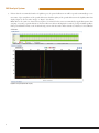
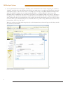
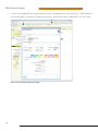
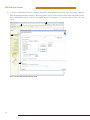
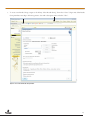
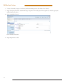
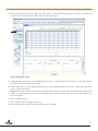
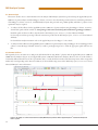
Use of the MSI Analysis System with the
Applied Biosystems® 3500 Gene c Analyzer
and POP-7™ Polymer
MSI Analysis System Application Note
Promega Corporation
Notes:
The MSI Analysis System is For
Research Use Only. Not For Use In
Diagnostic Procedures.
Guidelines for detecting
microsatellite instability
using the MSI Analysis
System, Applied Biosystems®
3500 Genetic Analyzer and
POP-7™ Polymer
1. Introduction
The MSI Analysis System was developed and optimized for use with Applied Biosystems®
Genetic Analyzers using POP-4™ polymer and a 36cm capillary array. However, amplification products can be detected using other instrument configurations. This Application
Note provides general guidelines for use of the MSI Analysis System with the Applied
Biosystems® 3500 and 3500xL Genetic Analyzers and POP-7™ polymer. Optimization
may be required for individual research needs.
2. General Considerations for Amplification
The MSI Analysis System is optimized to amplify 1–2ng of genomic DNA in a 10µl
reaction volume using the GeneAmp® PCR System 9600 and 9700 thermal cyclers. See
the MSI Analysis System, Version 1.2, Technical Manual #TM255 for amplification
conditions.
For thermal cyclers other than the GeneAmp® PCR System 9600 and 9700, we recommend that you replicate as closely as possible the thermal cycling conditions described
in the MSI Analysis System, Version 1.2, Technical Manual #TM255. Suboptimal thermal
cycling conditions can result in imbalanced profiles. Contact Promega Technical Services
at [email protected] for assistance with other thermal cyclers.
3. Materials to Be Supplied by the User
• MSI Analysis System, Version 1.2 (Cat.# MD1641)
• PowerPlex® Matrix Standards, 3100/3130 (Cat.# DG4650)
1
MSI Analysis System
4. Spectral Calibration Using the PowerPlex® Matrix Standards, 3100/3130
The PowerPlex® Matrix Standards, 3100/3130 (Cat.# DG4650), contain matrix fragments labeled with four fluorescent dyes: Fluorescein,
JOE, TMR and CXR. Once generated, the spectral calibration file is applied during collection of MSI Analysis System data to calculate
and compensate for spectral overlap between different fluorescent dye colors. These matrix standards are compatible with the Applied
Biosystems® 3500 and 3500xL Genetic Analyzers. For more information, see the PowerPlex® Matrix Standards, 3100/3130, Technical
Bulletin #TBD022 or the Applied Biosystems 3500/3500xL Genetic Analyzer User Guide.
!
The quality of formamide is critical. Use Hi-Di™ formamide. Freeze formamide in aliquots at –20°C. Multiple freeze-thaw cycles or
long-term storage at 4°C may cause breakdown of formamide. Poor-quality formamide may contain ions that compete with DNA
during injection, which results in lower peak heights and reduced sensitivity. A longer injection time may not increase the signal.
!
Formamide is an irritant and a teratogen; avoid inhalation and contact with skin. Read the warning label, and take the necessary
precautions when handling this substance. Always wear gloves and safety glasses when working with formamide.
4.A. Matrix Sample Preparation
There may be instrument-to-instrument variation in the sensitivity of detection. The dilutions described here may need to be optimized
in individual laboratories depending on the sensitivity of each Applied Biosystems® 3500 or 3500xL Genetic Analyzer.
1.
Thaw the matrix standards. Mix each matrix standard by vortexing for 5–10 seconds prior to use. Do not centrifuge the matrix
standards as this may cause the DNA to be concentrated at the bottom of the tube.
Note: While the matrix dyes thaw, preheat the oven of the capillary electrophoresis instrument to 60°C as described in Section 4.B,
Step 1.
2.
Initial dilution of concentrated fragments: Before combining the matrix standards, dilute the individual matrix standards 1:10 in
Nuclease-Free Water, which is supplied with the PowerPlex® Matrix Standards, 3100/3130, as described below. Vortex for
5–10 seconds to mix. Return the undiluted matrix standards to the freezer immediately after use.
FL
3.
JOE
TMR
CXR
Matrix Standard
5µl
5µl
5µl
5µl
Nuclease-Free Water
45µl
45µl
45µl
45µl
Fragment mix (using 1:10 dilutions of matrix standards): After the initial dilution in Step 2, combine the 1:10 dilutions of the
matrix standards as directed below. Vortex for 5–10 seconds to mix.
Component
Volume
Hi-Di™ formamide
652µl
FL from initial dilution
12.0µl
JOE from initial dilution
12.0µl
TMR from initial dilution
12.0µl
CXR from initial dilution
12.0µl
Note: The dilutions described here may need to be optimized in individual laboratories depending on the sensitivity of each Applied
Biosystems® 3500 or 3500xL Genetic Analyzer. The optimal dilution may differ for each dye.
4.
On the Applied Biosystems® 3500xL Genetic Analyzer (24 capillaries), only wells A1 to H3 of the 96-well plate are used for spectral
calibration. Load 25µl of the fragment mix prepared in Step 3 into each of the 24 wells. After placing the septa on the plate, briefly
centrifuge the plate to remove bubbles.
On the Applied Biosystems® 3500 Genetic Analyzer (8 capillaries), only wells A1 to H1 of the 96-well plate are used for spectral
calibration. Load 25µl of the fragment mix prepared in Step 3 into each of the 8 wells. After placing the septa on the plate, briefly
centrifuge the plate to remove bubbles.
2
5.
Denature samples at 95°C for 3 minutes, then immediately chill on crushed ice or in an ice-water bath for 3 minutes. Denature
samples just prior to loading the instrument. Dry the bottom of the plate to remove any liquid.
Note: If using a thermal cycler to denature samples, do not close the heated lid as this may melt the plate septa.
6.
Place the plate in the 3500 series 96-well standard plate base, and clamp the plate retainer squarely in place over the plate and base
until it clicks twice. Place the plate assembly in Position A on the autosampler with the labels facing you.
4.B. Instrument Preparation
If you have not already preheated the oven to 60°C, select “Start Pre-Heat” (Figure 1). Applied Biosystems recommends that you
preheat the oven for at least 30 minutes before you start a run if the instrument is cold.
11037TA
1.
Figure 1. The Dashboard.
3
MSI Analysis System
2.
To perform a spectral calibration with the Promega 4-dye chemistry for the first time, create a new dye set.
a. To create the new dye set, navigate to the Library, highlight “Dye Sets” and select “Create” (Figure 2).
9322TA
b. The Create New Dye Set window will appear. Name the Dye Set (e.g., Promega 4Dye), select “Matrix Standard” for the Chemistry and select “F Template” for the Dye Set Template. Leave all other settings as the default values. You may lock this dye set
to avoid making unintended changes. Select “Save” and “Close”.
Figure 2. The Create New Dye Set window.
4
c. To perform the spectral calibration with the Promega 4-dye chemistry, choose “Maintenance” from the Menu bar, then choose
“Spectral” in the navigation pane. Under the Calibration Settings choose “Matrix Standard” as the Chemistry Standard, and
select the name of the dye set you created in Step 2.b. Check the Allow Borrowing checkbox. Select other options as shown in
Figure 3.
11093TA
d. Select “Start Run”.
Figurre 3. Caliibration Run.
5
MSI Analysis System
3.
If fewer than the recommended number of capillaries pass, the spectral calibration run will be repeated automatically up to two
more times. Upon completion of the spectral calibration, check the quality of the spectral calibration in the Capillary Run Data
display (Figure 4). Choose either “Accept” or “Reject” (not shown).
9324TA
Note: Refer to the 3500 Series Data Collection Software User Manual for the criteria recommended by Applied Biosystems when
accepting or rejecting a spectral calibration. The data collection software will display the results for passing and failed capillaries.
If the spectral calibration fails, see the Troubleshooting section of the PowerPlex® Matrix Standards, 3100/3130, Technical Bulletin
#TBD022.
Figurre 4. The Capillarry Run Da
ata displa
ay.
6
5. Detection of Amplified Fragments Using the Applied Biosystems® 3500 or 3500xL Genetic Analyzer
The quality of formamide is critical. Use Hi-Di™ formamide. Freeze formamide in aliquots at –20°C. Multiple freeze-thaw cycles
or long-term storage at 4°C may cause breakdown of formamide. Poor-quality formamide may contain ions that compete with DNA
during injection, which results in lower peak heights and reduced sensitivity. A longer injection time may not increase the signal.
!
Formamide is an irritant and a teratogen; avoid inhalation and contact with skin. Read the warning label, and take appropriate
precautions when handling this substance. Always wear gloves and safety glasses when working with formamide.
!
5.A. Sample Preparation
Before preparing the samples, preheat the oven of the capillary electrophoresis instrument to 60°C as described in Section 5.B, Step 1,
if desired.
1.
Determine the number of samples to be analyzed, and add additional reactions to this number to compensate for pipetting error.
Prepare a loading cocktail by combining and mixing Internal Lane Standard 600 (ILS 600) and Hi-Di™ formamide as follows:
[(0.5µl ILS 600) × (# samples)] + [(9.5µl Hi-Di™ formamide) × (# samples)]
Note: The volume of internal lane standard used in the loading cocktail can be increased or decreased to adjust the intensity of the
size standard peaks.
2.
Vortex for 10–15 seconds to mix.
3.
Pipet 10µl of loading cocktail into each well.
4.
Add 1µl of amplified sample. Cover wells with appropriate septa.
Note: Instrument detection limits vary; therefore, injection time or injection voltage may need to be increased or decreased. To
modify the injection time or injection voltage in the run module, select “Instrument Protocol” from the Library menu in the data
collection software.
5.
Centrifuge plate briefly to remove air bubbles from the wells.
6.
Denature samples at 95°C for 3 minutes, then immediately chill on crushed ice or in an ice-water bath for 3 minutes. Denature
samples just prior to loading the instrument.
Note: If using a thermal cycler to denature samples, do not close the heated lid as this may melt the plate septa.
5.B. Instrument Preparation
Refer to the Applied Biosystems 3500/3500xL Genetic Analyzer User Guide for the instrument maintenance schedule and instructions to
install the capillary array, buffers and polymer pouch and perform a spatial calibration. This Application Note provides general guidelines
for use of the MSI Analysis System with the Applied Biosystems® 3500 Genetic Analyzers and POP-7™ polymer. Some optimization
may be required for individual research needs.
1.
Open the 3500 Data Collection Software. The Dashboard screen will launch (Figure 1). Ensure that the Consumables Information
and Maintenance Notifications are acceptable. Set the oven temperature to 60°C, then select “Start Pre-Heat”. Applied Biosystems
recommends that you preheat the oven for at least 30 minutes before you start a run if the instrument is cold.
7
MSI Analysis System
2.
To set up an Instrument Protocol, navigate to the Library, select “Instrument Protocols” from the navigation pane, and choose
“Fragment” as the Filter. Choose the default protocol that is the most similar to the one you will be using from the list of protocols,
and duplicate it (Figure 5). Do not choose “Create”. For example, if you are using an 8-capilllary 3500 Genetic Analyzer with a
50cm array and POP-7™ polymer, choose “FragmentAnalysis50_POP7_1.”, and select“Duplicate”. Edit the copy of this
Instrument Protocol, and fill in the drop-down menus as appropriate for your configuration. Make sure you choose “Fragment” as
the Application Type, and select the dye set that you created in Section 4.B. The Run Module will be “FragmentAnaysis50_POP7”.
Assign the Instrument Protocol a meaningful name so that it will be clear what the Instrument Protocol can be used for in future
runs. Leave all run options and Advanced Options as the default values. It is possible to change default injection parameters later
if necessary later. Select “Save” and “Close”.
10900TA
When you save this protocol with the Promega 4Dye dye set, the Instrument Protocol name will appear in the Library with the
Dye Set Name associated with it.
Figure 5. The Setup an Instrument Protocol window.
8
To create a new Size Standard for the Sizecalling protocol, navigate to the Library. Select “Size Standards”, then select “Create”.
Assign the size standard an appropriate name (Figure 6). Choose “Red” for the Dye Color. Enter the fragment sizes for the size
standard: 60, 80, 100, 120, 140, 160, 180, 200, 225, 250, 275, 300, 325, 350, 375, 400, 425, 450, 475, 500, 550 and 600 bases.
Select “Add Sizes”, “Save” then “Close”.
Note: Definition and detection of the 600bp fragment is optional.
10902TA
3.
Figure 6. The Create New Size Standard window.
9
MSI Analysis System
To create a new Sizecalling Protocol, navigate to the Library. Select “Sizecalling Protocols”, then select “Create”. Assign a descriptive
protocol name (Figure 7). Select the size standard created in Step 3. Leave all other settings as default. Select “Save” then “Close”.
10903TA
4.
Figure 7. The Create New Sizecalling Protocol window.
10
To create a new Assay, navigate to the Library. Select “Assays”, then select “Create”. In the Assay window (Figure 8), select the
application type “Fragment” and the appropriate Instrument Protocol and Sizecalling Protocol. Assign a descriptive assay name,
then select “Save” and “Close”.
10901TA
5.
Figure 8. The Assay window.
11
MSI Analysis System
To create a new File Name Convention, navigate to the Library. Select “File Name Conventions”, then select “Create” (Figure 9).
Select the File Name Attributes according to laboratory practices, and save with a descriptive name. Under “Select File Location”
choose “Default File Location”, or choose “Custom File Location” and navigate to your custom location. Select “Save” then
“Close”.
11024TA
6.
Figure 9. The Create New File Name Convention window.
12
To create a new Results Group, navigate to the Library. Select “Results Group”, then select “Create” (Figure 10). Select Results
Group Attributes according to laboratory practices. Save with a descriptive name, and select “Close”.
10906TA
7.
Figure 10. The Create New Results Group window.
13
MSI Analysis System
8.
To create a New Plate, navigate to the Library, and from the Manage menu, select “Plates”, then “Create”.
9.
Assign a descriptive plate name. Select the Plate Type “Fragment” from the drop-down menu (Figure 11). Select the appropriate
capillary length and polymer.
10907TA
10. Select “Assign Plate Contents”.
Figure 11. Defining plate properties.
11. Assign sample names to wells.
14
10908TA
12. In the lower left portion of the screen (Figure 12), under “Assays”, use the Add from Library option (not shown) to select the Assay
created in Step 5. Click on the Add to Plate button, and close the window.
Figure 12. Assigning plate contents.
13. Under “File Name Convention”, use the Add from Library option to select the File Name Convention created in Step 6. Click on
the Add to Plate button, and close the window.
14. Under “Results Groups”, use the Add from Library option to select the Results Group created in Step 7. Click on the Add to Plate
button, and close the window.
15. Highlight the sample wells, then select the boxes in the Assays, File Name Conventions and Results Groups that pertain to those
samples. To assign sample types, select the Custom Sample Info tab at the bottom left side of the window, assign sample types,
then save the plate.
16. Select “Link Plate for Run”.
17. The Load Plate window will appear. Select “Yes”.
18. In the Run Information window, assign a Run Name. Select “Start Run”.
15
MSI Analysis System
5.C. Data Analysis
This section describes how to obtain information for the analysis of MSI Analysis System data generated using the Applied Biosystems®
3500 series of genetic analyzers with GeneMapper® software, version 4.1. Note that Promega scientists have not verified the performance
of GeneMapper® software, version 4.1, with MSI Analysis System data generated using a 50cm capillary and POP-7™ polymer. Some
optimization may be required.
1.
To analyze .fsa files collected on the Applied Biosystems® 3500 series of genetic analyzers using GeneMapper® software, version 4.1,
download the panels and bins text files at: www.promega.com/resources/tools/msi-panels-and-bins-for-genemapper-software/
Install the panels and bins text files as described in the MSI Analysis System, Version 1.2, Technical Manual #TM255.
For data import and data processing, follow the instructions provided in the MSI Analysis System, Version 1.2, Technical Manual
#TM255.
For detailed data analysis instructions refer to the Applied Biosystems GeneMapper® 4.1 User Guide.
2.
To analyze .fsa files collected on the Applied Biosystems® 3500 series of genetic analyzers using GeneMapper® ID or GeneMapper® ID-X
software, contact Promega Technical Services by e-mail at: [email protected] to obtain the appropriate panels and bins text
files.
10909TA
5.D. Known Artifacts
We frequently observe an artifact at 87–90bp in the JOE channel when using POP-7™ polymer with the Applied Biosystems® 3500 and
3500xL Genetic Analyzers (Figure 13) and Applied Biosystems® 3100 and 3130 Genetic Analyzers. However, the size of this artifact is
considerably different than the sizes of the repeat markers and, as a result, should not interfere with data interpretation. Other nonspecific
artifacts may occur depending on the detection conditions. See the troubleshooting section of the MSI Analysis System, Version 1.2, Technical
Manual #TM255 for more information.
Figure 13. An example of an artifact observed when analyzing MSI Analysis System data with an Applied Biosystems® 3500 Genetic Analyzer and
POP-7™ polymer.
16
MSI Analysis System
Ordering Information
Product
MSI Analysis System, Version 1.2*
PowerPlex® Matrix Standards, 3100/3130**
Size
100 reactions
25μl (each dye)
Cat.#
MD1641
DG4650
*For Research Use Only. Not for Use in Diagnostic Procedures.
**Not For Medical Diagnostic Use.
PowerPlex is a registered trademark of Promega Corporation.
Applied Biosystems and GeneMapper are registered trademarks of Applied Biosystems. GeneAmp is a registered trademark of Applera Corporation. Hi-Di and POP-7 are trademarks of Applera Corporation.
Products may be covered by pending or issued patents or may have certain limitations. Please visit our Web site for more information.
PROMEGA CORPORATION • 2800 WOODS HOLLOW ROAD • MADISON, WI 53711 5399 USA • TELEPHONE 608 274 4330
www.promega.com • ©2013 PROMEGA CORPORATION • ALL RIGHTS RESERVED • PRICES AND SPECIFICATIONS SUBJECT TO CHANGE WITHOUT PRIOR NOTICE • PRINTED IN USA 8/13 • PART #AN192