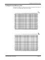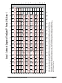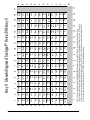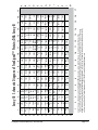
1
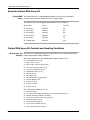
Protein/DNA Arrays
(Spin Column Separation Version)
User Manual
Cat # MA1210, MA1211, MA1212
MA1213, MA1214 & MA1215
P/N 13299 Rev. B 032007
DRAFT May 18, 2007 4:28 pm
PD ArrayTtile.fm
Panomics, Inc.
Protein/DNA Array Kit User Manual
Copyright
© Copyright 2006, Panomics, Inc. All rights reserved.
Trademarks
Hyperfilm and ECL are trademarks of GE Healthcare, Inc., FluorChem is trademark of AlphaInnotech, Inc.
Citing Protein/DNA Arrays in Publications
When describing a procedure for publication using this product, we would appreciate it if you would refer to it as the Protein/DNA Array Kit from
Panomics
If a paper cites the Protein/DNA Array kit and is published in a research journal, the lead author(s) may receive a travel stipend for use at a technology
conference or tradeshow by sending a copy of the paper to our technical support group at [email protected] or via fax at (510) 818-2610.
Disclaimer
Panomics, Inc. reserves the right to change its products and services at any time to incorporate technological developments. This manual is subject
to change without notice.
Although this manual has been prepared with every precaution to ensure accuracy, Panomics, Inc. assumes no liability for any errors or omissions, nor
for any damages resulting from the application or use of this information.
DRAFT May 18, 2007 4:28 pm
PD ArrayTtile.fm
Contents
About the User Manual . . . . . . . . . . . . . . . . . . . . . . . . . . . . . . . . . . . . . . . . . . . . . . . . . 5
Who Should Read this Manual. . . . . . . . . . . . . . . . . . . . . . . . . . . . . . . . . . . . . . . 5
What this Manual Covers . . . . . . . . . . . . . . . . . . . . . . . . . . . . . . . . . . . . . . . . . . . 5
Safety Warnings and Precautions . . . . . . . . . . . . . . . . . . . . . . . . . . . . . . . . . . . . 5
For More Information . . . . . . . . . . . . . . . . . . . . . . . . . . . . . . . . . . . . . . . . . . . . . . 5
About the TranSignal Protein/DNA Assay Kit . . . . . . . . . . . . . . . . . . . . . . . . . . . . . . . . 6
TranSignal Protein/DNA Assay . . . . . . . . . . . . . . . . . . . . . . . . . . . . . . . . . . . . . . 6
............................................................... 6
TranSignal Protein/DNA Assay Kit Contents and Handling Conditions . . . . . . . . . . . . . 6
Kit Contents and Storage . . . . . . . . . . . . . . . . . . . . . . . . . . . . . . . . . . . . . . . . . . . 6
Kit Handling . . . . . . . . . . . . . . . . . . . . . . . . . . . . . . . . . . . . . . . . . . . . . . . . . . . . . 6
Required Materials and Equipment Not Provided . . . . . . . . . . . . . . . . . . . . . . . . . . . . . 7
Equipment . . . . . . . . . . . . . . . . . . . . . . . . . . . . . . . . . . . . . . . . . . . . . . . . . . . . . . 7
Sample Type Specific Materials . . . . . . . . . . . . . . . . . . . . . . . . . . . . . . . . . . . . . . 7
Introduction . . . . . . . . . . . . . . . . . . . . . . . . . . . . . . . . . . . . . . . . . . . . . . . . . . . . . 7
Princicple of the Technology . . . . . . . . . . . . . . . . . . . . . . . . . . . . . . . . . . . . . . . . 7
Overview of Assay Workflow . . . . . . . . . . . . . . . . . . . . . . . . . . . . . . . . . . . . . . . . . . . . . 8
............................................................... 8
Guidelines for Assay Design and Analysis . . . . . . . . . . . . . . . . . . . . . . . . . . . . . . . . . . 9
Preparing Samples. . . . . . . . . . . . . . . . . . . . . . . . . . . . . . . . . . . . . . . . . . . . . . . . 9
General Guidelines . . . . . . . . . . . . . . . . . . . . . . . . . . . . . . . . . . . . . . . . . . . . . . . 9
Assay Procedures . . . . . . . . . . . . . . . . . . . . . . . . . . . . . . . . . . . . . . . . . . . . . . . . . . . . . 9
Before You Start . . . . . . . . . . . . . . . . . . . . . . . . . . . . . . . . . . . . . . . . . . . . . . . . . . 9
Preparing Nuclear Extracts . . . . . . . . . . . . . . . . . . . . . . . . . . . . . . . . . . . . . . . . . 9
Preparing the Membranes . . . . . . . . . . . . . . . . . . . . . . . . . . . . . . . . . . . . . . . . . . 9
Probe Mix . . . . . . . . . . . . . . . . . . . . . . . . . . . . . . . . . . . . . . . . . . . . . . . . . . . . . . 10
ISOLATING TF-BOUND PROBES. . . . . . . . . . . . . . . . . . . . . . . . . . . . . . . . . . . 10
HYBRIDIZATION . . . . . . . . . . . . . . . . . . . . . . . . . . . . . . . . . . . . . . . . . . . . . . . . .11
DETECTION . . . . . . . . . . . . . . . . . . . . . . . . . . . . . . . . . . . . . . . . . . . . . . . . . . . .11
RESULTS & ANALYSIS . . . . . . . . . . . . . . . . . . . . . . . . . . . . . . . . . . . . . . . . . . . 12
REFERENCES. . . . . . . . . . . . . . . . . . . . . . . . . . . . . . . . . . . . . . . . . . . . . . . . . . 12
APPENDIX I. . . . . . . . . . . . . . . . . . . . . . . . . . . . . . . . . . . . . . . . . . . . . . . . . . . . . . . . . 13
RECIPES AND INSTRUCTIONS . . . . . . . . . . . . . . . . . . . . . . . . . . . . . . . . . . . . 13
TranSignal Protein/DNA Arrays User Manual
iii
About the User Manual
About the User Manual
Who Should Read Anyone that has purchased a Protein/DNA Array Kit from Panomics to profile from 56
this Manual to 345 different Transcription Factors.
]
What this Manual This manual provides recommendations and step-by-step procedures for the
Covers following:
♦
Sample and assay preparation
♦
Assay procedure
♦
Troubleshooting
♦
Layouts
Safety Warnings CAUTION All chemicals should be considered potentially hazardous. We recommend that
and Precautions this product and its components be handled by those trained in laboratory techniques and be
used according to the principles of good laboratory practice.
For More For information about the Protein/DNA products mentioned in this manual, visit our
Information website at www.panomics.com.
Protein/DNA Arrays User Manual
Page 5
About the Protein/DNA Assay Kit
About the Protein/DNA Assay Kit
Protein/DNA The Protein/DNA kit is a highthroughput profiling assay that has the ability to
Assay mesaure the activation of multiple TF’s from a single sample .
The cytokine panels for TranSginal Protein/DNA kits are available as:\
Array Name
Cat. #
# of TF’s
♦
PD Array I
MA1210
56
♦
PD Array II
MA1211
96
♦
PD Array III
MA1212
94
♦
PD Array IV
MA1213
76
♦
PD Array V
MA1214
73
♦
Combo Array
MA1215
345
Protein/DNA Assay Kit Contents and Handling Conditions
Kit Contents and The contains the following components. Shelf life of kit is six months from date of
Storage receipt under proper storage conditions
Box 1: Array Membranes and Hybridization Reagents (Store at 4 °C)
♦
Protein/DNA Array (3 each)
♦
Spin Column (3 each)
♦
Spin Column collection Tube (6 each)
♦
1X Column Incubation Buffer (2 ml)
♦
1X Colunm Wash Buffer (10 ml)
♦
Hybridization Buffer (15 ml)
♦
20X SSC (32 ml)
♦
20% SDS (15 ml)
♦
Streptavidin-HRP conjugate (60 µl)
♦
4X Wash Buffer (45 ml)
♦
Solution I (750 µl)
♦
Solution II (750 µl)
♦
Solution III (8 ml)
Box 2: Reaction Kit (Store at -20 °C)
Page 6
♦
Probe Mix (30 µl)
♦
1X Column Elution Buffer (200 µl) Bring to room temperature before use
♦
Distilled H20, RNAse, DNAse free (500 µl)
♦
Control Nuclear Extract from HeLa Cells (5 µl)
♦
2X Blocking Buffer (30 ml)
♦
1X Detection Buffer (60 ml)
♦
5X Pre-Treatment Buffer I (20 ml)
♦
5X Pre-Treatment Buffer II (20 ml)
Protein/DNA Arrays User Manual
Protein/DNA Assay Kit Contents and Handling Conditions
Additional ♦
Materials Required ♦
Materials and
♦
Euipment
Panomics Nuclear Extraction Kit (Cat# AY2002)
Deionized H20
0.5 ml and 1.5ml microfuge tubes
♦
Pipetman and tips
♦
Microcentrifuge
♦
Hybridization oven and bottles
♦
Plastic containers (~4.5” x 3.5”; equivalent to the size of a 200 µl pipete tip box)
♦
Shaker
♦
Plastic sheet protectors
♦
Hyperfilm™ ECL™ (Amersham, Cat # RPN3114K)
♦
Chemiluminescent Imaging system, FluorChem™ from Alpha Innotech
Introduction Eukaryotic gene expression is regulated by a group of proteins called Transcription
Factors (TFs). By interacting with specific DNA-Binding elements present in the
promoters of certain genes, TFs modulate the frequency of transcriptional initiation.
The expression or activity of TFs may be regulated in a cell-type , tissue specific, or
cell cycle-dependent manner. Regulation can also be mediated by interactions with
other proteins. Through different combinations of these regulatory mechanisms,
eukaryotes are able to elicit a myriad of different gene expression patterns. The key
to a full understanding of how gene expression is regulated is the analysis of the
biochemical activity of TFs.
Panomics’ Protein/DNA Arrays simplify the functional analysis of eukaryotic TFs. Our
array-based technology is a significant improvement over cumbersome gel
mobility-shift assays, which permit the characterization of only a single TF at a time.
With these Protein/DNA Arrays, you can profile the activities of multiple TFs
simultaneously. These arrays can be used to study TF activation in a variety of
biological processes, including cell proliferation, differentiation, transformation,
apoptosis, and drug treatment.
We currently offer six versions of the TranSignal Protein/DNA Array. Version I for 56
transcription factors (TFs); Version II for 96; Version III for 94; Version IV for 76;
Version V for 73; and the Combo Array for 345 TFs. These transcription factors are
well characterized and widely available in published literature.
Princicple of the Protein/DNA Arrays use a proprietary, patent-pending technology developed by
Technology Panomics for the high-throughput analysis of TF activation. The procedure is simple
and straightforward (Figure 1). Three basic steps are involved: (1) a set of
biotin-labeled DNA binding oligonucleotides (Probe Mix) are preincubated with a
nuclear extract of interest to allow the formation of protein/DNA complexes; (2) the
protein/DNA complexes are separated from the free probes; and (3) the probes in the
complexes are then extracted and hybridized to the Protein/DNAl Array. Each kit
includes the reagents for HRP-based chemiluminescence detection.
Protein/DNA Arrays User Manual
Page 7
Overview of Assay Workflow
Overview of Assay Workflow
1. Mixture of pre-labeled TF probes
TF Probe 1
5' Biotin
TF Probe 2
3'
5' Biotin
TF Probe 3
3'
5' Biotin
TF Probe 4
3'
5' Biotin
3'
2. Incubate with nuclear extract
TF1
5' Biotin
TF2
3'
5' Biotin
TF3
3'
5' Biotin
TF4
3'
5' Biotin
3'
3. Separation of protein/DNA complexes
Spin column
Bind
Wash
Elute
4. Hybridization
Cis1
Cisn
Cis2
Figure 1: Workflow diagram of the Protein/DNA Array
Page 8
Protein/DNA Arrays User Manual
Guidelines for Assay Design and Analysis
Guidelines for Assay Design and Analysis
General Guidelines ♦
Read this user manual and all product inserts before performing the assay.
♦
Store all reagents at the recommended temperatures.
♦
Use Panomics’ Nuclear Extraction Kit for best results.
Assay Procedures
Before You Start ♦
♦
Preparing Nuclear ♦
Extracts ♦
♦
Thaw Positive Control and sample nuclear extracts on ice.
Thaw Probe mix on ice.
We recommend using Panomics’ Nuclear Extraction Kit (Cat. #AY2002).
The minimum protein concentration of nuclear extract should be 1.0 ug/ul or
greater and we recommend using between 4–10 µg of Nuclear Extract Protein.
You will need to adjust the amount of water accordingly when preparing the
samples with the probe mix.
When measuring protein concentration, we also recommend using the DC
Protein assay from Bio-Rad (Lowry assay) with a plate reader or
spectrophotometer.
Preparing the In this Section, you will prepare the array membranes for use. If you are reusing
Membranes membranes that have been previously stripped, skip steps , and begin from step 1.4.
1.1
Prepare Pre-Treatment Buffers I and II (see Appendix I for recipes) and warm
the Hybridization Buffer to 42 °C in a water bath or incubator. If you notice a
cloudy or soapy appearance for the Hybridization Buffer, make sure the
particulates are completely dissolved before proceeding. It may require
overnight heating in a water bath.
1.2
Place each array membrane into a hybridization bottle. Be sure to place the
membrane in the hybridization bottle such that the spotted oligos face the
center of the tube (away from the walls). Add 25 ml of 1X Pre-Treatment Buffer
I to each bottle and place in a hybridization oven at 45 °C for 5 minutes.
1.3
Thoroughly decant the solution from each bottle. Add 25 ml of 1X
Pre-Treatment Buffer II to each bottle and place in a hybridization oven at 45
°C for 10 minutes.
1.4
Decant the solution from each bottle. Rinse the entire surface of each
membrane with distilled water (not provided), by filling each bottle
approximately half full and swirling the bottle for 30 sec. Repeat this rinsing
step two additional times and decant water after final rinse.
1.5
To each hybridization bottle, add 5 ml of prewarmed Hybridization Buffer
(provided). Place each bottle into the rotating hybridization oven at 42 °C for 2
hr. If needed, you can stop at this point of the protocol and leave the
membrane overnight at 42 °C.
Protein/DNA Arrays User Manual
Page 9
Assay Procedures
Forming In this Section, you will allow the DNA probes to bind to transcription factors from
Protein-DNA your nuclear extract.
Complexes 2.1 For each nuclear extract sample, combine the following components into a
sterile 0.5-ml microcentrifuge tube (* = provided):
Nuclear extract (1-3 µg/µl)
5µl
TranSignal Probe Mix*
10µl
dH2O (RNase,DNase free)*
5µl
Total Volume
20µl
Note If the protein concentration of your nuclear extract is below the recommended
range, replace the dH2O with additional nuclear extract.
2.2
Mix well by pipeting. Incubate samples for 30 min at 15 °C.
IsolatingTF-Bound In this section, you will isolate the protein-bound probes from the non-bound probes.
Probes All centrifuge steps should be carried out on a regular benchtop centrifuge, at 7,000
rpm at 4 °C, unless otherwise stated.
3.1
Wash Spin Column by adding 500 µl of chilled 1X Column Incubation Buffer
and centrifuging at 10,000 rpm for 30 sec at 4 °C, or room temp.
3.2
Add 20 µl 1X Column Incubation Buffer to the TF-Probe mix, from Step 2.2.
Transfer all of this mix into the center of the Spin Column.
3.3
Incubate the Spin Column on ice for 30 min.
3.4
Centrifuge column at 7,000 rpm for 30 sec at 4 °C and discard the flow
through.
3.5
Place the Spin Column in a new Spin Column Collection Tube (provided).
3.6
Add 600µl of 1X Column Wash Buffer to the Spin Column and incubate for 10
min, on ice.
3.7
Centrifuge column at 7,000 rpm for 30 sec at 4 °C and discard the flow
through.
3.8
Wash the column by adding 600 µl of 1X Column Wash Buffer to the Spin
Column and centrifuging at 7,000rpm for 30 sec at 4 °C.
3.9
Repeat step 3.8 three more times.
3.10 Remove residual Wash Buffer by an additional centrifugation at 10,000 rpm for
30 sec at 4 °C
3.11 Add 60µl of 1X Column Elution Buffer to the center of the Spin Column and
incubate at room temperature for 5 min.
3.12 Place the Spin Column in a clean 1.5 ml microcentrifuge tube and centrifuge
for 1 minute at 10,000rpm at room temperature.
3.13 Place the microcentrifuge tube, containing the collected flow through, which
contains the eluted probe, on ice and use for further steps. If needed, you can
stop at this point and store the eluted probe at 20 °C overnight.
Hybridization In this Section, you will hybridize the eluted, labeled probe prepared in Section 3 to
the array membrane prepared in Section 1.
Page 10
Protein/DNA Arrays User Manual
Assay Procedures
4.1
Denature the eluted probe (from step 3.13) by heating it at 95 °C for 3 min and
quickly chill in ice for 2 min. Add the eluted probe to each hybridization bottle
and hybridize at 42 °C overnight in a rotating hybridization oven
4.2
Decant the hybridization mixture from each hybridization bottle from Step 4.1
and wash each membrane as follows (* = See Appendix I for recipes):
4.3
Add 50 ml of prewarmed Hybridization Wash I*, incubate at 42°C for 20 min in
a rotating hybridization oven. Decant liquid and repeat wash.
4.4
Add 50 ml of prewarmed Hybridization Wash II*, incubate at 42 °C for 20 min in
a rotating hybridization oven. Decant liquid and repeat wash.
Detection Important: Do not allow the membrane to dry during the detection.
5.1
Using forceps, carefully remove each membrane from its hybridization bottle
and transfer to a clean container containing 20 ml of 1X Blocking Buffer; each
membrane needs its own container. (We use a container that is equivalent to
the size of a 200-µl pipet-tip container, ~4.5" x 3.5".)
5.2
Block the membrane by incubating at room temperature with the 1X Blocking
Buffer for 15 min with gentle shaking.
5.3
Remove 1 ml of the 1X blocking buffer to a clean microcentrifuge tube. Dilute
20 µl of the stock Streptavidin-HRP conjugate with 1 ml of the 1X Blocking
Buffer. Vortex the diluted streptavidin-HRP and transfer back into the 1X
Blocking Buffer from Step 5.2 (containing the membrane). Add the diluted
conjugate to the container, making sure not to pour directly on the membrane.
Continue shaking the membrane for 15 min at room temperature.
5.4
Decant the diluted Streptavidin-HRP solution. Wash each membrane three
times at room temperature with 20 ml of 1X Wash Buffer, each for 8 min.
5.5
Add 20 ml of 1X Detection Buffer to each membrane and incubate at room
temperature for 5 min.
5.6
Overlay each blot with 2.5 ml of Working Substrate Solution, prepared by
mixing the following, in order: 250 µl Solution I with 250 µl Solution II. Briefly
vortex and add 2.0 ml Solution III. Thoroughly mix. Using a plastic sheet
protector or overhead transparency film (do not use thin plastic cling wraps as
these wrinkle and lead to uneven detection across the membrane). Place each
membrane on a plastic sheet. Then, pipet 2.5 ml of the mixed substrate
solution onto each membrane and overlay each with a second plastic sheet on
a flat surface as quick as possible. Ensure that substrate is evenly distributed
over the membrane with no air bubbles. Incubate at room temperature for 5
min.
5.7
Remove excess substrate by gently applying pressure over the top sheet.
Using a paper towel, remove excess substrate that might be remaining on the
surface of the sheets. Expose the membranes using either Hyperfilm ECL
(2-10 min) or a chemiluminescence imaging system (5-15 min), such as the
FluorChem imager from Alpha Innotech Corp. In either case, we recommend
that you try several different exposure times.
5.8
Obtain quantitative analysis, if desired. If you are using a chemiluminescence
imaging system, follow the instructions that are provided with that system’s
software. If you are using Hyperfilm ECL, you will need to scan the film to
obtain numerical data for comparison.
Protein/DNA Arrays User Manual
Page 11
Assay Procedures
Results & Analysis TranSignal Arrays give you quick answers when you want to identify those activated
transcription factors through comparison of two (or more) samples. Follow these
guidelines to analyze your results:
6.1
Acquire the images using either x-ray film or a chemiluminescence imaging
system.
6.2
Adjust the exposure time such that the majority of the positve control spots
have equal signal intensity at the same exposure time.
6.3
When comparing mutliple blots, ensure that the control spots have a similar
intensity.
Stripping Note: We do not encourage stripping the array membranes more than two times.
Procedure 7.1 Wash membranes in 0.4M NaOH at 45 °C for 30 min.
7.2
Wash membranes in 0.2M Tris-HCL, pH 7.6, 0.1X SSC, and 0.1% SDS at 45
°C for 15 min.
7.3
To ensure that stripping was successful, follow the standard
chemiluminescence detection procedure as described in the user manual.
7.4
After detection, wash the membrane twice in 1X TBST with 0.1%SDS at 42 °C
for 30 min.
7.5
Membranes are ready for hybridization or air dry the membrane at room
7.6
Store dry membrane protected between paper at room temperature unil ready
for further use.
Recipe for 10X TBST
1M Tris-HCL pH 7.5, 1.5M NaCl, 0.5% Tween-20
Possible Problems
and
Recommended
Solutions
Observation
Possible Cause
Recommended Action
Uneven Background
Substrate is note evenly
distributed on the
membrane
Do not use thin cling wrap
materials during the overlay
procedure
Exposure Time to Short
Re-detect with substrate
that is evenly covering the
membrane surface
Incubation with substrate is
too long
Incubate with shorter time
Non-specific protein bound
to membrane
Increase blocking or
washing times.
Not enough protein was
used in the assay.
Carefully measure the
concentration of the nuclear
extract using a Lowry or
Bradford assay
High background
Signal is too weak
Page 12
Protein/DNA Arrays User Manual
Assay Procedures
Observation
Possible Cause
Recommended Action
Don’t see difference
between treated and
untreated samples
Confirm concentration of
nuclear extract
Carefully measure
concentration
Cells cultured with growth
factors in serum
Culture cells overnight
without serum prior to
treatment.
TF not activated
Confirm with EMSA
References 1. Chodosh, L.A., Olesen, J., Hahn, S., Baldwin, A.S., Guarente, L., and Sharp, P.A.
(1988) A yeast and a human CCAAT-binding protein have heterologous subunits that
are functionally interchangeable. Cell 53:25–35.
2. Zeng, G., L., Gao, L., Xia, T., Tencomnao, T., and Yu, R. K. (2002) Characterization
of the 5’-flanking fragment of the human GM3-synthase gene. Biochimica et
Biophysica Acta 93745:1–6.
3.Miyoshi, K., Rzucidlo, S. J., Pratt, S. L., and Stice, S.L. (2002) Improvements in
Cloning Efficiencies May Be Possible by Increasing Uniformity in Recipient Oocytes
and Donor Cells. Biology of Reproduction, in press.
4. Govindarajan, B., Bai, X., Cohen, C., Zhong, H., Kilroy, S., Moses, M. and Arbiser,
J. (2003) Malignant transformation of melanocytes to melanoma by constitutive
activation of MAP kinase kinase signaling. J Biol Chem. Electronically published
1/03 - see www.jbc.org.
5. Wheeler, M., Smutney, O., Check, J., Rusyn, I., Schulte-Hermann, R. and Thurman,
R. (2003) Impaired Ras membrane association and activation in PPARa knockout
mice after partial hepatectomy. Am J Phsiol Gastrointest Liver Physiol 284:302–312.
6. Dignam, J.D., Lebovitz, R.M., and Roeder, R.G. (1983) Accurate transcription
initiation by RNA polymerase II in a soluble extract from isolated mammalian nuclei.
Nucleic Acids Research 11:1475–1489.
Protein/DNA Arrays User Manual
Page 13
APPENDIX I
APPENDIX I
RECIPES AND •75 ml of 1X Pre-Treatment Buffer I
INSTRUCTIONS
Warm 5X Pre-Treatment Buffer I to room temperature. If precipitate is visible, warm
gently to fully dissolve solution, and allow to cool to room temperature. To 60 mL of
deionized H2O, add 15 ml of 5x Pre-Treatment Buffer I. Mix well.
•75 ml of 1X Pre-Treatment Buffer II
Warm 5X Pre-Treatment Buffer II to room temperature. If precipitate is visible, warm
gently to fully dissolve solution, and allow to cool to room temperature. To 60 mL of
deionized H2O, add 15 ml of 5x Pre-Treatment Buffer II. Mix well.
•300 ml of 2X SSC/0.5% SDS (Hybridization Wash I)
To 262.5 ml of deionized H2O, add 30 ml of 20X SSC (provided) and 7.5 ml of 20%
SDS (provided). Mix well.
•300 ml of 0.1X SSC/0.5% SDS (Hybridization Wash II)
To 291 ml of deionized H2O, add 1.5 ml of 20X SSC (provided) and 7.5 ml of 20%
SDS (provided). Mix well.
•60 ml of 1X Blocking Buffer
To 30 ml of deionized H2O, add 30 ml of 2X Blocking Buffer (provided). Mix well and
store at 4°C.
•180 ml of 1X Wash Buffer
To 135 ml of deionized H2O, add 45 ml of 4X Wash Buffer (provided). Mix well and
store at room temperature.
Page 14
Protein/DNA Arrays User Manual
TranSignal Protein/DNA Array Grid
TranSignal Protein/DNA Array Grid
Photocopy this page onto a separate piece of paper or transparency film. Note that
the notch is on the upper-right hand corner of the grid.
1 2 3 4 5 6 7 8 9 10 11 12 13 14 15 16 17 18 19 20 21 22 23 24
A
B
C
D
E
F
G
H
I
J
K
L
M
N
O
P
1 2 3 4 5 6 7 8 9 10 11 12 13 14 15 16 17 18 19 20 21 22 23 24
A
B
C
D
E
F
G
H
I
J
K
L
M
N
O
P
1 2 3 4 5 6 7 8 9 10 11 12 13 14 15 16 17 18 19 20 21 22 23 24
A
B
C
D
E
F
G
H
I
J
K
L
M
N
O
P
1 2 3 4 5 6 7 8 9 10 11 12 13 14 15 16 17 18 19 20 21 22 23 24
Protein/DNA Arrays User Manual
A
B
C
D
E
F
G
H
I
J
K
L
M
N
O
P
Page 15
TranSignal Protein/DNA Array Grid
Page 16
Protein/DNA Arrays User Manual
TranSignal Protein/DNA Arrays User Manual
Page 15
TR
N
2
TR
SRE
SRE
E2F-1
3
4
TR/DR-4 TR/DR-4
TR/DR-4 TR/DR-4
SRE
SRE
E2F-1
6
USF-1
(1)
USF-1
(1)
USF-1
(1)
USF-1
(1)
5
Stat-1
Stat-1
Stat-1
Stat-1
PRE
PRE
11
Stat-3
7
8
VDR/DR-3 VDR/DR-3
VDR/DR-3 VDR/DR-3
Stat-3
Stat-3
9
HSE
HSE
Stat-4
Stat-4
10
HSE
HSE
Stat-4
Stat-4
11
MRE
MRE
Stat-5
Stat-5
SIE
RAR/DR-5 RAR/DR-5 RXR/DR-1 RXR/DR-1
Stat-3
12
13
14
15
16
12
MRE
MRE
Stat-5
Stat-5
SIE
SIE
AP-1
(2)
AP-1
(2)
AP-2
(2)
AP-2
(2)
AP-1
(2)
AP-1
(2)
AP-2
(2)
AP-2
(2)
13
Stat-6
Stat-6
15
TFIID
Stat-6
14
TFIID
Stat-6
16
TFIID
TFIID
O
N
M
L
K
J
Smad/SBE Smad/SBE SMAD-3/4 SMAD-3/4
H
G
F
E
D
C
B
A
I
17
18
17
Smad/SBE Smad/SBE SMAD-3/4 SMAD-3/4
CEBP
CDP
c-Myb
CDP
CBF
CBF
c-Myb
(1)
CEBP
CDP
c-Myb
CBF
CBF
CDP
c-Myb
(1)
Ets Ets-1/Pea-3 Ets-1/Pea-3
FAST-1 GAS/ISRE GAS/ISRE
FAST-1
(1)
(1)
(1)
Ets1/Pea3 Ets1/Pea3 FAST-1
Ets
FAST-1 GAS/ISRE GAS/ISRE
(1)
(1)
(1)
NF-1
NF-1
MEF-1
MEF-2 Myc/Max Myc/Max
MEF-2
(1)
(1)
(1)
(1)
(1)
NF-1
NF-1
MEF-2
MEF-1
MEF-2 Myc/Max Myc/Max
(1)
(1)
(1)
(1)
(1)
Pax-5
Pax-5
p53
OCT
p53
Pbx-1
Pbx-1
(1)
(1)
(1)
(1)
(1)
Pax-5
p53
Pax-5
OCT
p53
Pbx-1
Pbx-1
(1)
(1)
(1)
(1)
(1)
10
SIE
NFkB
(1)
NFkB
(1)
NFkB
(1)
NFkB
(1)
CEBP
(1)
CEBP
(1)
Ets
(1)
Ets
(1)
MEF-1
(1)
MEF-1
(1)
OCT
(1)
OCT
(1)
9
RAR/DR-5 RAR/DR-5 RXR/DR-1 RXR/DR-1
IRF-1
IRF-1
ER
ER
Brn-3
Brn-3
8
IRF-1
IRF-1
ER
ER
Brn-3
Brn-3
7
Array V: Grey rows and columns are spotted with biotinylated DNA and are used as postive controls for the assay. Pink shaded Rows are the same TF’s as the TF’s above
in the white rows except that they are spotted at a 1:10 dilution. For some Transcription Factors, mutliple consensus sequences for the same TF have been publsihed in the
literature and where appropriate, we have provided those variations on our arrays. For the TF’s with mutliple consensus sequences, they have been denoted with numbers
in parenthesis, ie (1), (2), (3) etc. To obtain the consensus sequences and references for the TF’s, used in our arrays, please call our Technical Support group at 877-726-6642 or
email at [email protected].
1
TR
M
O
Sp-1
Sp-1
L
TR
Sp-1
Sp-1
K
J
I
H
NFAT-1
(1)
NFAT-1
(1)
Pit-1
(1)
Pit-1
(1)
GATA
F
G
GATA
AR
6
EGR
(1)
EGR
(1)
HNF-4
(1)
HNF-4
(1)
NF-E2
(1)
NF-E2
(1)
AR
5
EGR
(1)
EGR
E2F-1
E2F-1
(1)
HNF-4
GR/PR
GR/PR
GATA
(1)
HNF-4
GR/PR
GR/PR
GATA
(1)
NF-E2
NFAT-1
NF-E1/YY1 NF-E1/YY1
(1)
(1)
NFAT-1 NF-E1/YY1 NF-E1/YY1 NF-E2
(1)
(1)
Pit-1
PPAR
PPAR
PRE
(1)
(1)
(1)
PPAR
Pit-1
PPAR
PRE
(1)
(1)
(1)
AP-2
(1)
AP-2
(1)
4
AR
AP-2
(1)
AP-2
(1)
3
AR
AP-1
(1)
AP-1
(1)
CREB
(1)
CREB
(1)
AP-1
(1)
AP-1
(1)
CREB
(1)
CREB
(1)
E
D
C
B
A
2
1
Array I: Schematic Diagram of TranSignal™ Protein/DNA Array I
Page 16
TranSignal Protein/DNA Arrays User Manual
6
MZF-1
PARP
RREB
(1)
HNF-3
ISRE
(3)
MUSF-1
p300
PPAR
(3)
ISRE
(3)
MUSF-1
p300
PPAR
(3)
HIF-1
ISRE
(2)
MT-Box
ORE
PPAR
(2)
HIF-1
ISRE
(2)
MT-Box
ORE
PPAR
(2)
G
H
I
J
K
M
2
3
4
TCF/LEF
8
9
10
5
6
XBP-1
RREB
(1)
PARP
MZF-1
L-III BP
8
PAX-2
(1)
RREB
(2)
XRE
(1)
PAX-2
(1)
RREB
(2)
XRE
(1)
7
NF-Y
KKLF
HNF-4
NF-Y
KKLF
HNF-4
10
11
ZIB
ZIA
ZIA
9
SAA
PAX-4
NPAS-2
RSRFC4
PAX-3
Nkx-2.5
MEF2
(2)
Ikaros
HFH-2
GATA-3
13
14
ZIC
ZIC
ZIB
12
Skn
HBS/
XBP-1
PAX-6
(1)
MEF-3
Skn
HBS/
XBP-1
PAX-6
(1)
MEF-3
SAA
PAX-4
NPAS-2
MEF2
(2)
IRF-1/2
Ikaros
IRF-1/2
HFH-3
(1)
HFH-3
(1)
HFH-2
Freac-4
E4F/ATF
GATA-4
Freac-4
E4F/ATF
DR-0
15
ZID
SRY
(1)
PAX-8
NZF-3
16
ZID
SRY
(1)
PAX-8
NZF-3
MSP-1
17
M
L
K
J
I
H
G
ISRE
(1)
ISRE
(1)
MSP-1
F
E
D
C
B
A
HFH-8
GATA-6
Freak-7
Elk-1
DR-1
17
HFH-8
GATA-6
Freak-7
Elk-1
DR-1
CdxA/
NKX2
β-response β-response CdxA/
element element NKX2
DR-0
16
14
15
13
GATA-4
GATA-3
E4BP4
(1)
Freac-2
(2)
E4BP-4
(1)
Freac-2
(2)
RSRFC4
PAX-3
Nkx-2.5
MEF2
(1)
HFH-1
(1)
HNF-4/
COUP-TF
HFH-1
(1)
HNF-4/
COUP-TF
MEF2
(1)
HBS/HAS HBS/HAS
GATA-2
GATA-1/2 GATA-1/2 GATA-2
E47
Freac-2
(1)
Afxh/
Foxo4
Afxh/
Foxo4
E47
Freac-2
(1)
DR-5
DR-5
c-REL
AP-4
(1)
AP-4
(1)
c-REL
12
11
Array II: Grey rows and columns are spotted with biotinylated DNA and are used as postive controls for the assay. For some Transcription Factors, mutliple
consensus sequences for the same TF have been publsihed in the literature and where appropriate, we have provided those variations on our arrays. For the TF’s with
mutliple consensus sequences, they have been denoted with numbers in parenthesis, ie (1), (2), (3) etc. To obtain the consensus sequences and references for the TF’s,
used in our arrays, please call our Technical Support group at 877-726-6642 or email at [email protected].
1
Tax/CREB Tax/CREB TCF/LEF
Gfi-1
GAG
XBP-1
L-III BP
HAS
HAS
Gfi-1
F
L
HNF-3β
HNF-3β
GAS
GAS
GAG
E
HNF-3
FKHR
(2)
GATA-1
(1)
HBS
(1)
FKHR
(2)
GATA-1
(1)
HBS
(1)
FKHR
(1)
FKHR
(1)
EVI-1
D
EVI-1
DR-3
DR-2
DR-2
C
DR-4
DR-4
DR-3
CEF-2
(1)
CEF-1
(1)
CEF-1
(1)
CEF-2
(1)
7
AP-3
ANG-IRE Antioxident Antioxident AP-3
(1)
(1)
RE
RE
COUP-TF COUP-TF
CETP/CRE CETP/CRE
CREB-BP1 CREB-BP1
(1)
(1)
5
B
4
ADR-1 AHR/ARNT AHR/ARNT ANG-IRE
3
ADR-1
2
A
1
Array II: Schematic Diagram of TranSignal™ Protein/DNA Array II
TranSignal Protein/DNA Arrays User Manual
Page 17
3
M
L
K
2
3
4
WTI
(2)
WTI
(2)
6
WTI
(3)
WTI
(3)
5
TFE-3
TFE-3
7
X2 BP
8
X2 BP
Tf-LF
SIF-1
MTF
(1)
NF-Gma
(2)
PEBP-2
(1)
LCR-F1
9
XBP-1/
X2 BP
TGT-3
10
XBP-1/
X2 BP
TGT-3
SIF-2
PPUR
(1)
PPUR
(1)
SIF-2
NF-IL-2
MyoD
LF-A1
HOXD8
(1)
HEN-1
CSBP
CCAC
AML-1
10
NF-IL-2
MyoD
LF-A1
HOXD8
(1)
HEN-1
CSBP
CCAC
AML-1
9
12
AREB-6
13
11
ZNF174
TIF-1
SIF-3
NF-Y
(2)
PPUR
(2)
MyoG
12
ZNF174
TIF-1
SIF-3
NF-Y
(2)
PPUR
(2)
MyoG
ARP
15
Pur-1
NRF-1
MyTI
HOXD9/10
LyF
(1)
HiNF
14
TREF-1/2 TREF-1/2
13
ARP
16
15
v-Maf
16
v-Maf
SRF/SAP
PYR
LyF
(2)
MZF-1
(1)
P53
(3)
LyF
(2)
MZF-1
(1)
P53
(3)
PYR
ICSBP
HLF
ICSBP
HLF
CD28RC
Cdx-2
Cdx-2
(2)
E12
EBP-40/45 EBP-40/45
(1)
AREB-6
14
SP-1/ASP SP-1/ASP SRF/SAP
Pur-1
NRF-1
MyTI
CD28RC CD28RC CD28RC
(2)
(1)
(1)
E12
CTCF
CTCF
(1)
HFH-8/ HFH-8/
HiNF
HNF3 LUN HNF3 LUN
HOXD8
HOXDHOXD8
(2)
9/10
(2)
LyF
LR-1
LR-1
(1)
AP-2/YY1 AP-2/YY1
11
Array III: Grey rows and columns are spotted with biotinylated DNA and are used as postive controls for the assay. For some Transcription Factors, mutliple
consensus sequences for the same TF have been publsihed in the literature and where appropriate, we have provided those variations on our arrays. For the TF’s with
mutliple consensus sequences, they have been denoted with numbers in parenthesis, ie (1), (2), (3) etc. To obtain the consensus sequences and references for the TF’s,
used in our arrays, please call our Technical Support group at 877-726-6642 or email at [email protected].
1
TCE
TCE
Tf-LF
SYR
(2)
WTI
(1)
PEBP
SYR
(2)
WTI
(1)
PEBP
SIF-1
NF-4FA
NF-4FA
RFX-1/2/3 RFX-1/2/3
RIPE-3a1 RIPE-3a1
(1)
(1)
MAZ
(2)
MAZ
(2)
RB
LCR-F1
RB
J
KTP-1
PEA3/HIP PEA3/HIP
p55
I
H
KTP-1
HOXD
8/9/10
HOXD
8/9/10
p55
LyF-1
(3)
MZF-1
(2)
KPF-1
HOX4C
HOX4C
NF-Gma
(1)
Isl-1
F
HNF-1A
H4TF
CREB-2
CCAAT
AIC/CBF
8
H4TF
NF-Gma
(1)
HMG
E
ELF
ELF
CREB-2
MTF
(1)
NF-Gma
(2)
PEBP-2
(1)
LyF-1
(3)
MZF-1
(2)
EKLF
(1)
D
CPE
CPE
CCAAT
AIC/CBF
7
MTB-Zf
KPF-1
Isl-1
CEA
C
CACC
AFP-1
6
CACC
AFP-1
5
MTB-Zf
HNF-1A
HMG
ATF/CRE
(1)
B
ADD-1
ADD-1
G
4
AF-1/
AF1/
ARP-1
ARP-1
ATF
ATF
ATF/CRE
(2)
(2)
(1)
CP1/CTF/
CP1/CTF/
CEA
CBTF
CBTF
EKLF
EKLF
EKLF
(1)
(2)
(2)
2
A
1
Array III: Schematic Diagram of TranSignal™ Protein/DNA Array III
17
17
M
L
K
J
I
H
G
F
E
D
C
B
A
Page 18
TranSignal Protein/DNA Arrays User Manual
AAF
L
ABF-1
3
ABF-1
4
7
ACP BP
6
ACF
5
ACF
AP-2
(2)
AP-2
(2)
2
3
4
LSF
5
LXRE-1
6
LXRE-1
7
8
9
CP-1B
CBFB
ATF-α
ALF-1
11
CP-1B
CBFB
ATF-α
ALF-1
12
α-PAL
14
CP-1
CP-1
βM-globin βM-globin
Factor B1 Factor B1
CBF
CBF
(2)
(2)
α-PAL
13
10
11
12
13
14
E2F-1
E2F-1
E2
E2
(2)
(2)
EGR-1
EGR-2 EIL-1/2/3 EIL-1/2/3
EGR-2
(2)
(2)
(2)
GATA-1 GBF-1/2/3 GBF-1/2/3
GKLF
GKLF
(2)
HY5
HY5
HiNF-B/
HNF-1
HNF-1
HiNF-D3 HiNF-D3
H1TF1
a/b/c
a/b/c
HSF (3)
HSF/HTF HSF/HTF IFI-56KBP IFI-56KBP
proximal
LF-A1
LF-A2
LF-A2
LF-B2
LF-B2
(2)
MBP-1
MBP-1
MBP-1
(1)
(2)
(2)
AIC2/3/4
ATFadelta
CEBP
(5)
COUP-TF
(2)
E12/E47
10
9
AIC2/3/4
ATFadelta
CEBP
(5)
COUP-TF
(2)
Array IV: Grey rows and columns are spotted with biotinylated DNA and are used as postive controls for the assay. For some Transcription Factors, mutliple
consensus sequences for the same TF have been publsihed in the literature and where appropriate, we have provided those variations on our arrays. For the TF’s with
mutliple consensus sequences, they have been denoted with numbers in parenthesis, ie (1), (2), (3) etc. To obtain the consensus sequences and references for the TF’s,
used in our arrays, please call our Technical Support group at 877-726-6642 or email at [email protected].
1
LSF
c-Myc
apo A1
PR
CEBP
(4)
ACP BP
8
dioxin
E12/E47
receptor
EGR-1
EGF
(2)
BP
gastrin EGF GATA-1
(2)
RE
HiNF-A HiNF-B/
H1TF1
HSF
HSF (3)
(2) distal proximal
lactoferrin LF-A1
BP
(2)
MBP-1
MASH-1 MASH-1
(1)
AP-3
AP-3
AP-4
AP-4
apo A1
(2)
(2)
(2)
(2)
PR
CEBP
CEPB
CEPB
CEBP
CEBP
BZP
BZP
(4)
(3)
(3)
(2)
(2)
c-fos
c-fos
c-myb
c-myb
c-Myc
c-Ets-1
c-Ets-1
enhancerBP enhancerBP (2)
(2)
dioxin
CREB
CREB
DE-1
DE-1
CYP1A1 CYP1A1
receptor
(2)
(2)
E4BP4
E4BP4
EBP-C2/NF EBP-C2/NF EGF
EBP-80
EBP-80
(2)
(2)
muE3/YEB3 muE3/YEB3
BP
ETV4/ Fra-1/JUN Fra-1/JUN gastrin EGF
ETV4/
ETF
ETF
E1A-F
E1A-F
RE
HFH-11β/ HFH-11β/ HFH-1
HFH-1
H4TF-1
H4TF-1
HiNF-A
11α
11α
(2)
(2)
HSF
HNF-4α HNF-4α HOXA4
HNF-1
HNF-1
HOXA4
(2) distal
2/1
2/1
lactoferrin
IL-6 RE-BP IL-6 RE-BP ISGF
kBF-A
kBF-A
ISGF
BP
AAF
2
K LH2/Lim1 LH2/Lim1
J
I
H
G
F
E
D
C
B
A
1
Array IV: Schematic Diagram of TranSignal™ Protein/DNA Array IV
15
15
L
K
J
I
H
G
F
E
D
C
B
A
TranSignal Protein/DNA Arrays User Manual
Page 19
SAP-1b
Stat-5b
tPA BP
XBP-1
(2)
SAP-1b
Stat-5b
tPA BP
XBP-1
(2)
H
I
J
K
L
Snail
3
4
Surf-2
Surf-2
(2)
(2)
transferrin transferrin
BP
BP
XRE
XRE
(2)
(2)
Snail
TTF-1
(1)
TTF-1
(1)
5
6
YB-1
T3R
YB-1
OCT
(3)
PBGD
BP
PCF
ODC
PCF
ODC
7
TTF-1
(2)
Tat
8
TTF-1
(2)
Tat
TFE-3L
9
10
TxREF NF TxREF NF
III
III
TFE-3L
RAR/RXR RAR/RXR
RFX-1/2/3 RFX-1/2/3
CF1/MB67 CF1/MB67
SRF
SRF
SRF
SRF
SPERM-1
(1)
(1)
(2)
(2)
PUR
T3R
SPERM-1
PUR
PO-B
PO-B
PU.1
PAX-6
PAX-6
PU.1
OCT
(3)
PBGD
BP
OCT
(2)
OCT
(2)
NF-Atx
PRDI-BFc PRDI-BFc PRDII-BF1 PRDII-BF1
NF-Atx
NF-1
(2)
NF-E2
(2)
NF-1
(2)
NF-E2
(2)
NCAM
BP
NCAM
BP
NF-Atp
NFAT-1
(2)
N-ras
BP
PAX-5
(2)
Pit-1
(2)
NFAT-1
(2)
N-ras
BP
PAX-5
(2)
Pit-1
(2)
Mfh-1
10
Mfh-1
9
MEF-2a
8
MEF-2a
7
NF-Atp
myc/CF1 myc/PRF myc/PRF
MDBP
(2)
6
myc/CF1
MDBP
(2)
5
MDBP
(1)
4
MDBP
(1)
3
12
13
14
11
URE
TFEB
SSAP
12
URE
TFEB
SSAP
RORE
PTF-1β
PREB
PREB
RORE
PEPCK
PR GR
PEBP-2
(2)
13
WAP BP
15
WAP BP
Thy-1 BP
Stat-1/3
Stat-1/3
Thy-1 BP
RVF
PTF-1β
PEPCK
PR GR
PAX1
NFkB
(2)
NF-A3
RVF
PAX1
PEBP-2
(2)
NFkB
(2)
NF-A3
NFE-6/
CP-1
p53
(2)
NF-1L
NFE-6/
CP-1
p53
(2)
NF-1L
MHC gene MHC gene
msx-1/2/3 msx-1/2/3
PR W Box PR W Box
11
Array V: Grey rows and columns are spotted with biotinylated DNA and are used as postive controls for the assay. For some Transcription Factors, mutliple
consensus sequences for the same TF have been publsihed in the literature and where appropriate, we have provided those variations on our arrays. For the TF’s with
mutliple consensus sequences, they have been denoted with numbers in parenthesis, ie (1), (2), (3) etc. To obtain the consensus sequences and references for the TF’s,
used in our arrays, please call our Technical Support group at 877-726-6642 or email at [email protected]. PR= Promoter, BP= Binding Protein.
2
PTF-1
PTF-1
G
F
E
1
NFkB
(3)
PAX-2
(2)
Pit-1
(1)
NFkB
(3)
PAX-2
(2)
Pit-1
(1)
D
NF-A
mck
PR E1
Myb
(2)
mck
PR E1
Myb
(2)
NF-A
C
B
A
2
1
16
15
Array V: Schematic Diagram of TranSignal™ Protein/DNA Array V
17
L
K
J
I
H
G
F
E
D
C
B
A
Page 20
TranSignal Protein/DNA Arrays User Manual
GATA
IRF-1
MEF-1
(1)
MEF-2
(1)
Myc-Max
AP-2
(1)
AR
Bm-3
CEBP
(1)
CBF
(1)
CDP
c-Myb
(1)
A
B
C
D
E
F
H
I
5
HIF-1
CEF-2
(1)
Stat-1
Stat-3
Stat-4
Stat-5 Beta-RE
NFkB
(1)
OCT
(1)
p53
(1)
PAX-5
(1)
ERE
Ets
(1)
Ets/PEA3
(1)
Fast-1
M
N
O
4
HFH-1
(1)
FKHR
(2)
AP-4
(1)
5
6
HFH-2
MEF-3
GFI-1
FKHR
(1)
AP-3
(1)
Afxh/
Foxo4
KKLF
GATA-6
EVI-1
Antioxident RE
7
MSP-1
L-III BP
GATA-4
ISRE
(2)
ISRE
(1)
IRF-I/2
ANG-IRE Elk-1
E4BP-4
GATA-2
(1)
8
PAX-8
PAX-6
(1)
PAX-4
9
HEN-1
HMG
HLF
HiNF
CPE
10
12
11
NF-4FA
MZF1
(2)
MZF1
(1)
MyTI
MyoG
MAZ
12
13
PEBP-2
(1)
PEBP
p55
14
Tf-LF
TFE3
SRF SAP
TCE
p53
(3)
LyF
(2)
LyF
(3)
SP-1/
ASP
SIF-3
SIF-2
NRF-1
NF-Y
(2)
NFIL-2
SIF-1
RIPE3a1
RFX-1/
2/3 (2)
RB
PYR
LyF
(1)
LR-1
LF-A1
TIF-1
TGT-3
15
alphaPAL
AP-2
(2)
16
CBF
(2)
CBFB
CEBP
(5)
CEBP
(4)
CEBP
(3)
16
HiNF-B/ MBP-1
HITF-1
(1)
LXRE-1
LSF
LH2/
Lim-1
LF-B2
LF-A2
HFN-1
a/b/c
EGR-2
(1)
17
MDBP
(2)
MDBP
(1)
18
IL-6RE-BP
19
Mfh-1
HOXA-4 MEF-2a
EGR-1 HNF-4a
(2)
2/1
EGF BP
MBP-1
EBP-80 HiNF/D3
(2)
E2
EILALF-1 c-Fos BP
1/2/3
15
HFH-1
(2)
HFH11B/11a
H4TF-1
GKLF
E12/E47 HiNF-A
DE-1
CYP1A1
CRE
(2)
β M glob
factor B1
CEBP
(2)
kBF-A
ISGF
19
CP-1B GBF-1/2 LF-A1
/3/HY5 (2)
CP-1
BZP
Fra-1/
JUN
ETF
18
COUP-TF GATA-1 Lacto(2)
(2) ferrin BP
c-Myc
c-myb BP
17
ATF-a
ATF
adelta
ACPBP c-Ets-1
ACF
ABF-1
AAF
ZNF174
XBP-1
X2 BP
X2 BP
WTI
(3)
WTI
(2)
WTI
(1)
v-Maf
Pur-1 TREF-1/2 AP-3
(2)
PPUR
(2)
PPUR
(1)
MTF
(1)
MyoD
14
13
LCR-F1 NF-Gma
(1)
KTP-1
KPF-1
Isl-1
ICSBP
HOXD9/10
HOXD-8
(2)
HOXD-8
(1)
HOXD- MTB-Zf
8/9/10
HOX4C
CP1/CTF HNF-1A
/CBTF
CEA
Cdx-2
CD28RC
(2)
CD28RC HFH-8/3
/LUN
(1)
CCAC
AIC/CBF CREB-2
AFP-1
AF-1
ADD-1
XRE
(1)
PAX-2
(1)
PAX-3
XBP-1
TCF/LEF
Tax/
CREB
PARP
p300
ORE
ELF
EKLF
(2)
EKLF
(1)
EBP40/45
E12
(1)
CTCF
CSBP
11
20
21
p53
(2)
ODC
OCT
(3)
Snail
RVF
RORE
PO-B
PEPCK
PR
PCF
20
NFkB
(2)
21
PRDIIBF1
24
I
TTF-1
(1)
22
Surf-2
(2)
23
YB-1
P
O
N
XBP-1
(2)
L
K
M
v-rel
50-55k
URE
J
H
G
F
E
D
C
B
A
Stat-1/3 WAP BP
SSAP
SRF
(2)
24
Transferrin BP
Thy-1BP
TFEB
TFE-3L
TEF1
TEF-1/
AP-5
PBGD SPERM-1 TxREF
NF-III
BP
PAX-6
(2)
PAX-5
(2)
PAX-2
(2)
RFX-1/
2/3 (3)
PUR
PU.1
PTF-1
NF-E6/ PRDI-BFc Stat-5b
CP1
NF-E2
(2)
NF-Atx
NF-Atp
NFAT-1
(2)
NF-A3
NF-1/L
NF-1
(2)
NCAM BP PAX-1
myc/PRF
myc/CF1
Myb
(2)
Tat
23
msxN-ras BP PTF-1β
1/2/3
22
T3R
NFkB
(3)
PREB
MHC
W box
ComboArray: Grey rows and columns are spotted with biotinylated DNA and are used as postive controls for the assay. For some Transcription Factors, mutliple consensus sequences for the same TF have been publsihed in the literature and whe
appropriate, we have provided those variations on our arrays. For the TF’s with mutliple consensus sequences, they have been denoted with numbers in parenthesis, ie (1), (2), (3) etc. To obtain the consensus sequences and references for the
TF’s, used in our arrays, please call our Technical Support group at 877-726-6642 or email at [email protected]. RE= Respnose Element BP= Binding Protein
P
L
3
SRE
NF-E2
(1)
EGR
(1)
K
2
Sp-1 AhR/Amt E4F/ATF GATA-3
NF-E1/
YY1
E2F-1
(1)
J
1
SMADADR-1
3/4
NFAT-1
(1)
CREB
(1)
GATA1/2
Ikaros
CCAAT
SRY
(1)
ATF
(2)
GATA-1 HNF-4/
NZF-3
(1) COUP-TF
SAA
CACC
MRE
E47
ARP
AREB-6
AML-1
10
RSRFC4 ATF/CRE
RREB
(1)
PPAR
(3)
PPAR
(2)
9
Skn
HNF-3
Nkx-2.5
(2)
NF-Y
(1)
MZF-1
MUSF-1
MT-Box
8
HNF-4
NPAS-2
(2)
GAS
SMAD/
SBE
c-Rel
CREBBP-1
COUP-TF
GAG
(1)
HNF-3
CETP-CRE Freac-7
(1)
Freac-4
Freac-2 HFH-8
(2)
NF-1
(1)
G
7
Freac-2
(1) HFH-3
6
CEF-1
(1)
CdxA/
NKX2
HSE
VDR/
DR-3
USF-1
(1)
RAR/
DR-5
RXR/
DR-1
TR/
DR-4
TR
PPAR
(1)
PRE
TFIID
Stat 6
4
Pit-1
(1)
Pbx1
3
SIE
GR/PR
2
GAS/
ISRE
1
AP-1
(1)
ComboArray: Schematic Diagram of Protein/DNA ComboArray