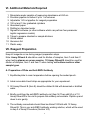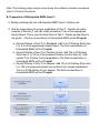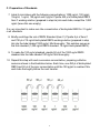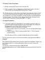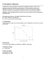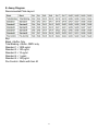
1
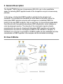
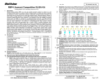
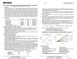
RayBio® Human/Mouse/Rat RBP4
Enzyme Immunoassay Kit
Catalog #: EIA-RBP, EIAM-RBP, EIAR-RBP
User Manual
Last revised December 1, 2015
Caution:
Extraordinarily useful information enclosed
ISO 13485 Certified
3607 Parkway Lane, Suite 100
Norcross, GA 30092
Tel: 1-888-494-8555 (Toll Free) or 770-729-2992, Fax:770-206-2393
Web: www.RayBiotech.com, Email: [email protected]
1
Table of Contents
Section
Page #
I.
Introduction
3
II.
General Description
4
III.
How It Works
4
IV.
Storage
5
V.
Reagents
5
VI.
Additional Materials Required
6
VII.
Reagent Preparation
A. Preparation of Plate and Anti-RBP4 Antibody
B. Preparation of Biotinylated Peptide (Item F)
C. Preparation of Standards
D. Preparation of Positive Control
E. Preparation of Samples
F. Preparation of Wash Buffer and HRP-Strep
6
6
7
8
9
9
10
VIII.
Assay Procedure
10
IX.
Assay Procedure Summary
11
X.
Calculation of Results
A. Typical Data
B. Sensitivity
C. Detection Range
D. Reproducibility
E. Assay Diagram
12
12
12
12
12
13
XI.
Specificity
14
XII.
Select Publications
14
XIII.
Troubleshooting Guide
15
Please read the entire manual carefully before starting your experiment
2
I. Introduction
RBP4 (retinol binding protein-4) is an adipocyte-secreted molecule that is elevated
in the serum before the development of frank diabetes. In obesity and type 2
diabetes, expression of the GLUT4 glucose transporter is decreased selectively in
adipocytes. Adipose-specific Glut4 knockout mice show insulin resistance
secondarily in muscle and liver. Recent studies have shown that serum RBP4
levels are elevated in insulin-resistant mice and humans with obesity and type 2
diabetes. RBP4 levels are normalized by rosiglitazone, an insulin-sensitizing drug.
Transgenic overexpression of human RBP4 or injection of recombinant RBP4 in
normal mice causes insulin resistance. Conversely, genetic deletion of RBP4
enhances insulin sensitivity. Fenretinide, a synthetic retinoid that increases urinary
excretion of RBP4, normalizes serum RBP4 levels and improves insulin resistance
and glucose intolerance in mice with obesity induced by a high-fat diet. Increasing
serum RBP4 induces hepatic expression of the gluconeogenic enzyme
phosphoenolpyruvate carboxykinase (PEPCK) and impairs insulin signalling in
muscle. Thus, RBP4 is an adipocyte-derived 'signal' that may contribute to the
pathogenesis of type 2 diabetes. Lowering RBP4 could be a new strategy for
treating type 2 diabetes. Thus, RBP4 appears to identify insulin resistance and
associated cardiovascular risk factors in subjects with varied clinical presentations.
3
II. General Description
The RayBio® RBP4 Enzyme Immunoassay (EIA) Kit is an in vitro quantitative
assay for detecting RBP4 peptide based on the competitive enzyme immunoassay
principle.
In this assay, a biotinylated RBP4 peptide is spiked into the samples and
standards. The samples and standards are then added to the plate, where the
biotinylated RBP4 peptide competes with endogenous (unlabeled) RBP4 for
binding to the anti-RBP4 antibody. After a wash step, any bound biotinylated RBP4
then interacts with horseradish peroxidase (HRP)-streptavidin, which catalyzes a
color development reaction. The intensity of the colorimetric signal is directly
proportional to the amount of captured biotinylated RBP4 peptide and inversely
proportional to the amount of endogenous RBP4 in the standard or samples. A
standard curve of known concentration of RBP4 peptide can be established and the
concentration of RBP4 peptide in the samples can be calculated accordingly.
III. How It Works
4
IV. Storage
The entire kit may be stored at -20°C to -80°C for up to 6 months from the date of
shipment. For extended storage, it is recommended to store at -80°C. Avoid
repeated freeze-thaw cycles. For prepared reagent storage, see table below.
V. Reagents
Component
Size / Description
Storage / Stability
After Preparation
RBP4 Microplate (Item A)
96 wells (12 strips x 8 wells) coated with
secondary antibody.
1 month at 4°C*
Wash Buffer Concentrate
(20X) (Item B)
25 ml of 20X concentrated solution.
1 month at 4°C
Standard RBP4 Peptide
(Item C)
2 vials of RBP4 Peptide. 1 vial is enough to run
each standard in duplicate.
The first standard:
2-3 days at 4°C
Additional dilutions:
Do not store
Anti-RBP4 Polyclonal
Antibody (Item N)
2 vials of anti-RBP4.
1 month at 4°C
Assay Diluent A (Item D)
30 ml, contains 0.09% sodium azide as
preservative. Diluent for standards and serum or
plasma.
N/A
Assay Diluent B (Item E)
15 ml of 5X concentrated buffer. Diluent for
standards, cell culture media or other sample
types, and HRP-Streptavidin.
1 month at 4°C
Biotinylated RBP4 Peptide
(Item F)
2 vials of Biotinylated RBP4 Peptide, 1 vial is
enough to assay the whole plate.
2-3 days at 4°C
HRP-Streptavidin
Concentrate (Item G)
600 µl 400X concentrated HRP-conjugated
streptavidin.
Do not store and
reuse
Positive Control (Item M)
1 vial of Positive Control.
2-3 days at 4°C
TMB One-Step Substrate
Reagent (Item H)
12 ml of 3,3,5,5'-tetramethylbenzidine (TMB) in
buffer solution.
N/A
Stop Solution (Item I)
8 ml of 0.2 M sulfuric acid.
N/A
*Return unused wells to the pouch containing desiccant pack, reseal along entire edge.
5
VI. Additional Materials Required
1.
2.
3.
4.
5.
6.
7.
8.
9.
10.
11.
Microplate reader capable of measuring absorbance at 450 nm
Precision pipettes to deliver 2 µl to 1 ml volumes
Adjustable 1-25 ml pipettes for reagent preparation
100 ml and 1 liter graduated cylinders
Absorbent paper
Distilled or deionized water
SigmaPlot software (or other software which can perform four-parameter
logistic regression models)
Tubes to prepare standard or sample dilutions
Orbital shaker
Aluminum foil
Plastic wrap
VII. Reagent Preparation
Keep kit reagents on ice during reagent preparation steps.
Note: Assay Diluent A should be used for dilution of samples, Item F and Item C
when testing plasma or serum samples. 1X Assay Diluent B should be used for
dilution of samples, Item F and Item C when testing cell culture media or other
sample types.
A. Preparation of Plate and Anti-RBP4 Antibody
1. Equilibrate plate to room temperature before opening the sealed pouch.
2. Label removable 8-well strips as appropriate for your experiment.
3. 5X Assay Diluent B (Item E) should be diluted 5-fold with deionized or distilled
water.
4. Briefly centrifuge the anti-RBP4 antibody vial (Item N) Then add 50 µl of 1X
Assay Diluent B to the vial to prepare the antibody concentrate. Pipette up and
down to mix gently.
5. The antibody concentrate should then be diluted 100-fold with 1X Assay
Diluent B. This is your anti-RBP4 antibody working solution, which will be used
in step 2 of Assay Procedure (Section VIII).
6
Note: The following steps may be done during the antibody incubation procedure
(step 2 of Assay Procedure)
B. Preparation of Biotinylated RBP4 (Item F)
5. Briefly centrifuge the vial of Biotinylated RBP4 (Item F) before use.
6. See the image below for proper preparation of Item F. Transfer the entire
contents of the Item F vial into a tube containing 10 ml of the appropriate
Assay Diluent. This is your Working Stock of Item F. Pipette up and down to
mix gently. The final concentration of biotinylated RBP4 will be 20 ng/ml.
a. Second Dilution of Item F for Standards: Add 2 ml of Working Stock Item
F to 2 ml of the appropriate Assay Diluent. The final concentration of
biotinylated RBP4 will be 10 ng/ml.
b. Second Dilution of Item F for Positive Control: Add 100 µl of Working
Stock Item F to 100 µl of the prepared Positive Control (Item M). (See
section D for Positive Control preparation) The final concentration of
biotinylated RBP4 will be 10 ng/ml.
c. Second Dilution of Item F for samples: Add 125 µl of Working Stock Item
F to 125 µl of prepared sample (see section E for sample preparation).
This is a 2-fold dilution of your sample. The final concentration of
biotinylated RBP4 will be 10 ng/ml.
7
C. Preparation of Standards
7. Label 6 microtubes with the following concentrations: 1000 ng/ml, 100 ng/ml,
10ng/ml, 1 ng/ml, 100 pg/ml and 0 pg/ml. Pipette 450 µl of biotinylated RBP4
Item F working solution (prepared in step 6a) into each tube, except the 1,000
ng/ml (leave this one empty).
It is very important to make sure the concentration of biotinylated RBP4 is 10 ng/ml
in all standards.
8. Briefly centrifuge the vial of RBP4 Standard (Item C). Pipette 8 µl of Item C
and 792 µl of 10 ng/ml biotinylated RBP4 working solution (prepared in step
6a) into the tube labeled 1000 ng/ml. Mix thoroughly. This solution serves as
the first standard (1,000 ng/ml RBP4 standard, 10 ng/ml biotinylated RBP4).
9. To make the 100 ng/ml standard, pipette 50 µl of the 1000 ng/ml RBP4
standard into the tube labeled 100 ng/ml. Mix thoroughly.
10. Repeat this step with each successive concentration, preparing a dilution
series as shown in the illustration below. Each time, use 450 µl of biotinylated
RBP4 and 50 µl of the prior concentration until the 100 pg/ml is reached. Mix
each tube thoroughly before the next transfer.
8
D. Positive Control Preparation
11. Briefly centrifuge the Positive Control vial (Item M).
12. Refer to step 6b. This is a 2-fold dilution of the Positive Control. The final
concentration of biotinylated RBP4 should still be 10 ng/ml.
The Positive Control is a cell culture media sample that serves as a system control
to verify that the kit components are working. The resulting OD will not be used in
any calculations; if no positive competition is observed please contact RayBiotech
Technical Support. The Positive Control may be diluted further if desired, but be
sure the final concentration of biotinylated RBP4 is 10 ng/ml.
E. Sample Preparation
13. If you wish to perform a 2-fold dilution of your sample, proceed to step 6c. If
you wish to perform a higher dilution of your sample, dilute your sample with
the appropriate Assay Diluent before performing step 6c.
EXAMPLE (to make a 4-fold dilution of sample):
a. Dilute sample 2-fold (62.5 µl of sample + 62.5 µl of the appropriate Assay
Diluent.).
b. Perform step 6c (125 µl of working solution Item F + 125 µl of sample
prepared above).
The total volume is 250 µl, enough for duplicate wells on the microplate.
It is very important to make sure the final concentration of the biotinylated RBP4 is
10 ng/ml.
Note: Optimal sample dilution factors should be determined empirically, however you
may reference below for recommended dilution factors for serum: Human=4X
Mouse=2X .
You may also contact technical support (888-494-8555;
[email protected]) to obtain additional recommended dilution factors for
serum.
9
F. Preparation of Wash Buffer and HRP
14. If Item B (20X Wash Concentrate) contains visible crystals, warm to room
temperature and mix gently until dissolved.
15. Dilute 20 ml of Wash Buffer Concentrate into deionized or distilled water to yield
400 ml of 1X Wash Buffer.
16. Briefly centrifuge the HRP-Streptavidin vial (Item G) before use.
17. Dilute the HRP-Streptavidin concentrate 400-fold with 1X Assay Diluent B.
Note: do not use Assay Diluent A for HRP-Streptavidin preparation in step 17
VIII. Assay Procedure
1. Keep kit reagents on ice during reagent preparation steps. It is recommended
that all standards and samples be run at least in duplicate.
2. Add 100 µl of Anti-RBP4 Antibody (Item N) (See Reagent Preparation step 3) to
each well. Incubate for 1.5 hours at room temperature with gentle shaking (1-2
cycle/sec). You may also incubate overnight at 4ºC.
3. Discard the solution and wash wells 4 times with 1X Wash Solution Buffer (200300 µl each). Washing may be done with a multichannel pipette or an automated
plate washer. Complete removal of liquid at each step is essential to good assay
performance. After the last wash, remove any remaining Wash Buffer by
aspirating or decanting. Invert the plate and blot it against clean paper towels.
4. Add 100 µl of each standard (see Reagent Preparation Section C), Positive
Control (see Reagent Preparation Section D) and sample (see Reagent
Preparation Section E) in appropriate wells. Be sure to include a blank well
(Assay Diluent only). Cover wells and incubate for 2.5 hours at room
temperature with gentle shaking (1-2 cycles/sec) overnight or at 4ºC.
5. Discard the solution and wash 4 times as directed in Step 3.
6. Add 100 µl of prepared HRP-Streptavidin solution (see Reagent Preparation
step 7) to each well. Incubate for 45 minutes at room temperature with gentle
10
shaking. It is recommended that incubation time should not be shorter or longer
than 45 minutes.
7. Discard the solution and wash 4 times as directed in Step 3.
8. Add 100 µl of TMB One-Step Substrate Reagent (Item H) to each well. Incubate
for 30 minutes at room temperature in the dark with gentle shaking (1-2
cycles/sec).
9. Add 50 µl of Stop Solution (Item I) to each well. Read at 450 nm immediately.
IX. Assay Procedure Summary
1. Prepare all reagents, samples and standards as instructed.
2. Add 100 µl anti-RBP4 to each well. Incubate 1.5 hours at room temperature or
overnight at 4ºC.
3. Add 100 µl standard or sample to each well. Incubate 2.5 hours at room
temperature or overnight at 4ºC.
4. Add 100 µl prepared Streptavidin solution. Incubate 45 minutes at room
temperature.
5. Add 100 µl TMB One-Step Substrate Reagent to each well. Incubate 30 minutes
at room temperature.
6. Add 50 µl Stop Solution to each well. Read at 450 nm immediately.
11
X. Calculation of Results
Calculate the mean absorbance for each set of duplicate stands, controls, and
samples and subtract the blank optical density. Plot the standard curve using
SigmaPlot software (or other software which can perform four-parameter logistic
regression models), with standard concentration on the x-axis and percentage of
absorbance (see calculation below) on the y-axis. Draw the best-fit curve through the
standard points.
Percentage absorbance = (B-blank OD)/B 0-blank OD) where
B = OD of sample or standard and
B0 = OD of zero standard (total binding)
A. Typical Data
These standard curves are for demonstration only. A standard curve must be run with
each assay.
B. Sensitivity
The minimum detectable concentrations of RBP4 is 460 pg/ml.
C. Detection Range
0.1-1,000 ng/ml
D. Reproducibility
Intra-Assay: CV<10%
Inter-Assay: CV<15%
12
E. Assay Diagram
Recommended Plate Layout:
Key:
Blank = Buffer Only
Total Binding = Biotin- RBP4 only
Standard 1 = 1000 ng/ml
Standard 2 = 100 ng/ml
Standard 3 = 10 ng/ml
Standard 4 = 1 ng/ml
Standard 5 = 100 pg/ml
Pos Control = Biotin with Item M
13
XI. Specificity
This kit detects RBP4 (183aa) and all other active forms including RBP4182,181,179,176.
Cross Reactivity: This EIA kit shows no cross-reactivity with any of the cytokines
tested: Ghrelin, Nesfatin, Angiotensin II, NPY and APC.
XIV. Publications Citing This Product
1. Anna P., Lech C., Jerzy J., Robert F. Assessment of serum IGF-1 and adipokines related
to metabolic dysfunction in HIV-infected adults. Cytokine 64 (2013) 97-102
Species: Human
Sample Type: Serum
14
XIII. Troubleshooting Guide
Problem
Cause
Solution
Inaccurate pipetting
Improper standard dilution
Check pipettes
Briefly centrifuge Item C and dissolve
the powder thoroughly by gently
mixing
Low signal
Improper preparation of
standard and/or
biotinylated antibody
Too brief incubation times
Inadequate reagent
volumes or improper dilution
Briefly spin down vials before
opening. Dissolve the powder
thoroughly.
Ensure sufficient incubation time;
assay procedure step 2 may be done
overnight
Check pipettes and ensure correct
preparation
Large CV
Inaccurate pipetting
Air bubbles in wells
Check pipettes
Remove bubbles in wells
High
background
Plate is insufficiently
washed
Contaminated wash buffer
Review the manual for proper wash.
If using a plate washer, ensure that
all ports are unobstructed.
Make fresh wash buffer
Improper storage of the
ELISA kit
Stop solution
Follow storage recomendations in
sections IV and V. Keep substrate
solution protected from light.
Add stop solution to each well before
reading plate
Poor standard
curve
Low sensitivity
15
RayBio® ELISA Kits
Over 2,000 ELISA kits available, visit www.RayBiotech.com/ELISA-Kits.html for
details.
This product is for research use only.
©2015 RayBiotech, Inc
16