1
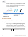
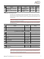
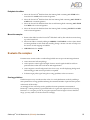
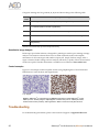
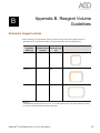
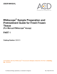
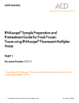
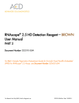
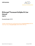
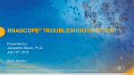
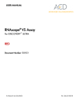
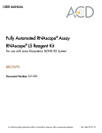
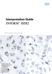
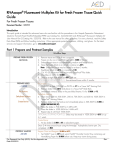
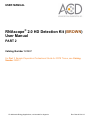
USER MANUAL
RNAscope® 2.0 HD Detection Kit (BROWN)
User Manual
PART 2
Catalog Number 320497
For Part 1, Sample Preparation Pretreatment Guide for FFPE Tissue, see Catalog
Number 320511.
For Molecular Biology Applications, not intended for diagnosis.
Rev. Date 20131113
For Molecular Biology Applications, not intended for diagnosis.
Trademarks
®
™
RNAscope and HybEZ are trademarks of Advanced Cell Diagnostics, Inc. All other trademarks belong to their
respective owners.
®
Citing RNAscope 2.0 in Publications
®
When describing a procedure for publication using this product, please refer to it as the RNAscope 2.0 Assay and cite:
Wang F, Flanagan J, Su N, Wang L-C, Bui S, Nielson A, Wu X, Vo H-T, Ma X-J and Luo Y. RNAscope: A Novel In Situ
RNA Analysis Platform for Formalin-Fixed Paraffin-Embedded Tissues. J. Mol. Diagnostics, 2012, 14:22–29.
Disclaimers
Advanced Cell Diagnostics, Inc. reserves the right to change its products and services at any time to incorporate
technological developments. This manual is subject to change without notice.
Although this manual has been prepared with every precaution to ensure accuracy, Advanced Cell Diagnostics, Inc.
assumes no liability for any errors, omissions, or for any damages resulting from the use of this information.
Copyright
© 2013. Advanced Cell Diagnostics, Inc. All rights reserved.
Contents
Chapter 1. Product Information .......................................................5
About this guide ............................................................................................... 5
Product description .......................................................................................... 5
Background.................................................................................................. 5
Overview ...................................................................................................... 5
Kit contents and storage .................................................................................. 6
®
RNAscope Probes ..................................................................................... 6
®
RNAscope 2.0 HD Detection Kit ................................................................ 7
Required materials and equipment ................................................................. 8
™
HybEZ Hybridization System .................................................................... 8
User-supplied materials ............................................................................... 8
Chapter 2. Before You Begin .........................................................11
Important procedural guidelines .................................................................... 11
Chapter 3. RNAscope® 2.0 Assay ..................................................13
Workflow ........................................................................................................ 13
Materials required for the assay .................................................................... 14
Prepare the materials .................................................................................... 14
Prepare 1X Wash Buffer............................................................................ 14
Prepare counterstaining reagents ............................................................. 15
Prepare dehydrating reagents ................................................................... 15
Equilibrate reagents ................................................................................... 15
Run the assay................................................................................................ 15
Hybridize probe ......................................................................................... 15
Hybridize Amp 1 ........................................................................................ 16
Hybridize Amp 2 ........................................................................................ 16
Hybridize Amp 3 ........................................................................................ 17
Hybridize Amp 4 ........................................................................................ 17
Hybridize Amp 5 ........................................................................................ 17
Hybridize Amp 6 ........................................................................................ 17
Detect the signal ........................................................................................ 18
Counterstain the slides .............................................................................. 18
Dehydrate the slides .................................................................................. 19
Mount the samples .................................................................................... 19
Evaluate the samples .................................................................................... 19
Scoring guidelines ..................................................................................... 19
Quantitative Image Analysis ...................................................................... 20
Control examples ....................................................................................... 20
Troubleshooting ............................................................................................. 20
Appendix A. Tissue Pretreatment Recommendation ................... 21
Tissue pretreatment recommendation........................................................... 21
Tissue-specific pretreatment conditions .................................................... 21
®
RNAscope 2.0 HD Detection Kit (BROWN) User Manual
3
Appendix B. Reagent Volume Guidelines ..................................... 23
Determine reagent volume ............................................................................ 23
Appendix C. Safety .........................................................................25
Chemical safety ............................................................................................. 25
Biological hazard safety................................................................................. 25
Documentation and support ..........................................................27
Obtaining MSDSs .......................................................................................... 27
Obtaining support .......................................................................................... 27
Contact information ....................................................................................... 27
Limited product warranty ............................................................................... 27
4
®
RNAscope 2.0 HD Detection Kit (BROWN) User Manual
Chapter 1. Product Information
1
Before using this product, please read the safety information in Appendix C. Safety
on page 25.
IMPORTANT! We recommend reading the entire user manual before beginning any
protocols.
About this guide
This user manual provides guidelines and protocols to use the RNAscope® 2.0 HD Detection
Kit – BROWN (Cat. No. 310035). RNAscope® Assays are compatible with a variety of sample
types.
You must use both an RNAscope® Detection Kit user manual and a Sample Preparation and
Pretreatment user guide to perform the entire assay.
IMPORTANT! For Part 1, Sample Preparation and Pretreatment Guide for FFPE Tissue, see
Catalog No. 320511.
Visit www.acdbio.com/support/technical-doc to download a sample preparation user guide.
Product description
Background
The RNAscope® Assays use a novel and proprietary method of in situ hybridization (ISH) to
visualize single RNA molecules per cell in samples mounted on slides. RNAscope® Assays do
not require the RNA-free environment used for traditional ISH. The assays are based on
ACD’s patented signal amplification and background suppression technology. Compared
with the RNAscope® 1.0 Assay, the 2.0 Assay incorporates an additional signal amplification
step, which enhances the signal for low expressing genes and RNA present in archived
samples and partially degraded specimens.
Overview
The RNAscope® Assay procedure is illustrated in Figure 1 on page 6. The procedure can be
completed in 7–8 hours or conveniently divided over two days. Most of the RNAscope® Assay
reagents are available in convenient Ready-To-Use (RTU) dropper bottles and provide a
simple, nearly pipette-free workflow.
Starting with properly prepared tissue samples, sections are first pretreated, and then RNAspecific probes are hybridized to target RNA. The signal is amplified using a multi-step
process, followed by hybridization to horseradish peroxidase (HRP)- or alkaline phosphatase
(AP)-labeled probes and detection using a chromogenic substrate. Each single RNA transcript
appears as a distinct dot of chromogen precipitate visible using a common bright field
microscope at 40–100X magnification. The RNAscope® 2.0 Assay has additional amplification
®
RNAscope 2.0 HD Detection Kit (BROWN) User Manual
5
steps that allow observable results under 10–20X magnification. RNAscope® 2.0 Assays offer
the choice of two Detection Kits: Brown (DAB) and Red (Fast Red), which enable RNA
molecules to be visualized as brown or red chromogenic dots, respectively.
The procedure can be automated using the Ventana® DISCOVERY XT or ULTRA Systems.
Refer to the RNAscope® VS Assay User Manual available at
www.acdbio.com/support/technical-doc for more details.
1: Tissue section
2: Hybridize to target RNA
Start with properly prepared
sections and pretreat to
allow access to target RNA.
Hybridize gene-specific probe
pairs to the target mRNA.
3: Amplify signal
4: Image
Probes are hybridized to a cascade of signal
amplification molecules, culminating in binding
of HRP- or AP-labeled probes. The 2.0 Assay
enhances signal further with additional
amplification steps. Add DAB or Fast Red
substrate to detect target RNA.
Visualize target
RNA using a
standard bright
field microscope.
Figure 1. Procedure overview
Kit contents and storage
The RNAscope® 2.0 Assay requires the RNAscope® Probes and the RNAscope® 2.0 HD
Detection Kit. Probes and Detection Kits are available separately.
RNAscope® Probes
The RNAscope® Probes consist of the user-specified Target Probe and the Positive and
Negative Control Probes. Visit www.acdbio.com/products/target-probes/search-product to
find a gene-specific probe from a searchable catalog of >27,000 predesigned Target Probes, or
order a custom probe. Visit www.acdbio.com/products/target-probes/controls-housekeeping
to find appropriate Control Probes. Each probe is sufficient for staining approximately
20 sections, each with an area of approximately 20 mm x 20 mm (0.75” x 0.75”). Larger tissue
sections will result in fewer tests. The probes have a shelf life of six months from the shipment
date when stored as indicated in the following table:
Target Probes
Reagent
®
Cat. No.
RNAscope Singleplex Target Probe – Various
[species] – [gene]
6
Content
Quantity
Probe targeting specific RNA 3 mL x 1 bottle
®
Storage
4°C
RNAscope 2.0 HD Detection Kit (BROWN) User Manual
Control Probes
Reagent
Cat. No.
Content
Quantity
Storage
®
Various
Probe targeting common
housekeeping gene
3 mL x 1 bottle
4°C
®
310043
Probe targeting bacterial
gene dapB
3 mL x 1 bottle
4°C
RNAscope Positive Control Probe –
[species] – PPIB
RNAscope Negative Control Probe –
DapB
RNAscope® 2.0 HD Detection Kit
Each RNAscope® 2.0 HD Detection Kit provides enough reagents to stain ~20 tissue sections,
each with an area of approximately 20 mm x 20 mm (0.75” x 0.75”). Larger tissue sections will
result in fewer tests. Each kit contains three sub-kits: a Pretreatment Kit, a Detection Kit, and a
Wash Buffer Kit.
IMPORTANT! Directions to use the Pretreatment Kit are included in separate sample
preparation and pretreatment user guides.
The reagents have a shelf life of six months from the shipment date when stored as indicated
in the following table:
Pretreatment Kit (Cat. No. 310020)
Reagent
Quantity
Storage
Pretreat 1
4 mL x 2 bottles
4°C
10X Pretreat 2*
70 mL x 4 bottles
Room temperature (20–25°C)
Pretreat 3
4.5 mL x 1 bottle
4°C
2.0 HD Detection Kit – BROWN (Cat. No. 310035)†
Reagent
Quantity
Storage
2.0 Amp 1
3 mL x 1 bottle
4°C
2.0 Amp 2
4.5 mL x 1 bottle
4°C
2.0 Amp 3
3 mL x 1 bottle
4°C
2.0 Amp 4
4.5 mL x 1 bottle
4°C
2.0 Amp 5–BROWN
4.5 mL x 1 bottle
4°C
2.0 Amp 6–BROWN
3 mL x 1 bottle
4°C
DAB-A
2 mL x 1 bottle
4°C
DAB-B
2 mL x 1 bottle
4°C
Wash Buffer Kit (Cat. No. 310091)
Reagent
50X Wash Buffer
Quantity
60 mL x 4 bottles
Storage
Room temperature (20–25°C)
* Comes in a separate box.
† Comes in two boxes.
IMPORTANT! RNAscope® HD Detection Kits share the same Pretreatment Kit and Wash
Buffer, but have unique Detection Kits. Do not interchange the reagent components of the
Detection Kits, even those having the same name.
®
RNAscope 2.0 HD Detection Kit (BROWN) User Manual
7
Required materials and equipment
The following materials and equipment are needed to perform the RNAscope® Assay.
HybEZ™ Hybridization System
IMPORTANT! The RNAscope® Assay has been validated using this system only.
The HybEZ™ Hybridization System (110 VAC, Cat. No. 310010; 220 VAC, Cat. No. 310013) is
designed for the hybridization and incubation steps in the RNAscope® Assays. Incubation
steps in the RNAscope® Assay require humid conditions to prevent sections from drying out.
For instructions on how to use the HybEZ™ Hybridization System, refer to the HybEZ™
Hybridization System User Manual available at: www.acdbio.com/support/technical-doc and
view the training video at www.acdbio.com/support/online-training-videos/. The system
contains the following components:
Component
Quantity
Cat. No.
™
1 oven
310010 or 310013
™
1 tray
310012
™
1 rack
310014
™
2 sheets
—
™
15 sheets
310015
HybEZ Oven (110 or 220 VAC)
HybEZ Humidity Control Tray (with lid)
HybEZ Slide Rack (20 slide capacity)
HybEZ Humidifying Paper
HybEZ Humidifying Paper Pack
User-supplied materials
Description
Supplier
Cat. No.
*
100% ethanol (EtOH)
American Master Tech Scientific/MLS
ALREAGAL
Gill’s Hematoxylin I
American Master Tech Scientific/MLS
HXGHE1LT
Xylene
Fisher Scientific/MLS
X3P-1GAL
®
American Master Tech Scientific/MLS
LWSRA24
®
American Master Tech Scientific/MLS
LWT4457EA
Tissue-Tek Clearing Agent Dish, xylene resistant (1 American Master Tech Scientific/MLS
required)
LWT4456EA
Cytoseal XYL xylene-based mounting medium
Richard-Allen Scientific/MLS
8312-4
Cover Glass 24 x 50 mm
Fisher Scientific/MLS
12--545-F
Ammonium hydroxide, 28–30%
Sigma-Aldrich/MLS
320145-500mL
Carboy (>3L)
MLS
—
Water bath or incubator, capable of holding
temperature at 40 +/- 1°C
MLS
—
Pipettors and tips, 1–1000 µL
MLS
—
Distilled water
MLS
—
Tubes (various sizes)
MLS
—
Tissue-Tek Vertical 24 Slide Rack
Tissue-Tek Staining Dish (3 required)
®
8
®
RNAscope 2.0 HD Detection Kit (BROWN) User Manual
Description
Supplier
Cat. No.
Fume hood
MLS
—
Graduated cylinder
MLS
—
Parafilm
MLS
—
Paper towel or absorbent paper
MLS
—
20% bleach
MLS
—
Microscope and accessories
MLS
—
* Major Laboratory Supplier in North America. For other regions, please check Catalog Numbers with your local lab supplier.
®
RNAscope 2.0 HD Detection Kit (BROWN) User Manual
9
10
®
RNAscope 2.0 HD Detection Kit (BROWN) User Manual
Chapter 2. Before You Begin
2
IMPORTANT! For Part 1, Sample Preparation and Pretreatment Guide for FFPE Tissue, see
Catalog No. 320511.
Prior to running the RNAscope® Assay on your samples for the first time, we recommend that
you:
• View the video demonstrations available at www.acdbio.com/support/online-trainingvideos/.
• Run the assay on FFPE RNAscope® Control Slides (Cat. No. 310045 for Human control
slide, Hela; Catalog No. 310023 for Mouse control slide, 3T3) using the Positive and
Negative Control Probes.
Important procedural guidelines
• Start with properly fixed and prepared sections. Refer to Appendix A. Tissue
Pretreatment Recommendation on page 21 and to our sample preparation and
pretreatment user guides available at www.acdbio.com/support/technical-doc.
• Use only samples mounted on SuperFrost Plus® Slides (Fisher Scientific;
Cat. No. 12-550-15).
• Follow the recommended pretreatment guidelines for your sample. Refer to our sample
preparation and pretreatment user guides available at
www.acdbio.com/support/technical-doc/.
• Always run positive and negative control probes on your sample to assess sample RNA
quality and optimal permeabilization.
• Do not substitute required materials. Assay has been validated with these materials
only.
• Follow the protocol exactly for best results.
• Do not let your sections dry out during the procedure.
• Use good laboratory practices and follow all necessary safety procedures. Refer to
Appendix C. Safety on page 25 for more information.
®
RNAscope 2.0 HD Detection Kit (BROWN) User Manual
11
12
®
RNAscope 2.0 HD Detection Kit (BROWN) User Manual
Chapter 3. RNAscope® 2.0 Assay
3
IMPORTANT! For Part 1, Sample Preparation and Pretreatment Guide for FFPE Tissue, see
Catalog No. 320511.
This procedure flows directly from sample preparation and pretreatment. Refer to the
appropriate sample preparation and pretreatment user guide for your specific sample type.
Workflow
Prepare the materials ~10–30 MIN
Run the assay ~4 HRS 45 MIN
Hybridize probe ~2 HRS
Hybridize Amp 1 ~30 MIN
Hybridize Amp 2 ~15 MIN
Hybridize Amp 3 ~30 MIN
Hybridize Amp 4 ~15 MIN
Hybridize Amp 5 ~30 MIN
Hybridize Amp 6 ~15 MIN
Detect the signal ~10 MIN
Counterstain the slides ~2 MIN
Dehydrate samples ~10 MIN
Mount samples ~5 MIN
Review results
®
RNAscope 2.0 HD Detection Kit (BROWN) User Manual
13
Materials required for the assay
Materials provided by the RNAscope
2.0 HD Detection Kit – BROWN
®
Materials provided by
®
RNAscope Probes
Other materials and equipment
• 50X Wash Buffer
• Target Probe
• Prepared sections
• 2.0 Amp 1
• Positive Control Probe
• Distilled water
• 2.0 Amp 2
• Negative Control Probe
• Carboy (>3L)
• 2.0 Amp 3
• Fume hood
• 2.0 Amp 4
• Xylene
• 2.0 Amp 5 – BROWN
• 100% ethanol (EtOH)
• 2.0 Amp 6 – BROWN
• Tissue-Tek Staining Dish (3)
• DAB-A
• Tissue-Tek Clearing Agent Dish,
xylene-resistant (1)
®
®
• DAB-B
• Gill’s Hematoxylin I
• Ammonium hydroxide, 28–30%
• Graduated cylinder
• Parafilm
• HybEZ Humidifying System
™
• Water bath or incubator
• Tissue-Tek Vertical 24 Slide Rack
®
• Tubes (various sizes)
• Paper towel or absorbent paper
• Pipettors and tips, 1–1000 µL
• Cytoseal XYL xylene-based
• 20% bleach
• Cover Glass, 24 mm x 50 mm
Prepare the materials
You may prepare the reagents at the same time you prepare pretreatment reagents. Refer to a
sample preparation and pretreatment user guide available at
www.acdbio.com/support/technical-doc.
Some of the materials may be prepared in advance and stored at room temperature.
Prepare 1X Wash Buffer
• Prepare 3 L of 1X WASH BUFFER by adding 2.94 L distilled water and 1 bottle (60 mL)
of 50X Wash Buffer to a large carboy. Mix well.
Note: Warm 50X Wash Buffer up to 40°C for 10–20 min before making 1X Wash Buffer. 1X
Wash Buffer may be prepared ahead of time and stored at room temperature for up to one
month.
14
®
RNAscope 2.0 HD Detection Kit (BROWN) User Manual
Prepare counterstaining reagents
• In the fume hood, prepare 50% HEMATOXYLIN staining solution by adding 100 mL
Gill’s Hematoxylin I to 100 mL distilled water in a Staining Dish.
Note: 50% Hematoxylin staining solution can be reused for up to 1 week.
• In the fume hood, prepare 0.02% (w/v) AMMONIA WATER (bluing reagent) by adding
1.43 mL of 1N ammonium hydroxide to 250 mL distilled water in a graduated cylinder
or other container.
• Seal the cylinder with parafilm. Mix well 3–5 TIMES.
Note: For assay quantitation, it is critical to use Ammonium Hydroxide.
Prepare dehydrating reagents
IMPORTANT! Do not reuse deparaffinization reagents for dehydration of the slides after the
assay.
• In the fume hood, add ~200 mL XYLENE to a Clearing Agent Dish.
• In the fume hood, fill two Staining Dishes with ~200 mL 100% ETOH.
• Prepare 70% ETOH by adding 140 mL 100% EtOH to 60 mL distilled water in a Staining
Dish. Seal the dish with parafilm, mix well, and place in fume hood.
Note: Reagents may be prepared ahead of time. Ensure all containers remain covered.
Equilibrate reagents
• Place AMP 1–6 reagents at ROOM TEMPERATURE (RT).
• Ensure HybEZ™ OVEN and prepared Humidity Control TRAY are at 40°C.
• Before each use, warm the Target and/or Control PROBES for 10 MIN at 40°C in a water
bath or incubator. Swirl gently to mix.
Run the assay
IMPORTANT! Do NOT let sections dry out between incubation steps. Work quickly and fill
barrier with solutions.
IMPORTANT! View the wash step video at www.acdbio.com/support/online-trainingvideos/wash-slides before proceeding.
Hybridize probe
IMPORTANT! Ensure probes are prewarmed to dissolve any precipitation prior to use.
1.
®
Tap and/or flick to remove excess liquid from slides and place in the HybEZ™ Slide
Rack. Add ~4 DROPS of the appropriate PROBE to entirely cover each section.
RNAscope 2.0 HD Detection Kit (BROWN) User Manual
15
Note: Refer to Appendix B. Reagent Volume Guidelines on page 23 to determine the
recommended number of drops needed per slide. For example, for a 0.75” x 0.75” barrier
add 4 drops of the appropriate probe.
2.
Place the HybEZ™ Slide Rack in the HybEZ™ Humidity Control Tray, cover with lid and
insert into the oven for 2 HRS at 40°C.
IMPORTANT! To prevent evaporation, make sure the turn nob is completely turned to lock
position.
3.
Remove the HybEZ™ Control Tray from the oven and remove HybEZ™ Slide Rack.
4.
One slide at a time, quickly remove excess liquid and place slide in a Tissue-Tek® Slide
Rack submerged in the Tissue-Tek® Staining Dish filled with 1X WASH BUFFER.
5.
Wash slides in 1X Wash Buffer for 2 MIN at RT. Agitate slides by moving the Slide Rack
up and down in the dish.
6.
Repeat Step 5 with fresh 1X Wash Buffer.
1.
Take each slide one at a time from the Tissue-Tek® Slide Rack and tap/and or flick to
remove the excess liquid before placing in the HybEZ™ Slide Rack. Add ~4 DROPS of
AMP 1 to entirely cover each section.
2.
Place the HybEZ™ Slide Rack in the HybEZ™ Humidity Control Tray. Close tray and
insert into the oven for 30 MIN at 40°C.
3.
Remove the HybEZ™ Control Tray from the oven and remove HybEZ™ Slide Rack.
4.
One slide at a time, quickly remove excess liquid and place slide in a Tissue-Tek® Slide
Rack submerged in the Tissue-Tek® Staining Dish filled with 1X WASH BUFFER.
5.
Wash slides in 1X Wash Buffer for 2 MIN at RT with occasional agitation.
6.
Repeat Step 5 with fresh 1X Wash Buffer.
1.
Take each slide one at a time from the Tissue-Tek® Slide Rack and tap/and or flick to
remove the excess liquid before placing in the HybEZ™ Slide Rack. Add ~4 DROPS of
AMP 2 to entirely cover each section.
2.
Place the HybEZ™ Slide Rack in the HybEZ™ Humidity Control Tray. Close tray and
insert into the oven for 15 MIN at 40°C.
3.
Remove the HybEZ™ Control Tray from the oven and remove HybEZ™ Slide Rack.
4.
One slide at a time, quickly remove excess liquid and place slide in a Tissue-Tek® Slide
Rack submerged in the Tissue-Tek® Staining Dish filled with 1X WASH BUFFER.
5.
Wash slides in 1X Wash Buffer for 2 MIN at RT with occasional agitation.
6.
Repeat Step 5 with fresh 1X Wash Buffer.
Hybridize Amp 1
Hybridize Amp 2
16
®
RNAscope 2.0 HD Detection Kit (BROWN) User Manual
Hybridize Amp 3
1.
Take each slide one at a time from the Tissue-Tek® Slide Rack and tap/and or flick to
remove the excess liquid before placing in the HybEZ™ Slide Rack. Add ~4 DROPS of
AMP 3 to entirely cover each section.
2.
Place the HybEZ™ Slide Rack in the HybEZ™ Humidity Control Tray. Close tray and
insert into the oven for 30 MIN at 40°C.
3.
Remove the HybEZ™ Control Tray from the oven and remove HybEZ™ Slide Rack.
4.
One slide at a time, quickly remove excess liquid and place slide in a Tissue-Tek® Slide
Rack submerged in the Tissue-Tek® Staining Dish filled with 1X WASH BUFFER.
5.
Wash slides in 1X Wash Buffer for 2 MIN at RT with occasional agitation.
6.
Repeat Step 5 with fresh 1X Wash Buffer.
1.
Take each slide one at a time from the Tissue-Tek® Slide Rack and tap/and or flick to
remove the excess liquid before placing in the HybEZ™ Slide Rack. Add ~4 DROPS of
AMP 4 to entirely cover each section.
2.
Place the HybEZ™ Slide Rack in the HybEZ™ Humidity Control Tray. Close tray and
insert into the oven for 15 MIN at 40°C.
3.
Remove the HybEZ™ Control Tray from the oven and remove HybEZ™ Slide Rack.
Hybridize Amp 4
IMPORTANT! Do not insert tray into the HybEZ™ Oven for the rest of the procedure.
4.
One slide at a time, quickly remove excess liquid and place slide in a Tissue-Tek® Slide
Rack submerged in the Tissue-Tek® Staining Dish filled with 1X WASH BUFFER.
5.
Wash slides in 1X Wash Buffer for 2 MIN at RT with occasional agitation.
6.
Repeat Step 5 with fresh 1X Wash Buffer.
1.
Take each slide one at a time from the Tissue-Tek® Slide Rack and tap/and or flick to
remove the excess liquid before placing in the HybEZ™ Slide Rack. Add ~4 DROPS of
AMP 5 to entirely cover each section.
2.
Place the HybEZ™ Slide Rack in the HybEZ™ Humidity Control Tray. Seal tray and
incubate for 30 MIN at RT.
3.
Remove the HybEZ™ Slide Rack from the HybEZ™ Humidity Control Tray.
4.
One slide at a time, quickly remove excess liquid and place slide in a Tissue-Tek® Slide
Rack submerged in the Tissue-Tek® Staining Dish filled with 1X WASH BUFFER.
5.
Wash slides in 1X Wash Buffer for 2 MIN at RT with occasional agitation.
6.
Repeat Step 5 with fresh 1X Wash Buffer.
1.
Take each slide one at a time from the Tissue-Tek® Slide Rack and tap/and or flick to
remove the excess liquid before placing in the HybEZ™ Slide Rack. Add ~4 DROPS of
AMP 6 to entirely cover each section.
Hybridize Amp 5
Hybridize Amp 6
®
RNAscope 2.0 HD Detection Kit (BROWN) User Manual
17
2.
The Amp 6 solution is a yellow color. This is normal. Place the HybEZ™ Slide Rack with
the slides in the HybEZ™ Humidity Control Tray, cover with lid and incubate for 15
MIN at RT.
3.
Remove the HybEZ™ Slide Rack from the HybEZ™ Humidity Control Tray.
4.
One slide at a time, quickly remove excess liquid and place slide in a Tissue-Tek® Slide
Rack submerged in the Tissue-Tek® Staining Dish filled with 1X WASH BUFFER.
5.
Wash slides in 1X Wash Buffer for 2 MIN at RT with occasional agitation.
6.
Repeat Step 5 with fresh 1X Wash Buffer.
1.
MIX EQUAL VOLUMES of BROWN-A and BROWN-B (DAB substrate) in an
appropriately sized tube by dispensing the same number of drops (2 drops of each
reagent total of 4) for each solution. Make ~120 μL DAB substrate PER SECTION. Mix
well 3–5 TIMES.
Detect the signal
CAUTION! DAB is toxic. Follow appropriate precautions and safety guidelines
when disposing of and handling this chemical.
2.
Take each slide one at a time from the Tissue-Tek® Slide Rack and tap and/or flick to
remove the excess liquid before placing in the HybEZ™ Slide Rack.
3.
Pipette ~120 μL of DAB onto each tissue section. Ensure sections are covered, and
incubate for 10 MIN at RT.
4.
Dispose the remaing DAB according to local regulation and insert the slide into a
Tissue-Tek® Slide Rack submerged in a Tissue-Tek® Staining Dish filled with
DISTILLED WATER.
5.
Wash slides in distilled water by moving the Tissue-Tek® Slide Rack up and down 3–5
TIMES. Replace with fresh distilled water.
Counterstain the slides
18
1.
Move the Tissue-Tek® Slide Rack into the Staining Dish containing
50% HEMATOXYLIN I staining solution for 2 MIN at RT. Slides will be purple.
2.
Immediately transfer the Slide Rack back into the Staining Dish containing distilled
water, and wash slides 3–5 TIMES by moving the rack up and down. Keep repeating
with fresh distilled water until the slides are clear, while sections remain purple.
3.
Replace distilled water in the Staining Dish with 0.02% AMMONIA WATER. Move
rack up and down 2–3 TIMES. Section should turn blue.
4.
Replace ammonia water with DISTILLED WATER. Wash slides 3–5 TIMES.
®
RNAscope 2.0 HD Detection Kit (BROWN) User Manual
Dehydrate the slides
1.
Move the Tissue-Tek® Slide Rack into the Staining Dish containing 70% ETOH in the
fume hood for 2 MIN with occasional agitation.
2.
Move the Tissue-Tek® Slide Rack into the first Staining Dish containing 100% ETOH for
2 MIN with occasional agitation.
3.
Move the Tissue-Tek® Slide Rack into the second Staining Dish containing 100% ETOH
for 2 MIN with occasional agitation.
4.
Move the Tissue-Tek® Slide Rack into the Staining Dish containing XYLENE for 5 MIN
with occasional agitation.
Mount the samples
1.
Remove the slides from the Tissue-Tek® Slide Rack and lay flat with the sections facing
up in the fume hood.
2.
Mount one slide at a time by adding 1–2 DROPS of CYTOSEAL or other xylene-based
mounting medium to each slide and carefully placing a 24 mm x 50 mm coverslip over
the section. Avoid trapping air bubbles.
3.
AIR DRY slides for >5 MIN.
Evaluate the samples
Examine tissue sections under a standard bright field microscope at 20–40X magnification:
• Assess tissue and cell morphology.
• Assess positive control signal strength. Positive control signal should be visible as
punctuate dots within cell nuclei at 20–40X magnification.
• Assess negative control background. One dot to every 10 cells displaying background
DAB staining per 20X microscope field is acceptable.
• Evaluate target probe signal using the scoring guidelines in the next section.
Scoring guidelines
The RNAscope® Assay can enhance the value of in situ hybridization results by enabling a
semi-quantitative scoring guideline utilizing the estimated number of punctate dots present
within each cell boundary.
An example of how to develop such a guideline for semi-quantitative assessment of
RNAscope® staining intensity is presented below for a gene with expression level varying
between 1 to > 10 copies per cell. If your gene expression level is higher or lower than this
range, you may need to scale the criteria accordingly.
®
RNAscope 2.0 HD Detection Kit (BROWN) User Manual
19
Categorize staining into five grades: 0, 1+, 2+, 3+ and 4+ according to the following table:
Staining
score
Microscope objective scoring*
0
No staining or less than 1 dot to every 10 cells (40X magnification)
1
1–3 dots/cell (visible at 20–40X magnification)
2
4–10 dots/cell. Very few dot clusters (visible at 20–40X magnification)
3
>10 dots/cell. Less than 10% positive cells have dot clusters (visible at 20X magnification)
4
>10 dots/cell. More than 10% positive cells have dot clusters (visible at 20X
magnification)
* Discount cells with artificially high nuclear background staining.
Quantitative Image Analysis
RNAscope® Spot Studio Software is designed for pathologists with no prior training in image
analysis. This intuitive software allows users to obtain statistical results with complete
information of cell-count/region and number of spots/cell. Simply load any image, select a
region of interest, define settings and run analysis, followed by a quality control review before
results are exported. Further information is available on our website at www.acdbio.com.
Control examples
Figure 2 is an example of HeLa cell pellet sections using DapB Negative Control Probe and
PPIB Positive Control Probe at 20X magnification.
2 (a). dapB
2 (b). HsPPIB
®
®
Figure 2. RNAscope 2.0 HD Detection Kit–BROWN performed on FFPE RNAscope Control
Slides (Cat. No. 310045) using the dapB Negative Control Probe (Cat. No. 310043) and PPIB
Positive Control Probe (313901), 20X magnification. Slides contain HeLa cell pellet sections.
Troubleshooting
For troubleshooting information, please contact technical support at [email protected].
20
®
RNAscope 2.0 HD Detection Kit (BROWN) User Manual
Appendix A. Tissue Pretreatment
Recommendation
A
Follow the recommended pretreatment conditions based on your tissue type for:
• Any new or previously untested FFPE tissue types
• Samples prepared differently than the sample preparation protocol found in Part 1,
Sample Preparation and Pretreatment Guide for FFPE Tissue (Cat. No. 320511).
Tissue pretreatment recommendation
1.
Stain representative samples using the positive and negative control probes.
2.
Fix sample in fresh 10% NBF for 16–32 HRS at RT.
Note: Perform tissue fixation step using the recommended amount of time. Over or
under-fixation will result in significant signal loss when performing the RNAscope®
Assay.
3.
Depending on your tissue type (see section below), vary the PRETREAT 2 and/or
PRETREAT 3 TIME.
Reagent
Mild
Standard
Extended
Pretreat 2
15 MIN
15 MIN
30 MIN
Pretreat 3
15 MIN
30 MIN
30 MIN
Note: Sample types such as certain Xenografts and Cell Pellets, require less time. For
these tissue types, vary the Pretreat 2 time to 8 min and Pretreat 3 time to 15 min. If you
have a tissue type not listed, contact support at [email protected].
Tissue-specific pretreatment conditions
If your sample fixation is successful in fresh 10% NBF (Step 2 above), then refer to the
following table for tissue-specific pretreatment conditions. For information about species or
tissue type not listed here, contact support at [email protected].
Species
Mouse/Rat
®
Tissue type
Pathology
Pretreatment
Condition
Intestine
Normal
Standard
Intestine
Tumor
Standard
Embryo
Normal
Standard
Brain
Normal
Standard
Spleen
Normal
Mild
Eye/Retina
Normal
Standard
RNAscope 2.0 HD Detection Kit (BROWN) User Manual
21
Species
Human
Tissue type
Pathology
Pretreatment
Condition
Liver
Normal
Extended
Kidney
Normal
Standard
Breast
Tumor
Standard
Colon
Tumor
Standard
Colon
Normal
Standard
Lung
Tumor
Standard
Lung
Normal
Standard
Prostate
Tumor
Standard
Prostate
Normal
Standard
Lymph node
Tumor
Mild
Lymph node
Normal
Mild
Tonsil
Normal
Mild
Pancreas
Normal
Standard
Cervical
Cancer
Standard
Cervical
Normal
Standard
Cervical dysplasia
Abnormal
Standard
Brain
Tumor
Standard
Brain
Normal
Standard
Head
Cancer
Standard
Neck
Cancer
Standard
Liver
Cancer
Standard
Kidney
Normal
Standard
Skin
Normal
Standard
Melanoma
Tumor
Standard
Nevus
Benign
Standard
Placenta
Normal
Standard
Skin (TMA*)
Normal
Standard
Breast (TMA)
Normal
Standard
Melanoma (TMA)
Normal
Standard
Nevus (TMA)
Benign
Standard
Stomach (TMA)
Normal
Standard
Stomach (TMA)
Tumor
Standard
Cell pellets, fixed with 10% NBF —
HeLa cells, fixed with 10%
Formaldehyde/PBS/ACD
Control
Mild
—
Standard
* Tissue Microarray
22
®
RNAscope 2.0 HD Detection Kit (BROWN) User Manual
Appendix B. Reagent Volume
Guidelines
B
Determine reagent volume
Before starting your experiment, measure the inner edge of the hydrophobic barrier to
determine the recommended number of drops needed per slide (see table below).
Size of
hyrophobic
barrier* (in)
Recommended
number of drops
per slide
Recommended
volume per slide
(µL)
0.75” x 0.75” †
4
120
0.75” x 1.0”
5
150
0.75” x 1.25”
6
180
Relative template size
* Hydrophobic barrier measured at inner edge. References in this user manual are for the 0.75” x 0.75” hydrophobic
barrier size.
† Recommended hydrophobic barrier size is 0.75” x 0.75”. With this barrier size, each probe is sufficient for staining
~20 sections. Larger tissue sections will result in fewer tests.
®
RNAscope 2.0 HD Detection Kit (BROWN) User Manual
23
24
®
RNAscope 2.0 HD Detection Kit (BROWN) User Manual
Appendix C. Safety
C
Chemical safety
WARNING! GENERAL CHEMICAL HANDLING. To minimize hazards,
ensure laboratory personnel read and practice the general safety guidelines for
chemical usage, storage, and waste provided below, and consult the relevant
SDS for specific precautions and instructions:
• Read and understand the Material Safety Data Sheets (MSDSs) provided by the chemical
manufacturer before you store, handle, or work with any chemicals or hazardous
materials. To obtain MSDSs, see Documentation and support in this document.
• Minimize contact with chemicals. Wear appropriate personal protective equipment
when handling chemicals (for example, safety glasses, gloves, or protective clothing).
• Minimize the inhalation of chemicals. Do not leave chemical containers open. Use only
with adequate ventilation (for example, fume hood).
• Characterize (by analysis if necessary) the waste generated by the particular
applications, reagents, and substrates used in your laboratory.
• Ensure that the waste is stored, transferred, transported, and disposed of according to
all local, state/provincial, and/or national regulations.
• IMPORTANT! Radioactive or biohazardous materials may require special handling,
and disposal limitations may apply.
Biological hazard safety
WARNING! BIOHAZARD. Biological samples such as tissues, body fluids,
infectious agents, and blood of humans and other animals have the potential to
transmit infectious diseases. Follow all applicable local, state/provincial, and/or
national regulations. Wear appropriate protective equipment, which includes but
is not limited to: protective eyewear, face shield, clothing/lab coat, and gloves.
All work should be conducted in properly equipped facilities using the
appropriate safety equipment (for example, physical containment devices).
Individuals should be trained according to applicable regulatory and
company/institution requirements before working with potentially infectious
materials. Read and follow the applicable guidelines and/or regulatory
requirements in the following:
®
RNAscope 2.0 HD Detection Kit (BROWN) User Manual
25
In the U.S.:
• U.S. Department of Health and Human Services guidelines published in Biosafety in
Microbiological and Biomedical Laboratories found at: www.cdc.gov/biosafety
• Occupational Safety and Health Standards, Bloodborne Pathogens (29 CFR§1910.1030),
found at: www.access.gpo.gov/nara/cfr/waisidx_01/%2029cfr1910a_01.html
• Your company’s/institution’s Biosafety Program protocols for working with/handling
potentially infectious materials.
• Additional information about biohazard guidelines is available at: www.cdc.gov/
In the EU:
• Check local guidelines and legislation on biohazard and biosafety precaution and refer
to the best practices published in the World Health Organization (WHO) Laboratory
Biosafety Manual, third edition, found at:
www.who.int/csr/resources/publications/biosafety/who_cds_csr_lyo_2004_11/en/
• Information about the Registration, Evaluation, Authorisation and Restriction of
Chemicals (REACH) can be found at: eurlex.europa.eu/LexUriServ/LexUriServ.do?uri=OJ:L:2010:133:0001:0043:EN:PDF
26
®
RNAscope 2.0 HD Detection Kit (BROWN) User Manual
Documentation and support
Obtaining MSDSs
Material Safety Data Sheets (MSDSs) are available at: www.acdbio.com/support/technicaldoc/category/msds. For the MSDSs of chemicals not distributed by Advanced Cell
Diagnostics, contact the chemical manufacturer.
Obtaining support
For the latest services and support information, go to: www.acdbio.com/support/
At the website, you can:
• Access telephone and fax numbers to contact Technical Support and Sales facilities.
• Search through frequently asked questions (FAQs).
• Submit a question directly to Technical Support.
• Search for user documents, MSDSs, application notes, citations, training videos, and
other product support documents.
• Find out information about customer training events.
Contact information
Advanced Cell Diagnostics, Inc.
3960 Point Eden Way
Hayward, CA 94545
Toll Free: 1-877-576-3636
Direct: 1-510-576-8800
Fax: 1-510-576-8801
Information: [email protected]
Orders: [email protected]
Support Email: [email protected]
Limited product warranty
Advanced Cell Diagnostics, Inc. and/or its affiliate(s) warrant their products as set forth in the
ACD General Terms and Conditions of Sale found on the ACD website at
www.acdbio.com/tos/terms-and-conditions-of-sale/. If you have any questions, please
contact Advanced Cell Diagnostics at www.acdbio.com/support/.
®
RNAscope 2.0 HD Detection Kit (BROWN) User Manual
27
Headquarters
3960 Point Eden Way Hayward, CA 94545
For support, email [email protected].
www.acdbio.com
Phone 1-510-576-8800
Toll Free 1-877-576-3636