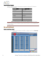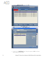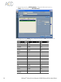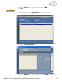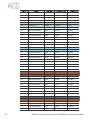
1
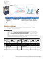
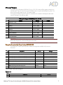
USER MANUAL
Fully Automated RNAscope® Assay
RNAscope® LS Reagent Kit
For use with Leica Biosystems’ BOND RX System
BROWN
Document Number 321038
For Molecular Biology Applications (MBA), not intended for diagnosis. Refer to appropriate regulations.
Rev. Date 20141112
For Molecular Biology Applications (MBA), not intended for diagnosis. Refer to appropriate regulations.
Trademarks
RNAscope® is a registered trademarks of Advanced Cell Diagnostics, Inc. Leica BOND RX is a registered trademark of Leica
Biosystems. All other trademarks belong to their respective owners.
Citing RNAscope® LS in Publications
When describing a procedure for publication using this product, please refer to it as the RNAscope® LS Assay and cite: Wang F,
Flanagan J, Su N, Wang L-C, Bui S, Nielson A, Wu X, Vo H-T, Ma X-J and Luo Y. RNAscope®: A Novel In Situ RNA Analysis
Platform for Formalin-Fixed Paraffin-Embedded Tissues. J. Mol. Diagnostics, 2012, 14:22–29.
Disclaimers
Advanced Cell Diagnostics, Inc. reserves the right to change its products and services at any time to incorporate technological
developments. This manual is subject to change without notice.
Although this manual has been prepared with every precaution to ensure accuracy, Advanced Cell Diagnostics, Inc. assumes no
liability for any errors, omissions, or for any damages resulting from the use of this information.
Copyright
© 2014. Advanced Cell Diagnostics, Inc. All rights reserved.
Contents
Chapter 1. Product Information ........................................................ 5
About this guide ............................................................................................ 5
Product description ........................................................................................ 5
Background............................................................................................... 5
Overview .................................................................................................. 5
Kit contents and storage ................................................................................. 6
RNAscope® LS Probes ................................................................................ 6
RNAscope® Reagents ................................................................................. 7
Required materials from Leica BOND RX ......................................................... 7
Equipment ................................................................................................. 7
User-supplied materials ................................................................................. 8
Chapter 2. Before You Begin ........................................................... 9
Important procedural guidelines ..................................................................... 9
Chapter 3. Prepare and Pretreat Samples ........................................ 11
Prepare FFPE sections .................................................................................. 11
Materials required ................................................................................... 11
Fix the sample ......................................................................................... 11
Dehydrate, embed, and cut the sample ..................................................... 11
Chapter 4. Fully Automated RNAscope® LS Assay ............................ 13
Workflow ................................................................................................... 13
Prepare the materials ................................................................................... 14
Materials required ................................................................................... 14
Prepare the reagents ................................................................................ 14
Prepare the instrument ............................................................................. 14
Register the reagents ................................................................................ 14
Prepare instrument reagents ..................................................................... 16
Set up the instrument ................................................................................... 16
Create a prestaining protocol ................................................................... 16
Create a staining protocol ........................................................................ 22
Register Probes ........................................................................................ 26
Set up a study ......................................................................................... 28
Run the RNAscope® Assay ........................................................................... 30
Materials required ................................................................................... 30
Load the reagents .................................................................................... 30
Start the run ............................................................................................ 31
Complete the run ..................................................................................... 31
RNAscope® LS Assay for Leica Biosystems’ BOND RX System User Manual-Brown
3
Dehydrate the slides................................................................................. 31
Mount the samples ................................................................................... 31
Chapter 5. Evaluate the results ....................................................... 33
Scoring guidelines ....................................................................................... 33
Quantitative Image Analysis ..................................................................... 33
Control example .......................................................................................... 34
Troubleshooting........................................................................................... 34
Appendix A. Safety ..................................................................... 35
Chemical safety ........................................................................................... 35
Biological hazard safety .............................................................................. 35
Documentation and support .......................................................... 37
Obtaining MSDSs........................................................................................ 37
Obtaining support ....................................................................................... 37
Contact information ..................................................................................... 37
Limited product warranty ............................................................................. 37
4
RNAscope® LS Assay for Leica Biosystems’ BOND RX System RX User Manual-Brown
1
Chapter 1. Product Information
Before using this product, read and understand the information in Appendix A. Safety on
page 35 in this document.
IMPORTANT! We recommend reading the entire user manual before beginning any protocols.
About this guide
This user manual provides guidelines and protocols to use the RNAscope® LS Reagent Kit for use with
Leica Biosystems’ BOND RX Research Advanced Staining System. RNAscope® LS Assays are
compatible with a variety of sample types.
Product description
Background
The RNAscope® LS Assays use a novel and proprietary method of in situ hybridization (ISH) to visualize
single RNA molecules per cell in formalin-fixed, paraffin-embedded (FFPE) tissue mounted on slides.
The assays are based on Advanced Cell Diagnostic’s patented signal amplification and background
suppression technology, and can detect RNA molecules in archival samples and partially degraded
specimens. The RNAscope® LS Assay allows users to automate the highly sensitive RNAscope® Assay
using Leica Biosystems’ BOND RX System.
Overview
The RNAscope® LS Assay procedure is illustrated in Figure 1 on page 6 and can be completed on the
instrument in ~9–10 hours. Starting with properly prepared samples, sections are first pretreated, and
then RNA-specific probes are hybridized to target RNA. The signal is amplified using multiple steps,
followed by hybridization to horseradish peroxidase (HRP)-labeled probes and detection using the 3,3'diaminobenzidine (DAB) chromogenic substrate. Each single RNA transcript appears as a distinct dot
of chromogen precipitate visible using a common bright-field microscope.
RNAscope® LS Assay for Leica Biosystems’ BOND RX System RX User Manual-Brown
5
Figure 1 Procedure overview
1: Tissue section
2: Hybridize to target RNA
3: Amplify signal
Start with properly
prepared sections and
load slides onto the
instrument. Pretreat tissue
to allow access to target
RNA.
Hybridize gene-specific probe
pairs to the target mRNA.
4: Image
Probes are hybridized to a cascade of
signal amplification molecules, culminating
in binding of HRP-labeled probes. Add DAB
substrate to detect target RNA.
Visualize target
RNA using a
standard bright
field microscope.
Kit contents and storage
The RNAscope® LS Assay requires the RNAscope® LS Probes and the RNAscope® LS Reagents,
available from Advanced Cell Diagnostics.
RNAscope® LS Probes
The RNAscope® LS Probes consist of the user-specified Target Probe and the Positive and Negative
Control Probes. Visit www.acdbio.com/products/target-probes/search-product to find a gene-specific Target
Probe. Visit www.acdbio.com/products/target-probes/controls-housekeeping to order appropriate Control
Probes.
Each probe is sufficient for staining ~30 standard slides. The probes have a shelf life of six months from
the shipment date when stored as indicated in the following table:
Target Probes
Reagent
Cat. No.
RNAscope LS — Target Probe –
[species] – [gene]
Content
Quantity
Storage
Various
Probe targeting specific RNA
11 mL x 1 bottle
4°C
Cat. No.
Content
Quantity
Storage
RNAscope LS — Positive Control Probe
– [species] – PPIB
Various
Probe targeting common
housekeeping gene
11 mL x 1 bottle
4°C
RNAscope® LS — Negative Control
Probe – DapB
312037
Probe targeting bacterial
gene dapB
11 mL x 1 bottle
4°C
®
Control Probes
Reagent
®
6
RNAscope® LS Assay for Leica Biosystems’ BOND RX System RX User Manual-Brown
RNAscope® Reagents
The RNAscope® LS Reagent Kit, BROWN (Cat. No. 321100) contains all the reagents needed to run
the RNAscope® Assay on Leica Biosystems’ BOND RX System, except for the RNA-specific probes. The
kits provide enough reagents to stain ~60 standard slides.
The reagents are Ready-To-Use (RTU) and have a shelf life of six months from the shipment date when
stored as indicated in the following table:
RNAscope® LS Reagent Kit–BROWN (Cat. No. 321100)
Reagent
Quantity
Storage
LS Pretreat1 – H202
10 mL x 1 bottle
4°C
LS Pretreat 3 – Protease
21 mL x 1 bottle
4°C
LS Amp 1
21 mL x 1 bottle
4°C
LS Amp 2
21 mL x 1 bottle
4°C
LS Amp 3
21 mL x 1 bottle
4°C
LS Amp 4
21 mL x 1 bottle
4°C
LS Amp 5 BROWN
21 mL x 1 bottle
4°C
LS Amp 6 BROWN
21 mL x 1 bottle
4°C
LS 10X Wash Buffer
5 mL x 1 bottle
4°C
IMPORTANT! Do not substitute the reagent components of the RNAscope® Reagent Kit with those of other
RNAscope® Reagent Kits, even those having the same name.
Required materials from Leica BOND RX
The RNAscope® LS Assay requires specific materials and equipment available only from Leica
Biosystems.
Component
Cat. No.
Storage
BOND Open Containers 30 mL
Op309700
Room temperature (20–25°C)
BOND Universal Covertiles 100 pack
S21.2001
Room temperature (20–25°C)
BOND Epitope Retrieval Solution 1-1L (RTU)
AR9961
2–8°C
BOND Epitope Retrieval Solution 2-1L (RTU)
AR9640
2–8°C
BOND Dewax Solution – 1L (RTU)
AR9222
2–8°C
AR9590
2–8°C
BOND Polymer Refine Detection (DAB) and Hematoxylin
DS9800
2–8°C
BOND Aspirating Probe Cleaning System
CS9100
2–8°C
BOND Mixing Stations
S21.1971
Room temperature (20–25°C)
BOND Wash Solution 10X Concentrate – 1L
*
* Do not substitute with any other chromogen kit.
Equipment
Component
Leica Biosystems’ BOND RX System — automated slide stainer
RNAscope® LS Assay for Leica Biosystems’ BOND RX System RX User Manual-Brown
Cat. No.
—
7
User-supplied materials
IMPORTANT! Do not substitute other materials for the SuperFrost® Plus Slides listed in the following table.
Description
Supplier
Cat. No.
SuperFrost Plus Slides (required)
Fisher Scientific
100% ethanol (EtOH)
American Master Tech Scientific/MLS
ALREAGAL
Xylene
Fisher Scientific/MLS
X3P-1GAL
10% neutral-buffered formalin (NBF)
MLS
—
Paraffin wax
MLS
—
1X PBS
MLS
—
Microtome
MLS
—
Drying oven, capable of holding temperature at 60
+/- 1°C (optional)
MLS
—
Water bath or incubator, capable of holding
temperature at 40 +/- 1°C
MLS
—
Cytoseal XYL xylene-based mounting medium
Richard-Allen Scientific/MLS
8312-4
Tissue-Tek Vertical 24 Slide Rack
American Master Tech Scientific/MLS
LWSRA24
Tissue-Tek Staining Dish (4 required)
American Master Tech Scientific/MLS
LWT4457EA
Tissue-Tek Clearing Agent Dish, xylene resistant (2
required)
American Master Tech Scientific/MLS
Cover Glass 24 x 50 mm
Fisher Scientific/MLS
12--545-F
Distilled water
MLS
—
Fume hood
MLS
—
®
*
®
®
®
12-550-15
LWT4456EA
* Major Laboratory Supplier in North America. For other regions, please check Catalog Numbers with your local lab supplier.
8
RNAscope® LS Assay for Leica Biosystems’ BOND RX System RX User Manual-Brown
2
Chapter 2. Before You Begin
Prior to running the RNAscope® LS Assay on your samples for the first time, we recommend that you:
•
Become familiar with Leica Biosystems’ BOND RX Research Advanced Staining System. Refer
to the Leica Biosystems’ BOND RX System Instructions For Use.
•
Run the assay on RNAscope® Control Slides (Cat. No. 310045 for Human Hela Cell Pellet,
and Cat. No. 310023 for Mouse 3T3 Cell Pellet) using the RNAscope® LS Positive and
Negative Control Probes.
Important procedural guidelines
•
Start with properly fixed and prepared sections. Refer to Chapter 3. Prepare and Pretreat Samples
on page 11 for preparation of FFPE slides. For preparation of other sample types, contact
[email protected].
•
Regularly maintain and clean your automated staining instrument.
•
Always run positive and negative control probes on your sample to assess sample RNA quality
and optimal permeabilization.
•
Do not substitute required materials. Assay has been validated with these materials only.
•
Follow the protocol exactly for best results.
•
Do not let your sections dry out during the procedure.
•
Use good laboratory practices and follow all necessary safety procedures. Refer to Appendix A.
Safety on page 35 for more information.
RNAscope® LS Assay for Leica Biosystems’ BOND RX System RX User Manual-Brown
9
10
RNAscope® LS Assay for Leica Biosystems’ BOND RX System RX User Manual-Brown
3
Chapter 3. Prepare and
Pretreat Samples
Formalin-fixed, paraffin-embedded (FFPE) sample preparation and pretreatment are described in the
following protocols.
IMPORTANT! We highly recommend following these guidelines. We cannot guarantee assay results with
other preparation methods.
Prepare FFPE sections
Materials required
• 10% neutral buffered formalin (NBF)
• 1X PBS
• Paraffin wax
• 100% EtOH
• Xylene
• Microtome
• Water bath
• SuperFrost® Plus slides
Fix the sample
1. Immediately following dissection, fix tissue in 10% NBF for 16–32 HRS at ROOM TEMPERATURE
(RT). Fixation time will vary depending on tissue type and size.
CAUTION! Handle biological specimens appropriately.
IMPORTANT! Fixation for <16 HRS or >32 HRS will impair the performance of the RNAscope® Assay.
Dehydrate, embed, and cut the sample
IMPORTANT! Use fresh reagents.
1. Wash sample with 1X PBS.
2. Dehydrate sample using a standard ethanol series, followed by xylene.
3. Embed sample in paraffin using standard procedures.
Note: Embedded samples may be stored at RT for years.
4. Trim paraffin blocks as needed, and cut embedded tissue into 5 +/- 1 µm sections using a
microtome.
RNAscope® LS Assay for Leica Biosystems’ BOND RX System RX User Manual-Brown
11
Name
Case ID
5. Place paraffin ribbon in a 40–45°C water bath, and mount sections on SUPERFROST® PLUS
SLIDES. Place tissue as shown for optimal staining:
Tissue section location
6. Air dry slides OVERNIGHT at RT.
OPTIONAL STOPPING POINT. Use sectioned tissue within 3 months. Store sections with
dessicants at RT.
12
RNAscope® LS Assay for Leica Biosystems’ BOND RX System RX User Manual-Brown
4
Chapter 4. Fully Automated
RNAscope® LS Assay
IMPORTANT! We strongly recommend you run the RNAscope® Control Slides (Cat. No. 310045 or Cat.
No. 310023) using the RNAscope® LS positive and negative control probes along with your samples in
every run.
Workflow
Prepare the materials
Set up the instrument
Run the RNAscope® Assay ~9 hours
RNAscope® LS Assay for Leica Biosystems’ BOND RX System RX User Manual-Brown
13
Prepare the materials
Materials can be prepared ahead of time, unless otherwise stated.
Materials required
Materials provided by Advanced Cell Diagnostics
• RNAscope® LS Target Probe
• RNAscope LS Positive Control Probe
®
• RNAscope LS Negative Control Probe
®
• LS Pretreat 1
Materials provided by Leica Biosystems
Stainer
• Leica Biosystems’ BOND RX System
Bulk Reagents
• BOND Wash Solution 10X
• LS Pretreat 3
• BOND Dewax Solution
• LS Amp 1
• BOND Epitope Retrieval Solution 1
• LS Amp 2
• BOND Epitope Retrieval Solution 2
Materials provided by user
• Distilled water
• Conical tube 50 mL
• Drying oven
Reagents
• LS Amp 3
• BOND PolymerRefine Detection (DAB)
plus Hematoxylin
• LS Amp 4
• LS Amp 5 BROWN
• LS Amp 6 BROWN
• LS 10X Wash Buffer
Prepare the reagents
•
Warm up LS Amp 1, LS Amp 3, LS 10X Wash Buffer, and all LS target probes in a 40°C oven
for 30 MIN before the run.
Note: Loss of signal will occur if precipitates do not dissolve.
•
Prepare two conical tubes of 30 mL of LS 1X Wash Buffer by adding 27 mL distilled water
and 3 mL of LS 10X Wash Buffer to each tube. Mix well by inverting the tubes slowly at least
five times. Do not shake the tube.
Prepare the instrument
•
Fill the large containers located in the bottom of the instrument with the Leica BOND RX bulk
reagents. Dilute BOND Wash Solution 1:10.
Note: Insufficient bulk reagent volumes may lead to run failure.
IMPORTANT! Do not introduce bubbles into the solutions by shaking the containers. To mix
reagents, gently invert the containers several times. If bubbles are present, leave the containers
out at room temperature until the bubbles dissipate.
•
Use clean, dry covertiles for every run. Clean used covertiles with water, bleach, and ethanol
Air dry before reuse.
•
Ensure waste bulk containers are emptied before starting a run. Discard waste according to all
local, state/provincial, and/or national regulations.
Register the reagents
1. Select the Reagent Setup icon at the top of the screen.
14
RNAscope® LS Assay for Leica Biosystems’ BOND RX System RX User Manual-Brown
2. Select Add to enter reagent information.
3. Enter ACD Amp 1 in the Name text box.
Enter ACD Amp 1 in the Abbreviated name text box.
Select Ancillary in the Type drop-down menu.
Enter ACD in the Supplier text box.
Select Save.
Repeat steps 2–7 for Amp 2 – Amp 6 Brown and 1X LS Wash Buffer.
Note: Do not add reagents for LS Pretreat 1 and LS Pretreat 3. They will be directly scanned
and registered as *Open 0 Haz and *Enzyme 1, respectively. See Prepare instrument reagents.
4.
5.
6.
7.
8.
RNAscope® LS Assay for Leica Biosystems’ BOND RX System RX User Manual-Brown
15
Prepare instrument reagents
Fill the Leica BOND RX containers with the appropriate reagents from the RNAscope® LS Reagent Kit
according to the following table:
Reagents
Container Name
LS Pretreat 1
*Open 0 Haz
LS Pretreat 3
*Enzyme 1
LS Amp 1
ACD Amp 1
LS Amp 2
ACD Amp 2
LS Amp 3
ACD Amp 3
LS Amp 4
ACD Amp 4
LS Amp 5
ACD Amp 5 Brown
LS Amp 6
ACD Amp 6 Brown
LS 1X Wash Buffer
ACD Wash Buffer
LS Target Probe
Variable
Note: Leica BOND DAB and Hematoxylin come in pre-filled Leica BOND RX containers.
IMPORTANT! Do not introduce bubbles into the solutions by shaking the containers. To mix reagents, gently
invert the containers several times. If bubbles are present, leave the containers out at room temperature until
the bubbles dissipate.
Set up the instrument
Create a prestaining protocol
1. Open the instrument software (version BDZ 6.0 or higher) and click on the Protocol setup
icon as shown.
16
RNAscope® LS Assay for Leica Biosystems’ BOND RX System RX User Manual-Brown
2. Select Prestaining under the Protocol group menu located in the bottom left corner of the
screen to access the Enzyme Pretreatment, Heat pretreatment, and ISH Hybridization
protocols.
Antigen Retrieval
1. Under the Protocol type menu select Heat pretreatment.
RNAscope® LS Assay for Leica Biosystems’ BOND RX System RX User Manual-Brown
17
2. Highlight the *HIER 10 min with ER2 protocol. Select Copy.
3. Rename the protocol (e.g. ACD 15 min ER2 @ 95°C). Change the Abbreviated name (e.g.
ER2-15) and the Description (e.g. ACD 15 min ER2 @ 95°C).
4. Highlight the third *Bond ER Solution 2 step. Change the temperature to 95°C and incubation
time according to the following table:
18
RNAscope® LS Assay for Leica Biosystems’ BOND RX System RX User Manual-Brown
Tissue Type
ER2 Incubation Time
Temperature
Brain and spinal cord
15 MIN
95°C
Breast cancer
15 MIN
95°C
Cell pellet
15 MIN
95°C
Colon
15 MIN
95°C
GI tract
15 MIN
95°C
Head and neck cancer
15 MIN
95°C
Heart
15 MIN
95°C
Kidney
15 MIN
95°C
Liver
20 MIN
95°C
Lung
15 MIN
95°C
Lymphoma
15 MIN
95°C
Placenta
15 MIN
95°C
Prostate
15 MIN
95°C
Skin
15 MIN
95°C
Stomach
15 MIN
95°C
Thymus
15 MIN
95°C
Tonsil
15 MIN
95°C
Xenograft
15 MIN
95°C
5. Select Save.
Protease and H202 Treatment
1. Under the Protocol type menu select Enzyme Pretreatment.
2. Highlight the *Protease 20 min and fix protocol. Select Copy.
RNAscope® LS Assay for Leica Biosystems’ BOND RX System RX User Manual-Brown
19
3. Rename the protocol (e.g. ACD 15min Protease). Change the Abbreviated name (e.g.
15mPro) and the Description (e.g. ACD 15min Protease).
4. Highlight the second *Enzyme 1 step. Change the temperature to 40°C and incubation time
according to the following table:
Tissue Type
20
Enzyme 1 Incubation Time
Temperature
Brain and spinal cord
15 MIN
40°C
Breast cancer
15 MIN
40°C
Cell pellet
15 MIN
40°C
Colon
15 MIN
40°C
GI tract
15 MIN
40°C
Head and neck cancer
15 MIN
40°C
Heart
15 MIN
40°C
Kidney
15 MIN
40°C
Liver
25 MIN
40°C
Lung
15 MIN
40°C
Lymphoma
15 MIN
40°C
Placenta
15 MIN
40°C
Prostate
15 MIN
40°C
Skin
15 MIN
40°C
Stomach
15 MIN
40°C
Thymus
15 MIN
40°C
Tonsil
15 MIN
40°C
Xenograft
15 MIN
40°C
RNAscope® LS Assay for Leica Biosystems’ BOND RX System RX User Manual-Brown
5. Highlight the *Open 0 Haz step. Change the incubation time to 10 MIN.
6. Select Save.
Probe Hybridization
1. In the Protocol setup screen select ISH Hybridization under the Protocol type menu.
2. Highlight the *ISH Hybridization (2Hr) protocol. Select Copy.
3. Change the Name to ACD 2 Hour Hybridization, the Abbreviated Name to Hyb-2hr, and the
Description to ACD 2 Hour Hybridization.
4. Highlight the *No Reagent step. Change the incubation time to 120 MIN and temperature to
RNAscope® LS Assay for Leica Biosystems’ BOND RX System RX User Manual-Brown
21
40°C.
5. Select Save.
Create a staining protocol
1. In the Protocol setup screen select Staining under the Protocol group menu.
2. Highlight the *ViewRNA 1- FFPE protocol. Select Copy.
3. Change the name to ACD ISH DAB Procol in the Name text box, ACD-DAB in the Abbreviated
name text box, and ACD ISH DAB Protocol in the Description text box.
4. Select Bond Polymer Refine Detection under the Preferred detection system menu.
22
RNAscope® LS Assay for Leica Biosystems’ BOND RX System RX User Manual-Brown
5. Highlight and select on each Reagent step to edit each step. Set up the protocol steps
(highlighted rows) according to the following table:
Step No.
Reagent
Step Type
Incubation Time
Temperature
1
*ACD Amp 1
Reagent
1 MIN
42°C
2
*ACD Amp 1
Reagent
30 MIN
42°C
3
*Bond Wash Solution
Wash
0 MIN
Ambient
4
*Bond Wash Solution
Wash
0 MIN
Ambient
5
*Bond Wash Solution
Wash
0 MIN
Ambient
6
*Bond Wash Solution
Wash
3 MIN
Ambient
7
*Bond Wash Solution
Wash
3 MIN
Ambient
8
*Bond Wash Solution
Wash
0 MIN
Ambient
9
*Bond Wash Solution
Wash
0 MIN
Ambient
10
*Bond Wash Solution
Wash
0 MIN
Ambient
11
*ACD 1X Wash Buffer
Reagent
5 MIN
Ambient
12
*ACD 1X Wash Buffer
Reagent
5 MIN
Ambient
13
*Bond Wash Solution
Wash
0 MIN
Ambient
14
*Bond Wash Solution
Wash
0 MIN
Ambient
15
*Bond Wash Solution
Wash
0 MIN
Ambient
16
*Bond Wash Solution
Wash
0 MIN
Ambient
17
*ACD Amp 2
Reagent
1 MIN
42°C
18
*ACD Amp 2
Reagent
15 MIN
42°C
19
*Bond Wash Solution
Wash
0 MIN
Ambient
20
*Bond Wash Solution
Wash
0 MIN
Ambient
21
*Bond Wash Solution
Wash
0 MIN
Ambient
22
*Bond Wash Solution
Wash
1 MIN
Ambient
RNAscope® LS Assay for Leica Biosystems’ BOND RX System RX User Manual-Brown
23
Step No.
24
Reagent
Step Type
Incubation Time
Temperature
23
*Bond Wash Solution
Wash
1 MIN
Ambient
24
*Bond Wash Solution
Wash
1 MIN
Ambient
25
*Bond Wash Solution
Wash
1 MIN
Ambient
26
*Bond Wash Solution
Wash
1 MIN
Ambient
27
*ACD Amp 3
Reagent
1 MIN
42°C
28
*ACD Amp 3
Reagent
30 MIN
42°C
29
*Bond Wash Solution
Wash
0 MIN
Ambient
30
*Bond Wash Solution
Wash
0 MIN
Ambient
31
*Bond Wash Solution
Wash
0 MIN
Ambient
32
*Bond Wash Solution
Wash
3 MIN
Ambient
33
*Bond Wash Solution
Wash
3 MIN
Ambient
34
*Bond Wash Solution
Wash
1 MIN
Ambient
35
*Bond Wash Solution
Wash
1 MIN
Ambient
36
*Bond Wash Solution
Wash
1 MIN
Ambient
37
*ACD Amp 4
Reagent
1 MIN
42°C
38
*ACD Amp 4
Reagent
15 MIN
42°C
39
*Bond Wash Solution
Wash
0 MIN
Ambient
40
*Bond Wash Solution
Wash
0 MIN
Ambient
41
*Bond Wash Solution
Wash
0 MIN
Ambient
42
*Bond Wash Solution
Wash
1 MIN
Ambient
43
*Bond Wash Solution
Wash
1 MIN
Ambient
44
*Bond Wash Solution
Wash
1 MIN
Ambient
45
*Bond Wash Solution
Wash
1 MIN
Ambient
46
*Bond Wash Solution
Wash
1 MIN
Ambient
47
*ACD Amp 5 Brown
Reagent
1 MIN
Ambient
48
*ACD Amp 5 Brown
Reagent
30 MIN
Ambient
49
*Bond Wash Solution
Wash
0 MIN
Ambient
50
*Bond Wash Solution
Wash
0 MIN
Ambient
51
*Bond Wash Solution
Wash
0 MIN
Ambient
52
*Bond Wash Solution
Wash
1 MIN
Ambient
53
*Bond Wash Solution
Wash
1 MIN
Ambient
54
*Bond Wash Solution
Wash
1 MIN
Ambient
55
*Bond Wash Solution
Wash
1 MIN
Ambient
56
*Bond Wash Solution
Wash
1 MIN
Ambient
57
*ACD Amp 6 Brown
Reagent
1 MIN
Ambient
58
*ACD Amp 6 Brown
Reagent
15 MIN
Ambient
59
*Bond Wash Solution
Wash
0 MIN
Ambient
60
*Bond Wash Solution
Wash
0 MIN
Ambient
61
*Bond Wash Solution
Wash
0 MIN
Ambient
62
*Bond Wash Solution
Wash
1 MIN
Ambient
RNAscope® LS Assay for Leica Biosystems’ BOND RX System RX User Manual-Brown
Step No.
Reagent
Step Type
Incubation Time
Temperature
63
*Bond Wash Solution
Wash
1 MIN
Ambient
64
*Bond Wash Solution
Wash
1 MIN
Ambient
65
*Bond Wash Solution
Wash
1 MIN
Ambient
66
*Bond Wash Solution
Wash
1 MIN
Ambient
67
*ACD 1X Wash Buffer
Reagent
5 MIN
Ambient
68
*ACD 1X Wash Buffer
Reagent
5 MIN
Ambient
69
*Mixed DAB Refine
Reagent
1 MIN
Ambient
70
*Mixed DAB Refine
Reagent
20 MIN
Ambient
71
*De-ionized Water
Wash
0 MIN
Ambient
72
*De-ionized Water
Wash
0 MIN
Ambient
73
*De-ionized Water
Wash
0 MIN
Ambient
74
*De-ionized Water
Wash
0 MIN
Ambient
75
*De-ionized Water
Wash
0 MIN
Ambient
76
*De-ionized Water
Wash
0 MIN
Ambient
77
*Hematoxylin
Reagent
5 MIN
Ambient
78
*De-ionized Water
Wash
0 MIN
Ambient
79
*De-ionized Water
Wash
0 MIN
Ambient
80
*De-ionized Water
Wash
0 MIN
Ambient
81
*De-ionized Water
Wash
0 MIN
Ambient
Note: The temperature for these steps cannot be changed. You may only change the
incubation times.
RNAscope® LS Assay for Leica Biosystems’ BOND RX System RX User Manual-Brown
25
6. Click Show wash steps to view the washing steps in between each reagent. Insert BOND
Washes to match the protocol steps shown in the table above.
7. Make sure that Preferred is selected at the bottom right corner of the window.
8. Select Save.
Register Probes
1. Click the Reagent setup icon to register each probe.
2. Select Add.
26
RNAscope® LS Assay for Leica Biosystems’ BOND RX System RX User Manual-Brown
3. Enter the name of the probe in the Name and Abbreviated name text boxes.
4. Select Probe in the Type drop-down menu. Enter ACD in the Supplier text box.
5. Check RNA for Probe Type.
6. Select ACD DAB as the Default staining protocol.
7. Select ACD 15min ER2 @ 95 C as the Default HIER protocol.
8. Select ACD 15min Protease as the Default enzyme protocol.
9. Leave the Default denaturation protocol blank.
10. Select ACD 2 Hour Hybridization as the Default hybridization protocol.
11. Select Save.
12. Repeat steps 2 –11 for each additional probe.
RNAscope® LS Assay for Leica Biosystems’ BOND RX System RX User Manual-Brown
27
Set up a study
1. To build a study, select the Slide setup icon at the top of the screen.
2. Select Add study and enter a name in the Study ID field.
3. Select 150 µl for Dispense volume.
4. Select *Bake and Dewax for Preparation protocol.
5. Select OK.
6. Select Add slide to assign a protocol to each slide.
28
RNAscope® LS Assay for Leica Biosystems’ BOND RX System RX User Manual-Brown
7. Enter the name of the tissue under the Comments field. Then select ISH. Select the marker
(Target Probe).
8. Under the Preparation drop-down menu, select the protocol *Bake and Dewax.
9. Select Add slide for each target probe and the slides used for the run. After adding all the
slides to the study, select Close to return to the Slide setup screen.
RNAscope® LS Assay for Leica Biosystems’ BOND RX System RX User Manual-Brown
29
10. Select Print labels to print barcodes that will be attached to the slides.
Run the RNAscope® Assay
Materials required
• Distilled Water
• Fume hood
• Xylene
• 100% EtOH
• Tissue-Tek® Staining Dish (4)
• Tissue-Tek® Clearing Agent Dish, xylene-resistant (2)
• Tissue-Tek® Vertical 24 Slide Rack
• Cytoseal
• Cover Glass, 24 mm x 50 mm
Load the reagents
1. Label each Leica BOND RX container with the corresponding reagent using a
water-resistant marker.
2. Use the barcode scanner to scan the barcode located on the front of each container
(including Leica Bond DAB and Hematoxylin containers). A pop-up menu will appear.
IMPORTANT! Do NOT scan the barcode located on the top of each container.
3. Choose the appropriate Reagent Name, and enter the lot number and expiration date in
their respective text boxes. Select OK.
4. Load the containers onto the reagent tray and slide the tray into the Leica BOND RX
module.
30
RNAscope® LS Assay for Leica Biosystems’ BOND RX System RX User Manual-Brown
Start the run
1. Once the barcodes have been attached to the slides, add the slides to the slide tray with
the label sides facing up.
Note: Each tray can accommodate only one study. If a different protocol is used, it must be
placed in a separate tray. Only three different parameters may be used for a complete run for a
total of 30 slides.
2. Add a covertile on top of each slide. The rectangular-shaped neck of the covertile should fit
into the groove of the slide tray.
3. Place the tray in the Leica Bond RX™ and press the button to load the tray onto the
machine.
4. Once the slides have been scanned, select the triangular (PLAY) button on the screen
located under the start tray to start the run. Alternatively, right-click on scanned label images
and select Delayed Start to start the run at a future time.
IMPORTANT! Before leaving the instrument unattended, ensure that the instrument is running successfully.
Note: The following sections may also be performed using an automated coverslipper.
Complete the run
1. After the run is complete press the button on the instrument to unload the slides.
2. Place the slides onto the Tissue-Tek® Slide Rack and move the rack into a staining dish
containing distilled water.
3. Wash the slides by lifting the slide rack up and down several times.
Dehydrate the slides
1. Move the Tissue-Tek® Slide Rack into the a staining dish containing 100% EtOH in the fume
hood for 2 MIN. Agitate the slides by occasionally lifting the slide rack up and down.
2. Move the slide rack into a second staining dish containing 100% EtOH for 2 MIN with
occasional agitation.
3. Move the slide rack into the third staining dish containing 100% EtOH for 2 MIN with
occasional agitation.
4. Move the slide rack into a clearing agent dish containing xylene for 1 MIN with occasional
agitation.
5. Move the Tissue-Tek® Slide rack into a second clearing agent dish containing xylene for
1 MIN with occasional agitation.
Mount the samples
1. Remove the slides from the Tissue-Tek® Slide Rack and lay flat with the sections facing up in
the fume hood.
2. Mount one slide at a time by adding 1–2 DROPS of Cytoseal or other xylene-based
mounting medium to each slide and carefully placing a 24 mm x 50 mm coverslip over the
section. Avoid trapping air bubbles.
3. Air dry slides for 5 MIN.
4. Proceed to Chapter 5. Evaluate the results on page 33.
RNAscope® LS Assay for Leica Biosystems’ BOND RX System RX User Manual-Brown
31
32
RNAscope® LS Assay for Leica Biosystems’ BOND RX System RX User Manual-Brown
5
Chapter 5. Evaluate the results
Examine tissue sections under a standard bright field microscope at 20–40X magnification:
•
Assess tissue and cell morphology.
•
Assess positive control signal strength. Positive control signal should be visible as punctate dots
within cell nuclei at 20–40X magnification.
•
Assess negative control background. One dot to every 10 cells displaying background DAB
staining per 20X microscope field is acceptable.
•
Evaluate target probe signal using the scoring guidelines in the next section.
Scoring guidelines
The RNAscope® Assay enables a semi-quantitative scoring guideline utilizing the estimated number of
punctate dots present within each cell boundary.
An example of how to develop such a guideline for semi-quantitative assessment of RNAscope®
staining intensity is presented below for a gene with expression level varying between 1 to > 10
copies per cell.
Note: If your gene expression level is higher or lower than this range, you may need to scale the criteria
accordingly.
Categorize staining into five grades: 0, 1+, 2+, 3+ and 4+ according to the following table:
Staining
score
Microscope objective scoring*
0
No staining or less than 1 dot/cell (40X magnification)
1
1–3 dots/cell (visible at 20–40X magnification)
2
4–10 dots/cell. No or very few dot clusters (visible at 20–40X magnification)
3
>10 dots/cell. Less than 10% positive cells have dot clusters (visible at 20X magnification)
4
>10 dots/cell. More than 10% positive cells have dot clusters (visible at 20X magnification)
* Discount cells with artificially high nuclear background staining.
Quantitative Image Analysis
RNAscope® Spot Studio Software is designed for pathologists with no prior training in image analysis.
This intuitive software allows users to get statistical results with complete information of cell-count/region
and number of spots/cell. Simply load any image, select a region of interest, define settings and run
analysis, followed by a quality control review before results are exported.
RNAscope® LS Assay for Leica Biosystems’ BOND RX System RX User Manual-Brown
33
Control example
If the assay is successful, the staining should look like the following images:
Figure 2 RNAscope® Assay detection of PPIB mRNA in mouse brain FFPE tissue.
Troubleshooting
If you obtain less than satisfactory results, troubleshoot your assay by following these simple guidelines:
•
If you observe the presence of background staining, increase the Epitope Retrieval 2 (ER2) in
increments of 5 minutes and increase the Enzyme (Protease) time in increments of 10 minutes.
Keep the temperatures for each step constant (e.g. 20 min ER2 at 95°C and 25 min Protease
at 40°C; 25 min ER2 at 95°C and 35 min Protease at 40°C).
•
Use the above process for over-fixed tissues.
•
LS Amp 1, LS Amp 3, 10X LS Wash Buffer, and all target probes require warming up prior to
running the assay to remove crystals that form during refrigeration. Incubate the reagents in an
oven or water bath at 40°C for 30 minutes. Failure to warm the reagents properly will lead to
weak or intermittent staining.
•
The RNAscope® LS Brown and LS Red assays utilize Leica Biosystems’ BOND Polymer Refine
Detection and Bond Polymer Refine Red Detection kits, respectively. Do not use any other
chromogen kits.
•
Do not shake the contents in the dispensers as this will form bubbles and may lead to weak or
no staining. To mix reagents, gently invert the dispensers several times. If bubbles are present,
leave the containers out at room temperature until the bubbles dissipate.
•
Do not alter the staining protocol in any way except the hematoxylin incubation time. The
parameters in the staining protocol have been optimized to run the RNAscope® assay on the
instrument.
For troubleshooting information, please contact technical support at [email protected].
34
RNAscope® LS Assay for Leica Biosystems’ BOND RX System RX User Manual-Brown
A
Appendix A. Safety
Chemical safety
WARNING! GENERAL CHEMICAL HANDLING. To minimize hazards, ensure
laboratory personnel read and practice the general safety guidelines for chemical
usage, storage, and waste provided below, and consult the relevant SDS for specific
precautions and instructions:
•
Read and understand the Material Safety Data Sheets (MSDSs) provided by the chemical
manufacturer before you store, handle, or work with any chemicals or hazardous materials. To
obtain MSDSs, see Documentation and support in this document.
•
Minimize contact with chemicals. Wear appropriate personal protective equipment when
handling chemicals (for example, safety glasses, gloves, or protective clothing).
•
Minimize the inhalation of chemicals. Do not leave chemical containers open. Use only with
adequate ventilation (for example, fume hood).
•
Characterize (by analysis if necessary) the waste generated by the particular applications,
reagents, and substrates used in your laboratory.
•
Ensure that the waste is stored, transferred, transported, and disposed of according to all
local, state/provincial, and/or national regulations.
•
IMPORTANT! Radioactive or biohazardous materials may require special handling, and
disposal limitations may apply.
Biological hazard safety
WARNING! BIOHAZARD. Biological samples such as tissues, body fluids, infectious
agents, and blood of humans and other animals have the potential to transmit
infectious diseases. Follow all applicable local, state/provincial, and/or national
regulations. Wear appropriate protective equipment, which includes but is not limited
to: protective eyewear, face shield, clothing/lab coat, and gloves. All work should be
conducted in properly equipped facilities using the appropriate safety equipment (for
example, physical containment devices). Individuals should be trained according to
applicable regulatory and company/institution requirements before working with
potentially infectious materials. Read and follow the applicable guidelines and/or
regulatory requirements in the following:
In the U.S.:
•
U.S. Department of Health and Human Services guidelines published in Biosafety in
Microbiological and Biomedical Laboratories found at: www.cdc.gov/biosafety
•
Occupational Safety and Health Standards, Bloodborne Pathogens (29 CFR§1910.1030),
found at: www.access.gpo.gov/nara/cfr/waisidx_01/%2029cfr1910a_01.html
•
Your company’s/institution’s Biosafety Program protocols for working with/handling
potentially infectious materials.
RNAscope® LS Assay for Leica Biosystems’ BOND RX System RX User Manual-Brown
35
•
Additional information about biohazard guidelines is available at: www.cdc.gov
•
Check local guidelines and legislation on biohazard and biosafety precaution and refer to the
best practices published in the World Health Organization (WHO) Laboratory Biosafety
Manual, third edition, found at:
In the EU:
www.who.int/csr/resources/publications/biosafety/who_cds_csr_lyo_2004_11/en/
•
Information about the Registration, Evaluation, Authorisation and Restriction of Chemicals
(REACH) can be found at:
eur-lex.europa.eu/LexUriServ/LexUriServ.do?uri=OJ:L:2010:133:0001:0043:EN:PDF
36
RNAscope® LS Assay for Leica Biosystems’ BOND RX System RX User Manual-Brown
Documentation and support
Obtaining MSDSs
Material Safety Data Sheets (MSDSs) are available at: www.acdbio.com/product_literature.html. For the
MSDSs of chemicals not distributed by Advanced Cell Diagnostics, contact the chemical manufacturer.
Obtaining support
For the latest services and support information, go to: www.acdbio.com/product_literature.html
At the website, you can:
•
Access telephone and fax numbers to contact Technical Support and Sales facilities.
•
Search through frequently asked questions (FAQs).
•
Submit a question directly to Technical Support.
•
Search for user documents, MSDSs, application notes, citations, training videos, and other
product support documents.
•
Find out information about customer training events.
Contact information
Advanced Cell Diagnostics, Inc.
3960 Point Eden Way
Hayward, CA 94545
Toll Free: 1-877-576-3636
Direct: 1-510-576-8800
Fax: 1-510-576-8801
Information: [email protected]
Orders: [email protected]
Support Email: [email protected]
Limited product warranty
Advanced Cell Diagnostics, Inc. and/or its affiliate(s) warrant their products as set forth in the ACD
General Terms and Conditions of Sale found on the ADC website at
www.acdbio.com/product_literature.html. If you have any questions, please contact Advanced Cell
Diagnostics at www.acdbio.com/about/contact.
RNAscope® LS Assay for Leica Biosystems’ BOND RX System RX User Manual-Brown
37
Headquarters
3960 Point Eden Way
Hayward, CA 94545
For support, email [email protected].
www.acdbio.com
Phone 1-510-576-8800
Toll Free 1-877-576-3636