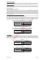
1
COBAS® AmpliScreen HBV Test
FOR IN VITRO DIAGNOSTIC USE.
COBAS® AmpliScreen HBV Test
COBAS® AmpliScreen Multiprep Specimen Preparation and Control Kit
COBAS®
HBV
96 Tests
P/N: 03599779 190
MULTIPREP/CTL
96 Tests
P/N: 03302555 018
500 Tests
P/N: 20759899 123
ART: 07 5989 9
US: 83314
AMPLICOR Wash Buffer
WB
O
N
LY
INTENDED USE
The COBAS® AmpliScreen HBV Test is a qualitative in vitro test for the direct detection of Hepatitis B Virus (HBV) DNA in human plasma.
The COBAS® AmpliScreen HBV Test is intended to be used to screen donors for HBV DNA in addition to the currently recommended serology tests.
This product is intended for use as a donor screening test to detect HBV in plasma samples from individual human donors, including donors of whole
blood and blood components, source plasma and other living donors. It is also intended for use to screen organ donors when specimens are obtained
while the donor's heart is still beating, and to detect HBV DNA in blood specimens from cadaveric (non-heart-beating) organ and tissue donors. This
test is not intended for use on samples of cord blood.
Plasma from all donors may be screened as individual samples. For donations of whole blood and blood components, plasma may be tested in pools
comprised of equal aliquots of not more than 24 individual donations in conjunction with licensed tests for detecting hepatitis B surface antigen
(HBsAg) and antibody to hepatitis B core antigen (anti-HBc). For donations of hematopoietic stem/progenitor cells (HPCs) sourced from bone marrow,
peripheral blood or cord blood, and donor lymphocytes for infusion (DLI), plasma may be tested in pools comprised of equal aliquots of not more
than 24 individual donor specimens. For donations of source plasma, plasma samples of the donations may be tested in pools comprised of equal
aliquots of not more than 96 individual donations.
The COBAS® AmpliScreen HBV Test can be considered a supplemental test that confirms HBV infection for specimens that are repeatedly reactive
on a licensed donor screening test for hepatitis B surface antigen, and reactive on the COBAS® AmpliScreen HBV Test.
This assay is not intended for use as an aid in diagnosis.
R
EF
ER
EN
C
E
U
SE
SUMMARY AND EXPLANATION OF THE TEST
Hepatitis B Virus is considered to be one of the major etiologic agents that cause chronic and acute hepatitis, cirrhosis and hepatocellular carcinoma.1-4 HBV is one of the most infectious disease causing agents, with about 350 million chronic hepatitis B carriers worldwide.9 The prevalence
of HBV infection in the US is approximately 200-300 thousand and in Europe it is over 1 million.10, 11 In the US, there has been a steady decline in
hepatitis B rates with the implementation of a national vaccination strategy.27 The global prevalence of chronic HBV infection, as determined by
immunoserology, ranges from < 2% in western countries to ≥ 8% in Asian and African countries.12
HBV is a partially double-stranded circular DNA virus with a genome of approximately 3,200 bases that contains four overlapping open reading frames
encoding for all viral proteins.1,5,6 The current risk of transfusion transmitted HBV is higher than that of HCV and HIV.7,8
The presence of HBV antigens or antibodies in patients infected with HBV has led to the development of immunoserological tests that are specific for these antigens or antibodies. Implementation of these tests has reduced, but not completely eliminated, the incidence of post-transfusion hepatitis B.7,8 Currently, all blood units are screened for HBsAg and anti-HBc. However, it has been reported that some blood units from
HBsAg and anti-HBc negative donors were associated with post-transfusion hepatitis B in the recipients.13,14 HBV DNA was prospectively
detected in HBsAg and anti-HBc negative blood units by Polymerase Chain Reaction (PCR).13 Screening of blood donations for HBV DNA will
further reduce the residual transmission risk. PCR tests can also detect viremic units donated by carriers who are in the window period, early
acute infection, or late resolving infection that may not be detectable by the existing immunological assays.13,15-17 A number of proposals have
been made for performing nucleic acid tests on mini-pools composed of small aliquots from many individual units.18-20
The COBAS® AmpliScreen HBV Test uses a generic DNA specimen preparation technique in a mini-pool testing format. HBV DNA in blood donations is detected by automated amplification by PCR on the COBAS® AMPLICOR Analyzer. The assay incorporates an Internal Control for monitoring
assay performance in each individual test as well as the AmpErase (uracil-N-glycosylase) enzyme to reduce potential contamination by previously
amplified material (amplicon).
PRINCIPLES OF THE PROCEDURE
The COBAS® AmpliScreen HBV Test is based on four major processes:
Sample Processing
2.
PCR amplification of target DNA using HBV-specific complementary primers
3.
Hybridization of the amplified products to oligonucleotide probes specific to the target(s)
4.
Detection of the probe-bound amplified products by colorimetric determination
FO
R
1.
Specimen Processing
Two specimen processing procedures are used with the COBAS® AmpliScreen HBV Test as follows:
•
Multiprep Specimen Processing Procedure for preparation of mini-pool specimens and individual cadaveric specimens
•
Standard Specimen Processing Procedure for preparation of individual specimens
NOTE: For testing of cadaveric specimens, the specimen should be first diluted 1:5 in Multiprep Specimen Diluent (MP DIL) prior to
processing using the Multiprep Specimen Processing Procedure.
NOTE: In order to detect 2 International Units (IU/mL) (2IU≅ 10 copies), triplicate testing using the Multiprep Specimen Processing Procedure should
be performed (i.e., three amplification/detections are performed requiring 50 µL each of the 200 µL of eluate, following a single extraction).
A positive result in one or more of the replicates indicates that the specimen is positive, and contains at least 2 IU/mL of HBV DNA.
In the Standard Specimen Processing Procedure, HBV DNA is isolated directly from plasma by lysis of the virus particles with Multiprep Lysis Reagent
followed by precipitation of the DNA with alcohol. In the Multiprep Specimen Processing Procedure, HBV viral particles are first pelleted from the
plasma sample by high speed centrifugation, followed by lysis of the pelleted virus with a chaotropic agent (Multiprep Lysis Reagent) and precipitation of the DNA with alcohol.
The Multiprep Internal Control (MP IC), containing the HBV Internal Control, is introduced into each specimen with the Multiprep Lysis Reagent and
serves as an extraction and amplification control for each processed specimen and control. The HBV Internal Control is a DNA plasmid with primer
binding regions identical to those of the HBV target sequence, a randomized internal sequence of similar length and base composition as the HBV
target sequence, and a unique probe binding region that differentiates the HBV Internal Control amplicon from target amplicon. These features were
selected to ensure equivalent amplification of the HBV Internal Control and the HBV target DNA.
PCR Amplification
The amplification reactions are performed with the thermostable recombinant enzyme Thermus aquaticus DNA Polymerase (Taq pol). The reaction
mixture is heated to separate double-stranded DNA. As the mixture cools, primers anneal to the target DNA and in the presence of Mg2+ and excess
deoxynucleotide triphosphates (dNTPs), the Taq pol extends the annealed primers along the target templates to produce a double-stranded DNA
molecule termed an amplicon. The COBAS® AMPLICOR Analyzer automatically repeats this process for a designated number of cycles, each cycle
effectively doubling the amount of amplicon DNA. The required number of cycles is preprogrammed in the COBAS® AMPLICOR Analyzer.
Selective Amplification
To ensure selective amplification of nucleic acid target in the sample and prevent amplification of pre-existing amplicon, the AmpErase (uracil-N-glycosylase) enzyme is added to the COBAS® AmpliScreen HBV Test. The AmpErase enzyme recognizes and catalyzes the destruction of DNA strands
The Document Revision Information section is located at the end of this document.
03599833001-07EN
1
Doc Rev. 7.0
containing deoxyuridine23, but not DNA containing deoxythymidine. Deoxyuridine is not present in naturally occurring DNA, but is always present in
amplicon because of the use of deoxyuridine triphosphate in place of thymidine triphosphate as one of the dNTPs in the Master Mix reagent; therefore, only amplicon contain deoxyuridine. Deoxyuridine renders contaminating amplicon susceptible to destruction by AmpErase enzyme before amplification of the target DNA. AmpErase enzyme, which is included in the Master Mix reagent, catalyzes the cleavage of deoxyuridine-containing DNA
rendering the DNA non-amplifiable. The AmpErase enzyme is inactive at temperatures above 55°C, i.e., throughout the thermal cycling steps, and
therefore does not destroy target amplicon. Following amplification, any residual enzyme is denatured by the addition of the Denaturation Solution,
thereby preventing the degradation of any target amplicon.
Hybridization Reaction
Following PCR amplification, the COBAS® AMPLICOR Analyzer automatically adds Denaturation Solution to the A-tubes to chemically denature the
HBV amplicon and the HBV Multiprep Internal Control amplicon to form single-stranded DNA. Aliquots of denatured amplicon are then transferred to
two detection cups (D-cups). A suspension of magnetic particles coated with an oligonucleotide probe specific for HBV amplicon (HBV DET) or HBV
Internal Control amplicon is added to the individual D-cups. The biotin-labeled HBV and HBV Internal Control amplicon are hybridized to the targetspecific oligonucleotide probes bound to the magnetic particles. This hybridization of amplicon to the target-specific probe increases the overall specificity of the COBAS® AmpliScreen HBV Test.
Detection Reaction
Following the hybridization reaction, the COBAS® AMPLICOR Analyzer washes the magnetic particles in the D-cups to remove unbound material and
then adds avidin-horseradish peroxidase conjugate. The avidin-horseradish peroxidase conjugate binds to the biotin-labeled amplicon. The COBAS®
AMPLICOR Analyzer removes unbound conjugate by washing the magnetic particles and then adds a substrate solution containing hydrogen peroxide
and 3,3',5,5'-tetramethylbenzidine (TMB) to each D-cup. In the presence of hydrogen peroxide, the particle-bound horseradish peroxidase catalyzes the
oxidation of TMB to form a colored complex. The absorbance is measured by the COBAS® AMPLICOR Analyzer at a wavelength of 660 nm.
MATERIALS PROVIDED BY ROCHE
The COBAS® AmpliScreen Multiprep Specimen Preparation and Control Kit and the COBAS® AMPLICOR Wash Buffer are provided as stand alone
kits to be used in conjunction with the COBAS® AmpliScreen HBV Test, as well as the COBAS® AmpliScreen HIV-1 Test, v1.5, and the COBAS®
AmpliScreen HCV Test.
COBAS® AmpliScreen Multiprep Specimen Preparation and Control Kit
(P/N: 03302555 018)
96 Tests
MULTIPREP/CTL
MP (–) C
[Multiprep Negative (–) Control]
N
LY
MP (+) C
[Multiprep Positive (+) Control]
MP LYS
(Multiprep Lysis Reagent)
O
MP DIL
(Multiprep Specimen Diluent)
SE
MP IC
(Multiprep Internal Control)
NHP
(Negative Plasma [Human])
96 Tests
U
COBAS® AmpliScreen HBV Test
(P/N: 03599779 190)
HBV
COBAS® AmpliScreen HBV Amplification Reagents
C
E
HBV AMP
HBV Mg2+
(HBV Magnesium Solution)
ER
EN
HBV MMX
(HBV Master Mix)
COBAS® AmpliScreen HBV Detection Reagents
BH PS1
(HBV Probe Suspension 1)
BH4
(HBV Probe Suspension 2)
R
EF
BI PS1
(HBV IC Probe Suspension 1)
HBV DK
BI4
(HBV IC Probe Suspension 2)
DN4
(Denaturation Solution)
FO
R
CN4
(Avidin-Horseradish Peroxidase Conjugate)
SB3
(Substrate A)
SB
(Substrate B)
COBAS® AMPLICOR Wash Buffer
(P/N: 20759899 123; ART: 07 5989 9; US: 83314)
500 Tests
WB
WB
(10X-Wash Concentrate)
OTHER MATERIALS REQUIRED BUT SOLD SEPARATELY
(MAY BE PURCHASED FROM ROCHE)
•
COBAS® AMPLICOR Analyzer with software version 0022B, Printer, and Operator’s Manual for the COBAS® AMPLICOR Analyzer
•
COBAS® AMPLICOR A-rings
•
COBAS® AMPLICOR D-cups
•
AMPLILINK Software, version 1.4 and Operator’s Manual for the AMPLILINK Software
•
Hamilton MICROLAB® AT plus 2 Pipettor (with Hamilton SUNPLUS and RUNENDE Software, and the Roche Pooling Methods Software
version 1.3), the COBAS® AmpliScreen Pooling System Guide (Roche Pooling Methods Software, version 1.3 and the COBAS® AmpliScreen
Pooling System Guide are validated to prepare pools of equal aliquots of not more than 24 individual plasma donations using the Hamilton
MICROLAB® AT plus 2 Pipettor with Hamilton SUNPLUS and RUNENDE Software).
•
Additional MP DIL from the COBAS® AmpliScreen Multiprep Specimen Preparation and Control Kit is required for testing of cadaveric
specimens.
NOTE: The user must validate all pooling algorithms and equipment other than those supplied by Roche.
•
Sarstedt 1.5-mL tube Barcode Labels
•
Hamilton Archive and Intermediate Plate Barcode Labels
•
Refrigerated high speed centrifuge with fixed angle rotor (45 degrees, capacity for at least 24 x 1.5-mL tubes) with an RCF of 23,600 x g
(Heraeus Centrifuge 17RS or Biofuge 28RS with HFA 22.1 rotor, Heraeus Biofuge Stratos with the 3331 rotor or equivalent)
03599833001-07EN
Doc Rev. 7.0
2
MATERIALS REQUIRED BUT NOT PROVIDED BY ROCHE
Microcentrifuge, (max. RCF 16,000 x g; min. RCF 12,500 x g) (Eppendorf® 5415C, HERMLE Z230M, or equivalent)
Eppendorf® 1.25-mL Combitips® Reservoir (sterile) or equivalent
Eppendorf Multipette® pipette or equivalent
Ethanol, 90% or 95%, reagent grade for Molecular Biology or Histology use
Distilled or deionized water
Powderless disposable gloves
Isopropyl alcohol, reagent grade
Disposable, Sterile, Polystyrene Pipettes (5 mL, 10 mL and 25 mL)
Sterile, RNase-free, fine-tip transfer pipettes
Pipettors (capacity 20 µL to 1000 µL capable of providing ± 3% accuracy and precision ≤ 5%) with aerosol barrier or positive displacement RNase-free tips
Tube racks (Sarstedt P/N 93.1428 or equivalent)
1.5-mL sterile, non-siliconized, conical polypropylene screw-cap tubes, (Sarstedt 72.692.105 or equivalent)
Vortex mixer
Hamilton Slotted Deepwell Archive Plate, 2.2 mL and Sealing Capmat
Hamilton Slotted Intermediate Plate
•
•
•
•
•
•
•
•
•
•
•
•
•
•
•
REAGENTS
COBAS® AmpliScreen Multiprep Specimen Preparation and Control Kit
(P/N: 03302555 018)
MP (–) C
(Multiprep Negative (–) Control)
96 Tests
MULTIPREP/CTL
8 x 0.1 mL
< 0.005% Poly rA RNA (synthetic)
EDTA
0.05% Sodium azide
8 x 0.1 mL
N
LY
MP (+) C
(Multiprep Positive (+) Control)
SE
O
Tris-HCl buffer
< 0.001% Non-infectious linearized plasmid DNA (microbial) containing HBV sequences
< 0.001% Non-infectious in vitro transcribed RNA (microbial) containing HCV sequences
< 0.001% Non-infectious in vitro transcribed RNA (microbial) containing HIV-1 sequences
< 0.005% Poly rA RNA (synthetic)
EDTA
0.05% Sodium azide
MP LYS
(Multiprep Lysis Reagent)
Harmful
MP DIL
(Multiprep Specimen Diluent)
C
E
8 x 4.8 mL
R
EF
Tris-HCl buffer
< 0.005% Poly rA RNA (synthetic)
EDTA
0.05% Sodium azide
ER
EN
60% (w/w) Guanidine thiocyanate
+
Xn
U
Tris-HCl buffer
60% Guanidine thiocyanate
3% Dithiothreitol
< 1% Glycogen
8 x 9.0 mL
8 x 0.1 mL
FO
R
MP IC
(Multiprep Internal Control)
Tris-HCl buffer
< 0.001% Non-infectious plasmid DNA containing HBV primer binding sequences
and a unique probe binding region
< 0.001% Non-infectious in vitro transcribed RNA (microbial) containing HCV primer
binding sequences and a unique probe binding region
< 0.001% Non-infectious in vitro transcribed RNA (microbial) containing HIV-1 primer
binding sequences and a unique probe binding region
< 0.005% Poly rA RNA (synthetic)
EDTA
<0.1% Amaranth dye
0.05% Sodium azide
NHP
(Negative Plasma [Human])
16 x 1.6 mL
Human plasma, non-reactive by US FDA licensed tests for antibody to HIV-1/2,
antibody to HCV, HBsAg, HCV RNA, HIV-1 RNA and HBV DNA.
0.1% ProClin® 300 preservative
COBAS® AmpliScreen HBV Test
(P/N: 03599779 190)
COBAS®
96 Tests
HBV
AmpliScreen HBV Amplification Reagents
HBV MMX
HBV AMP
(HBV Master Mix)
Tris-HCl buffer
Glycerol
< 0.001% AmpliTaq® DNA Polymerase (microbial)
Ammonium sulfate
0.05% dATP, dCTP, dGTP, dUTP
< 0.001% HBV-104UB and HBV-104D, HW016TBB1 primers (HBV – 104UB is biotinylated)
< 0.001% AmpErase (uracil-N glycosylase) enzyme (microbial)
< 0.5% Tween 20 surfactant
0.09% Sodium azide
HBV Mg2+
(HBV Magnesium Solution)
< 1% Magnesium chloride
Amaranth dye
0.05% Sodium azide
03599833001-07EN
8 x 0.7 mL
8 x 0.1 mL
Doc Rev. 7.0
3
COBAS® AmpliScreen HBV Detection Reagents
BH PS1
(HBV Probe Suspension 1)
MES buffer
< 0.4% Suspension of Dynabeads® (paramagnetic particles)
coated with HBV-specific oligonucleotide (HBV DET) capture probe
0.09% Sodium azide
BH4
(HBV Probe Suspension 2)
96 Tests
1 x 100 Tests
HBV DK
1 x 100 Tests
Sodium phosphate buffer
24.9% Sodium thiocyanate
0.2% Solubilizer
24.9% (w/w) Sodium thiocyanate
+
Xn
Harmful
BI PS1
(HBV IC Probe Suspension 1)
1 x 100 Tests
MES buffer
< 0.4% Suspension of Dynabeads (paramagnetic particles)
coated with HBV IC-specific oligonucleotide capture probe (SK535)
0.09% Sodium azide
BI4
(HBV IC Probe Suspension 2)
1 x 100 Tests
Sodium phosphate buffer
24.9% Sodium thiocyanate
0.2% Solubilizer
N
LY
24.9% (w/w) Sodium thiocyanate
+
Xn
SE
1.6% (w/w) Sodium hydroxide
Irritant
CN4
(Avidin-Horseradish Peroxidase Conjugate)
2 x 100 Tests
ER
EN
Tris-HCl buffer
< 0.001% Avidin-horseradish peroxidase conjugate
Bovine serum albumin (mammalian)
Emulsit 25 (Dai-ichi Kogyo Seiyaku Co., Ltd.)
0.1% Phenol
1% ProClin® 150 preservative
SB3
(Substrate A)
Citrate solution
0.01% Hydrogen peroxide
0.1% ProClin® 150 preservative
SB
(Substrate B)
C
E
U
+
Xi
1 x 100 Tests
O
Harmful
DN4
(Denaturation Solution)
1.6% Sodium hydroxide
EDTA
Thymol blue
R
EF
10 x 75 Tests
10 x 75 Tests
(10 x 5 mL)
40% (w/w) Dimethylformamide (DMF)
FO
T
R
0.1% 3,3’,5,5’-Tetramethylbenzidine (TMB)
40% Dimethylformamide (DMF)
Toxic
R: 61-20/21-36 May cause harm to the unborn child. Harmful by inhalation
and in contact with skin. Irritating to eyes.
S: 53-45
Avoid exposure – obtain special instructions before use. In
case of accident or if you feel unwell, seek medical advice
immediately (show the label where possible).
COBAS® AMPLICOR Wash Buffer
(P/N: 20759899 123; ART: 07 5989 9; US: 83314)
WB
(10X-Wash Concentrate)
500 Tests
WB
2 x 250 Tests
< 2% Phosphate buffer
< 9.0% Sodium chloride
EDTA
< 2% Detergent
0.5% ProClin® 300 preservative
STORAGE INSTRUCTIONS
A.
B.
C.
Room temperature is defined as 15-30°C.
Do not freeze reagents.
Store the following reagents at 2-8°C. Unopened, these reagents are stable until the expiration date indicated.
MP LYS, MP IC, MP (+) C, MP (–) C, MP DIL, and NHP
HBV MMX, HBV Mg2+
BH PS1, BH4, BI PS1, BI4
CN4, SB3, SB
D.
Store DN4 at 2-25°C. Store WB at 2-30°C. DN4 and WB are stable until the expiration dates indicated.
03599833001-07EN
Doc Rev. 7.0
4
E.
Do not expose SB3, SB or Working Substrate to metals, oxidizing agents, or direct sunlight.
F.
The following reagents are one-time use. Discard any unused portion.
MP IC, MP (+) C, MP (–) C, MP DIL, and NHP
HBV Mg2+, and SB
ER
EN
C
E
U
SE
O
N
LY
PRECAUTIONS
FOR IN VITRO DIAGNOSTIC USE.
A.
Specimens may be infectious. Use Universal Precautions when performing the assay.24-25 Only personnel proficient in the use of the COBAS®
AmpliScreen System and trained in handling infectious materials should perform this procedure. Thoroughly clean and disinfect all work surfaces
with a freshly prepared solution of 0.5% sodium hypochlorite in distilled or deionized water. Follow by wiping down the surface with 70% ethanol.
B.
CAUTION: The Negative Human Plasma of this kit contains human blood products non-reactive by US FDA licensed tests for antibody
to HIV-1/2, antibody to HCV, HBsAg, HCV RNA, HIV-1 RNA and HBV DNA. No known test method can offer complete assurance that
products derived from human blood will not transmit infectious agents. All human blood-sourced materials should be considered
potentially infectious and should be handled with Universal Precautions. If spillage occurs, immediately disinfect, then wipe up with a
0.5% (final concentration) sodium hypochlorite solution (diluted bleach) or follow appropriate site procedures.
C.
Use routine laboratory precautions. Do not pipette by mouth. Do not eat, drink or smoke in designated work areas. Wear disposable gloves, laboratory coats and eye protection when handling specimens and kit reagents. Wash hands thoroughly after handling specimens and kit reagents.
D.
This product contains sodium azide as a preservative. Do not use metal tubing for reagent transfer. If solutions containing azide compounds
are disposed of in a plumbing system, they should be diluted and flushed with generous amounts of running water. These precautions are recommended to avoid accumulation of deposits in metal piping in which explosive conditions could develop.
E.
Heparin has been shown to inhibit PCR. Do not use heparinized plasma with this procedure.
F.
Use only supplied or specified required disposables to ensure optimal assay performance.
G.
Screw-cap tubes must be used for specimen and control preparation to prevent splashing and potential cross-contamination of specimens
and controls. Do not use snap cap tubes.
H.
Adequately vortex, where specified, to ensure optimal assay performance.
I.
Handle all materials containing specimens or controls according to Good Laboratory Practices in order to prevent cross-contamination of specimens or controls.
J.
Before use, visually inspect each reagent bottle to ensure that there are no signs of leakage and/or abnormal color. If there is any evidence of
leakage and/or abnormal color, do not use that bottle for testing.
K.
Dispose of all materials that have come in contact with specimens and reagents in accordance with country, federal, state and local regulations.
L.
Do not use a kit after its expiration date. DO NOT interchange, mix, or combine reagents from kits with different master lot numbers. Do not
use expired reagents.
M.
Material Safety Data Sheets (MSDS) are available on request.
N.
Supplies and equipment must be dedicated to each pre-amplification activity and should not be used for other activities or moved between
areas. Fresh, clean gloves must be worn in each area and must be changed before leaving that area. Equipment and supplies used for
reagent preparation must not be used for specimen preparation activities or for pipetting or processing amplified DNA or other sources of target
DNA. Post-amplification supplies and equipment must remain in the Post-Amplification Area at all times.
O.
Avoid contact of MP LYS, HBV MMX, HBV Mg2+, BH4, BI4, DN4, CN4, SB3, SB and Working Substrate (mixed SB3 and SB reagent) with
the skin, eyes or mucous membranes. If contact does occur, immediately wash with large amounts of water, otherwise, burns can occur.
If these reagents are spilled, dilute with water before wiping dry. Do not allow MP LYS, which contains guanidine thiocyanate, or BH4 and
BI4, which contain sodium thiocyanate, to contact sodium hypochlorite (bleach) solution. This mixture can produce a highly toxic gas.
P.
SB and Working Substrate contain dimethylformamide, which has been reported to be toxic in high oral doses, and may be harmful
to the unborn child. Skin contact, inhalation of fumes and ingestion should be avoided. If skin contact occurs, wash thoroughly with
soap and water and seek medical advice immediately.
Q.
Refer to “Precautions” in the package inserts accompanying other COBAS® AmpliScreen products, COBAS® AmpliScreen Pooling System
Guide, and the Operator’s Manuals for the AMPLILINK Software and COBAS® AMPLICOR Analyzer.
R.
Closely follow procedures and guidelines provided to ensure that the specimen and control preparation is performed correctly. Any deviation
from the given procedures and guidelines may affect optimal assay performance.
S.
The use of excessively hemolyzed cadaveric specimens should be avoided.
REAGENT PREPARATION
A.
MP IC, MP (+) C, MP (–) C, MP DIL and NHP
B.
Working Lysis Reagent
R
EF
1. Warm MP IC, MP(+) C, MP(–) C, MP DIL and NHP to room temperature before use by using a 37°C incubator or on laboratory bench top.
1. Warm MP LYS to 25-37°C to dissolve precipitate (maximum 30 minutes). Mix thoroughly until the crystals are dissolved. Prior to use,
visually verify that crystals are dissolved and examine each bottle of MP LYS against a white background for the appearance of a
yellow color or leakage. If there is any yellow color or signs of leakage do not use that bottle for testing. Contact your local Roche
office for replacement.
R
2. Vortex MP IC briefly before use. Tap vial to collect the solution in the base. Pipette 100 µL MP IC into 1 bottle MP LYS. Cap the MP
LYS bottle and vortex briefly. The pink color confirms that the MP IC has been added to the MP LYS. Discard the remaining MP IC.
FO
3. Store Working Lysis Reagent at room temperature. Use within 4 hours of preparation.
C.
Working Amplification Master Mix
1. Prepare Working Master Mix in a template-free area (e.g., in a dead air box). Reagent preparation area must be clean and disinfected in accordance with methods outlined in Precautions (Item A). Failure to do so may result in reagent contamination.
2. Pipette 100 µL HBV Mg2+ into 1 bottle HBV MMX. Recap HBV MMX bottle and mix well by inverting 10-15 times. The pink color
confirms that the HBV Mg2+ has been added to the HBV MMX. Discard the remaining HBV Mg2+. Do not vortex the Working Master
Mix. These reagents do not need to be at room temperature before use.
3. Store at 2-8°C and use within 4 hours of preparation.
D.
Working Probe Suspension Detection Reagents
1. Prepare Working HBV Probe Suspension: Mix BH PS1 well by vortexing briefly to suspend the microparticles. Pipette 2.5 mL BH PS1
into one BH4 cassette.
2. Prepare Working IC Probe Suspension: Mix BI PS1 well by vortexing briefly to suspend the microparticles. Pipette 2.5 mL BI PS1 into
one BI4 cassette.
3. Both Working Probe Suspension Detection Reagents are stable for 30 days at 2-8°C. Working Reagents can be used for a maximum
of ten instrument cycles (12 hours per cycle). Mixing occurs automatically on the COBAS® AMPLICOR Analyzer.
4. Store Working Probe Suspension Detection Reagents at 2-8°C between instrument cycles. Remove from refrigerator 30 minutes before
use on the COBAS® AMPLICOR Analyzer.
E.
DN4 — Denaturation Reagent and CN4 Conjugate Reagent
1. Once opened, DN4 and CN4 are stable for 30 days at 2-8°C, or until the expiration date, whichever comes first. Both DN4 and CN4
can be used for a maximum of ten instrument cycles (12 hours per cycle).
2. Store DN4 and CN4 at 2-8°C between instrument cycles. Remove from refrigerator 30 minutes before use on the COBAS® AMPLICOR Analyzer.
F.
Working Substrate Reagent
1. Working Substrate must be prepared each day by pipetting 5 mL SB into one SB3 cassette. Pipette up and down at least 5 times to mix.
2. Working Substrate is stable on the COBAS® AMPLICOR Analyzer for a maximum of 16 hours.
3. Do not expose SB3, SB, or Working Substrate to metals, oxidizing agents or direct light.
03599833001-07EN
Doc Rev. 7.0
5
G.
Wash Buffer Reagent
1. Examine WB before dilution and if necessary, warm at 30-37°C to dissolve any precipitate. Add 1 volume of WB to 9 volumes of distilled or deionized water. Mix well. Keep a minimum of 3-4 liters of Working Wash Buffer (1X) in the Wash Buffer Reservoir of the
COBAS® AMPLICOR Analyzer at all times.
2. Working Wash Buffer (1X) should be stored at 2-25°C in the COBAS® AMPLICOR Wash Buffer Reservoir and is stable for 2 weeks
from the date of preparation.
H.
70% Ethanol
1. Prepare 70% ethanol fresh daily.
2. One mL 70% ethanol is needed for each specimen and control processed. For example, mix 11.7 mL 90% ethanol and 3.3 mL of distilled or deionized water for every 12 specimens and controls to be processed.
SPECIMEN COLLECTION, STORAGE AND POOLING
NOTE: Handle all specimens as if they are potentially infectious agents.
Living Donor Specimens
A.
EDTA, CPD, CPDA-1, CP2D, ACD-A and 4% Sodium Citrate may be used with the COBAS® AmpliScreen HBV Test. Follow sample tube
manufacturer’s instructions.
B.
Blood collected in EDTA may be stored at 2-30ºC for up to 72 hours from time of draw, followed by an additional two days at 2-8ºC. For
storage longer than five days, remove the plasma from the red blood cells by centrifugation at 800 - 1600 x g for 20 minutes. Following removal,
plasma may be stored at 2-8ºC for an additional seven days. Alternatively, plasma may be stored at ≤ -18ºC for up to one month.
2 to 30˚C
Whole
Blood
2 to 8˚C
8
Plasma
1
2
3
4
5
6
7
8
9 10 11 12 13 14 15
SE
0
O
2
N
LY
Temperature (˚C)
30
Days Post Collection
Blood collected in CPD, CPDA-1, or CP2D may be stored for up to 72 hours at 1-24ºC. Following centrifugation of the CPD, CPDA-1, or CP2D
samples at 800 - 1600 x g for 20 minutes, plasma may be stored at 1-6°C for an additional 7 days from the date the plasma was removed
from the red blood cells. Plasma separated from the cells may be stored at ≤ -18ºC for up to one month.
D.
ACD-A or 4% sodium citrate anticoagulated apheresis plasma can be stored at 1-6ºC for up to 6 hours, followed by subsequent storage at ≤ -18ºC
for up to one month.
E.
Do not freeze whole blood.
F.
Heparin has been shown to inhibit PCR. Use of heparinized specimens is not recommended.
ER
EN
C
E
U
C.
G.
Warm pooled or individual specimens to room temperature before using.
H.
Covered Archive Plates may be stored at 2-8ºC for up to 7 days from the date the plasma was removed from the red blood cells.
I.
No adverse effect on assay performance was observed when plasma specimens were subjected to three freeze-thaw cycles.
Thaw frozen specimens at room temperature before using.
K.
The user should validate other collection and storage conditions. If specimens are to be shipped, they should be packaged and labeled in
compliance with applicable federal and international regulations covering the transport of clinical specimens and etiologic agents.26
L.
False positive results may occur if cross contamination of specimens is not adequately controlled during specimen handling and processing.
M.
SPECIMEN POOLING
R
EF
J.
R
NOTE: Pooling of specimens should only be performed on individual whole blood and source plasma donations, or on plasma specimens
from donors of hematopoietic stem/progenitor cells or donor lymphocytes for infusion. Cadaveric specimens must be tested individually and not as part of a pool.
FO
1. The COBAS® AmpliScreen Pooling System performs barcode scanning and pooling operations that combine aliquots from 24 individual specimens into a single Primary Pool that is used for testing. The pooling algorithm requires preparation of Secondary Pools
as well as individual specimens for follow-up testing in the event a Primary Pool tests positive. If less than 24 specimens are available, testing is performed using the individual specimens.
2. For Source Plasma, the Hamilton MICROLAB AT plus 2 Pipettor performs barcode scanning and pooling operations that combine aliquots
from 96 individual specimens into a single Primary Pool that is used for testing. Positive Primary Pools are traced to the positive individual
using an overlapping pool testing matrix. Minipools are prepared from the eight individual specimens for columns 1-12 and from the 12 individual specimens for row 1-8. The positive specimen is identified by the intersection of the positive column and positive row. Confirmatory
testing is conducted on the implicated specimens using Standard Specimen Processing Procedure. (Hamilton MICROLAB AT plus 2 Pipettor
with SUNRISE PLUS v3.3 software was used to prepare pools of up to 96 equal aliquots of plasma during the clinical trials).
NOTE: The user must validate other pooling algorithms and equipment other than those supplied by Roche.
Cadaveric Blood Specimens
N.
Cadaveric blood specimens can be collected in serum or EDTA anticoagulant tubes.
NOTE: A serum or plasma specimen collected from a donor prior to death may be tested instead of a cadaveric blood specimen using
either the instruction for cadaveric donor specimens or the instruction for living donor blood specimens.
O.
For collection, storage and handling of specimens from deceased donors, follow general standards and/or regulations. Cadaveric samples may
be stored for up to 72 hours at refrigerated conditions (2-8°C), or up to 48 hours at ambient temperature (15-30°C). Other storage and handling conditions must be validated by the user.
NOTE: Cadaveric samples should be placed at 2-8°C as soon as possible after collection. The use of excessively hemolyzed cadaveric
specimens should be avoided.
PROCEDURAL NOTES
A.
Run Size
1. Each kit contains reagents sufficient for eight 12-specimen runs, which may be performed separately or simultaneously. At least one
preparation of the COBAS® AmpliScreen Multiprep Negative (–) Control and one preparation of the COBAS® AmpliScreen Multiprep
Positive (+) Control must be included in each run (see “Quality Control” section).
2. The Specimen Preparation and Amplification Reagents are packaged in eight single-use bottles. The Multiprep Negative (–) and
Multiprep Positive (+) Controls are packaged in single-use vials. For the most efficient use of reagents, specimens and controls should
be processed in batches that are multiples of 12.
3. The use of sterile gauze, when uncapping sample tubes may reduce the potential for cross contamination between specimens.
03599833001-07EN
Doc Rev. 7.0
6
C.
D.
E.
F.
U
C
E
I.
ER
EN
H.
SE
O
G.
Equipment
1. Prepare the COBAS® AMPLICOR Analyzer and AMPLILINK Data Station for use according to instructions in the Operator’s Manual
for the AMPLILINK software and the Operator’s Manual for the COBAS® AMPLICOR Analyzer.
2. Prepare the Hamilton Microlab AT plus 2 System and SUNPLUS Data Station for use according to instructions in the Operator’s
Manuals.
3. Pre-cool the high-speed centrifuge and rotor to 2-8°C. See operating instructions for the high-speed centrifuge for details.
4. Perform manufacturer recommended maintenance and calibration on all instruments, including pipettors, to ensure proper functioning.
Reagents
1. All reagents, except HBV MMX and HBV Mg2+ must be at room temperature before use. Visually examine reagents for sufficient
volume before beginning the test procedure. See section “Reagent Preparation” for specific reagent storage conditions.
2. Add all reagents using a pipettor capable of delivering specified volume with ± 3% accuracy and a precision of ± 5% CV. Check
pipettor functionality and calibrate as recommended by pipettor manufacturer.
3. Prepare Working Master Mix in a template-free area (e.g., in a dead air box). Reagent preparation area must be clean and disinfected
in accordance with methods outlined in “Precautions” (Item A). Failure to do so may result in reagent contamination.
4. Prepare 70% ethanol fresh each day.
5. Check expiration date of opened or Working Reagents before loading on the COBAS® AMPLICOR Analyzer.
6. Check to ensure that all reagents used are of the same master lot of kit reagents.
Workflow
1. To minimize the possibility of laboratory areas becoming contaminated with amplicon, the laboratory area should be separated into
several distinct areas organized around Pre-Amplification and Post-Amplification. Personnel should use proper anti-contamination
safeguards when moving between areas.
2. The Pre-Amplification Area should have a template-free area for preparation of Working Master Mix and an amplicon free area for
specimen and control preparation.
3. The Post-Amplification Area should have a COBAS® AMPLICOR Analyzer(s) and AMPLILINK Data Station(s) with additional area for
preparing Working Amplification and Detection Reagents.
4. Pipettors and other supplies should be dedicated to a specific area. Specimens, equipment and reagents should not be returned to
the area where a previous step was performed.
Temperature
1. Room temperature is defined as 15º to 30ºC.
Vortexing
1. Proper vortexing during sample preparation is important to ensure homogeneous mixture after additions of reagents.
Pipetting
1. Pooled or individual plasma specimens must be at room temperature before pipetting.
2. Use a clean pipette tip or disposable transfer pipette with each specimen or control. Use aerosol barrier or positive displacement
RNase-free tips.
3. Confirm that all pipettors are correctly set to dispense the specified volumes in accordance with the specimen preparation procedures
and guidelines.
Specimen Processing
1. Screw-cap tubes must be used for specimen and control preparation to prevent splashing and potential cross-contamination of specimens and controls. Do not use snap cap tubes.
2. Avoid contaminating gloves when manipulating specimens.
3. Specimens and controls should be prepared in a laminar flow hood. Failure to do so may result in specimen contamination. Specimen
and control preparation area must be cleaned and disinfected in accordance with methods outlined in “Precautions” (Item A).
Decontamination
1. Thoroughly clean and disinfect all work surfaces with a freshly prepared solution of 0.5% sodium hypochlorite in distilled or deionized water. Follow by wiping down the surface with 70% ethanol.
N
LY
B.
INSTRUCTIONS FOR USE
The Multiprep Specimen Processing Procedure is used for extracting nucleic acid from pooled specimens and from individual cadaveric specimens, and
for testing individual specimens in triplicate. The Standard Specimen Processing Procedure is used for extracting nucleic acid from individual specimens.
R
EF
The Multiprep and the Standard Specimen Processing Procedures are generic nucleic acid extraction procedures and can be used for the extraction
of HIV-1 RNA, HCV RNA, and/or HBV DNA. A single extraction is sufficient for multiple assays. Workflow can be performed on the same day or over
multiple days under the following conditions:
Amplification, Hybridization and Detection of Stored Processed Specimens
FO
R
Amplification, hybridization and detection can occur on the same day as specimen processing or on a subsequent day. If amplification, hybridization and
detection are to be done on a subsequent day, perform the Multiprep Specimen Processing Procedure described in steps B1 through B21 or the Standard
Specimen Processing Procedure described in steps B22 through B38. Store the processed specimens and controls as indicated. On the subsequent day,
begin with Step A (Reagent Preparation – Working Master Mix), thaw processed specimens and controls at room temperature, and continue with Step B39.
Hybridization and Detection of Stored Denatured Amplicon
Hybridization and detection of the denatured amplicon may occur on the same day as amplification or on a subsequent day. If hybridization and
detection are to be done on a subsequent day, the denatured amplicon may be left on-board the COBAS® AMPLICOR Analyzer for not more than
24 hours before starting the hybridization and detection steps. Alternatively, the denatured amplicon may be stored at 2 – 8°C for not more than five
days before starting the hybridization and detection steps.
A.
Reagent Preparation – Working Master Mix
Performed in: Pre-Amplification – Reagent Preparation Area (e.g., dead air box)
A1.
Determine the appropriate number of A-ring(s) needed for specimen and control testing.
A2.
Place the A-ring(s) on the A-ring holder(s).
A3.
For each A-ring, prepare one Working Master Mix.
A4.
Pipette 50 µL Working Master Mix into each A-tube. Discard unused Working Master Mix. Do not close the covers of the A-tubes at this time.
A5.
Place the A-ring containing Working Master Mix in a sealable bag and seal the plastic bag. Record the assay name (HBV) and the time the
Working Master Mix was prepared.
A6.
Store the A-ring(s) containing Working Master Mix at 2 – 8°C until specimen and control preparation is completed. The A-rings with Working
Master Mix must be used within 4 hours of preparation.
A7.
Decontaminate area. See “Procedural Notes”, Item I.
B.
Specimen and Control Preparation
Performed in Pre-Amplification – Specimen and Control Preparation Area
Multiprep Specimen Processing Procedure (Pooled Specimens, Individual Cadaveric Specimens and For Testing Individual Specimens in Triplicate)
B1. For pooled specimens, pipette 1000 µL of each pool into an appropriately labeled screw-cap tube using the COBAS® AmpliScreen Pooling
System, a hand-held pipettor or other user-validated method. Cap the tubes. Proceed to Step B2.
For individual cadaveric specimens, pipette 200 µL into an appropriately labeled screw-cap tube and add 800 µL Multiprep Diluent (MP DIL)
using a hand-held pipettor or other user-validated method. Cap the tubes. Vortex each specimen tube briefly. Proceed to Step B2.
For testing in triplicate, pipette 1000 µL of each specimen into an appropriately labeled screw-cap tube using the COBAS® AmpliScreen
Pooling System, a hand-held pipettor or other user-validated method. Cap the tubes. Proceed to Step B2.
B2.
Vortex NHP briefly.
03599833001-07EN
Doc Rev. 7.0
7
B3.
For each Negative and Positive Control pipette 1000 µL NHP into an appropriately labeled screw-cap tube. Cap the tubes.
For cadaveric testing, pipette 200 µL NHP into an appropriately labeled screw-cap tube and add 800 µL Multiprep Diluent (MP DIL) using a
hand-held pipettor or other user-validated method. Cap the tubes. Vortex each specimen tube briefly.
B4.
Use a permanent marker to make an orientation mark on each tube.
B5.
Place the specimen and control tubes into the pre-cooled high-speed centrifuge with the orientation marks facing outward, so that the orientation marks will align with the pellets formed during centrifugation.
B6.
Centrifuge specimens and control tubes at 23,000 - 24,000 x g for 60 ± 4 minutes at 2 – 8°C. The pellet will form on the outer wall as indicated by the orientation mark.
NOTE: The 60 ± 4 minutes begins when the centrifuge reaches 23,000 - 24,000 x g.
B7.
Remove the tubes from the centrifuge and remove the caps. Slowly aspirate 900 µL of the supernatant from each centrifuged tube leaving
approximately 100 µL of supernatant. Avoid contact with the pellet. Discard the supernatant and pipette tip appropriately. Use a fresh pipette
tip for each tube.
B8.
Prepare a Working Lysis Reagent bottle for every batch of 12 specimens and controls to be processed.
B9.
Pipette 600 µL Working Lysis Reagent into each specimen and control tube. Cap and vortex tubes briefly.
B10. Prepare Controls as follows:
a. Negative Control
Vortex MP (–) C briefly. Tap vial to collect the solution in the base. Pipette 20 µL MP (–) C to the tube labeled “MP (–) C” containing
Working Lysis Reagent and NHP. Cap the tube and vortex briefly.
b. Positive Control
Vortex MP (+) C briefly. Tap vial to collect the solution in the base. Pipette 20 µL MP (+) C to the tube labeled “MP (+) C” containing
Working Lysis Reagent and NHP. Cap the tube and vortex briefly.
B11. Incubate all tubes for 10 to 15 minutes at room temperature after adding Working Lysis Reagent to the last tube. After the incubation period,
briefly vortex all tubes.
B12. Pipette 700 µL of isopropanol into each tube. Cap the tubes and vortex briefly.
N
LY
B13. Place the tubes into a microcentrifuge with the orientation marks facing outward to align with the pellets that will form. Centrifuge at
14,250 ± 1750 x g for 15-20 minutes at room temperature.
B14. Slowly aspirate the supernatant from each tube. Remove as much liquid as possible without disturbing the pellet.
B15. Pipette 1.0 mL of 70% ethanol into each tube. Cap the tubes and vortex briefly.
B16. Place the tubes into a microcentrifuge with the orientation marks facing outward to align with the pellets that will form. Centrifuge at
14,250 ± 1750 x g for 5-10 minutes at room temperature.
O
B17. Slowly aspirate the supernatant from each tube using a fine-tip disposable transfer pipette. Remove as much liquid as possible without disturbing the pellet. Use a new transfer pipette for each tube.
SE
B18. Using a new transfer pipette for each tube, repeat Step B17 to remove as much of the remaining supernatant as possible without disturbing
the pellet. Residual ethanol can inhibit amplification.
U
B19. Pipette 200 µL MP DIL into each tube. Use a pipette tip to break apart the pellet. This can be done by aspirating 30-40 µL of the diluent in
the tip and scraping the sides and base of the tube in an up/down motion for at least 10 seconds and dispensing 30-40 µL. Cap the tubes
and vortex briefly to resuspend the extracted DNA. Note that some insoluble material may remain.
C
E
B20. At this point amplification of the processed specimens and controls must be started within 2 hours. If not, the processed specimens and controls can be stored at -70°C or colder for up to one month. Thawing should be completed within one hour at room temperature.
B21. Proceed to step B39, Loading the A-ring.
Standard Specimen Processing Procedure (Individual Specimens [Non-Cadaveric] and Source Plasma Minipools)
FO
R
R
EF
ER
EN
B22. Pipette 200 µL of each specimen into an appropriately labeled screw-cap tube using the COBAS® AmpliScreen Pooling System, a hand-held
pipettor or other user-validated method. Cap the tubes.
B23. Vortex NHP briefly.
B24. For each Negative and Positive Control pipette 200 µL NHP into appropriately labeled screw-cap tubes. Cap the tubes.
B25. Use a permanent marker to make an orientation mark on each tube.
B26. Prepare a Working Lysis Reagent bottle for every 12 specimens and controls to be processed.
B27. Pipette 600 µL Working Lysis Reagent into each tube. Cap and vortex tubes briefly.
B28. Prepare Controls as follows:
a. Negative Control
Vortex MP (–) C briefly. Tap vial to collect the solution in the base. Pipette 20 µL MP (–) C into the tube labeled “MP (–) C” containing
Working Lysis Reagent and NHP. Cap the tube and vortex briefly.
b. Positive Control
Vortex MP (+) C briefly. Tap vial to collect the solution in the base. Pipette 20 µL MP (+) C into the tube labeled “MP (+) C” containing
Working Lysis Reagent and NHP. Cap the tube and vortex briefly.
B29. Incubate all tubes for 10-15 minutes at room temperature after adding Working Lysis Reagent to the last tube. After the incubation period,
briefly vortex all tubes.
B30. Pipette 800 µL of isopropanol into each tube. Cap the tubes and vortex briefly.
B31. Place the tubes into a microcentrifuge with the orientation marks facing outward to align with the pellets that will form. Centrifuge at
14,250 ± 1750 x g for 15-20 minutes at room temperature.
B32. Slowly aspirate the supernatant from each tube. Remove as much liquid as possible without disturbing the pellet.
B33. Pipette 1.0 mL of 70% ethanol into each tube. Cap the tubes and vortex briefly.
B34. Place the tubes into a microcentrifuge with the orientation marks facing outward to align with the pellets that will form. Centrifuge at
14,250 ± 1750 x g for 5-10 minutes at room temperature.
B35. Slowly aspirate the supernatant from each tube using a fine-tip disposable transfer pipette. Remove as much liquid as possible without disturbing the pellet. Use a new transfer pipette for each tube.
B36. Using a new transfer pipette for each tube, repeat Step B35 to remove as much of the remaining supernatant as possible without disturbing
the pellet. Residual ethanol can inhibit amplification.
B37. Pipette 200 µL MP DIL into each tube. Use a pipette tip to break apart the pellet. This can be done by aspirating 30-40 µL of the diluent in
the tip and scraping the sides and base of the tube in an up/down motion for at least 10 seconds and dispensing 30-40 µL. Cap the tubes
and vortex briefly to resuspend the extracted DNA. Note that some insoluble material may remain.
B38. At this point amplification of the processed specimens and controls must be started within 2 hours. If not, the processed specimens and controls can be stored at -70°C or colder for up to one month. Thawing should be completed within one hour at room temperature.
Loading the A-ring
B39. Create an A-ring worklist record for each A-ring to identify the A-tube with the appropriate control or specimen to be pipetted.
B40. If processed specimens and controls were stored frozen, thaw at room temperature before proceeding. Briefly vortex the processed specimens and controls.
B41. Pipette 50 µL of each processed specimen and control into the appropriate A-tube containing HBV Working Master Mix. Immediately cap the
A-tube and repeat this step for all the 12 A-tubes to complete the A-ring loading. Use the A-ring worklist record to ensure the appropriate
specimen or control is added to the correct A-tube position for each A-ring.
B42. Transfer the A-ring with sealed tubes containing the processed specimens and controls in Working Master Mix to the Amplification/Detection
Area. Proceed to Part C.
03599833001-07EN
Doc Rev. 7.0
8
NOTE: Amplification must begin within 45 minutes from when the first specimen or control in the A-ring is added to the Working Master Mix.
C.
Amplification and Detection
Performed in: Post-Amplification – Amplification/Detection Area
C1.
Perform Daily Instrument Maintenance as outlined in the Operator’s Manual for the COBAS® AMPLICOR Analyzer including:
a. Wipe D-cup handler tip with a lint-free moist cloth and dry.
b. Wipe initialization post with a lint-free moist cloth and dry.
C2.
Before each run:
a. Check waste container and empty if necessary.
b. Check Wash Buffer Reservoir and add prepared Wash Buffer if necessary.
c. Replace used D-cup racks.
d. Prime the COBAS® AMPLICOR Analyzer.
C3.
Instrument Loading and System Operation
a. Prepare enough of the following detection reagent cassettes to complete the workload: Working HBV Probe Suspension Reagent (BH4),
Working IC Probe Suspension Reagent (BI4), Working Substrate (SB3), Denaturation Reagent (DN4), and Conjugate Reagent (CN4).
b. Place the BH4 and BI4 cassettes in the test-specific reagent rack.
c. Place DN4, CN4 and SB3 cassettes in the generic reagent rack. Record on the cassette the date when each cassette was opened.
d. Identify the reagent racks as generic or test specific using the COBAS® AMPLICOR Analyzer barcode scanner for the AMPLILINK
software, as described in the Operator’s Manual for AMPLILINK software.
e. Configure the reagent racks by entering the reagent positions and lots using the COBAS® AMPLICOR Analyzer barcode scanner for
the AMPLILINK software, as described in the Operator’s Manual for AMPLILINK software.
f.
Load the reagent racks onto the COBAS® AMPLICOR Analyzer using the COBAS® AMPLICOR Analyzer barcode scanner for the
AMPLILINK software, as described in the Operator’s Manual for AMPLILINK software. Make sure that each reagent cassette is in its
assigned position and that each cassette fits tightly into its rack.
N
LY
g. Place the D-cup rack on the D-cup platform. Two D-cups are required for each A-tube and two D-cups are required for each Working
Substrate cassette to allow for blanking by the COBAS® AMPLICOR Analyzer, as described in the Operator’s Manual for the COBAS®
AMPLICOR Analyzer.
h. Place the A-ring into the thermal cycler segment of the COBAS® AMPLICOR Analyzer and close the cover on the thermal cycler segment.
Load the A-ring into the COBAS® AMPLICOR Analyzer using the COBAS® AMPLICOR Analyzer barcode scanner for the AMPLILINK
software, as described in the Operator’s Manual for AMPLILINK software.
j.
Create an A-ring order, using the AMPLILINK software, as described in the Operator’s Manual for AMPLILINK software. Use the A-ring
worklist record created for specimen processing to assist in entering the A-ring order.
O
i.
k. Repeat steps h. through j. above to load a second A-ring on the COBAS® AMPLICOR Analyzer.
Start the COBAS® AMPLICOR Analyzer as described in the Operator’s Manual for AMPLILINK software.
SE
l.
m. Wait for the COBAS® AMPLICOR Analyzer to indicate that the load check has passed.
U
NOTE: The required quantity of each detection reagent is automatically calculated by the COBAS® AMPLICOR Analyzer during
the Load Check to determine if sufficient reagents are available for the requested tests.
C
E
n. The COBAS® AMPLICOR Analyzer automatically performs amplification and detection. Results are expressed as absorbance values
at 660 nm and as positive or negative.
QUALITY CONTROL PROCEDURES
1.
ER
EN
o. As a Quality Control measure, the AMPLILINK A-ring Results Report and the Run Log may be printed (e.g. daily, weekly or monthly)
and retained along with the respective A-ring worklist. A selection of A-ring worklist records should be periodically compared with the
AMPLILINK A-ring Results Report to verify that the A-ring ID, instrument serial number, and specimen IDs are identical. Reconcile the
Run Log with the selected A-ring worklist to account for all A-ring IDs associated with the run. If there are discrepancies, perform
follow-up investigation.
At least one Multiprep (–) Control and one Multiprep (+) Control must be processed with each A-ring.
a. Negative Control
b. Positive Control
R
EF
The absorbance for the MP (–) C should be less than 0.2 at 660 nm and its associated MP IC should be greater than or equal to 0.2
for the Negative Control to be valid. If the absorbance value for the MP (–) C is greater than or equal to 0.2 and/or its associated MP
IC is less than 0.2, the entire A-ring is invalid, and the entire test procedure for that A-ring (specimen and control preparation, amplification and detection) must be repeated.
FO
R
The absorbance for the MP (+) C should be greater than or equal to 1.0 at 660 nm and its associated MP IC should be greater than
or equal to 0.2 at 660 nm for the Positive Control to be valid. If the absorbance value for the MP (+) C is less than 1.0 and/or its associated MP IC is less than 0.2, the entire A-ring is invalid, and the entire test procedure for that A-ring (specimen and control preparation, amplification and detection) must be repeated.
Summary of Control Acceptance Criteria
HBV Result
IC Result
A660
Comment
A660
Comment
Negative Control
< 0.2
Negative
≥ 0.2
Valid
Positive Control
≥ 1.0
Positive
≥ 0.2
Valid
2.
Flags and comments may be generated by the COBAS® AMPLICOR Analyzer during a run. The Operator must check the run printout(s) for
flags and comments to verify that the run is valid. Refer to the Operator’s Manual for the AMPLILINK software and the Operator’s Manual for
the COBAS® AMPLICOR Analyzer for interpretation of flags and comments.
3.
External Control
If an External Control (i.e., an additional run control other than the Multiprep (+) Control or Multiprep (–) Control) is required by the
laboratory, the External Control should meet regulatory requirements for such controls. The absorbance of the HBV External Control
should be equal to or greater than 0.2 at 660 nm, irrespective of the MP IC absorbance. If the absorbance of the HBV External Control
does not meet the above criterion, the negative results for specimens in the associated run may be invalidated. However, positive
results for specimens in such a run should not be invalidated solely on the basis of the results obtained for an External Control; those
positive results should remain the test of record. The laboratory should follow its established Standard Operating Procedure for the
appropriate action.
INTERPRETATION OF RESULTS
1.
Flags and comments may be generated by the COBAS® AMPLICOR Analyzer during a run. The Operator must check the run printout(s) for
flags and comments to verify that the run is valid. Refer to the Operator’s Manual for the AMPLILINK software and the Operator’s Manual for
the COBAS® AMPLICOR Analyzer for interpretation of flags and comments.
2.
Specimen Results
Two absorbance values are obtained for each specimen: one for the HBV target and one for the internal control MP IC. For a specimen with
an absorbance less than 0.2, the MP IC absorbance for that specimen must be greater than or equal to 0.2 at 660 nm for a valid negative
specimen test result. If the absorbance for the HBV target is greater than or equal to 0.2, the MP IC result is disregarded and the test result
is valid and positive.
03599833001-07EN
Doc Rev. 7.0
9
3.
For a valid run, results are interpreted as follows:
HBV Result
IC Result
A660
Comment
A660
Comment
< 0.2
NEGATIVE
≥ 0.2
VALID
< 0.2
NEGATIVE
< 0.2
INVALID
≥ 0.2
POSITIVE
ANY
VALID
Interpretation
Specimen is negative for HBV DNA.
Invalid result. Repeat entire test procedure for invalid specimen.
Specimen is positive for HBV DNA.
Invalid Test Runs
When invalid Positive or Negative Control results are obtained on an A-ring, that A-ring is invalid. Repeat the entire test procedure for the associated specimens (including specimen and control preparation, amplification and detection) in the A-ring by processing another aliquot of the original plasma specimens.
With the exception of instrument failures subsequent to denaturation of amplicon, an instrument failure during a test run, as indicated by system error
messages, also constitutes an invalid test run. In such instances, repeat the test procedure for the associated controls and specimens (amplification
and detection) in the run by processing another aliquot of the processed specimen.
For instrument failures subsequent to successful denaturation of amplicon, it is not necessary to repeat the entire test procedure for the associated
specimens. In such instances, the denatured amplicon may be redetected by the COBAS® AMPLICOR Analyzer. The denatured amplicon may be
left on the COBAS® AMPLICOR Analyzer for not more than 24 hours before continuing with the hybridization and detection steps. Alternatively, the
denatured amplicon may be stored at 2-8°C for not more than five days before continuing with the hybridization and detection steps.
Invalid Specimen Results
For plasma specimen(s) that are invalid, repeat entire test procedure in single on the remaining replicate tubes. The test result for the pool or individual specimen is based only on the repeat valid test result. If the last available replicate of a pooled specimen gives an invalid result, each individual specimen in that pool should be tested. If an individual specimen gives an invalid result, the test result for that individual specimen should be
considered invalid for HBV DNA.
For cadaveric specimens that are invalid, additional cadaveric specimen is diluted 1:5 with MP DIL reagent and retested in duplicate using the Multiprep
Specimen Processing Procedure. The test result for the cadaveric specimen is based on the repeat valid test results.
Results of Pooled Donor Specimens (Pools of up to 24 Individual Specimens)
N
LY
The testing algorithm for testing of pooled specimens for the COBAS® AmpliScreen HBV Test requires a single level of testing for Primary Pools that are
negative for HBV DNA and three levels of testing (Primary Pool, Secondary Pool and tertiary resolution) for Primary Pools that are positive for HBV DNA.
Negative Primary Pools
When the Primary Pool is negative, report the results for all associated individual specimens in that Primary Pool as “HBV DNA Negative.”
Positive Primary Pools – Secondary Pool Testing
O
When the Primary pool is positive, each of the 4 Secondary Pools prepared from the Archive Plate using specimens comprising the Primary Pool is
tested by the COBAS® AmpliScreen HBV Test, using the Multiprep Specimen Processing Procedure.
If one or more of the Secondary Pools tests positive, report the results for the specimens in the negative Secondary Pools as “HBV DNA
negative.” For positive Secondary Pools, proceed to the section entitled “Positive Primary Pool, Positive Secondary Pools – Tertiary
Resolution Testing.”
•
If all 4 Secondary Pools are negative, the individual specimens in that Primary Pool may be reported as “HBV DNA Negative.”
•
As part of an overall Quality Assurance program, you may wish to conduct additional testing to determine the cause of the initial positivity
of the Primary Pool.
U
SE
•
C
E
Positive Primary Pool, Positive Secondary Pools – Tertiary Resolution Testing
For a positive Secondary Pool, test each of the individual specimens in that Secondary Pool. The individual specimens must be processed using the
Standard Specimen Processing procedure.
If one or more of the individual specimens is positive, the positive specimen(s) is reported as “HBV DNA positive” and the remaining negative specimens associated with the positive Secondary Pool are reported as “HBV DNA negative.”
ER
EN
•
•
If all of the individual specimens in that Secondary Pool test negative, the specimens in the Secondary Pool may be reported as “HBV DNA negative.”
•
As part of an overall Quality Assurance program, you may wish to conduct additional testing to determine the cause of the initial positivity
of the Primary and Secondary Pools.
Results of Individual Specimens
If an individual specimen is positive, the specimen is reported as “HBV DNA positive.”
•
If an individual specimen is negative, the specimen is reported as “HBV DNA negative.”
R
EF
•
Results of Pooled Source Plasma Specimens (Pools of up to 96 Individual Specimens)
The testing algorithm for testing of pooled specimens for the COBAS® AmpliScreen HBV Test requires a single level of testing for Primary Pools that are
negative for HBV DNA and three levels of testing (Primary Pool, Minipool, and confirmatory testing) for Primary Pools that are positive for HBV DNA.
FO
R
Negative Primary Pools
When the Primary Pool is negative, report the results for all associated individual specimens in that Primary Pool as “HBV DNA Negative.”
Positive Primary Pools – Minipool Testing
Positive Primary Pools are traced to the positive individual.
If using an overlapping pool testing matrix, minipools are prepared from the eight individual specimens for columns 1-12 and from the 12 individual specimens for row 1-8. The 20 minipools are tested using the Standard Specimen Processing Procedure. The positive specimen is identified by the intersection of the positive column and positive row. Confirmatory testing is conducted on the implicated specimens from the positive
subpool using the Standard Specimen Processing Procedure.
Results of Triplicate Testing with the Multiprep Specimen Processing Procedure: Detection of 2 IU/mL of HBV DNA
• If an individual specimen is tested in triplicate, following the Multiprep Specimen Processing Procedure, a positive result for one or more
replicates indicates that the specimen is HBV DNA positive.
• If an individual specimen is tested in triplicate, following the Multiprep Specimen Processing Procedure, a negative result for all of the replicates indicates that the specimen is HBV DNA negative.
NOTE: The 95% hit rate, using the Multiprep Specimen Processing Procedure can be improved to 99.99% by testing in triplicate and considering the test result positive if at least one of the three replicates is positive. Dilutions of the WHO HBV International Standard (97/746)
were prepared at concentrations of 30, 10, 3 and 1 copies/mL (1IU≅ 5 copies) respectively. Forty replicates of each dilution were tested.
For each replicate, the sample was tested in triplicate and the result for the particular replicate was considered positive if at least one
or more of the replicates was positive. All 40 replicates of the 10 copies/mL (2 IU/mL) dilution were detected with this algorithm.
Results of Individual Cadaveric Specimens
If an individual cadaveric specimen is positive, the positive cadaveric specimen is reported as "HBV DNA Positive."
If an individual cadaveric specimen is negative, the negative cadaveric specimen is reported as "HBV DNA Negative."
For cadaveric specimens that had an initial invalid result and were repeated in duplicate, if either or both the duplicate samples are positive, the specimen is reported as “HBV DNA Positive.” If both duplicate specimens were negative, or if one duplicate is negative and one is invalid, the specimen
is reported as “HBV DNA Negative.” If both replicates are invalid, it is most likely due to inhibitory substances in the specimen and the results should
be marked as invalid or unresolved.
PROCEDURAL LIMITATIONS
1.
This test has been evaluated only for use in combination with the COBAS® AmpliScreen Multiprep Specimen Preparation and Control Kit,
COBAS® AMPLICOR Analyzer, and the Hamilton MICROLAB AT plus 2 Pipettor for the automated preparation of plasma pools.
2.
Heparin inhibits PCR; specimens collected using heparin as the anticoagulant should not be used with the COBAS® AmpliScreen
HBV Test.
03599833001-07EN
Doc Rev. 7.0
10
3.
Reliable results are dependent on adequate specimen collection and proper transport procedures.
4.
Detection of HBV DNA is dependent on the number of virus particles present in the specimen and may be affected by specimen collection
methods, patient factors (i.e., age, presence of symptoms), and/or stage of infection and pool size.
5.
Only the Hamilton MICROLAB AT plus 2 Pipettor has been validated for use with the COBAS® AmpliScreen HBV Test, for the automated preparation of plasma pools. Adhere to the hardware instructions and safety precautions outlined in the User Manual for the Hamilton MICROLAB
AT plus 2 Pipettor.
6.
Though rare, mutations within the highly conserved regions of the viral genomes covered by the COBAS® AmpliScreen HBV Test primers and/or
probes may result in failure to detect a virus.
7.
Due to inherent differences between technologies, it is recommended that, prior to switching from one technology to the next; users perform
method correlation studies in their laboratory to qualify technology differences.
PERFORMANCE CHARACTERISTICS
Reproducibility
The reproducibility of the COBAS® AmpliScreen HBV Test was established by testing two 6-member EDTA plasma panels with known concentrations of HBV. Panel One, which was tested by using the Multiprep Specimen Processing Procedure, was composed of HBV DNA positive
specimens at concentrations of 25, 90, 150, and 25,000 copies/mL and two HBV negative specimens. Panel Two, which was tested by using
the Standard Specimen Processing Procedure was composed of HBV positive specimens at concentrations of 75, 300, 500 and 25,000
copies/mL and two HBV negative specimens.
Testing was performed at three sites with two operators at each site using three COBAS® AmpliScreen HBV Test kit lots and analyzed in 5 different
days. Each operator used a dedicated COBAS® AMPLICOR Analyzer throughout the study. Each operator was provided panel sets that had been
randomized and labeled in blinded fashion.
All valid reproducibility data were evaluated by calculating the percentage of correct results for each panel member. The data were analyzed by site,
lot, testing day, run, and operator for each Specimen Processing Procedure (Multiprep and Standard).
The reproducibility study for the COBAS® AmpliScreen HBV Test demonstrated consistency by lot and site for both the Multiprep and Standard
Specimen Processing Procedures as seen in Table 1 and Table 2 below. The reproducibility by operator is shown in Table 3 and Table 4 below.
Results By Lot (# Positive / # Tested)
N
LY
Table 1
Reproducibility Results
Multiprep Specimen Processing Procedure
25 c/mL
90 c/mL
150 c/mL
25,000 c/mL
Lot #1
(%)
Lot #2
(%)
Lot #3
(%)
0/180
(0%)
0/178
(0%)
1/179
(1%)
75/90
(83%)
75/90
(83%)
76/89
(85%)
89/90
(99%)
87/88
(99%)
88/89
(99%)
89/90
(99%)
89/90
(99%)
90/90
(100%)
90/90
(100%)
90/90
(100%)
90/90
(100%)
Site #1
(%)
Site #2
(%)
Site #3
(%)
0/180
(0%)
0/177
(0%)
1/180
(1%)
SE
O
Negative
U
Results By Site (# Positive / # Tested)
90/90
(100%)
84/87
(97%)
90/90
(100%)
ER
EN
C
E
80/89
(90%)
79/90
(84%)
70/90
(78%)
90/90
(100%)
88/90
(98%)
90/90
(100%)
90/90
(100%)
90/90
(100%)
90/90
(100%)
Table 2
Reproducibility Results
Standard Specimen Processing Procedure
R
EF
Results By Lot (# Positive / # Tested)
FO
R
Lot #1
(%)
Lot #2
(%)
Lot #3
(%)
Negative
75 c/mL
300 c/mL
500 c/mL
25,000 c/mL
0/179
(0%)
1/179
(1%)
0/180
(0%)
76/89
(85%)
73/90
(81%)
78/90
(87%)
89/89
(100%)
88/89
(99%)
90/90
(100%)
90/90
(100%)
88/90
(98%)
90/90
(100%)
90/90
(100%)
90/90
(100%)
90/90
(100%)
Results By Site (# Positive / # Tested)
Site #1
(%)
Site #2
(%)
Site #3
(%)
0/180
(0%)
1/179
(1%)
0/179
(0%)
72/89
(81%)
76/90
(84%)
79/90
(88%)
89/89
(100%)
88/89
(99%)
90/90
(100%)
90/90
(100%)
88/90
(98%)
90/90
(100%)
90/90
(100%)
90/90
(100%)
90/90
(100%)
Table 3
Operator Variability Data
Multiprep Specimen Processing Procedure
Results By Operator / Instrument (# Positive / # Tested)
Operator
1
2
3
4
5
6
Negative
25 c/mL
90 c/mL
150 c/mL
25,000 c/mL
0/90
(0%)
0/90
(0%)
0/90
(0%)
0/90
(0%)
0/90
(0%)
1/90
(1.1%)
39/44
(88.6%)
41/45
(91.1%)
44/45
(97.8%)
32/45
(71.1%)
35/45
(77.8%)
35/45
(77.8%)
45/45
(100%)
45/45
(100%)
45/45
(100%)
39/42
(92.9%)
45/45
(100%)
45/45
(100%)
45/45
(100%)
45/45
(100%)
45/45
(100%)
43/45
(95.6%)
45/45
(100%)
45/45
(100%)
45/45
(100%)
45/45
(100%)
45/45
(100%)
45/45
(100%)
45/45
(100%)
45/45
(100%)
03599833001-07EN
Doc Rev. 7.0
11
Table 4
Operator Variability Data
Standard Specimen Processing Procedure
Results By Operator / Instrument (# Positive / # Tested)
Operator
1
2
3
4
5
6
Negative
75 c/mL
300 c/mL
500 c/mL
25,000 c/mL
0/90
(0%)
0/90
(0%)
0/90
(0%)
1/89
(1.1%)
0/89
(0%)
0/90
(0%)
39/44
(88.6%)
33/45
(73.3%)
41/45
(91.1%)
35/45
(77.8%)
41/45
(91.1%)
38/45
(84.4%)
45/45
(100%)
44/44
(100%)
45/45
(100%)
43/44
(97.7%)
45/45
(100%)
45/45
(100%)
45/45
(100%)
45/45
(100%)
45/45
(100%)
43/45
(95.6%)
45/45
(100%)
45/45
(100%)
45/45
(100%)
45/45
(100%)
45/45
(100%)
45/45
(100%)
45/45
(100%)
45/45
(100%)
Analytical Sensitivity – WHO HBV International Standard
The Limit of Detection of the COBAS® AmpliScreen HBV Test was determined by using the WHO HBV International Standard (97/746). The WHO
HBV International Standard was serially diluted in Negative Human Plasma to final concentrations of 100, 30, 10, 5, 4, and 3 IU/mL for the Multiprep
Specimen Processing Procedure and 300, 100, 30, 20, 15, and 10 IU/mL for the Standard Specimen Processing Procedure. Each dilution was tested
using two lots of COBAS® AmpliScreen HBV Test at 60 replicates per lot.
When evaluated using PROBIT analysis, the combined data from all specimens tested with the Multiprep Sample Processing Procedure indicate an
average 95% LOD of 4.41 IU/mL, with lower and upper 95% confidence limits of 3.56 IU/mL and 6.13 IU/mL, respectively. The LOD of 4.41 IU/mL
corresponds to approximately 22 copies/mL.
N
LY
When evaluated using PROBIT analysis, the combined data from all specimens tested with the Standard Sample Processing Procedure indicate an
average 95% LOD of 15.99 IU/mL, with lower and upper 95% confidence limits of 13.78 IU/mL and 20.06 IU/mL, respectively. The LOD of 15.99
IU/mL corresponds to approximately 80 copies/mL.
Table 5 and Table 6 summarize the overall results for the Multiprep and Standard Specimen Processing Procedures, respectively.
O
Table 5
Serial Dilution Testing Summary for Multiprep Procedure with HBV DNA WHO International Standard (97/746)
Combined Input Values with Lower 95% Confidence Limit (One-Sided)
Number of
Individual Tests
100
120
120
30
120
120
10
120
5
115
4
108
112
95% Lower
Confidence Limit
(one-sided)
100.0%
97.5%
97.5%
120
100.0%
97.5%
120
95.8%
91.4%
120
90.0%
84.3%
93.3%
88.3%
C
E
100.0%
ER
EN
3
% Positive
SE
Number of
Positives
U
HBV DNA
Concentration
(IU/mL)
120
R
EF
Table 6
Serial Dilution Testing Summary for Standard Method with HBV DNA WHO International Standard (97/746)
Combined Input Values with Lower 95% Confidence Limit (One-Sided)
HBV DNA
Concentration
(IU/mL)
Number of
Positives
Number of
Individual Tests
% Positive
95% Lower
Confidence Limit
(one-sided)
300
120
120
100.0%
97.5%
120
120
100.0%
97.5%
118
119
99.2%
96.1%
20
116
120
96.7%
92.5%
15
115
120
95.8%
91.4%
10
101
120
84.2%
77.6%
R
100
FO
30
Genotype Sensitivity and Inclusivity
One hundred fifteen specimens were diluted to 60 copies/mL and 200 copies/mL with HBV Negative Human Plasma (16 Genotype A, 22 Genotype
B, 16 Genotype C, 8 Genotype D, 16 Genotype E, 22 Genotype F, 1 Genotype G, 3 Genotype A/C, 2 Genotype A/D, 2 Genotype C/D, 2 Genotype
D/E, and 1 each of Genotypes A/E, B/C, C/E, D/F, and E/F). All the genotypes tested positive by the COBAS® AmpliScreen HBV Test at 60 copies/mL
with the Multiprep Specimen Processing Procedure, and at 200 copies/mL with the Standard Specimen Processing Procedure. An additional 13 specimens (2 Genotype A, 2 Genotype B, 2 Genotype C, 2 Genotype D, 2 Genotype E and 3 Genotype F) were diluted in HBV Negative Human Plasma
and tested in replicates of 22 with the COBAS® AmpliScreen HBV Test at a level which resulted in a ≥ 95% detection rate (2 to 100 copies/mL for
the Multiprep Specimen Processing Procedure and 15 to 400 copies/mL for the Standard Specimen Processing Procedure). All of the genotypes
tested positive by the COBAS® AmpliScreen HBV Test. As a result of limited availability, only one Genotype G was evaluated. Therefore, performance
of the COBAS® AmpliScreen HBV Test with this one Genotype G specimen may not be representative of all Genotype G specimens. Data are provided in Table 7.
03599833001-07EN
Doc Rev. 7.0
12
Table 7
HBV Genotypes Tested
Reactive/Total
Quantity
(MultiPrep)
Genotypes
Reactive/Total
(Standard Prep)
A
18
18/18
18/18
B
24
24/24
24/24
C
18
18/18
18/18
D
10
10/10
10/10
E
18
18/18
18/18
F
25
25/25
25/25
G
1
1/1
1/1
A/C
3
3/3
3/3
A/D
2
2/2
2/2
C/D
2
2/2
2/2
D/E
2
2/2
2/2
A/E
1
1/1
1/1
B/C
1
1/1
1/1
C/E
1
1/1
1/1
D/F
1
1/1
1/1
E/F
1
1/1
1/1
O
N
LY
Seroconversion Panels
Forty commercially available HBV seroconversion panels were tested with the COBAS® AmpliScreen HBV Test. Each panel member was tested undiluted with the Standard Specimen Processing Procedure and diluted 1:24 with HBV Negative Human Plasma for testing with the Multiprep Specimen
Processing Procedure. The COBAS® AmpliScreen HBV Test results were compared to the results from an FDA-licensed HBV surface antigen (HBsAg)
assay (Ortho HBsAg ELISA Test System 3). Of the 40 panels tested, one was removed from the analysis which was detected by the COBAS®
AmpliScreen HBV Test 108+ days prior to the Ortho HBsAg ELISA Test System 3. In addition, 6 panels tested with the Multiprep Specimen Processing
Procedure and 3 panels tested by the Standard Specimen Processing Procedure that were intermittently HBV DNA positive up to 100+ days to the
Ortho test were removed, resulting in a total of 33 panels tested with the Multiprep Specimen Processing Procedure and 36 panels tested with the
Standard Specimen Processing Procedure in the final analysis.
SE
When compared to the Ortho HBsAg ELISA Test System 3 assay, HBV DNA was consistently detected earlier than HBsAg by the Ortho Test in all 33 panels
using the Multiprep Specimen Processing Procedure (specimens diluted 1:24). With the Standard Specimen Processing Procedure (specimens undiluted), HBV
DNA was consistently detected earlier than HBsAg by the Ortho Test in all 36 panels. In no instance was there a specimen that showed HBsAg reactivity yet
was negative for HBV DNA. The COBAS® AmpliScreen HBV Test detected HBV DNA a mean of 16 days prior to detection of HBsAg by the Ortho HBsAg test
using the Multiprep Specimen Processing Procedure and 22 days when using the Standard Specimen Processing procedure, see Table 8.
U
Table 8
Summary of the Pre-Seroconversion Detection of HBV DNA vs. Ortho HBsAg
C
E
Days Prior to Ortho HBsAg Test System 3
Standard Specimen
Processing Procedure
16
22
Median
14
19
Maximum
33
53
Minimum
7
7
ER
EN
Mean
MultiPrep Specimen
Processing Procedure
R
EF
* One seroconversion panel was not included in the calculations which was detected
by the COBAS® AmpliScreen HBV Test 108+ days prior to the Ortho HBsAg ELISA
Test System 3. In addition, the calculations do not included the results for six panels
tested with the Multiprep Specimen Processing Procedure and three panels tested
by the Standard Specimen Processing Procedure that were intermittently HBV DNA
positive up to 100+ days prior to the Ortho test.
FO
R
The results of these studies provide objective evidence that the COBAS® AmpliScreen HBV Test, using either the Multiprep or Standard Specimen
Processing Procedures, demonstrates greater sensitivity than observed with current HBsAg serology assays in detecting early HBV infection.
Analytical Specificity – Potentially Cross Reactive and Interfering Microorganisms
The analytical specificity of the COBAS® AmpliScreen HBV Test was evaluated by testing a panel of 38 microorganisms, including 33 viral isolates
and 5 bacterial strains, using the Multiprep Specimen Processing Procedure. No cross-reactivity was observed with the COBAS® AmpliScreen HBV
Test. Table 9 provides a list of organisms tested and the level at which they were tested.
Table 9
Analytical Specificity – Microorganisms Tested
Adenovirus, Human Type 2
Chlamydia trachomatis
HCV 1a
Adenovirus, Human Type 3
Neisseria gonorrhoeae
HCV 1b
Adenovirus, Human Type 7
Epstein Barr Virus (Burkitt's lymphoma)
HCV 2a/2c
Cytomegalovirus (3 strains)
Epstein Barr Virus
(RAJI Human Burkitt's lymphoma)
HCV 2b
Herpes Simplex type 1
Echovirus 1
HCV 3a
Herpes Simplex type 2
Echovirus 5
HIV Subtype A
Hepatitis A
Coxsackievirus B1
HIV Subtype B
Human Papilloma Virus, Type 6a
Varicella-Zoster
HIV Subtype C
Human Papilloma Virus, Type 16
Varicella
HIV Subtype D
Human Papilloma Virus, Type 18
Propionibacterium acnes
HIV Subtype E
HTLV-I
Staphylococcus aureus
HIV Subtype F
HTLV-II
Staphylococcus epidermidis
HIV Subtype G
Up to 25 individual patient plasma specimens from each of the following disease categories were spiked with HBV positive plasma to a level of 90
copies/mL for the Multiprep Specimen Processing Procedure and 300 copies/mL for the Standard Specimen Processing Procedure or non-spiked:
specimens that are serologically positive for HIV-1, HIV-2, HAV, HCV, autoimmune disease, EBV, CMV, and negative human plasma spiked with Candida
albicans. No false negative test results were observed with the HBV spiked specimens, and no cross-reactivity was observed with the non-spiked
specimens, using both the Multiprep and Standard Specimen Processing Procedures.
03599833001-07EN
Doc Rev. 7.0
13
Potentially Interfering Substances
Endogenous Interfering Substances
HBV spiked and non-spiked plasma samples derived from whole blood containing abnormally high concentrations of bilirubin (up to 20 mg/dL), triglycerides (up to 3000 mg/dL), hemoglobin (up to 5.0 g/dL), and albumin (up to 6 g/dL) were tested. These endogenous substances did not interfere with
the sensitivity or specificity of the COBAS® AmpliScreen HBV Test using either the Multiprep or Standard Specimen Processing Procedures.
Exogenous Interfering Substances
HBV spiked and non-spiked plasma samples derived from whole blood containing abnormally high concentrations of aspirin (up to 50 mg/dL), pseudoephedrine-HCl (up to 3 mg/dL), ascorbic acid (up to 20 mg/dL), acetaminophen (up to 40 mg/dL), or ibuprofen (up to 40 mg/dL) were tested. These
exogenous substances did not interfere with the sensitivity or specificity of the COBAS® AmpliScreen HBV Test using either the Multiprep or Standard
Specimen Processing Procedures.
CLINICAL PERFORMANCE
Detection of HBV DNA in Seropositive Specimens
A total of 1177 known HBV seropositive (HBsAg positive) specimens were tested by the COBAS® AmpliScreen HBV Test. These HBV seropositive
specimens included the following specimens and sources: 723 HBV seropositive specimens, including 49 acute patients (HBsAg and HBeAg positive) obtained from commercial vendors and blood banks in the US, Japan and China; 100 chronic HBV patient specimens (HBsAg positive for at
least 6 months) obtained from a commercial vendor; 60 HBsAg seropositive specimens collected from patients at high risk for hepatitis, and 189
HBsAg positive specimens from 40 seroconversion panels (also described in Seroconversion Panels). These specimens were tested using both the
Multiprep Specimen Processing Procedure (specimens diluted 1:24 in negative human plasma) and Standard Specimen Processing Procedure (specimens tested undiluted). An additional 105 HBsAg positive specimens were tested in the Clinical Performance study. These specimens were initially
tested in primary mini-pools of 24 specimens using the Multiprep Specimen Processing Procedure, and tested individually using the Standard
Specimen Processing Procedure.
Seropositive Specimens (Including 49 Acute Patients) from Commercial Vendors
A total of 723 HBV seropositive specimens, including 49 acute patients (HBsAg and HBeAg positive) obtained from commercial vendors and blood
banks in the US, Japan, and China were tested with the COBAS® AmpliScreen HBV Test.
N
LY
Of the 723 HBsAg positive specimens that were tested using the Multiprep Specimen Processing Procedure, 694 specimens (96.0%) tested HBV
DNA positive and 29 specimens (4.0%) were HBV DNA negative. Of the 723 HBsAg positive specimens that were tested individually using the Standard
Specimen Processing Procedure, 708 specimens (97.9%) tested HBV DNA positive and 15 specimens (2.1%) tested HBV DNA negative. The specimens discordant with serology when tested individually using the Standard Specimen Processing Procedure were a subset of the specimens that
were discordant with serology when tested using the Multiprep Specimen Processing Procedure. See Table 10 and Table 11.
Table 10
HBV Seropositive Specimens (Including 49 Acute Patients) from Commercial Vendors –
Results for Multiprep Specimen Processing Procedure (Specimens Diluted 1:24)
–
Total
694 (96.0%)
29*
SE
MultiPrep Procedure
O
HBV DNA Results
+
723
U
* 22 of the 29 HBV DNA negative specimens were tested by alternate HBV
DNA tests and were negative (< 300 copies/mL). Includes 7 Specimens which
were not retested by alternate HBV DNA tests due to insufficient volume.
ER
EN
C
E
Table 11
HBV Seropositive Specimens Including 49 Acute Patients from Commercial Vendors –
Results for Standard Specimen Processing Procedure (Specimens Undiluted)
Standard Procedure
+
–
Total
HBV DNA Results
708 (97.9%)
15*
723
Chronic HBV Patients
R
EF
* All HBV DNA negative specimens were tested by alternate HBV DNA tests
and contained < 300 copies/mL HBV DNA.
FO
R
A total of 100 HBV seropositive specimens from chronic HBV patients were tested with the COBAS® AmpliScreen HBV Test. Of the 100 HBsAg positive specimens, there were 78 specimens (78.0%) HBV DNA positive and 22 specimens (22.0%) HBV DNA negative with the Multiprep Specimen
Processing Procedure, and 95 specimens (95.0%) HBV DNA positive and 5 specimens (5.0%) HBV DNA negative with the Standard Specimen
Processing Procedure. The specimens discordant with serology when tested individually using the Standard Specimen Processing Procedure were
a subset of the specimens which were discordant with serology when tested using the Multiprep Specimen Processing Procedure. See Table 12 and
Table 13.
Table 12
HBV Seropositive Specimens from Chronic HBV Patients –
Results for Multiprep Specimen Processing Procedure (Specimens Diluted 1:24)
HBV DNA Results
MultiPrep Procedure
+
–
Total
78 (78.0%)
22*
100
* All HBV DNA negative specimens were tested by alternate HBV DNA tests
and contained < 300 copies/mL HBV DNA.
Table 13
HBV Seropositive Specimens from Chronic HBV Patients –
Results for Standard Specimen Processing Procedure (Specimens Undiluted)
HBV DNA Results
Standard Procedure
+
–
Total
95 (95%)
5*
100
* All HBV DNA negative specimens were tested by alternate HBV DNA tests
and contained < 300 copies/mL HBV DNA.
03599833001-07EN
Doc Rev. 7.0
14
High Risk Patients
There were 60 HBV seropositive specimens from patients at high risk for liver disease tested with the COBAS® AmpliScreen HBV Test. There were
59 specimens (98.3%) which tested HBV DNA positive and 1 specimen (1.7%) HBV DNA negative using the Multiprep Specimen Processing
Procedure. All 60 specimens (100%) were HBV DNA positive using the Standard Specimen Processing Procedure. See Table 14 and Table 15.
Table 14
HBV Seropositive Specimens from Patients at High Risk for Liver Disease –
Results for Multiprep Specimen Processing Procedure (Specimens Diluted 1:24)
HBV DNA Results
+
MultiPrep Procedure
59 (98.3%)
–
1*
Total
60
* The HBV DNA negative specimen was tested by alternate HBV DNA tests
and contained < 300 copies/mL HBV DNA.
Table 15
HBV Seropositive Specimens from Patients at High Risk for Liver Disease –
Results for Standard Specimen Processing Procedure (Specimens Undiluted)
HBV DNA Results
+
Standard Procedure
60 (100%)
–
0
Total
60
Seropositive Specimens from Seroconversion Panels
N
LY
For the 40 HBV seroconversion panels, each panel was consistently positive by the COBAS® AmpliScreen HBV Test with both the Multiprep and
Standard Specimen Processing Procedures. Combining all 40 panels, a total of 189 HBsAg positive specimens were tested and shown to be 100%
concordant (HBV DNA positive) with serologic results.
Seropositive Specimens from Blood Donors
SE
O
During the clinical studies, there were 105 HBV donor specimens confirmed positive for HBsAg which were also tested with the COBAS® AmpliScreen
HBV Test. There were 87 specimens (82.9%) HBV DNA positive and 18 specimens (17.1%) HBV DNA negative using the Multiprep Specimen
Processing Procedure. Of these 105 HBsAg positive specimens, 104 specimens were tested individually using the Standard Specimen Processing
Procedure, and 97 specimens (93.3%) tested DNA positive and 7 specimens (6.7%) tested DNA negative. The specimens discordant with serology
when tested individually using the Standard Specimen Processing Procedure were a subset of the specimens which were discordant with serology
when tested using the Multiprep Specimen Processing Procedure. See Table 16 and Table 17.
U
Table 16
HBV Seropositive Specimens –
Results for Multiprep Specimen Processing Procedure (Specimens Diluted 1:24)
HBV DNA Results
87 (82.9%)
–
18*
105
ER
EN
Total
C
E
MultiPrep Procedure
+
* 6 of 18 specimens were negative for HBV DNA by alternate qualitative
NAT and specimens from 3 of 18 were not available for testing by alternate NAT. 11 of 18 were quantitated for HBV DNA by alternate quantitative NAT and 7 of these contained < 300 copies/mL HBV DNA.
R
EF
Table 17
HBV Seropositive Donor Specimens –
Results for Standard Specimen Processing Procedure (Specimens Undiluted)
Standard Procedure
HBV DNA Results
+
–
97 (93.3%)
7*
104
FO
R
Total
* 4 of 7 specimens were negative for HBV DNA by alternate NAT, 1 was not
tested by alternate NAT and the remaining 2 quantified by alternate NAT
had HBV DNA < 100 copies/mL).
Pool Reactivity in Whole Blood
A random selection of 25,845 pools revealed that 150 Primary Pools were reactive for an initial reactive rate of 0.58%. There were 85/150 (56.7%)
positive pools that were concordant with confirmed positive serology status. Two of these pools were identified as having a window period case.
There were 51/25,845 (0.197%) pools that were initially reactive but determined to be HBV DNA negative upon resolution testing. A total of 9/25,845
(0.035%) pools were found positive but were not confirmed positive by serology or by subsequent testing of individual specimens by the COBAS®
AmpliScreen HBV Test. There were 3 pools with two positive units, one with concordant serology and one without concordant serology. Results are
summarized in the Table 18.
Table 18
Pool Reactivity in Whole Blood
Category
No. of Pools
Percentage
Pools tested
25,845
100%
Non-Reactive pools
25,695
99.42%
Initially Reactive pools
150
0.58%
Initially reactive pools with positive individual specimen testing
and with concordant serology
85
0.33%
Positive pools due to window period case
2
0.008%
Initially reactive pools with negative serology and negative individual specimen AmpliScreen Testing (false positive)
51
0.2%
9
0.03%
3
0.001%
Initially reactive pools with positive individual specimen testing
and without concordant serology
Initially reactive pools with 2 positive units by individual testing;
one concordant with serology and one without concordant
serology
03599833001-07EN
Doc Rev. 7.0
15
Of the 25,845 pools, a total of 581,790 specimens were tested from 5 geographically divergent sites. The results from these specimens were used
to determine the specificity and sensitivity of COBAS® AmpliScreen HBV Test. The HBV serology status of each specimen was determined using
each specimens antigen and antibody results. HBV serology status-negative included specimens that were HBsAg and anti-HBc negative unless the
subject was enrolled in the follow-up study and had test results that changed this assessment. HBV serology status positive included those specimens that were HBsAg positive regardless of the anti-HBc results unless the subject was enrolled in the follow-up study and had test results that
changed this assessment. HBV serology status unknown included those specimens that were anti-HBc positive and HBsAg negative.
There were 578,694 specimens that were determined to be HBV serology status negative. Of these, 578,673 were also HBV DNA negative (21 specimens were false positive). The specificity of the COBAS® AmpliScreen HBV Test in this study was 578,673/578,694 or 99.9964% with 95% confidence limits of 99.9948% to 99.9979%.
There were 105 specimens that were determined to be HBV serology status positive (HBsAg confirmed positive). Of these 105 HBsAg specimens,
87 specimens (82.9%) were also HBV DNA positive when tested in primary mini-pools using the Multiprep Specimen Processing Procedure and 18
specimens (17.1%) were negative by this procedure. Of these 18 HBV DNA negative specimens, 17 specimens were also tested individually by the
Standard Specimen Processing Procedure and 10 of these specimens were positive by this procedure and 7 specimens were negative.
It should be noted that three of the specimens with quantitative detection values of 1200 copies/mL, 2600 copies/mL and 5900 copies/mL of DNA
that were also positive for HBsAg and anti-HBc were negative on mini-pool NAT.
COBAS® AmpliScreen HBV Test Results in HBsAg Repeatedly Reactive Donations
From November 11, 2002 until June 13, 2007, 903,349 donations were tested. There were 212 HBsAg Repeatedly Reactive specimens, of these, 109
were also positive by the COBAS® AmpliScreen HBV Test. None of these were negative by neutralization.
Table 19
HBsAg Neutralization and COBAS® AmpliScreen HBV Test Results for HBsAg Repeatedly Reactive Donations
COBAS® AmpliScreen HBV Test Result
HBsAg Neutralization
Positive
Negative
Positive
109
29
Negative
0
74
U
SE
O
N
LY
Detection of Window Period Cases
A confirmed window period case is defined as an enrolled individual from whom the index donation was positive in the COBAS® AmpliScreen HBV Test
but tested negative by HBsAg and anti-HBc and a follow-up specimen tested positive either by COBAS® AmpliScreen HBV Test, HBsAg, or anti-HBc. Two
window period cases were found during the clinical trial for a detection rate of 1:290,895 with exact 95% confidence limits of 1:11,497,826 to 1:52,083.
Single Donation Testing Performance
A total of 1754 specimens for which serology results were available were tested individually in the COBAS® AmpliScreen HBV Test clinical trial. The
results are shown in Table 20. There were 89/1754 classified as HBV status positive (87 were HBsAg, anti-HBc and HBV DNA positive and 2 were
HBV window period cases based on follow-up testing). In the absence of follow-up testing 13/1754 were classified as HBV status unknown (12 were
HBsAg negative, anti-HBc positive and HBV DNA negative, and 1 was HBsAg negative, anti-HBc positive and HBV DNA positive). There were
1652/1754 classified as HBV status negative and of these, 1625/1652 were COBAS® AmpliScreen HBV Test negative. Of the remaining 27 specimens that tested positive by the COBAS® AmpliScreen HBV Test and negative by serology testing, 12 were enrolled in the follow-up study and determined to be false positives. Based on these results, all 27 were presumed to be false positives on the COBAS® AmpliScreen HBV Test. The specificity
of the COBAS® AmpliScreen HBV Test in this study was 98.4% (1625/1652) with a 95% confidence interval of 97.6% to 98.9%.
Table 20
Paired Specimen Results for Individual Samples with Assigned HBV Status
Anti-HBc
Result
Negative
NR
Negative
Negative
Negative
Positive
Total
Negative
1625
Negative
Unknown
12
NR
Positive
Negative
27
NR
Positive
Positive*
2
RR
Positive
Unknown
1
Any
Negative
Positive
0
Any
Positive
Positive
87
Total
1754
R
EF
Positive
Negative
Status
RR
ER
EN
Negative
COBAS®
AmpliScreen
DNA Result
C
E
HbsAg
Result
* Status Positive reclassified due to follow-up results.
FO
Clinical Performance
R
Performance Characteristics Source Plasma
A total of 1,080 primary pools tested in the 96-member mini-pool format representing 103,680 specimens from 40,230 donors revealed that 8 pools
were reactive with the COBAS® AmpliScreen HBV Test for an initial reactive rate of 0.74%. Of the 8 reactive pools, there were 3 identified HBV DNA
positive pools and 2 pools were positive due to apparent window period cases. The remaining 3 pools were reactive for HBV DNA, but were not confirmed. The data are presented in Table 21.
Table 21
Pool Reactivity in Source Plasma Donors
Category
1
No. of Pools
Percentage
Pools tested
1080
100%
Non-Reactive pools
1072
99.26%
Initially Reactive pools
8
0.74%
Initially pools containing a reactive individual specimen with
concordant serology
3
0.28%
Positive pools due to window period case1
2
0.18%
Initially Reactive pools with negative resolution COBAS®
AmpliScreen Testing (false positive)
3
0.28%
Two HBsAg negative specimens were in one 96-member mini-pool.
There were 1075 pools used to determine the specificity of the COBAS® AmpliScreen HBV Test. Of these pools, 1072 were HBV DNA negative and
3 were initially Reactive with negative resolution COBAS® AmpliScreen Testing (false positive). The specificity of the COBAS® AmpliScreen HBV Test
in this study was 1072/1075 or 99.7209% with 95% confidence limits of 99.19% to 99.94%.
HBV Seroconversion Panels
Ten commercially available HBV seroconversion panels were tested using the Multiprep Specimen Processing Procedure and compared to results
obtained with licensed HBsAg tests. Blinded panel members were diluted 1:96 with HBV negative human plasma.
In two panels, COBAS® AmpliScreen HBV Test detected HBV DNA on the same day as HBsAg was detected by the Ortho Antibody to HBsAg ELISA
Test System 2. In three panels, COBAS® AmpliScreen HBV Test detected HBV DNA on the same day as HBsAg was detected by the Ortho HBsAg
Test System 3. In two panels, COBAS® AmpliScreen HBV Test detected HBV DNA on the same day as HBsAg was detected by the Abbott Auszyme
Test. Data are presented in Table 22.
03599833001-07EN
Doc Rev. 7.0
16
Table 22
Summary of Pre-Seroconversion Detection of HBV DNA vs. FDA Licensed Serology Tests –
Multiprep Specimen Processing Procedure (Specimen diluted 1:96)
Days Before Ortho
HBsAg ELISA Test
System 2
(8 panels tested*)
Days Before Ortho
HBsAg ELISA Test
System 3
(9 panels tested*)
Days Before Abbott
Auszyme overnight
for HBsAg
(9 panels tested*)
11.0
Mean
8.8
8.3
Median
8
7
9
Maximum
27
27
27
Minimum
0
0
0
* One seroconversion panel was not included in the calculations which was detected by the
COBAS® AmpliScreen HBV Test 108+ days prior to detection of HBsAg by the Ortho HBsAg
ELISA Test System 2, 101 days prior to the Ortho HBsAg ELISA Test System 3, and 87 days prior
to the Abbott Auszyme test.
The seroconversion study demonstrates the COBAS® AmpliScreen HBV Test used with the Multiprep Specimen Processing Procedure and pools of
96 specimens, identified HBV infected specimens at the same time or earlier than the U.S. FDA licensed HBsAg tests.
NON-CLINICAL PERFORMANCE CHARACTERISTICS FOR CADAVERIC SPECIMENS
Sensitivity Study
Sixty pre-mortem EDTA plasma and 58 cadaveric specimens non-reactive for HBV were divided into 5 groups. Specimens within each group were
spiked with HBV viral target to a concentration of 3X the LOD using a different clinical viral isolate for each group. The spiked specimens were equally
divided and tested with three COBAS® AmpliScreen HBV Test kit lots.
Post-Mortem EDTA
Plasma Specimen
58
+
59
56
-
1
Inhib.
0
SE
2
0
Sensitivity
98.3%
96.6%
Upper
99.9%
99.6%
Lower
91.1%
88.1%
C
E
Specificity Study
U
95% Confidence
Interval
O
Pre-Mortem EDTA
Plasma Specimen
60
Replicates
Test Results
N
LY
The COBAS® AmpliScreen HBV Test, using samples diluted 1:5 and the Multiprep Specimen Processing Procedure, correctly detected 98.3% (59/60)
pre-mortem EDTA plasma specimens with a 95% confidence interval of 91.1% to 99.9%, and 96.6% (56/58) of cadaveric specimens with a 95%
confidence interval of 88.1% to 99.6% that were spiked with HBV DNA at 3X the LOD of the COBAS® AmpliScreen HBV Test. The summary of the
final test results of this study is presented in Table 23 below.
Table 23
Summary of Sensitivity Test Results
Sixty pre-mortem and 58 post-mortem specimens that were negative for HBV DNA were divided into three groups, diluted 1:5 in MP DIL, processed
using the Multiprep Specimen Processing Procedure, and tested using 3 lots of the COBAS® AmpliScreen HBV Test.
ER
EN
The COBAS® AmpliScreen HBV Test using samples diluted 1:5 and the Multiprep Specimen Processing Procedure yielded negative results on 100%
(60/60) of the pre-mortem EDTA plasma specimens with a 95% confidence interval of 94% to 100%, and 100% (58/58) of the post-mortem EDTA
plasma specimens with a 95% confidence interval of 93.8% to 100%. The summary of the specificity test results is presented in Table 24 below.
R
EF
Table 24
Summary of Specificity Test Results
R
Test Results
FO
95% Confidence
Interval
Replicates
Pre-Mortem EDTA
Plasma Specimen
60
Post-Mortem EDTA
Plasma Specimen
58
+
0
0
-
60
58
Inhib.
0
0
Final Specificity
100%
100%
Upper
100%
100%
Lower
94%
93.8%
Reproducibility Study
Twenty pre-mortem EDTA plasma and 20 individual cadaveric specimens were spiked with HBV viral target using a secondary standard to a final concentration of 3X the LOD. Each of the 20 pre- and post-mortem specimens were tested using three different COBAS® AmpliScreen HBV Test kit lots
at three different testing sites in this study. At each testing site, each specimen was tested singly in two separate runs using each of the three different kit lots (total of six valid test results for each specimen at each site). There were a total of 18 valid test results (six results per site x 3 testing
sites) for each specimen.
All valid reproducibility data for post-mortem and pre-mortem specimens were evaluated by calculating the percentage of correct results for each
assay. The data were analyzed by lot and by testing site. The summary of results of the reproducibility study test is presented in Table 25 below.
Table 25
Summary of Reproducibility Study Test Results — Post-Mortem versus Pre-Mortem
Post-Mortem
Pre-Mortem
Results by Lot (# Positive / # Tested, Percent Hit Rate)
Lot # 1
Lot # 2
Lot # 3
120/120
100%
120/120
100%
120/120
100%
120/120
100%
119/120
99.2%
119/120
99.2%
Results by Site (# Positive / # Tested, Percent Hit Rate)
Site # 1
Site # 2
Site # 3
120/120
100%
120/120
100%
120/120
100%
03599833001-07EN
120/120
100%
119/120
99.2%
119/120
99.2%
Doc Rev. 7.0
17
REFERENCES
Fagan, EA and Harrison, TJ. 2000. Viral Hepatitis: A Handbook for Clinicians and Scientists, Springer-Verlag New York Inc., 89-130.
2.
Beasley RP. 1982. Hepatitis B virus as etiologic agent in hepatocellular carcinoma – epidemiological considerations. Hepatology 2
(suppl):21S-26S.
3.
Feitelson M. 1992. Hepatitis B virus infection and primary hepatocellular carcinoma. Clin Microbiol Rev 14:257-301
4.
Mast EE and Later MJ. 1993. Epidemiology of viral hepatitis: an overview. Semin Virol 4:273-283
5.
Robinson WS, Clayton DA, and Greenman RL. 1974. DNA of a human hepatitis B virus candidate. J Virol 14:384-391
6.
Galibert F, Mandart E, Fitoussi F, Tiollais P, and Charney P. 1979. Nucleotide sequence of the hepatitis B virus genome (subtype ayw)
cloned in E. coli. Nature 281:646-650
7.
Kitchen A. 1998. Hepatitis B and blood safety. Vaccine 16:s34-s37.
8.
Schreiber GB, Busch MP, Kleinman SH, and Korelitz JJ. 1996. The risk of transfusion-transmitted viral infections. N Engl J Med 334:16851690.
9.
Zuckerman AJ. 1999. More than third of the world’s population has been infected with hepatitis B virus [letter]. Br Med J 318:1213
10.
McQuillan GM, Coleman PJ, Kruszon-Moran D, Moyer LA, Lambert SB, and Margolis HS. 1999. Prevalence of hepatitis B virus infection
in the United States: The National Health and Nutrition Examination Survey, 1976 through 1994. Am J Pub Health 89:14-18
11.
Van DP and Vellinga A. 1998. Epidemiology of hepatitis B and C in Europe. Acta Gastroenterol. Belg. 61:175-182
12.
Maddrey WC. 2000. Hepatitis B: An important public health issue. J Med Viol 61:362-366
13.
Larsen J, Hetland G, and Skaug K. 1990. Posttransfusion hepatitis B transmitted by blood from a hepatitis B surface antigen-negative
hepatitis B virus carrier. Transfusion 30:431
14.
Saraswat S, Banerjee K, Chaudhury N, Mahant T, Khandekar P, Gupta RK, and Naik S. 1996. Post-transfusion hepatitis type B following
multiple transfusions of HBsAg-negative blood. J of Hepatol 25:639-643
15.
Yotsuyanagi H, Yasuda K, Iino S, Moriya K, Shintani Y, Fujie H, Tsutsumi T, Kimura S, and Koike K. 1998. Persistent Viremia After Recovery
From Self-Limited Acute Hepatitis B. Hepatology 27:1377-1382
16.
Michalak TI, Pasquinelli C, Guilhot S, and Chisari FV. 1994. Hepatitis B virus persistence after recovery from acute viral hepatitis. J Clin
Invest 93:230-239
17.
Cabrerizo M, Bartolomé J, Caramelo C, Barril G, and Carreño V. 2000. Molecular Analysis of Hepatitis B Virus DNA in Serum and Peripheral
Blood Mononuclear Cells From Hepatitis B Surface Antigen–Negative Cases. Hepatology 32:116-123
18.
Busch MP, Stramer SL, Kleinman SH. 1997. Evolving applications of nucleic acid amplification assays for prevention of virus transmission
by blood components and derivatives. In: Garratty G, ed. Applications of molecular biology. Bethesda, American Association of Blood
Banks 1997:123-76. (presented at a workshop during the 50th Annual Meeting of the AABB, October 1997, Denver, CO)
19.
Mortimer J. 1997. Intersecting pools and their potential application in testing donated blood for viral genomes. Vox Sanguinis 73:93-96
20.
Kanemitsu K, Tomono T, Murozuka T, Emura H, Yugi H, Miyamoto, and Nishioka K. 2000. National NAT screening of HBV, HCV, and HIV
for blood transfusion. Vox Sanguinis 78 S1:0081
21.
Yang Y, Xu D, Mendoza M, Lamendola M, Wu Y, Yeh S, Kung K. 2000. A prototype of Hepatitis B virus (HBV) PCR assay with a semiautomated instrument system. Transfusion 40 No.10S:SP207
22.
Rosenstraus M, Gutekunst K, Herman S et al. 1998. Improved COBAS® AMPLICOR® viral assays as a basis for mini-pool screening of
viruses in blood or plasma. Infusionsther Tranfusionsmed 25:153-9
23.
Longo M.C., Berninger, M.S. and Hartley, J.L. 1990. Use of uracil DNA glycosylase to control carry-over contamination in polymerase
chain reactions. Gene 93:125-128.
24.
Richmond, J.Y. and McKinney, R.W. eds. 1999. Biosafety in Microbiological and Biomedical Laboratories. HHS Publication Number (CDC)
93-8395.
25.
Clinical and Laboratory Standards Institute (CLSI). Protection of Laboratory Workers from Occupationally Acquired Infections. Approved
Guideline Third Edition. CLSI Document M29-A3 Wayne, PA:CLSI, 2005.
26.
International Air Transport Association. Dangerous Goods Regulations, 41st Edition. 2000. 704 pp.
27.
Centers for Disease Control and Prevention. Summary of Notifiable Diseases, United States, 2000, Morbidity and Mortality Weekly Report,
2002;49(53).
FO
R
R
EF
ER
EN
C
E
U
SE
O
N
LY
1.
03599833001-07EN
Doc Rev. 7.0
18
N
LY
O
SE
U
C
E
ER
EN
R
EF
R
FO
03599833001-07EN
Doc Rev. 7.0
19
Document Revision Information
Doc Rev. 7.0
1/2010
The PRINCIPLES OF THE PROCEDURE section and the RESULTS section have been updated to indicate international
Units/mL and the approximate equivalent copies/mL
Corrections were made to the sero-conversion panel testing data in the PERFORMANCE CHARACTERISTICS section.
In the Distributed by section addresses have been updated.
Trademarks have been updated throughout.
Please contact your local Roche Representative if you have any questions.
"
Roche Diagnostics
9115 Hague Road
Indianapolis, IN 46250-0457 USA
(For Technical Assistance call the
Roche Response Center
Toll-free: 1-800 526-1247)
N
LY
Distributed by
Roche Molecular Systems, Inc., Branchburg, NJ 08876 USA
U.S. License No. 1636
A Member of the Roche Group
O
ROCHE, AMPERASE, COBAS, AMPLICOR, AMPLISCREEN, AMPLILINK and AMPLITAQ are trademarks of Roche.
ROCHE RESPONSE CENTER is a service mark of Roche.
C
E
U
SE
DYNABEADS paramagnetic particles are licensed under patents owned by Dynal Biotech ASA, Oslo, Norway. Dynabeads is a trademark of Dynal
Biotech ASA, Oslo, Norway, licensed to Roche.
EPPENDORF, EPPENDORF MULTIPETTE and EPPENDORF COMBITIPS are trademarks of Eppendorf-Netheler-Hinz GmbH, Hamburg, Germany.
MICROLAB is a trademark of Hamilton Company.
PROCLIN is a trademark of Rohm and Haas Company.
Copyright 2010, Roche Molecular Systems, Inc. All rights reserved.
1/2010
03599833001-07
FO
R
R
EF
ER
EN
Doc Rev. 7.0
03599833001-07EN
Doc Rev. 7.0
20