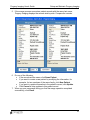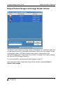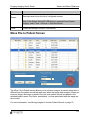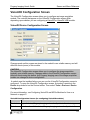
1
Progeny Imaging 1.2
User’s Guide
Part Number: 00-02-1598
ECN: P1568
REV. C
Copyright ©2008
Progeny Dental
675 Heathrow Drive
Lincolnshire, Illinois 60069 U.S.A.
Phone: (888) 924-3800 Fax: (847) 415-9801
WWW.progenydental.com
Progeny Imaging User's Guide
Table of Contents
Contents
Section 1: About This Manual ......................................................................7
Text Conventions.................................................................................. 7
Section 2: Welcome to Progeny Imaging .................................................. 8
Progeny Imaging Features and Functions ............................................ 8
System Components and Configurations ............................................. 8
Progeny Imaging Contact Information ................................................ 11
Section 3: Tour Progeny Imaging............................................................. 12
Progeny Imaging Screen Layout......................................................... 12
Main Menu Bar ................................................................................... 13
Toolbars.............................................................................................. 15
Tooth Panel ........................................................................................ 16
Image Container ................................................................................. 18
Study Surface ..................................................................................... 22
Template Manager.............................................................................. 23
Section 4: Installing Progeny Imaging..................................................... 25
Recommended System Requirements ............................................... 25
Installing Progeny Imaging ................................................................. 26
Logging in as Administrator ................................................................ 30
Configuring the Progeny Imaging Database for Use on a Network .... 31
Configuring Progeny Imaging to Use a Networked Database............. 38
Uninstalling Progeny Imaging ............................................................. 39
Section 5: Installing Image Acquisition Modules ................................... 41
Installing VisionDX Modules ............................................................... 41
Progeny Imaging User's Guide
Table of Contents
Installing MPSe Modules .................................................................... 42
Configuring VisionDX and MPSe Modules for Use on a Network ....... 43
Configuring Progeny Imaging to Use a Networked Image
Acquisition Module.............................................................................. 51
Installing VisionDX USB Modules ....................................................... 54
Installing the Vivid USB Camera......................................................... 58
Section 6: Setup and Maintain Progeny Imaging.................................... 59
Managing Users.................................................................................. 59
Creating and Modifying Image Acquisition Templates ........................ 62
Backing up and Exporting a Patient Database ................................... 63
Restoring and Importing a Patient Database ...................................... 65
Removing the Login Screen ............................................................... 68
Section 7: Use Progeny Imaging .............................................................. 69
Logging in as a User........................................................................... 69
Creating a Patient Record .................................................................. 70
Opening a Patient Record .................................................................. 70
Adding Files to a Patient Record ........................................................ 71
Modifying a Patient Record................................................................. 71
Acquiring X-ray Image Sequences ..................................................... 72
Streamlining Image Acquisition with Templates ................................. 74
Acquiring Images Using a TWAIN-compliant Device .......................... 76
Displaying Images .............................................................................. 77
Annotating Images.............................................................................. 77
Moving Images to Another Patient Record ......................................... 79
Exporting Patient Images.................................................................... 79
4
Progeny Imaging User's Guide
Table of Contents
Deleting Images.................................................................................. 81
Creating Studies ................................................................................. 81
Section 8: Screen and Menu Reference................................................... 83
Add Sensor Screen............................................................................. 83
Annotate and Measure Toolbar .......................................................... 84
Backup and Restore Screen............................................................... 85
Correct Tooth Numbers Screen .......................................................... 88
Device Controls Toolbar ..................................................................... 90
Export Patient Images and Image Viewer Screen .............................. 91
File Menu............................................................................................ 92
Filter Menu.......................................................................................... 93
Filter Toolbar ...................................................................................... 94
Help Menu .......................................................................................... 97
Image Container ................................................................................. 97
Image Menu...................................................................................... 101
Image Operations Toolbar ................................................................ 104
Main Menu Bar ................................................................................. 105
Manage VisionDX Sensors Screen................................................... 106
Move File to Patient Screen.............................................................. 107
Options Screen ................................................................................. 109
Patient Controls Toolbar ................................................................... 116
Patient Menu .................................................................................... 117
Patient Properties Screen................................................................. 119
Print Preview Screen ........................................................................ 122
Select Patient Screen ....................................................................... 123
5
Progeny Imaging User's Guide
Table of Contents
Select Source Screen ....................................................................... 125
Study Surface ................................................................................... 125
Study Surface Menu ......................................................................... 126
Template Controls Toolbar ............................................................... 127
Template Manager............................................................................ 128
Tools Menu....................................................................................... 130
Tooth Panel ...................................................................................... 132
User Manager Screen....................................................................... 133
Video Screen .................................................................................... 136
VisionDX Configuration Screen ........................................................ 137
Section 9: Keyboard Shortcuts .............................................................. 141
Keyboard Command Sequences ...................................................... 141
Index ......................................................................................................... 143
DICOM Conformance Statement ............................................................ 149
6
Progeny Imaging User's Guide
About this Manual
Section 1: About This Manual
This section explains how to use this manual.
Text Conventions
The following typographic conventions are used in this manual.
Type of Information
Convention
Example
Menu selection
Bold font,
menus in path
connected by
‘>’
Select Tools > User Management
User interface
objects and
controls
Bold font
Click Next
Program
information and
information typed
by the user
Fixed-width font Change directories to
C:/program_files/ProgenyDental
User-specific
information typed
by the user
Fixed-width
fontwith italics
and ‘< >’
Type C:/program_files/<user_database>,
substituting the name of your database for
<user_database>
7
Progeny Imaging User's Guide
Getting Started
Section 2: Welcome to Progeny Imaging
This section introduces Progeny Imaging features and configurations.
Progeny Imaging Features and Functions
Progeny Imaging acquires, displays, and stores digital dental X-rays and intraoral video
images. Progeny Imaging stores digital sensor images in DICOM format (Digital
Imaging and Communications in Medicine). The DICOM format assures that each
image contains patient identification and acquisition information.
Progeny Imaging works with the following image acquisition modules and devices:
•
•
•
•
Progeny MPSe and VisionDX digital X-Ray image sensor systems
Progeny VisionDX USB digital X-Ray image sensor systems
TWAIN-compliant devices
Progeny Vivid USB Video Camera
You can use Progeny Imaging to:
•
•
•
•
Configure devices to work with Progeny Imaging
Create login IDs for users of Progeny Imaging
Manage patient records
Acquire, manipulate, and communicate images
System Components and Configurations
When you use Progeny Imaging, you work in several related contexts. This topic
describes the following:
•
•
•
Progeny Imaging Components
Image Acquisition Modules
3rd Party Applications, such as practice management software
Progeny Imaging Components
Progeny Imaging consists of two main components: a graphical user interface and a
database. The database stores image acquisition module configurations, user
information, patient information, and patient images. Patient and user information is
stored in an MS SQL Server database, while patient images are stored in computer
files.
8
Progeny Imaging User's Guide
Getting Started
Progeny Imaging is installed on every computer where you want to use it. The database
component of Progeny Imaging is installed by default on the same computer that runs
the Progeny Imaging graphical user interface. You can instead choose to locate the
database on another computer on the same dental office network and configure other
computers on the network to use this database.
Image Acquisition Modules
Progeny Imaging works with Progeny MPSe, VisionDX, and VisionDX USB digital XRay image acquisition modules and sensors as well as with the Progeny Vivid USB
Video Camera.
Options for installing MPSe and VisionDX modules and sensors are:
•
•
Install a module directly to a computer with Progeny Imaging for use only from that
computer
Install a module directly to a hub in the dental office network and enable other
computers with Progeny Imaging on the network to use the module
You must install the VisionDX USB and the Progeny Vivid USB Video Camera directly
to a computer. They can be used only from the computer to which they are installed.
Standalone Configuration
In a standalone configuration, Progeny Imaging is installed on a computer that is not
connected to any other computers. The patient database is located on the computer.
Imaging modules are connected directly to the computer. No other computers use the
database or modules that are installed on this computer.
Networked Configuration
In a networked configuration, computers are connected to a Peer-to-Peer (P2P)
Windows network or a Windows network that is managed by a domain server.
Progeny Imaging is installed on each computer and on the dental office server.
Computers A, B, and C use the Progeny Imaging database on the dental office server.
Computer A only uses the VisionDX and VisionDX USB modules connected directly to
9
Progeny Imaging User's Guide
Getting Started
it. Computers A, B, and C all use the image acquisition module connected to the
network hub.
Bridge to Third-party Applications
PIBridge is an additional software application from Progeny Dental that enables you to
integrate Progeny Imaging with 3rd-party applications, such as practice management
software. With PIBridge, you can add Progeny Imaging's image acquisition and analysis
capability seamlessly to your practice management software. After accessing a patient's
records in your practice management application, you use PIBridge commands to "call"
Progeny Imaging. At your command, Progeny Imaging opens for you to acquire images
and create studies.
For information on PIBridge and integrating a 3rd party application with Progeny
Imaging, contact Progeny Technical Support. For more information, see Progeny
Imaging Contact Information on page 11.
10
Progeny Imaging User's Guide
Getting Started
Progeny Imaging Contact Information
For product technical questions, call Technical Support. For literature, location of your
nearest Progeny Sales Representative, and all other questions, call Progeny Customer
Service. Hours of operation are 8am-5pm Central Time, Monday-Friday.
For Technical Support
+1-847-415-9800
+Toll Free: 888-924-3800
Press Option 2
For Customer Service
+1-847-415-9800
+Toll Free: 888-924-3800
Press Option 1
11
Progeny Imaging User's Guide
Tour Progeny Imaging
Section 3: Tour Progeny Imaging
This section introduces key features of the Progeny Imaging user interface.
Progeny Imaging Screen Layout
Progeny Imaging’s screen layout is easy to learn and use.
Main Menu Bar (1)
The Main Menu bar contains menus of commands to perform most Progeny Imaging
functions.
For more information, see Main Menu Bar on page 105.
Toolbars
Toolbars are groups of icons to select frequently performed actions. Progeny Imaging
contains the following toolbars:
12
Progeny Imaging User's Guide
•
•
•
•
•
Tour Progeny Imaging
Patient Toolbar (2)
Image Operations Toolbar (3)
Device Controls Toolbar (4)
Template Toolbar (5)
Filter Toolbar (6)
For more information, see Toolbars on page 15.
Tooth Panel (7)
The Tooth Panel allows you to select sequences of teeth and to acquire images.
Note: To activate and use the Tooth Panel, a patient record must be open and an
image acquisition module must be selected.
For more information, see Tooth Panel on page 132.
Study Surface (8)
The Study Surface is where you display, filter, and annotate images that are part of a
patient record.
For more information, see Study Surface on page 125.
Image Container (9)
The Image Container contains thumbnail images and information that are part of a
patient record.
For more information, see Image Container on page 97.
Progress and Status Bar (10)
Information at the bottom of the Progeny Imaging screen gives you information on the
progress of image acquisition, image acquisition module readiness, and current user
and server.
Main Menu Bar
The Main Menu bar contains menus of the major functions provided by Progeny
Imaging.
13
Progeny Imaging User's Guide
Tour Progeny Imaging
Main Menu Bar
Menu
Description
File
The File menu contains options for basic tasks in Progeny Imaging. For
more information, see File Menu on page 92.
Patient
The Patient menu contains options for working with patient records. For
more information, see Patient Menu on page 117.
Image
The Image menu contains settings that allow you to work with an image that
is displayed in the study surface. For more information, see Image Menu on
page 101.
Filter
The Filter menu contains a subset of the image manipulation options that
are found on the Filter toolbar. For more information, see Filter Menu on
page 93.
Tools
The Tools menu contains settings that allow you to modify how Progeny
Imaging looks and functions. For more information, see Tools Menu on
page 130.
Study
Surface
The Study Surface menu contains options for working with studies. For
more information, see Study Surface Menu on page 126.
Help
The Help menu contains options for displaying Progeny Imaging user
assistance and product information. For more information, see Help Menu
on page 97.
14
Progeny Imaging User's Guide
Tour Progeny Imaging
Toolbars
Toolbars are groups of icons to select frequently performed actions.
Toolbars
Toolbar
Patient
Controls
Image
Operations
Description
The Patient Controls toolbar allows you to work with patient records.
For more information, see Patient Controls Toolbar on page 116.
The Image Operations toolbar allows you to perform basic operations
on images. For more information, see Image Operations Toolbar on
page 104.
Filter
The Filter Toolbar has controls to change or manipulate the way an
image is displayed. For more information, see Filter Toolbar on page
94.
Device
Controls
Template
Controls
Annotate and
Measure
The Device Controls Toolbar allows you to select and configure image
acquisition modules. For more information, see Device Controls
Toolbar on page 90.
The Template Controls toolbar allows you to select a template and
open the Template Manager. For more information, see Template
Controls Toolbar on page 127.
The Annotate and Measure Toolbar contains the tools to highlight or
mark up an image. For more information, see Annotate and Measure
Toolbar on page 84.
15
Progeny Imaging User's Guide
Tour Progeny Imaging
Tooth Panel
The Tooth Panel allows you to select sequences of teeth and to acquire images.
For more information, see Acquiring X-ray Image Sequences on page 72.
If the Tooth Panel is not displayed, click the Tooth icon, select File > Toggle Tooth
Panel, or ALT + 1.
Note: To activate and use the Tooth Panel, a patient record must be open and an
image acquisition module must be selected.
16
Progeny Imaging User's Guide
Tour Progeny Imaging
Tooth Panel
Item
Description
Teeth
Selects teeth to add to an image sequence. Selected teeth are
highlighted.
BWR2, R1, L1,
BWL2
Selects predefined bitewing and incisor sequences. Teeth that are
included in the sequence are highlighted.
Acquire
Acquires the selected sequence(s) of teeth.
Add Sequence
Adds sequence for imaging. Teeth that are part of the sequence
change shade to indicate that the sequence is selected for acquisition.
Remove
Sequences
Removes a highlighted sequence.
Pause
Pauses image acquisition.
Select None
Deselects teeth that had been selected.
17
Progeny Imaging User's Guide
Tour Progeny Imaging
Image Container
The Image Container consists of four tabs with the information and images that are part
of a patient record. You must open a patient record to view information in the Image
Container.
For more information, see Opening a Patient Record on page 70.
Folder Tab
The Folder tab contains thumbnail images of X-rays and other files in the patient’s
record. The number in the tab is the number of items in the patient's record. Scroll down
to see all items. The slider at the bottom of the Folder tab adjusts the view so that you
can more easily find images. For X-ray images, the Folder tab lists the tooth or teeth in
the image sequence and the date and time that the image was acquired. For other
items, the Folder tab lists the file name and time of creation. Drag an image from the
Folder tab to the study surface to view the image at actual size. To delete an image
from the Folder tab, select the image and click Delete, or ALT + D.
18
Progeny Imaging User's Guide
Tour Progeny Imaging
For more information, see Acquiring X-ray Image Sequences on page 72.
Studies Tab
The Studies tab contains studies that have been saved in the patient’s record. The
number in the tab is the number of studies in the patient's record. Scroll down to see all
items.The slider at the bottom of the Studies tab adjusts the view so that you can more
easily find studies. The Studies tab shows a thumbnail image of the study and provides
the name or number of the study, a description of the study, and the date on which the
image in the study was created. Select a study and click Open to display it in the study
surface. To save a study, display an image in the study surface, then click Save, or
ALT + S. To delete a study from the Studies tab, select the study and click Delete, or
ALT + D.
For more information, see Creating Studies on page 81.
19
Progeny Imaging User's Guide
Tour Progeny Imaging
Patient Tab
The Patient tab contains information from the patient’s Patient Properties screen. To
edit the information, select the row and type in the new information. Changes made to
patient information in the Patient tab are automatically saved to the patient’s record and
displayed in the Patient Properties screen.
For more information, see Patient Properties Screen on page 119.
For more information, see Creating a Patient Record on page 70.
20
Progeny Imaging User's Guide
Tour Progeny Imaging
Photo Tab
The Image Container Photo tab displays the patient’s picture that was included in the
patient’s Patient Properties screen.
For more information, see Creating a Patient Record on page 70.
21
Progeny Imaging User's Guide
Tour Progeny Imaging
Study Surface
The Study Surface is where you display, filter, and annotate images that are part of a
patient record.
For more information, see Displaying Images on page 77.
For more information, see Creating Studies on page 81.
22
Progeny Imaging User's Guide
Tour Progeny Imaging
Template Manager
Templates are pre-defined groupings of image acquisition sequences that you can use
to streamline image acquisition. You use the Template Manager to create and modify
templates.
Open the Template Manager by selecting Tools > Template or click the Templates
icon in the Template toolbar.
For more information, see Creating and Modifying Image Acquisition Templates on
page 62.
Template Manager
Item
Template
Menu
Description
Options for working with templates
•
New: Opens the New Template dialog box where you name and
save a new (blank) template. The new template will be open in the
23
Progeny Imaging User's Guide
Item
Tour Progeny Imaging
Description
•
•
•
•
•
design surface.
Save: Saves changes to the template currently open in the design
surface.
Save As: Opens a Save As dialog box where you select a new
name or location for a template that is open.
Delete: Deletes the template currently open in the design surface.
The template no longer appears in the Open list.
Remove All Sequences: Removes all sequences from the template.
Exit: Closes the Template Manager.
Sequence
Menu
Remove: Deletes the first or selected sequence from the template.
Help Menu
Displays the help file.
Open
Selects a template to display in the design surface. Templates listed are
those that come with Progeny Imaging and templates you create using the
Template Manager.
Save
Saves changes to the template currently open in the design surface.
New
Opens a box where you name and save the template that you are
creating.
Delete
Deletes the template currently open in the design surface. The template
no longer appears in the Open list.
Sequence
Panel
Sequences of teeth that can be included in the template. Tool tips show
the tooth number for teeth that are part of the sequence. Drag one or
more sequences to the design surface to create the template.
Design
Surface
Layout area of the Template Manager where you arrange sequences of
teeth to create the template.
24
Progeny Imaging User's Guide
Install Progeny Imaging
Section 4: Installing Progeny Imaging
This section tells how to install Progeny Imaging.
Recommended System Requirements
The performance of Progeny Imaging software is affected by the amount of RAM and
storage memory available to the system for acquisition, displaying, storing, and printing
digital X-ray images. The recommended requirements are listed below as a guideline
only.
As you review these guidelines, please be aware that your patient volume, and the
specific demands of your practice, may require you to adjust these guidelines
accordingly. The system requirements of other programs operating on the same
computer or network may affect these guidelines as well.
Recommended System Requirements
System
Component
Requirement
Computer
Hardware
PC-compatible Pentium 4 / 1.4 GHz or greater computer
Memory
System
1 GB RAM or higher (minimum 512 MB)
Operating
System
Microsoft Windows XP ® with Service Pack 2
Microsoft Vista (all editions)
Disk Space
450 MB minimum
You will need additional disk space depending on the size of your practice
and the number of images and other information you plan to store. Each
DICOM image is approximately 4 MB. For example, if you plan to store
75,000 images, you will need approximately 300 GB disk space.
25
Progeny Imaging User's Guide
System
Component
Display
Settings
Install Progeny Imaging
Requirement
800 x 600 (16-bit or higher) with 32 MB of Video RAM
Note: It is possible for you to increase these settings based on the actual
settings defined by the video adapter and/or capture card installed in the
system.
Installing Progeny Imaging
Progeny Imaging contains the patient database and screens for creating and managing
patient records and acquiring images as well as drivers that enable Progeny Imaging to
work with image acquisition modules.
You must install Progeny Imaging on every computer where you want to use it to view
patient records or acquire images, even if you later decide to use a patient database or
modules located elsewhere in your dental office network.
After installing Progeny Imaging, you open the software. If you are installing Progeny
Imaging for the first time on the computer, Progeny Imaging will create the database.
Installation of Progeny Imaging requires only a few minutes.Progeny Imaging is installed
from the Progeny Installation CD-ROM, which also contains the help file and the
database software, MS SQL Server 2005 Express Edition. If MS SQL Server is not
already installed on your computer, the Progeny Software Installer will install it.
Before Installing Progeny Imaging
•
•
•
If you are upgrading from a previous version of Progeny Imaging, you must
uninstall Progeny Device Service. For more information, see Uninstalling Progeny
Imaging on page 39.
Locate the Progeny Imaging Installation CD-ROM.
Ensure that you are logged into your computer using an account that has
Windows computer administrator privileges.
Note: Progeny Imaging currently requires that all users be logged into Windows
as a computer administrator. This will be changed in a future release.
26
Progeny Imaging User's Guide
Install Progeny Imaging
To Install Progeny Imaging
1. Insert the Progeny Imaging Installation CD-ROM into the computer’s CD-ROM
drive. The Progeny Software Installer starts automatically.
Note: If the Progeny Software Installer does not start automatically, use the
Windows Start menu and select Run. Then type the path to the program on the
Progeny Imaging Installation CD-ROM.
2. In the Progeny Imaging Software Installer, click Install Progeny Imaging.
3. In the pop-up message, click Yes to confirm that you want to start the installation.
4. If your computer does not have MS SQL Server 2005 Express Edition installed,
you will see a license agreement. Click Accept. Clicking Don't Accept ends the
installation. You will see the Progeny Imaging screen.
27
Progeny Imaging User's Guide
Install Progeny Imaging
5. In the Progeny Imaging screen, click Next.
28
Progeny Imaging User's Guide
Install Progeny Imaging
6. In the Welcome screen, click Next.
7. In the Confirm Installation screen, click Next to launch the installation. During
installation, you will see the Installing Progeny Imaging screen and a black
command window may appear briefly. This is normal. When the installation is
complete, you will see the Installation Complete screen.
8. Click Close.
9. In the Progeny Software Installer, click Exit.
10. In the pop-up message, click Yes to confirm that you want to exit.
To Open Progeny Imaging
1. On your computer's desktop, double-click on the Progeny Imaging icon, or select
Progeny Imaging from your Windows Start menu.
2. If this is the first time Progeny Imaging has been installed on the computer, you
will see a message stating that the database was not found, and you will be asked
to create a new database. Click Yes, even if you will be using a database on
another computer.
3. Log into Progeny Imaging as the Administrator.
For more information, see Logging in as Administrator on page 30.
29
Progeny Imaging User's Guide
Install Progeny Imaging
Logging in as Administrator
Every time Progeny Imaging is launched, the Login window appears. You must log in to
use Progeny Imaging. Immediately after installing Progeny Imaging, you will log in as
Administrator. For security purposes, add a password for Administrator the first time that
you log in.
Note: While other users can be given application administrator privileges within
Progeny Imaging, you use the Administrator login in the event that you need to
contact Technical Support.
You can configure Progeny Imaging to open without requiring users to log in. For more
information, see Removing the Login Screen on page 68.
Before Logging in as Administrator for the First Time
Ensure that you are logged into an account that has Windows computer administrator
privileges.
Note: Progeny Imaging currently requires that all users be logged into Windows
as a computer administrator. This will be changed in a future release.
To Log in as Administrator for the First Time
1. On your computer's desktop, double-click the Progeny Imaging icon, or select
Progeny Imaging from your Windows Start menu.
2. In the Login screen User ID field, type Administrator.
3. Leave the Password field blank.
4. Click Login.
30
Progeny Imaging User's Guide
Install Progeny Imaging
To Set a Password for Administrator
CAUTION!
In order to ensure the security of patient data, you must set an Administrator
password.
1. Log in as Administrator.
2. Select File > User Manager. The User Manager screen will appear.
3. In the User Manager screen, click the Password button to the left of the
Administrator user ID.
4. In the User Password screen, enter a password in the Password field. When
creating a password, remember the following password rules:
• Passwords must be at least 5 characters long
• Passwords are case sensitive
5. Retype the password in the Re-Enter Password field.
6. Click OK.
When you next log in, you will enter the User ID, Administrator, and the password that
you just created.
Configuring the Progeny Imaging Database for Use on
a Network
The Progeny Imaging database stores image acquisition module configurations, user
information, patient information, and patient images. Patient and user information is
stored in an MS SQL Server database, while patient images are stored in computer
files.
By default, Progeny Imaging uses the database that is installed on the same computer
that runs the Progeny Imaging graphical user interface. Image files are stored in
C:\Program Files\Progeny\Progeny Imaging\Images.
You can instead choose to have all computers running Progeny Imaging use a Progeny
Imaging database on one computer on the dental office network. Configuring the
database for use on a network requires the following tasks:
•
•
•
On the computer that hosts the Progeny Imaging database, configure sharing for
the Progeny Imaging directory.
• Configure sharing on a Windows domain network
• Configure sharing on a Peer-to-Peer network
On the computer that hosts the Progeny Imaging database, configure the MS
SQL Server database to be accessed over the network.
On every other computer that runs Progeny Imaging, configure Progeny Imaging
to use the networked database.
31
Progeny Imaging User's Guide
•
Install Progeny Imaging
For more information, see Configuring Progeny Imaging to Use a
Networked Database on page 38.
Note: Progeny Imaging requires that your dental office network be a setup as a Peer-toPeer (P2P) or Windows domain (client-server) network. In a P2P or domain network, all
users must have local administrator privileges on all clients in the network. On a domain
network, a domain server is required for authentication.
Before Configuring the Progeny Imaging Database for Use on the
Network
•
Install Progeny Imaging on all computers where it will be used.
To Configure Sharing for the Progeny Imaging Directory on a
Windows Domain Network
1. Navigate to C:\Program Files\Progeny.
2. In the Progeny folder, highlight the Progeny Imaging folder.
3. Right click and select Sharing and Security.
4. In the Progeny Imaging Properties box, select the Sharing tab.
5. Click Share this folder.
6. Click Permissions.
7. Click Add.
8. Click Advanced.
32
Progeny Imaging User's Guide
Install Progeny Imaging
9. In the Select Users, Computers, or Groups box, click Find Now.
10. Select Network Service.
11. Click OK.
12. Click OK.
33
Progeny Imaging User's Guide
Install Progeny Imaging
13. In the Permissions for Progeny Imaging box, select Allow for Full Control.
14. Click OK.
15. Click OK. The Progeny Imaging folder icon should indicate that the folder is now
shared.
To Configure Sharing for the Progeny Imaging Directory on a Peer-toPeer Network
1. Navigate to C:\Program Files\Progeny.
2. In the Progeny folder, highlight the Progeny Imaging folder.
3. Right click and select Sharing and Security.
34
Progeny Imaging User's Guide
Install Progeny Imaging
4. In the Progeny Imaging Properties box, select the Sharing tab.
5. Click the sentence link: If you understand the security risks but want to share
files without running the wizard, click here.
6. Select Just enable file sharing and click OK.
7. The Sharing tab now shows that Progeny Imaging is shared. Select Allow
network users to change my files and click OK.
8. In the warning about share name length, click OK.
To Configure MS SQL Server for Network Access
1. From the Windows Start menu, select Programs > Microsoft SQL Server 2005 >
Configuration Tools > SQL Server Configuration Manager.
2. In the SQL Server Configuration Manager, expand SQL Server 2005 Network
Configuration.
35
Progeny Imaging User's Guide
Install Progeny Imaging
3. Click Protocols for SQLEXPRESS.
4. Verify that Shared Memory, Named Pipes, and TCP/IP are Enabled. If any of
these items are not enabled, select, right-click, and enable them. Ignore any
warnings that are displayed.
5. Expand SQL Native Client Configuration.
6. Click Client Protocols.
7. Verify that Shared Memory, Named Pipes, and TCP/IP are Enabled. If any of
these items are not enabled, select, right-click, and enable them. Ignore any
warnings that are displayed.
8. Close the SQL Server Configuration Manager.
9. From the Windows Start menu, select Programs > Microsoft SQL Server 2005 >
Configuration Tools > SQL Server Surface Area Configuration.
10. Near the bottom of the screen, click Surface Area Configuration for Services
and Connections.
11. Expand SQLEXPRESS.
12. Click Service.
36
Progeny Imaging User's Guide
Install Progeny Imaging
13. In the Service status field, verify that the service is running. If the service is not
running, click Start. Also, if the service is running, but you made changes in the
SQL Server Configuration Manager, click Stop, then click Start.
14. Click Remote Connections.
15. Click Local and remote connections.
37
Progeny Imaging User's Guide
Install Progeny Imaging
16.
17.
18.
19.
20.
Click Using both TCP/IP and named pipes.
Click Apply.
Click SQL Server Browser.
Click Service.
In the Service status field, verify that the service is running. If the service is not
running, select Automatic from the Startup Type drop-down list. Click Apply, then
click Start.
21. Click OK to close Surface Area Configuration for Services and Connections.
Next Steps
On every other computer that runs Progeny Imaging, configure Progeny Imaging to use
the networked database. For more information, see Configuring Progeny Imaging to
Use a Networked Database on page 38.
Configuring Progeny Imaging to Use a Networked
Database
The Progeny Imaging database stores image acquisition module configurations, user
information, patient information, and patient images. Patient and user information is
stored in an MS SQL Server database, while patient images are stored in computer
files.
38
Progeny Imaging User's Guide
Install Progeny Imaging
By default, Progeny Imaging uses the database that is installed on the same computer
that runs the Progeny Imaging graphical user interface. Image files are stored in
C:\Program Files\Progeny\Progeny Imaging\Images.
You can instead set the options for each computer that is running Progeny Imaging to
use a Progeny Imaging database that is located on another computer on the dental
office network.
Note: Progeny Imaging requires that your dental office network be a setup as a Peer-toPeer (P2P) or Windows domain (client-server) network. In a P2P or domain network, all
users must have local administrator privileges on all clients in the network. On a domain
network, a domain server is required for authentication.
Before Configuring Progeny Imaging to Use a Database on the
Network
•
•
Install Progeny Imaging on all computers where it will be used.
Configure the Progeny Imaging database to be used on a network.
• For more information, see Configuring the Progeny Imaging Database for
Use on a Network on page 31.
To Configure Progeny Imaging to Use a Database on the Network
1.
2.
3.
4.
Log into Progeny Imaging.
Select Tools > Options.
In the Options screen, select the Database tab.
Select Networked to indicate that you are using a Progeny Imaging database that
is located on another computer in the dental office network.
5. Click Find Servers and select the computer that is running the Progeny Imaging
database that you want to connect to.
6. Click Test Database to verify that your computer can connect to the database.
7. Click OK. If you change the database, you must restart Progeny Imaging.
For more information, see Options Screen on page 115.
Uninstalling Progeny Imaging
To remove Progeny Imaging or Progeny Device Service
The steps below assume you are running Windows XP. If you are running Windows
Vista, use the Programs and Features icon in the Control Panel.
1. From the Windows Start menu, select Control Panel. Then click on Add or
Remove Programs.
2. In the Add or Remove Programs screen, select Progeny Device Service.
3. Click Remove.
39
Progeny Imaging User's Guide
Install Progeny Imaging
4. In the Add or Remove Programs screen, select Progeny Imaging.
5. Click Remove.
Note: Uninstalling Progeny Imaging and the Progeny Device Service does not remove
the Progeny Imaging database or MS SQL Server 2005 Enterprise Edition, because
these components will be used if you re-install Progeny Imaging. However, if MS SQL
Server 2005 is removed or becomes corrupted, later installation of Progeny Imaging will
fail unless you remove the Progeny Imaging database. To remove the Progeny Imaging
database after removing MS SQL Server, navigate to C:\Program Files\Microsoft SQL
Server\MSSQL.1\MSSQL\Data. Remove the file ProgenyStudyLab.mdf.
40
Progeny Imaging User's Guide
Install Image Acquisition Modules
Section 5: Installing Image Acquisition
Modules
This section explains how to install the VisionDX, MPSe, VisionDX USB, and Progeny
Vivid USB Video Camera to work with Progeny Imaging.
Installing VisionDX Modules
Progeny VisionDX is an intraoral system for digital imaging of teeth and the oral cavity.
The VisionDX image acquisition module can be used from the computer where you
install it. This topic describes how to install VisionDx modules directly to your computer.
Alternatively, you can install the VisionDX on a computer, configure it for use over the
dental office network, then reconnect it to a network hub so that it can be used from
other computers on the dental office network.
For more information, see Configuring VisionDX and MPSe Modules for Use on a
Network on page 43.
Before Installing VisionDX Modules
•
•
•
Install Progeny Imaging on your computer.
Examine the contents of your VisionDX kit. You should have the following items:
• Sensor(s)
• VisionDX Module
• Power Cable
• Power Supply (Power over Ethernet Injector, IEEE802.3af compatible)
• CAT5 Cross-over Cable (shorter cable; usually orange, but may be purple,
yellow, or green)
• CAT5 Standard Ethernet Network Cable (longer cable; usually white, but
may be gray or blue)
• Progeny VisionDX User Manual
• Sensor Sheaths
You must have a free network connection on your computer.
To Install the VisionDX Directly to your Computer
1. Plug the cross-over cable into the LAN-IN connection on the power supply.
2. Plug the standard Ethernet cable into the PWR LAN-OUT connection on the
power supply.
41
Progeny Imaging User's Guide
Install Image Acquisition Modules
3. Connect the other end of the cross-over cable into the network connection on the
back of your computer.
4. Connect the other end of the Ethernet cable into the Ethernet port on the
VisionDX.
5. Connect the sensor to the VisionDX.
6. Connect the power cord to the power supply and to an electrical outlet.
7. Power on the VisionDX. Once the module is powered up, one or more pop-up
messages will appear in the lower-right hand side of the screen as the Windows
network detects that a new module is connected.
8. Log into Progeny Imaging. Progeny Imaging will detect the VisionDX and you can
begin acquiring images immediately.
• For more information, see Logging in as a User on page 69.
9. Select Tools > Devices > Device Configuration to configure a name for your
VisionDX module.
• For more information, see VisionDX Configuration Screen on page 137.
Installing MPSe Modules
Progeny MPSe is an intraoral system for digital imaging of teeth and the oral cavity. The
MPSe image acquisition module can be used from the computer where you install it.
This topic describes how to install MPSe modules directly to your computer.
Alternatively, you can install the MPSe on a computer, configure it for use over the
dental office network, then reconnect it to a network hub so that it can be used from
other computers on the dental office network.
For more information, see Configuring VisionDX and MPSe Modules for Use on a
Network on page 43.
42
Progeny Imaging User's Guide
Install Image Acquisition Modules
Before Installing MPSe Modules
•
•
•
Install Progeny Imaging on your computer.
Examine the contents of your MPSe kit. You should have the following items:
• Sensor(s)
• MPSe Module
• Power Cable
• Power Supply
• CAT5 Standard Ethernet Network Cable (longer cable; usually white, but
may be gray or blue)
• Progeny MPSe User Manual
• Sensor Sheaths
You must have a free network connection on your computer.
To Install the MPSe Directly to your Computer
1. Plug the standard Ethernet cable into the Ethernet connection on the MPSe
module.
2. Connect the other end of the Ethernet cable into the network connection on the
back of your computer.
3. Connect the sensor to the MPSe module.
4. Connect the power cord to the power supply and to an electrical outlet.
5. Power on the MPSe module. Once the module is powered up, one or more pop-up
messages will appear in the lower-right hand side of the screen as the Windows
network detects that a new module is connected.
6. Log into Progeny Imaging. Progeny Imaging will detect the MPSe and you can
begin acquiring images immediately.
• For more information, see Logging in as a User on page 69.
7. Select Tools > Devices > Device Configuration to configure a name for your
MPSe module.
• For more information, see VisionDX Configuration Screen on page 137.
Configuring VisionDX and MPSe Modules for Use on a
Network
Progeny VisionDX and MPSe image acquisition modules can be configured on the
dental office network so that they can be used from any computer on the network that is
running Progeny Imaging. This topic describes how to configure a VisionDX or MPSe
module that is connected to a hub on the dental office network.
Configuring VisionDX and MPSe image acquisition modules for use on the network
requires the following tasks:
•
•
Install the image acquisition device directly to any computer
Configure the image acquisition module with information about the network
43
Progeny Imaging User's Guide
•
•
Install Image Acquisition Modules
Reconnect the image acquisition module to the network hub
Direct each computer with Progeny Imaging to recognize the networked module.
• For more information, see Configuring Progeny Imaging to Use a Networked
Image Acquisition Module on page 51.
Before Configuring VisionDX and MPSe Modules for Use on a
Network
•
•
•
Install Progeny Imaging
Install the VisionDX or MPSE module
• For more information, see Installing VisionDX Modules on page 41.
• For more information, see Installing MPSe Modules on page 42.
Obtain the following information that you will need to configure the module so that
it can be recognized on the network:
• Name
• Description
• IP address
• Subnet mask
• Gateway
To Configure a VisionDX or MPSe Module for Use on a Network
1. Select Tools > Devices > Add New Device Wizard.
44
Progeny Imaging User's Guide
Install Image Acquisition Modules
2. In the Device Installation Wizard, click Next.
3. In the Device Selection screen, use the Product drop-down to select your image
acquisition module. You can also enter a name and description for the module.
Click Next.
45
Progeny Imaging User's Guide
Install Image Acquisition Modules
4. Be sure the device that you are configuring is already connected to the computer
where you are running the Add New Device wizard. If the device is not connected,
use the cross-over cable provided to connect the device.
5. Click Next. The Device Properties screen will display the version and serial
number of the device.
46
Progeny Imaging User's Guide
Install Image Acquisition Modules
6. Click Next.
7. In the Select your connection type screen, select Network Connection.
47
Progeny Imaging User's Guide
Install Image Acquisition Modules
8. Click Next.
9. In the Device Network Properties screen, change the information to be correct for
the dental office network. The IP Address must be a number that is not used by
any other device on the network.
10. Click Next to save the information to the device.
48
Progeny Imaging User's Guide
Install Image Acquisition Modules
11. Click Next.
49
Progeny Imaging User's Guide
Install Image Acquisition Modules
12. Follow the instructions in the Restart Device screen to remove the image
acquisition module from the computer. Reconnect it to the network hub.
13. Turn the module on and wait for one minute.
14. In the Restart Device screen, click Next. The Device Detection screen verifies that
the module is now recognized on the network.
15. Click Next.
16. Click Finish.
Next Steps
Direct each computer with Progeny Imaging to recognize the networked module. For
more information, see Configuring Progeny Imaging to Use a Networked Image
Acquisition Module on page 51.
50
Progeny Imaging User's Guide
Install Image Acquisition Modules
Configuring Progeny Imaging to Use a Networked
Image Acquisition Module
After you install a VisionDX or MPSe acquisition module for use on the dental office
network, you must configure Progeny Imaging to recognize it. You do this on each
computer that is running Progeny Imaging.
Before Configuring Progeny Imaging to Use a Networked Acquisition
Module
•
•
•
Install an MPSe or VisionDX acquisition module and configure it for use on the
network.
•
For more information, see Configuring VisionDX and MPSe Modules for
Use on a Network on page 43.
Obtain the following information about the networked VisionDX or MPSe
acquisition module:
• Name
• Description
• IP address
• Subnet mask
• Gateway
Log into Progeny Imaging
To Configure Progeny Imaging to Use a VisionDX or MPSe
Acquisition Module on the Network
1. Select Tools > Devices > Add Existing Device.
51
Progeny Imaging User's Guide
Install Image Acquisition Modules
2. In the Device Installation Wizard, click Next.
52
Progeny Imaging User's Guide
Install Image Acquisition Modules
2. In the Select Your Connection Type screen, select Network Connection. Click
Next.
3. In the Device Configuration Properties, enter the properties of the Vision DX or
MPSe acquisition module that has been configured for use on the dental office
network.
4. Click Connect to connect to the image acquisition module. If Progeny Imaging
can connect with the module, the Version and Serial # fields will show information
about the module.
5. Click Next. The device configuration will be saved.
6. Click Next.
7. Click Finish.
53
Progeny Imaging User's Guide
Install Image Acquisition Modules
Installing VisionDX USB Modules
Progeny VisionDX USB is an intraoral system for digital imaging of teeth and the oral
cavity. The VisionDX USB image acquisition module must be used from the computer
where you install it. Installing the VisionDX Module means connecting it to the computer
and using Progeny Imaging and the Sensor Calibration Files CD-ROM to add and
calibrate the sensor. This topic describes how to connect and calibrate the VisionDX
USB module.
Before Installing VisionDX USB Modules
•
•
•
Install Progeny Imaging on your computer.
Examine the contents of your VisionDX USB kit. You should have the following
items:
• Sensor(s)
• VisionDX USB Interface Module
• USB Cable
• Sensor Calibration Files CD-ROM
• Sensor Sheaths
• Progeny VisionDX USB User Manual
You must have a free USB 2.0 connection on your computer.
To Connect the VisionDX USB Module
1. Plug the square end of the USB cable into the VisionDX USB Interface Module.
2. Plug the sensor into the VisionDX USB Interface Module.
3. Plug the flat end of the USB cable into the computer's USB connection. Pop-up
windows will display on your computer indicating that the computer recognizes the
new device. The green light on the Device toolbar will indicate that Progeny
Imaging recognizes the device, but you must add and calibrate the device before
you can acquire images.
To Add and Calibrate the VisionDX USB Module
1. Log into Progeny Imaging. Progeny Imaging will display the Manage VisionDX
Sensors screen. The screen shows the serial number for the sensor at the top, but
the serial number does not appear in the Serial Number list.
• For more information, see Logging in as a User on page 69.
54
Progeny Imaging User's Guide
Install Image Acquisition Modules
2. In the Manage VisionDX Sensors screen, click Add Sensor. Progeny Imaging will
display the Add Sensor screen.
55
Progeny Imaging User's Guide
Install Image Acquisition Modules
3. Place the Sensor Calibration Files CD-ROM in the computer's CD-ROM drive.
4. In the Add Sensor screen, click the '...' button to open the Browse for Folder
screen.
5. In the Browse for Folder screen, browse and select the Progeny Sensor C.
56
Progeny Imaging User's Guide
Install Image Acquisition Modules
6. Click OK. The Add Sensor screen will now contain the sensor serial number
highlighted in blue.
7. In the Add Sensor screen, click OK.
8. In the confirmation pop-up, click Yes. The Manage VisionDX Sensors screen will
now contain the sensor serial number highlighted in blue.
57
Progeny Imaging User's Guide
Install Image Acquisition Modules
9. Click Close. You can now acquire images with the VisionDX USB module.
10. Select Tools > Devices > Device Configuration to configure a name for your
VisionDX USB module.
• For more information, see VisionDX Configuration Screen on page 139.
Installing the Vivid USB Camera
Follow the installation instructions in the Progeny Vivid USB Camera Installation and
User’s Guide. The Vivid USB Camera will automatically be recognized by Progeny
Imaging. No additional installation or configuration is required.
For more information, see Video Screen on page 136.
58
Progeny Imaging User's Guide
Setup and Maintain Progeny Imaging
Section 6: Setup and Maintain Progeny
Imaging
This section contains instructions for getting ready to use Progeny Imaging and
safeguarding patient records.
Managing Users
Everyone who uses Progeny Imaging must log in. When Progeny Imaging is installed,
only one user, the Administrator, can log in. In order to implement security for patient
records, you must create additional users.
For more information, see Logging in as Administrator on page 30.
Progeny Imaging has three types of users, each with specific privileges.
•
•
•
Administrator -- Administrator is a special user that Progeny Imaging requires for
Technical Support. The Administrator user can create and manage other users
and is a primary dentist with access to all patient records.
Application Administrator -- Application Administrator users create and manage
other users and are primary dentists with access to all patient records. You make
a user an application administrator by checking the Administrator box in the User
Manager screen.
Ordinary Users -- Ordinary users are primary dentists who create and manage
only their own patients' records. In the User Manager screen, leave the
Administrator box unchecked to create an ordinary user.
Before Creating Users
To create a user, you must be logged in to Progeny Imaging as the Administrator or as
an application administrator user.
59
Progeny Imaging User's Guide
Setup and Maintain Progeny Imaging
To Create a User
1. Select File > User Manager, or CTRL + U.
2. In the User Manager screen, click New. A blank row is added to the User Manager
screen.
3. In the new row, configure the following information for the user:
• Type a User ID.
• (Optional) Select the Administrator box to make the user an application
administrator.
• (Optional) Click the Password box to assign a password for the user. In the
User Password screen, enter and re-enter the password. When creating a
password, remember the following password rules:
• Passwords must be at least 5 characters long
• Passwords are case sensitive
• (Optional) In the remaining fields, enter other information about the user.
4. Click Close to save the user information and close the User Manager screen.
60
Progeny Imaging User's Guide
Setup and Maintain Progeny Imaging
Modifying User Information
To modify user information, you must be logged in to Progeny Imaging as an application
administrator user.
To Modify User Information
1. Select File > User Manager, or CTRL + U.
2. In the User Manager screen, select the user whose information you want to
change.
3. Change the user’s information.
4. Click Close to save the user information and close the User Manager screen.
Deleting Users
Before Deleting Users
•
•
If the user to be deleted has patients assigned to him or her, assign the patients to
another user.
To delete users, you must be logged in to Progeny Imaging as an application
administrator user.
To Delete a User
1.
2.
3.
4.
Select File > User Manager , or CTRL + U.
In the User Manager screen, select the user to delete.
Click Delete.
Click OK to delete the user.
To Assign Patients to Another User
1. Log in as the user whose patients you are reassigning.
2. Click Open.
3. Write down the names of the user’s patients that appear in the Select Patient
screen.
4. Select File > Log Out.
5. Log into Progeny Imaging as an application administrator user.
6. Select Patient > Open, or click the Open icon.
7. In the Select Patient screen, select a patient who is assigned to the user whom
you are deleting.
8. Click Properties.
9. In the Patient Properties screen, change the patient’s primary dentist to a dentist
other than the user whom you are deleting.
10. Click Apply to save your changes and continue working in the Patient Properties
screen.
11. When you have reassigned all the patients, click OK.
61
Progeny Imaging User's Guide
Setup and Maintain Progeny Imaging
Creating and Modifying Image Acquisition Templates
Templates are pre-defined groupings of image acquisition sequences that you can use
to streamline image acquisition. You use the Template Manager to create, modify, and
delete templates. On the left side of the Template Manager are sequences of teeth to
include in the template. On the right side of the Template Manager is the design surface
where you assemble sequences for the template. The design surface is oriented from
the patient’s point of view.
For more information, see Streamlining Image Acquisition with Templates on page 74.
To Create a Template
1. Select Tools > Templates, or click the Template icon in the Template toolbar.
2. In the Template Manager, select Template > New, or click New.
3. In the New Template screen, enter a name for the template and click OK. The
template name appears as the open template.
4. Drag sequences of teeth to the design surface, positioning the sequences in the
order in which they will be acquired.
62
Progeny Imaging User's Guide
Setup and Maintain Progeny Imaging
•
To remove a sequence from the design surface, select the sequence, then
select Sequence > Remove. To remove all sequences, select Template >
Remove All Sequences.
5. Select Template > Save, or click Save.
6. To close the Template Manager, select Template > Exit.
To Modify a Template
1. Select Tools > Templates, or click the Template icon.
2. In the Template Manager, use the drop-down list to select the template to modify.
3. In the design surface, select a sequence. Then click the right mouse button to
display a menu of actions that you can perform on the sequence.
4. Select an action to perform on the sequence. You can perform the following
actions on sequences in a template:
• Make First in Sequence: Reorders the sequences in the template so that the
selected sequence will be acquired first
• Make Last in Sequence: Reorders the sequences in the template so that the
selected sequence will be acquired last
• Move Up in Sequence: Reorders the sequences in the template so that the
selected sequence will be acquired before the immediately preceding
sequence
• Move Down in Sequence: Reorders the sequences in the template so that
the selected sequence will be acquired after the immediately following
sequence
• Background Color: Displays a color palette from which you select the color
of the background for the template
• Remove: Removes the selected sequence from the template
5. Select Template > Save, or click Save.
6. To close the Template Manager, select Template > Exit.
Backing up and Exporting a Patient Database
Progeny Imaging stores patient data in a Microsoft SQL Server database on the
computer where Progeny Imaging is installed or on another computer on the same
network.
Backing up the patient database regularly is important to ensure that patient data is not
lost in case of computer failure. By default, the database backup file is written to
C:\Progeny Imaging\Database\database.bak. For security, specify a backup location on
removable media or on another computer on your network.
Note: Progeny Imaging does not currently support Backup and Restore for Windows
Vista operating systems. Vista support for Backup and Restore is planned for a future
release.
63
Progeny Imaging User's Guide
Setup and Maintain Progeny Imaging
You can also export a patient database for later merge with another Progeny Imaging
patient database. This is useful if you see some patients in one office and wish to merge
their records with a Progeny Imaging patient database in another office. To merge
records, you first use the Backup and Restore screen to export the records, selecting
Export for Merge. Then you use the Restore tab to merge the records.
For more information, see Backup and Restore Screen on page 85.
For more information, see Restoring and Importing a Patient Database on page 65.
To Backup the Patient Database
1. Select File > Backup and Restore.
2. In the Backup tab of the Backup and Restore screen, use the Destination field to
select a location for the backup file. Use the Browse button to find additional
destinations.
CAUTION!
For security reasons, specify a backup destination on removable media or on
another computer. If you create the backup on your computer, be sure to transfer
it to removable media or another computer.
3. Click Backup.
64
Progeny Imaging User's Guide
Setup and Maintain Progeny Imaging
4. When you see a message telling you that the backup completed successfully,
click Close.
To Export the Patient Database for Merge
1. Select File > Backup and Restore.
2. In the Backup tab of the Backup and Restore screen, select a destination for the
backup file. Use the Browse button to find additional destinations.
3. Select Export for Merge.
4. Click Backup.
5. When you see a message telling you that the backup completed successfully,
click Close.
Restoring and Importing a Patient Database
You can restore a patient database from a backup file that was created by Progeny
Imaging. The backup file must exist on the computer with Progeny Imaging or on
another computer on the same network. By default, the database backup file is written
to C:\Progeny Imaging\Database\database.bak.
Note: Progeny Imaging does not currently support Backup and Restore for Windows
Vista operating systems. Vista support for Backup and Restore is planned for a future
release.
You can also merge records from one Progeny Imaging patient database into another
Progeny Imaging patient database. This is useful if you see some patients in one office
and wish to merge their records with a Progeny Imaging patient database in another
office. To merge records, you first export the records, selecting Export for Merge in the
Backup and Restore screen Backup tab. Then you use the Restore tab to merge the
records.
For more information, see Backup and Restore Screen on page 85.
For more information, see Backing up and Exporting a Patient Database on page 63.
To Restore a Patient Database
1. Select File > Backup and Restore.
2. In the Backup and Restore screen, select the Restore tab.
65
Progeny Imaging User's Guide
Setup and Maintain Progeny Imaging
3. In the Source field, select the location of the backup file. Use the Browse button to
find additional locations.
4. Click Restore.
5. When you see a message telling you that the restore operation completed
successfully, click Close.
To Merge Records into a Patient Database
1. Select File > Backup and Restore.
2. In the Backup and Restore screen, select the Restore tab.
3. In the Source field, select the location of the file that was created by exporting the
database for merge. Use the Browse button to find additional locations.
4. Click Merge.
66
Progeny Imaging User's Guide
Setup and Maintain Progeny Imaging
5. If the merge process encounters patient records with the same last name,
Progeny Imaging displays the records side-by-side. Compare the records.
6. Do one of the following:
• If the records are the same, click Same Patient.
• If you want to create a new patient by modifying the information, for
example, for two members of the same family, click New Patient.
• If you want to update information in the patient's record, click Update.
• Click Cancel to skip updates to this patient record.
7. When you see a message telling you that the merge operation completed
successfully, click Close.
67
Progeny Imaging User's Guide
Setup and Maintain Progeny Imaging
Removing the Login Screen
Every time you launch Progeny Imaging, the Login window appears. If you want to use
Progeny Imaging without requiring users to log in, you must create a new desktop
shortcut.
Note: When the Login screen is not used, the only Progeny Imaging user is
Administrator and you cannot create other users. The Administrator user is a primary
dentist with access to all patient records. For more information, see Managing Users on
page 59.
To Remove the Login Screen
1. Select the Progeny Imaging executable file. By default, the file is located in:
C:\Program Files\Progeny\Progeny Imaging\ProgenyImaging.exe
2. Copy ProgenyImaging.exe.
3. On your computer's desktop, right click and select Paste Shortcut from the option
menu.
4. With the shortcut selected, right click and select Properties.
5. In the Properties dialog box, select the Shortcut tab.
6. In the Target text field, place your cursor to the right of the last character.
7. Type a space, and then type login=false.
8. Click Apply.
9. Click OK.
68
Progeny Imaging User's Guide
Use Progeny Imaging
Section 7: Use Progeny Imaging
This section contains procedures for using Progeny Imaging to manage patient records,
acquire images, and create studies.
Logging in as a User
Every time Progeny Imaging is launched, the Login window appears. You must log in to
use Progeny Imaging.
You can configure Progeny Imaging to open without requiring users to log in. For more
information, see Removing the Login Screen on page 68.
Before Logging In
•
Ensure that you are logged onto your computer using an account that has
Windows computer administrator privileges.
Note: Progeny Imaging currently requires that all users be logged into Windows
as a computer administrator. This will be changed in a future release.
•
Obtain a user ID from your system administrator. You may also be given a
password.
To Log In
1. On your computer's desktop, double-click the Progeny Imaging icon, or select
Progeny Imaging from your Windows Start menu.
2. In the Login screen, enter your user ID and password, if you have one. Remember
that passwords are case sensitive.
3. Click Login.
69
Progeny Imaging User's Guide
Use Progeny Imaging
Creating a Patient Record
Progeny Imaging associates X-Ray images and other patient files with a patient record.
You need a record for each patient for whom you want to acquire images.
Before Creating a Patient Record
•
•
•
You must be logged into Progeny Imaging to create a patient record.
When you create a patient record, you must assign the patient a primary dentist.
The person who will be the primary dentist must already be set up as a user of
Progeny Imaging.
• For more information, see Managing Users on page 59.
If you want to include the patient’s picture in their record, place a JPEG image file
of the patient in a directory on your computer or on a computer that you can reach
from Progeny Imaging.
To Create a Patient Record
1. Select Patient > New or click the New icon or ALT + N.
2. In the Patient Properties screen, enter patient information. Fields marked with an
asterisk ‘*’ are required.
3. (Optional) Click Browse to locate and include a picture of the patient. Pictures
must be JPEG image files.
4. Click Apply to save your changes and continue working in the Patient Properties
screen. Click OK to save your changes and close the screen.
For more information, see Patient Properties Screen on page 119.
Opening a Patient Record
Progeny Imaging requires that you open a patient record in order to acquire images and
create studies.
Before Opening a Patient Record
The patient record must be created before you can open it.
To Open a Patient Record
1. Select Patient > Open, or ALT + O, or click the Open icon.
2. In the Select Patient screen, select a patient. To search for a patient by last name,
enter all or part of the patient’s last name in the Last Name Filter field.
3. Click Open, or double-click next to the left of the patient's information, to open the
patient’s record. When the record is open, you will see the patient’s name at the
70
Progeny Imaging User's Guide
Use Progeny Imaging
top of the Progeny Imaging window and the patient’s information will appear in the
Image Container.
Note: If the Image Container is not displayed, select Patient > Show Patient
Panel, or ALT + 2.
For more information, see Select Patient Screen on page 123.
Adding Files to a Patient Record
While most images in your patient’s records will be X-rays, you can also add files
created in other applications to patients’ records. For example, if you find a Web page
or a PDF that contains information related to a patient, you can store this information in
the patient’s record. Storing these files in a patient’s record creates a copy of the file.
When you open these files from the Image Container, the application associated with
the file opens so that you can edit the copy of the file that Progeny Imaging stored in the
patient’s record.
Before Adding Files to a Patient’s Record
•
•
•
•
Files to be added to a patient’s record must be located in a directory on your
computer or on a computer that you can reach from Progeny Imaging.
Files to be added must be Word (.doc) files, Acrobat (.pdf) files, Web (.htm or
.html) files, Excel files (.xls), or text (.txt) files.
The patient’s record must be open.
The application associated with the file must be located on your computer if you
want to open and edit these files from the Image Container.
To Add Files to a Patient Record
1.
2.
3.
4.
Select Image > Import.
In the file selection box, navigate to the folder where the file is located.
Select the file.
Click Open. The file is added to the patient's record and an icon representing the
file type and the name of the file appear in the Folder tab of the Image Container.
Modifying a Patient Record
Before Modifying a Patient Record
The patient record must be created before you can modify it.
71
Progeny Imaging User's Guide
Use Progeny Imaging
To Modify a Patient Record
1. Select Patient > Open, or ALT + O.
2. In the Select Patient screen, highlight the patient record and click Properties. You
can also open the patient's record, then select Patient > Properties, or
CTRL + ALT + P, or click the Properties icon. You can also modify the patient
record using the Patient tab in the Image Container.
3. In the Patient Properties screen, modify the patient’s information.
4. Click Apply to save your changes and continue working in the Patient Properties
screen. Click OK to save your changes and close the screen.
For more information, see Patient Properties Screen on page 119.
Acquiring X-ray Image Sequences
Progeny Imaging allows you to acquire images of sequences of teeth. The sequence
defines the teeth that will be imaged and the order in which the images will be acquired.
In the Tooth Panel, you can use the preset sequences or create your own sequences by
selecting individual teeth. To streamline image acquisition, you can use the Template
Manager to save frequently used sequences as templates.
For more information, see Streamlining Image Acquisition with Templates on page 74.
Before Acquiring X-ray Image Sequences
•
•
•
Open a patient record.
Verify that the image acquisition module you want to use is installed and available.
Dipslay the Tooth Panel. If the Tooth Panel is not displayed, click the Tooth icon,
select File > Toggle Tooth Panel, or ALT + 1. The Patient Panel must be displayed
in order to display the Tooth Panel.
For more information, see Tooth Panel on page 16.
To Image a Sequence of Teeth
1. Select an X-ray image acquisition module from the Device Controls drop-down list.
The Connection Indicator will become green showing that the module is ready.
2. In the Tooth Panel, select one or more teeth to image, or click BWR1, R1, BWL2,
or L1 to select the sequence to image. The teeth that are included in the selected
sequence will become white.
3. Click Add Sequence. The selected teeth change to orange to indicate that they
are now part of a sequence.
To select more than one sequence, repeat steps 2 and 3.
To remove the sequence, click Remove Sequence. Then click Select None.
72
Progeny Imaging User's Guide
Use Progeny Imaging
4. Click Acquire. The teeth in the first sequence will flash to indicate that Progeny
Imaging is ready to accept an image from the X-ray sensor. At any time during
acquisition, click Cancel to stop the acquisition.
CAUTION!
Before clicking Cancel to stop the acquisition, ensure that the patient will not be
unnecessarily exposed to X-ray radiation.
5. Visually verify that the VisionDX is ready to accept an X-ray exposure. The light on
the front of the VisionDX control module should be yellow.
6. Insert the X-ray sensor into a protective sheath and position the X-ray sensor in
the patient's mouth.
7. Select the appropriate exposure and prepare the X-ray source to produce the
selected X-ray exposure.
8. Align the X-ray source with the X-ray sensor as appropriate for the desired
radiographic technique.
9. Check again that Progeny Imaging, the X-ray sensor and the X-ray source are
ready for an X-ray exposure.
CAUTION!
The X-ray sensor is active and waiting for X-Ray exposure for a limited time. At the end
of the time period, the sensor times out, requiring you to start the procedure again. You
should always verify that Progeny Imaging and the X-ray sensor are ready before
exposing the patient to X-ray radiation. If you need more time, change the sensor
timeout by in the VisionDX Configuration screen.
10. Activate the X-ray source to expose the sensor.
During acquisition, the teeth in the first sequence change shade to yellow and
flash to indicate that the image transfer is in progress. When the acquisition is
complete, the teeth change shade to green, the image is automatically saved, and
appears in the study surface and in the Folder tab of the Image Container.
11. If you selected more than one sequence for acquisition, Progeny Imaging will
automatically begin to acquire the next sequence. Repeat steps 5 to 10.
12. To pause acquisition between two sequences, click Pause during the first
acquisition before the second acquisition begins. To continue acquiring the next
sequence, click Resume.
13. Record the X-ray exposure parameters or other information related to the acquired
image as an image note. To create a note, click the yellow note icon in the bottom
right corner of the image. Click Save to save the added text with the image. If the
note icon is not visible, select Study Surface > Expanded View.
73
Progeny Imaging User's Guide
Use Progeny Imaging
Streamlining Image Acquisition with Templates
Templates are pre-defined groupings of image acquisition sequences that you can use
to streamline image acquisition. For example, the 2 BW template that is delivered with
Progeny Imaging sets up two bitewing sequences. When you select this template, the
template is displayed in the design surface and the sequences are added in the Tooth
Panel. When you acquire images using this template, Progeny Imaging creates a twoimage study, with the images displayed on the design surface in the order in which the
sequences appeared in the template.
Progeny Imaging is delivered with several templates. In addition, you can create and
modify templates. Templates can also be imported and exported for use in Progeny
Imaging on other computers.
Before Acquiring Images Using a Template
•
•
•
Open a patient record.
Verify that the image acquisition module you want to use is installed and available.
Verify that the template you want to use is available.
• For more information, see Creating and Modifying Image Acquisition
Templates on page 62.
To Acquire Images Using a Template
1. Select an X-ray sensor image acquisition module from the Device Controls dropdown list. The Connection Indicator will become green showing that the module is
ready.
2. In the Template toolbar, select the template from the template drop-down list. The
sequences in the template appear in the study surface. In the Tooth Panel, the
teeth in the template sequences change to orange.
3. Click Acquire. The teeth in the first sequence will flash to indicate that Progeny
Imaging is ready to accept an image from the X-ray sensor. At any time during
acquisition, click Cancel to stop the acquisition.
CAUTION!
Before clicking Cancel to stop the acquisition, ensure that the patient will not be
unnecessarily exposed to X-ray radiation.
4. Visually verify that the VisionDX is ready to accept an X-ray exposure. The light on
the front of the VisionDX control module should be yellow.
5. Insert the X-ray sensor into a protective sheath and position the X-ray sensor in
the patient’s mouth.
6. Select the appropriate exposure and prepare the X-ray source to produce the
selected X-ray exposure.
7. Align the X-ray source with the X-ray sensor as appropriate for the desired
radiographic technique.
74
Progeny Imaging User's Guide
Use Progeny Imaging
8. Check again that Progeny Imaging, the X-ray sensor and the X-ray source are
ready for an X-ray exposure.
CAUTION!
The X-ray sensor is active and waiting for X-Ray exposure for a limited time. At the end
of the time period, the sensor times out, requiring you to start the procedure again. You
should always verify that Progeny Imaging and the X-ray sensor are ready before
exposing the patient to X-ray radiation. If you need more time, change the sensor
timeout by in the VisionDX Configuration screen.
9. Activate the X-ray source to expose the sensor.
During acquisition, the teeth in the first sequence change shade to yellow and
flash to indicate that the image transfer is in progress. When the acquisition is
complete, the teeth change shade to green, the image is automatically saved, and
appears in the study surface, replacing the template sequence, and in the Folder
tab of the Image Container.
10. If the template contains more than one sequence, Progeny Imaging will
automatically begin to acquire the next sequence. Repeat steps 4 to 9.
11. To pause acquisition between two sequences, click Pause during the first
acquisition before the second acquisition begins. To continue acquiring the next
sequence, click Resume.
When all images for the template have been acquired, the images appear in the
Image Container Folder tab, and a study, which includes all the images specified
by the template, appears in the Image Container Studies tab.
12. Progeny Imaging will ask you if you wish to close the template. Closing the
template removes the sequences from the Tooth Panel. Click Yes to close the
template or No to leave the sequences selected.
13. Record the X-ray exposure parameters or other information related to the acquired
image as an image note. To create a note, click the yellow note icon in the bottom
right corner of the image. Click Save to save the added text with the image. If the
note icon is not visible, select Study Surface > Expanded View.
14. If you are satisfied with the images, click Save in the Image Container Studies tab
to save the study. You can enter a name for the study in the text field next to the
Save button.
75
Progeny Imaging User's Guide
Use Progeny Imaging
Acquiring Images Using a TWAIN-compliant Device
TWAIN is a cross-platform interface for acquiring images. The TWAIN-compliant device
must be on the dental office network. TWAIN-compliant devices include digital intraoral
X-Ray sensor systems,TWAIN-compliant intraoral video cameras, and certain scanners.
The TWAIN-compliant device driver must be present on your computer before you can
acquire images in Progeny Imaging using the TWAIN-compliant device. For information
on the TWAIN-compliant device, refer to the device manufacturer's installation
information.
Images that you acquire using a TWAIN-compliant device are displayed in Progeny
Imaging as DICOM images. You can then annotate the images and save them to the
patient's record.
Before Acquiring Images Using a TWAIN-compliant Device
•
•
Open a patient record.
Verify that the TWAIN-compliant device is available on the dental office network.
To Acquire Images Using a TWAIN-compliant Device
1. In the Device Controls drop-down list, select TWAIN Device.
2. In the Tooth Panel, select one or more teeth to image, or click BWR1, R1, BWL2,
or L1 to select the sequence to image. The teeth that are included in the selected
sequence will become white.
3. Click Add Sequence. The selected teeth change to orange to indicate that they
are now part of a sequence.
To select more than one sequence, repeat steps 2 and 3.
To remove the sequence, click Remove Sequence. Then click Select None.
4. Click Acquire. The Select Source screen will appear.
5. In the Select Source screen, highlight the TWAIN-compliant device that you want
to use as the image source.
6. Click Select. The image acquisition window of the source device that you selected
will open. You will now be using the source device and the features that it provides
to acquire the image.
For more information, see Select Source Screen on page 125.
76
Progeny Imaging User's Guide
Use Progeny Imaging
Displaying Images
You can display previously acquired images in the study surface.
Before Displaying Images
•
•
Open a patient record that contains images.
Display the Patient Panel. If the Patient Panel is not displayed, select Patient
> Show Panel.
To Load a Previously Saved Image
1. In the Image Container, select the Folder tab.
2. Select the image to display. Use the horizontal slider to adjust the view of the
Image Container to help you find the image.
3. Drag the image to the study surface.
Annotating Images
Annotations are lines, measurements, and text that you add to images. When you
annotate an image, Progeny Imaging stores the annotations in separate files so that the
original image remains intact. You can apply several annotations simultaneously to the
image or you can clone the image and annotate each copy separately.
CAUTION!
The accuracy of measurements made with Progeny Imaging virtual measurement tools
is not guaranteed and depends upon accurate calibration of the tool object.
For more information, see Annotate and Measure Toolbar on page 84.
To Annotate an Image
1. Display an image or study in the study surface.
2. Select the image.
3. In the Filter toolbar, click the Annotate icon, or ALT + A, or select Image >
Annotate.
4. In the Annotate and Measure toolbar, you can:
• Select an existing annotation, use the Select tool.
• Add a virtual measurement tool, such as a ruler, tape, or protractor, by
selecting the Ruler, Distance, or Angle tool. Then click the image where you
want to begin the measurement. Hold the left mouse button down and drag
the tool to complete the measurement.
CAUTION!
The accuracy of measurements made with Progeny Imaging virtual measurement tools
is not guaranteed and depends upon accurate calibration of the tool object.
77
Progeny Imaging User's Guide
Use Progeny Imaging
•
Add a marker by selecting the Marker tool. Then click the image where you
want to place the marker.
• Add text to the image by selecting the Text tool. Then, holding down the left
mouse button, draw a text box on the image. Enter text in the text box. Text
will be formatted according to settings in the Options screen Annotation
Defaults tab.
• Add an arrow, rectangle, or circle, by selecting the Arrow, Rectangle, or
Circle tool. Then click the image where you want the line or shape to begin.
Hold the left mouse button down and drag the line or shape to the desired
size.
5. Click Save to save the image with the annotation.
To Modify Annotations in an Image
1. Display an image containing annotations in the study surface.
2. Place your cursor over the image and click the right mouse button. A pop-up menu
appears with options for modifying annotations and filters.
3. Select the Annotations option to display a list of annotations in the image.
4. Select the annotation that you want to modify. The Annotation Properties box for
that annotation appears.
5. Modify properties for the annotation.
6. Click Save to save the image with the changes that you made.
To Remove Annotations from an Image
1. Display an image containing annotations in the study surface.
2. Open the Annotate and Measure Toolbar by selecting Image > Annotate, or by
clicking the Annotate and Measure icon on the Filter Toolbar.
3. In the Annotate and Measure Toolbar, click the Select icon.
4. In the image, click the annotation that you want to delete. A box will appear
around the annotation.
5. Click the Delete key on your computer.
6. Click Save to save the image with the changes that you made.
78
Progeny Imaging User's Guide
Use Progeny Imaging
Moving Images to Another Patient Record
You can use the Move File to Patient screen in the event that you acquire an image, but
need to store it in another patient record.
Before Moving an Image
•
•
The patient record that contains the image to be moved must be open.
The patient record to which the image will be moved must exist.
Moving an Image
1. Open the Move File to Patient screen by selecting Image > Move to Patient, or
ALT + M.
2. In the Move File to Patient screen, select the patient whose record will contain the
image.
3. In the Image Container Folder tab, select the image to move.
4. Drag the image from the Image Container Folder tab to the Drag here icon in the
Move File to Patient screen.
5. When Progeny Imaging asks you to confirm that you want to move the image to
the selected patient's record, click Yes.
For more information, see Move File to Patient Screen on page 107.
Exporting Patient Images
Progeny Imaging allows you to export images in various formats.
•
•
•
Exporting DICOM Images from a Patient Record
Exporting JPEG Images from the Study Surface
Exporting Various Image Formats from the Image Menu
Exporting DICOM Images from a Patient Record
You can use the Export Patient Images and Image Viewer screen to copy images from
a patient record to a removable media device (flash drive). Images are exported as
DICOM image files. When you export images, a copy of the ImageJ viewer, a DICOMcompliant image viewer, is also written to the removable media device. Using ImageJ,
the recipient of images from Progeny Imaging can view the DICOM image information.
When you export patient images, Progeny Imaging creates a Removable Progeny folder
on the removable media device. The folder contains the ImageJ Viewer, viewer
instructions, and a folder with the image files that you exported. The names of the
image files are the names of the files assigned by Progeny Imaging.
79
Progeny Imaging User's Guide
Use Progeny Imaging
Before Exporting DICOM Images
•
•
The patient record that contains the images to be exported must be open.
A removable media device must be present on the computer from which you are
exporting the patient images.
To Export DICOM Images
1. Open the Export Patient Images and Image Viewer screen by selecting Patient >
Export Patient Images.
2. Select the images that you want to export. To select several images, hold down
the CTRL key while selecting the images. To select all images, click Select All.
3. In the Select a Removable Media Device drop-down list, select the device where
the images will be copied.
4. Click Export.
For more information, see Export Patient Images and Image Viewer Screen on page 91.
Exporting JPEG Images from the Study Surface
You can export the images in a study as JPEG files to a location on your computer, on a
removable media, or on the dental office network. The file names are the patient's name
followed by the name of the image file that is assigned by Progeny Imaging.
Before Exporting Study Images
•
The study must be open in the Study Surface.
To Export Study Images
1. From the Study Surface menu, select Export All. The Browse for Folder screen
appears.
2. In the Browse for Folder screen, select the location to copy the files.
3. Click OK. The images will be copied to the location that you specified.
Exporting Various Image Formats from the Image Menu
You can export images from a patient record in various image formats, such as BMP or
JPEG, to a location on your computer, on a removable media, or on the dental office
network. You assign the file name and select the image format when you export the
image.
Before Exporting Images from a Patient Record
•
The Image must be open in the Study Surface.
80
Progeny Imaging User's Guide
Use Progeny Imaging
To Export an Image from a Patient Record
1. From the Images menu, select Export > Other Format. The Save As screen will
appear.
2. In the Save As screen, select the location to copy the image.
3. In the Filename field, enter the name to use when saving the image.
4. In the Save as type drop-down list, select the image format.
5. Click OK. The image will be saved in the location and with the name that you
specified.
Deleting Images
Deleting images permanently removes the image and its associated files.
CAUTION!
Do not delete the image if regulations for your jurisdiction require you to save all X-ray
exposures.
Before Deleting Images
•
Display the image in the study surface.
To Delete Images
1. In the study surface, select the image.
2. Click Delete in the Folder tab of the Image Container.
3. If prompted, confirm that you want to delete the file.
Creating Studies
Studies are saved to the Study Manager located in the Image Container Studies tab.
Images acquired using a template automatically appear in the Study Manager and can
be saved as a study. In addition, you can save any image(s) that is displayed in the
study surface as a study.
Progeny Imaging offers a large number of filters and annotation options to enhance the
images in your study. When you annotate or filter an image, Progeny Imaging stores the
annotations and filters in separate files so that the original image remains intact.
CAUTION!
The accuracy of measurements made with Progeny Imaging virtual measurement tools
is not guaranteed and depends upon accurate calibration of the tool object.
For more information, see Image Container on page 99.
81
Progeny Imaging User's Guide
Use Progeny Imaging
To Save a Study
1. In the study surface, display the image or images to be included in the study.
2. (Optional) Use filters or annotations to modify the image(s). When you annotate or
filter an image, Progeny Imaging stores the annotations and filters in separate files
so that the original image remains intact.
3. In the Image Container, select the Studies tab.
4. In the Studies tab, enter a name and description for the study in the text fields and
click Save, or select Study Surface > Save as Study, or ALT + S.
To Load a Previously Saved Study
1. In the Image Container, select the Studies tab.
2. Select the study to open. Use the horizontal slider to adjust the view of the Image
Container to help you find the study.
3. Click Open. The study images will appear in the study surface.
Using Filters on Images
Filters allow you to modify or add information to an image. When you use filters,
Progeny Imaging stores the filter settings in separate files so that the original image
remains intact. You can apply several filters simultaneously to the image or you can
clone the image and apply filters individually to each copy of the image.
To Apply Filters to an Image
1. Display an image or study in the study surface.
2. Select the image.
3. In the Filter toolbar, click on one of the filter icons, or select the filter from the Filter
menu.
• For more information, see Filter Toolbar on page 94.
4. Adjust the filter controls. You will see a preview of your image with the filter
applied.
5. Click OK to apply the filter and close the filter controls. Click Cancel to close the
filter without applying it.
6. Click Save to save the filter defined for the image.
82
Progeny Imaging User's Guide
Screen and Menu Reference
Section 8: Screen and Menu Reference
This section describes each menu, toolbar, and screen in the Progeny Imaging user
interface. User interface elements are listed in alphabetical order.
Add Sensor Screen
The Add Sensor screen allows you to add a VisionDX USB image acquisition device to
the list of devices available in Progeny Imaging. Adding a sensor means that Progeny
Imaging recognizes the sensor and that the sensor's calibration files have been
installed.
To open the Add Sensor screen, click Add Sensor in the Manage VisionDX Sensors
screen.
For more information, see Installing VisionDX USB Modules on page 54.
83
Progeny Imaging User's Guide
Screen and Menu Reference
Add Sensor Screen
Item
Description
Location
Path to the sensor calibration files. Clicking the "..." button opens the
Browse for Folders screen that you use to navigate to the location of the
sensor calibration files.
Serial
Number
Serial number of the device. Each sensor has a unique, factory-installed
serial number.
Description
Type of the sensor. Each sensor has factory-installed type (type 1 or type
2).
Annotate and Measure Toolbar
The Annotate and Measure Toolbar contains the tools to highlight or mark up an image.
Open the Annotate and Measure Toolbar by selecting Image > Annotate, or by clicking
the Annotate and Measure icon on the Filter Toolbar.
CAUTION!
The accuracy of measurements made with Progeny Imaging virtual measurement tools
is not guaranteed and depends upon accurate calibration of the tool object.
To calibrate measurement tools, such as the Ruler, place the tool in an image. Then
right-click on the tool and display the Annotation Properties dialog box.
To view and modify the default properties of text and lines created with the Annotate
and Measure toolbar, Select Tools > Options and then click Annotation Defaults.
For more information, see Annotating Images on page 77.
Annotate and Measure Toolbar
Item
Select
Description
Allows for easy selection of any annotation applied to an image. Click Select
then click on the annotation you want to manipulate.
Ruler
Adds a virtual ruler to the image.
84
Progeny Imaging User's Guide
Item
Screen and Menu Reference
Description
Distance
Adds a virtual tool that sums the total distance of all lines within the
annotation.
Angle
Allows you to us a virtual protractor to mark up your image.
Marker
Marks a point on an image. Select Marker, then click on the image to insert
the marker.
Text
Adds an editable text box to an image allowing you to make a note on the
image. Select Text, then click and hold the left mouse button to draw the
text box on the image.
Arrow
Adds a line with an arrowhead to an image. Select Arrow, then click and
hold the left mouse button to draw the arrow on the image.
Rectangle Adds a rectangular shape to an image. Select Rectangle, then click and
hold the left mouse button draw the shape on the image.
Round
Adds a circular shape to an image. Select Round, then click and hold the
left mouse button draw the shape on the image.
Backup and Restore Screen
The Backup and Restore screen allows you to backup a patient database, export
records from a patient database, restore a patient database, and merge records from a
85
Progeny Imaging User's Guide
Screen and Menu Reference
patient database into another patient database. By default, the database backup file is
written to C:\Progeny Imaging\Database\database.bak.
Note: Progeny Imaging does not currently support Backup and Restore for Windows
Vista operating systems. Vista support for Backup and Restore is planned for a future
release.
Open the Backup and Restore screen by selecting File > Backup and Restore. Click
the Backup or Restore tab to display controls for backing up and restoring.
Backup Tab
Item
Description
Destination
Displays the available drives where Progeny Imaging will create the
backup or export file.
Browse
Opens a folder selection screen for you to select a location for the
backup or export file.
Eject After
Burn
When selected, ejects the CD-ROM when the backup is complete.
Export for
Merge
When selected, exports patient records that can be imported into an
existing Progeny Imaging patient database.
Backup
Starts the backup or export.
Cancel
Cancels the backup or export.
Eject
Ejects the CD-ROM.
Close
Closes the Backup and Restore screen.
86
Progeny Imaging User's Guide
Item
Help
Screen and Menu Reference
Description
Opens the help file.
For more information, see Backing up and Exporting a Patient Database on page 63.
Restore Tab
Item
Description
Source Displays the available drives where the backup or export file is located.
Browse Opens a folder selection screen for you to select where the backup or export
file is located.
Restore Starts importing the backup file. If Progeny Imaging finds a file created by
exporting the patient database, this button will be labeled 'Merge'.
Cancel Cancels the restore or import.
Eject
Ejects the CD-ROM.
Close
Closes the Backup and Restore screen.
Help
Opens the help file.
For more information, see Restoring and Importing a Patient Database on page 65.
87
Progeny Imaging User's Guide
Screen and Menu Reference
Correct Tooth Numbers Screen
The Correct Tooth Numbers screen allows you to correct the teeth associated with an
image's DICOM information. This screen is useful if you acquire an image for a tooth but
had selected a different tooth in the Tooth Panel. Using the Correct Tooth Numbers
screen, you can assign the correct tooth information to the image's DICOM information.
Open the Correct Tooth Numbers screen by selecting Image > Correct Tooth
Numbers. An image must be displayed in the Study Surface. Click column headers for
ADA, FDI, or DICOM to order the teeth according to the selected tooth numbering
scheme. Check the boxes for the correct tooth numbers, then click OK.
88
Progeny Imaging User's Guide
Screen and Menu Reference
Default Filters Screen
The Default Filters screen allows you to select and configure default filters. When you
select a default filter, Progeny Imaging will apply the filter to an image automatically
after the image has been acquired. By default, no default filter is applied. Once a default
filter is selected, all images that you acquire will have this filter applied until you remove
or change the default filter option. Filters applied by default cannot be removed from
images as can filters that are applied to images after acquisition.
Open the Default Filters screen from the VisionDX Configuration screen. Select Tools >
Devices > Device Configuration. Then click Default Filters.
After selecting a default filter, you can use Progeny Imaging's default settings for the
filter or configure your own. Click Default to reset filter settings to Progeny Imaging
default settings.
Default Filters
Item
Description
No Filtering
No default filter will be applied.
Apply Smooth Filter
The Median filter will be applied by default.
89
Progeny Imaging User's Guide
Screen and Menu Reference
Item
Description
Apply Smooth, Sharp Filters
Median and Sharp filters will be applied by default.
Apply Smooth, Sharp, Gamma
Filters
Median, Sharp, and Gamma filters will be applied by
default.
Smooth Filter
Set the smooth filter radius.
Sharp Filter
Set the sharp filter radius, percentage, and threshold.
Gamma Filter
Set the gamma filter gamma.
For more information, see VisionDX Configuration Screen on page 137.
Device Controls Toolbar
The Device Controls Toolbar allows you to select and configure image acquisition
modules.
Device Controls Toolbar
Item
Description
Connection
Indicator
Shows whether the selected module is ready for image acquisition.
Green indicates that the module is ready; red indicates that the module
is not ready. The Connection Indicator flickers as Progeny Imaging
checks for the presence of the module.
Device List
Displays default and installed VisionDX, MPSe, and VisionDX USB
modules. You can also select TWAIN Device to open the TWAIN Device
Selection screen.
Video
Opens the Video screen. This control is available only when the Progeny
Vivid USB Camera is installed.
For more information, see Video Screen on page 136.
For more information, see Select Source Screen on page 125.
90
Progeny Imaging User's Guide
Screen and Menu Reference
Export Patient Images and Image Viewer Screen
The Export Patient Images and Image Viewer screen allows you to transfer images from
an open patient record to a removable media device. When you export images, a copy
of the ImageJ viewer, a DICOM-compliant image viewer, is also written to the
removable media device. Using ImageJ, the recipient of images from Progeny Imaging
can view the DICOM image information.
For more information, see Exporting Patient Images on page 79.
Open the Export Patient Images and Image Viewer screen by selecting Patient >
Export Patient Images.
91
Progeny Imaging User's Guide
Screen and Menu Reference
Export Patient Images and Image Viewer
Item
Description
Image
Thumbnail image of images available for export.
Tooth Number
Tooth number (for DICOM images) or file type for other
images.
Date/Time
Date and time when the image was acquired.
Select a Removable Media
Device
Lists devices to which the images and the ImageJ viewer
can be exported.
Select All
Selects all the available images for export.
Export
Export the selected images to the removable media
device.
Close
Closes the Export Patient Images and Image Viewer.
File Menu
The File menu contains options for basic tasks in Progeny Imaging.
File Menu
Menu Option
Description
Toggle Tooth Hides and redisplays the Tooth Panel. The Patient Panel must be
Panel
displayed in order to display the Tooth Panel.
User
Manager
Displays the User Manager where you can add and delete users and
modify user profiles. For more information, see User Manager Screen on
page 133.
Backup and
Restore
Displays the Backup and Restore screen where you backup the patient
database. For more information, see Backup and Restore Screen on
page 85.
92
Progeny Imaging User's Guide
Menu Option
Screen and Menu Reference
Description
Log Out
Logs out the current user and redisplays the login screen.
Exit
Closes the Progeny Imaging application.
Filter Menu
The Filter menu contains a subset of the image manipulation options that are found on
the Filter toolbar. These options are applied to images displayed in the study surface.
Filter Menu
Menu
Option
Description
Adjust
Displays a filter with controls to adjust the brightness, contrast or intensity
of the selected image.
Edge
Displays a filter to mark the points in the image where the luminous
intensity changes sharply.
Gamma
Displays a filter to make changes to the overall brightness (and color
saturation) of an image as it is displayed on a monitor.
Invert
Reverses the hue, saturation, and brightness values of the content of the
image.
Level
Uses histogram information to remove extraneous information from the
image.
Sharpen
Displays a filter to enhance the sharpness and crispness of image details.
Smooth
Displays a filter to soften the focus of the image.
93
Progeny Imaging User's Guide
Menu
Option
Emboss
Screen and Menu Reference
Description
Displays a filter to add texture to an image using grayscale values.
Filter A, B, Apply user-configurable filters, A, B, C, and D. You configure these filters
by selecting Tools > Options and clicking the ABCD Filters tab. Only one
C, D
customized filter can be applied to an image.
For more information, see Filter Toolbar on page 94.
Filter Toolbar
The Filter Toolbar has controls to change or manipulate the way an image is displayed.
You must display an image in the study surface to use these controls.
Filter Toolbar
Item
Icon
Description
Hide/Show
Patient Panel
Hides and redisplays the Patient Panel.
Close Image
Closes the selected image in the study surface.
Zoom In
Enlarges the view of the image contents. Use Zoom Out to
restore your view.
Zoom Out
Reduces the view of the image contents.
Zoom To
Enlarges a user selected area of the image. To define an
enlarged area, click the Zoom To tool. Then click and drag the
tool anywhere on the image to identify a specific area to zoom
in on. Use Zoom Out to restore your view.
Magnifying
Displays a virtual magnifying glass. Click the Magnifying Glass
94
Progeny Imaging User's Guide
Item
Icon
Screen and Menu Reference
Description
Glass
tool. Then click an area of the image that you want to magnify.
Continue holding the mouse button down and dragging to
magnify other areas of the image. Release the left mouse
button to return to normal view. Set the level of magnification
for the Magnifying Glass tool by selecting Tools > Options,
then clicking the General tab.
Pan
On an image that is zoomed in, moves the image so that you
can view different parts.
UnZoom All
Restores all enlarged areas to their original view.
Annotate &
Measure
Show/Hide
Notes
Displays the Annotate and Measure toolbar with controls to add
virtual tools, markers, text, lines, and shapes to images.
Displays and hides a text area below an image where you can
view, add, and modify notes on the image.
Rotate 90º
Changes the orientation of an image 90º in the clockwise
direction. Clicking Rotate 90º again returns the image to its
original orientation.
Adjust
Brightness,
Contrast,
Intensity
Displays a filter with controls to adjust the brightness, contrast
or intensity of the selected image.
Edge Detection
Displays a filter to mark the points in the image where the
luminous intensity changes sharply.
Gamma
Displays a filter to make changes to the overall brightness (and
color saturation) of an image as it is displayed on a monitor.
95
Progeny Imaging User's Guide
Item
Icon
Screen and Menu Reference
Description
Invert
Reverses the hue, saturation, and brightness values of the
content of the image.
Image Leveling
Uses histogram information to remove extraneous information
from the image.
Sharpen
Displays a filter to enhance the sharpness and crispness of
image details.
Smooth
Displays a filter to soften the focus of the image.
Emboss
Displays a filter to add texture to an image using grayscale
values.
IsoDensity
Colorization
Uses histogram information to change the saturation and hue
so that you can highlight specific areas of interest in the image.
Customized
Filters
Apply user-configurable filters, A, B, C, and D. You configure
these filters by selecting Tools > Options and clicking the
ABCD Filters tab.
96
Progeny Imaging User's Guide
Screen and Menu Reference
Help Menu
The Help menu contains options for displaying Progeny Imaging user assistance and
product information.
Help Menu
Item
Description
Contents
(ALT + H)
Opens the help file with the contents pane selected.
Index
Opens the help file with the index pane selected.
About
Progeny
Imaging
Displays Progeny Imaging version information.
Check for
Updates
Compares your version of Progeny Imaging with the latest version
available on the Progeny Dental website. If a newer version is available,
you can download it from the Progeny Dental website.
Image Container
The Image Container consists of four tabs with the information and images that are part
of a patient record. You must open a patient record to view information in the Image
Container.
For more information, see Opening a Patient Record on page 70.
97
Progeny Imaging User's Guide
Screen and Menu Reference
Folder Tab
The Folder tab contains thumbnail images of X-rays and other files in the patient’s
record. The number in the tab is the number of items in the patient's record. Scroll down
to see all items. The slider at the bottom of the Folder tab adjusts the view so that you
can more easily find images. For X-ray images, the Folder tab lists the tooth or teeth in
the image sequence and the date and time that the image was acquired. For other
items, the Folder tab lists the file name and time of creation. Drag an image from the
Folder tab to the study surface to view the image at actual size. To delete an image
from the Folder tab, select the image and click Delete, or ALT + D.
For more information, see Acquiring X-ray Image Sequences on page 72.
98
Progeny Imaging User's Guide
Screen and Menu Reference
Studies Tab
The Studies tab contains studies that have been saved in the patient’s record. The
number in the tab is the number of studies in the patient's record. Scroll down to see all
items.The slider at the bottom of the Studies tab adjusts the view so that you can more
easily find studies. The Studies tab shows a thumbnail image of the study and provides
the name or number of the study, a description of the study, and the date on which the
image in the study was created. Select a study and click Open to display it in the study
surface. To save a study, display an image in the study surface, then click Save, or
ALT + S. To delete a study from the Studies tab, select the study and click Delete, or
ALT + D.
For more information, see Creating Studies on page 81.
99
Progeny Imaging User's Guide
Screen and Menu Reference
Patient Tab
The Patient tab contains information from the patient’s Patient Properties screen. To
edit the information, select the row and type in the new information. Changes made to
patient information in the Patient tab are automatically saved to the patient’s record and
displayed in the Patient Properties screen.
For more information, see Patient Properties Screen on page 119.
For more information, see Creating a Patient Record on page 70.
100
Progeny Imaging User's Guide
Screen and Menu Reference
Photo Tab
The Image Container Photo tab displays the patient’s picture that was included in the
patient’s Patient Properties screen.
For more information, see Creating a Patient Record on page 70.
Image Menu
The Image menu contains settings that allow you to work with an image that is
displayed in the study surface. Select the image, and then select an option from the
Image menu.
101
Progeny Imaging User's Guide
Screen and Menu Reference
Image Menu
Menu Option
Description
Undo Filter
(ALT + U)
Removes the last filter applied.
Redo Filter
(ALT + R)
Reapplies the last filter removed.
Undo All
Filters
Removes all filters.
Annotate
(ALT + A)
Displays the Annotate and Measure toolbar with controls for annotating
the image.
Correct Tooth
Opens the Select Correct Tooth Numbers screen so that you can
Numbers
change the tooth number stored in the image's DICOM information. For
more information, see Correct Tooth Numbers Screen on page 88.
Notes
Displays and hides a text area below an image where you can view,
add, and modify notes on the image.
Flip Vertical
Flips the selected image along the vertical axis.
Flip
Horizontal
Flips the selected image along the horizontal axis.
Rotate
Displays a menu of options for rotating the selected image.
Full Screen
(ALT + F)
Expands the display of the selected image to fill the computer monitor.
Double-click on the image to return to the Progeny Imaging application.
102
Progeny Imaging User's Guide
Menu Option
Screen and Menu Reference
Description
Maximize
Expands the display of the selected image to fill the study surface.
Double-click on the image to display it in Full Screen mode. Select
Image > Restore Down to display the image again in its actual size.
Restore
Down
Displays the current image in a window.
Import
Adds an image or other file to a patient folder.
Export
Displays options for copying an image to a specified location on your
computer or removable media. Export copies all supported image
formats except DICOM images. To export DICOM images, use Patient
> Export Patient Images.
For more information, see Exporting Patient Images on page 79.
Move to
Patient
(ALT + M)
Allows a user with administrative privileges to move a patient’s image to
another patient. The moved image appears in the target patient folder
and the DICOM information associated with the image is updated to
reflect the new owner of the image. You use Move to Patient the wrong
patient folder was open during image acquisition.
Clone
Creates a copy of the selected image and places the copy on the study
surface.
Copy to
Clipboard
Places a copy of the selected image on the Windows clipboard. You can
then paste the image into another application such as Microsoft Word.
Expanded
View
(ALT + E)
Hides and displays a border on the image containing the name of the
image file, date of acquisition, and other information.
103
Progeny Imaging User's Guide
Screen and Menu Reference
Menu Option
Description
Print
(ALT + P)
Opens the Print Preview screen where you preview and print the
selected image.
Show Image
Information
Displays DICOM information for the selected image.
Close
(ALT + C)
Removes the selected image from the study surface. The image is not
deleted and can be reopened.
Image Operations Toolbar
The Image Operations toolbar allows you to perform basic operations on images.
Image Operations Toolbar
Item
Description
Save Save the image to the patient’s record.
Undo Undo the last filter applied to the image.
Redo Redo the last filter applied to the image.
Print Opens the Print Preview window from where you can view and print the image.
Copy Copy the image to the clipboard. The copied image can be pasted into another
document.
104
Progeny Imaging User's Guide
Screen and Menu Reference
Main Menu Bar
The Main Menu bar contains menus of the major functions provided by Progeny
Imaging.
Main Menu Bar
Menu
Description
File
The File menu contains options for basic tasks in Progeny Imaging. For
more information, see File Menu on page 92.
Patient
The Patient menu contains options for working with patient records. For
more information, see Patient Menu on page 117.
Image
The Image menu contains settings that allow you to work with an image that
is displayed in the study surface. For more information, see Image Menu on
page 101.
Filter
The Filter menu contains a subset of the image manipulation options that
are found on the Filter toolbar. For more information, see Filter Menu on
page 93.
Tools
The Tools menu contains settings that allow you to modify how Progeny
Imaging looks and functions. For more information, see Tools Menu on
page 130.
Study
Surface
The Study Surface menu contains options for working with studies. For
more information, see Study Surface Menu on page 126.
Help
The Help menu contains options for displaying Progeny Imaging user
assistance and product information. For more information, see Help Menu
on page 97.
105
Progeny Imaging User's Guide
Screen and Menu Reference
Manage VisionDX Sensors Screen
The Manage VisionDX Sensors screen shows the installed VisionDX USB image
acquisition devices and allows you to add or remove VisionDX USB sensors. The
Manage VisionDX Sensors screen opens automatically when Progeny Imaging detects
a new sensor. You can also open the Manage VisionDX Sensors screen by clicking
Sensors in the Device Configuration screen.
For more information, see Installing VisionDX USB Modules on page 54.
For more information, see Add Sensor Screen on page 83.
Manage VisionDX Sensors Screen
Item
Description
Check
Sensor
Click to display the sensor type and serial number of a sensor that is
currently connected to your computer.
Serial
Number
Serial number of the device. Each sensor has a unique, factory-installed
serial number.
Description
Type of the sensor. Each sensor has factory-installed type (type 1 or
type 2).
Add Sensor
Opens the Add Sensor screen.
106
Progeny Imaging User's Guide
Item
Remove
Sensor
Screen and Menu Reference
Description
Removes sensor from the list of configured sensors.
Note: If the Default VisionDX USB device is removed from Progeny
Imaging, select Tools > Devices > Add New Device.
Close
Closes the Manage VisionDX USB screen.
Move File to Patient Screen
The Move File to Patient screen allows you to move an image to a patient record that is
different than the patient record that was open when the image was acquired. When you
move an image, the image is deleted from the open patient record and added to the
selected patient record. Any filters, annotations, or notes associated with the image are
also moved.
For more information, see Moving Images to Another Patient Record on page 79.
107
Progeny Imaging User's Guide
Screen and Menu Reference
Open the Move File to Patient screen by selecting Image > Move to Patient, or
ALT + M. Most fields in the Move File to Patient screen are informational only. To
change patient information, use the Patient Properties screen.
For more information, see Patient Properties Screen on page 119.
Move File to Patient Screen
Item
Description
Med Alert
Indicates that the patient has a medical condition. Turn the medical alert
on for the patient in the patient’s Patient Properties screen.
Last
Patient’s last name as entered in the patient’s Patient Properties screen.
First
Patient’s first name as entered in the patient’s Patient Properties screen.
Folder
Double click to open the patient’s folder in Windows. Patient folders
contain DICOM images, associated files, and any other files that have
been imported into the patient’s folder.
Gender
Patient’s gender as entered in the patient’s Patient Properties screen.
Birth Date
Patient’s birth date as entered in the patient’s Patient Properties screen.
Last X-Ray
Date on which an X-ray acquisition was last performed for this patient.
Progeny Imaging automatically updates this field.
This option is not currently used.
Start Date
Filter by
Last Name
Enter all or part of a last name to limit the patients displayed in the Move
File to Patient screen to those whose names match the name or letters
entered. You can use an asterisk ‘*’ to represent any letters.
108
Progeny Imaging User's Guide
Item
Screen and Menu Reference
Description
Name of patient to whose record the image will be moved.
First Name,
Last Name
Options Screen
Open the Options screen by selecting Tools > Options. Then select the tab for the type
of information to edit.
The Options screen is used to set and edit the following system information:
•
•
•
•
•
•
Clinic information
General image acquisition defaults
Histogram stretch
Customized filters
Annotation defaults
Database settings
Clinic Information
109
Progeny Imaging User's Guide
Screen and Menu Reference
The Clinic Information tab describes your clinic. The information entered here appears
in the DICOM Image Information and on printed images.
Clinic Information
Item
Description
ID
String of characters you define to identify your
clinic.
Name
Name of your practice or clinic.
Street
Street address of the clinic.
City, State/Province, Zip,
Country
City, state, zip code, and country of the clinic.
Name
Name of the contact for the clinic.
Phone
Phone number for the clinic.
Fax
Fax number for the clinic.
Email
Email address of the clinic.
110
Progeny Imaging User's Guide
Screen and Menu Reference
General Tab
The General tab is used to enter default properties for image acquisition, language
support, grid display, tooth numbering, and system log recording.
General Settings
Item
Description
Image Mode
(bit/pixel)
Progeny Imaging acquires images using the 16 bit image mode, which
provides unmatched image quality for radiographic images at lowest
patient radiation dose. At 16 bit, digital detectors will acquire images
with over 65,000 grey levels, yielding improved diagnostic information.
Max
Resolution
(DPI)
Selects the image resolution. Image resolution, measured in dots per
inch, or DPI, is an indication of the quality of an image. Progeny
Imaging supports raw, 300, or 600 DPI. Raw files represent the closest
image format to the original sensor data, and, therefore, the most
"pure" way to retain your image data.
Sound
Selects the sound that Progeny Imaging plays to indicate image
acquisition. Use the drop-down to select the sound. The triangle button
allows you to hear a sample of the sound.
Language
Displays a menu of language options. The default language is English.
If Progeny Imaging has been localized for additional languages, you
can select another language option. After selecting a language, you
111
Progeny Imaging User's Guide
Item
Screen and Menu Reference
Description
must exit and restart Progeny Imaging.
Grid On
Controls whether a grid will be displayed in the study surface each time
Progeny Imaging is launched. If the grid is on by default, select Study
Surface > Grid to toggle it off and on.
Snap to Grid
Sets the default behavior of the grid so that images placed on the study
surface automatically align with grid points.
Grid Size
Sets the density of grid points on the screen.
Tooth
Numbering
The Tooth Numbering System tab allows you to select the numbering
system that is displayed in the Tooth Panel and recorded in image
information. Progeny Imaging uses the American Dental Association
(ADA) or FDI World Dental Federation Two-Digit Notation (FDI) for
identifying the patient’s teeth.
Magnifying
Glass
Sets the image magnification for the magnifying glass tool on the Filter
toolbar. Two is the least magnification, five is the greatest.
System Log
Recording
Level
Selects the level of detail to be recorded in Progeny Imaging log files.
Log files assist Technical Support in diagnosing the behavior of the
application. Do not change the System Log Recording level unless
directed to do so by Technical Support.
112
Progeny Imaging User's Guide
Screen and Menu Reference
Histogram Stretch Tab
The Histogram Stretch tab is used to enable the Histogram Stretch Filter and set the
pixel saturation percentage.
Histogram Stretch
Item
Descriptionngs
Histogram Stretch
Filter Enabled
When enabled, this option captures additional data so that a
histogram can be displayed with the Image Leveling filter.
Saturated Pixels
Percentage
113
Progeny Imaging User's Guide
Screen and Menu Reference
Customized Filters Tab
The Customized Filters tab allows you to define four custom filters. You apply your
custom filters to images displayed in the study surface after acquisition by selecting the
image and then clicking the A, B, C, or D icons in the Filter toolbar. To define a custom
filter, select the filter that you are defining, then adjust the Smooth, Sharpen, and
Gamma filter controls to the desired settings. By default, the image displays as a 16-bit
image, but you can select to view it as an 8-bit image. Click Set to Defaults to cancel
and redefine the custom filter.
114
Progeny Imaging User's Guide
Screen and Menu Reference
Annotation Defaults Tab
The Annotation Defaults tab allows you to adjust characteristics of text blocks and lines
that you add to images using the Annotate and Measure toolbar.
Database Tab
The Database tab allows you to specify where the patient database for Progeny
Imaging will be located. By default, Progeny Imaging assumes that the patient database
115
Progeny Imaging User's Guide
Screen and Menu Reference
is located on the machine where you are running Progeny Imaging. You also use this
tab to test connectivity to the database.
For more information, see Configuring Progeny Imaging to Use a Networked Database
on page 38.
Database
Item
Description
Standalone
Select to indicate that the Progeny Imaging patient database is located
on the computer where you are running Progeny Imaging.
Networked
Select to indicate that you are using a Progeny Imaging patient
database located on a computer other than the one where you are
running Progeny Imaging. The computer with the patient database must
be on the same network as the computer where you are running
Progeny Imaging.
Find Servers Click to locate computers on your network.
Server Drop- Select the computer where the Progeny Imaging patient database that
down List
you will connect to is located.
Test
Database
Click to verify that Progeny Imaging can connect to the Progeny Imaging
patient database on the computer that is selected in the Server Dropdown List.
Patient Controls Toolbar
Patient Controls allow you to work with patient records.
116
Progeny Imaging User's Guide
Screen and Menu Reference
Patient Control Toolbar
Item
Tooth
Description
Toggles the display of the Tooth Panel.
Note: The Patient Panel must be displayed in order to display the Tooth
Panel.
Open
Opens the Select Patient screen so that you can select a patient.
Close
Closes the patient record that is currently open.
New
Opens the Patient Properties screen where you can create a patient folder.
Properties Opens the Patient Properties screen for the patient record that is currently
open.
Patient Menu
The Patient menu contains options for working with patient records. Options marked
with '*' require that a patient record be open.
Patient Menu
Menu Option
Description
Toggle Patient Hides and redisplays the Patient Panel.
Panel
Open
Displays the Select Patient screen where you select a patient record to
open.
*Close
Closes the currently open patient record.
117
Progeny Imaging User's Guide
Menu Option
Screen and Menu Reference
Description
New
Opens the Patient Properties screen where you create a new patient
record.
*Save
Saves the annotations or filtering for all images on the study surface.
*Add Patient
Photo
Opens a file selection box where you locate and select a patient’s
image file to add to their record. Images must be JPEG files.
*Export
Patient
Images
Copies selected DICOM images to a removable media device (flash
drive). DICOM Export copies all files associated with the image and a
copy of ImageJ, a DICOM-compliant image viewer.
For more information, see Exporting Patient Images on page 79.
*Send Email
to Patient
Opens a blank email message using the computer’s default email client.
The email address in the message is the email address stored in the
patient’s record. You can type a message to the patient and add
attachments.
*Delete
Patient
Deletes the open patient record and any images, studies, and other
documents that have been created for this patient. Progeny Imaging will
ask you to confirm your selection to delete.
CAUTION!
To preserve patient data, be sure to backup the patient database before
deleting patients.
*Properties
Opens the Patient Properties screen for the currently selected patient.
118
Progeny Imaging User's Guide
Screen and Menu Reference
Patient Properties Screen
You use the Patient Properties screen to add and maintain patient data.
To open the Patient Properties screen and create a new patient, select Patient > New,
or ALT + N. To open the Patient Properties screen for an existing patient, open the
patient’s record, then click the Properties icon or select CTRL + ALT + P. You can also
open the Patient Properties screen for an existing patient by selecting the patient in the
Select Patient screen and clicking Properties.
For more information, see Creating a Patient Record on page 70.
119
Progeny Imaging User's Guide
Screen and Menu Reference
Patient Properties Screen
Item
Description
First Name Patient’s first name. You must enter a first name to create a patient.
Last Name Patient’s last name. You must enter a last name to create a patient.
Birth Date
Patient’s date of birth. Enter the birth date using mm/dd/yyyy or select the
date from the drop-down calendar.
SSN
Patient’s Social Security number.
Male,
Female
Patient’s gender.
Browse
Opens a file selection box where you locate and include the patient’s
picture. Pictures must be JPEG image files.
Street
Patient’s street address.
City, State, Patient’s city, state, zip code, and country.
Zip,
Country
Home,
Work,
Mobile
Patient’s telephone numbers.
E-Mail
Patient’s email address. This address is used when you select Patient >
Send Email to Patient.
120
Progeny Imaging User's Guide
Screen and Menu Reference
Item
Description
Medical
Alert
Flags the patient record for a medical alert.
Primary
Dentist
Progeny Imaging user to whom this patient record is assigned. When
users are added to Progeny Imaging they appear in the Primary Dentist
drop-down list. You must select a primary dentist in order to create a
patient.
Bridge ID
Identifier for a 3rd party application, for example, practice management
software, that is integrated with Progeny Imaging using the PIBridge
application. For information on PIBridge and integrating a 3rd party
application, contact Progeny Technical Support. For more information, see
Progeny Imaging Contact Information on page 11.
Notes
Notes regarding the patient.
121
Progeny Imaging User's Guide
Screen and Menu Reference
Print Preview Screen
You use the Print Preview screen to view and print images.
To open the Print Preview screen, select an image in the design surface. Then select
Image > Print, or ALT + P, or click the Print icon.
Print Preview Screen
Item
Description
Print
Sends the image to the printer.
Zoom
Magnifies the image byy the percentage you select in the drop-down menu.
Page
icons
Selects the number of pages to display in the Print Preview screen.
122
Progeny Imaging User's Guide
Item
Close
Screen and Menu Reference
Description
Closes the Print Preview screen.
Page field
Selects the page to display in the Print Preview Screen.
Note: Progeny Imaging currently supports printing only a single image per
page.
Select Patient Screen
You use the Select Patient screen to open a patient record.
Note: If you are logged in to Progeny Imaging as an ordinary user, you will see
only your own patients the Select Patient screen. If you are logged in as a user
with administrator privileges, you will see all patients.
For more information, see Opening a Patient Record on page 70.
Open the Select Patient screen by selecting Patient > Open, or ALT + O, or by clicking
the Open icon.
123
Progeny Imaging User's Guide
Screen and Menu Reference
Select Patient Screen
Item
Description
Med Alert
Indicates that the patient has a medical condition. Turn the medical alert
on for the patient in the patient’s Patient Properties screen.
Last
Patient’s last name as entered in the patient’s Patient Properties screen.
First
Patient’s first name as entered in the patient’s Patient Properties screen.
Folder
Double click to open the patient’s folder in Windows. Patient folders
contain DICOM images, associated files, and any other files that have
been imported into the patient’s folder.
Gender
Patient’s gender as entered in the patient’s Patient Properties screen.
Birth Date
Patient’s birth date as entered in the patient’s Patient Properties screen.
Last X-Ray
Date on which an X-ray acquisition was last performed for this patient.
Progeny Imaging automatically updates this field.
This option is not currently used.
Start Date
Last
Changed
Date on which the patient’s properties were last modified. Progeny
Imaging automatically updates this field.
Last Name
Filter
Enter all or part of a last name to limit the patients displayed in the Select
Patient screen to those whose names match the name or letters entered.
You can use an asterisk ‘*’ to represent any letters.
124
Progeny Imaging User's Guide
Item
Screen and Menu Reference
Description
Open
Opens the selected patient’s folder. Only one patient folder can be open
at a time.
Properties
Opens the selected patient’s Patient Properties screen.
Select Source Screen
The Select Source screen allows you to select a TWAIN-compliant device so that you
can use Progeny Imaging to acquire images using this device. TWAIN is a crossplatform interface for acquiring images. The TWAIN-compliant device must be on the
dental office network. TWAIN-compliant devices include digital intraoral X-Ray sensor
systems,TWAIN-compliant intraoral video cameras, and certain scanners. The TWAINcompliant device driver must be present on your computer before you can acquire
images in Progeny Imaging using the TWAIN-compliant device. For information on the
TWAIN-compliant device, refer to the device manufacturer's installation information.
For more information, see Acquiring Images Using a TWAIN-compliant Device on page
76.
Study Surface
The Study Surface is where you display, filter, and annotate images that are part of a
patient record.
For more information, see Displaying Images on page 77.
For more information, see Creating Studies on page 81.
125
Progeny Imaging User's Guide
Screen and Menu Reference
Study Surface Menu
The Study Surface menu contains options for working with studies.
Study Surface Menu
Menu
Option
Expanded
View
Description
Hides and displays additional image information for images in the study.
Additional image information appears as a border on the image containing
the name of the image file, date of acquisition, and other information. By
default, Progeny Imaging displays the expanded view of images. When
you click Expanded View (CTRL + ALT+ E), Progeny Imaging hides a
border on all images in the Study Surface. If the information is hidden,
126
Progeny Imaging User's Guide
Menu
Option
Screen and Menu Reference
Description
clicking Expanded View (CTRL + ALT + E) displays the additional
information. To show or hide additional information for a single image,
select the image and choose Image > Expanded View (ALT + E).
Export All
Opens a folder selection box where you choose a location to save the
study image(s) as JPEG files. To export DICOM images, use Patient
> Export Patient Images. To export in other image formats, use Image >
Export.
For more information, see Exporting Patient Images on page 79.
Grid
Toggles on and off the grid in the study surface. Grid properties are set by
selecting Tools > Options, then selecting the General tab.
Print All
Sends the images or study to the printer.
Remove
All
Closes an open study or all open images.
Save as
Study
(ALT + S)
Saves the open study or saves images as a study.
Tile
(ALT + T)
Repositions images that are displayed in the study surface.
Template Controls Toolbar
Template controls allow you to select a template and open the Template Manager.
127
Progeny Imaging User's Guide
Screen and Menu Reference
Template Manager
Templates are pre-defined groupings of image acquisition sequences that you can use
to streamline image acquisition. You use the Template Manager to create and modify
templates.
Open the Template Manager by selecting Tools > Template or click the Templates
icon in the Template toolbar.
For more information, see Creating and Modifying Image Acquisition Templates on
page 62.
Template Manager
Item
Template
Menu
Description
Options for working with templates
•
New: Opens the New Template dialog box where you name and
save a new (blank) template. The new template will be open in the
128
Progeny Imaging User's Guide
Item
Screen and Menu Reference
Description
•
•
•
•
•
design surface.
Save: Saves changes to the template currently open in the design
surface.
Save As: Opens a Save As dialog box where you select a new
name or location for a template that is open.
Delete: Deletes the template currently open in the design surface.
The template no longer appears in the Open list.
Remove All Sequences: Removes all sequences from the template.
Exit: Closes the Template Manager.
Sequence
Menu
Remove: Deletes the first or selected sequence from the template.
Help Menu
Displays the help file.
Open
Selects a template to display in the design surface. Templates listed are
those that come with Progeny Imaging and templates you create using the
Template Manager.
Save
Saves changes to the template currently open in the design surface.
New
Opens a box where you name and save the template that you are
creating.
Delete
Deletes the template currently open in the design surface. The template
no longer appears in the Open list.
Sequence
Panel
Sequences of teeth that can be included in the template. Tool tips show
the tooth number for teeth that are part of the sequence. Drag one or
more sequences to the design surface to create the template.
Design
Surface
Layout area of the Template Manager where you arrange sequences of
teeth to create the template.
129
Progeny Imaging User's Guide
Screen and Menu Reference
Tools Menu
The Tools menu contains settings that allow you to modify how Progeny Imaging looks
and functions.
For more information, see VisionDX Configuration Screen on page 137.
For more information, see Configuring VisionDX and MPSe Modules for Use on a
Network on page 43.
For more information, see Template Manager on page 128.
For more information, see Options Screen on page 109.
Tools Menu
Menu
Option
Description
Video
(ALT + V)
Opens the Video screen if the Progeny Vivid USB Camera is installed. You
use the Video screen to capture video images and configure video.
Devices
Displays a menu with options for configuring installed image acquisition
modules or adding new modules.
Tools > Devices > Device Configuration opens the Device Configuration
screen where you change settings of an installed VisionDX, VisionDX USB,
or MPSe module.
Tools > Devices > Add New Device Wizard runs the Device Installation
Wizard so you can configure a VisionDX or MPSe module for use on a
dental office network or reinstall a VisionDX USB module.
Tools > Devices > Add Existing Device Wizard runs the Device Installation
Wizard so you can configure Progeny Imaging to recognize a VisionDX or
MPSe module that is installed on a dental office network.
Templates Opens the Template Manager. Templates are image acquisition
sequences used to streamline the image capture process for a dental
study.
130
Progeny Imaging User's Guide
Menu
Option
Screen and Menu Reference
Description
Light Box
The Light Box is used to light X-ray images using the computer screen.
When you select Tools > Light Box, the computer screen will go completely
white so X-ray images can be displayed. Press Esc to return to Progeny
Imaging.
Options
Opens the Options Screen where you can customize program defaults
including Clinic Information, Default Filter, Annotation Defaults, and Tooth
Numbering System.
Color
Theme
Displays a menu of color themes. Color themes change the colors used in
the Progeny Imaging screen.
License
Displays the currently installed Progeny Imaging license.
131
Progeny Imaging User's Guide
Screen and Menu Reference
Tooth Panel
The Tooth Panel allows you to select sequences of teeth and to acquire images.
For more information, see Acquiring X-ray Image Sequences on page 72.
If the Tooth Panel is not displayed, click the Tooth icon, select File > Toggle Tooth
Panel, or ALT + 1.
Note: To activate and use the Tooth Panel, a patient record must be open and an
image acquisition module must be selected.
Tooth Panel
Item
Description
Teeth
Selects teeth to add to an image sequence. Selected teeth are
highlighted.
BWR2, R1, L1,
BWL2
Selects predefined bitewing and incisor sequences. Teeth that are
included in the sequence are highlighted.
132
Progeny Imaging User's Guide
Item
Screen and Menu Reference
Description
Acquire
Acquires the selected sequence(s) of teeth.
Add Sequence
Adds sequence for imaging. Teeth that are part of the sequence
change shade to indicate that the sequence is selected for acquisition.
Remove
Sequences
Removes a highlighted sequence.
Pause
Pauses image acquisition.
Select None
Deselects teeth that had been selected.
User Manager Screen
The User Manager screen allows you to add users and create and maintain user
profiles.
133
Progeny Imaging User's Guide
Screen and Menu Reference
For more information, see Managing Users on page 59.
Open the User Manager screen by selecting File > User Manager, or CTRL + U.
Note: You must be logged in as the Administrator or as an application administrator
user in order to open the User Manager screen.
User Manager Screen
Item
Description
Administrator
Checkbox that grants the user administrator privileges. Users with
administrator privileges create and manage other users and have
access to all patients. Users without administrator privileges can
access only their own patients.
Password
Password for the user. If a password is set, the user must enter the
password each time they log in. Passwords are optional. Passwords
must contain a minimum of 5 characters and are case-sensitive.
User ID
User’s login ID. User ID is required to create a user.
First
User’s first name. This field is optional.
MI
User’s middle initial. This field is optional.
Last Name
User’s last name. This field is optional.
Address 1, 2
User’s address. This field is optional.
City, State, Zip,
Country
User’s city, state, zip code, and country. These fields are optional.
Home, Work,
User’s phone numbers. These fields are optional.
134
Progeny Imaging User's Guide
Item
Screen and Menu Reference
Description
Mobile Phone
Email
User’s email address. This field is optional.
New
Adds a blank row to the User Manager screen where you can create
a new user.
Delete
Deletes the selected user. If the user has patients assigned to him or
her, you must reassign them before Progeny Imaging will allow you
to delete the user.
135
Progeny Imaging User's Guide
Screen and Menu Reference
Video Screen
The Video screen allows you to capture and view images from the Progeny Vivid USB
Camera. You use controls on the Video screen to configure capture settings. You must
have a Progeny Vivid USB Camera installed in order to use the Video screen.
To open the Video screen, click Video on the Device Configuration toolbar, or select
Tools > Video, or ALT + V.
136
Progeny Imaging User's Guide
Screen and Menu Reference
VisionDX Configuration Screen
The VisionDX Configuration screen allows you to configure an image acquisition
module. The controls that appear on the VisionDX Configuration screen differ
depending upon whether you are configuring a VisionDX or VisionDX USB module.
VisionDX Device Configuration Screen
Changes made on this screen are stored in the module's non-volatile memory and will
affect all clients (users) of this module.
CAUTION!
The VisionDX Configuration screen allows you to configure the image acquisition
module's non-volatile memory. Changes made in the VisionDX Configuration screen
affect all clients using this device. Any Progeny Imaging user of the image acquisition
module can change its device configuration.
A module must be installed before you can use the VisionDX Configuration screen to
configure it. To open the VisionDX Configuration screen, first select a module in the
Device drop-down list on the Device toolbar. Then select Tools > Devices > Device
Configuration.
For more information, see Configuring VisionDX and MPSe Modules for Use on a
Network on page 43.
VisionDX Configuration Screen (for configuring VisionDX modules)
Item
Description
Product
Type of device; Progeny Imaging automatically detects the device type.
Serial
Serial number of the device; Progeny Imaging automatically detects the
137
Progeny Imaging User's Guide
Item
Screen and Menu Reference
Description
Number
serial number.
Name
Name you assign to the module. When a device is installed initially
Progeny Imaging assigns it a default name.
Description Description for the module. When a device is installed initially, its
description is ‘Default factory set device’.
IP Address Internet Protocol address for the module as connected to the computer
where Progeny Imaging is installed or on the dental office network. By
default, the IP address is 192.168.100.190.
Subnet
Mask
Internet Protocol address for data allowed into the dental office network.
By default the subnet mask is 255.255.255.0.
Gateway
Internet Protocol address of the dental office network gateway. By default
the gateway address is 0.0.0.0.
Timeout
Number of seconds allowed for image acquisition. The timeout period
begins when you click Acquire in Progeny Imaging. At the end of the
timeout period, if no X-ray exposure has been made, the digital sensor
generates a gray-scale image. The factory default timeout setting is 90
seconds. Progeny recommends that you use this setting to ensure that
you have enough time to acquire your image.
Default
Filters
Click this to open the Default Filters screen to define filters that Progeny
Imaging will automatically apply to all images that you acquire. By default,
no default filters are configured.
For more information, seeon page 89.
Remove
Deletes the module configuration.
Reset
Turns off and restarts the module.
Export
Stores the module configuration in a file. When you click Export, Progeny
Imaging opens a dialog box for you to name and save the file. You can
then move the module configuration file to another computer.
Apply
Applies the changes that you made to the device configuration. If you
change the name of the device, Progeny Imaging will ask you to confirm
138
Progeny Imaging User's Guide
Item
Screen and Menu Reference
Description
the name change. Click Yes to create a new device configuration; click No
to simply rename the current configuration.
Close
Click to close the VisionDX Configuration screen.
VisionDX USB Device Configuration Screen
A module must be installed before you can use the VisionDX Configuration screen to
configure it. To open the VisionDX Configuration screen, first select a module in the
Device drop-down list on the Device toolbar. Then select Tools > Devices > Device
Configuration.
VisionDX Configuration Screen (for configuring VisionDX USB modules)
Item
Description
Product
Type of device; Progeny Imaging automatically detects the device type.
Name
Name you assign to the module. When a device is installed initially
Progeny Imaging assigns it a default name.
Description Description for the module. When a device is installed initially, its
description is ‘Default factory set device’.
Timeout
Number of seconds allowed for image acquisition. The timeout period
begins when you click Acquire in Progeny Imaging. At the end of the
timeout period, if no X-ray exposure has been made, the digital sensor
139
Progeny Imaging User's Guide
Item
Screen and Menu Reference
Description
generates a gray-scale image. The factory default timeout setting is 90
seconds. Progeny recommends that you use this setting to ensure that
you have enough time to acquire your image.
Refresh
Rate
Check
Sensor
Click this to retrieve the type and serial number of the image acquisition
module. The device must be ready.
Sensor
Type
Size of the sensor. Type 1 sensors are smaller than Type 2 sensors.
Serial
Number
Serial number of the device; Progeny Imaging automatically detects the
serial number.
Sensors
Click this to open the Manage VisionDX Sensors screen where you add or
remove sensors.
For more information, see Manage VisionDX Sensors Screen on page
106.
Advanced
Default
Filters
Click this to show or hide offset correction controls.
Click this to open the Default Filters screen to define filters that Progeny
Imaging will automatically apply to all images that you acquire. By default,
no default filters are configured.
For more information, seeon page 89.
Remove
Deletes the module configuration.
Apply
Applies the changes that you made to the device configuration. If you
change the name of the device, Progeny Imaging will ask you to confirm
the name change. Click Yes to create a new device configuration; click No
to simply rename the current configuration.
Close
Click to close the VisionDX Configuration screen.
140
ProgenyImaging User's Guide
Keyboard Shortcuts
Section 9: Keyboard Shortcuts
Keyboard Command Sequences
Use the commands below for efficient access to Progeny Imaging functions.
Keyboard Shortcuts
Shortcut Key
Command
File Menu
ALT + 1
Show or Hide the Tooth Panel
CTRL + U
Open the User Manager
ALT + L
Logout of Progeny Imaging and redisplay the Login screen
ALT + X
Exit Progeny Imaging
Patient Menu
ALT + 2
Show or Hide the Patient Panel
ALT + O
Open the Select Patient screen
ALT + N
Open the Patient Properties screen to create a new patient record
CTRL + ALT +
P
Open the Patient Properties screen for a patient whose record is
open
ALT + D
Delete the selected image in the Image Container Folder tab
Image Menu
ALT + U
Undo filter
ALT + R
Redo filter
ALT + A
Open the Annotate and Measure toolbar
ALT + P
Open the Print Preview screen
ALT + F
Full-screen display of an image in the Study Surface
ALT + M
Open the Move to Patient screen
ALT + E
Hide and display an Expanded View of the image in the Study
Surface
ALT + C
Close an image in the Study Surface
Tools Menu
ALT + V
Display the Video screen if a video capture device is available
141
ProgenyImaging User's Guide
Keyboard Shortcuts
Study Surface Menu
ALT + T
Tiles (repositions) images in the Study Surface
CTRL + ALT +
E
Hides and displays an Expanded View of all images in the Study
Surface
ALT + D
Delete Study that is selected in the Image Container Study tab
ALT + S
Save images in the Study Surface as a study
Help Menu
ALT + H
Displays Progeny Imaging Help
142
Progeny Imaging User's Guide
Index
Index
Configuring
A
Acquiring X-Ray Images, 70, 72
ADA Numbering, 106
Adding
Devices, 49, 88, 133
Connection Indicator, 88
Contrast, 92
Creating
Patients, 68, 114
Image Sequences, 23, 125
Users, 57, 130
Patient Records, 68, 114, 116
Adding Files to Patient Records, 69
Adding Text to Images, 75
Administrator Log In, 30
Annotating Images, 75, 82
Annotation and Measure Toolbar, 82
Studies, 79
Templates, 59
Users, 57
Customer Service Phone Numbers, 10
Customized Filters, 92, 106
Annotation Defaults, 106
D
B
Backing up Patient Data, 61, 83
Backup and Restore Screen, 83
C
Deleting
Patient Records, 114
Users, 57
X-Ray Images, 78
Calibration, 82
Design Surface, 59
Clinic Information, 106
Device Configuration Screen, 133
Clone Images, 98
Device Controls Toolbar, 88
Color Themes, 127
Devices, 49
Computer Requirements, 25
IP Address, 133
143
Progeny Imaging User's Guide
Index
MPSe, 41
Filter Toolbar, 92
Properties, 133
Filters
VisionDX, 40
Customized, 92, 106
Dialog Boxes
Default, 106
See Screens, 83
G
DICOM Image Information
Displaying, 98
Gateway, 133
Getting Help, 10
DICOM Images, 8
Grid
Exporting, 77, 89
Displaying, 123
Disk Space, 25
Grid Settings, 106
Display Settigs, 25
H
Displaying
Help Menu, 94
Images, 74
I
Studies, 79
E
Image Acquisition Templates, 23, 59,
72, 125
E-mailing Patients, 114
Image Container, 17, 95
Exiting, 90
Image Menu, 98
Export Patient Images and Image
Viewer Screen, 89
Image Operations Toolbar, 101
Image Resolution, 106
Exporting Images, 77, 89
Image Sequences, 16, 23, 70, 125, 129
F
FDI Numbering, 106
File Menu, 90
Filter Menu, 91
Images
Annotate, 92
Clone, 98
Contrast, 92
144
Progeny Imaging User's Guide
Index
Filtering, 92
Log Levels, 106
Flip, 98
Logging In as a User, 67
Magnifying, 92
Logging in as an Administrator, 30
Maximize, 98
Logging Out, 90
Notes, 92
M
Pan, 92
Magnifying Glass, 92
Rotate, 92, 98
Magnifying Glass Size, 106
Zoom, 92
Main Menu Bar, 13, 101
Installation Requirements, 25
Installing
File, 90
Progeny Imaging, 26
VisionDX Devices, 40
Vivid USB Camera, 56
IP Address, 133
Keyboard Shortcuts, 138
L
License, 127
Light Box, 127
Loading
Studies, 79
X-Ray Images, 74
Filter, 91
Help, 94
Image, 98
Main Menu Bar, 13, 101
K
Language, 127
Menus
Patient, 114
Study Surface, 123
Tools, 127
Modifying
User Information, 57
Modifying Patient Records, 17, 69, 95
Move File to Patient Screen, 104
Images, 76
X-Ray Images, 76
Moving Images, 76
145
Progeny Imaging User's Guide
MPSe
Index
Network Configuration, 31, 37
Configuring, 49
Network Installation, 106
Patient Database Backup, 61
MPSe Devices
Patient Information, 68, 116
Installing, 41
N
Network Installation
Patient Menu, 114
Patient Photo, 17, 95, 114
Patient Properties, 114
Configuration, 8
Database Configuration, 31
Patient Database, 106
Patient Properties Screen, 116
Patient Records
Adding Files, 69
New Patients, 116
Notes on Images, 92
Backing Up, 61
Creating, 68
O
Modifying, 17, 69, 95, 120
Opening
Opening, 68
Studies, 79
Pause, 16, 129
Opening a Patient Record, 68, 120
Primary Dentist, 57
Operating System, 25
Print Preview Screen, 119
Options Screen, 106
Printing, 119
Overview of Progeny Imaging, 8
Progeny Imaging
P
Pan, 92
Passwords, 30, 130
Installing, 26
Removing, 38
System Requirements, 25
Patient Controls Toolbar, 113
R
Patient Database
RAM, 25
146
Progeny Imaging User's Guide
Index
Annotating, 75
Removing
Users, 57
Creating, 79
Removing Progeny Imaging, 38
Saving, 17, 95, 123
Removing X-Ray Images, 78
Study Surface, 21, 122
Resolution, 106
Study Surface Menu, 123
Restoring Patient Data, 61, 83
Subnet Mask, 133
Rotate Images, 98
System Configuration, 8
S
System Requirements, 25
T
Screens
Backup and Restore, 83
Taking X-Ray Images, 70
Device Configuration, 133
Technical Support Phone Numbers, 10
Options, 106
Teeth
Patient Properties, 116
Selecting, 16, 70, 129
Print Preview, 119
Template Controls Toolbar, 124
Select Patient, 68, 120
Template Manager Screen, 23, 59, 125
Template Manager, 23, 125
Timeout, 133
User Manager, 130
Toolbars
Video, 133
Device Contorls, 88
Select Patient Screen, 120
Filter, 92
Sensors
Image Operations, 101
MPSe, 41
Patient Controls, 113
VisionDX, 40
Template Controls, 124
Shortcuts, 138
Tools Menu, 127
Studies
Tooth Numbering System, 106
147
Progeny Imaging User's Guide
Index
Tooth Panel
W
Using, 16, 70, 129
U
Windows
See Screens, 83
Uinstalling Progeny Imaging, 38
Updating Patient Records, 69
X
X-Ray Images
User Information, 130
Acquiring, 70
User Manager Screen, 57, 130
Annotating, 75
V
Video Screen, 133
VisionDX
Configuring, 49
Deleting, 78
Displaying, 74
Saving, 17, 95
Timeout, 133
Z
Installing, 40
Vivid USB Camera, 133
Zoom, 92
Installing, 56
148
Progeny Imaging User's Guide
DICOM Performance Statement
DICOM Conformance Statement
149
Progeny Imaging
DICOM Conformance Statement
Revision: 0.2
September 5, 2006
Document Status: Draft
*Dan Wolf
Credits and Trademarks: ACR/NEMA and DICOM are registered trademarks of the American
College of Radiology (ACR) and the National Electrical Manufacturers Association (NEMA).
Progeny Imaging is a registered trademark of Progeny Dental, Inc. © Progeny Dental, Inc., 2005,
2006.
Progeny Dental, Inc. reserves the right to change any part of this document without prior notice. This
publication is protected by International Copyright law, with all rights reserved.
Disclaimer: The electronic copy is the official copy and supersedes any printed copy.
Keywords: Progeny, Dental, DICOM Conformance Statement, DICOM
© Copyright 2005, Progeny Dental, Inc., Proprietary Information
DICOM Conformance Statement
Progeny Imaging
1. DICOM CONFORMANCE STATEMENT OVERVIEW
1.1
Progeny Imaging
The Progeny Imaging software application is designed to facilitate the acquisition, analysis and
organization of digital images and documents from a variety of other sources. Progeny Imaging
supports multiple sensor systems for the acquisition of digital radiograph images. It also supports
images of many types from a variety of sources and other input devices. Images are stored in an open
architecture and related to a patient. A large number of diagnosis specific image processing functions
are provided.
The application can be run standalone or integrated in a multi-user network environment to support
typical dental clinics or scaled up to support larger dental organizations (e.g., Dental School).
Progeny Imaging has its own patient database. Progeny Imaging can also be integrated with a thirdparty Patient Management Software system (which controls the patient related billing and scheduling
information).
This conformance statement represents the functionality of Progeny Imaging with respect to its
communication with other DICOM compliant components and systems. The document is formatted
according to DICOM PS 3.2-2004
PS 3.2 (Part 2) of the DICOM Standard: http://medical.nema.org/dicom/2004/04_02PU.PDF
ADA: Working Group 22 - ADA SCDI WG 12.1
1.2
Network Services
SOP Classes
Transfer
Stored Print Storage SOPClass
Hardcopy Grayscale Image Storage SOP
Class
Hardcopy Color Image Storage SOP Class
Computed Radiography Image Storage
Digital X-Ray Image Storage – For
© Copyright 2006, Progeny Dental, Inc.
User of Service
(SCU)
Provider of Service
(SCP)
Stored only
Stored and Viewed
No
Yes
Stored and Viewed
Stored and Viewed
Stored and Viewed
Yes
Yes
Yes
Page: 2 of 21
DICOM Conformance Statement
Progeny Imaging
Presentation
Digital X-Ray Image Storage – For
Processing
Digital Intra-oral X-Ray Image Storage –
For Presentation
Digital Intra-oral X-Ray Image Storage –
For Processing
CT Image Storage
Standalone Overlay Storage
Standalone Modality LUT Storage
Standalone VOI LUT Storage
Grayscale Softcopy Presentation State
Storage SOP Class
Raw Data Storage
RT Image Storage
RT Dose Storage
RT Structure Set Storage
RT Beams Treatment Record Storage
RT Plan Storage
RT Brachy Treatment Record Storage
RT Treatment Summary Record Storage
Query/Retrieve
Study Root Information Model FIND
Study Root Information Model MOVE
Workflow Management
None
Print Management
None
1.3
Stored only
Yes
Stored and Viewed
Yes
Stored only
Yes
Stored and Viewed
Stored only
Stored only
Stored only
Stored and Viewed
Yes
Yes
Yes
Yes
Yes
Stored only
Stored and Viewed
Stored only
Stored only
Stored only
Stored only
Stored only
Stored only
Yes
Yes
Yes
Yes
Yes
Yes
Yes
Yes
Yes – Hierarchical only
Yes – Hierarchical only
No
No
No
No
Media Services
Media Storage Application Profile
Compact Disk - Recordable
General Purpose CD-R
DVD
General Purpose DVD-RAM
© Copyright 2006, Progeny Dental, Inc.
Write Files
(FSC or FSU)
Read Files
(FSR)
Yes (on Backup Disks Only)
Yes
Yes (on Backup Disks Only)
Yes
Page: 3 of 21
DICOM Conformance Statement
Progeny Imaging
TABLE OF CONTENTS
1.
DICOM CONFORMANCE STATEMENT OVERVIEW ................................................... 2
1.1
PROGENY IMAGING ..................................................................................................... 2
1.2
NETWORK SERVICES .................................................................................................. 2
1.3
MEDIA SERVICES ........................................................................................................ 3
2.
INTRODUCTION ............................................................................................................ 5
2.1
REVISION HISTORY ..................................................................................................... 5
2.2
AUDIENCE .................................................................................................................. 5
2.3
IMPORTANT CONSIDERATIONS FOR THE READER ........................................................... 5
2.4
GLOSSARY OF TERMS ................................................................................................. 6
2.5
REFERENCES AND LINKS ........................................................................................... 10
3.
NETWORKING............................................................................................................. 11
3.1
IMPLEMENTATION MODEL.................................................................................. 11
3.1.1
Application Data Flow Diagram....................................................................... 11
3.1.2
Functional Definition of AE’s (Application Entities).......................................... 12
3.1.3
Sequencing of Real World Activities ............................................................... 12
3.2
AE SPECIFICATIONS............................................................................................ 12
3.2.1
Progeny Imaging............................................................................................. 12
3.3
NETWORK INTERFACES ..................................................................................... 13
3.3.1
Physical Network Interface ............................................................................. 13
3.4
CONFIGURATION................................................................................................. 14
3.4.1
AE Title/Presentation Address Mapping ......................................................... 14
4.
MEDIA INTERCHANGE............................................................................................... 15
4.1
IMPLEMENTATION MODEL.................................................................................. 15
4.1.1
Application Data Flow Diagram....................................................................... 15
4.1.2
Functional definitions of AE’s.......................................................................... 16
4.1.3
Sequencing of Real World Activities ............................................................... 16
4.1.4
File Meta Information for Implementation Class and Version.......................... 16
4.2
AE SPECIFICATIONS............................................................................................ 17
4.3
AUGMENTED AND PRIVATE APPLICATION PROFILES..................................... 17
4.3.1
Augmented Application Profiles ...................................................................... 17
4.3.2
Private Application Profiles ............................................................................. 18
4.4
MEDIA CONFIGURATION..................................................................................... 18
5.
SUPPORT OF EXTENDED CHARACTER SETS ........................................................ 19
5.1
CODES AND CONTROLLED TERMINOLOGY ................................................................... 19
6.
SECURITY PROFILES................................................................................................. 20
7.
IOD CONTENTS .................................................ERROR! BOOKMARK NOT DEFINED.
8.
COVER LETTER .......................................................................................................... 21
9. DICOM PS 3.2-2004 - PART 2: CONFORMANCE REQUIREMENTS .............. ERROR!
BOOKMARK NOT DEFINED.
10. RELEASE NOTICE .............................................ERROR! BOOKMARK NOT DEFINED.
© Copyright 2006, Progeny Dental, Inc.
Page: 4 of 21
DICOM Conformance Statement
Progeny Imaging
2. INTRODUCTION
2.1
Revision History
Revision
Revision 0.1
Revision 0.2
Revision 0.3
Revision 1.0
2.2
Date
June 5, 2006
September 6, 2006
Author
Dan Wolf
Dan Wolf
Change
Initial creation
SOP changes
Audience
Conformance of Progeny Imaging to the DICOM 3.0 Standard is discussed in this document. It
specifies the Service Classes, Information Objects, and Communication Protocols supported by the
implementation. This statement is intended to provide information about the functionality of Progeny
Imaging in a clinical environment and aid the system integrator in designing the software and
hardware infrastructure connecting Progeny Imaging to other components which make use of the
DICOM 3.0 Standard for inter-network communication. The reader of this document should be
familiar with the DICOM 3.0 Standard, the components being interconnected, and other references
listed in Section 0.3 of this document.
2.3
Important Considerations for the Reader
There is no concept in DICOM of a singular “monolithic” compliance with the Standard. The
DICOM Conformance Statement, is a document whose organization and content are mandated by the
Standard (PS 3.2-2004) and which allows users to communicate how they comply with the Standard
in their implementations. The presence of specific DICOM functionality in a Conformance Statement
is not sufficient to guarantee inter-operability between components. When evaluating network interoperability between the Progeny Imaging and some other DICOM component, the following should
be considered:
The Progeny Imaging Conformance Claim is an appropriate starting point for ascertaining whether
the Progeny Imaging software can communicate with a particular component on a protocol level.
The only way to know for certain whether Progeny Imaging can inter-operate with other DICOM
components is to perform a connectivity test. This test must be completed before a field installation
can occur. The developers or distributors of Progeny Imaging OEM normally do such testing in
cooperation with the suppliers of other DICOM components.
The Progeny Imaging Conformance Claim represents a best effort at documenting the DICOM
functionality of the software, but is not a functional specification of any image processing procedure.
Oral Diagnostic Systems reserves the right to make changes at any time to the functionality of
components described herein. Both Oral Diagnostic Systems and its regional distributors are
© Copyright 2006, Progeny Dental, Inc.
Page: 5 of 21
DICOM Conformance Statement
Progeny Imaging
committed to following the evolution of the DICOM Standard with either modifications or additions
to the DICOM functionality provided by the software.
2.4
Glossary of Terms
Term
Abstract Syntax
ACR
ACR-NEMA
Annotation Box
ANSI
Application Entity
(AE)
Association
Association
Negotiation
Attribute
Big Endian
Calling
(Requesting) AE
Title
Called (Receiving)
AE Title
Command Element
Composite
Information Object
Conformance
Definition
A DICOM term which is identical to a DICOM SOP Class; it identifies a
set of SOPs which, when taken together, represent a logical grouping. An
Abstract Syntaxidentifies one SOP Class or Meta SOP Class.
American College of Radiology.
American College of Radiology - National Electrical Manufacturers
Association
A DICOM name for annotation text printed on the film or other media.
American National Standards Institute
A DICOM term for defining a particular user at an IP address
A DICOM term for a communication context which is used by two
Application Entities that communicate to one another.
The software handshaking that occurs between two DICOM Application
Entities to set up an Association
Each DICOM information object has its own set of characteristics or
attributes. Each attribute has a name and may have a value (see IOD),
depending on its category.
A term for encoding data where the most-significant byte appears first and
remaining bytes follow in descending order of significance; sometimes
known as “Motorola” format (see Little Endian). (The term is used
because of an analogy with the story Gulliver's Travels, in which Jonathan
Swift imagined a never-ending fight between the kingdoms of the BigEndians and the Little-Endians, whose only difference is in where they
crack open a hard-boiled egg.)
The name used by the receiver in a DICOM Association to indicate which
Application Entity it received the data from. It is the AE Title of the AE
that is initiating the transfer
The name used by the sender in a DICOM Association to indicate which
Application Entity it wants to transmit its data to. It is the AE Title of the
AE
that is receiving the transfer.
An encoding of a parameter of a command which conveys this parameter's
value.
Command Stream: The result of encoding a set of DICOM Command
Elements using the DICOM encoding scheme.
A DICOM information object (see IOD) whose attributes contain multiple
real world objects.
Conformance in the DICOM sense means to be in compliance with the
parts of the DICOM Standard.
© Copyright 2006, Progeny Dental, Inc.
Page: 6 of 21
DICOM Conformance Statement
Progeny Imaging
Conformance
Statement
Combined Print
Image
CR
Data Dictionary
Data Element
Data Set
Data Stream
DICOM
DICOM File
DICOM File
Format
DIMSE
DIMSE-C
DIMSE-C Services
DIMSE-N
DIMSE-N Services
Film Box
Film Session
FSC
FSR
A document whose organization and content are mandated by the DICOM
Standard, which allows users to communicate how they have chosen to
comply with the Standard in their implementations (see Section 8)
a pixel matrix created by superimposing an image and an overlay, the size
of which is defined by the smallest rectangle enclosing the superimposed
image and overlay
Computed Radiography
A registry of DICOM Data Elements which assigns a unique tag, a name,
value characteristics, and semantics to each Data Element (see the
DICOM Data Element Dictionary in DICOM PS 3.6-1999)
A unit of information as defined by a single entry in the data dictionary.
An encoded Information Object Definition (IOD) Attribute that is
composed of, at a minimum, three fields: a Data Element Tag, a Value
Length, and a Value Field. For some specific Transfer Syntaxes, a Data
Element also contains a VR Field where the Value Representation of that
Data Element is specified explicitly
Exchanged information consisting of a structured set of Attribute values
directly or indirectly related to Information Objects. The value of each
Attribute in a Data Set is expressed as a Data Element
The result of encoding a Data Set using the DICOM encoding scheme
(Data Element Numbers and representations as specified by the Data
Dictionary)
Digital Imaging and Communications in Medicine
A DICOM File is a file with a content formatted according to the
requirements of DICOM PS 3.10-1999
The DICOM File Format provides a means to encapsulate in a File the
Data Set representing a SOP Instance related to a DICOM Information
Object.
DICOM Message Service Element. This represents an abstraction of a
common set of things that a user would do to a data element, would likely
use over and over, and would appear in various different contexts.
DICOM Message Service Element—Composite
A subset of the DIMSE services which supports operations on Composite
SOP Instances related to composite Information Object Definitions with
peer DIMSE-service-users
DICOM Message Service Element—Normalized.
A subset of the DIMSE services which supports operations and
notifications on Normalized SOP Instances related to Normalized
Information Object Definitions with peer DIMSE-service-users.
A Normalized Information Object which is the DICOM name for the
equivalent of a sheet of physical film
A Normalized Information Object which is the DICOM name for the
equivalent of a typical “study” or “series”
File-set Creator
File-set Reader
© Copyright 2006, Progeny Dental, Inc.
Page: 7 of 21
DICOM Conformance Statement
Progeny Imaging
FSU
HIS
Image Box
File-set Updater
Hospital Information System
A Normalized Information Object which is the DICOM name for the
equivalent of a typical “frame” or “image”.
Information Object A software representation of a real object (e.g., CT Image, Study, etc.). An
Class or
Information Object is generally a list of characteristics (Attributes) which
Information Object completely describe the object as far as the software is concerned. The
[Definition] (IOD)
formal description of an Information Object generally includes a
description of its purpose and the Attributes it possesses
Information Object Instance or Instance (of an IOD): A software representation of a specific
occurrence of a real object or entity, including values for the Attributes of
the Information Object Class to which the entity belongs
IOD
Information Object Definition
Little Endian
A term for encoding data where the least-significant byte appears first and
remaining bytes follow in ascending order of significance; sometimes
known as ”Intel” format (see Big Endian)
LUT
Lookup Table
Message
A data unit of the Message Exchange Protocol exchanged between two
cooperating DICOM Application Entities. A Message is composed of a
Command Stream followed by an optional Data Stream
Meta SOP Class
A collection or group of related SOP Classes identified by a single
Abstract Syntax UID, which, when taken together, represent a logical
grouping and which are used together to provide a high-level
functionality, e.g., for the purpose of negotiating the use of the set with a
single item
Module
A logical group of the valid attributes of DICOM information objects
NEMA
National Electrical Manufacturers Association
Normalized
A DICOM Information Object (see IOD) whose attributes contain a single
Information Object real world object. Note: the differentiation of normalized versus composite
information object definitions is not strongly enforced in DICOM 3.0
OSI
Open Systems Interconnection
PACS
Picture Archive and Communication System
PDU
Protocol Data Unit
Presentation
A Presentation Context consists of an Abstract Syntax plus a list of
Context
acceptable Transfer Syntaxes. The Presentation Context defines both what
data will be sent (Abstract Syntax) and how the data are encoded to be
sent (Transfer Syntax).
Print Job SOP Class A DICOM representation of a Print Job which consists of a set of IODs
which describe a Print Job and a set of services which can be performed
on those IODs
Print Management
A DICOM term for a logical grouping of Service Classes which all
Service Class or
involve printing, also referred to as Print Management Service Class (an
Print Service Class example of a Meta SOP Class)
(PSC)
Printer SOP Class
A DICOM representation of a Printer which consists of a set of IODs
© Copyright 2006, Progeny Dental, Inc.
Page: 8 of 21
DICOM Conformance Statement
Progeny Imaging
Protocol Data Unit
(PDU)
Real-World
Activity
Real-World Object
RIS
SCP
SCU
Service Class (SC)
Service Class
Provider (SCP,
Provider, Server)
Service Class User
(SCU, User, Client)
Service-Object Pair
(SOP)
SOP
SOP Class
Storage Service
Class (SSC)
Tag
TCP/IP
Transfer Syntax
Unique Identifier
(UID)
which describe a Printer and a set of services which can be performed on
those IODs
A data object which is exchanged by software protocol devices (entities,
machines) within a given layer of the protocol stack
Something which exists in the real world and which pertains to specific
area of information processing within the area of interest of the DICOM
Standard. A Real-World Activity may be represented by one or more SOP
Classes
Something which exists in the real world and upon which operations may
be performed which are within the area of interest of the DICOM
Standard. A Real-World Object may be represented through a SOP
Instance
Radiology Information System
Service Class Provider
Service Class User
A group of operations that a user might want to perform on particular
Information Objects. Formally, a structured description of a service which
is supported by cooperating DICOM Application Entities using specific
DICOM Commands acting on a specific class of Information Object
A device which provides the services of a DICOM Service Class or
Classes which are utilized by another device (SCU) and which performs
operations and invokes notifications on a specific Association
A device which utilizes the DICOM Service Class or Classes which are
provided by another device (SCP) and which invokes operations and
performs notifications on a specific Association
The combination of a DICOM Information Object and the Service Class
which operates upon that object.
Service-Object Pair
A DICOM term which is identical to an Abstract Syntax; it identifies a set
of SOPs which, when taken together, represent a logical grouping (see
Meta SOP Class)
A DICOM term for a logical grouping of Service Classes
which all involve storage of images
A unique identifier for an element of information composed of an ordered
pair of numbers (a Group Number followed by an Element Number),
which is used to identify Attributes and corresponding Data Elements
Transmission Control Protocol / Internet Protocol
A part of the DICOM Presentation Context which specifies a set of
encoding rules that allow Application Entities to unambiguously negotiate
the encoding techniques (e.g., Data Element structure, byte ordering,
compression) they are able to support, thereby allowing these Application
Entities to communicate.
A globally unique identifier (based on the structure defined by ISO 8824
for OSI Object Identifiers) which is assigned to every DICOM
information object as specified by the DICOM Standard (see Section
© Copyright 2006, Progeny Dental, Inc.
Page: 9 of 21
DICOM Conformance Statement
Progeny Imaging
Value
Representation
(VR)
2.5
2.1.1.4) and which guarantees global unique identification for objects
across multiple countries, sites, vendors and equipment
A VR is the defined format of a particular data element.
References and Links
Item
[1]
Reference No.
PS 3.x
[2]
PS 3.2-2004
Title
ACR-NEMA DICOM 3.0 Standard, Parts 1 through 14
http://medical.nema.org/
Digital Imaging and Communications in Medicine (DICOM)
Part 2: Conformance Document
http://medical.nema.org/dicom/2004/04_02PU.PDF
[3]
© Copyright 2006, Progeny Dental, Inc.
Page: 10 of 21
DICOM Conformance Statement
Progeny Imaging
3. NETWORKING
3.1
IMPLEMENTATION MODEL
The Implementation model consists of three sections: the Application Data Flow Diagram, specifying
the relationship between the Application Entities and the “external world” or Real-World activities, a
functional description of each Application Entity, and the sequencing constraints among them.
3.1.1
Application Data Flow Diagram
The relationships between Real-World Activities and Application Entities are illustrated.
Create
DICOM
Image
View
DICOM
Image
Remote
Real-World
Activity
Progeny
Imaging
Update
DICOM
Tags
DICOM
Storage
Medium
Functional Overview
© Copyright 2006, Progeny Dental, Inc.
Page: 11 of 21
DICOM Conformance Statement
Progeny Imaging
3.1.2
Functional Definition of AE’s (Application Entities)
The Progeny Imaging application entity can perform these functions:
It can write a new DICOM file-set.
It can read and existing DICOM file-set and display the image.
It can update and existing DICOM file-set.
3.1.2.1
Progeny Imaging Application Entity Specification
The Progeny Imaging application entity provides standard conformance to DICOM Interchange
Option of the Media Storage Service Class. The Application Profiles and Roles are listed:
Application Profiles Supported
STD-DEN-CD
STD-DEN-CD
AUG-PRO-01
STD-DEN-CD
AUG-PRO-01
Real world activity
Export DICOM file-set
View DICOM file-set
Role
FSC
FSR
SC Option
Interchange
Interchange
Update DICOM file-set
FSU
Interchange
3.1.2.2
File Meta Information for the Progeny Imaging Application Entity
The Application Entity (AE) title is “Progeny Imaging”.
3.1.3
Sequencing of Real World Activities
If applicable, this section shall contain a description of sequencing as well as potential constraints, of
Real-World Activities, including any applicable user interactions, as performed by all the Application
Entities. A UML sequence diagram, which depicts the Real-World Activities as vertical bars and
shows the events exchanged between them as arrows, is strongly recommended.
3.2
3.2.1
AE SPECIFICATIONS
Progeny Imaging
3.2.1.1
SOP Classes
The specification for an Application Entity shall contain a statement of the form: "This Application
Entity provides Standard Conformance to the following SOP Class(es):”
3.2.1.2
Association Policies
Each AE Specification shall contain a description of the General Association Establishment and
Acceptance policies of the AE.
© Copyright 2006, Progeny Dental, Inc.
Page: 12 of 21
DICOM Conformance Statement
Progeny Imaging
3.2.1.2.1
General
The DICOM standard Application context shall be specified.
3.2.1.2.2
Number of Associations
The number of simultaneous associations, which an Application Entity may support as a SCU or SCP,
shall be specified. Any rules governing simultaneity of associations shall be defined here.
Note: For example an AE may have the capability to have up to 10 simultaneous associations, but
may limit itself to have no more than 2 with any particular other AE. There may also be policies based
upon combinations of simultaneous Real-World Activities.
3.2.1.2.3
Asynchronous Nature
If the implementation supports negotiation of multiple outstanding transactions, this shall be stated
here, along with the maximum number of outstanding transactions supported.
3.2.1.2.4
Implementation Identifying Information
The value supplied for Implementation Class UID shall be documented here. If a version name is
supplied, this fact shall be documented here. Policies defining the values supplied for version name
may be stated here.
3.2.1.3
Association Initiation Policy
This describes the conditions under which the AE will initiate an association.
3.2.1.4
Association Acceptance Policy
Each AE Specification shall contain a description of the Association Acceptance policies of the AE.
This describes the conditions under which the AE will accept an association.
3.3
3.3.1
NETWORK INTERFACES
Physical Network Interface
If applicable, specifies what physical network interface(s) are supported.
© Copyright 2006, Progeny Dental, Inc.
Page: 13 of 21
DICOM Conformance Statement
Progeny Imaging
3.4
3.4.1
CONFIGURATION
AE Title/Presentation Address Mapping
An important installation issue is the translation from AE title to Presentation Address. How this is to
be performed shall be described in this section.
© Copyright 2006, Progeny Dental, Inc.
Page: 14 of 21
DICOM Conformance Statement
Progeny Imaging
4. MEDIA INTERCHANGE
4.1
IMPLEMENTATION MODEL
The Implementation Model shall identify the DICOM Application Entities in a specific
implementation and relate the Application Entities to Real-World Activities.
4.1.1
Application Data Flow Diagram
Create
DICOM
Image
View
DICOM
Image
Remote
Real-World
Activity
Progeny
Imaging
Update
DICOM
Tags
DICOM
Storage
Medium
© Copyright 2006, Progeny Dental, Inc.
Page: 15 of 21
DICOM Conformance Statement
Progeny Imaging
4.1.2
4.1.2.1
Functional definitions of AE’s
Progeny Imaging
4.1.2.2
Create DICOM Image
The Progeny Imaging application acts as a FSC when acquiring images into the DICOM format.
4.1.2.3
Display DICOM Image
The Progeny Imaging application acts as a FSR when using the viewing and image in DICOM format.
Application Profile AUG-PRO-01.
4.1.2.4
Update DICOM Image
The Progeny Imaging application acts as a FSU when updating image “notes”.
Application Profile AUG-PRO-01.
4.1.2.4.1
Export DICOM Image
The Progeny Imaging application acts as an FSC when using the export function located in the file
menu and subsequently selecting as the export format. When the Progeny Imaging Application is
requested to export an image in the DICOM format, it will create a new File-set containing the
information corresponding to the Application Profile STD-DEN-CD.
4.1.2.4.2
Media Storage Application Profile
No Application Profiles that invoke this AE for the Export DICOM image.
There are no extensions or specializations.
4.1.2.4.3
Display Directory
DICOMDIR IS NOT SUPPORTED
Future -> The Progeny Imaging application acts as a FSR using the open DICOMDIR from the file
menu. When the Progeny Imaging Application is requested to read out a DICOMDIR it will read the
File-set and display the DICOMDIR directory for those SOP Instances in the File-set that correspond
to the AUG_EMG_01 Application Profile.
4.1.3
Sequencing of Real World Activities
4.1.4
File Meta Information for Implementation Class and Version
This section shall be used to list the values assigned to the File Meta Information attributes (see PS
3.10)
that pertain to the Implementation Class and Version. These are:
File Meta Information Version
Implementation Class UID
© Copyright 2006, Progeny Dental, Inc.
Page: 16 of 21
DICOM Conformance Statement
Progeny Imaging
Implementation Version Name
4.2
AE SPECIFICATIONS
The next section in the DICOM Conformance Statement is a set of Application Entity Specifications.
There shall be one such specification for each Application Entity type.
4.3
4.3.1
AUGMENTED AND PRIVATE APPLICATION PROFILES
Augmented Application Profiles
The Progeny Imaging application supports one augmented Application Profile: AUG-PRO-01.
4.3.1.1
Progeny Imaging
This Application Profile is an augmentation of the STD-DEN-CD Standard Application profile
defined in supplement 92 of the dicom standard.
4.3.1.1.1
SOP Class Augmentations
The following IODs are part of the AUG-PRO-01. There are no requirements or restrictions on
SOP options for these IODs beyond those in their standard definitions.
Table 3-1 - IODs for AUG-PRO-01
Information Object Definition (IOD)
Secondary Capture Image Storage
Computed Radiography Image Storage
SOP Class UID
1.2.840.10008.5.1.4.1.1.7
1.2.840.10008.5.1.4.1.1.1
All the transfer syntaxes AUG-PRO-01 support are described in table 3-2.
Table 3-2 – Transfer syntaxes for AUG-PRO-01
Transfer Syntax
Implicit VR Little Endian
Explicit VR Little Endian
Explicit VR Big Endian
Transfer Syntax UID
1.2.840.10008.1.2
1.2.840.10008.1.2.1
1.2.840.10008.1.2.2
JPEG Lossless, Non-Hierarchical
(Process 14)
JPEG Lossless, Non-Hierarchical FirstOrder Prediction (Process 14[Selection
Value 1])
1.2.840.10008.1.2.4.57
© Copyright 2006, Progeny Dental, Inc.
1.2.840.10008.1.2.4.70
Page: 17 of 21
DICOM Conformance Statement
Progeny Imaging
JPEG-LS Lossless Image Compression
JPEG-LS Lossy (Near-Lossless) Image
Compression
RLE Lossless
1.2.840.10008.1.2.4.80
1.2.840.10008.1.2.4.81
1.2.840.10008.1.2.5
4.3.1.1.2
Directory Augmentations
There are no additional directory keys, records, or options as part of this profile. None will be
written as either FSU or FSC.
4.3.1.1.3
None.
4.3.2
Other Augmentations
Private Application Profiles
None.
4.4
MEDIA CONFIGURATION
Any implementation’s DICOM conformance may be dependent upon configuration that takes place at
the time of installation. Issues concerning configuration shall be addressed in this section (e.g. the
configuration of the Source AE Title in File Meta Information).
© Copyright 2006, Progeny Dental, Inc.
Page: 18 of 21
DICOM Conformance Statement
Progeny Imaging
5. SUPPORT OF EXTENDED CHARACTER SETS
The Progeny Imaging Application Entity will only support copy of SOP Instances containing the
DICOM
default character set as defined in PS 3.5.
5.1
Codes and Controlled Terminology
This implementation uses the SNOMED DICOM Microglossary as a Mapping Resource.
No Private Mapping Resources or Coding Schemes are used.
© Copyright 2006, Progeny Dental, Inc.
Page: 19 of 21
DICOM Conformance Statement
Progeny Imaging
6. SECURITY PROFILES
None.
© Copyright 2006, Progeny Dental, Inc.
Page: 20 of 21
DICOM Conformance Statement
Progeny Imaging
7. COVER LETTER
This document provides written affirmation that the imaging software known as Progeny Imaging is
DICOM compliant.
Progeny Dental, Inc., the maker of Progeny Imaging, is committed to delivering DICOM
functionality. Progeny Imaging is currently producing DICOM compliant images following the
ACR-NEMA DICOM standards parts 1 – 14 and supplement 32 (Digital X-ray). In addition, the
implementation of the DICOM standard is in compliance with ADA WG 12.1. This involves the
DICOM substandard for dental digital applications.
Thank you for your interest in Progeny Imaging
Sincerely,
Progeny Dental, Inc.
© Copyright 2006, Progeny Dental, Inc.
Page: 21 of 21