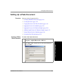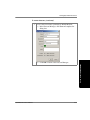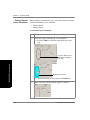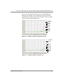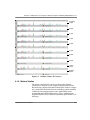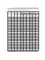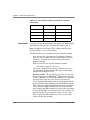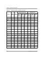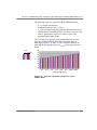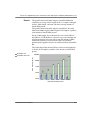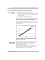
1
Quantifiler™ Kits
Quantifiler™ Human DNA Quantification Kit and
Quantifiler™ Y Human Male DNA Quantification
Kit
User’s Manual
© Copyright 2006, Applied Biosystems. All rights reserved.
For Research, Forensic, or Paternity Use Only. Not for use in diagnostic procedures.
Information in this document is subject to change without notice. Applied Biosystems assumes no responsibility for any errors that
may appear in this document.
APPLIED BIOSYSTEMS DISCLAIMS ALL WARRANTIES WITH RESPECT TO THIS DOCUMENT, EXPRESSED OR
IMPLIED, INCLUDING BUT NOT LIMITED TO THOSE OF MERCHANTABILITY OR FITNESS FOR A PARTICULAR
PURPOSE. IN NO EVENT SHALL APPLIED BIOSYSTEMS BE LIABLE, WHETHER IN CONTRACT, TORT, WARRANTY,
OR UNDER ANY STATUTE OR ON ANY OTHER BASIS FOR SPECIAL, INCIDENTAL, INDIRECT, PUNITIVE,
MULTIPLE OR CONSEQUENTIAL DAMAGES IN CONNECTION WITH OR ARISING FROM THIS DOCUMENT,
INCLUDING BUT NOT LIMITED TO THE USE THEREOF.
NOTICE TO PURCHASER: LIMITED LICENSE
Use of this product is covered by one or more of the following US patents and corresponding patent claims outside the US:
5,079,352, 6,127,155, 5,677,152 (claims 1–23), 5,773,258 (claims 1–10), 5,210,015, 5,487,972, 5,804,375, 5,538,848, 5,723,591,
5,876,930, 6,030,787, and 6,258,569. The purchase of this product includes a limited, non-transferable immunity from suit under
the foregoing patent claims for using only this amount of product solely in forensic and paternity testing, including reporting results
of purchaser’s activities for a fee or other commercial consideration, and also for the purchaser's own internal research. No right
under any other patent claims (such as apparatus or system claims in U.S. Patent No. 6,814,934) is conveyed expressly, by
implication, or by estoppel. Further information on purchasing licenses may be obtained from the Director of Licensing, Applied
Biosystems, 850 Lincoln Centre Drive, Foster City, California 94404, USA.
TRADEMARKS:
Applied Biosystems, AB (Design), ABI PRISM, AmpFlSTR, Genotyper, GeneScan, Identifiler, MicroAmp, Profiler Plus,
QuantiBlot, Quantifiler, and VIC are registered trademarks, and Applera, FAM, MicroAmp, and ROX are trademarks of
Applera Corporation or its subsidiaries in the U.S. and/or certain other countries.
AmpliTaq Gold and TaqMan are registered trademarks of Roche Molecular Systems, Inc.
All other trademarks are the sole property of their respective owners.
Part Number 4344790 Rev. D
04/2006
DRAFT
April 12, 2006 3:32 pm, 7x9_Title.fm
Contents
Preface
How to Use This Guide . . . . . . . . . . . . . . . . . . . . . . . . . . . . . . . . . . . . . . vii
Safety . . . . . . . . . . . . . . . . . . . . . . . . . . . . . . . . . . . . . . . . . . . . . . . . . . . ix
How to Obtain More Information . . . . . . . . . . . . . . . . . . . . . . . . . . . . . . xiii
How to Obtain Support . . . . . . . . . . . . . . . . . . . . . . . . . . . . . . . . . . . . . . xiii
Chapter 1
Overview
Product Overview . . . . . . . . . . . . . . . . . . . . . . . . . . . . . . . . . . . . . . . . . 1-2
Chemistry Overview . . . . . . . . . . . . . . . . . . . . . . . . . . . . . . . . . . . . . . . 1-3
Instrument Overview . . . . . . . . . . . . . . . . . . . . . . . . . . . . . . . . . . . . . . . 1-7
SDS Software Overview . . . . . . . . . . . . . . . . . . . . . . . . . . . . . . . . . . . . 1-8
Real-Time Data Analysis . . . . . . . . . . . . . . . . . . . . . . . . . . . . . . . . . . . 1-10
Procedural Overview . . . . . . . . . . . . . . . . . . . . . . . . . . . . . . . . . . . . . . 1-15
Materials and Equipment . . . . . . . . . . . . . . . . . . . . . . . . . . . . . . . . . . 1-16
Chapter 2
Software Setup
Section 2.1 7000 SDS Software Setup . . . . . . . . . . . . . . . . . . . . . . . . 2-3
Overview . . . . . . . . . . . . . . . . . . . . . . . . . . . . . . . . . . . . . . . . . . . . . . . . 2-4
Starting the 7000 SDS . . . . . . . . . . . . . . . . . . . . . . . . . . . . . . . . . . . . . 2-5
About Plate Documents . . . . . . . . . . . . . . . . . . . . . . . . . . . . . . . . . . . . 2-7
Setting Up a Plate Document . . . . . . . . . . . . . . . . . . . . . . . . . . . . . . . 2-10
Setting Up a Plate Document Template . . . . . . . . . . . . . . . . . . . . . . . 2-22
Section 2.2 7900HT SDS Software Setup . . . . . . . . . . . . . . . . . . . . 2-25
Overview . . . . . . . . . . . . . . . . . . . . . . . . . . . . . . . . . . . . . . . . . . . . . . . 2-26
Starting the 7900HT Real-Time PCR System . . . . . . . . . . . . . . . . . . . 2-27
About Plate Documents . . . . . . . . . . . . . . . . . . . . . . . . . . . . . . . . . . . 2-28
Setting Up a Plate Document . . . . . . . . . . . . . . . . . . . . . . . . . . . . . . . 2-31
Setting Up a Plate Document Template . . . . . . . . . . . . . . . . . . . . . . . 2-40
Quantifiler Kits User’s Manual
iii
Chapter 3
PCR Amplification
Preparing the DNA Quantification Standard . . . . . . . . . . . . . . . . . . . . 3-2
Preparing the Reactions . . . . . . . . . . . . . . . . . . . . . . . . . . . . . . . . . . . . 3-5
Running the Reactions . . . . . . . . . . . . . . . . . . . . . . . . . . . . . . . . . . . . . 3-7
Chapter 4
Data Analysis and Results
Section 4.1 7000 SDS Data Analysis . . . . . . . . . . . . . . . . . . . . . . . . . 4-3
Analyzing the Plate Document . . . . . . . . . . . . . . . . . . . . . . . . . . . . . . . 4-3
Viewing Results . . . . . . . . . . . . . . . . . . . . . . . . . . . . . . . . . . . . . . . . . . 4-4
Section 4.2 7900HT SDS Data Analysis . . . . . . . . . . . . . . . . . . . . . . 4-7
Analyzing the Plate Document . . . . . . . . . . . . . . . . . . . . . . . . . . . . . . . 4-7
Viewing Results . . . . . . . . . . . . . . . . . . . . . . . . . . . . . . . . . . . . . . . . . . 4-8
Chapter 5
Interpretation of Results
Checking Analysis Settings . . . . . . . . . . . . . . . . . . . . . . . . . . . . . . . . . 5-2
Examining the Standard Curve . . . . . . . . . . . . . . . . . . . . . . . . . . . . . . 5-4
Troubleshooting the Standard Curve . . . . . . . . . . . . . . . . . . . . . . . . . . 5-6
Using the Internal PCR Control System . . . . . . . . . . . . . . . . . . . . . . . 5-10
Troubleshooting Amplification Plots . . . . . . . . . . . . . . . . . . . . . . . . . . 5-12
Assessing Quantity . . . . . . . . . . . . . . . . . . . . . . . . . . . . . . . . . . . . . . . 5-16
Chapter 6
Experiments and Results
Overview . . . . . . . . . . . . . . . . . . . . . . . . . . . . . . . . . . . . . . . . . . . . . . . . 6-2
Section 6.1 ABI PRISM® 7000 Sequence Detection System Validation
(SDS Software v1.0) . . . . . . . . . . . . . . . . . . . . . . . . . . . . . . . . . . . . . . 6-3
Precision . . . . . . . . . . . . . . . . . . . . . . . . . . . . . . . . . . . . . . . . . . . . . . . . . 6-4
Reproducibility . . . . . . . . . . . . . . . . . . . . . . . . . . . . . . . . . . . . . . . . . . . . 6-7
Specificity with a Human DNA Panel . . . . . . . . . . . . . . . . . . . . . . . . . . 6-10
Specificity with a Non-Human Panel. . . . . . . . . . . . . . . . . . . . . . . . . . . 6-11
Specificity with a Bacterial Pools Panel . . . . . . . . . . . . . . . . . . . . . . . . 6-14
iv
Quantifiler Kits User’s Manual
Sensitivity . . . . . . . . . . . . . . . . . . . . . . . . . . . . . . . . . . . . . . . . . . . . . . . .6-16
Stability . . . . . . . . . . . . . . . . . . . . . . . . . . . . . . . . . . . . . . . . . . . . . . . . .6-17
Mixture Studies . . . . . . . . . . . . . . . . . . . . . . . . . . . . . . . . . . . . . . . . . . .6-21
Degraded DNA Studies . . . . . . . . . . . . . . . . . . . . . . . . . . . . . . . . . . . . .6-23
Comparisons with Other Methods . . . . . . . . . . . . . . . . . . . . . . . . . . . . .6-27
Comparison with A260 and Quantiblot Kit . . . . . . . . . . . . . . . . . . . . . . .6-27
Comparison with A260 and Dye Intercalation . . . . . . . . . . . . . . . . . . . . .6-31
Assay Background . . . . . . . . . . . . . . . . . . . . . . . . . . . . . . . . . . . . . . . . .6-35
Section 6.2 Applied Biosystems
7900HT Real-Time PCR System Validation (SDS Software v2.0) . . 6-37
Precision (7900HT SDS). . . . . . . . . . . . . . . . . . . . . . . . . . . . . . . . . . . . .6-37
Mixture Studies (7900HT SDS) . . . . . . . . . . . . . . . . . . . . . . . . . . . . . . .6-40
Comparisons with Other Methods (7900HT SDS) . . . . . . . . . . . . . . . . .6-41
Section 6.3 Casework Sample Analysis . . . . . . . . . . . . . . . . . . . . . . 6-45
Section 6.4 Applied Biosystems
7500 Real-Time PCR System Validation (SDS Software v1.2.3) . . . 6-50
Materials and Methods . . . . . . . . . . . . . . . . . . . . . . . . . . . . . . . . . . . . .6-50
Experimental Setup . . . . . . . . . . . . . . . . . . . . . . . . . . . . . . . . . . . . . . . .6-51
Data Collection. . . . . . . . . . . . . . . . . . . . . . . . . . . . . . . . . . . . . . . . . . . .6-54
Data Analysis . . . . . . . . . . . . . . . . . . . . . . . . . . . . . . . . . . . . . . . . . . . . .6-54
Discussion . . . . . . . . . . . . . . . . . . . . . . . . . . . . . . . . . . . . . . . . . . . . . . .6-63
Conclusion . . . . . . . . . . . . . . . . . . . . . . . . . . . . . . . . . . . . . . . . . . . . . . .6-64
Section 6.5 ABI PRISM® 7000 Sequence Detection System Validation
(SDS Software v1.2.3) . . . . . . . . . . . . . . . . . . . . . . . . . . . . . . . . . . . . 6-66
Materials and Methods . . . . . . . . . . . . . . . . . . . . . . . . . . . . . . . . . . . . .6-66
Experimental Setup . . . . . . . . . . . . . . . . . . . . . . . . . . . . . . . . . . . . . . . .6-67
Data Collection. . . . . . . . . . . . . . . . . . . . . . . . . . . . . . . . . . . . . . . . . . . .6-69
Data Analysis . . . . . . . . . . . . . . . . . . . . . . . . . . . . . . . . . . . . . . . . . . . . .6-69
Discussion . . . . . . . . . . . . . . . . . . . . . . . . . . . . . . . . . . . . . . . . . . . . . . .6-79
Conclusion . . . . . . . . . . . . . . . . . . . . . . . . . . . . . . . . . . . . . . . . . . . . . . .6-80
Bibliography
Index
Quantifiler Kits User’s Manual
v
vi
Quantifiler Kits User’s Manual
Preface
This preface contains:
How to Use This Guide . . . . . . . . . . . . . . . . . . . . . . . . . . . . . . . . . . vii
Safety. . . . . . . . . . . . . . . . . . . . . . . . . . . . . . . . . . . . . . . . . . . . . . . . ix
How to Obtain More Information . . . . . . . . . . . . . . . . . . . . . . . . . xiv
How to Obtain Support . . . . . . . . . . . . . . . . . . . . . . . . . . . . . . . . . xiv
How to Use This Guide
Purpose of This
Guide
Text Conventions
The Quantifiler® Kits User’s Manual provides information about and
instructions for using the Quantifiler® Human DNA Quantification
Kit and the Quantifiler® Y Human Male DNA Quantification Kit.
This guide uses the following conventions:
• Bold indicates user action. For example:
Type 0, then press Enter for each of the remaining fields.
• Italic text indicates new or important words and is also used for
emphasis. For example:
Before analyzing, always prepare fresh matrix.
• A right arrow bracket (>) separates successive commands you
select from a drop-down or shortcut menu. For example:
Select File > Open > Spot Set.
Right-click the sample row, then select View Filter > View All
Runs.
Quantifiler Kits User’s Manual
vii
Preface
User Attention
Words
Two user attention words appear in Applied Biosystems user
documentation. Each word implies a particular level of observation
or action as described below:
Note: Provides information that may be of interest or help but is not
critical to the use of the product.
IMPORTANT! Provides information that is necessary for proper
instrument operation, accurate chemistry kit use, or safe use of a
chemical.
Examples of the user attention words appear below:
Note: The size of the column affects the run time.
Note: The Calibrate function is also available in the Control Console.
IMPORTANT! To verify your client connection to the database, you
need a valid Oracle user ID and password.
IMPORTANT! You must create a separate Sample Entry Spreadsheet
for each 96-well plate.
Safety Alert
Words
viii
Safety alert words also appear in user documentation. For more
information, see “Safety Alert Words” on page ix.
Quantifiler Kits User’s Manual
Safety
Safety
Safety Alert
Words
Four safety alert words appear in Applied Biosystems user
documentation at points in the document where you need to be aware
of relevant hazards. Each alert word–IMPORTANT, CAUTION,
WARNING, DANGER–implies a particular level of observation or
action, as defined below:
IMPORTANT! – Indicates information that is necessary for proper
instrument operation, accurate chemistry kit use, or safe use of a
chemical.
– Indicates a potentially hazardous situation that,
if not avoided, may result in minor or moderate injury. It may also be
used to alert against unsafe practices.
– Indicates a potentially hazardous situation that,
if not avoided, could result in death or serious injury.
– Indicates an imminently hazardous situation
that, if not avoided, will result in death or serious injury. This signal
word is to be limited to the most extreme situations.
Chemical Hazard
Warning
CHEMICAL HAZARD. Some of the chemicals
used with Applied Biosystems instruments and protocols are
potentially hazardous and can cause injury, illness, or death.
Quantifiler Kits User’s Manual
ix
Preface
Chemical Safety
Guidelines
About MSDSs
To minimize the hazards of chemicals:
• Read and understand the Material Safety Data Sheets (MSDS)
provided by the chemical manufacturer before you store, handle,
or work with any chemicals or hazardous materials. (See “About
MSDSs.”)
• Minimize contact with chemicals. Wear appropriate personal
protective equipment when handling chemicals (for example,
safety glasses, gloves, or protective clothing). For additional
safety guidelines, consult the MSDS.
• Minimize the inhalation of chemicals. Do not leave chemical
containers open. Use only with adequate ventilation (for
example, fume hood). For additional safety guidelines, consult
the MSDS.
• Check regularly for chemical leaks or spills. If a leak or spill
occurs, follow the manufacturer’s cleanup procedures as
recommended on the MSDS.
• Comply with all local, state/provincial, or national laws and
regulations related to chemical storage, handling, and disposal.
Chemical manufacturers supply current Material Safety Data Sheets
(MSDSs) with shipments of hazardous chemicals to new customers.
They also provide MSDSs with the first shipment of a hazardous
chemical to a customer after an MSDS has been updated. MSDSs
provide the safety information you need to store, handle, transport,
and dispose of the chemicals safely.
Each time you receive a new MSDS packaged with a hazardous
chemical, be sure to replace the appropriate MSDS in your files.
Obtaining MSDSs
The MSDS for any chemical supplied by Applied Biosystems is
available to you free 24 hours a day. To obtain MSDSs:
1. Go to https://docs.appliedbiosystems.com/msdssearch.html
2. In the Search field of the MSDS Search page:
a. Type in the chemical name, part number, or other
information that you expect to appear in the MSDS of
interest.
b. Select the language of your choice.
c. Click Search.
x
Quantifiler Kits User’s Manual
Safety
3. To view, download, or print the document of interest:
a. Right-click the document title.
b. Select:
• Open – To view the document
• Save Target As – To download a PDF version of the
document to a destination that you choose
• Print Target – To print the document
4. To have a copy of an MSDS sent by fax or e-mail, in the Search
Results page:
a. Select Fax or Email below the document title.
b. Click RETRIEVE DOCUMENTS at the end of the
document list.
c. Enter the required information.
d. Click View/Deliver Selected Documents Now.
Note: For the MSDSs of chemicals not distributed by Applied
Biosystems, contact the chemical manufacturer.
Chemical Waste
Hazard
Chemical Waste
Safety Guidelines
CHEMICAL WASTE HAZARD. Some wastes
produced by the operation of the instrument or system are potentially
hazardous and can cause injury, illness, or death.
To minimize the hazards of chemical waste:
Quantifiler Kits User’s Manual
• Read and understand the Material Safety Data Sheets (MSDSs)
provided by the manufacturers of the chemicals in the waste
container before you store, handle, or dispose of chemical waste.
• Provide primary and secondary waste containers. (A primary
waste container holds the immediate waste. A secondary
container contains spills or leaks from the primary container.
Both containers must be compatible with the waste material and
meet federal, state, and local requirements for container
storage.)
• Minimize contact with chemicals. Wear appropriate personal
protective equipment when handling chemicals (for example,
safety glasses, gloves, or protective clothing). For additional
safety guidelines, consult the MSDS.
xi
Preface
• Minimize the inhalation of chemicals. Do not leave chemical
containers open. Use only with adequate ventilation (for
example, fume hood).For additional safety guidelines, consult
the MSDS.
• Handle chemical wastes in a fume hood.
• After emptying the waste container, seal it with the cap
provided.
• Dispose of the contents of the waste tray and waste bottle in
accordance with good laboratory practices and local,
state/provincial, or national environmental and health
regulations.
Waste Disposal
If potentially hazardous waste is generated when you operate the
instrument, you must:
• Characterize (by analysis if necessary) the waste generated by
the particular applications, reagents, and substrates used in your
laboratory.
• Ensure the health and safety of all personnel in your laboratory.
• Ensure that the instrument waste is stored, transferred,
transported, and disposed of according to all local,
state/provincial, and/or national regulations.
IMPORTANT! Radioactive or biohazardous materials may require
special handling, and disposal limitations may apply.
Biological Hazard
Safety
BIOHAZARD. Biological samples such as
tissues, body fluids, and blood of humans and other animals have the
potential to transmit infectious diseases. Follow all applicable local,
state/provincial, and/or national regulations. Wear appropriate
protective eyewear, clothing, and gloves. Read and follow the
guidelines in these publications:
• U.S. Department of Health and Human Services guidelines
published in Biosafety in Microbiological and Biomedical
Laboratories (stock no. 017-040-00547-4;
http://bmbl.od.nih.gov)
xii
Quantifiler Kits User’s Manual
Safety
• Occupational Safety and Health Standards, Bloodborne
Pathogens
(29 CFR§1910.1030; http://www.access.gpo.gov/nara/cfr/
waisidx_01/29cfr1910a_01.html).
Additional information about biohazard guidelines is available at:
http://www.cdc.gov
Quantifiler Kits User’s Manual
xiii
Preface
How to Obtain More Information
Related
Documentation
• ABI PRISM® 7000 Sequence Detection System User Guide –
Describes the 7000 SDS hardware and software and provides
information on preparing, maintaining, and troubleshooting the
system.
• ABI PRISM® 7900HT Sequence Detection System User Guide –
Describes the 7900HT SDS hardware and software and provides
information on preparing, maintaining, and troubleshooting the
system.
Note: For additional documentation, see “How to Obtain Support”
below.
Send Us Your
Comments
Applied Biosystems welcomes your comments and suggestions for
improving its user documents. You can e-mail your comments to:
[email protected]
How to Obtain Support
For the latest services and support information for all locations, go to
http://www.appliedbiosystems.com, then click the link for
Support.
At the Support page, you can:
• Obtain worldwide telephone and fax numbers to contact
Applied Biosystems Technical Support and Sales facilities
• Search through frequently asked questions (FAQs)
• Submit a question directly to Technical Support
• Order Applied Biosystems user documents, MSDSs, certificates
of analysis, and other related documents
• Download PDF documents
• Obtain information about customer training
• Download software updates and patches
xiv
Quantifiler Kits User’s Manual
Overview
1
1
This chapter covers:
Product Overview . . . . . . . . . . . . . . . . . . . . . . . . . . . . . . . . . . . . . .1-2
Chemistry Overview . . . . . . . . . . . . . . . . . . . . . . . . . . . . . . . . . . . .1-3
Instrument Overview. . . . . . . . . . . . . . . . . . . . . . . . . . . . . . . . . . . .1-7
SDS Software Overview . . . . . . . . . . . . . . . . . . . . . . . . . . . . . . . . .1-8
Real-Time Data Analysis . . . . . . . . . . . . . . . . . . . . . . . . . . . . . . .1-10
Procedural Overview. . . . . . . . . . . . . . . . . . . . . . . . . . . . . . . . . . .1-15
Materials and Equipment . . . . . . . . . . . . . . . . . . . . . . . . . . . . . . .1-16
Quantifiler Kits User’s Manual
1-1
Chapter 1 Overview
Product Overview
Purpose
The Quantifiler® Human DNA Quantification Kit (PN 4343895) and
the Quantifiler® Y Human Male DNA Quantification Kit
(PN 4343906) are designed to quantify the total amount of
amplifiable human (and higher primate) DNA or human male DNA
in a sample. The results from using the kits can aid in determining:
• If sufficient human DNA or human male DNA is present to
proceed with short tandem repeat (STR) analysis
• How much sample to use in STR analysis applications
Product
Description
The Quantifiler kits contain all the necessary reagents for the
amplification, detection, and quantification of a human-specific
DNA target or a human male-specific DNA target.
The reagents are designed and optimized for use with the following
Applied Biosystems instruments and software:
• ABI PRISM® 7000 Sequence Detection System and SDS
Software v1.0
• ABI PRISM® 7900HT Sequence Detection System (no
automation module) and SDS Software v2.0.
See Chapter 6, “Experiments and Results,” for validation studies
performed using the Applied Biosystems 7500 Real-Time PCR
System with SDS Software v1.2.3 and the ABI PRISM® 7000
Sequence Detection System with SDS Software v1.2.3.
1-2
Quantifiler Kits User’s Manual
Chemistry Overview
Chemistry Overview
Assay Overview
The DNA quantification assay combines two 5′ nuclease assays:
• A target-specific (human DNA or human male DNA) assay
• An internal PCR control (IPC) assay
Target-Specific
Assay
Components
About the Targets
Table 1-1
The target-specific assay consists of:
• Two primers for amplifying human DNA or human male DNA
• One TaqMan® MGB probe labeled with FAM™ dye for
detecting the amplified sequence
Table 1-1 provides information about the targets of PCR
amplification in the Quantifiler Human kit and the Quantifiler Y kit.
Targets of Quantifiler kits
Kit
Gene Target
Location
Amplicon
Length
Region
Amplified
Ploidy
Quantifiler Human kit
Human telomerase
reverse
transcriptase gene
(hTERT)
5p15.33
62 bases
Nontranslated
region (intron)
Diploid a
Quantifiler Y kit
Sex-determining
region Y gene (SRY)
Yp11.3
64 bases
Nontranslated
region
Haploida
a. Single-copy target.
IPC Assay
Components
The IPC assay consists of:
Quantifiler Kits User’s Manual
• IPC template DNA (a synthetic sequence not found in nature)
• Two primers for amplifying the IPC template DNA
• One TaqMan® MGB probe labeled with VIC® dye for detecting
the amplified IPC DNA
1-3
Chapter 1 Overview
About the Probes
The TaqMan MGB probes contain:
• A reporter dye (FAM™ dye or VIC® dye) linked to the 5′ end of
the probe
• A minor groove binder (MGB) at the 3´ end of the probe
This modification increases the melting temperature (Tm)
without increasing probe length (Afonina et al., 1997; Kutyavin
et al., 1997), which allows the design of shorter probes.
• A nonfluorescent quencher (NFQ) at the 3′ end of the probe
Because the quencher does not fluoresce, Applied Biosystems
sequence detection systems can measure reporter dye
contributions more accurately.
5′ Nuclease
Assay Process
The 5′ nuclease assay process (Figures 1-1 through 1-5) takes place
during PCR amplification. This process occurs in every cycle and
does not interfere with the exponential accumulation of product.
NFQ
= Nonfluorescent quencher
MGB
= Minor groove binder
R
= Reporter
P
= AmpliTaq Gold ®
DNA Polymerase
Figure 1-1
Legend for 5′ nuclease assay process figures
During PCR, the TaqMan MGB probe anneals specifically to a
complementary sequence between the forward and reverse primer
sites (Figure 1-2).
When the probe is intact (Figures 1-2 and 1-3), the proximity of the
reporter dye to the quencher dye results in suppression of the reporter
fluorescence primarily by Förster-type energy transfer (Förster, 1948;
Lakowicz, 1983).
1-4
Quantifiler Kits User’s Manual
Chemistry Overview
5′
Forward
Primer
R
P
TaqMan
MGB probe
NFQ
MGB
3′
3′
5′
5′
3′
P
Figure 1-2
5′
Reverse
Primer
Polymerization
R
5′
Forward
Primer
TaqMan
MGB probe
P
NFQ
MGB
3′
3′
5′
5′
3′
P
Figure 1-3
5′
Reverse
Primer
Strand displacement
AmpliTaq Gold® DNA polymerase cleaves only probes that are
hybridized to the target (Figure 1-4). Cleavage separates the reporter
dye from the quencher dye, which results in increased fluorescence
by the reporter. The increase in fluorescence signal occurs only if the
target sequence is complementary to the probe and is amplified
during PCR. Because of these requirements, nonspecific
amplification is not detected.
5′
Forward
Primer
R
P
TaqMan
MGB probe
NFQ
MGB
3′
3′
5′
5′
3′
P
Figure 1-4
Quantifiler Kits User’s Manual
5′
Reverse
Primer
Cleavage
1-5
Chapter 1 Overview
Polymerization of the strand continues, but because the 3′ end of the
probe is blocked, there is no extension of the probe during PCR
(Figure 1-5).
5′
Forward
Primer
R
TaqMan
MGB probe
NFQ
MGB
3′
3′
5′
5′
3′
5′
Reverse
Primer
Figure 1-5
1-6
Completion of polymerization
Quantifiler Kits User’s Manual
Instrument Overview
Instrument Overview
Fluorescence
Detection
Detection on the ABI PRISM 7000 Sequence Detection System
1. A tungsten-halogen lamp directs light to each well on the
reaction plate. The light passes through the ABI PRISM™
Optical Adhesive Cover and excites the fluorescent dyes in each
well of the plate.
2. A system of lenses, filters, and a dichroic mirror focuses the
fluorescence emission into a charge-coupled device (CCD)
camera.
3. Based on wavelength, the filters separate the light into a
predictably spaced pattern across the CCD camera.
4. During the run, the CCD camera detects the fluorescence
emission between 500 nm and 660 nm from each well.
5. The SDS software obtains the fluorescence emission data from
the CCD camera and applies data analysis algorithms.
Detection on the ABI PRISM 7900HT Sequence Detection System
1. An argon ion laser directs light to each well on the microplate.
The light passes through the ABI PRISM Optical Adhesive Cover
and excites the fluorescent dyes in each well of the plate.
2. A system of lenses, filters, and a dichroic mirror focuses the
fluorescence emission into a grating.
3. Based on wavelength, the grating separates the light into a
predictably spaced pattern across the CCD camera.
4. During the run, the CCD camera detects the fluorescence
emission between 500 nm and 660 nm from each well.
5. The SDS software obtains the fluorescence emission data from
the CCD camera and applies data analysis algorithms.
Quantifiler Kits User’s Manual
1-7
Chapter 1 Overview
SDS Software Overview
This section describes how the SDS software analyzes raw run data
from real-time runs. Raw data consists of the spectral data between
500 nm to 660 nm collected by the SDS software during a sequence
detection run.
Composite
Spectrum
Figure 1-6 shows a composite fluorescence spectrum from a single
well containing the passive reference, one probe labeled with FAM™
dye and a nonfluorescent quencher, and one probe labeled with VIC®
dye and a nonfluorescent quencher. The example shows how the
overlapping component dye spectra contribute to the composite
spectrum.
0.110
FAM™ dye
Composite spectrum
(all dyes +
Background + MSE)
ROX™ dye
VIC® dye
Pure Dye components
0.083
0.055
0.027
0.000
500
550
600
Wavelength
Figure 1-6
1-8
650
Background
(from background
calibration run)
Example of a composite spectrum
Quantifiler Kits User’s Manual
SDS Software Overview
Processing
Multicomponent
Data
During the multicomponent transformation, the SDS software uses
algorithms to determine the contribution of each dye:
• An algorithm removes the background component stored in the
background calibration file to eliminate the contribution of
background fluorescence in the raw data.
• The software uses the extracted pure dye standards to express
the composite spectrum in terms of the pure dye components.
• Then, an algorithm applies matrix calculations to determine the
contributions of each component dye to the composite spectrum.
The software uses the pure dye spectra, generated as part of
instrument calibration, to solve for coefficients a, b, and c and to
calculate the mean standard error (MSE) in the following equation:
Measured spectrum = a ( FAM ) + b ( VIC ) + c ( ROX ) + d ( Background ) + MSE
where coefficients a, b, and c represent the contribution of each dye
to the composite spectrum. The MSE value indicates how closely the
collective multicomponent spectrum conforms to the raw spectra.
Note: The example equation above assumes that pure dye
components exist for FAM dye, VIC dye, and ROX dye and for the
instrument background.
Temperature
Fluorescence
FAM dye component
12000
8000
VIC dye component
4000
ROX dye component
0
93
Background
component
83
MSE
73
63
53
0:10
0:30
Figure 1-7
reaction
Quantifiler Kits User’s Manual
0:50 6978 1:10
Time
1:30
Typical component contributions in a multiplex
1-9
Chapter 1 Overview
Normalization of
Reporter Signals
The SDS software displays cycle-by-cycle changes in normalized
reporter signal (Rn). The SDS software normalizes each reporter
signal by dividing it by the fluorescent signal of the passive reference
dye. Because the passive reference is one component of the PCR
master mix, it is present at the same concentration in all wells of the
reaction plate. By normalizing the reporter signal using the passive
reference, the software can account for minor variations in signal
caused by pipetting inaccuracies and make better well-to-well
comparisons of reporter signal.
Real-Time Data Analysis
The 7000 SDS and the 7900HT SDS can be used to determine the
relative quantity of a target nucleic acid sequence in a sample by
analyzing the cycle-to-cycle change in fluorescent signal as a result
of amplification during a PCR (Figure 1-8).
Amplification Plot
Example
When using TaqMan probes with the 7000 SDS or 7900HT SDS, the
fluorescent signal (or normalized reporter, Rn) increases as the
amount of specific amplified product increases. Figure 1-8 shows the
amplification of PCR product in a plot of Rn vs. cycle number during
PCR. This amplification plot contains three distinct phases that
characterize the progression of the PCR.
8.0
Phase 3, plateau
2.0
1.0
Phase 2, linear
Rn
Phase 1, geometric
0.0
0
10
20
30
40
Cycle Number
Figure 1-8
1-10
Phases of PCR amplification
Quantifiler Kits User’s Manual
Real-Time Data Analysis
Phases of
Amplification
Initially, Rn appears as a flat line because the fluorescent signal is
below the detection limit of the sequence detector.
Phase 1: Geometric (Exponential)
Signal is detected and increases in direct proportion to the increase of
PCR product. As PCR product continues to increase, the ratio of
AmpliTaq Gold polymerase to PCR product decreases.
During the geometric phase, amplification is characterized by a high
and constant efficiency. It occurs between the first detectable rise in
fluorescence and before the beginning of the linear phase. During the
geometric phase, a plot of DNA concentration versus cycle number
on a log scale should approximate a straight line with a slope.
Typically, the SDS system is sufficiently sensitive to detect at least
3 cycles in the geometric phase, assuming reasonably optimized PCR
conditions. When the template concentration reaches 10−8 M, PCR
product stops accumulating exponentially.
Phase 2: Linear
During the linear phase, the slope of the amplification plot decreases
steadily. At this point, one or more components of the PCR has
decreased below a critical concentration, and the amplification
efficiency begins to decrease. This phase is termed linear because
amplification approximates an arithmetic progression, rather than a
geometric increase. Because the amplification efficiency is
continually decreasing during the linear phase, it exhibits low
precision.
Phase 3: Plateau
The amplification plot achieves the plateau phase when the PCR
stops, the Rn signal remains relatively constant, and the template
concentration reaches a plateau at about 10−7 M (Martens and Naes,
1989).
Quantifiler Kits User’s Manual
1-11
Chapter 1 Overview
Relationship of
Amplified PCR
Product to Initial
Template
Concentration
Because of the progressive cleavage of TaqMan fluorescent probes
during the PCR, as the concentration of amplified product increases
in a sample, so does the Rn value. The following equation describes
the relationship of amplified PCR product to initial template during
the geometric phase:
Nc = N ( 1 + E ) c
where Nc is the concentration of amplified product at any cycle, N is
the initial concentration of target template, E is the efficiency of the
system, and c is the cycle number.
For example, with the dilutions of RNase P target in the TaqMan®
RNase P Instrument Verification Plate, the ratio of template
concentration to detectable signal is preserved in the geometric phase
for all dilutions (Figure 1-9). As the rate of amplification approaches
a plateau, the amount of product is no longer proportional to the
initial number of template copies.
Copy
Number
20000
10000
5000
2500
1250
8.0
Rn
2.0
1.0
0.0
0
10
20
30
40
Cycle Number
Figure 1-9 Amplification plot from a real-time run of an RNase P
Instrument Verification Plate
1-12
Quantifiler Kits User’s Manual
Real-Time Data Analysis
About the
Threshold
About the
Threshold Cycle
The SDS software uses a threshold setting to define the level of
detectable fluorescence. Based on the number of cycles required to
reach the threshold, the SDS software can compare test samples
quantitatively: A sample with a higher starting template copy number
reaches the threshold earlier than a sample with a lower starting
template copy number.
The threshold cycle (CT) for a specified amplification plot occurs
when the fluorescent signal increases beyond the value of the
threshold setting. The CT value depends on:
• Starting template copy number
• Efficiency of DNA amplification by the PCR system
How CT Values
Are Determined
To determine the CT value, the SDS software uses the Rn values
collected from a predefined range of PCR cycles called the baseline
(the default baseline occurs between cycles 6 and 15 on the
7000 SDS and between cycles 3 and 15 on the 7900HT SDS):
1. The software generates a baseline-subtracted amplification plot
of ∆Rn versus cycle number.
2. An algorithm defines the cycle where the ∆Rn value crosses the
threshold setting (the default threshold setting is 0.2) as the
threshold cycle (CT).
Quantifiler Kits User’s Manual
1-13
Chapter 1 Overview
Relationship of
Threshold Cycles
to Initial Template
Amount
The following equation describes the exponential amplification of
the PCR:
Xn = Xm ( 1 + EX )
n–m
where:
Xn = number of target molecules at cycle n (so that n > m)
Xm = number of target molecules at cycle m
EX = efficiency of target amplification (between 0 and 1)
n − m = number of cycles elapsed between cycle m and cycle n
Amplicons designed and optimized according to Applied Biosystems
guidelines (amplicon size <150 bp) have amplification efficiencies
that approach 100%. Therefore EX = 1 so that:
n–m
Xn = Xm ( 1 + 1 )
n–m
= Xm ( 2 )
To define the significance in amplified product of one thermal cycle,
set n − m = 1 so that:
Xn = Xm ( 2 )
1
= 2X m
Therefore, each cycle in the PCR reaction corresponds to a two-fold
increase in product. Likewise, a difference in CT values of 1 equates
to a two-fold difference in initial template amount.
1-14
Quantifiler Kits User’s Manual
Procedural Overview
Procedural Overview
Use of the Quantifiler kits involves the following workflow:
Software Setup
PCR Amplification
Data Analysis
Interpretation of Results
Quantifiler Kits User’s Manual
1-15
Chapter 1 Overview
Materials and Equipment
Kit Contents and
Storage
Each Quantifiler® kit contains materials sufficient to perform 400
reactions at a 25-µL reaction volume.
Table 1-2 Quantifiler kit contents
Reagent
Contents
Quantity
Storage
• Forward and reverse
primers to amplify
human DNA or human
male DNA target
• Probe to detect
human DNA or human
male DNA target
• IPC system primers,
template, and probe
3 tubes,
1.4 mL
each
−15 to −25 °C
Quantifiler Human DNA Standard
200 ng/µL purified DNA
standard
1 tube,
120 µL
−15 to −25 °C
Quantifiler PCR Reaction Mix
AmpliTaq Gold® DNA
Polymerase, dNTPs with
dUTP, Passive Reference,
and optimized buffer
components
1 tube,
5 mL
2 to 8 °C
Quantifiler® Human Primer Mix or
Quantifiler® Y Human Male Primer Mix
Additional
Storage
Guidelines For
Primer Mixes
1-16
Follow the additional guidelines for storing the primer mixes:
• Minimize freeze-thaw cycles.
• Keep protected from direct exposure to light. Excessive
exposure to light may affect the fluorescent probes.
Quantifiler Kits User’s Manual
Materials and Equipment
Equipment and
Materials Not
Included
Tables 1-3 through 1-5 list required and optional equipment and
materials not supplied with the Quantifiler kits. Unless otherwise
noted, many of the items are available from major laboratory
suppliers (MLS).
Table 1-3
Equipment
Equipment
Applied Biosystems 7900HT Real-Time PCR
System (no automation)
Source
Contact your local
Applied Biosystems
sales representative.
ABI PRISM® 7000 Sequence Detection System
Tabletop centrifuge with 96-well plate
adapters (optional)
Table 1-4
MLS
User-supplied materials
Material
Source
Quantifiler® Human DNA Quantification Kit
Applied Biosystems
(PN 4343895)
Quantifiler® Y Human Male DNA
Quantification Kit
Applied Biosystems
(PN 4343906)
Glycogen, 20 mg (1 mL)
Roche Applied Science
(PN 901 393)
High-Throughput Setup
Quantifiler Kits User’s Manual
96-Well Optical Reaction Plates
Applied Biosystems
(PN 4306737)
Optical Adhesive Covers Starter Kit (20
covers, 1 compression pad, 1 applicator)
Applied Biosystems
(PN 4313663)
Optical Adhesive Covers (100 covers)
Applied Biosystems
(PN 4311971)
MicroAmp™ Splash Free Support Base
Applied Biosystems
(PN 4312063)
1-17
Chapter 1 Overview
Table 1-4
User-supplied materials (continued)
Material
Source
Mid-to-Low-Throughput Setup
MicroAmp® Optical Tubes (8 tubes/strip, 125
strips)
Applied Biosystems
(PN 4316567)
MicroAmp™ 96-Well Tray/Retainer Set
Applied Biosystems
(PN 403081)
Optical Caps (8 caps/strip, 300 strips)
Applied Biosystems
(PN 4323032)
Compression pad from Optical Adhesive
Covers Starter Kit
Applied Biosystems
(PN 4313663)
Note: Not necessary if using Optical Caps
Table 1-5
1-18
Documents
Document
Applied Biosystems
Part Number
ABI PRISM® 7000 Sequence Detection System
User Guide
4330228
ABI PRISM® 7900HT Sequence Detection
System User Guide
4317596
Quantifiler Kits User’s Manual
Chapter 2
Software Setup
Quantifiler Kits User’s Manual
I
Software Setup
2
2
This chapter covers:
Section 2.1 7000 SDS Software Setup . . . . . . . . . . . . . . . . . . . . .2-3
Overview . . . . . . . . . . . . . . . . . . . . . . . . . . . . . . . . . . . . . . . . . . . . .2-4
Starting the 7000 SDS. . . . . . . . . . . . . . . . . . . . . . . . . . . . . . . . . . .2-5
About Plate Documents. . . . . . . . . . . . . . . . . . . . . . . . . . . . . . . . . .2-7
Setting Up a Plate Document . . . . . . . . . . . . . . . . . . . . . . . . . . . .2-10
Setting Up a Plate Document Template . . . . . . . . . . . . . . . . . . . .2-22
Section 2.2 7900HT SDS Software Setup . . . . . . . . . . . . . . . . .2-25
Overview . . . . . . . . . . . . . . . . . . . . . . . . . . . . . . . . . . . . . . . . . . . .2-26
Starting the 7900HT Real-Time PCR System. . . . . . . . . . . . . . . .2-27
About Plate Documents. . . . . . . . . . . . . . . . . . . . . . . . . . . . . . . . .2-28
Setting Up a Plate Document . . . . . . . . . . . . . . . . . . . . . . . . . . . .2-31
Setting Up a Plate Document Template . . . . . . . . . . . . . . . . . . . .2-40
Quantifiler Kits User’s Manual
2-1
7000 SDS Software Setup
Chapter 2 Software Setup
2-2
Quantifiler Kits User’s Manual
Section 2.1 7000 SDS Software Setup
Section 2.1 7000 SDS Software Setup
This section covers:
Quantifiler Kits User’s Manual
2-3
7000 SDS Software Setup
Overview . . . . . . . . . . . . . . . . . . . . . . . . . . . . . . . . . . . . . . . . . . . . .2-4
Starting the 7000 SDS. . . . . . . . . . . . . . . . . . . . . . . . . . . . . . . . . . .2-5
About Plate Documents. . . . . . . . . . . . . . . . . . . . . . . . . . . . . . . . . .2-7
Setting Up a Plate Document . . . . . . . . . . . . . . . . . . . . . . . . . . . .2-10
Setting Up a Plate Document Template . . . . . . . . . . . . . . . . . . . .2-22
Chapter 2 Software Setup
7000 SDS Software Setup
Overview
2-4
Purpose
During software setup, you start up the ABI PRISM® 7000 Sequence
Detection System (7000 SDS) and set up a plate document for DNA
quantification using the Quantifiler® kits.
Configuration
The Quantifiler® kits are supported using the 7000 SDS and
Sequence Detection Systems (SDS) Software v1.0 for real-time data
collection and analysis.
Quantifiler Kits User’s Manual
Starting the 7000 SDS
Starting the 7000 SDS
Overview
Starting the 7000 SDS involves:
1. Starting the Computer
2. Powering On the Instrument (page 2-6)
3. Starting SDS Software (page 2-6)
7000 SDS Software Setup
Starting the
Computer
1. If you are using the laptop computer, open it by pushing in
the front, center button, holding it, and lifting up the lid.
Instrument power button
GR2078
2. Press the power button on the computer.
Laptop power button
3. In the Enter User name field of the login window, type your
name or the user name associated with the computer.
4. If required, type your password in the Password field.
Quantifiler Kits User’s Manual
2-5
Chapter 2 Software Setup
Powering On the
Instrument
Note: Wait for the computer to finish starting up before powering on
the 7000 instrument.
PHYSICAL INJURY HAZARD. Moving parts
can crush and cut. Keep hands clear of moving parts while operating.
Disconnect power before servicing the instrument.
7000 SDS Software Setup
Press the power button on the lower left front of the instrument.
Power button
Starting SDS
Software
2-6
Select Start > ABI Prism 7000 > ABI Prism 7000 SDS Software.
The software attempts to initialize the instrument and displays a
message in the status bar for a few seconds. Then the computer
attempts to establish communication with the 7000 instrument. If the
connection is successful, the software displays
in the
status bar.
Quantifiler Kits User’s Manual
About Plate Documents
About Plate Documents
How Plate
Documents Are
Used
Running a reaction plate on the 7000 SDS requires creating and
setting up a plate document using the SDS software. A plate
document is a representation of the arrangement of samples
(standards and unknowns) and detectors on the reaction plate. The
SDS software uses the plate document to:
Plate Document
Types
You can use the SDS software to create two types of plate document
files.
Plate Document Type
File Extension
Description
SDS document
*.sds
Primary file to use when performing a run. Required for
all experiments.
SDS template
*.sdt
File that already contains run parameters that are
commonly used in plate documents, such as detectors,
thermal cycler conditions, and so on. Streamlines the
creation of the SDS document (*.sds) file.
Quantifiler Kits User’s Manual
2-7
7000 SDS Software Setup
• Coordinate the instrument operation, such as thermal cycling
and data collection
• Organize and store the data gathered during the run
• Analyze the data from the run
Chapter 2 Software Setup
Example Plate
Document Setup
You can arrange the reactions in any well of the reaction plate, but
you need to set up the plate document so that it corresponds exactly
to the arrangement of the standards and unknown samples in the
wells of the reaction plate.
Figure 2-1 shows one example of arranging reactions when running
two Quantifiler kits on one 96-well reaction plate:
7000 SDS Software Setup
• Wells A1 through D12 (gray) correspond to reactions using the
Quantifiler Human kit
• Wells E1 through H12 (white) correspond to reactions using the
Quantifiler Y kit
Note: For each Quantifiler kit assay, there are eight DNA
quantification standards and two reactions for each standard. See
“Preparing the DNA Quantification Standard” on page 3-2 for more
information about the DNA quantification standards.
A
1
2
3
4
5
6
7
8
9
10
11
12
Std 1
Std 1
Std 2
Std 2
Std 3
Std 3
Std 4
Std 4
Std 5
Std 5
Std 6
Std 6
Std 7
Std 8
Std 8 UNKN UNKN UNKN UNKN UNKN UNKN UNKN UNKN
B
Std 7
C
UNKN UNKN UNKN UNKN UNKN UNKN UNKN UNKN UNKN UNKN UNKN UNKN
D
UNKN UNKN UNKN UNKN UNKN UNKN UNKN UNKN UNKN UNKN UNKN UNKN
E
Std 1
Std 1
Std 2
Std 2
Std 7
Std 8
Std 8 UNKN UNKN UNKN UNKN UNKN UNKN UNKN UNKN
Std 3
Std 3
Std 4
Std 4
Std 5
Std 5
Std 6
Std 6
F
Std 7
G
UNKN UNKN UNKN UNKN UNKN UNKN UNKN UNKN UNKN UNKN UNKN UNKN
H
UNKN UNKN UNKN UNKN UNKN UNKN UNKN UNKN UNKN UNKN UNKN UNKN
Figure 2-1
2-8
Example plate setup of reactions with two kits
Quantifiler Kits User’s Manual
About Plate Documents
Figure 2-2 shows another example of arranging reactions when
running two Quantifiler kits on one 96-well reaction plate if you are
using repeat pipettors:
• Wells A1 through D6 (gray) correspond to reactions using the
Quantifiler Human kit
• Wells A7 through H12 (white) correspond to reactions using the
Quantifiler Y kit
Note: For each Quantifiler kit assay, there are eight DNA
1
2
3
4
5
6
7
8
9
10
11
12
A
Std 1
Std 1 UNKN UNKN UNKN UNKN Std 1
Std 1 UNKN UNKN UNKN UNKN
B
Std 2
Std 2 UNKN UNKN UNKN UNKN Std 2
Std 2 UNKN UNKN UNKN UNKN
C
Std 3
Std 3 UNKN UNKN UNKN UNKN Std 3
Std 3 UNKN UNKN UNKN UNKN
D
Std 4
Std 4 UNKN UNKN UNKN UNKN Std 4
Std 4 UNKN UNKN UNKN UNKN
E
Std 5
Std 5 UNKN UNKN UNKN UNKN Std 5
Std 5 UNKN UNKN UNKN UNKN
F
Std 6
Std 6 UNKN UNKN UNKN UNKN Std 6
Std 6 UNKN UNKN UNKN UNKN
G
Std 7
Std 7 UNKN UNKN UNKN UNKN Std 7
Std 7 UNKN UNKN UNKN UNKN
H
Std 8
Std 8 UNKN UNKN UNKN UNKN Std 8
Std 8 UNKN UNKN UNKN UNKN
Figure 2-2
pipettors
Quantifiler Kits User’s Manual
Example plate setup of reactions using repeat
2-9
7000 SDS Software Setup
quantification standards and two reactions for each standard. See
“Preparing the DNA Quantification Standard” on page 3-2 for more
information about the DNA quantification standards.
Chapter 2 Software Setup
Setting Up a Plate Document
Overview
Setting up a plate document to run Quantifiler kit assays involves:
1. Creating a Blank Plate Document (page 2-10)
2. Creating Detectors (the first time only, page 2-11)
7000 SDS Software Setup
3. Adding Detectors to the Plate Document (page 2-14)
4. Applying Detectors for Standards (page 2-15)
5. Applying Detectors for Unknown Samples (page 2-17)
6. Adding Sample Names for Unknown Samples (page 2-18)
7. Setting Thermal Cycler Conditions (page 2-19)
8. Saving the Plate Document (page 2-21)
Creating a Blank
Plate Document
To create a blank plate document:
1. If the SDS software is not already started, select Start >
Programs > ABI Prism 7000 > ABI Prism 7000 SDS
Software.
2. In the SDS software, select File > New to open the New
Document dialog box.
Default
settings
2-10
Quantifiler Kits User’s Manual
Setting Up a Plate Document
To create a blank plate document: (continued)
3. Click OK to use the default settings and to view a blank
plate document:
7000 SDS Software Setup
Creating
Detectors
Before you set up the plate document, you need to create detectors in
the SDS software for running Quantifiler kit assays. After the
detectors are created, you do not need to create detectors for
subsequent runs of Quantifiler kit assays and you can skip to “Adding
Detectors to the Plate Document” on page 2-14.
To create detectors:
1. Select Tools > Detector Manager.
2. In the lower left part of the Detector Manager dialog box,
select File > New to open the New Detector dialog box.
Quantifiler Kits User’s Manual
2-11
Chapter 2 Software Setup
To create detectors: (continued)
3. Create a detector for the Quantifiler Human kit:
Enter Quantifiler Human
Select FAM
7000 SDS Software Setup
Make sure (none) is
selected
Click to select a color
4. Click Create Another to add the Quantifiler Human
detector and to reset the New Detector dialog box.
5. Create a detector for the Quantifiler Y kit:
Enter Quantifiler Y
Select FAM
Make sure (none) is
selected
Click to select a color
6. Click Create Another to add the Quantifiler Y detector and
to reset the New Detector dialog box.
2-12
Quantifiler Kits User’s Manual
Setting Up a Plate Document
To create detectors: (continued)
7. Create a detector for the IPC assay:
Enter IPC
Select VIC
Make sure (none) is
selected
8. Click OK to add the IPC detector and to return to the
Detector Manager dialog box.
Quantifiler Kits User’s Manual
2-13
7000 SDS Software Setup
Click to select a color
Chapter 2 Software Setup
To add detectors to the plate document:
1. In the SDS software, select Tools > Detector Manager. If
the detectors for the Quantifiler kits have been created, they
are listed in the Detector Manager:
7000 SDS Software Setup
Adding Detectors
to the Plate
Document
2. In the Detector Manager, select the Quantifiler Human,
Quantifiler Y, and the IPC detectors by clicking them while
pressing the Ctrl key.
3. Click Add To Plate Document.
4. Click Done to close the Detector Manager.
2-14
Quantifiler Kits User’s Manual
Setting Up a Plate Document
Applying
Detectors for
Standards
You need to apply detectors to the plate document for the wells on the
reaction plate that contain DNA quantification standards. Repeat the
procedure until you complete applying detector tasks, quantities, and
sample names for all quantification standards.
IMPORTANT! Set up detectors for each quantity and for each kit
separately. For example, set up detectors for quantification standard 1
for the Quantifiler Human kit first, and then for quantification
standard 2 for the Quantifiler Human kit, and so on, until you finish
setting up the detectors for all wells containing quantification
standards
7000 SDS Software Setup
To apply detectors for quantification standards:
1. Select View > Well Inspector to open the dialog box:
Make sure that
ROX is selected
Note: The Well Inspector displays the detectors that were
added to the plate document.
2. On the Plate tab, select wells that correspond to a specific
quantification standard for one kit.
Wells selected
Quantifiler Kits User’s Manual
2-15
Chapter 2 Software Setup
To apply detectors for quantification standards: (continued)
3. With the wells selected, go to the Well Inspector and:
a. Select the Use boxes for the applicable detectors:
• IPC
• Quantifiler Human or Quantifiler Y
7000 SDS Software Setup
b. For the Quantifiler Human or Quantifiler Y detector,
click Unknown in the Task column, then select
Standard from the drop-down list.
c. For the Quantifiler Human or Quantifiler Y detector,
select the Quantity field for the appropriate detector and
enter the quantity of DNA in the well.
IMPORTANT! Although you do not enter units for
Quantity, you must use a consistent unit (for example,
ng/µL) for all standard quantities. The units used for
standard quantities defines the quantification units for
analysis results.
Note: Leave the IPC detector Task for standard
reactions set to Unknown. Quantity values are not
needed for IPC detectors.
d. Enter the Sample Name (for example, Std. 1, Std. 2, and
so on).
For example:
Task for IPC set
to Unknown
(default)
2-16
Quantifiler Kits User’s Manual
Setting Up a Plate Document
Applying
Detectors for
Unknown
Samples
You need to apply detectors to the plate document for the wells on the
reaction plate that contain unknown samples.
IMPORTANT! If you run reactions for the Quantifiler Human kit and
the Quantifiler Y kit on the same plate, apply detectors for unknown
samples for each kit separately.
To apply detectors for unknown samples:
1. On the Plate tab, select the wells that correspond to all
unknown samples for one Quantifiler kit.
For example:
Make sure
that ROX is
selected
3. If you are running both kits on the reaction plate, repeat
steps 1 and 2 for the unknown samples for the other kit.
4. Select View > Well Inspector to close the Well Inspector.
Quantifiler Kits User’s Manual
2-17
7000 SDS Software Setup
2. With the well(s) selected, select View > Well Inspector and
check the Use boxes for the applicable detectors:
• Quantifiler Human or Quantifiler Y
• IPC
Chapter 2 Software Setup
Adding Sample
Names for
Unknown
Samples
Repeat the procedure to add sample names for all unknown samples.
To add sample names for unknown samples:
1. On the Plate tab, select one well containing an unknown
sample.
7000 SDS Software Setup
2. With the well selected, select View > Well Inspector and
enter the Sample Name.
For example:
Note: Samples with identical sample names are treated as
replicates by the SDS software. Results for replicate
reactions are grouped together automatically for data
analysis.
2-18
Quantifiler Kits User’s Manual
Setting Up a Plate Document
Setting Thermal
Cycler Conditions
Before running a Quantifiler kit assay, you need to make two changes
to the default thermal cycler conditions:
• Thermal profile
• Sample volume
To set thermal cycler conditions:
1. In the plate document, select the Instrument tab.
Press the Shift
key while you
click within the
Stage 1 hold step
3. After the hold step is selected, press the Delete key.
Stage 1 hold step
is selected
Quantifiler Kits User’s Manual
2-19
7000 SDS Software Setup
2. Press the Shift key and click within the Stage 1 hold step
(50 °C for 2 minutes) to select it.
Chapter 2 Software Setup
To set thermal cycler conditions: (continued)
7000 SDS Software Setup
4. Make sure that the thermal profile appears as follows:
5. Change the Sample Volume to 25 (µL) and make sure that
the 9600 Emulation box is selected.
Note: Selecting the 9600 Emulation box reduces the ramp
rate.
Set the volume to
25 µL
Make sure that this box is selected
2-20
Quantifiler Kits User’s Manual
Setting Up a Plate Document
Saving the Plate
Document
Before running the reaction plate, save the plate document as an SDS
Document (*.sds) file.
Note: To save the plate document as a template, see “Setting Up a
Plate Document Template” on page 2-22.
To save the plate document:
1. Select File > Save.
2. Select the location for the plate document.
7000 SDS Software Setup
3. Enter a file name.
4. For Save as type, select SDS Documents (*.sds).
5. Click Save.
Quantifiler Kits User’s Manual
2-21
Chapter 2 Software Setup
Setting Up a Plate Document Template
Purpose
7000 SDS Software Setup
Template Settings
A plate document template reduces the time required to set up a plate
document. This section describes how to create an SDS Template
Document (*.sdt) for running Quantifiler kit assays.
In addition to plate document settings (assay and container),
templates can contain:
• Assay-specific detectors
• Well assignments for quantification standards, with detectors,
tasks, and quantity
• Well assignments for unknown samples, with detectors and tasks
• Instrument settings: thermal cycler conditions and reaction
volume settings
Creating a Plate
Document
Template
This procedure assumes that you have created the detectors for
running reactions using the Quantifiler kits (page 2-11).
To create a plate document template:
1. If the SDS software is not already started, select Start >
Programs > ABI Prism 7000 > ABI Prism 7000 SDS
Software.
2. Select File > New, complete the New Document dialog box,
then click OK.
3. Apply the desired template settings to the plate document:
• Add detectors to the plate document (page 2-14)
• Apply detectors for standards and for unknown samples
(page 2-15 and page 2-17)
• Set thermal cycler conditions (page 2-19)
2-22
Quantifiler Kits User’s Manual
Setting Up a Plate Document Template
To create a plate document template: (continued)
4. Select File > Save As and complete the Save As dialog box:
a. For Save as type, select SDS Templates (*.sdt).
b. Locate and select the Templates folder within the
software folder:
X:Program Files > ABI Prism 7000 > Templates,
where X is the hard drive on which the SDS software is
installed.
makes the template available in the Template dropdown list of the New Document dialog box (see step 2
in “Creating a Plate Document from a Template” on
page 2-24).
c. For File name, enter a name for the template. For
example, enter Quantifiler Template:
Save the template file in the Templates folder
Enter a name for the template
d. Click Save.
Quantifiler Kits User’s Manual
2-23
7000 SDS Software Setup
Note: Saving the template file in the Templates folder
Chapter 2 Software Setup
Creating a Plate
Document from a
Template
After you create a template, you can use it to create a plate document.
To create a plate document from a template:
1. If the SDS software is not already started, select Start >
Programs > ABI Prism 7000 > ABI Prism 7000 SDS
Software.
7000 SDS Software Setup
2. Select File > New and in the New Document dialog box and
make the following selections:
• For Assay, select Absolute Quantitation.
• For Container, select 96-Well Clear.
• For Template, select an appropriate template from the
list.
Note: If the template is not available in the list, click Browse
to locate and select an appropriate template.
3. Complete the plate document setup:
• Add detectors to the plate document (page 2-14)
• Apply detectors for standards and for unknown samples
(page 2-15 and page 2-17)
• Set thermal cycler conditions (page 2-19)
Note: The tasks that you perform vary according to which
settings were defined in the template.
4. Save the plate document (page 2-21).
Note: For Save as type, select SDS Documents (*.sds).
2-24
Quantifiler Kits User’s Manual
Section 2.2 7900HT SDS Software Setup
Section 2.2 7900HT SDS Software Setup
This section covers:
Overview . . . . . . . . . . . . . . . . . . . . . . . . . . . . . . . . . . . . . . . . . . . .2-26
Starting the 7900HT Real-Time PCR System. . . . . . . . . . . . . . . .2-27
About Plate Documents. . . . . . . . . . . . . . . . . . . . . . . . . . . . . . . . .2-28
Setting Up a Plate Document . . . . . . . . . . . . . . . . . . . . . . . . . . . .2-31
Setting Up a Plate Document Template . . . . . . . . . . . . . . . . . . . .2-40
7900HT SDS Software Setup
Quantifiler Kits User’s Manual
2-25
Chapter 2 Software Setup
Overview
Purpose
During software setup, you start up the Applied Biosystems 7900HT
Real-Time PCR System and set up a plate document for DNA
quantification using the Quantifiler® kits.
Configuration
The Quantifiler® kits are supported using the following configuration
of the 7900HT Real-Time PCR System for real-time data collection
and analysis:
• 96-well reaction plates
• Manual setup
• Sequence Detection Systems (SDS) software v2.0
Note: Use of the robotic microplate handler and/or 384-well reaction
7900HT SDS Software Setup
plates is not supported.
2-26
Quantifiler Kits User’s Manual
Starting the 7900HT Real-Time PCR System
Starting the 7900HT Real-Time PCR System
Overview
Starting the Applied Biosystems 7900HT Real-Time PCR System
involves:
1. Powering on the computer.
2. Powering on the instrument.
3. Starting the SDS software.
Starting the
7900HT System
To start the 7900HT System:
1. Press the power buttons on the computer and on the monitor.
2. In the login screen, enter the User Name and Password.
3. Press the power button below the status lights on the front of
the instrument.
Red
Orange
Green
Status lights
GR2010
At startup, the instrument:
• Emits a high-pitched tone, indicating that the system is
initialized
• Cycles the status lights (red > orange > green),
indicating that the instrument is active
4. Select Start > Programs > Applied Biosystems > SDS 2.0.
At startup, the software attempts to establish communication
with the 7900HT instrument. If the connection is successful,
the software displays
in the status bar.
Quantifiler Kits User’s Manual
2-27
7900HT SDS Software Setup
Power button
Chapter 2 Software Setup
About Plate Documents
How Plate
Documents Are
Used
Running a reaction plate on the 7900HT Real-Time PCR System
requires creating and setting up a plate document using the SDS
software. A plate document is a representation of the arrangement of
samples (standards and unknowns) and reagents on the reaction plate.
The SDS software uses the plate document to:
• Coordinate the instrument operation, such as thermal cycling
and data collection
• Organize and store the data gathered during the run
• Analyze the data from the run
You can use SDS software to create two types of plate document
files.
Plate Document Type
File Extension
Description
Single plate document
*.sds
Primary file to use when performing a run. Required for
all experiments.
Template plate
document
*.sdt
File that already contains run parameters that are
commonly used in plate documents, such as detectors,
thermal cycler conditions, and so on. Streamlines the
creation of the SDS document (*.sds) file.
7900HT SDS Software Setup
Plate Document
Types
2-28
Quantifiler Kits User’s Manual
About Plate Documents
Example Plate
Document Setup
You can arrange the reactions in any well of the reaction plate, but
you need to set up the plate document so that it corresponds exactly
to the arrangement of the standards and unknown samples in the
wells of the reaction plate.
Figure 2-3 shows one example of arranging reactions when running
two Quantifiler kit assays on one 96-well plate:
• Wells A1 through D12 (gray) correspond to reactions using the
Quantifiler Human kit
• Wells E1 through H12 (white) correspond to reactions using the
Quantifiler Y kit
Note: For each Quantifiler kit assay, there are eight DNA
quantification standards and two reactions for each standard. See
“Preparing the DNA Quantification Standard” on page 3-2 for more
information about the DNA quantification standards.
A
1
2
3
4
5
6
7
8
9
10
11
12
Std 1
Std 1
Std 2
Std 2
Std 3
Std 3
Std 4
Std 4
Std 5
Std 5
Std 6
Std 6
Std 7
Std 8
Std 8 UNKN UNKN UNKN UNKN UNKN UNKN UNKN UNKN
B
Std 7
C
UNKN UNKN UNKN UNKN UNKN UNKN UNKN UNKN UNKN UNKN UNKN UNKN
D
UNKN UNKN UNKN UNKN UNKN UNKN UNKN UNKN UNKN UNKN UNKN UNKN
E
Std 1
Std 1
Std 2
Std 2
Std 7
Std 8
Std 8 UNKN UNKN UNKN UNKN UNKN UNKN UNKN UNKN
Std 3
Std 3
Std 4
Std 4
Std 5
Std 5
Std 6
Std 6
Std 7
UNKN UNKN UNKN UNKN UNKN UNKN UNKN UNKN UNKN UNKN UNKN UNKN
H
UNKN UNKN UNKN UNKN UNKN UNKN UNKN UNKN UNKN UNKN UNKN UNKN
Figure 2-3
Quantifiler Kits User’s Manual
Example arrangement of reactions with two kits
2-29
7900HT SDS Software Setup
F
G
Chapter 2 Software Setup
Figure 2-4 shows another example of arranging reactions when
running two Quantifiler kits on one 96-well reaction plate if you are
using repeat pipettors:
• Wells A1 through D6 (gray) correspond to reactions using the
Quantifiler Human kit
• Wells A7 through H12 (white) correspond to reactions using the
Quantifiler Y kit
Note: For each Quantifiler kit assay, there are eight DNA
quantification standards and two reactions for each standard. See
“Preparing the DNA Quantification Standard” on page 3-2 for more
information about the DNA quantification standards.
7900HT SDS Software Setup
1
2
3
4
5
6
7
8
9
10
11
12
A
Std 1
Std 1 UNKN UNKN UNKN UNKN Std 1
Std 1 UNKN UNKN UNKN UNKN
B
Std 2
Std 2 UNKN UNKN UNKN UNKN Std 2
Std 2 UNKN UNKN UNKN UNKN
C
Std 3
Std 3 UNKN UNKN UNKN UNKN Std 3
Std 3 UNKN UNKN UNKN UNKN
D
Std 4
Std 4 UNKN UNKN UNKN UNKN Std 4
Std 4 UNKN UNKN UNKN UNKN
E
Std 5
Std 5 UNKN UNKN UNKN UNKN Std 5
Std 5 UNKN UNKN UNKN UNKN
F
Std 6
Std 6 UNKN UNKN UNKN UNKN Std 6
Std 6 UNKN UNKN UNKN UNKN
G
Std 7
Std 7 UNKN UNKN UNKN UNKN Std 7
Std 7 UNKN UNKN UNKN UNKN
H
Std 8
Std 8 UNKN UNKN UNKN UNKN Std 8
Std 8 UNKN UNKN UNKN UNKN
Figure 2-4
pipettors
2-30
Example arrangement of reactions using repeat
Quantifiler Kits User’s Manual
Setting Up a Plate Document
Setting Up a Plate Document
Overview
Setting up a plate document involves:
1. Creating a Blank Plate Document (page 2-31)
2. Creating Detectors (page 2-32)
3. Copying Detectors to the Plate Document (page 2-34)
4. Applying Detectors for Standards (page 2-35)
5. Applying Detectors for Unknown Samples (page 2-36)
6. Adding Sample Names for Unknown Samples (page 2-37)
7. Setting Thermal Cycler Conditions (page 2-38)
8. Saving the Plate Document (page 2-39)
Creating a Blank
Plate Document
To create a blank plate document:
1. If the SDS software is not already started, select Start >
Programs > Applied Biosystems > SDS 2.0.
Quantifiler Kits User’s Manual
2-31
7900HT SDS Software Setup
2. Select File > New, complete the New Document dialog box,
then click OK.
Chapter 2 Software Setup
Creating
Detectors
Before you set up the plate document, you need to create detectors in
the SDS software for running Quantifiler kit assays. After the
detectors are created, you do not need to create detectors for
subsequent runs of Quantifiler kit assays and you can skip to
“Copying Detectors to the Plate Document” on page 2-34.
To create detectors:
1. With a new plate document open, select Tools > Detector
Manager.
2. Create a detector for the Quantifiler Human kit:
7900HT SDS Software Setup
a. In the lower left part of the Detector Manager, click
New, then complete the dialog box:
b. Click OK to return to the Detector Manager.
2-32
Quantifiler Kits User’s Manual
Setting Up a Plate Document
To create detectors: (continued)
3. Create a detector for the Quantifiler Y Human Male kit:
a. In the Detector Manager, click New and complete the
dialog box:
b. Click OK to return to the Detector Manager.
7900HT SDS Software Setup
Quantifiler Kits User’s Manual
2-33
Chapter 2 Software Setup
To create detectors: (continued)
4. Create a detector for the IPC assay:
a. In the Detector Manager, click New, then complete the
Add Detector dialog box:
7900HT SDS Software Setup
b. Click OK to return to the Detector Manager.
Copying
Detectors to the
Plate Document
To copy detectors to the plate document:
1. If the Detector Manager is not already open, select Tools >
Detector Manager.
2. Select the Quantifiler Human, Quantifiler Y, and the IPC
detectors by clicking them while pressing the Ctrl key.
Note: If the detectors are not available, create them first (see
page 2-32 for the procedure).
3. With the three detectors selected, click Copy To Plate
Document.
4. Click Done to close the Detector Manager and return to the
plate window.
2-34
Quantifiler Kits User’s Manual
Setting Up a Plate Document
Applying
Detectors for
Standards
You need to apply the detectors to the plate document for the wells on
the reaction plate that contain DNA quantification standards. Repeat
the procedure until you complete applying detector tasks, quantities,
and sample names for all quantification standards.
IMPORTANT! Set up detectors for each quantity and for each kit
separately. For example, set up detectors for Std. 1 for the Quantifiler
Human kit first, and then for Std. 2 for the Quantifiler Human kit,
and so on, until you finish setting up the detectors for all wells
containing quantification standards.
To apply detectors for quantification standards:
1. In the plate grid, press the Ctrl key while you select the wells
that correspond to a specific quantification standard for one
kit.
2. Complete the Well Inspector:
a. Select the Use boxes for the applicable detectors:
• IPC
• Quantifiler Human or Quantifiler Y
IMPORTANT! Although you do not enter units for
Quantity, you must use a consistent unit (for example,
ng/µL) for all standard quantities. The units used for
standard quantities defines the quantification units for
analysis results.
Note: Leave the IPC detector Task for standard
reactions set to Unknown. Quantity values are not
needed for IPC detectors.
Quantifiler Kits User’s Manual
2-35
7900HT SDS Software Setup
b. For the Quantifiler Human or Quantifiler Y detector:
• Click Unknown in the Task column, then select
Standard from the drop-down list.
• Select the Quantity field and enter the quantity of
DNA in the well.
Chapter 2 Software Setup
To apply detectors for quantification standards: (continued)
Step 2 continued:
c. Enter the Sample Name (for example, Std. 1, Std. 2, and
so on).
d. Make sure that ROX is selected for the Passive
Reference.
For example:
Task for IPC set to Unknown
(default)
7900HT SDS Software Setup
Applying
Detectors for
Unknown
Samples
You need to apply detectors to the plate document for the wells on the
reaction plate that contain unknown samples.
IMPORTANT! If you run reactions for the Quantifiler Human kit and
the Quantifiler Y kit on the same plate, apply detectors for unknown
samples for each kit separately.
To apply detectors for unknown samples:
1. In the plate grid, press the Ctrl key and select the wells that
contain unknown samples for one kit.
2. In the Well Inspector, select the Use boxes for the detectors
in the selected wells:
• IPC
• Quantifiler Human or Quantifiler Y
For example:
2-36
Quantifiler Kits User’s Manual
Setting Up a Plate Document
To apply detectors for unknown samples: (continued)
3. In the Well Inspector, make sure that ROX is selected for the
Passive Reference.
Adding Sample
Names for
Unknown
Samples
Repeat this procedure to enter the names for all unknown samples.
To add sample names for unknown samples:
1. In the plate grid, select a reaction well containing an
unknown sample.
2. In the Well Inspector panel, enter a name in the Sample
Name field.
Note: Samples with identical sample names are treated as
replicates by the SDS software. Results for replicate
reactions are grouped together automatically for data
analysis.
7900HT SDS Software Setup
Quantifiler Kits User’s Manual
2-37
Chapter 2 Software Setup
Setting Thermal
Cycler Conditions
Before running a Quantifiler kit assay, you need to make two changes
to the default thermal cycler conditions:
• Thermal profile
• Sample volume
To set thermal cycler conditions:
1. In the plate window, select the Instrument tab.
2. Delete the Stage 1 hold step (50 °C for 2 minutes):
a. Press the Shift key and click within the Stage 1 hold
step.
7900HT SDS Software Setup
Press the Shift key and
click within the Stage 1
hold step
Hold step is selected
b. After the hold step is selected, press the Delete key.
3. Make sure that the thermal profile appears as follows:
2-38
Quantifiler Kits User’s Manual
Setting Up a Plate Document
To set thermal cycler conditions: (continued)
4. Set the Sample Volume to 25 µL and make sure that the
9600 Emulation box is selected.
Note: Selecting the 9600 Emulation box reduces the ramp
rate.
Set the volume to
25 µL
Make sure that this
box is selected
Saving the Plate
Document
Before running the reaction plate, save the plate document as an ABI
PRISM SDS Single Plate (*.sds) file.
Note: To save the document as a template, see “Setting Up a Plate
Document Template” on page 2-40.
To save the plate document:
1. Select File > Save As.
2. For Files of Type, select ABI PRISM SDS Single Plate
(*.sds).
Quantifiler Kits User’s Manual
2-39
7900HT SDS Software Setup
5. Make sure that the default settings are kept on the remaining
tabs:
• Auto Increment
• Ramp Rate
• Data Collection
Chapter 2 Software Setup
To save the plate document: (continued)
3. Navigate to where you want to save the plate document file.
4. In the File Name field, enter a name for the plate document.
5. Click Save.
Setting Up a Plate Document Template
Purpose
7900HT SDS Software Setup
Template Settings
A plate document template reduces the time required to set up a plate
document. This section describes how to create an SDS Template
Document (*.sdt) set up for running Quantifiler kit assays.
In addition to plate document settings (assay and container),
templates can contain:
• Assay-specific detectors
• Well assignments for quantification standards, with detectors,
tasks, and quantity
• Well assignments for unknown samples, with detectors and tasks
• Instrument settings: thermal cycler conditions and reaction
volume settings.
Creating a Plate
Document
Template
This procedure assumes that you have created the detectors for
running reactions using the Quantifiler kits (page 2-32).
To create a plate document template:
1. If the SDS software is not already started, select Start >
Programs > Applied Biosystems > SDS 2.0.
2-40
Quantifiler Kits User’s Manual
Setting Up a Plate Document Template
To create a plate document template: (continued)
2. Select File > New, then complete the New Document dialog
box:
3. Apply the desired template settings to the plate document:
• Copy detectors (page 2-34)
• Apply detectors for standards (page 2-35)
• Apply detectors for unknown samples (page 2-36)
• Set thermal cycler conditions (page 2-38)
a. For Files of Type, select ABI PRISM SDS Template
Document (*.sdt).
b. Locate and select the Templates folder within the
software folder:
X:Program Files > Applied
Biosystems>7900HTSDS>Templates, where X is the
hard drive on which the SDS software is installed.
Note: Saving the template file in the Templates folder
makes it available in the Template drop-down list of the
New Document dialog box (see step 2 in “Creating a
Plate Document from a Template” on page 2-42).
c. Enter a name for the template. For example, enter
Quantifiler Template.
d. Click Save.
Quantifiler Kits User’s Manual
2-41
7900HT SDS Software Setup
4. Select File > Save As and complete the Save As dialog box:
Chapter 2 Software Setup
Creating a Plate
Document from a
Template
After you create a template, you can use it to create a plate document.
To create a plate document from a template:
1. If the SDS software is not already started, select Start >
Programs > Applied Biosystems > SDS 2.0.
2. Select File > New and in the New Document dialog box and
make the following selections:
• For Assay, select Absolute Quantitation.
• For Container, select 96-Well Clear Plate.
• For Template, select an appropriate template from the
list.
Note: If the template is not available in the list, click Browse
to locate and select an appropriate template.
3. Complete the plate document setup:
• Copy detectors (page 2-34)
• Apply detectors for standards (page 2-35)
• Apply detectors for unknown samples (page 2-36)
• Set thermal cycler conditions (page 2-38)
7900HT SDS Software Setup
Note: The tasks that you perform vary according to which
settings were defined in the template.
4. Save the plate document (page 2-39).
Note: For Files of Type, select ABI PRISM SDS Single
Plate (*.sds).
2-42
Quantifiler Kits User’s Manual
04/2006
Part Number 4344790 Rev. D
Chapter 3
PCR Amplification
Quantifiler Kits User’s Manual
I
PCR Amplification
3
3
This chapter covers:
Preparing the DNA Quantification Standard . . . . . . . . . . . . . . . . .3-2
Preparing the Reactions. . . . . . . . . . . . . . . . . . . . . . . . . . . . . . . . . .3-5
Running the Reactions . . . . . . . . . . . . . . . . . . . . . . . . . . . . . . . . . .3-7
Quantifiler Kits User’s Manual
3-1
Chapter 3 PCR Amplification
Preparing the DNA Quantification Standard
Required
Materials
• Pipettors
• Pipette tips
• Quantifiler® Human DNA Standard
Note: The same standard can be used for both Quantifiler® kits.
• T10E0.1 buffer:
– 10 mM Tris-HCl (pH 8.0)
– 0.1 mM Na2EDTA
– 20 µg/mL glycogen (optional)
Note: If you use T10E0.1 buffer with glycogen, you can store the
DNA quantification standards for up to 2 weeks at 2 to 8 °C.
Guidelines for
Calculating the
Standards
Dilution Series
The standard dilution series example shown in Table 3-1 on page 3-3
is suitable for general use.
IMPORTANT! Applied Biosystems recommends:
• Three-fold dilution series with eight concentration points in the
standard series for each assay
• Minimum input volume of 10 µL DNA for dilutions (to ensure
accuracy of pipetting)
3-2
Quantifiler Kits User’s Manual
Preparing the DNA Quantification Standard
Standards
Dilution Series
Example
Table 3-1
Standard
Table 3-1 shows an example of one standards dilution series with the
concentrations ranging from 50 ng/µL (Std. 1) to 0.023 ng/µL, or
23 pg/µL (Std. 8). A sample at the lowest concentration (2 µL per
reaction) contains on average 14 to 16 copies of a diploid single-copy
locus and 7 to 8 copies of a haploid single-copy locus.
Standards dilution series example
Concentration
(ng/µL)
Example Amounts
Minimum Amounts
Dilution
Factor
Std. 1
50.000
50 µL [200 ng/µL stock] +
150 µL T10E0.1/glycogen buffer
10 µL [200 ng/µL stock]
+ 30 µL T10E0.1 buffer
4✕
Std. 2
16.700
50 µL [Std. 1] +
100 µL T10E0.1/glycogen buffer
10 µL [Std. 1] +
20 µL T10E0.1 buffer
3✕
Std. 3
5.560
50 µL [Std. 2] +
100 µL T10E0.1/glycogen buffer
10 µL [Std. 2] +
20 µL T10E0.1 buffer
3✕
Std. 4
1.850
50 µL [Std. 3] +
100 µL T10E0.1/glycogen buffer
10 µL [Std. 3] +
20 µL T10E0.1 buffer
3✕
Std. 5
0.620
50 µL [Std. 4] +
100 µL T10E0.1/glycogen buffer
10 µL [Std. 4] +
20 µL T10E0.1 buffer
3✕
Std. 6
0.210
50 µL [Std. 5] +
100 µL T10E0.1/glycogen buffer
10 µL [Std. 5] +
20 µL T10E0.1 buffer
3✕
Std. 7
0.068
50 µL [Std. 6] +
100 µL T10E0.1/glycogen buffer
10 µL [Std. 6] +
20 µL T10E0.1 buffer
3✕
Std. 8
0.023
50 µL [Std. 7] +
100 µL T10E0.1/glycogen buffer
10 µL [Std. 7] +
20 µL T10E0.1 buffer
3✕
Quantifiler Kits User’s Manual
3-3
Chapter 3 PCR Amplification
Preparation
Guidelines
Preparing the
DNA Quantification Standards
While preparing the standards, keep in mind that:
• DNA quantification standards are critical for accurate analysis
of run data
• Any mistakes or inaccuracies in making the dilutions directly
affect the quality of results
• The quality of pipettors and tips and the care used in measuring
and mixing dilutions affect accuracy
If you use T10E0.1 Buffer:
• With glycogen, you can store the prepared DNA quantification
standards for up to 2 weeks at 2 to 8 °C.
• Without glycogen, long-term stability of the prepared DNA
quantification standards may not be assured.
To prepare the DNA quantification standards dilution series:
1. Label eight microcentrifuge tubes: Std. 1, Std. 2, Std. 3, and
so on.
2. Dispense the required amount of diluent (T10E0.1 Buffer with
or without glycogen) to each tube.
3. Prepare Std. 1:
a. Vortex the Quantifiler Human DNA Standard
3 to 5 seconds.
b. Using a new pipette tip, add the calculated amount of
Quantifiler Human DNA Standard to the tube for Std. 1.
c. Mix the dilution thoroughly.
4. Prepare Std. 2 through 8:
a. Using a new pipette tip, add the calculated amount of
the prepared standard to the tube for the next standard.
b. Mix the standard thoroughly.
c. Repeat steps 4a and 4b until you complete the dilution
series.
3-4
Quantifiler Kits User’s Manual
Preparing the Reactions
Preparing the Reactions
• Quantifiler® Human Primer Mix or Quantifiler® Y Human Male
Primer Mix
• Quantifiler® PCR Reaction Mix
• 10-mL polypropylene tube
• 96-well reaction plate
• Extracted DNA samples
• DNA quantification standards dilutions series
• T10E0.1 Buffer (with or without glycogen for negative controls)
• Optical Adhesive Cover
Required
Materials
Preparing the
Reactions
While preparing the reactions, keep the 96-well reaction plate in its
base and do not place it on the counter.
To prepare the reactions:
1. Calculate the volume of each component needed to prepare
the reactions, using the table below.
Component
Volume Per
Reaction (µL)
Quantifiler Human Primer Mix or Quantifiler Y
Human Male Primer Mix
10.5
Quantifiler PCR Reaction Mix
12.5
Note: Include additional reactions in your calculations to
provide excess volume for the loss that occurs during reagent
transfers.
CHEMICAL HAZARD. Quantifiler
PCR Reaction Mix may cause eye and skin irritation.
Exposure may cause discomfort if swallowed or inhaled.
Read the MSDS, and follow the handling instructions. Wear
appropriate protective eyewear, clothing, and gloves.
Quantifiler Kits User’s Manual
3-5
Chapter 3 PCR Amplification
To prepare the reactions: (continued)
2. Prepare the reagents:
• Thaw the primer mix completely, then vortex
3 to 5 seconds and centrifuge briefly before opening the
tube.
• Swirl the Quantifiler PCR Reaction Mix gently before
using. Do not vortex it.
3. Pipette the required volumes of components into an
appropriately sized polypropylene tube.
4. Vortex the PCR mix 3 to 5 seconds, then centrifuge briefly.
5. Dispense 23 µL of the PCR mix into each reaction well.
6. Add 2 µL of sample, standard, or control to the appropriate
wells. For plate setup examples, see page 2-8, page 2-9,
page 2-29, and page 2-30.
IMPORTANT! Applied Biosystems recommends running
duplicates of the eight DNA quantification standards for
each assay and on each reaction plate (see page 3-4).
7. Seal the reaction plate with the Optical Adhesive Cover.
8. Centrifuge the plate at 3000 rpm for about 20 seconds in a
tabletop centrifuge with plate holders to remove any bubbles.
Note: If a tabletop centrifuge with 96-well plate adapters is
not available, this step can be omitted.
9. If you are using a 7000 or 7900HT instrument, place the
compression pad over the Optical Adhesive Cover with the
gray side down and the brown side up and with the holes
positioned directly over the reaction wells.
IMPORTANT! Do not use a compression pad if you are using
a 7500 instrument.
3-6
Quantifiler Kits User’s Manual
Running the Reactions
Running the Reactions
Before You Run
the Reactions
Running the Plate
on the 7000 SDS
Before you run the reactions, make sure that you have:
• Powered on the SDS instrument, computer, and software:
– For 7000 SDS setup procedures, see page 2-5
– For 7900HT SDS setup procedures, see page 2-27
• Set up a plate document for the run:
– For 7000 SDS software procedures, see page 2-7
– For 7900HT SDS software procedures, see page 2-31
To run the plate on the 7000 SDS:
1. Lift the handle at the bottom of the door on the front of the
instrument until the door is raised completely. Gently push
the carriage back until it stops and locks into place.
Do not push here to open
Lift handle to open
2. Position the plate in the instrument thermal block so that:
• Well A1 is in the upper-left corner
• The notched corner of the plate is in the upper-right
corner
Quantifiler Kits User’s Manual
3-7
Chapter 3 PCR Amplification
To run the plate on the 7000 SDS: (continued)
3. Gently push then release the carriage to unlatch it. The
carriage automatically slides forward into position over the
sample plate.
Do not pull the door forward
by the handle
Gently push carriage back
and release
4. After the door moves to the front, pull the handle down into
place to close the cover.
Do not pull the door handle to move the
carriage forward. This may cause serious damage to the door
or the door mechanism.
PHYSICAL INJURY HAZARD.
During instrument operation, the temperature of the heated
cover can be as high as 108 °C, and the temperature of the
sample block can be as high as 100 °C. Before performing
the procedure, keep hands away until the heated cover and
sample block reach room temperature.
5. In the SDS software, open the plate document that you set up
for the run.
6. Select the Instrument tab, then click Start.
3-8
Quantifiler Kits User’s Manual
Running the Reactions
Running the Plate
on the
7900HT SDS
To run the plate on the 7900HT SDS:
1. In the SDS software, select the Instrument tab for the plate
document.
2. In the Real-Time tab, click Open/Close to rotate the
instrument tray to the OUT position.
3. Place the plate in the instrument tray so that:
• Well A1 is in the upper-left corner
• The notched corner is in the upper-right corner
4. Click Start to rotate the instrument tray to the IN position
and to start the run.
Note: The instrument may pause to allow the heated cover to
heat to the appropriate temperature before beginning the run.
The SDS software collects and saves the run data and the
Real-Time tab displays the instrument status and run
progress.
5. After the run is complete, remove the plate from the
instrument:
a. Click Open/Close in the Instrument tab of the plate
document that is open and connected to the 7900HT
instrument. The instrument tray rotates to the OUT
position.
b. Remove the plate from the instrument.
c. Click Open/Close in the Instrument tab to rotate the
instrument tray to the IN position.
Quantifiler Kits User’s Manual
3-9
04/2006
Part Number 4344790 Rev. D
Chapter 4
Data Analysis and Results
Quantifiler Kits User’s Manual
I
Data Analysis and Results
4
4
This chapter covers:
Section 4.1 7000 SDS Data Analysis . . . . . . . . . . . . . . . . . . . . . .4-3
Analyzing the Plate Document . . . . . . . . . . . . . . . . . . . . . . . . . . . .4-3
Viewing Results. . . . . . . . . . . . . . . . . . . . . . . . . . . . . . . . . . . . . . . .4-4
Section 4.2 7900HT SDS Data Analysis. . . . . . . . . . . . . . . . . . . .4-7
Analyzing the Plate Document . . . . . . . . . . . . . . . . . . . . . . . . . . . .4-7
Viewing Results. . . . . . . . . . . . . . . . . . . . . . . . . . . . . . . . . . . . . . . .4-8
Quantifiler Kits User’s Manual
4-1
7000 SDS Data Analysis
Chapter 4 Data Analysis and Results
4-2
Quantifiler Kits User’s Manual
Section 4.1 7000 SDS Data Analysis
Section 4.1 7000 SDS Data Analysis
Analyzing the Plate Document
Analyze a run after it is complete and reanalyze after you make any
changes to the plate document, such as sample names.
7000 SDS Data Analysis
To analyze a plate document:
1. Open the plate document to analyze.
2. Verify the analysis settings:
a. Select Analysis > Analysis Settings to open the
Analysis Settings dialog box.
b. Verify that the settings are as shown below, then click
OK.
IMPORTANT! If the analysis settings differ from those
shown here, change them to match the settings before
clicking OK.
3. Select Analysis > Analyze.
Quantifiler Kits User’s Manual
4-3
Chapter 4 Data Analysis and Results
Viewing Results
7000 SDS Data Analysis
Overview
Viewing the results of data analysis can involve one or more of the
following:
•
•
•
•
Viewing the
Standard Curve
Viewing the Standard Curve (page 4-4)
Viewing the Amplification Plot (page 4-5)
Viewing the Report (page 4-5)
Printing or Exporting the Report (page 4-6)
For information about interpreting and troubleshooting the standard
curve, see “Examining the Standard Curve” on page 5-4 and
“Troubleshooting the Standard Curve” on page 5-6.
To view the standard curve:
1. In the Results tab, select the Standard Curve tab.
2. In the Detector drop-down list, select the detector that
corresponds to the kit that you are using:
• Quantifiler Human or
• Quantifiler Y
3. View the CT values for the quantification standard reactions
and the calculated regression line, slope, y-intercept, and R2
values.
Amplification Plot
Results
The amplification plot can display one of the following:
• Plot of normalized reporter signal (Rn) versus cycle number for
each reaction
• CT versus well position on the assay plate
For more information about the amplification plot, see “Real-Time
Data Analysis” on page 1-10.
4-4
Quantifiler Kits User’s Manual
Viewing Results
Viewing the
Amplification Plot
For troubleshooting information, see “Troubleshooting Amplification
Plots” on page 5-12.
To view the amplification plot:
1. In the Results tab, select the Amplification Plot tab.
3. Select the appropriate samples in the table below the
amplification plot.
4. Make sure that the Threshold is set to 0.20, the default
setting.
Note: If you move the threshold bar, it changes from green
to red to indicate reanalysis is needed. After reanalysis, it
changes from red to green.
Viewing the
Report
The report summarizes the quantity of DNA present in the samples.
For information about the quantities reported, see “Assessing
Quantity” on page 5-16.
To view the report:
1. In the analyzed plate document, select the Results tab, then
select the Report tab.
2. Select the reactions in the 96-well plate representation below
the report to display the results in the report.
3. View the Qty column to determine the quantity of DNA in
each sample.
Note: Quantities are calculated only if quantification
standards were run and set up correctly in the software.
Otherwise, only CT values are shown.
Quantifiler Kits User’s Manual
4-5
7000 SDS Data Analysis
2. In the Detector drop-down list, select a detector:
• Quantifiler Human or Quantifiler Y
• IPC
Chapter 4 Data Analysis and Results
Printing or
Exporting the
Report
For more information about exporting data, see the ABI PRISM® 7000
Sequence Detection System User Guide (PN 4330228).
To print or export the report:
7000 SDS Data Analysis
1. In the Report tab of the Results window, select Tools >
Report Settings, then set up how the report is printed and
exported:
2.
• Select File > Print to print the report.
• Select File > Export to export the report as tabdelimited text.
Note: You can later open the exported file using
spreadsheet software.
4-6
Quantifiler Kits User’s Manual
Section 4.2 7900HT SDS Data Analysis
Section 4.2 7900HT SDS Data Analysis
Analyzing the Plate Document
Analyze a run after it is complete and reanalyze the run:
• Each time that you open a plate document to convert the saved
raw data into analyzed data
• After you make changes to the plate document, such as sample
names
To analyze the plate document:
1. Open the plate document to analyze.
2. Verify the analysis settings:
a. Select Analysis > Analysis Settings to open the
Analysis Settings dialog box.
b. Verify that the settings are as shown below, then click
OK:
7900HT SDS Data Analysis
IMPORTANT! If the analysis settings differ from those
shown here, change them to match the settings before
clicking OK.
3. Select Analysis > Analyze for the software to convert the
raw data to analyzed data.
4. Select the Results tab to view the results.
Quantifiler Kits User’s Manual
4-7
Chapter 4 Data Analysis and Results
Viewing Results
Overview
Viewing the results of data analysis can involve one or more of the
following:
•
•
•
•
•
Viewing the
Standard Curve
Viewing the Standard Curve (page 4-8)
Viewing the Amplification Plot (page 4-9)
Viewing the Results Table (page 4-9)
Printing the Results (page 4-10)
Exporting the Results (page 4-10)
For information about interpreting and troubleshooting the standard
curve, see “Examining the Standard Curve” on page 5-4 and
“Troubleshooting the Standard Curve” on page 5-6.
To view the standard curve:
7900HT SDS Data Analysis
1. In the Results tab, select the Standard Curve tab.
2. In the Detector drop-down list, select the detector that
corresponds to the kit that you are using:
• Quantifiler Human or
• Quantifiler Y
3. View the CT values for the quantification standard reactions
and the calculated regression line, slope, intercept, and R2
values.
Amplification Plot
Results
The amplification plot can display one of the following:
• Plot of normalized reporter signal (Rn) versus cycle number for
each reaction
• CT versus well position on the assay plate
For more information about the amplification plot, see “Real-Time
Data Analysis” on page 1-10.
4-8
Quantifiler Kits User’s Manual
Viewing Results
Viewing the
Amplification Plot
For troubleshooting information, see “Troubleshooting Amplification
Plots” on page 5-12.
To view the amplification plot:
1. After the run is finished, select the Results tab, then select
the Amplification Plot tab.
2. In the Detector drop-down list, select the detector:
• Quantifiler Human or Quantifiler Y
• IPC
3. Select the appropriate samples in the 96-well grid or the
sample table to the left of the amplification plot.
4. Make sure that the Threshold is set to 0.20, the default
setting.
Results Table
The results table displays:
Viewing the
Results Table
Well position of samples
Sample names
Detector assignments
Task assignments
CT values
Quantity
Mean and standard deviation for CT values and Quantity, if
replicate groups were defined in assay setup
View the Qty column to determine the quantity of DNA present in
each sample.
Note: Units for calculated quantities are not displayed but are the
same as those specified for the quantification standards when you set
up the plate document.
Note: Quantities are calculated only if quantification standards were
run and set up correctly in the software. Otherwise, only CT values
are shown.
For more information about the quantities reported, see “Assessing
Quantity” on page 5-16.
Quantifiler Kits User’s Manual
4-9
7900HT SDS Data Analysis
•
•
•
•
•
•
•
Chapter 4 Data Analysis and Results
Printing the
Results
To print the results:
1. Select File > Print Report.
2. Select the data to include in the report by selecting the
corresponding boxes for:
• Document Information
• Thermal Cycler Conditions
• Detector Information
• Well Status Summary
• Raw Data Plot
• Multicomponent Data Plot
• Amplification Plot
3. Click Page Setup, then select:
• Header/footer information and placement
• Layout orientation and size
4. Click Print to print the report.
7900HT SDS Data Analysis
Exporting the
Results
You can export the results in tab-delimited (*.txt) format and later
open the exported files using spreadsheet software.
To export the results:
1. Select File > Export.
2. Select the results to export:
• Setup Table
• Results Table
• Multicomponent
• Clipped
3. Select whether you want to export data from all wells or
selected wells.
4. Select the SDS format of data to export.
5. Select Group by replicates if you want the replicates to be
grouped together in the exported results.
4-10
Quantifiler Kits User’s Manual
Viewing Results
To export the results: (continued)
6. Locate, then select the folder where you want to save the
exported results file.
7. Enter the File Name, then click Export.
7900HT SDS Data Analysis
Quantifiler Kits User’s Manual
4-11
04/2006
Part Number 4344790 Rev. D
Interpretation of Results
5
5
This chapter covers:
Checking Analysis Settings. . . . . . . . . . . . . . . . . . . . . . . . . . . . . . .5-2
Examining the Standard Curve . . . . . . . . . . . . . . . . . . . . . . . . . . . .5-4
Troubleshooting the Standard Curve. . . . . . . . . . . . . . . . . . . . . . . .5-6
Using the Internal PCR Control System . . . . . . . . . . . . . . . . . . . .5-10
Troubleshooting Amplification Plots . . . . . . . . . . . . . . . . . . . . . .5-12
Assessing Quantity . . . . . . . . . . . . . . . . . . . . . . . . . . . . . . . . . . . .5-16
Quantifiler Kits User’s Manual
5-1
Chapter 5 Interpretation of Results
Checking Analysis Settings
The validity of the results requires correct analysis settings.
Checking
Analysis Settings
on the 7000 SDS
To check analysis settings on the 7000 SDS:
1. If the SDS software is not already started, select Start >
Programs > ABI Prism 7000 > ABI Prism 7000 SDS
Software.
2. Select File > Open.
3. Locate the plate document for the assay run of interest, select
it, then click Open.
4. Select Analysis > Analysis Settings.
5. For all detectors, confirm that the settings are as shown
below:
6. If the analysis settings differ from those shown in step 5:
a. Change the settings to match those in step 5.
b. Click Apply.
c. Click OK & Reanalyze to close the dialog box and
reanalyze the plate document.
d. View the results using Chapter 4, “Data Analysis and
Results.”
5-2
Quantifiler Kits User’s Manual
Checking Analysis Settings
Checking
Analysis Settings
on the 7900HT
SDS
To check analysis settings on the 7900HT SDS:
1. If the SDS software is not already started, select Start >
Programs > Applied Biosystems > SDS 2.0.
2. Select File > Open.
3. Locate the plate document for the assay run of interest, select
it, then click Open.
4. Select Analysis > Analysis Settings and confirm that the
settings are as shown below:
5. If the analysis settings differ from those shown in step 4:
a. Change the settings to match those in step 4.
b. Click OK.
c. Select Analysis > Analyze for the software to reanalyze
the data.
d. View the results using Chapter 4, “Data Analysis and
Results.”
Quantifiler Kits User’s Manual
5-3
Chapter 5 Interpretation of Results
Examining the Standard Curve
Examine the standard curve results to evaluate the quality of the
results from the quantification standard reactions.
About Standard
Curve Results
The standard curve is a graph of the CT of quantification standard
reactions plotted against the starting quantity of the standards. The
software calculates the regression line by calculating the best fit with
the quantification standard data points. The regression line formula
has the form:
CT = m [log (Qty)] + b
where m is the slope, b is the y-intercept, and Qty is the starting DNA
quantity. The values associated with the regression analysis can be
interpreted as follows:
• R2 value – Measure of the closeness of fit between the standard
curve regression line and the individual CT data points of
quantification standard reactions. A value of 1.00 indicates a
perfect fit between the regression line and the data points.
• Regression coefficients:
– Slope – Indicates the PCR amplification efficiency for the
assay. A slope of −3.3 indicates 100% amplification
efficiency.
– Y-intercept – Indicates the expected CT value for a sample
with Qty = 1 (for example, 1 ng/µL)
R2 Value
An R2 value ≥ 0.99 indicates a close fit between the standard curve
regression line and the individual CT data points of quantification
standard reactions
If the R2 value is <0.98 check the following:
• Quantity values entered for quantification standards in the Well
Inspector during plate document setup
• Making of serial dilutions of quantification standards
• Loading of reactions for quantification standards
• Failure of reactions containing quantification standards
• CT value for Std. 8 of the DNA quantification standard
(23 pg/µL), if using the Quantifiler® Y kit
5-4
Quantifiler Kits User’s Manual
Examining the Standard Curve
R2 Value < 0.98
for Quantifiler Y
Kit Only
If the R2 value is <0.98 for the Quantifiler Y kit only, you may choose
to omit Std. 8 of the DNA quantification standard (23 pg/µL) from
analysis.
At the lowest concentration point, there are only 7 to 8 copies per
2 µL reaction of the haploid target locus for the Quantifiler Y kit.
Because of stochastic effects when using the lowest concentration
point with Quantifiler Y kits, the CT values are more variable at the
lowest concentration point and may affect the closeness of fit
between the standard curve regression line and the individual data
points of the quantification standard.
To omit Std. 8 from analysis (for Quantifiler Y kits only):
1. Select the wells in the plate document that correspond to Std.
8 and open the Well Inspector.
2. Change the Task assignment for the Quantifiler Y detector
from Standard to Unknown.
3. Reanalyze the plate to incorporate the change.
Slope
A slope close to −3.3 indicates optimal, 100% PCR amplification
efficiency.
Table 5-1
Range and average of standard curve slope values
Kit
Typical Slope (range)
Average Slope
Quantifiler Human
−2.9 to −3.3
−3.1
Quantifiler Y
−3.0 to −3.6
−3.3
If the slope varies beyond the typical range indicated in Table 5-1,
check the following:
•
•
•
•
Quantifiler Kits User’s Manual
Assay setup
Software setup
Reagents
Instrument
5-5
Chapter 5 Interpretation of Results
Troubleshooting the Standard Curve
Table 5-2
Troubleshooting the standard curve
Observation
Slope for the standard curve
differs significantly from −3.33
or
R2 value significantly less than
0.98 to 0.99
Possible Cause
When applying detectors for
standards, the Task and
Quantity were applied to the
wrong detector (see “Example
1” on page 5-7).
Recommended Action
1. From the plate document,
double-click a well
containing a DNA
quantification standard to
view the Well Inspector.
2. Verify that the Task and
Quantity were applied to the
correct detector and
reanalyze.
When applying detectors for
the standards, the incorrect
Quantity was entered (see
“Example 2” on page 5-8).
1. From the plate document,
double-click a well
containing a DNA
quantification standard to
view the Well Inspector.
2. Verify that the correct
Quantity was entered and
reanalyze.
At each concentration in the
standard curve:
• There are four replicates
• There is a large difference in
CT between the replicates
Note: This observation applies
only when Quantifiler Human kit
reactions and Quantifiler Y kit
reactions are run together on
the same reaction plate.
5-6
Stochastic effects when using
the lowest concentration point
with the Quantifiler Y kit.
Omit Std. 8 of the DNA
quantification standard
(23 pg/µL) from analysis.
The same detector was applied
for the Quantifiler Human kit
standard reactions and for the
Quantifiler Y kit standard
reactions (see “Example 3” on
page 5-9).
1. From the plate document,
double-click a well
containing a DNA
quantification standard to
view the Well Inspector.
2. Verify that the correct
detector is in use and that
the Task and Quantity were
applied to the correct
detector and reanalyze.
Quantifiler Kits User’s Manual
Troubleshooting the Standard Curve
The examples shown in the following sections can be caused by
errors made in applying the detectors for standards when setting up
the plate document. For instructions on how to apply the detectors for
standards, see:
• Page 2-15 (7000 SDS)
• Page 2-35 (7900HT SDS)
Note: The standard curves shown in these examples are not optimal
and should not be used.
Example 1
Observation
Almost all of the CT values for the DNA quantification standard
reactions lie outside of the standard curve and form a straight
horizontal line.
Quantifiler Kits User’s Manual
5-7
Chapter 5 Interpretation of Results
Possible Cause
When applying detectors for the standards, the Task and Quantity
were applied to the IPC detector instead of to the Quantifiler Human
detector.
Task and
Quantity
applied
to wrong
detector
Example 2
Observation
One point lies outside of the standard curve.
Outlier
Possible Cause
When applying detectors for the standards, the incorrect Quantity
was entered. In the example shown above, 0.062 was entered for the
Quantity instead of 0.62.
5-8
Quantifiler Kits User’s Manual
Troubleshooting the Standard Curve
Incorrect
Quantity
entered
for
standard
Example 3
Observation
At each concentration in the standard curve:
• There are four replicates
• There is a large difference in the CT between the replicates
Possible Cause
The Quantifiler® Human kit assay and the Quantifiler® Y kit assay
were performed on the same reaction plate and when applying
detectors for standards, the same detector was applied for Quantifiler
Human kit standard reactions and for the Quantifiler Y kit standard
reactions.
Quantifiler Kits User’s Manual
5-9
Chapter 5 Interpretation of Results
Using the Internal PCR Control System
Purpose
Use the Internal PCR Control (IPC) system to distinguish between
true negative sample results and reactions affected by:
• The presence of PCR inhibitors
• Assay setup
• A chemistry or instrument failure
Components
The following components of the IPC system are present in the
Quantifiler PCR mix:
• Synthetic DNA template
• Primers that hybridize specifically to the synthetic DNA
template
• Probe labeled with VIC® dye
Interpreting IPC
Results
Table 5-3
In the amplification plot window of the SDS software, observe
amplification of the FAM™ dye (Quantifiler Human detector or
Quantifiler Y detector) and the VIC® dye (IPC detector), then use
Table 5-3 to interpret the IPC results.
Interpreting IPC amplification results
Quantifiler Human or
Quantifiler Y (FAM Dye)
IPC (VIC Dye)
Interpretation
No amplification
Amplification
True negative
No amplification
No amplification
Invalid result
Amplification
(low CT and high ∆Rn)
No amplification
Disregard IPC result
Amplification
(high CT and low ∆Rn)
No amplification
Partial PCR inhibition
Note: Positive amplification is when the CT value for the detector is
<40. Because samples contain unknown amounts of DNA, a large
range of CT values is possible. Because the IPC system template
DNA is added to the reaction at a fixed concentration, the CT VIC
should range from 20 to 30.
5-10
Quantifiler Kits User’s Manual
Using the Internal PCR Control System
True Negative
Results
With a true negative result:
• FAM dye signal indicates that the human-specific target failed to
amplify
• VIC dye signal (CT VIC between 20 and 30) indicates that the IPC
target was amplified; so, the PCR was not inhibited
Invalid IPC
Results
If the human-specific target and the IPC target failed to amplify, it is
not possible to distinguish between the absence of DNA and PCR
inhibition.
Disregarding IPC
Results
With extremely high concentrations of human genomic DNA
(>10 ng/µL), competition between the human-specific and IPC PCR
reactions appears to suppress IPC amplification for that sample. If
the target amplifies with low CT and high ∆Rn results, it is unlikely
that PCR inhibitors are present. In these cases, appearance of
suppression or failure of IPC amplification can be disregarded.
Partial PCR
Inhibition
Weak amplification (high CT value and low ∆ Rn value) of the human
target and no amplification of the IPC may indicate partial PCR
inhibition in the sample.
Determining the
Normal Range for
IPC
To determine the normal range of CT values for the IPC, view the
VIC dye signal in the amplification plots for the quantification
standards. If the assays were set up properly and the buffer used to
dilute the quantification standards was free of PCR inhibitors, the
reactions should show normal IPC amplification across a broad range
of input DNA.
Evaluating PCR
Inhibition
If the IPC amplification for certain samples appears reduced relative
to IPC amplification for quantification standards, the decreased IPC
amplification may be interpreted as partial PCR inhibition. The IPC
results can help you decide the next step:
• Proceed directly to an STR assay of the sample
• Repeat the DNA extraction from the sample
• Perform additional cleanup of the sample
Quantifiler Kits User’s Manual
5-11
5-12
Observation
∆Rn and CT values inconsistent with
replicates
Possible Cause
Incorrect volume of Quantifiler
PCR Reaction Mix added to
some reactions
Evaporation of reaction mixture
from some wells because the
Optical Adhesive Cover was
not sealed to the reaction plate
properly or the compression
pad was not used during the
run
Troubleshooting amplification plots
∆Rn and CT values inconsistent with
replicates
Table 5-4
Troubleshooting Amplification Plots
2. Select the Spectra tab.
Wells with the incorrect volume of Quantifiler
PCR Reaction Mix should generate
significantly different amounts of
fluorescence compared to unaffected wells.
1. Select the Component tab.
Affected wells should generate significantly
different amounts of fluorescence compared
to unaffected replicates.
Confirm the cause:
For subsequent runs, make sure that the Optical
Adhesive Cover is sealed to the reaction plate
properly and that the compression pad is used.
2. Check the amount of solution in each well of
the reaction plate.
Wells affected by evaporation should contain
less solution compared to unaffected wells
and should correspond with the inconsistent
results.
1. Select the Component tab.
Affected wells should generate significantly
less fluorescence compared to unaffected
replicates.
Confirm the cause:
Recommended Action
Chapter 5 Interpretation of Results
Quantifiler Kits User’s Manual
Quantifiler Kits User’s Manual
Observation
Baseline spikes with certain reactions and
normal amplification with other reactions
Mechanical or optical
misalignment
Weak lamp or improper
replacement
Possible Cause
Troubleshooting amplification plots (continued)
Jagged amplification plots
Table 5-4
instructions on how to perform instrument
function tests.
Note: See your instrument user guide for
3. Perform the instrument function tests.
– If a function test fails, contact your
Applied Biosystems Service
Representative.
– If all functional tests pass, the reaction
plate or the door of the instrument may not
have been aligned properly during the run.
2. Run the TaqMan® RNase P Instrument
Verification Plate (PN 4310982).
1. Localize the wells that contain baseline
spikes.
Replace the lamp or make sure that the lamp
was replaced properly.
Recommended Action
Troubleshooting Amplification Plots
5-13
5-14
Observation
Abnormal ∆Rn values and some negative Rn
values
No defined amplification plots
Incorrect passive reference
was selected when setting up
the plate document
Incorrect detector selected on
the amplification plot or
incorrect detector applied to
the reactions when setting up
the plate document
If the pure dyes are not calibrated, run the pure
dyes and recalibrate.
Uncalibrated pure dyes,
damage to the lens, or dust on
the filters and/or mirror
Reference.
Note: ROX should be selected as the Passive
2. Observe which Passive Reference is
selected.
1. From the plate document, double-click a well
to view the Well Inspector.
Confirm the diagnosis:
2. If the amplification plots are still not defined:
a. From the plate document, double-click a
well to view the Well Inspector.
b. Verify that the detector settings are correct
and reanalyze.
1. Make sure that the correct detector is
selected on the amplification plot.
instructions on how to run pure dyes and
recalibrate.
Note: See your instrument user guide for
Recommended Action
Possible Cause
Troubleshooting amplification plots (continued)
Abnormal amplification plots in one column
of reactions
Table 5-4
Chapter 5 Interpretation of Results
Quantifiler Kits User’s Manual
Quantifiler Kits User’s Manual
Observation
1. Localize the wells that show poor
amplification.
Instrument door was not
aligned properly on the
reaction plate
instructions on how to check ROI calibration and
to perform instrument function tests.
Note: See your instrument user guide for
4. Perform the instrument function tests.
– If a function test fails, contact your
Applied Biosystems Service
Representative.
– If all functional tests pass, the reaction
plate or the door of the instrument may not
have been aligned properly during the run.
3. Check the calibration of the regions of
interest (ROI).
2. Run the TaqMan® RNase P Instrument
Verification Plate (PN 4310982).
Recommended Action
Possible Cause
Troubleshooting amplification plots (continued)
Reactions in rows B, C, and D show poor
amplification and reactions in the rows E, F,
and G show good amplification
Table 5-4
Troubleshooting Amplification Plots
5-15
Chapter 5 Interpretation of Results
Assessing Quantity
Purpose
After viewing the results and assessing the quality of the results, the
analyst should determine whether sufficient DNA is present to
proceed with a short tandem repeat (STR) assay.
Assay Sensitivity
Quantifiler kit assays can detect < 23 pg/µL of human genomic DNA
in samples. For samples loaded at 2.0 µL per reaction, this
concentration corresponds to < 13 copies of the Quantifiler Human
target DNA and < 7 copies of the Quantifiler Y target locus (Y
chromosome loci are haploid).
Stochastic
Effects
In the 23-pg/µL concentration range, stochastic effects, or the
statistical effect of sampling low-copy loci, may cause significant
variability in assay results.
Validity
The detection and quantification of low-copy DNA samples with the
Quantifiler kits is valid. However, the amounts present in the sample
may be below the working range of certain genotyping methods.
If Insufficient
DNA Is Present
If the results from Quantifiler kit reactions indicate that insufficient
DNA is present to perform an STR assay, the analyst may decide to:
• Extract the DNA again, then repeat the test with the Quantifiler
kit before performing STR analysis
• Concentrate the sample, then repeat the test with the Quantifiler
kit before performing STR analysis
5-16
Quantifiler Kits User’s Manual
Experiments and Results
6
6
This chapter covers:
Overview . . . . . . . . . . . . . . . . . . . . . . . . . . . . . . . . . . . . . . . . . . . . .6-2
Section 6.1 ABI PRISM® 7000 Sequence Detection System
Validation (SDS Software v1.0) . . . . . . . . . . . . . . . .6-3
Section 6.2 Applied Biosystems 7900HT Real-Time PCR System
Validation (SDS Software v2.0) . . . . . . . . . . . . . . .6-37
Section 6.3 Casework Sample Analysis. . . . . . . . . . . . . . . . . . .6-46
Section 6.4 Applied Biosystems 7500 Real-Time PCR System
Validation (SDS Software v1.2.3) . . . . . . . . . . . . .6-50
Section 6.5 ABI PRISM® 7000 Sequence Detection System
Validation (SDS Software v1.2.3) . . . . . . . . . . . . .6-65
Note: The information in Sections 6.4 and 6.5 is also contained in
the Quantifiler® Kits User Bulletin: Validation Using SDS Software
Version 1.2.3 on the Applied Biosystems 7500 Real-Time PCR System
and the ABI PRISM® 7000 Sequence Detection System (PN 4374659
Rev. A, 4/2006).
Quantifiler Kits User’s Manual
6-1
Chapter 6 Experiments and Results
Overview
About This
Chapter
This chapter provides results of the validation experiments performed
by Applied Biosystems using the Quantifiler® Human DNA
Quantification Kit and the Quantifiler® Y Human Male DNA
Quantification Kit.
Importance of
Validation
Although the Quantifiler kits are not DNA genotyping assays, they
are intended for use before performing genotyping assays such as the
AmpFlSTR® PCR Amplification kits. By testing the procedure with
samples commonly encountered in forensic and parentage
laboratories, the validation process clarifies attributes and limitations
that are critical for sound data interpretation in casework.
Experiments
Experiments to evaluate the performance of the Quantifiler kits were
performed at Applied Biosystems, according to the DNA Advisory
Board (DAB) Quality Assurance Standards For Forensic DNA
Testing Laboratories (DAB, 1998). These DAB standards describe
the quality assurance requirements that a laboratory should follow to
ensure the quality and integrity of the data and competency of the
laboratory. The DAB defines a laboratory as a facility in which
forensic DNA testing is performed. Additional validation was
performed according to guidelines from the Scientific Working
Group on DNA Analysis Methods (SWGDAM).
The experiments focused on kit performance parameters relevant to
the intended use of the kits as human-specific DNA quantification
assays and as a part of a forensic DNA genotyping procedure.
Each laboratory using the Quantifiler® Human DNA Quantification
Kit or the Quantifiler® Y Human Male DNA Quantification Kit
should perform appropriate validation studies.
6-2
Quantifiler Kits User’s Manual
Section 6.1 ABI PRISM® 7000 Sequence Detection System Validation (SDS Software v1.0)
Section 6.1 ABI PRISM® 7000 Sequence
Detection System Validation
(SDS Software v1.0)
DAB (DNA Advisory Board) Guideline 8.1.1
“Developmental validation that is conducted shall be appropriately
documented.” (DAB, 1998).
DAB Guideline 8.1.2
“Novel forensic DNA methodologies shall undergo developmental
validation to ensure the accuracy, precision and reproducibility of the
procedure.” (DAB, 1998).
6.1.1 Precision . . . . . . . . . . . . . . . . . . . . . . . . . . . . . . . . . . . . . . . .6-4
6.1.2 Reproducibility . . . . . . . . . . . . . . . . . . . . . . . . . . . . . . . . . . .6-7
DAB Guideline 8.1.2.2
“Species specificity, sensitivity, stability and mixture studies are
conducted.” (DAB, 1998).
6.1.3 Specificity with a Human DNA Panel . . . . . . . . . . . . . . . .6-10
6.1.4 Specificity with a Non-Human Panel . . . . . . . . . . . . . . . . .6-11
6.1.5 Specificity with a Bacterial Pools Panel . . . . . . . . . . . . . . .6-14
6.1.6 Sensitivity . . . . . . . . . . . . . . . . . . . . . . . . . . . . . . . . . . . . . .6-16
6.1.7 Stability . . . . . . . . . . . . . . . . . . . . . . . . . . . . . . . . . . . . . . . .6-17
6.1.8 Mixture Studies . . . . . . . . . . . . . . . . . . . . . . . . . . . . . . . . . .6-21
6.1.9 Degraded DNA Studies . . . . . . . . . . . . . . . . . . . . . . . . . . . .6-23
6.1.10 Comparisons with Other Methods . . . . . . . . . . . . . . . . . . .6-27
6.1.11 Comparison with A260 and Quantiblot Kit . . . . . . . . . . .6-27
6.1.12 Comparison with A260 and Dye Intercalation . . . . . . . . .6-31
6.1.13 Assay Background . . . . . . . . . . . . . . . . . . . . . . . . . . . . . . .6-35
Quantifiler Kits User’s Manual
6-3
Chapter 6 Experiments and Results
6.1.1 Precision
The precision of the Quantifiler® Human kit and the Quantifiler® Y
kit was tested by performing runs on different instruments and on
different days.
Experiment
One set of eight serial dilutions of the Quantifiler Human DNA
Standard was prepared. The dilutions ranged from 50 ng/µL to
23 pg/µL in three-fold increments.
Three different reaction plates were prepared and each plate
contained duplicate reactions of the dilutions using the Quantifiler
Human kit and the Quantifiler Y kit.
The three plates were run on three different 7000 SDS instruments,
using standard thermal cycler conditions for the Quantifiler kits. The
multiple runs were performed on two different days, using the same
three 7000 SDS instruments.
The CT FAM values were recorded and the means and standard
deviations of the CT FAM values were calculated for each of the eight
dilutions using the Quantifiler Human kit and the Quantifiler Y kit.
Results
Table 6-1 shows the means and standard deviations of the CT FAM
values calculated for all 12 reactions of each quantification standard
dilution for the Quantifiler Human and Quantifiler Y kits.
Table 6-1
6-4
Precision: CT values
Quantifiler Human Kit
Quantifiler Y Kit
Quantification
Standard Dilution
(ng/µL)
CT (Mean)
Standard
Deviation
CT (Mean)
Standard
Deviation
50
23.09
0.10
23.94
0.21
16.7
24.64
0.17
25.38
0.17
5.56
26.19
0.16
26.91
0.13
1.85
27.67
0.17
28.35
0.15
0.62
29.09
0.17
29.84
0.26
0.21
30.31
0.19
31.38
0.31
Quantifiler Kits User’s Manual
Section 6.1 ABI PRISM® 7000 Sequence Detection System Validation (SDS Software v1.0)
Table 6-1
Precision: CT values (continued)
Quantifiler Human Kit
Quantifiler Y Kit
Quantification
Standard Dilution
(ng/µL)
CT (Mean)
Standard
Deviation
CT (Mean)
Standard
Deviation
0.068
31.90
0.28
33.38
0.44
0.023
33.45
0.48
35.19
0.73
Figure 6-1 and Figure 6-2 show the CT FAM results for all 8
quantification standard dilutions reactions using the Quantifiler
Human kit and the Quantifiler Y kit.
Day 1
40
Day 2 Mean
35
30
25
CT
1 2 3 1 2 3
Instruments
20
15
10
5
0
50
16.7
5.56
1.85
0.62
0.21
0.068
0.023
DNA quantity (ng/µL)
Figure 6-1
Quantifiler Kits User’s Manual
Precision using the Quantifiler Human kit
6-5
Chapter 6 Experiments and Results
Day 1
Day 2 Mean
40
35
30
25
CT
1 2 3 1 2 3
Instruments
20
15
10
5
0
50
16.7
5.56
1.85
0.62
0.21
0.068
0.023
DNA quantity (ng/µL)
Figure 6-2
Precision using the Quantifiler Y kit
The data show that at lower DNA concentrations, the CT values
increased and the standard deviation increased, most likely because
of stochastic effects.
For each sample, the CT values obtained using the Quantifiler Human
kit are lower than those obtained using the Quantifiler Y kit because
there are two copies of the autosomal human target locus and only
one copy of the Y chromosome target locus.
The CT values do not vary significantly from run to run or from
instrument to instrument. The CT value from one sample run on three
different 7000 instruments varies with an average standard deviation
of 0.3. Systematic differences between instruments, which are
normally insignificant, are not expected to affect final sample
quantification results because, when samples and quantification
standards are run on the same plate and instrument, the CT values are
affected equally.
6-6
Quantifiler Kits User’s Manual
Section 6.1 ABI PRISM® 7000 Sequence Detection System Validation (SDS Software v1.0)
6.1.2 Reproducibility
Experiment
Six different human DNA samples were tested for reproducibility of
the quantification results.
Table 6-2
Human DNA samples tested for reproducibility
DNA
Sex
Extraction Source
007
Male
Blood
9948
Male
Cell line
Human genomic
Male
Blood
Raji (Lot 1)
Male
Cell line
Raji (Lot 2)
Male
Cell line
K-562
Female
Cell line
Using the concentrations provided by the supplier, the DNA samples
were diluted to 2.0 ng/µL (A), 0.5 ng/µL (B), and 0.1 ng/µL (C).
Note: All dilutions were made in T10E0.1 Buffer with 20 µg/mL
glycogen added as a carrier and stabilizer.
All samples and dilutions were tested in successive runs using the
Quantifiler Human kit and the Quantifiler Y kit. Three different runs
were performed. Each assay contained two reactions for each of the
quantification standards and one reaction for each of the samples.
For each sample reaction the CT FAM values were obtained and the
DNA quantity calculated. The mean quantity and standard deviations
were calculated for each sample. The 95% confidence interval values
were calculated as the mean of the DNA quantity ± two standard
deviation units for each sample and expressed as a percentage of the
mean quantification result.
Quantifiler Kits User’s Manual
6-7
Chapter 6 Experiments and Results
Results
Table 6-3
The following tables show the DNA quantity calculated for all
samples and dilutions tested for all three runs using the Quantifiler
Human kit (Table 6-3) and the Quantifiler Y kit (Table 6-4).
Reproducibility using the Quantifiler Human kit
DNA Quantity (ng/µL)
Sample
Standard
Deviation
95%
Confidence
(± percent)
Run 1
Run 2
Run 3
Mean
007 A
2.580
2.830
2.900
2.770
0.168
12.15
007 B
0.894
0.779
0.892
0.855
0.066
15.40
007 C
0.216
0.160
0.192
0.189
0.028
29.68
9948 A
2.300
2.240
2.210
2.250
0.046
4.07
9948 B
0.504
0.481
0.573
0.519
0.048
18.44
9948 C
0.123
0.132
0.132
0.129
0.005
8.06
Human genomic A
1.810
1.790
2.240
1.947
0.254
26.12
Human genomic B
0.495
0.468
0.504
0.489
0.019
7.66
Human genomic C
0.128
0.106
0.106
0.113
0.013
22.41
K-562 A
1.360
1.350
1.360
1.357
0.006
0.85
K-562 B
0.379
0.425
0.460
0.421
0.041
19.28
K-562 C
0.096
0.126
0.096
0.106
0.017
32.42
Raji-1 A
1.920
1.800
1.770
1.830
0.079
8.67
Raji-1 B
0.484
0.402
0.466
0.451
0.043
19.13
Raji-1 C
0.149
0.120
0.104
0.124
0.023
36.69
Raji-2 A
1.720
1.860
1.700
1.760
0.087
9.91
Raji-2 B
0.419
0.407
0.408
0.411
0.007
3.24
Raji-2 C
0.113
0.088
0.061
0.087
0.026
59.50
6-8
Quantifiler Kits User’s Manual
Section 6.1 ABI PRISM® 7000 Sequence Detection System Validation (SDS Software v1.0)
Table 6-4
Reproducibility using the Quantifiler Y kit
DNA Quantity (ng/µL)
Sample
Standard
Deviation
95%
Confidence
Run 1
Run 2
Run 3
Mean
007 A
3.760
3.600
3.840
3.733
0.122
6.55
007 B
1.180
0.898
1.040
1.039
0.141
27.13
007 C
0.238
0.185
0.172
0.198
0.035
35.26
9948 A
2.590
2.540
2.670
2.600
0.066
5.04
9948 B
0.810
0.612
0.709
0.710
0.099
27.88
9948 C
0.146
0.130
0.151
0.142
0.011
15.41
Human genomic A
2.010
1.770
1.760
1.847
0.142
15.33
Human genomic B
0.577
0.462
0.591
0.543
0.071
26.06
Human genomic C
0.081
0.053
0.052
0.062
0.017
54.04
K-562 A
—
—
—
n.d. a
n.d.
n.d.
K-562 B
—
—
—
n.d.
n.d.
n.d.
K-562 C
—
—
—
n.d.
n.d.
n.d.
Raji-1 A
2.500
2.090
2.400
2.330
0.214
18.35
Raji-1 B
0.679
0.481
0.565
0.575
0.099
34.57
Raji-1 C
0.123
0.096
0.148
0.122
0.026
42.80
Raji-2 A
2.630
2.050
2.190
2.290
0.303
26.43
Raji-2 B
0.574
0.536
0.612
0.574
0.038
13.24
Raji-2 C
0.091
0.123
0.160
0.125
0.034
55.02
a. n.d. = not determined
Quantifiler Kits User’s Manual
6-9
Chapter 6 Experiments and Results
The 95% confidence interval shows the approximate range expected
for results when using the Quantifiler kits. The average 95%
confidence interval for each kit:
• Quantifiler Human kit: ±18.5%
• Quantifiler Y kit: ±26.9%
The data show that as the DNA concentration decreases, the amount
of variability in the quantification results increases. This results from
stochastic effects—the statistical principles involved when testing
DNA samples with low concentrations. Stochastic effects may cause
imbalance or dropouts of alleles when performing STR analysis of
DNA samples with low concentrations.
6.1.3 Specificity with a Human DNA Panel
Purified genomic DNA samples from 500 human individuals were
obtained from two different commercial sources. Many of the
samples were extracted from cell lines that provide distinct genotypes
for forensic validation work; other samples were extracted from
blood specimens. The sex of all samples was confirmed by genotypic
analysis using the AmpFlSTR® Identifiler® PCR Amplification Kit
(amelogenin locus).
Experiment
Approximately 20 to 40 ng of purified genomic DNA from the
Human DNA Panel was used for each Quantifiler kit reaction.
Sequence Detection Systems (SDS) software was used to analyze the
data and calculate the CT FAM value:
6-10
CT FAM Value
Result
CT FAM <40
+
No amplification after 40 cycles
−
Quantifiler Kits User’s Manual
Section 6.1 ABI PRISM® 7000 Sequence Detection System Validation (SDS Software v1.0)
Results
The results in Table 6-5 show that:
• The Quantifiler Human kit detected all 500 human DNA
samples.
• The Quantifiler Y kit detected all 240 male DNA samples and
none of the female DNA samples.
Table 6-5
Specificity with human DNA panel
Result
Sex
Quantifiler Human Kit
Quantifiler Y Kit
Male (240)
+
+
Female (260)
+
−
6.1.4 Specificity with a Non-Human Panel
Samples were obtained either as purified DNA or as whole blood
from individual animals. For some of the purified DNA samples, the
sex of the donor animals was unknown; for remaining samples, the
sex and identity of the animals was known. For some species,
multiple individuals were tested.
Experiment
For many of the reactions, approximately 0.25 to 1.0 ng of DNA was
used in each reaction. For a few reactions, up to 40 ng of DNA was
used in one reaction.
SDS software was used to analyze the data and calculate the CT FAM
value:
Quantifiler Kits User’s Manual
CT FAM Value
Result
CT FAM <40
+
No amplification after 40 cycles
−
6-11
Chapter 6 Experiments and Results
Results
The two human control samples that were tested show expected
results (as shown in Table 6-5 on page 6-11).
Quantifiler Human Kit Results
The Quantifiler Human kit detected DNA from humans and apes,
with some less-efficient detection of one other primate. The
Quantifiler Human kit:
• Detected DNA from all of the higher ape DNA samples
(chimpanzee, gorilla, and orangutan) at an efficiency similar to
that of humans
• Detected DNA from macaque monkeys at a significantly
reduced efficiency, possibly because of partial homology
between the primers and probe and the macaque DNA
• Did not detect DNA from the remaining species
Quantifiler Y Kit Results
The Quantifiler Y kit detected DNA from male humans and
chimpanzees but from no other species tested.
Of the DNA samples that were detected using the Quantifiler Human
Kit (gorilla, chimpanzee, orangutan, and macaque), the Quantifiler Y
kit:
• Detected DNA from the chimpanzees
• Did not detect DNA from the male gorilla
• Did not detect DNA from the female orangutans or macaques
Table 6-6
Specificity with non-human panel
Result
6-12
Organism
Sex
Gorillla (2)
Quantifiler
Human Kit
Quantifiler Y
Kit
Female a
+
−
Chimpanzee (2)
Unknown
+
+
Orangutan (2)
Femalea
+
−
Macaque (2)
Femalea
±b
−
Quantifiler Kits User’s Manual
Section 6.1 ABI PRISM® 7000 Sequence Detection System Validation (SDS Software v1.0)
Table 6-6
Specificity with non-human panel (continued)
Result
Quantifiler Kits User’s Manual
Organism
Sex
Cat
Quantifiler
Human Kit
Quantifiler Y
Kit
Unknown
−
−
Dog
Unknown
−
−
Pig
Unknown
−
−
Cow
Unknown
−
−
Mouse
Unknown
−
−
Rabbit
Unknown
−
−
Hamster
Unknown
−
−
Rat
Unknown
−
−
Chicken
Unknown
−
−
Fish
Unknown
−
−
Gorilla
Male
+
−
Cat
Male
−
−
Dog (2)
Male
−
−
Mouse
Male
−
−
Rabbit
Male
−
−
Rat
Male
−
−
Horse (2)
Male
−
−
Bovine
Male
−
−
Sheep
Male
−
−
Pig
Male
−
−
Deer
Male
−
−
6-13
Chapter 6 Experiments and Results
Table 6-6
Specificity with non-human panel (continued)
Result
Organism
Sex
Chicken
Quantifiler
Human Kit
Quantifiler Y
Kit
Male
−
−
Human
Female
+
−
Human
Male
+
+
a. Sex confirmed by STR analysis.
b. Weak but positive amplification with higher CT values and lower Rn values
than normal for the input amount of DNA in the reaction.
6.1.5 Specificity with a Bacterial Pools Panel
The bacterial pools panel contained purified genomic DNA from 53
bacterial species and one yeast species. The panel included:
• Common gram-negative and gram-positive species
• Species associated with the human gut (for example, Proteus,
Providencia, Alcaligenes)
• Species associated with food (Lactobacillus spp.)
• Species associated with spoilage and decomposition (for
example, Pseudomonas, Flavobacterium, Clostridium,
Candida)
• Species associated with human enteric disease (for example,
Salmonella, Escherichia coli, Yersinia).
• Several species of Bacillus, a common and pervasive bacterial
genus
Experiment
There were approximately 1 × 10⁵ genome copies of each species in
each reaction.
SDS software was used to analyze the data and calculate the CT FAM
value:
6-14
CT FAM Value
Result
CT FAM <40
+
No amplification after 40 cycles
−
Quantifiler Kits User’s Manual
Section 6.1 ABI PRISM® 7000 Sequence Detection System Validation (SDS Software v1.0)
Results
Table 6-7
The Quantifiler Human Kit and the Quantifiler Y kit did not detect
DNA from any of the bacterial or yeast species tested.
Specificity with bacterial pools panel
Result
Species Composition
Quantifiler
Human Kit
Quantifiler
Y Kit
−
−
Brochothrix thermosphacta, Brochothrix campestris, Aerococcus
viridians, Kurthia gibsonii, Alcaligenes faecalis
−
−
Bacillus subtilis, Bacillus cereus, Bacillus licheniformis, Bacillus
mycoides, Bacillus stearothermophilus
−
−
Pseudomonas fluorescens, Flavobacterium odoratum, Clostridium
sporogenes, Candida kefyr (yeast), Deinococcus radiodurans
−
−
Lactococcus lactis, Bordetella bronchiseptica, Acinetobacter
baumannii, Aeromonas caviae, Corynebacterium varibile
−
−
Nocardia asteroides, Stenotrophomonas maltophilia, Bacillus
coagulans, Rhodococcus equi, Acinetobacter calcoaceticus
−
−
Propionibacterium acnes, Clostridium difficile, Fusebacterium
necrophorum, Burkholderia cepacia, Delftia acidovorans
−
−
Micrococcus luteus, Streptomyces rimosus, Gordonia sputi,
Legionella ansia, Pasteurella aerogenes
−
−
Citrobacter freundii, Klebsiella pneumoniae, Escherichia hermanii,
Enterobacter cloacae, Escherichia coli O157:H7
−
−
Salmonella enteritidis, Shigella dysenteriae, Proteus vulgaris,
Pseudomonas aeruginosa, Hafnia alvei
−
−
Yersinia enterocolitica, Campylobacter coli, Providencia stuartii,
Vibrio parahaemolyticus, Alcaligenes faecalis
−
−
Lactobacillus acidophilus, Lactobacillus delbrueckii (2), Lactobacillus
rhamnosus, Lactobacillus casei
Quantifiler Kits User’s Manual
6-15
Chapter 6 Experiments and Results
6.1.6 Sensitivity
Human genomic DNA samples were obtained from different
commercial sources. For each DNA sample, a dilution series was
made and each dilution was tested with the Quantifiler Human kit
and the Quantifiler Y kit.
DNA Samples
Tested
Five different human DNA samples were tested.
Table 6-8
Human DNA samples tested for sensitivity
Sample
007
Human male blood
9948
Human male cell line
Human genomic
Raji
K-562
Experiment
Extraction Source
Human male blood
Human male cell line
Human female cell line
Using the concentrations provided by the suppliers, five-fold serial
dilutions of the DNA samples were made. Concentrations ranged
from 10 ng/µL to 0.016 ng/µL (16 pg/µL).
Note: All dilutions were made in T10E0.1 Buffer with 20 µg/mL
glycogen added as a carrier and stabilizer.
For each 25-µL reaction, 2.0 µL of DNA sample was used.
Results
6-16
A plot of the CT values versus the known DNA quantities showed the
expected log-linear relationship between the two quantities. All
dilutions, including samples at the lowest concentration (16 pg/µL),
gave positive results for the Quantifiler Human kit and the
Quantifiler Y kit. For each dilution series, the data points formed an
acceptable standard curve. The small differences in CT values among
the dilutions of different DNA samples likely reflect differences in
the quantification measurements made by each supplier.
Quantifiler Kits User’s Manual
Section 6.1 ABI PRISM® 7000 Sequence Detection System Validation (SDS Software v1.0)
007
40.00
38.00
36.00
9948
Human genomic
K-562
CT FAM
Raji
34.00
32.00
30.00
28.00
26.00
24.00
22.00
20.00
0.01
0.1
1
10
100
DNA Quantity (ng/µL)
Figure 6-3
007
Sensitivity using the Quantifiler Human kit
40.00
38.00
36.00
9948
Human genomic
CT FAM
Raji
34.00
32.00
30.00
28.00
26.00
24.00
22.00
20.00
0.01
0.1
1
10
100
DNA Quantity (ng/µL)
Figure 6-4
Sensitivity using the Quantifiler Y kit
6.1.7 Stability
DNA samples from various origins are commonly contaminated with
organic and inorganic compounds that inhibit the amplification of
nucleic acids by PCR. These PCR inhibitors can interfere with the
reaction and cause varying levels of reduced PCR efficiency,
including complete inhibition of PCR. A wide variety of PCR
inhibitors has been reported, including in DNA samples extracted
Quantifiler Kits User’s Manual
6-17
Chapter 6 Experiments and Results
from blood stains. One example is hematin, which has been found in
DNA samples extracted from blood stains. Because the solubility of
hematin is similar to that of DNA, it is thought that it is extracted and
purified with the DNA. The presence of hematin in DNA samples
may interfere with PCR by inhibiting polymerase activity.
Bovine serum albumin (BSA) is used in enzymatic reactions because
it appears to increase the efficiency of the PCR reaction, most likely
acting as a chelating agent with many inhibitors. BSA is added to the
Quantifiler kit and AmpFlSTR® kit reaction mixes specifically to
counteract the presence of PCR inhibitors.
Experiment
Human genomic DNA was mixed with varying concentrations of
hematin: 0 µM, 10 µM, 12 µM, 14 µM, 16 µM, 18 µM, 20 µM, and
40 µM. 2.0 µL of each DNA/hematin mix, containing 1.0 ng total of
human DNA, was quantified using the Quantifiler Human kit and
Quantifiler Y kit; the same amounts of samples were added to
reactions using the AmpFlSTR® Identifiler® PCR Amplification Kit.
Identifiler kit reactions were analyzed on a 3100 instrument. Data
were analyzed with GeneScan® Software v3.7.1 and Genotyper®
Software v3.7, for use with the Windows NT® operating system.
Results
Amplification plots (Figures 6-5 and 6-6) showed lower ∆Rn values
and higher CT values as the concentration of hematin increased. CT
results and corresponding quantification results were relatively stable
up to 14 µM hematin, with results more affected at higher
concentrations. As the concentration of hematin increased, the PCR
efficiency in the Quantifiler kit reactions and the Identifiler kit
reactions decreased. For the Quantifiler Human kit, complete
inhibition occurred at 40 µM, and for the Quantifiler Y kit, complete
inhibition occurred at 18 µM, 20 µM, and 40 µM. The inhibition may
be stronger with the Quantifiler Y kit because there is only one copy
of the haploid Y chromosome target locus for the Quantifiler Y kit
and two copies of the diploid autosomal target locus for the
Quantifiler Human kit.
The IPC system is more sensitive to PCR inhibition. For the
Quantifiler Human kit, in samples containing more than 16 µM
hematin, amplification of IPC detectors failed. In samples containing
less hematin, amplification of IPC detectors was inhibited
(Figure 6-7). Although the Human detector amplified for the 16 µM,
18 µM and 20 µM hematin samples, the failure of IPC amplification
6-18
Quantifiler Kits User’s Manual
Section 6.1 ABI PRISM® 7000 Sequence Detection System Validation (SDS Software v1.0)
in those reactions indicates that the presence of PCR inhibitors is
likely. Because the IPC system components are the same in both
Quantifiler kits, the IPC results for the Quantifiler Y kit were similar
to those for the Quantifiler Human kit.
[Hematin]
0 µM
10 µM
12 µM
14 µM
16 µM
18 µM
20 µM
40 µM
Figure 6-5
Inhibition studies: Quantifiler Human kit
[Hematin]
0 µM
10 µM
12 µM
14 µM
16 µM
18 µM
20 µM
40 µM
Figure 6-6
Quantifiler Kits User’s Manual
Inhibition studies: Quantifiler Y kit
6-19
Chapter 6 Experiments and Results
[Hematin]
0 µM
10 µM
12 µM
14 µM
16 µM
18 µM
20 µM
40 µM
Figure 6-7
Inhibition studies: IPC detector
The results of STR analysis using the Identifiler kit (Figure 6-8) were
consistent with the results from the Quantifiler kits: as the
concentration of hematin increased, the overall STR peak profile
decreased. Complete STR profiles were obtained at hematin
concentrations up to 20 µM. The STR amplification reaction was
completely inhibited by 40 µM hematin. The results from the
Quantifiler kits provided reasonable predictions of samples that
would fail STR analysis because of the presence of the PCR inhibitor.
The STR profiles for the positive and negative controls are included
for reference.
6-20
Quantifiler Kits User’s Manual
Section 6.1 ABI PRISM® 7000 Sequence Detection System Validation (SDS Software v1.0)
[Hematin]
0 µM
10 µM
12 µM
14 µM
16 µM
18 µM
20 µM
40 µM
Positive
control
Negative
control
Figure 6-8
Inhibition studies: STR analysis
6.1.8 Mixture Studies
The mixture studies in this section were designed to simulate
circumstances in which a small component of male DNA must be
discerned from a high background of female DNA. Evidence samples
may contain DNA from more than one individual, and this should be
considered when interpreting the results. Applied Biosystems
recommends that individual laboratories assign a minimum peak
height threshold based on validation experiments performed in each
laboratory.
Quantifiler Kits User’s Manual
6-21
Chapter 6 Experiments and Results
Experiment
Purified genomic DNA from the Raji (male) and K-562 (female) cell
lines were mixed in ratios of 1:1, 1:4, 1:16, 1:64, 1:256 and 1:1024
(Raji:K-562). The male DNA was added at a constant level of
0.05 ng/µL in all samples, and the female DNA was present at
amounts ranging from 0.05 ng/µL in the 1:1 sample to 50 ng/µL in
the 1:1024 sample. The DNA amounts were calculated based only on
the DNA concentrations provided by the suppliers and were not
calibrated with the Quantifiler kits.
The mixtures were tested with the Quantifiler Human kit and the
Quantifiler Y kit to determine the concentrations of total human
genomic DNA (Quantifiler Human kit) and male DNA only
(Quantifiler Y kit). For each sample, three replicate reactions were
performed for each assay. Each assay used the same set of 8 human
genomic DNA quantification standards run in duplicate reactions for
each assay and both assays were run on the same reaction plate. The
reaction plates were run on a 7000 instrument.
Results
The quantification results (Figure 6-9 on page 6-23) from using the
Quantifiler Human kit varied from an average of 0.16 ng/µL for the
1:1 sample to 38 ng/µL for the 1:1024 sample, consistent with the
increasing amounts of female DNA present.
The quantification results from using the Quantifiler Y kit varied
from between 0.034 ng/µL to 0.063 ng/µL for all samples, regardless
of the amount of female DNA present.
For the 1:1024 sample, the results showed a ratio of male DNA to
total DNA of 1:974. Differences between target concentrations and
actual measurements were expected because the amounts of DNA
added to the mixtures were based only on the DNA concentrations
provided by the suppliers and were not calibrated with the Quantifiler
kits.
In all samples, the male DNA was detected and quantified accurately,
regardless of the amount of female DNA present.
6-22
Quantifiler Kits User’s Manual
Section 6.1 ABI PRISM® 7000 Sequence Detection System Validation (SDS Software v1.0)
100.000
DNA Quantity (ng/µL)
Quantifiler Y kit
Quantifiler Human kit
10.000
1.000
0.100
0.010
1:1
Figure 6-9
1:5
1:16
1:64
1:256
Male:Female Mixture Ratio
1:1024
DNA quantities determined in mixture studies
6.1.9 Degraded DNA Studies
Forensic samples may be exposed to environmental conditions that
degrade DNA molecules and reduce their amplification efficiency in
PCR reactions. Exposure to environmental conditions can reduce the
overall DNA concentration and may cause fragmentation of fulllength DNA molecules into smaller fragments. DNA fragmentation
makes it difficult to amplify longer segments such as the larger STR
loci. Because of such potential occurrences, the validation of forensic
DNA methods often involves studies of the effects of degradation on
the amplification and detection of DNA.
The Quantifiler® kits were tested with DNA degraded with the DNA
nuclease DNase I. The degraded DNA samples were tested with the
Quantifiler Human kit and the Quantifiler Y kit to determine the
quantity of amplifiable DNA in each time point. Results obtained
using the Quantifiler kits were used to calculate DNA input for an
STR assay using an ABI PRISM® 3100 Genetic Analyzer.
Experiment
A time-course of exposure to DNase I was performed on a sample of
high molecular weight human genomic DNA to generate a series of
samples with varying levels of degradation. The time points in the
DNase I treatment were 0 minutes (untreated), 1 minute, 2 minutes,
3 minutes, 4 minutes, 5 minutes, 15 minutes and 60 minutes.
Samples from all time points were run on a 2% agarose gel for 25
minutes and visualized by staining with ethidium bromide. The
Quantifiler Kits User’s Manual
6-23
Chapter 6 Experiments and Results
treated DNA samples were examined by agarose gel electrophoresis
to determine the average size of the DNA fragments at each time
point. The degraded DNA samples were tested with the Quantifiler
Human kit and the Quantifiler Y kit to determine the quantity of
amplifiable DNA in each time point.
Using the results from the Quantifiler kits, the volumes of DNA
required for AmpFlSTR® Identifiler® kit reactions were calculated so
that 1.0 ng/µL was added for each reaction. The PCR products were
run on an ABI PRISM® 3100 Genetic Analyzer.
Results
Agarose gel electrophoresis showed that the DNase I treatment
reduced the average size of DNA fragments to 100 basepairs (bp) or
less within the first 5 minutes (Figure 6-10).
0
1
Minutes degraded
2 3 4 5 15 60
High molecular
weight DNA
1000 bp
700 bp
525/500 bp
400 bp
300 bp
200 bp
100 bp
50 bp
Figure 6-10
DNase I degradation of human genomic DNA
The results from the Quantifiler kits (Figures 6-11 and 6-12) showed
higher CT values with longer DNase exposure times, corresponding
to lower amounts of amplifiable DNA in the samples. According to
results from the Quantifiler Human kit, the amount of amplifiable
DNA decreased from 12.0 ng/µL to 1.2 ng/µL at the 5-minute time
point and to 0.11 ng/µL at the 15-minute time point. At the
60-minute time point, no amplifiable DNA was detected.
6-24
Quantifiler Kits User’s Manual
Section 6.1 ABI PRISM® 7000 Sequence Detection System Validation (SDS Software v1.0)
Minutes
0
1
2
3
4
5
15
60
Figure 6-11
plot
Degraded DNA: Quantifiler Human kit amplification
Minutes
0
1
2
3
4
5
15
60
Figure 6-12
Degraded DNA: Quantifiler Y kit amplification plot
Using the DNA quantification results from the Quantifiler Human
kit, 1.0 ng of each DNA sample was added to Identifiler kit reactions.
As the concentration of amplifiable DNA decreased because of
degradation, the sample volume required in the reaction increased.
Identifiler kit results at 1.0 ng/µL produced complete STR profiles
up to the 5-minute time point, although the amount of amplifiable
DNA (according to the Quantifiler kit) was reduced by 90% relative
to the untreated control (Figure 6-13). The peak heights were reduced
for the more degraded samples, but profiles were still detected. The
15-minute time point contained only 1% of the original amount of
Quantifiler Kits User’s Manual
6-25
Chapter 6 Experiments and Results
amplifiable DNA and produced only a partial STR profile of mostly
smaller molecular weight loci. At 60 minutes, no DNA was detected
by the Quantifiler kits (Figures 6-11 and 6-12) or the Identifiler kit
(Figure 6-13).
The Quantifiler kits can be used to report the amount of amplifiable
DNA in a sample but not the amount of DNA degradation. Using the
quantification data from the kits to determine the amount of sample
input for STR analysis may help to correct for the loss of amplifiable
DNA because of degradation, but if the level of DNA degradation is
so high that the remaining DNA fragments are too small, the sample
will not amplify by using the Quantifiler kits or the STR kits.
Minutes
0
1
2
3
4
5
15
60
Figure 6-13
6-26
STR analysis using degraded DNA
Quantifiler Kits User’s Manual
Section 6.1 ABI PRISM® 7000 Sequence Detection System Validation (SDS Software v1.0)
6.1.10 Comparisons with Other Methods
Purified DNA samples were quantified using the Quantifiler Human
kit and the Quantifiler Y kit. The results were compared to results
obtained from measuring absorbance at 260 nm (A260), using a dye
intercalation method, and using the Quantiblot® Human DNA
Quantitation Kit (Applied Biosystems).
The methods tested show different sensitivity ranges and different
specificities.
Table 6-9
Comparison: sensitivity and specificity of methods
Method
A260
Sensitivity
Cannot detect DNA in the picogram
range.
Dye intercalation
25 pg/mL a
Quantiblot kit
2 ng/µL to 0.03125 ng/µL
Specificity
Not specific for human genomic DNA.
Detects single-stranded DNA,
double-stranded DNA, and RNA.
Not specific for human genomic DNA
Specific for human genomic DNA
a. Value obtained from the manufacturer’s documentation.
6.1.11 Comparison with A260 and Quantiblot Kit
The concentration of DNA was measured for 50 human genomic
DNA samples using a A260 method, the Quantiblot kit, and the
Quantifiler kits. The DNA quantification results were compared.
Resolution Panel
The resolution panel, a set of 50 human genomic DNA samples
purified from blood, was tested. The samples were database type
samples because they were extracted from blood specimens and had
uniform high concentrations of DNA between approximately 10 and
20 ng/µl. All samples were within the range of sensitivity for the A260
method.
Quantifiler Kits User’s Manual
6-27
Chapter 6 Experiments and Results
Experiment
Each DNA sample was quantified using:
• A260 method – Absorbance at 260 nm was measured. DNA
concentration was calculated using the formula:
Concentration (µg/mL) = 50 × A260
• Quantiblot kit – DNA was quantified using a protocol for
chemiluminescence detection with film autoradiography.
• Quantifiler kits – DNA was quantified using the standard
procedure.
For each sample, the percent differences between Quantifiler kit
results and results from the other two methods were calculated. The
differences were expressed as a percentage of the reference method.
For each method, the average percent differences from Quantifiler kit
results were calculated. For comparisons with the Quantifiler Y kit,
only results from male samples were used.
Results
Table 6-10
Table 6-10 shows the DNA quantification results for all 50 samples
in the resolution panel and for the three methods. The table also
shows the percent differences between the results from the
Quantifiler kits and the other two methods. There is no A260 data for
two samples (13 and 17), and all female samples were excluded from
the comparisons to the Quantifiler Y kit results.
Comparison with A260 and Quantiblot kit
Quantifiler Human Kit
Sample
Sex
A260
Result
(ng/µL)
1
M
17.5
2
M
3
6-28
QB a
Result
(ng/µL)
Quantifiler Y Kit
Result
(ng/µL)
% Diff.
from
A260
% Diff.
from
QB
Result
(ng/µL)
% Diff.
from
A260
% Diff.
from
QB
20
6.69
61.7
66.6
10.13
41.9
49.4
15.4
20
14.3
7.1
28.5
16.78
9.0
16.1
M
13.9
30
15.48
11.4
48.4
14.30
2.9
52.3
4
M
11.4
20
12.44
9.6
37.8
12.45
9.7
37.8
5
M
10.3
20
12.69
23.2
36.6
11.00
6.8
45.0
6
M
13.9
20
12.54
9.8
37.3
13.56
2.4
32.2
7
M
11.5
40
13.78
20.1
65.6
12.28
7.1
69.3
Quantifiler Kits User’s Manual
Section 6.1 ABI PRISM® 7000 Sequence Detection System Validation (SDS Software v1.0)
Table 6-10
Comparison with A260 and Quantiblot kit (continued)
Quantifiler Human Kit
Sample
Sex
A260
Result
(ng/µL)
8
M
11.2
9
M
10
QB a
Result
(ng/µL)
Quantifiler Y Kit
Result
(ng/µL)
% Diff.
from
A260
% Diff.
from
QB
Result
(ng/µL)
% Diff.
from
A260
% Diff.
from
QB
20
13.51
21.2
32.5
11.77
5.6
41.2
9.8
20
15.09
54.0
24.6
13.06
33.3
34.7
M
9.7
20
13.98
44.1
30.1
12.29
26.7
38.6
11
M
13.0
20
11.27
13.3
43.7
12.85
1.2
35.8
12
M
13.3
30
9.92
25.1
66.9
11.59
12.5
61.4
13
M
nd
14
13.90
n.d.
0.7
11.31
n.d.
19.2
14
F
12.8
16
13.90
9.0
13.1
neg
n.d.
n.d.
15
M
15.7
16
12.62
19.4
21.1
13.89
11.2
13.2
16
M
12.1
24
13.09
8.2
45.5
10.78
10.9
55.1
17
M
nd
20
12.81
n.d.
36.0
14.36
n.d.
28.2
18
M
13.5
24
8.18
39.4
65.9
10.25
24.1
57.3
19
M
13.2
20
10.37
21.4
48.2
13.12
0.6
34.4
20
M
12.9
16
12.69
1.2
20.7
12.36
3.8
22.8
21
M
11.0
14
13.48
22.9
3.7
13.00
18.5
7.1
22
M
11.5
24
12.23
6.6
49.0
12.85
12.0
46.5
23
M
10.9
14
10.91
0.6
22.1
11.73
8.1
16.2
24
M
12.4
20
15.19
22.8
24.1
14.38
16.2
28.1
25
M
10.8
20
15.21
41.5
24.0
18.07
68.1
9.7
26
F
13.9
20
14.00
1.1
30.0
−b
n.d.
n.d.
27
F
11.5
32
13.16
14.4
58.9
−
n.d.
n.d.
28
F
11.5
40
10.51
8.6
73.7
−
n.d.
n.d.
Quantifiler Kits User’s Manual
6-29
Chapter 6 Experiments and Results
Table 6-10
Comparison with A260 and Quantiblot kit (continued)
Quantifiler Human Kit
Sample
Sex
A260
Result
(ng/µL)
29
F
11.2
30
F
31
QB a
Result
(ng/µL)
Quantifiler Y Kit
Result
(ng/µL)
% Diff.
from
A260
% Diff.
from
QB
Result
(ng/µL)
% Diff.
from
A260
% Diff.
from
QB
20
10.45
6.3
47.8
−
n.d.
n.d.
16.0
20
12.56
21.5
37.2
−
n.d.
n.d.
F
10.9
20
12.12
11.7
39.4
−
n.d.
n.d.
32
F
10.9
40
9.42
13.6
76.5
−
n.d.
n.d.
33
F
11.5
20
13.95
21.3
30.3
−
n.d.
n.d.
34
F
10.4
20
12.14
16.7
39.3
−
n.d.
n.d.
35
F
11.1
40
12.38
11.3
69.1
−
n.d.
n.d.
36
F
10.5
20
13.38
28.0
33.1
−
n.d.
n.d.
37
F
12.0
24
12.50
4.2
47.9
−
n.d.
n.d.
38
F
10.8
20
9.59
11.0
52.1
−
n.d.
n.d.
39
F
11.4
16
10.42
8.8
34.9
−
n.d.
n.d.
40
F
10.4
40
11.16
7.3
72.1
−
n.d.
n.d.
41
F
12.6
20
12.49
0.9
37.6
−
n.d.
n.d.
42
F
12.5
28
8.68
30.3
69.0
−
n.d.
n.d.
43
F
12.2
20
13.57
11.5
32.2
−
n.d.
n.d.
44
F
9.8
16
9.42
3.9
41.1
−
n.d.
n.d.
45
F
12.4
16
10.96
11.6
31.5
−
n.d.
n.d.
46
F
12.2
16
11.49
5.4
28.2
−
n.d.
n.d.
47
F
10.4
40
12.93
24.1
67.7
−
n.d.
n.d.
48
F
12.3
20
12.23
0.6
38.9
−
n.d.
n.d.
6-30
Quantifiler Kits User’s Manual
Section 6.1 ABI PRISM® 7000 Sequence Detection System Validation (SDS Software v1.0)
Table 6-10
Comparison with A260 and Quantiblot kit (continued)
Quantifiler Human Kit
Sample
Sex
A260
Result
(ng/µL)
49
F
10.7
50
F
12.8
QB a
Result
(ng/µL)
Quantifiler Y Kit
Result
(ng/µL)
% Diff.
from
A260
% Diff.
from
QB
Result
(ng/µL)
% Diff.
from
A260
% Diff.
from
QB
40
15.02
40.4
62.5
−
n.d.
n.d.
32
13.50
5.5
57.8
−
n.d.
n.d.
a. Quantiblot kit method
b. Negative (−) result
The different methods produced similar quantification results.
Table 6-11
Average differences from A260 and Quantiblot kit
Average Difference (%)
Method
Quantifiler Human Kit
Quantifiler Y Kit
A260
16.9
15.1
Quantiblot
42.0
35.5
6.1.12 Comparison with A260 and Dye Intercalation
The concentration of DNA was measured for 13 human genomic
DNA samples using the A260 method, a dye intercalation method, and
the Quantifiler kits.
DNA Samples
Tested
Six human genomic DNA samples were obtained from different
commercial sources.
Table 6-12 Human DNA samples tested with A260 and dye
intercalation
Quantifiler Kits User’s Manual
DNA
Sex
Extraction Source
007
Male
Blood
9948
Male
Cell line
6-31
Chapter 6 Experiments and Results
Table 6-12 Human DNA samples tested with A260 and dye
intercalation
Experiment
DNA
Sex
Extraction Source
Human genomic
Male
Blood
Raji-1
Male
Cell line
Raji-2
Male
Cell line
K-562
Female
Cell line
Using the concentrations provided by the supplier, the DNA samples
were diluted to 2.0 ng/µL (A), 0.5 ng/µL (B), and 0.1 ng/µL (C).
Note: All dilutions were made in T10E0.1 Buffer with 20 µg/mL
glycogen added as a carrier and stabilizer.
All sample dilutions were quantified using the following methods:
• A260 – Because the concentrations of the dilutions extended
below the detection limit of the spectrophotometer, ultraviolet
absorbance at 260 nm was measured for only the highest
dilution (2.0 ng/µL).
DNA concentration was calculated from the formula:
Concentration (µg/mL) = 50 × A260
The results calculated for the 2.0 ng/µL dilutions were then
extrapolated for the other dilutions (0.5 ng/µL and 0.1 ng/µL),
using the known dilution factors.
• Dye intercalation – The microplate assay mode was used and
the plate was read on an ABI PRISM® 7700 Sequence Detection
System (7700 SDS). All of the sample dilutions were within the
detection range of the assay. The assay was run using the
λ bacteriophage DNA quantification standard supplied with the
kit and a quantification standard based on Raji human genomic
DNA. There were significant differences between the standard
curves from the λ DNA and Raji DNA. The results obtained
from using the Raji DNA standard were used in this experiment
because the Raji DNA was considered to be more similar to the
DNA measured in these experiments and because the results
from using the Raji DNA standard were closer to the results
obtained by the other methods.
6-32
Quantifiler Kits User’s Manual
Section 6.1 ABI PRISM® 7000 Sequence Detection System Validation (SDS Software v1.0)
• Quantifiler kits – DNA was quantified using the standard
procedure. The Quantifiler® Human DNA standard provided
with the kits was used as recommended, with duplicate reactions
for each of eight serial dilutions.
For each sample, the percent differences between Quantifiler kit
results and results from the other two methods were calculated. The
differences were expressed as a percentage of the reference method.
For each method, the average percent differences from Quantifiler kit
results were calculated. For comparisons with the Quantifiler Y kit,
only results from male samples were used.
Results
Table 6-13 shows the DNA concentrations calculated for all samples
using the A260 method, the dye intercalation method, Quantifiler
Human kit, and Quantifiler Y kit. It also shows the percent
differences calculated for the comparisons between the Quantifiler
Human kit or the Quantifiler Y kit and the A260 method and the dye
intercalation method.
Quantifiler Kits User’s Manual
6-33
Chapter 6 Experiments and Results
Table 6-13
Comparison with A260 and dye intercalation
Quantifiler Human Kit
A260
Result
(ng/µL)
DI a
Result
(ng/µL)
007 A
2.74
007 B
Sample
Quantifiler Y Kit
Result
(ng/µL)
% Diff.
from
A260
% Diff.
from
DI
Result
(ng/µL)
% Diff.
from
A260
% Diff.
from
DI
2.502
2.580
5.8
3.1
3.760
37.2
50.3
0.685
0.756
0.894
30.5
18.3
1.180
72.3
56.2
007 C
0.137
0.176
0.216
57.7
22.6
0.238
73.7
35.1
9948 A
1.9
2.286
2.300
21.1
0.6
2.590
36.3
13.3
9948 B
0.475
0.496
0.504
6.1
1.5
0.810
70.5
63.2
9948 C
0.095
0.103
0.123
29.5
19.4
0.146
53.7
41.7
Human
genomic A
2.2
2.270
1.810
17.7
20.3
2.010
8.6
11.5
Human
genomic B
0.55
0.584
0.495
10.0
15.2
0.577
4.9
1.1
Human
genomic C
0.11
0.134
0.128
16.4
4.8
0.081
26.2
39.6
Raji-1 A
2
1.271
1.920
4.0
51.0
2.500
25.0
96.7
Raji-1 B
0.5
0.351
0.484
3.2
38.1
0.679
35.8
93.7
Raji-1 C
0.1
0.085
0.149
49.0
76.1
0.123
23.0
45.4
Raji-2 A
1.98
1.262
1.720
13.1
36.3
2.630
32.8
108.4
Raji-2 B
0.495
0.357
0.419
15.4
17.3
0.574
16.0
60.7
Raji-2 C
0.099
0.110
0.113
14.1
2.5
0.091
7.7
17.1
K-562 A
2.76
1.317
1.360
50.7
3.3
neg
n.d.
n.d.
K-562 B
0.69
0.365
0.379
45.1
3.9
neg
n.d.
n.d.
K-562 C
0.138
0.104
0.096
30.4
7.9
neg
n.d.
n.d.
a. Dye intercalation method
6-34
Quantifiler Kits User’s Manual
Section 6.1 ABI PRISM® 7000 Sequence Detection System Validation (SDS Software v1.0)
The different methods produced similar quantification results.
Table 6-14
Average differences from A260 and dye intercalation
Average Difference (%)
Method
Quantifiler Human Kit
Quantifiler Y Kit
A260
23.3
34.9
Dye intercalation
19.0
48.0
6.1.13 Assay Background
An experiment was performed to check the assay system for falsepositive results that would indicate the presence of human DNA in a
sample that contained none.
Experiment
Results
For each Quantifiler kit, 48 negative control reactions were set up.
PCR Mixes were prepared and dispensed into wells of the reaction
plate according to the standard procedure. For each negative control
reaction, 2 µL of T10E0.1 Buffer was added. All standard assay
parameters were used, except that the number of thermal cycles was
extended from 40 to 50 for increased stringency.
Figures 6-14 and 6-15 show that all 48 reactions with each assay were
negative for their respective human DNA targets. The IPC reactions
amplified for all reactions in both assays, indicating that the assay
systems performed normally. These data show that there is no
inherent false-positive background associated with the Quantifiler
kits. However, the assays are extremely sensitive, and achieving clean
results requires care in assay setup and good contamination control
for reagents, instruments, and laboratory work surfaces.
Quantifiler Kits User’s Manual
6-35
Chapter 6 Experiments and Results
IPC amplification
Human DNA
amplification
Figure 6-14
Assay background with the Quantifiler Human kit
IPC amplification
Human Y DNA
amplification
Figure 6-15
6-36
Assay background with the Quantifiler Y kit
Quantifiler Kits User’s Manual
Section 6.2 Applied Biosystems 7900HT Real-Time PCR System Validation (SDS Software v2.0)
Section 6.2 Applied Biosystems
7900HT Real-Time PCR System
Validation (SDS Software v2.0)
Overview
Certain performance parameters for the Quantifiler kits were also
tested separately using the ABI PRISM® 7900HT Sequence Detection
System (7900HT SDS). The experiments performed for the 7900HT
SDS were less exhaustive than those for the 7000 instrument (see
previous section) and were performed to test and compare the most
sensitive parameters of assay performance between the two
instrument platforms.
Experiments
6.2.1 Precision (7900HT SDS) . . . . . . . . . . . . . . . . . . . . . . . . . . .6-37
6.2.2 Mixture Studies (7900HT SDS) . . . . . . . . . . . . . . . . . . . . .6-40
6.2.3 Comparisons with Other Methods (7900HT SDS) . . . . . . .6-42
6.2.1 Precision (7900HT SDS)
Experiment
One set of eight serial dilutions of the Quantifiler Human DNA
Standard was prepared. The dilutions ranged from 50 ng/µL to
23 pg/µL in three-fold increments.
Three identical runs containing both Quantifiler Human and
Quantifiler Y kits were performed, each containing duplicate
reactions of the dilutions for each assay. The three runs were
performed on different days on the same 7900HT SDS instrument, all
using standard thermal cycler conditions for the Quantifiler kits.
The CT FAM values were recorded and the means and standard
deviations of the CT FAM values were calculated for each of the eight
dilutions using the Quantifiler Human kit and the Quantifiler Y kit.
Quantifiler Kits User’s Manual
6-37
Chapter 6 Experiments and Results
Results
Table 6-15 shows the means and standard deviations of the CT FAM
values calculated for all reactions of each quantification standard
dilution for the Quantifiler Human kit and the Quantifiler Y kit.
Table 6-15
DNA
Quantity
(ng/µL)
6-38
Means and standard deviations for CT results
Quantifiler Human Kit
Quantifiler Y Kit
CT (Mean)
Standard
Deviation
CT (Mean)
Standard
Deviation
50
23.83
0.13
24.50
0.09
16.7
25.36
0.08
26.08
0.09
5.56
26.79
0.08
27.50
0.06
1.85
28.14
0.08
29.03
0.08
0.62
29.56
0.14
30.68
0.30
0.21
31.00
0.06
32.54
0.42
0.068
32.51
0.25
34.41
0.56
0.023
33.86
0.49
35.59
0.58
Quantifiler Kits User’s Manual
Section 6.2 Applied Biosystems 7900HT Real-Time PCR System Validation (SDS Software v2.0)
The following results are consistent with the 7000 SDS results:
• CT vs. sample concentration
• Standard deviations of the CT values
• CT value calculated using the Quantifiler Human kit was lower
than that for the Quantifiler Y kit because there is only one copy
of the Y chromosome target locus and two copies of the
autosomal human target locus.
The CT results for all quantification standard dilutions reactions
using the Quantifiler Human kit and the Quantifiler Y kit are
displayed in Figures 6-16 and 6-17. For each of the dilutions, the
mean and the standard deviation of CT FAM for the repeated runs is
shown.
40
35
CT
Day
1 2 3 Mean
30
25
20
15
10
5
0
50
16.7
5.56
1.85
0.62
0.21
0.068 0.023
DNA quantity (ng/µL)
Figure 6-16 Precision: Quantifiler Human kit CT results
(7900HT SDS)
Quantifiler Kits User’s Manual
6-39
Chapter 6 Experiments and Results
40
35
CT
Day
1 2 3 Mean
30
25
20
15
10
5
0
50
16.7
5.56
1.85
0.62
0.21
0.068 0.023
DNA quantity (ng/µL)
Figure 6-17
Precision: Quantifiler Y kit CT results (7900HT SDS)
6.2.2 Mixture Studies (7900HT SDS)
An experiment was performed to demonstrate the specificity of the
Quantifiler Human kit and the Quantifiler Y kit in analyzing
mixtures of human genomic DNA from male and female sources.
The mixture studies were designed to simulate circumstances in
which a small component of male DNA must be discerned from a
high background of female DNA.
Experiment
Purified genomic DNA from the Raji (male) and K-562 (female) cell
lines were combined in ratios of 1:1, 1:4, 1:16, 1:64, 1:256 and
1:1024 (Raji:K-562). The male DNA was added at a constant level of
0.05 ng/µL in all samples, and the female DNA was present at
amounts ranging from 0.05 ng/µL in the 1:1 sample to 50 ng/µL in
the 1:1024 sample. The DNA amounts were based on the DNA
concentrations provided by the suppliers and were not calibrated with
the Quantifiler kits.
The mixtures were tested with the Quantifiler Human kit assay and
the Quantifiler Y kit assay to determine the concentrations of total
human genomic DNA (Quantifiler Human kit) and male DNA only
(Quantifiler Y kit). For each sample, three replicate reactions were
performed for each assay. Each assay used the same set of 8 human
genomic DNA quantification standards run in duplicate reactions for
each assay and both assays were run on the same reaction plate. The
reaction plates were run on a 7900HT instrument.
6-40
Quantifiler Kits User’s Manual
Section 6.2 Applied Biosystems 7900HT Real-Time PCR System Validation (SDS Software v2.0)
Results
The quantification results from using the Quantifiler Human kit
varied from an average of 0.12 ng/µL for the 1:1 sample to 60 ng/µL
for the 1:1024 sample, consistent with the increasing amounts of
female DNA present.
The quantification results from using the Quantifiler Y kit varied
from between 0.023 ng/µL to 0.058 ng/µL for all samples, regardless
of the amount of female DNA present.
For the 1:1024 sample, the results showed a ratio of male DNA to
total DNA of 1:1700. Differences between target concentrations and
actual measurements were expected because the amounts of DNA
added to the mixtures were based on the DNA concentrations
provided by the suppliers and were not calibrated with the Quantifiler
kits.
The results showed that the male DNA was detected and quantified
accurately in all samples, regardless of the amount of female DNA
present.
Quantifiler Y kit
Quantifiler Human kit
DNA Quantity (ng/µL)
100.000
10.000
1.000
0.100
0.010
1:1
Quantifiler Kits User’s Manual
1:4
1:16
1:64
1:256
Male:Female Mixture Ratio
1:1024
6-41
Chapter 6 Experiments and Results
6.2.3 Comparisons with Other Methods (7900HT SDS)
Experiment
Six human genomic DNA samples were obtained from different
commercial sources.
Table 6-16 DNA samples tested with A260 and dye intercalation
(7900HT SDS)
DNA
Sex
Extraction Source
007
Male
Blood
9948
Male
Cell line
Human genomic
Male
Blood
Raji-1
Male
Cell line
Raji-2
Male
Cell line
K-562
Female
Cell line
Using the concentrations provided by the supplier, the DNA samples
were diluted to 2.0 ng/µL (A), 0.5 ng/µL (B), and 0.1 ng/µL (C).
Note: All dilutions were made in T10E0.1 Buffer with 20 µg/mL
glycogen added as a carrier and stabilizer.
All sample dilutions were quantified using the following methods:
• A260 – Because the concentrations of the dilutions extended
below the detection limit of the spectrophotometer, absorbance
at 260 nm was measured only for the highest dilution
(2.0 ng/µL).
DNA concentration was calculated from the formula:
Concentration (µg/mL) = 50 × A260
The results calculated for the 2.0 ng/µL dilutions were then
extrapolated for the higher dilutions (0.5 ng/µL and 0.1 ng/µL)
using the known dilution factors.
• Dye intercalation – The microplate assay mode was used and
the plate was read on a 7700 SDS. All of the sample dilutions
were within the detection range of the assay. The assay was run
using the λ bacteriophage DNA quantification standard supplied
with the kit and a quantification standard based on Raji human
genomic DNA. There were significant differences between the
6-42
Quantifiler Kits User’s Manual
Section 6.2 Applied Biosystems 7900HT Real-Time PCR System Validation (SDS Software v2.0)
standard curves from the λ DNA and Raji DNA. The results
obtained from using the Raji DNA standard were used in these
experiments because the Raji DNA was considered to be more
similar to the DNA measured and because the results from using
the Raji DNA standard were closer to the results obtained by the
other methods.
• Quantifiler kits – DNA was quantified using the standard
procedure. The Quantifiler Human DNA standard provided with
the kits was used as recommended, with duplicate reactions for
each of eight serial dilutions.
For each sample, the percent differences between Quantifiler kit
results and results from the other two methods were calculated. The
differences were expressed as a percentage of the reference method.
For each method, the average percent differences from Quantifiler kit
results were calculated. For comparisons with the Quantifiler Y kit,
only results from male samples were used.
Results
Table 6-17
Table 6-17 shows the DNA concentrations calculated for all samples
using the A260 method, the dye intercalation method, the Quantifiler
Human kit, and the Quantifiler Y kit. It also shows the percent
differences calculated for the comparisons.
Comparison with A260 and dye intercalation (7900HT SDS)
Quantifiler Human Kit
Sample
A260
Result
(ng/µL)
DI a
Result
(ng/µL)
Quantifiler Y Kit
Result
(ng/µL)
% Diff.
from
A260
% Diff.
from DI
Result
(ng/µL)
% Diff.
from
A260
% Diff.
from DI
007 A
2.74
2.502
2.094
23.6
16.3
3.547
29.4
41.8
007 B
0.685
0.756
1.007
47.0
33.2
0.950
38.6
25.7
007 C
0.137
0.176
0.272
98.8
54.6
0.220
60.3
24.6
9948 A
1.9
2.286
2.215
16.6
3.1
2.562
34.8
12.1
9948 B
0.475
0.496
0.677
42.5
36.4
0.634
33.5
27.8
9948 C
0.095
0.103
0.144
51.1
39.3
0.115
21.1
11.6
2.2
2.270
2.887
31.2
27.2
1.792
18.6
21.1
Human
genomic A
Quantifiler Kits User’s Manual
6-43
Chapter 6 Experiments and Results
Table 6-17
Comparison with A260 and dye intercalation (7900HT SDS)
Quantifiler Human Kit
Sample
A260
Result
(ng/µL)
DI a
Result
(ng/µL)
Quantifiler Y Kit
Result
(ng/µL)
% Diff.
from
A260
% Diff.
from DI
Result
(ng/µL)
% Diff.
from
A260
% Diff.
from DI
Human
genomic B
0.55
0.584
0.805
46.3
37.9
0.379
31.0
35.0
Human
genomic C
0.11
0.134
0.184
67.4
36.9
0.105
4.3
21.7
K-562 A
2.76
1.317
1.631
40.9
23.9
0.000
n.d.
n.d.
K-562 B
0.69
0.365
0.474
31.4
29.9
0.000
n.d.
n.d.
K-562 C
0.138
0.104
0.060
56.2
42.1
0.000
n.d.
n.d.
Raji-1 A
2
1.271
1.702
14.9
33.9
2.101
5.0
65.2
Raji-1 B
0.5
0.351
0.483
3.4
37.7
0.547
9.5
56.1
Raji-1 C
0.1
0.085
0.094
6.4
10.6
0.109
8.5
28.3
Raji-2 A
1.98
1.262
1.555
21.5
23.2
2.134
7.8
69.1
Raji-2 B
0.495
0.357
0.446
9.9
24.9
0.606
22.4
69.7
Raji-2 C
0.099
0.110
0.081
17.7
26.1
0.126
27.0
14.0
a. Dye intercalation method
6-44
Quantifiler Kits User’s Manual
Section 6.2 Applied Biosystems 7900HT Real-Time PCR System Validation (SDS Software v2.0)
The different methods produced similar quantification results.
Table 6-18
(7900HT)
Average differences from A260 and dye intercalation
Average Difference (%)
Method
Quantifiler Kits User’s Manual
Quantifiler Human Kit
Quantifiler Y Kit
A260
34.8
23.5
Dye intercalation
29.8
34.9
6-45
Chapter 6 Experiments and Results
Section 6.3 Casework Sample Analysis
Case Type
Studies
There is a recommended optimal DNA concentration range for using
AmpFlSTR® PCR Amplification kits. The recommended amount of
DNA input for the AmpFlSTR® Identifiler® PCR Amplification Kit
is 0.5 to 1.25 ng human DNA (total per reaction), and for four-dye
assays such as the AmpFlSTR® Profiler Plus® PCR Amplification
Kit, 1.0 to 2.5 ng.
DNA quantification is specified as a requirement by the Scientific
Working Group on DNA Analysis Methods (SWGDAM) as a
preliminary step to STR genotyping (Scientific Working Group on
DNA Analysis Methods, 2000).
Experiment
A set of samples consisting of both non-casework and casework
samples was tested. Of the sample set, 6 samples were non-casework,
consisting primarily of blood sample extracts from single sources,
and 22 were casework DNA extracts from fabric, clothing, or surface
swabs. All DNA samples were prepared by organic extraction.
The DNA samples were quantified using the QuantiBlot® Human
DNA Quantitation Kit (Applied Biosystems) and the Quantifiler
Human kit performed on both the 7000 SDS and 7900HT SDS. The
QuantiBlot kit was used in the chemiluminescent autoradiography
mode. Tests with the Quantifiler kits for the 7000 SDS and 7900HT
SDS were performed according to the standard procedure.
Using the results from the Quantifiler Human kit and the 7000 SDS,
between 0.8 and 1.4 ng human genomic DNA was added to each
Identifiler kit reaction, with many of the samples added at
approximately 1.0 ng per reaction. Identifiler kit reactions were
processed on the ABI PRISM® 3100 Genetic Analyzer and analyzed
using GeneScan® Software v3.7.1 and Genotyper® Software v3.7, for
use with the Windows NT® operating system. The STR profiles
obtained from using the Identifiler kit were analyzed. Successful
STR profiles produced complete profiles with peak heights between
200 and 4000 relative fluorescence units (RFU).
6-46
Quantifiler Kits User’s Manual
Section 6.3 Casework Sample Analysis
Results
According to the results from the Quantifiler Human kit reactions run
on the 7000 SDS, the range of DNA concentrations was 0.06 ng/µL
to 2.61 ng/µL (Table 6-19).
Successful STR profiles were obtained for the 28 samples that were
analyzed (Figure 6-18). These samples contained the minimum
amount of DNA recommended for optimal Identifiler kit results
(50 pg/µL in a 10-µL reaction). For some samples in the original set,
the volume of DNA sample remaining after DNA quantification was
insufficient to perform STR assays; these samples were not included
in the data presented.
Table 6-19
Input for STR analysis of casework samples
Quantity (ng/µL)
STR a
1
2
3
4
5
6
7
8
9
10
11
12
Sample
Input Amount
for Identifiler
Kit (ng)
QuantiBlot Kit
Quantifiler
Human Kit
and 7000 SDS
Quantifiler
Human Kit and
7900HT SDS
Non-casework
0.4
0.42
0.4
1.0
Non-casework
0.4
0.50
0.50
1.2
Non-casework
0.4
0.23
0.38
1.3
Non-casework
0.4
0.54
0.56
1.3
Non-casework
0.16
0.17
0.23
1.0
Non-casework
0.4
0.67
0.65
1.4
Positive control
n.d.
n.d.
n.d.
1.0
Negative control
n.d.
n.d.
n.d.
0.0
Cutting from shirt
0.4
0.78
0.88
1.1
Cutting from shirt
0.4
0.66
0.99
1.1
Cutting from fabric
0.06
0.093
0.11
1.2
Cutting from fabric
0.06
0.060
0.087
0.8
Quantifiler Kits User’s Manual
6-47
Chapter 6 Experiments and Results
Table 6-19
Input for STR analysis of casework samples (continued)
Quantity (ng/µL)
STR a
13
14
15
16
17
18
19
20
21
22
23
24
25
26
27
28
29
Sample
Input Amount
for Identifiler
Kit (ng)
QuantiBlot Kit
Quantifiler
Human Kit
and 7000 SDS
Quantifiler
Human Kit and
7900HT SDS
Cutting from denim
0.16
0.10
0.13
1.3
Cutting from sock
0.04
0.11
0.15
1.1
Cutting from
sweatshirt
1.2
2.61
3.75
1.4
Cutting from cotton
0.4
0.52
0.87
1.1
Cutting from
sweatshirt
0.4
0.94
0.97
1.0
Cutting from cloth
0.4
0.31
0.56
1.1
Cutting from fabric
0.04
0.23
0.34
1.1
Cutting from leather
0.08
0.10
0.18
1.2
Cutting from carpet
0.4
0.76
0.95
1.3
Cutting from cloth
1.6
1.89
2.95
1.1
Cutting from shirt
1.2
2.29
3.10
1.2
Swab from hammer
0.6
0.47
0.58
1.1
Cutting from cloth
0.4
0.45
0.58
1.1
Cutting from fabric
0.08
0.16
0.18
1.3
Cutting from carpet
0.4
1.45
1.62
1.3
Cutting from cap
0.4
0.45
0.46
1.0
Cutting from shirt
1.2
2.29
3.10
1.3
a. See Figure 6-18 for STR profiles.
6-48
Quantifiler Kits User’s Manual
Section 6.3 Casework Sample Analysis
1
2
3
4
5
6
7
8
9
10
11
12
13
14
15
Quantifiler Kits User’s Manual
6-49
Chapter 6 Experiments and Results
16
17
18
19
20
21
22
23
24
25
26
27
28
29
Figure 6-18
6-50
STR profiles of casework samples
Quantifiler Kits User’s Manual
Section 6.4 Applied Biosystems 7500 Real-Time PCR System Validation (SDS Software v1.2.3)
Section 6.4 Applied Biosystems
7500 Real-Time PCR System
Validation (SDS Software v1.2.3)
Overview
The Quantifiler® Human DNA Quantification Kit and the
Quantifiler® Y Human Male DNA Quantification Kit were tested
(see the experiments listed below) using the Applied Biosystems
7500 Real-Time PCR System with SDS Software v1.2.3, running on
the Windows® XP operating system. The results were then compared
to the previously validated ABI PRISM® 7000 Sequence Detection
System with SDS Software v1.0.
The experimental data generated demonstrate that the 7500 System
(SDS Software v1.2.3):
• Provides accurate results when used with the Quantifiler kits for
the analysis of genomic DNA samples.
• Produced results that are statistically similar to the results
produced on the previously validated 7000 System (SDS
Software v1.0).
Validation
Experiments
Performed
•
•
•
•
Precision and Accuracy
Reproducibility and Sensitivity
Background
Auto Baseline versus Manual analysis
6.4.1 Materials and Methods
6.4.1.1 Reagents
To minimize variables from hand pipetting and lot-to-lot reagent
differences, the following set up procedures were used throughout the
study:
• Eight serial dilutions were made with one lot of standard DNA
provided with the Quantifiler kits (first dilution prepared with
500 µL DNA and 1,000 µL 10 mM Tris-HCl (pH 8.0) and 0.1
mM Na2EDTA (T10E0.1 buffer)).
Quantifiler Kits User’s Manual
6-51
Chapter 6 Experiments and Results
• One manufactured lot of each kit was used for all validation
studies:
Kit
Part Number
Lot Number
Quantifiler Human Kit
4343895
0501020
Quantifiler Y Human Male Kit
4343906
0501018
6.4.1.2 Instruments
Three 7500 systems (SDS Software v1.2.3) and three 7000 systems
(SDS Software v1.0) were used for this study (six instruments total).
Before the study, each instrument was calibrated by an Applied
Biosystems service engineer (ROI calibration, background
calibration, optical calibration, pure dye calibration, RNase P run).
The Biomek® FX Laboratory Automation Workstation was used to
set up the real-time PCR reaction plates to minimize hand-pipetting
variations:
• The PCR master mixes (PCR reagents with standard or sample
DNA mixed together) were aliquoted into a 96-well plate
(PCR master mix plate).
• Six empty 96-well plates and the PCR master mix plate were
placed on the Biomek FX work surface.
• The Biomek FX aspirated 25 µL from the PCR master mix
plate, then slowly dispensed it into the corresponding well in an
empty 96-well plate. The plates were sealed, spun down, then
quickly loaded onto a 7500 or 7000 system. This process
ensured timely and precise replication of real-time PCR plates
for six instruments at a time.
6.4.2 Experimental Setup
Precision and
Accuracy Testing
On each 96-well reaction plate, six sets of standard dilutions for each
Quantifiler kit were set up for real-time PCR. Figure 6-19 shows the
experimental plate layout.
For each instrument, six replicate plates were run consecutively. The
cycle threshold (CT), R2, and slope values were compared statistically
to determine precision and accuracy, which established 95%
confidence intervals for each instrument type.
6-52
Quantifiler Kits User’s Manual
Section 6.4 Applied Biosystems 7500 Real-Time PCR System Validation (SDS Software v1.2.3)
Quantifiler®
Human DNA
Quantification Kit
7500 Systems (3)
Quantifiler® Y Human
Male DNA
Quantification Kit
7000 Systems (3)
Figure 6-19 Plate layout – Precision and accuracy testing on the
7500 System (SDS Software v1.2.3) and 7000 System (SDS
Software v1.0)
Quantifiler Kits User’s Manual
6-53
Chapter 6 Experiments and Results
Reproducibility
and Sensitivity
Testing
On each 96-well reaction plate, the following were set up for
real-time PCR:
• Standard dilution series (two replicates of each dilution point)
• Five replicate serial dilution sets of two sample DNAs (Raji and
9948B)
The experimental plate layout is shown in Figure 6-20.
Quantifiler Human
Kit or Quantifiler Y
Human Male Kit
Standard DNA 1
curve
7500 Systems (3)
DNA 2
Standard
7000 Systems (3)
Figure 6-20 Plate Layout – Reproducibility and sensitivity testing
on the 7500 System (identical plate layout for both kits)
On each instrument, six replicate plates were run consecutively with
each Quantifiler kit (for a total of 18 plates on 7500 systems and
18 plates on 7000 systems).
To demonstrate reproducibility and sensitivity, the replicate DNA
samples were quantitated, and the results were compared statistically
between instrument types.
6-54
Quantifiler Kits User’s Manual
Section 6.4 Applied Biosystems 7500 Real-Time PCR System Validation (SDS Software v1.2.3)
Background
Testing
Ninety-five no template controls (NTCs) and one positive control
(the 50 ng/µL standard DNA dilution sample) were set up on a
96-well plate. One plate from each Quantifiler kit was run on each
instrument (for a total of 12 plates).
6.4.3 Data Collection
The standard thermal cycling protocol (9600 Emulation mode)
described in the Chapter 3, “PCR Amplification,” was used for all
instrument runs.
6.4.4 Data Analysis
Initial Data
Compiling and
Analysis
All runs were analyzed initially using Manual analysis mode, with the
baseline set to 3 to 15 and the threshold set at 0.2.
Average values and standard deviations for CT, slope, and R2 were
calculated for all replicate samples in a run.
For Auto-Baseline-to-Manual analysis comparisons, the run files
from the 7500 System (SDS Software v1.2.3) were reanalyzed using
Auto Baseline mode and a threshold of 0.2.
Statistical Data
Analysis
For statistical analysis, the Stat-Ease Design-Expert® Software was
used for all ANOVA (analysis of variance) calculations. For paired
t-Tests analysis, MicroSoft® Excel Analysis ToolPak software was
used.
6.4.4.1 Precision and Accuracy
For the precision and accuracy tests between the two instrument
types, the following values were determined:
•
•
•
•
Quantifiler Kits User’s Manual
Average CT
Average Slope
Average R2
95% confidence intervals (CI) by ANOVA analysis
6-55
Chapter 6 Experiments and Results
CT Results
Table 6-20
Standard
Curve
Dilution
(ng/µL)
Table 6-20 shows the average CT values (95% CI) for the 7500
System (SDS Software v1.2.3) and the 7000 System (SDS Software
v1.0) at each standard curve dilution.
CT Values (95% CI)
7500 System
7000 System
Average
CT Value
(95% CI)
CT Value Range (95% CI)
Average
CT Value
(95% CI)
50
23.29
23.21 to 23.37
23.05
22.97 to 23.13
16.7
24.98
24.90 to 25.06
24.56
24.48 to 24.64
5.56
26.53
26.44 to 26.61
26.08
26.00 to 26.16
1.85
28.05
27.97 to 28.14
27.53
27.45 to 27.61
0.62
29.44
29.36 to 29.53
29.00
28.92 to 29.09
0.21
30.86
30.78 to 30.94
30.33
30.25 to 30.41
0.068
32.40
32.32 to 32.48
31.61
31.53 to 31.70
0.023
33.98
33.88 to 34.05
33.03
32.95 to 33.11
CT Value Range (95% CI)
Statistically, the two instrument types resulted in significantly
different CT values (p <0.0001) when compared with the ANOVA
analysis. No significant difference in CT values was observed when
comparing results from instruments of the same type.
6-56
Quantifiler Kits User’s Manual
Section 6.4 Applied Biosystems 7500 Real-Time PCR System Validation (SDS Software v1.2.3)
Slope Results
Figure 6-21 shows the average slope values obtained for replicate
standard curves run on each instrument. The slope values obtained
for the 7500 System (SDS Software v1.2.3) are listed below and are
within the ranges previously established on the 7000 System (SDS
Software v1.0):
Slope
Established Slope
Range
Quantifiler Human Kit
−2.93 to −3.18
−2.9 to −3.3
Quantifiler Y Human Male Kit
−3.05 to −3.36
−3.0 to −3.6
Kit
Average Slope per Instrument
Ave Slope Value
Quantifiler Human kit
Quantifiler Y kit
-4.50
-4.00
-3.50
-3.00
-2.50
-2.00
-1.50
-1.00
-0.50
0.00
01
02
03
7000 Instrument
01
02
03
7500 System
Instrument
Figure 6-21
Quantifiler Kits User’s Manual
Average slope values – Replicate standard curves
6-57
Chapter 6 Experiments and Results
R2 Results
Figure 6-22 shows the average R2 values obtained for replicate
standard curves on each instrument. All R2 values were greater than
0.98 and are within the established range.
R2 Average per Instrument
Quantifiler Human kit
Quantifiler Y kit
1.000
0.995
2
Ave R value
1.005
0.990
0.985
0.980
01
02
03
7000 Instrument
01
02
03
7500 System
Instrument
Figure 6-22
Average R2 values – Replicate standard curves
6.4.4.2 Reproducibility and Sensitivity
Two sample DNAs were quantitated for this experiment. Eight 3-fold
serial dilutions for each sample were run (five replicates per dilution,
40 wells per sample). The CT values were generated in Manual
analysis mode, then the quantities were calculated using the standard
curve on each plate.
Figure 6-23 shows average CT values (each point n = 90 replicates)
across a set of four serial dilutions (2 ng/µL to 0.5 ng/µL) with the
Quantifiler Human Kit and the corresponding quantitated
concentrations for one DNA sample. Similar results were obtained
for the second DNA sample and the Quantifiler Y Human Male Kit
(data not shown).
As the data show, differences in CT values do not affect calculated
quantities (calculated quantities were normalized resulting in
comparable concentrations on both instrument types).
6-58
Quantifiler Kits User’s Manual
Section 6.4 Applied Biosystems 7500 Real-Time PCR System Validation (SDS Software v1.2.3)
DNA CT values with Quantifiler Human kit
DNA Calculated Quantitation with Quantifiler Human
kit
7000 Instruments
7500 Systems
7000 Instruments
Calculated Quantities (ng/ul)
7500 Systems
CT
31
28
3
-2
25
0
40
Sample
80
0
40
80
Sample
Figure 6-23 CT values and quantitated concentrations –
Quantifiler Human Kit (comparable data were obtained for the
Quantifiler Y Human Male Kit)
Quantifiler Kits User’s Manual
6-59
Chapter 6 Experiments and Results
Table 6-21 shows the average calculated quantities for each DNA
sample obtained with the Quantifiler Human Kit. For sample
concentrations between 2 ng/µL and 0.5 ng/µL, the percent
difference between the quantitated values between instrument types
did not exceed 16%. No statistically significant difference was
observed for calculated quantities obtained using the Quantifiler
Human Kit on the two instrument types.
Table 6-21
DNA
Sample
Raji
9948
6-60
Average Calculated DNA Quantities – Quantifiler Human Kit
7000 Avg
Calculated
Qty. (ng/µL)
7000
Std.
Dev.
7500 Avg
Calculated
Qty.
(ng/µL)
7500
Std.
Dev.
Difference
Between 7000 &
7500 Calculated
Qty. (ng/µL)
% Difference
of 7000 Qty.
Value from
7500 Qty. Value
9.33
0.51
9.14
0.33
0.19
2.04
4.58
0.15
4.24
0.12
0.34
7.72
2.30
0.11
2.09
0.04
0.21
9.63
1.16
0.05
1.07
0.03
0.09
8.01
0.59
0.03
0.55
0.01
0.04
6.91
0.27
0.01
0.26
0.01
0.01
3.43
0.15
0.01
0.15
0.01
0.00
3.24
0.08
0.00
0.07
0.00
0.01
8.04
4.65
0.15
5.02
0.20
−0.37
7.58
2.33
0.02
2.34
0.05
−0.01
0.36
1.16
0.05
1.09
0.03
0.07
5.98
0.59
0.02
0.50
0.03
0.08
15.52
0.31
0.02
0.27
0.01
0.04
12.31
0.17
0.01
0.15
0.01
0.02
10.80
0.08
0.01
0.06
0.00
0.03
38.59
0.05
0.01
0.04
0.00
0.01
18.14
Quantifiler Kits User’s Manual
Section 6.4 Applied Biosystems 7500 Real-Time PCR System Validation (SDS Software v1.2.3)
Table 6-22 shows the average calculated quantities for each DNA
sample obtained with the Quantifiler Y Human Male Kit. For sample
concentrations of 2 ng/µL to 0.5 ng/µL, the percent difference
between the quantitated values between instrument types did not
exceed 18%. A minimal statistical difference was observed for
calculated quantities obtained using the Quantifiler Y Human Male
Kit on the two instrument types (p = 0.0027).
Table 6-22
DNA
Sample
Raji
9948
Average Calculated DNA Quantities – Quantifiler Y Human Male Kit
7000 Ave.
Calculated
Qty. (ng/µL)
7000
Std.
Dev.
7500 Ave.
Calculated
Qty. (ng/µL)
7500 Std.
Dev.
Difference
Between
7000 & 7500
Calculated
Qty. (ng/µL)
% Difference
of 7000 Qty.
Value from
7500 Qty.
Value
9.12
0.40
9.09
0.07
0.03
0.34
4.60
0.20
4.66
0.04
−0.06
1.29
2.53
0.07
2.36
0.05
0.17
7.04
1.29
0.09
1.19
0.03
0.10
8.12
0.66
0.05
0.62
0.03
0.05
7.36
0.33
0.02
0.30
0.02
0.02
7.89
0.15
0.02
0.14
0.01
0.02
11.55
0.070
0.02
0.057
0.01
0.01
19.85
4.71
0.12
4.56
0.06
0.15
3.19
2.43
0.14
2.30
0.06
0.12
5.14
1.34
0.09
1.13
0.05
0.21
17.34
0.68
0.03
0.62
0.03
0.06
9.93
0.33
0.03
0.28
0.03
0.05
15.60
0.18
0.01
0.14
0.01
0.04
24.87
0.08
0.00
0.05
0.00
0.02
34.65
0.04
0.00
0.03
0.00
0.01
38.29
Quantifiler Kits User’s Manual
6-61
Chapter 6 Experiments and Results
6.4.4.3 Background
Figure 6-24 shows background amplification plots for 95 NTCs and
one positive control for both kits (one plate each) run on the 7000
System (SDS Software v1.0). Figure 6-25 shows background
amplification plots for the 7500 System (SDS Software v1.2.3).
On all instruments, the 95 NTC samples yielded negative results
(all CT values >40) with both Quantifiler kits.
Quantifiler Human Kit
Quantifiler Y Human Male Kit
Figure 6-24 Background amplification plots – 7000 System (SDS
Software v1.0)
Quantifiler Human Kit
Quantifiler Y Human Male Kit
Figure 6-25 Background amplification plots – 7500 System (SDS
Software v1.2.3)
6-62
Quantifiler Kits User’s Manual
Section 6.4 Applied Biosystems 7500 Real-Time PCR System Validation (SDS Software v1.2.3)
6.4.4.4 Auto Baseline Analysis Versus Manual Analysis
CT Precision and
Accuracy
For Auto-Baseline-to-Manual analysis comparisons:
• The SDS software v1.2.3 data from the experiments described
on the previous pages were reanalyzed in Auto Baseline mode
(default threshold 0.2).
• The CT values were compared to each other.
Figure 6-26 shows the CT values obtained using the Auto Baseline
and Manual analysis modes with the Quantifiler Human Kit. Similar
data were obtained for the Quantifiler Y Human Male Kit.
No statistically significant differences were observed for CT values
generated using the Auto Baseline and Manual analysis modes with
either Quantifiler kit.
Raji DNA CT values with Quantifiler Human
Auto Baseline
Manual
32.00
CT
29.00
26.00
23.00
0
40
80
120
160
Sample
Figure 6-26 Comparison of CT values between Auto Baseline
and Manual analysis modes
CT
Reproducibility
and Sensitivity
Figure 6-27 shows the CT values and calculated quantities obtained
using the Auto Baseline and Manual analysis modes with the
Quantifiler Human Kit. Similar data were obtained for the
Quantifiler Y Human Male Kit.
No statistically significant differences were observed for CT values
and calculated quantities derived using the Auto Baseline and
Manual analysis modes with either Quantifiler kit.
Quantifiler Kits User’s Manual
6-63
Chapter 6 Experiments and Results
DNA CT values with Quantifiler Human
DNA Calculated Quantitation with Quantifiler Human
Auto Baseline
Manual
Auto Baseline
Manual
32
10
CT
(ng/ul)
29
5
26
23
0
40
80
Sample
0
0
40
80
Sample
Figure 6-27 Comparison of CT values and the corresponding
calculated quantities – Auto Baseline and Manual analysis modes
– Quantifiler Human Kit
6.4.5 Discussion
6.4.5.1 Precision and Accuracy
7500 System Comparison: No statistically significant differences
were observed in CT, slope, and R2 values between replicate samples
run on the 7500 System (SDS Software v1.2.3) using both
Quantifiler kits.
7500-to-7000 System Comparison: Statistically significant
differences in CT, slope, and R2 values were observed in samples run
on the 7500 System (SDS Software v1.2.3) versus the 7000 System
(SDS Software v1.0) using both Quantifiler kits.
However, the data obtained from both instrument types are within the
previously established parameter ranges published in the Quantifiler®
User's Manual, Chapter 5, Table 5-1.
6.4.5.2 Reproducibility and Sensitivity
Sensitivity: Similar CT values and calculated DNA quantities were
obtained at each of the standard curve concentrations, demonstrating
similar sensitivity results between the 7000 System (SDS Software
v1.0) and 7500 System (SDS Software v1.2.3).
6-64
Quantifiler Kits User’s Manual
Section 6.4 Applied Biosystems 7500 Real-Time PCR System Validation (SDS Software v1.2.3)
Calculated Quantities: Data obtained using the Quantifiler Human
Kit showed no statistically significant difference when the calculated
quantities obtained from the 7000 and 7500 systems were compared
(p = 0.22, with 95% confidence). However, minimally significant
differences were observed between the two instrument types for
calculated quantities using the Quantifiler Y Human Male Kit.
To further explore the extent of the difference between the two
instrument types, the percent differences between the calculated
quantities within the concentration range of 2 ng/µL to 0.5 ng/µL
were determined. This range was selected because it represents the
optimal input range for most STR kits. In this range, there was, at
most, an 18% concentration difference between calculated quantities
using the 7000 and the 7500 systems. The impact of the slight
differences in calculated quantities should have minimal effect on
results of STR analysis. However, laboratories should perform the
appropriate studies to verify optimal input amounts for amplification.
6.4.5.3 Auto Baseline Analysis Versus Manual Analysis
No statistically significant difference was observed for CT values and
calculated quantities derived using the Auto Baseline and Manual
analysis modes on the 7500 System (SDS Software v1.2.3).
6.4.6 Conclusion
This validation study demonstrates that the Applied Biosystems
7500 Real-Time PCR System with SDS Software v1.2.3 is a robust,
reliable, and reproducible system for performing DNA quantification
using the Quantifiler kits.
When statistically comparing 7500 System (SDS Software v1.2.3)
results (CT, slope, and R2 values) to results obtained using previously
validated ABI PRISM® 7000 Sequence Detection System with SDS
Software v1.0:
• Differences in calculated quantities are minimal (Quantifiler Y
Human Male Kit) or insignificant (Quantifiler Human Kit) for
unknown samples using the 7500 and 7000 systems.
• The differences observed should have little effect on resulting
STR amplification based on calculated DNA quantities.
• No significant difference is observed between CT values and
calculated quantities derived by using Auto Baseline and
Manual analysis modes.
Quantifiler Kits User’s Manual
6-65
Chapter 6 Experiments and Results
Section 6.5 ABI PRISM® 7000 Sequence
Detection System Validation (SDS
Software v1.2.3)
Overview
The Quantifiler® Human DNA Quantification Kit and Quantifiler® Y
Human Male DNA Quantification Kit were tested (see the
experiments listed below) using the ABI PRISM® 7000 Sequence
Detection System with SDS Software v1.2.3, running on the
Windows® 2000 operating system., then compared to the previously
validated ABI PRISM® 7000 Sequence Detection System with SDS
Software v1.0.
The experimental data generated demonstrate that the 7000 System
(SDS Software v1.2.3):
• Provides accurate results when used with the Quantifiler kits for
the analysis of genomic DNA samples.
• Produced results that are similar to the results produced on the
previously validated 7000 System (SDS Software v1.0).
Validation
Experiments
Performed
•
•
•
•
Precision and Accuracy
Reproducibility and Sensitivity
Background
Auto Baseline versus Manual analysis
6.5.1 Materials and Methods
6.5.1.1 Reagents
To minimize variables from hand-pipetting and lot-to-lot reagent
differences, the following set-up procedures were used throughout
the study:
• Eight serial dilutions were made with one lot of standard DNA
provided with the Quantifiler kits (first dilution prepared with
500 µL DNA and 1,000 µL 10mM Tris-HCl (pH 8.0) and 0.1
mM Na2EDTA (T10E0.1 buffer)).
6-66
Quantifiler Kits User’s Manual
Section 6.5 ABI PRISM® 7000 Sequence Detection System Validation (SDS Software v1.2.3)
• One manufactured lot of each kit was used for all validation
studies:
Kit
Part Number
Lot Number
Quantifiler Human Kit
4343895
0501022
Quantifiler Y Human Male Kit
4343906
0501020
6.5.1.2 Instruments
One ABI PRISM® 7000 Sequence Detection System was used for this
study under the following conditions:
• All experiments were run initially using SDS Software v1.0.
• The 7000 system computer was upgraded to
SDS Software v1.2.3.
• The 7000 System (SDS Software v1.2.3) was calibrated by an
Applied Biosystems service engineer (background calibration,
pure dye calibration, RNase P run).
• For the following experiments, v1.0 data was reanalyzed using
SDS Software v1.2.3:
– Precision and Accuracy
– Reproducibility and Sensitivity
– Background
• For Auto Baseline versus Manual analysis experiments, new
data were collected using SDS Software v1.2.3, analyzed in
Auto Baseline mode, then reanalyzed in Manual mode.
6.5.2 Experimental Setup
Precision and
Accuracy Testing
On each 96-well reaction plate, six sets of standard dilutions for each
Quantifiler kit were set up for real-time PCR. The experimental plate
layout is shown in Figure 6-28.
Three replicate plates were run consecutively. The CT, slope, and R2
values were compared to determine precision and accuracy.
Quantifiler Kits User’s Manual
6-67
Chapter 6 Experiments and Results
Quantifiler®
Human DNA
Quantification Kit
7000 System
(SDS Software v1.0)
Quantifiler® Y Human
Male DNA
Quantification Kit
7000 System
(SDS Software v1.2.3)
Figure 6-28 Plate Layout – Precision and accuracy testing on the
7000 System
Reproducibility
Sensitivity, and
Background
Testing
On each 96-well reaction plate, the following were set up for
real-time PCR:
• Standard dilution series (two replicates of each dilution point)
• Four replicate serial dilution sets of two sample DNAs (007 and
9948B)
• Sixteen no template controls (NTCs), which served as
background samples
Figure 6-29 shows the experimental plate layout.
6-68
Quantifiler Kits User’s Manual
Section 6.5 ABI PRISM® 7000 Sequence Detection System Validation (SDS Software v1.2.3)
Quantifiler
Human Kit
Standard DNA 1
curve
Standard
DNA 2
NTC
Quantifiler Y
Human Male Kit
Standard DNA 1
curve
DNA 2
NTC
Figure 6-29 Plate Layout – Reproducibility and sensitivity
experiments – 7000 Systems
One plate was run with each type of Quantifiler kit.
To demonstrate reproducibility and sensitivity, the replicate DNA
samples were quantitated and the results were compared between
each software version.
6.5.3 Data Collection
The standard thermal cycling protocol (9600 Emulation mode)
described in the Chapter 3, “PCR Amplification,” was used for both
studies.
6.5.4 Data Analysis
Initial Data
Compiling and
Analysis
All runs were analyzed initially using Manual analysis mode, with the
baseline set to 3 to 15 and the threshold set at 0.2.
Average values and standard deviations for CT, slope, and R2 were
calculated for all replicate samples in a run.
The instrument was then upgraded to SDS Software v1.2.3, then the
same run files were reanalyzed and exported with the same analysis
settings.
Quantifiler Kits User’s Manual
6-69
Chapter 6 Experiments and Results
For Manual-to-Auto-Baseline analysis comparisons, the run files
from the 7000 System (SDS Software v1.2.3) were reanalyzed using
the Auto Baseline mode and a threshold of 0.2.
6.5.4.1 Precision and Accuracy
For the precision and accuracy tests between the two software
versions, the average CT, average slope, and average R2 values were
determined.
CT Results
Figures 6-30 to 6-32 show CT values obtained using the SDS
Software v1.0 and v1.2.3. The data consistently show that SDS
Software v1.2.3 yields lower CT values (2% difference).
Average CT values fromSoftware v1.0 and v1.2.3
34
32
CT
30
Quantifiler Human v1.0
28
Quantifiler Human v1.2.3
26
24
22
-2
-1.5
-1
-0.5
0
0.5
1
1.5
2
Log Concentration
Figure 6-30 Average CT values – Quantifiler Human Kit – SDS
Software v1.0 and v1.2.3 (error bars indicate standard deviations)
6-70
Quantifiler Kits User’s Manual
Section 6.5 ABI PRISM® 7000 Sequence Detection System Validation (SDS Software v1.2.3)
Average CT values from Software v1.0 and v1.2.3
36
34
32
30
CT
Quantifiler Y v1.0
Quantifiler Y v1.2.3
28
26
24
22
-2
-1.5
-1
-0.5
0
0.5
1
1.5
2
Log Concentration
Figure 6-31 Average CT values – Quantifiler Y Human Male Kit –
SDS Software v1.0 and v1.2.3 (error bars indicate standard
deviations)
Software v1.0 and v1.2.3 analysis C T values from 3 plates of Quantifiler® Human
Standard Curves
36
34
32
CT
30
Software v1.0
Software v1.2.3
28
26
24
22
20
0
20
40
60
80
100
120
140
160
Wells
Figure 6-32 CT Values per Sample – v1.0 compared to v1.2.3 –
Quantifiler Human Kit
Quantifiler Kits User’s Manual
6-71
Chapter 6 Experiments and Results
Slope Results
Figure 6-33 shows the average slope values obtained using the SDS
software v1.2.3 compared to v1.0. The slope values obtained for the
7000 System (SDS Software v1.2.3) are within the established
ranges.
Kit
Established Slope
Range
Slope
Quantifiler Human Kit
−2.90 to −2.97
−2.9 to −3.3
Quantifiler Y Human Male Kit
−3.0 to −3.09
−3.0 to −3.6
A 1% slope difference is observed between the v1.2.3 and v1.0
software.
Average Slopes Per Run
-3.60
-3.10
-2.60
Slope
-2.10
Software v1.0
Software v1.2.3
-1.60
-1.10
-0.60
-0.10
Run 01
Run 02
Quantifiler Human
Figure 6-33
v1.2.3
6-72
Run 03
Run 01
Run 02
Run 03
Quantifiler Y
Average slope values – SDS Software v1.0 and
Quantifiler Kits User’s Manual
Section 6.5 ABI PRISM® 7000 Sequence Detection System Validation (SDS Software v1.2.3)
R2 Results
Figure 6-34 shows that SDS software v1.2.3 yields data that are
within the acceptable range of R2 values: 0.98 to 1 for both kits
(<0.5% difference).
2
Average R Per Run
1.0050
1.0000
0.9950
R2
Software v1.0
Software v1.2.3
0.9900
0.9850
0.9800
Run 01
Run 02
Run 03
Quantifiler Human
Run 01
Run 02
Run 03
Quantifiler Y
Figure 6-34 Average R2 values – Quantifiler Human Kit and
Quantifiler Y Human Male Kit – SDS Software v1.0 and v1.2.3.
6.5.4.2 Reproducibility and Sensitivity
Two sample DNAs were quantitated for this experiment. Eight 2-fold
serial dilutions for each sample were run (four replicates per dilution,
32 wells per sample). The CT values were generated in Manual
analysis mode, then the quantities were calculated using the standard
curve on each plate.
Figure 6-35 shows the CT values for 007 and 9948B across a set of
eight serial dilutions (~30 ng/µL to 0.1 ng/µL) with the Quantifiler
Human Kit and the corresponding quantitated concentrations.
As the data show, differences in CT values do not affect calculated
quantities (calculated quantities were normalized, resulting in
comparable concentrations from results generated with both software
versions.)
Quantifiler Kits User’s Manual
6-73
Chapter 6 Experiments and Results
Software v1.0 and Software v1.2.3 CT values
34
Quantity Calculations between software v1.0 and software
v1.2.3 analysis
100
32
Quantifiler Human 007 v1.0
Quantifiler Human 9948 v1.0
Quantifiler Human 007 v1.2.3
Quantifiler Human 9948 v1.2.3
10
ng/ul
CT
30
28
Quantifiler Human 007 v1.0
Quantifiler Human 9948 v1.0
Quantifiler Human 007 v1.2.3
Quantifiler Human 9948 v1.2.3
26
1
24
22
0.1
0
5
10
15
20
25
30
35
0
5
10
Well location
15
20
25
30
35
Well location
Figure 6-35 Average CT values and quantitated DNA
concentrations – 007 and 9948B – Quantifiler Human Kit
Figure 6-36 shows CT results for the Quantifiler Y Human Male Kit
that differ slightly between the v1.0 analysis and the v1.2.3 analysis.
However, differences in CT values do not affect calculated quantities
(calculated quantities were normalized resulting in comparable
concentrations from results generated with both software versions.)
Software v1.0 and Software v1.2.3 CT values
34
Quantity Calculations between software v1.0 and software
v1.2.3 analysis
100
32
Quantifiler Y
Quantifiler Y
Quantifiler Y
Quantifiler Y
10
28
ng/ul
CT
30
007 v1.0
9948 v1.0
007 v1.2.3
9948 v1.2.3
Quantifiler Y
Quantifiler Y
Quantifiler Y
Quantifiler Y
26
007 v1.0
9948 v1.0
007 v1.2.3
9948 v1.2.3
1
24
22
0.1
0
5
10
15
20
Well location
25
30
35
0
5
10
15
20
25
30
35
Well location
Figure 6-36 Average CT values and quantitated DNA
concentrations – 007 and 9948B – Quantifiler Y Human Male Kit
6-74
Quantifiler Kits User’s Manual
Section 6.5 ABI PRISM® 7000 Sequence Detection System Validation (SDS Software v1.2.3)
Figure 6-37 shows that there was a ≤6% quantity difference between
results obtained with v1.0 and v1.2.3 software.
% QTY differences between v1.0 and v1.2.3 analysis
%Difference (((QTYv1.0-QTYv1.2.3)/QTYv1.0)%)
8%
6%
4%
Quantifiler Human 007
2%
Quantifiler Human 9948
Quantifiler Y 007
0%
Quantifiler Y 9948
-2%
-4%
-6%
0
5
10
15
20
25
30
35
Well location
Figure 6-37 Percent DNA quantity differences – SDS Software
v1.0 and v1.2.3
6.5.4.3 Background
Figure 6-38 shows the background results for 16 NTCs and one
positive control for both kits run on the 7000 System (SDS Software
v1.0). One out of 16 NTCs for the Quantifiler Human Kit resulted in
a <40 CT result (36.81 CT). Remaining NTCs resulted in >40 CT
values (negative results).
Quantifiler Human Kit
Quantifiler Y Human Male Kit
Figure 6-38 Background amplification plots – 7000 System (SDS
Software v1.0)
Quantifiler Kits User’s Manual
6-75
Chapter 6 Experiments and Results
Figure 6-39 shows the background results for 16 NTCs and one
positive control for both kits reanalyzed on the 7000 System (SDS
Software v1.2.3). One out of 16 NTCs for the Quantifiler Human Kit
resulted in a <40 CT value (38.26 CT). Overall, the NTC results do
not change when analyzed with version 1.2.3.
Quantifiler Human Kit
Quantifiler Y Human Male Kit
Figure 6-39 Background amplification plots – 7000 System (SDS
Software v1.2.3)
6.5.4.4 Auto Baseline Analysis Versus Manual Analysis
CT Precision and
Accuracy
For Manual-to-Auto-Baseline analysis comparisons:
• Data from initial runs were collected with SDS Software v 1.2.3
and analyzed in Manual analysis mode, then reanalyzed in Auto
Baseline analysis mode (default threshold 0.2).
• The CT values were compared to each other.
Figures 6-40 and 6-41 show the average CT values between Auto
Baseline analysis and Manual analysis. There is a <2% difference
between the two analysis methods for both kits.
6-76
Quantifiler Kits User’s Manual
Section 6.5 ABI PRISM® 7000 Sequence Detection System Validation (SDS Software v1.2.3)
Average CT values from AutoBaseline and Manual Analysis
Quantifiler Human AutoBaseline
Quantifiler Human Manual
34
32
CT
30
28
26
24
22
-2
-1.5
-1
-0.5
0
0.5
1
1.5
2
Log Concentration
Figure 6-40 Comparison of CT values between Auto Baseline
and Manual analysis – Quantifiler Human Kit (error bars indicate
standard deviations)
Average CT values from AutoBaseline and Manual Analysis
Quantifiler Y AutoBaseline
Quantifiler Y Manual
36
34
32
CT
30
28
26
24
22
-2
-1.5
-1
-0.5
0
0.5
1
1.5
2
Log Concentration
Figure 6-41 Comparison of CT values between Auto Baseline
and Manual analysis – Quantifiler Y Human Male Kit (error bars
indicate standard deviations)
Quantifiler Kits User’s Manual
6-77
Chapter 6 Experiments and Results
CT
Reproducibility
and Sensitivity
Figure 6-42 shows the CT values obtained using the Auto Baseline
and Manual analysis modes with the Quantifiler Human Kit.
No significant differences were observed for CT values generated
using the Auto Baseline and Manual analysis modes with either
Quantifiler kit.
Quantifiler Human Auto Baseline and Manual Quantity
values
Quantifiler Human Auto Baseline and Manual CT values
Quantifiler Human 007 Auto Baseline
Quantifiler Human 9948 Auto Baseline
Quantifiler Human 007 Manual
Quantifiler Human 9948 Manual
34
100
Quantifiler Human
Quantifiler Human
Quantifiler Human
Quantifiler Human
32
10
ng/ul
CT
30
007 Auto Baseline
9948 Auto Baseline
007 Manual
9948 Manual
28
26
1
24
22
0.1
0
10
20
Well location
30
40
0
5
10
15
20
25
30
35
Well location
Figure 6-42 Average CT values and average calculated quantities
for 9948 and 007 – Quantifiler Human Kit (~30 ng/µL to 0.1 ng/µL)
Figure 6-43 shows the CT values obtained using the Auto Baseline
and Manual analysis modes with the Quantifiler Human Kit.
6-78
Quantifiler Kits User’s Manual
Section 6.5 ABI PRISM® 7000 Sequence Detection System Validation (SDS Software v1.2.3)
No significant differences were observed for CT values generated
using the Auto Baseline and Manual analysis modes with either
Quantifiler kit. Auto Baseline CT values overlap the manual CT
values. The corresponding quantities also overlap.
Quantifiler Y Auto Baseline and Manual Quantity values
Quantifiler Y Auto Baseline and Manual CT values
Quantifiler Y
Quantifiler Y
Quantifiler Y
Quantifiler Y
34
007 Auto Baseline
9948 Auto Baseline
007 Manual
9948 Manual
100
Quantifiler Y
Quantifiler Y
Quantifiler Y
Quantifiler Y
32
10
ng/ul
30
CT
007 Auto Baseline
9948 Auto Baseline
007 Manual
9948 Manual
28
1
26
24
0.1
22
0
5
10
15
20
25
30
35
Well location
0
5
10
15
20
25
30
35
Well location
Figure 6-43 Average CT values and average calculated quantities
for 9948 and 007 – Quantifiler Y Human Male Kit
6.5.5 Discussion
6.5.5.1 Precision and Accuracy
The results from SDS Software v1.0 and v1.2.3 on a 7000 System
slightly differ in CT value (2% difference), slope (1%), and R2
(<0.5%) for both Quantifiler kits. All v1.0 data and v1.2.3 data are
within the Quantifiler® User’s Manual published parameter ranges.
6.5.5.2 Reproducibility and Sensitivity
For both Quantifiler kits, there was a maximum difference of 6%
when the calculated quantities using v1.0 and v1.2.3 were compared.
Such minor differences in calculated quantities should not affect the
ability to obtain interpretable STR profiles using the optimal input
amount determined by individual laboratories during validation of the
Quantifiler kits.
Quantifiler Kits User’s Manual
6-79
Chapter 6 Experiments and Results
6.5.5.3 Manual Analysis Versus Auto Baseline Analysis
CT values and their corresponding calculated quantities showed a
maximum 8% difference between Auto Baseline and Manual analysis
modes on the 7000 System (SDS Software v1.2.3). However, the
differences observed should have little effect on resulting STR
amplification based on calculated DNA quantities.
6.5.6 Conclusion
This validation study demonstrates that the ABI PRISM® 7000
Real-Time PCR system with SDS Software v1.2.3 is a robust,
reliable, and reproducible system for performing DNA quantification
using the Quantifiler kits.
When comparing 7000 System (SDS Software v1.2.3) results (CT,
slope, and R2 values) to results obtained using the previously
validated 7000 System (SDS Software v1.0):
• Small percentage differences are observed in CT, slope, and R2
values.
• Differences in calculated quantities are minimal for unknown
samples using the 7000 System (SDS Software v1.2.3) and 7000
System (SDS Software v1.0).
• The differences observed should have little effect on resulting
STR amplification based on calculated DNA quantities.
• No significant difference is observed between CT values and
calculated quantities derived using Auto Baseline and Manual
analysis modes.
6-80
Quantifiler Kits User’s Manual
Bibliography
Afonina, I., Zivarts, M., Kutyavin, I., et al. 1997. Efficient priming of
PCR with short oligonucleotides conjugated to a minor groove
binder. Nucleic Acids Res. 25:2657–2660.
DNA Advisory Board. Quality assurance standards for forensic DNA
testing laboratories (approved October1998). Forensic Science
Communications 2000;2(3). Available at:
http//www.fbi.gov/programs/lab/fsc/backissu/july2000/
codispre.htm.
Förster, V.T. 1948. Zwischenmolekulare Energiewanderung und
Fluoreszenz. Ann. of Phys. (Leipzig) 2:55–75.
Green, R., Roinestad, I., Boland, C., Hennessy, L., Developmental
Validation of the Quantifiler Real-Time PCR Kits for the
Quantification of Human Nuclear DNA Samples. Journal of Forensic
Science, July, 2005, Vol 50, No. 4.
Kutyavin, I.V., Lukhtanov, E.A., Gamper, H.B., and Meyer, R.B.
1997. Oligonucleotides with conjugated dihydropyrroloindole
tripeptides:base composition and backbone effects on hybridization.
Nucleic Acids Res. 25:3718–3723.
Lakowicz, J.R. 1983. Energy Transfer. In Principles of Fluorescence
Spectroscopy, New York: Plenum Press 303–339.
Martens, H. and Naes, T. 1989. Multivariate Calibration, Chichester:
John Wiley & Sons.
Scientific Working Group on DNA Analysis Methods. Revised
validation guidelines (approved July 2003). Available at:
http//www.fbi.gov/hq/lab/fsc/backissu/july2004/standards/
2004_03_standards02.htm.
Quantifiler Kits User’s Manual
Bibliography-1
Bibliography-2
Quantifiler Kits User’s Manual
Index
Numerics
A
5´ nuclease assay 1-3, 1-4
7000 SDS
analysis settings, checking 5-2
baseline settings 4-3
detectors, creating 2-11
detectors, settings 5-2
fluorescence detection on 1-7
PCR reactions, running on 3-7
plate document, analyzing 4-3
plate document, setting up 2-10
results, viewing 4-4
starting 2-5
supported configuration 2-4
template, setting up 2-22
threshold settings 4-3
validation SDS, v1.0 software 6-3
validation, SDS v1.2.3 software 6-66
7500 Real-Time PCR System, validation, SDS
v1.2.3 software 6-51
7900HT SDS
analysis settings, checking 5-3
baseline settings 4-7
fluorescence detection on 1-7
PCR reactions, running on 3-9
plate document, analyzing 4-7
plate document, setting up 2-31
results, viewing 4-8
starting 2-27
supported configuration 2-26
template, setting up 2-40
threshold settings 4-7
validation, SDS v2.0 software 6-37
9600 Emulation box
selection on the 7000 SDS 2-20
selection on the 7900HT SDS 2-39
amplification plot
abnormal plots in one column, example
of 5-14
about 1-10
baseline spikes, example of 5-13
example 1-10
inconsistent replicates, example of 5-12
jagged plot, example of 5-13
phases of 1-11
print setup 4-6
printing on the 7900HT SDS 4-10
troubleshooting 5-12
undefined plots, example of 5-14
viewing on the 7000 SDS 4-5
viewing on the 7900HT SDS 4-9
analysis settings
checking on the 7000 SDS 5-2
checking on the 7900HT SDS 5-3
verifying on the 7000 SDS 4-3
verifying on the 7900HT SDS 4-7
Applied Biosystems
contacting xiv
customer feedback on documentation xiv
Information Development department xiv
Services and Support xiv
Technical Support xiv
Quantifiler Kits User’s Manual
B
background, assay 6-35
baseline
about 1-13
settings for the 7000 SDS 4-3
settings for the 7900HT SDS 4-7
biohazard warning xii
biohazardous waste, handling xii
biological hazard safety. See biohazard warning
bold text, when to use vii
Index-1
C
casework sample analysis 6-46
CAUTION, description ix
chemical safety guidelines x
chemical waste
hazards xi
safety guidelines xi
cleavage, in 5´ nuclease assay 1-5
clipped results, exporting (7900HT SDS) 4-10
comparisons with other methods
on the 7000 SDS 6-27
on the 7900HT SDS 6-42
composite spectrum 1-8
computer, starting
for the 7000 SDS 2-5
for the 7900HT SDS 2-27
contents of kit 1-16
conventions
bold text vii
for describing menu commands vii
IMPORTANTS! viii
in this guide vii
italic text vii
Notes viii
user attention words viii
CT. See threshold cycle
customer feedback, on Applied Biosystems
documents xiv
D
DANGER, description ix
data collection 1-8
degraded DNA studies 6-23
Detector Manager 2-14
detectors (7000 SDS)
adding to plate document 2-14
analysis settings for 5-2
applying for standards 2-15
applying for unknown samples 2-17
creating 2-11
Index-2
detectors (7900HT SDS)
applying for standards 2-35
applying for unknown samples 2-36
copying to plate document 2-34
creating 2-32
DNA quantification standards
dilution series, example 3-3
dilution series, guidelines for 3-2
guidelines for preparing 3-4
materials required to prepare 3-2
omitting Std. 8 5-5
preparing 3-4
reaction recommendation 3-6
See also standards
documentation, related xiv
E
equipment, not included with Quantifiler
kits 1-17
exponential phase. See geometric phase
exporting results
on the 7000 SDS 4-6
on the 7900HT SDS 4-10
F
fluorescence, detection of 1-7
G
geometric phase, amplification plot 1-11
glycogen, adding to T10E0.1 buffer 3-2
guidelines
chemical safety x
chemical waste safety xi
waste disposal xii
H
hazards
biological xii
chemical waste
xi
Quantifiler Kits User’s Manual
I
N
IMPORTANT, description ix
Information Development department,
contacting xiv
instrument, powering on
for the 7000 SDS 2-6
for the 7900HT SDS 2-27
Internal PCR Control system, See IPC system
IPC system
about assay 1-3
components 5-10
detectors for, creating on the 7000
SDS 2-13
detectors for, creating on the 7900HT
SDS 2-34
interpreting results of 5-10
invalid results from 5-11
italic text, when to use vii
negative results 5-11
New Document dialog box
7000 SDS 2-10
7900HT SDS 2-31
nonfluorescent quencher, description
L
linear phase, amplification plot
1-11
M
manuals. See documentation, related
materials, not included with Quantifiler
kits 1-17
menu commands, conventions for
describing vii
minor groove binder, description 1-4
mixture studies
on the 7000 SDS 6-21
on the 7900HT SDS 6-40
MSDSs
description x
obtaining x, xiv
referring to x, xi
multicomponent data
exporting (7900HT SDS) 4-10
printing (7900HT SDS) 4-10
processing of 1-9
Quantifiler Kits User’s Manual
1-4
O
Optical Adhesive Cover, sealing plate
with 3-6
P
passive reference
multicomponent analysis, use in 1-9
normalization using 1-10
selecting in Well Inspector (7000
SDS) 2-15, 2-17
selecting in Well Inspector (7900HT
SDS) 2-36, 2-37
PCR
inhibition 5-11
kinetic analysis of 1-10
phases of 1-11
process in 5´ nuclease assay 1-4
reactions, preparing 3-5
reactions, running on the 7000 SDS 3-7
reactions, running on the 7900HT SDS 3-9
standard, preparing 3-2
plate document (7000 SDS)
analyzing 4-3
creating blank 2-10
creating from a template 2-24
detectors, adding 2-14
detectors, applying for standards 2-15
detectors, applying for unknown
samples 2-17
detectors, creating 2-11
how used 2-7
sample names, adding 2-18
saving 2-21
setup examples 2-8
template, creating 2-22
template, setting up 2-22
thermal cycler conditions, setting 2-19
types 2-7
Index-3
plate document (7900HT SDS)
analyzing 4-7
creating blank 2-31
creating from a template 2-42
detectors, applying for standards 2-35
detectors, applying for unknown
samples 2-36
detectors, copying 2-34
detectors, creating 2-32
how used 2-28
sample names, adding 2-37
saving 2-39
setup examples 2-29
template, creating 2-40
template, setting up 2-40
thermal cycler conditions, setting 2-38
types 2-28
plateau phase, amplification plot 1-11
polymerization, in 5´ nuclease assay
completion of 1-6
process 1-5
precision
on the 7000 SDS 6-4
on the 7900HT SDS 6-37
printing results
on the 7000 SDS 4-6
on the 7900HT SDS 4-10
probes, about 1-4
pure dye spectra, how used 1-9
Q
Quantifiler Human detector
creating (7000 SDS) 2-12
creating (7900HT SDS) 2-32
Quantifiler Y detector
creating (7000 SDS) 2-12
creating (7900HT SDS) 2-33
quantity
assessing 5-16
entering for detector (7000 SDS)
exporting (7000 SDS) 4-6
printing (7000 SDS) 4-6
viewing (7000 SDS) 4-5
viewing (7900HT SDS) 4-9
Index-4
2-16
R
R2 value
interpreting 5-4
viewing (7000 SDS) 4-4
viewing (7900HT SDS) 4-8
radioactive waste, handling xii
raw data
about 1-8
printing (7000 SDS) 4-6
printing (7900HT SDS) 4-10
reactions
examples of arranging 2-8, 2-29
real-time data analysis 1-10
regression line formula 5-4
replicates
grouping in exported results (7900HT
SDS) 4-10
results, how grouped 2-18, 2-37
report
exporting and printing on the 7000
SDS 4-6
exporting and printing on the 7900HT
SDS 4-10
viewing on the 7000 SDs 4-5
reporter signal, normalized
about 1-10
viewing in amplification plot (7000
SDS) 4-4
viewing in amplification plot (7900HT
SDS) 4-8
reproducibility 6-7
results
exporting (7900HT SDS) 4-10
viewing (7000 SDS) 4-4
viewing (7900HT SDS) 4-8
Rn. See reporter signal, normalized
ROX
selecting in Well Inspector (7000
SDS) 2-15, 2-17
selecting in Well Inspector (7900HT
SDS) 2-36, 2-37
Quantifiler Kits User’s Manual
S
safety
biological hazards
chemical waste xi
safety alert words
CAUTIONS ix
DANGERS ix
description ix
IMPORTANTS ix
WARNINGS ix
sample name
adding for unknown samples (7000
SDS) 2-18
adding for unknown samples (7900HT
SDS) 2-37
entering for detector (7000 SDS) 2-16
entering for detector (7900HT SDS) 2-36
SDS document. See *.sds file
*.sds file
description (7000 SDS) 2-7
description (7900HT SDS) 2-28
saving (7000 SDS) 2-21
saving (7900HT SDS) 2-39
See also plate document (7000 SDS) and
plate document (7900HT SDS)
SDS software
starting for 7000 SDS 2-6
starting for 7900HT SDS 2-27
SDS template. See *.sdt file
*.sdt file
description (7000 SDS) 2-7
description (7900HT SDS) 2-28
saving (7000 SDS) 2-23
See also template
sensitivity
of assay 5-16
tests 6-16
Services and Support, obtaining xiv
setup table, exporting (7900HT SDS) 4-10
single plate document. See *.sds file (7900HT
SDS)
slope of standard curve
about 5-4
differs significantly from -3.33 5-6
interpreting 5-5
Quantifiler Kits User’s Manual
viewing (7000 SDS) 4-4
viewing (7900HT SDS) 4-8
specificity
with bacterial pools panel 6-14
with human DNA panel 6-10
with non-human panel 6-11
stability 6-17
standard curve
about results 5-4
differences in CT values of replicates 5-9
interpreting 5-4
outlier in, example of 5-8
print setup (7000 SDS) 4-6
replicates, example of four 5-9
straight horizontal line 5-7
troubleshooting 5-6
viewing (7000 SDS) 4-4
viewing (7900HT SDS) 4-8
standards
applying detectors for (7000 SDS) 2-15
applying detectors for (7900HT
SDS) 2-35
See also DNA quantification standards
stochastic effects 5-16
storage recommendations, for kits 1-16
strand displacement, in 5´ nuclease assay 1-5
T
T10E0.1 buffer, contents of 3-2
TaqMan® MGB probe 1-4
targets, about 1-3
task
selecting for detector (7000 SDS) 2-16
selecting for detector (7900HT SDS) 2-35
Technical Support, contacting xiv
Index-5
template
creating plate document from (7000
SDS) 2-24
creating plate document from (7900HT
SDS) 2-42
saving (7000 SDS) 2-23
saving (7900HT SDS) 2-41
setting up for plate documents (7000
SDS) 2-22
setting up for plate documents (7900HT
SDS) 2-40
template plate document. See *.sdt file
text conventions vii
thermal cycler conditions
printing (7900HT SDS) 4-10
setting (7000 SDS) 2-19
setting (7900HT SDS) 2-38
threshold
settings for the 7000 SDS 4-3
settings for the 7900HT SDS 4-7
threshold cycle
calculation of 1-13
exporting (7000 SDS) 4-6
in standard curve 5-4
normal range for IPC system 5-11
printing (7000 SDS) 4-6
relationship to initial template
amount 1-14
viewing in amplification plot (7000
SDS) 4-4
viewing in amplification plot (7900HT
SDS) 4-8
viewing in standard curve (7000 SDS) 4-4
viewing in standard curve (7900HT
SDS) 4-8
training, information on xiv
V
validation
7000 SDS, SDS v1.0 software 6-3
7000 SDS, SDS v1.2.3 software 6-66
7500 Real-Time PCR System, SDS v1.2.3
software 6-51
7900HT Real-Time PCR System, SDS v2.0
software 6-37
background, assay 6-35
comparisons with other methods 6-27
comparisons with other methods (7900HT
SDS) 6-42
degraded DNA studies 6-23
importance of 6-2
mixture studies 6-21
mixture studies (7900HT SDS) 6-40
precision 6-4
precision (7900HT SDS) 6-37
reproducibility 6-7
sensitivity 6-16
specificity with bacterial pools panel 6-14
specificity with human DNA panel 6-10
specificity with non-human panel 6-11
stability 6-17
W
WARNING, description ix
waste disposal, guidelines xii
Y
y-intercept of standard curve
interpreting 5-4
viewing (7000 SDS) 4-4
viewing (7900HT SDS) 4-8
U
unknown samples
detectors, applying for (7000 SDS) 2-17
detectors, applying for (7900HT
SDS) 2-36
names, adding (7000 SDS) 2-18
names, adding (7900HT SDS) 2-37
user attention words, described viii
Index-6
Quantifiler Kits User’s Manual
Headquarters
850 Lincoln Centre Drive
Foster City, CA 94404 USA
Phone: +1 650.638.5800
Toll Free (In North America): +1 800.345.5224
Fax: +1 650.638.5884
Worldwide Sales and Support
Applied Biosystems vast distribution and
service network, composed of highly trained
support and applications personnel, reaches
150 countries on six continents. For sales office
locations and technical support, please call our
local office or refer to our Web site at
www.appliedbiosystems.com.
Applera Corporation is committed to providing the
world’s leading technology and information for life
scientists. Applera Corporation consists of the
Applied Biosystems and Celera Genomics
businesses.
Printed in the USA, 04/2006
Part Number 4344790 Rev. D