1
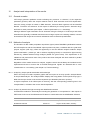
USER MANUAL
LCD Array Kit
SPECIES 2.0
DNA-based identification of
Animal species
Code: A-750-04
Code: A-750-12
1
Manual : SPECIES 2.0 Vers 1.0-2012
1
Product information ................................................................................................................................. 3
1.1
Intended use ................................................................................................................................. 3
1.2
Test principle................................................................................................................................. 4
2
Storage conditions & kit content.............................................................................................................. 5
3
Equipment and reagents - not supplied with the kit ................................................................................ 6
4
Preparation of reagents........................................................................................................................... 6
5
Warnings and precautions....................................................................................................................... 7
6
General safety information ...................................................................................................................... 7
7
Method description .................................................................................................................................. 8
7.1
PCR............................................................................................................................................... 8
7.2
Hybridisation / Detection ............................................................................................................... 8
8
Array description ..................................................................................................................................... 9
9
Protocol ................................................................................................................................................. 10
9.1
9.2
9.3
10
11
PCR amplification........................................................................................................................ 10
9.1.1
PCR set-up ..................................................................................................................... 10
9.1.2
Template ......................................................................................................................... 10
9.1.3
Cycler settings ................................................................................................................ 11
9.1.4
Agarose gel examination ................................................................................................ 11
LCD Array hybridisation and detection ....................................................................................... 11
9.2.1
Hybridisation ................................................................................................................... 12
9.2.2
Labelling.......................................................................................................................... 13
9.2.3
Staining ........................................................................................................................... 13
Short protocol.............................................................................................................................. 14
Analysis and interpretation of the results .............................................................................................. 15
10.1
General remarks ........................................................................................................................ 15
10.2
Methods of analysis ................................................................................................................... 15
10.2.1
Analysis by simple optical examination with naked eye ............................................... 15
10.2.2
Analysis by transmission light scanning and SlideReader software ............................ 15
Device performance .............................................................................................................................. 16
11.1
Sensitivity ................................................................................................................................... 16
11.2
Determination of device performance........................................................................................ 17
12
Device limits .......................................................................................................................................... 18
13
Trouble shooting.................................................................................................................................... 18
14
Literature ............................................................................................................................................... 19
15
Order information................................................................................................................................... 20
16
Contact manufacturer............................................................................................................................ 20
17
Symbol key ............................................................................................................................................ 20
2
Manual : SPECIES 2.0 Vers 1.0-2012
Instructions for use
Instructions version:
1
Revised :
26-July-2012
Product information
Product name:
1.1
SPECIES 2.0 V-1.0-2012-ENG
LCD Array Kit SPECIES 2.0
Order code
Kit size
A-750-04
32 Tests / 04 Chips
A-750-12
96 Tests / 12 Chips
Intended use
This array has been developed for rapid, easy and reliable identification of animal DNA in fresh meat
preparations and products manufactured thereof. DNA of animal species most commonly used in
food production will be detected in parallel using extracted DNA as starting material.
Beef
Bos taurus
Mallard Duck
Anas platyrhyncos
Buffalo
Bubalus bubalis
Muscovy Duck Cairina moschata
Pork
Sus scrofa
Dog
Canis lupus (familiaris)
Sheep
Ovis aries
Cat
Felis catus
Goat
Capra hircus
Kangaroo
M. giganteus / M. rufus
Horse
Equus caballus
Ostrich
Struthio camelus
Donkey
Equus asinus
Bison
Bos bison
Rabbit
Oryctolagus cuniculus
Red Deer
Cervus elaphus
Hare
Lepus europaeus
Fallow Deer
Dama dama
Chicken
Gallus gallus
Roe Deer
Capreolus capreolus
Turkey
Meleagris gallopavo
Camel
Camelus sp.
Goose
Ansa albifrons
Seagull
Larus sp.
List of species which can be detected by hybridisation of specific capture probes.
3
Manual : SPECIES 2.0 Vers 1.0-2012
1.2
Test principle
Using extracted DNA as starting material (1), small fragments of the mitochondrial 16S rRNA genes
of the target organisms will be amplified by the initial PCR. During this amplification the generated
PCR fragments are labelled with Biotin (2).
PCR amplicons are hybridised to specific capture probes on the surface of the array (3). Nonspecifically bound PCR amplicons will be removed by short washes under high stringency. The
remaining, specifically bound amplicons can be visualized by incubation with a StreptavidinPeroxidase conjugate and the subsequent formation of a dark precipitate after incubation with
peroxidase substrate (TMB) provided as staining solution (4).
extracted
PCR with biotin
Array
Detection by
DNA
incorporation
hybridisation
TMB precipitation
Figure 1: Test principle
4
Manual : SPECIES 2.0 Vers 1.0-2012
2
Storage conditions & kit content
When stored at the indicated temperature and not handled otherwise as specifically described, all
components are stable until their expiry date. Expire dates are printed on the components label or
the labels of the surrounding packages. More than 6 freezing and thawing cycles should be avoided
for components which require frozen storage. Prepare aliquots if necessary.
Component
A-300-04 (32 tests)
A-300-12 (96 tests)
Storage
LCD-Arrays
1 box, 04 chips
3 boxes, 12 chips
+4°C to +28°
1 bottle
2 bottles
+4°C to +28°
1 x 100 µl
2 x 100 µl
-10°C to –25°C
1 x 300 µl
2 x 300 µl
+4°C to +28°
Dilution Buffer
1 x 2000 µl
2 x 2000 µl
+4°C to +28°
Stain
1 x 2000 µl
2 x 2000 µl
1 x 1750 µl
2 x 1750 µl
-10°C to –25°C
1 x 20 µl
2 x 20 µl
-10°C to –25°C
Wash Powder
Primer
MEAT Primer Mix
Detection Kit I
Modulator
(*)
+4°C to +28°
Detection Kit II
Hybridisation Buffer .B.
Label
Additional supplies
Analysis Matrix
---
Manual, Pattern File, MSDS
Chip-Box Connectors
(*) =
1 CD
1 CD
---
2 connectors
6 connectors
---
Keep the ‚Stain’ solution always dark, protected from direct light.
5
Manual : SPECIES 2.0 Vers 1.0-2012
3
Equipment and reagents - not supplied with the kit
Instrumentation
-
Thermal cycler
-
Water bath
(adjustable to 35°C)
-
Centrifuge for LCD-Arrays
(cat. no HS-500-01, Chipron GmbH)
-
Micro pipettes
(range from 2 µl to 1000 µl)
optional:
-
SlideScanner PF3650u
(cat. no HS-300-01, Chipron GmbH)
-
SlideReader Analysis Software
(cat. no HS-200-01, Chipron GmbH)
Reagents & Materials
4
-
Reagents for DNA extraction
-
PCR chemicals
-
PCR grade water
-
Deionised water
-
Disposable gloves
-
Sterile filter tips
-
PCR reaction vessels
-
3 wash containers
-
1l-bottles
( Taq Polymerase, Buffer, dNTPs)
( 150 to 400 ml each)
Preparation of reagents
Wash Buffer
To prepare the 20 x wash buffer concentrate dissolve the content of one wash powder bottle in 1 liter
of deionised water. To prepare the 1 x wash buffer add 50 ml of 20 x wash buffer concentrate to 950
ml of deionised water. Prepare the 1 x wash buffer always fresh.
Stability of Wash-Buffer solutions:
20 x Concentrate
2 month at room temperature, 12 month at +4°C
If formation of precipitate occurs, warm the solution to 42°C and let
equilibrate to room temperature prior to use
1 x Working solution
min. 10 days at room temperature
6
Manual : SPECIES 2.0 Vers 1.0-2012
5
Warnings and precautions
-
All steps described in the protocol should only be carried out by well trained lab personnel.
-
Read the manual carefully and completely before starting.
-
Avoid any exposure to light of the ‘Stain’ solution. Always keep it dark.
-
All reagents in the kit are optimised for this particular test. Substitution of kit reagents may
effect the performance.
-
The same general safety guidelines which apply to the sample material should be followed
during the whole protocol.
-
The PCR products generated in the first protocol step have to be considered as
contamination sources for further PCRs. Therefore, all hybridisation, washing, staining,
drying and analysis steps should be carried out in the ‘post-PCR’ area.
-
Observe the standard guidelines for working in a PCR molecular diagnostic laboratory to
prevent contaminations.
6
General safety information
When working with chemicals, make sure that you always wear a suitable lab coat, disposable
gloves and protective goggles. For more information about our products, please refer to the
appropriate material safety data sheets (MSDS) which can be found on the CD provided with the kit.
Hybridisation Buffer
contains formamide ( >50%) and N-Dodecanoyl-N-methylglycin-Sodium salt.
Hybridisation buffer should therefore be handled as formamide.
Toxic, harmful, irritant
Risk and safety phrases: R61, R36, R38, S24, S26
Wash Powder
contains N-Dodecanoyl-N-methylglycin-Sodium salt.
Harmful, irritant
Risk and safety phrases: R36, S24, S26
Stain
contains 3,3´,5,5´-Tetramethylbenzidin (> 0.5 %).
Harmful, irritant
Risk and safety phrases: R20/21/22, R36/37/38, R40
(Self Assessment)
Modulator
Harmful, irritant
Risk and safety phrases: R36/37/38 S26/36
(Self Assessment)
7
Manual : SPECIES 2.0 Vers 1.0-2012
7
Method description
The test consists of two main procedure steps.
7.1
-
PCR amplification of DNA fragments with biotin incorporation
-
Hybridisation of PCR fragments to LCD-Arrays and detection
PCR
One primer mix is provided with the kit. The multiple primer pairs in this mix target a portion of the
16S RNA gene of a broad range of Vertebrate species. When using the primer mix ‘MEAT’ distinct
amplification products of 115 – 125 bp can be expected (species dependent):
Examples of fragment sizes:
7.2
Species
Size
Bos taurus
116 bp
Sus scrofa
119 bp
Equus caballus
124 bp
Gallus gallus
121 bp
Struthio camelus
122 bp
Hybridisation / Detection
The labelled PCR fragments are combined with the hybridisation buffer (provided) and hybridised to
the individual array fields of one chip. During hybridisation the labelled PCR fragments will bind to
the specific capture probes immobilized as spots on the bottom of each field. Following a short
washing procedure, each field is incubated with a secondary label solution (enzyme-conjugate). After
a second washing step, the positions (spots) where PCR fragments and secondary label are bound
can be visualized as blue precipitate formed by the enzyme substrate provided as “STAIN”. The data
read-out can either be done by simple ‘naked-eyed’ examination, using the ‘Analysis Matrix’
provided with the kit or, alternatively, with the scanner and software from the “Analysis-Package”
which can be obtained from Chipron GmbH.
For experienced users the whole procedure will take 3-4 hrs (depending on the duration of the PCR
amplification).
8
Manual : SPECIES 2.0 Vers 1.0-2012
8
Array description
Each LCD-Chip contains eight identical arrays in rectangular reaction chambers which can be
addressed individually. Functional controls to monitor hybridisation, secondary labeling and staining
are located in three corners. The arrays of the SPECIES 2.0 kit consist of an 8 x 8 pattern with
average spot diameters of 350 μm. The species specific capture probes are positioned as vertical
duplicates..
No. Name
Species
Name
Species
01
Hyb-Ctrl
Hybridization Control
02
Beef
Bos taurus
14
Mall. Duck
Anas platyrhyncos
03
Buffalo
Bubalus bubalis
15
Musc. Duck
Cairina moschata
04
Pork
Sus scrofa
16
Dog 3)
Canis lupus (familiaris)
05
Sheep
Ovis aries
17
Cat 4)
Felis catus
06
Goat
Capra hircus
18
Kangaroo
M. giganteus / M. rufus
07
Horse
Equus caballus
19
Ostrich
Struthio camelus
08
Donkey 2)
Equus asinus
20
Bison
Bos bison
09
Rabbit
Oryctolagus cuniculus
21
Red Deer
Cervus elaphus
10
Hare
Lepus europaeus
22
Fallow Deer
Dama dama
11
Chicken
Gallus gallus
23
Roe Deer
Capreolus capreolus
12
Turkey
Meleagris gallopavo
24
Camel 5)
Camelus sp.
13
Goose
Ansa albifrons
25
Seagull
Larus sp.
1)
2)
3)
4)
5)
6)
1)
No.
6)
Cross reactivity of the capture probe for Beef with 100% Bison may occur
Cross reactivity of the capture probe for Donkey with 100% Horse may occur
Capture probe detects Canis lupus, C. lupus familiaris, C. lupus chanco and Canis indica
Capture probe detects several species from the subfamily Felinae (Felis, Puma, Leopardus, Lynx)
Capture probe detects both species of the Genus Camelus ( C. bactrianus & C. dromedarius).
Capture probe detects several species from the Genus Larus (L.argentatus, L.cachinnans, L.occidentalis,
L.dominicanus, L.glaucoides).
9
Manual : SPECIES 2.0 Vers 1.0-2012
9
Protocol
9.1
PCR amplification
9.1.1
PCR set-up
We recommend the HotStarTaq Plus Master Mix Kit
[Qiagen, Code: 203645 ]
Using a 2 x PCR Master Mix (incl. Taq-Polymerase, dNTPs, Buffer and MgCl2)
Number of reactions (25 μl each)
1
4 (set up 5)
8 (set up 9)
12.5 μl
62.5 μl
112.5 μl
Primer Mix ‘MEAT’
1.5 μl
7.5 μl
13.5 μl
PCR grade water
6.0 μl
30.0 μl
54,0 μl
20.0 μl
100.0 μl
180.0 μl
4 x 20.0 μl
8 x 20.0 μl
1
4 (set up 5)
8 (set up 9)
2.5 μl
12.5 μl
22.5 μl
dNTP mix (10mM each)
1.0 μl
5.0 μl
9.0 μl
Primer Mix ‘MEAT’
1.5 μl
7.5 μl
13.5 μl
Taq Polymerase (5 U/μl)
0.3 μl
1.5 μl
2.7 μl
PCR grade water
14.7 μl
73.5 μl
132.3 μl
Total volume
20.0 μl
100.0 μl
180.0 μl
4 x 20.0 μl
8 x 20.0 μl
2x Master Mix
( incl. MgCl2 final 1.5-2.0 mM )
Total volume
Aliqout into
Using Taq-Polymerase, dNTPs, and Buffer as separate components
Number of reactions (25 μl each)
10x PCR Buffer
(incl. 15-20 mM MgCl2)
Aliquot into
9.1.2
Template
Add 5.0 μl of extracted DNA as template to each reaction tube.
Note:
If larger or smaller volumes of template need to be added, reduce the amount of added
water accordingly
10
Manual : SPECIES 2.0 Vers 1.0-2012
9.1.3
Cycler settings
The cycle regime given below has been optimised for the use of
-
HotStarTaq Plus Master Mix
Qiagen GmbH
[Code 203645]
-
TProfessional Standard Cycler
Analytik Jena AG
[Code 070-951]
When other combinations of PCR cycler and enzyme will be used, slight modifications of the given
protocol could become necessary. Please contact your local distributor or the Chipron support team
if you wish assistance for any kind of assay optimization.
Step
Duration
Temperature
1)
5:00 min
95°C
Initial denaturation
Ramp
Note
longer with some
“Hot-Start” enzymes
2)
3)
* 35 repetitions of:
Denaturation
0:30 min
94°C
3°C / sec
Annealing
0:45 min
57°C
3°C / sec
Elongation
0:45 min
72°C
3°C / sec
2:00 min
72°C
Strand completion
* Increase cycle number for higher sensitivity, or decrease cycle number for lower sensitivity.
9.1.4
Agarose gel examination
When the arrays are used for the first time or optimisation becomes necessary, it is recommended to
analyse the PCR amplification by agarose gel electrophoreses ( 2.0 % agarose gel ).
9.2
LCD Array hybridisation and detection
Detailed protocol for first time user – advanced users refer to short protocol (chapter 9.3.)
General Remarks
Since the working principle of LCD-Arrays is DNA/DNA hybridisation, the specificity and sensitivity of
the assays are mainly controlled by the hybridisation stringency during the 30 minute hybridisation
step. Apart from the concentration of the interacting molecules, the two factors with the biggest
influence on hybridisation stringency are temperature and buffer composition (e.g. concentration of
salts, formamide, urea etc.). It is crucial that the temperature during hybridisation and the pipetting
volumes are precisely controlled. The use of calibrated thermometers and micro pipettes is strongly
recommended.
Deviations of more than 1 °C or 1 µl can have severe effects.
11
Manual : SPECIES 2.0 Vers 1.0-2012
9.2.1
Hybridisation
1)
Set water bath temperature to 35 °C and add one droplet of water to each corner of the
humidity chamber. Do NOT preheat the LCD-Chip. The chip should be equilibrated to room
temperature.
2)
Prepare the Hybridisation Mix.
Make sure that all components are equilibrated to room temperature.
Number of reactions
Hybridisation buffer .B.
Modulator
Aliquot per*
1
4 (set up 5)
8 (set up 9)
22 µl
110 µl
198 µl
2 µl
10 µl
18 µl
-
24 µl
24 µl
* 0.2 ml reaction vials or 8-well PCR strips are well suited for setting up the reactions
3)
Combine the PCR product with the Hybridisation Mix.
Add 10 µl of the PCR product to the respective aliquot of Hybridisation Mix. Mix well by
pipetting up and down for several times – do not vortex!
4)
Initiate the hybridisation.
Place the slide in the humidity chamber and pipette 28 μl of the PCR-Hybridisation mixes to
the respective fields. Make sure that the PCR-Hybridisation mixes come into contact with the
entire reaction zone of the respective array field – avoid any contact of the pipetting tip with
the chip surface. Transfer the closed humidity chamber as quickly as possible to the preset
water bath (the chamber will float on the surface).
5)
Incubate the slide at 35°C for 30 minutes
6)
Prepare three wash containers with 1 x wash buffer (~ 150 ml each).
Prepare the 1 x wash buffer from the 20 x wash buffer concentrate, by adding 50 ml of
concentrate to 950 ml of deionized water. Mix thoroughly.
7)
Submerge the slide completely in the first container with the wash buffer and move it 3 times
slowly backward and forward. Submerge the slide quickly, to avoid field to field cross talk.
Repeat the procedure in wash container 2. Subsequently, transfer the slide to container 3
and incubate for 1 minute
8)
Dry the slide by spinning for 15 seconds in the CHIP Spin FVL2400N (Cat.No. HS-500-01).
12
Manual : SPECIES 2.0 Vers 1.0-2012
9.2.2
Labelling
8)
Prepare the labelling mix
Number of reactions
1
4 (setup 5)
8 (setup 10)
27.0 µl
135 µl
270 µl
Modulator
3.0 µl
15 µl
30 µl
Label
0.2 µl
1 µl
2 µl
Total
30.2 µl
151 µl
302 µl
Dilution Buffer
Mix well by vortex or intensive pipetting
9)
Apply 28 μl of the label mix to each field of the slide.
Make sure that the label mix has contact with the entire reaction zone of the respective array
field – avoid any contact of the pipette tip with the chip surface.
9.2.3
10)
Incubate the slide at room temperature for 5 min
11)
Replace the wash buffer in all three containers and repeat the wash procedure from step 7).
12)
Dry the slide as in step 8).
Staining
13)
Apply 28 μl of ‘Stain’ solution to each field and incubate for 5 minutes at room temperature.
Make sure that the fields do not contain any traces of washing solution – wait until fields are
completely dry.
Avoid any contamination of the ‘Stain’ stock solution! Prepare aliquots if necessary.
14)
Stop the staining process by rinsing the slide in the last wash container from step 11) for 15
seconds.
15)
Dry the slide as in step 8).
The slides are now ready for read out and can be kept in the dark for several years without a
significant loss of signal intensity.
13
Manual : SPECIES 2.0 Vers 1.0-2012
9.3
Short protocol
1)
Set water bath temperature to 35°C
2)
For each reaction mix 22 μl Hybridisation buffer .B., 2 μl of Modulator and 10 μl of PCR
product. Apply 28 µl of this solution to the respective array field.
3)
Incubate the slide at 35°C for 30 minutes (in humidity chamber)
4)
Prepare 3 wash containers with ~ 150 ml each of 1x wash solution.
(1 x wash buffer freshly prepared from 20x concentrate)
5)
Rinse slide in wash container 1 and 2 for 10 seconds each and incubate in wash container 3
for 1 minute.
6)
Dry the slide by spinning it for 15 seconds in the CHIP-Spin Centrifuge
7)
Prepare the labelling mix by combining 27 μl of Dilution buffer, 3 μl of Modulator and 0.2 μl of
Label per array field.
8)
Apply 28 μl of label mix to each field of the slide and incubate for 5 minutes at room
temperature.
9)
Replace the wash solution in all containers and repeat the wash procedure from step 5).
10)
Dry the slide as in step 6).
11)
Apply 28 μl of Stain solution to each field of the slide and incubate for 5 minutes at room
temperature.
Avoid contamination of the STAIN solution with residues of Label solution
12)
Stop the staining after 5 minutes by rinsing the slide in the last wash container from step 9)
for 10 seconds.
13)
Dry the slide as in step 6) – the slide is now ready for analysis.
14
Manual : SPECIES 2.0 Vers 1.0-2012
10
Analysis and interpretation of the results
10.1
General remarks
LCD-Arrays generate qualitative results indicating the ‘presence’ or ‘absence’ of the respective
parameter (species) within the sample material used for DNA extraction and PCR amplification.
Since the assay principle is based on DNA detection, vital and dead organisms will be identified
likewise. ‘Presence’ and ‘absence’ in this respect is defined by the assay specific detection range
and limits for each parameter (see chapter 11 Device performance, 11.1 Sensitivity).
Although different signal intensities can be observed during the analysis of LCD-Arrays and these
intensities are generally correlated with the amount of target copies in the starting material, it should
be noted that LCD-Arrays have not been validated as tools for absolute quantification.
10.2
Methods of analysis
The formation of dark visible precipitates at positions (spots) where DNA/DNA hybridisation between
the PCR amplicons and the immobilized capture probes took place, combined with the crystal clear
polymer support (LCD chip), offers the opportunity to use two different analysis methods. Simple
optical examination ( naked eye, with or without magnifying lenses) or transmission light scanning
followed by software-assisted image analysis (details are given below). The experiments of all
validations and performance tests of the product have been analysed with both methods in parallel
with identical results.
Regardless of the method chosen for analysis, negative controls should always be included into the
experimental scheme to ensure that no artificial background ‘signals’ are detected due to cross
contamination or ‘over staining’ (see chapter 13, Trouble shooting).
10.2.1 Analysis by simple optical examination with naked eye
Each LCD-Array Kit contains a pattern graphic with the imprint of an array specific ‘Analysis Matrix’
for simple data analysis. The ‘Analysis Matrix’ displays the array pattern for the specific array type, a
table with the spot number code and the respective spot identities (Genera or species).
A simple comparison of all visible spots on the array, with the corresponding positions given in the
‘Analysis Matrix’, will lead to the correct data interpretation.
10.2.2 Analysis by transmission light scanning and SlideReader software
An alternative method for data analysis including the generation of comprehensive data reports in
PDF format is the use of the SlideScanner PF3650u in combination with the SlideReader Software.
Product
Order No.
Company
SlideScanner PF3650u
HS-300-01
Chipron GmbH, Berlin, Germany
SlideReader Software
HS-200-01
Chipron GmbH, Berlin, Germany
15
Manual : SPECIES 2.0 Vers 1.0-2012
Detailed instructions for the use of SlideScanner and SlideReader software are given in the
SlideReader Manual.
The pattern file for automatic analysis with the SlideReader software is provided as data file on the
CD which is part of every LCD-Array kit. Choose the directory \ Pattern File and dependent on the
SlideReader software version in use one of the two following files.
SlideReader Vers 6 to 9:
use the file
Gal-00377 SPECIES 2.0.txt
SlideReader Version 11 (and higher)
use the file
0377 SPECIES 2.0.gal
Required software settings:
Cut off:
set to 2000
Please contact your local distributor or the Chipron support team to learn more about the automatic
analysis options with the SlideReader software.
11
Device performance
All DNA sequences underlying the design of primer and probes have been carefully chosen and
thoroughly confirmed by broad comparisons in publicly available databases (BLAST analysis). No
indication for cross reactivity apart from those as described below the specificity table on page 9
could be revealed by such in silico analysis.
It should be noted that, as for all PCR and hybridisation-based molecular tests, it can not be
excluded that rare genotypes, comprising variations within the sequence regions used for the assay
design, may remain undetected. Additionally, other, yet unknown / yet unpublished species (
especially among the Bovidae) might contain highly similar sequence regions in their genomes and
could therefore interfere with the assay performance. The assay in its present form reflects the
current state of knowledge of Chipron GmbH.
11.1
Sensitivity
The sensitivity of the assay is mainly determined by the number of PCR cycles and the binding
affinity of the capture probes during hybridisation. Under the given protocol conditions (hybridisation
buffer, temperature and duration), the binding affinity of the capture probes is well standardised and
allows for the detection of ~ 10 fmol of single-stranded, biotinylated target sequences. The number of
PCR cycles, on the other hand, can easily be adjusted to modulate the sensitivity range according to
the experimental demands. The maximal sensitivity of the SPECIES 2.0 LCD-Array kit for 16S rRNA
sequences of the target organisms represented by specific capture probes is approx. 0.1 Genome
Equivalent (GE) / µl and will be reached with 42 PCR cycles.
Note:
Detection limits below 1 GE / reaction are not unusual for mitochondrial target genes like the
16S rRNA gene which are present in high copy numbers / cell (tissue dependent).
16
Manual : SPECIES 2.0 Vers 1.0-2012
The assay sensitivity has been measured with serial dilutions of standardized genomic DNA extracts
or calibrated plasmid solutions containing fragments of the 16S rRNA genes.
Example:
Bos Taurus
Genome size: ~ 2.65 x 10 9 base pairs
Î
3 ng
equal
1 x 10 3 GE
Î
3 pg
equal
1 GE
For most users of the SPECIES 2.0 LCD-Array kit the relative detectable amounts of species DNA in
mixed meat preparations will be more interesting than the absolute sensitivity.
It should be noted, that due to competition the simultaneous amplification of several different target
sequences in one PCR reaction (Multiplex PCR) will constrict the dynamic range of the assay and
the detection limits for the individual targets. The bigger the difference in the starting concentration of
two targets, the stronger the effects of competition for the target of lower concentration.
The sensitivity values given above (in GE/µl) are valid for amplifications of individual targets or
equimolar mixtures thereof. In unequimolar mixtures of multiple target sequences the detection limit
for targets of lower concentration will be raised by competition with targets of higher concentration.
For DNA-detection assays based on multiplex PCR amplification (like the SPECIES 2.0 Kit) this
implies that the DNA concentration of the most prominent species in a sample determines the
experimental detection limits for species of lower abundance. A typical dynamic range of ‘Multiplex’
amplifications with respect to the targets of the highest and the lowest abundance overspans 3-4
logs (e.g. 105 – 102). For the analysis of meat preparations with the SPECIES 2.0 LCD-Array in
samples consisting of meat from mainly one species (~99%) additions of other meat species in the
range of 0.1% should be detectable. An experimental proof has been provided by Bush and
colleagues [1].
11.2
Determination of device performance
The type of Taq polymerase and the Theromcycler instrument used can have a strong influence on
the
assay performance. Therefore, all performance tests and assay evaluations have been
undertaken with various combinations of the following instruments and enzyme preparations. No
significant differences in the test performance have been observed with any combination of the
instruments and enzymes listed below.
Preparations of Taq-polymerase
- AmpliTaq Gold® PCR Master Mix
Applied Biosystems GmbH
[Code 4318739]
- HotStar Taq Plus Master Mix
Qiagen GmbH
[Code 203645]
- Mastercycler®
Eppendorf AG
[Code 5333 000.018]
- TProfessional Standard (Biometra)
Analytik Jena AG
[Code 070-951]
- labcycler Basic
SensoQuest GmbH
[Code 011-103]
PCR-Thermocycler
AmpliTaq Gold® is a registeredtrade mark of Roche Molecular Systems Inc.,
MasterCycler® is a registeredtrade mark of Eppendorf AG.
17
Manual : SPECIES 2.0 Vers 1.0-2012
12
Device limits
The following factors might limit the assay performance:
-
total concentration of extracted DNA is too low or DNA is degraded
-
PCR amplification is hampered by PCR inhibitors which have been co-extracted
-
use of PCR additives which can interfere with the hybridisation
(avoid additives like Urea, DMSO, Betaine etc.)
13
-
use of non calibrated or functionally impaired instruments (pipettes, water bath, PCR cycler)
-
any variation from the protocol given in this user instruction
Trouble shooting
General remarks
In cases where unexpected results are obtained or when signal intensities or background levels
appear to be different from usual observations, check the following list first
-
kit components and PCR chemicals were not expired
-
agarose gel examination of PCR products is positive
-
correct results with positive and negative controls
-
correct combination of primer mixes, chip types, hybridisation buffer and protocol
Detailed trouble shooting
Observation
No signals detected (including guide dots)
Probable causes
Wrong reaction set-up, wrong temperature, use of wrong hybridisation buffer .
Compare the experimental set-up with the given protocol, check temperature of
Solution
the water bath, compare the letters printed on the hybridisation buffer label and
the chip label (should be B) .
Observation
Probable causes
Solution
Guide dots detected, but no other signals visible
PCR failure, DNA extraction inefficient, template DNA degraded, DNA content
too low or test result for target organisms is negative.
Use fresh PCR chemicals, make sure to run the controls provided with kit,
check the PCR cycler program, try an alternative DNA extraction procedure.
Observation
Signals detected , but background is too high
Probable causes
Wrong reaction set up or incubation times too long
18
Manual : SPECIES 2.0 Vers 1.0-2012
Make sure the Modulator has been added at all stages according to the
Solution
protocol, monitor incubation times and temperatures for hybridisation, labelling
and staining carefully.
Observation
‘Bleeding’ guide dots and very strong control signals
Probable causes
Staining or secondary labeling time is too long
Solution
Reduce the incubation time gradually in 1 minute steps
Observation
Lots of small dark particles on chip surface
Probable causes
1 x wash buffer is too old (formation of precipitates)
Solution
Prepare a fresh solution of 1 x wash buffer from 20 x concentrate
Observation
Negative control shows species specific signals
Probable causes
Contamination of PCR chemicals or primer mixes or sample carryover during
the hybridisation protocol
Use fresh PCR chemicals, make sure to follow the general guide lines for PCR
Solution
set up to avoid laboratory born contaminations. If necessary, use new primer
mixes. Make sure that no well to well cross talk occurs during the hybridisation
steps.
Observation
Probable causes
Only single spots are seen for some features ( not duplicates)
The missing spots have been mechanically removed during the pipetting or
drying steps or by touching the surface of the reaction chambers.
Use solely the Chip-Spin FVL2400N centrifuge for drying steps. Higher g-forces
Solution
can result in spot dislocations. Avoid any contact with the reaction zone surface
of the array fields
If none of the above given solutions solves your problem, please get in contact with your local
distributor or contact our support team at
14
[email protected] .
Literature
[1]
Iwobi A., Huber I., Hauner G., Miller A. and Busch U., (2011):
Biochip Technology for the Detection of Animal Species in Meat Products;
Food Analytical Methods, Vol. 4, No. 3, pp. 389-398
19
Manual : SPECIES 2.0 Vers 1.0-2012
15
Order information
Product:
Quantity
Order No.
SPECIES 2.0
32 Tests / 04 Chips
A-750-04
SPECIES 2.0
96 Tests / 12 Chips
A-750-12
Product
Description
Order No.
Slide Reader Software
1 License
HS-200-01
Scanner PF 3650
Slide Scanner w. 10µm resolution
HS-300-01
CHIP Spin FVL 2400N
Mini-Centrifuge for LCD-Arrays
HS-500-01
Related Products:
16
17
Contact manufacturer
Chipron GmbH
Tel.:
+ 49 (0) 30 787994 70
Eresburgstrasse 22-23
Fax:
+ 49 (0) 30 787994 99
D-12103 Berlin
Email:
Germany
Support:
[email protected]
[email protected]
Symbol key
The following symbols are used on labels of the kit box and components therein
Manufacturer
Number of tests
Lot number
Date of expiry [ month – year ]
Order number
Storage temperature [ range °C ]
20
Manual : SPECIES 2.0 Vers 1.0-2012