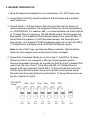
1
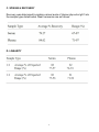
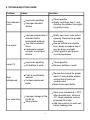
RayBio® Human IgG1 ELISA Kit
Catalog #: ELH-IGG1
User Manual
Last revised December 7, 2015
Caution:
Extraordinarily useful information enclosed
ISO 13485 Certified
3607 Parkway Lane, Suite 100
Norcross, GA 30092
Tel: 1-888-494-8555 (Toll Free) or 770-729-2992, Fax:770-206-2393
Web: www.RayBiotech.com, Email: [email protected]
1
RayBiotech, Inc.
________________________________________
RayBio® Human IgG1 ELISA Kit Protocol
Table of Contents
Section
Page #
I.
Introduction
3
II.
Storage
4
III.
Reagents
4
IV.
Additional Materials Required
4
V.
Reagent Preparation
5
VI.
Assay Procedure
6
VII.
Assay Procedure Summary
7
VIII.
Calculation of Results
A. Typical Data
B. Sensitivity
C. Spiking & Recovery
D. Linearity
E. Reproducibility
8
8
8
9
9
10
IX.
Specificity
10
X.
Troubleshooting Guide
11
Please read the entire manual carefully before starting your experiment
2
I. INTRODUCTION
The human immune system consists of two functional components classified as the
innate system (the physical, biochemical and cellular barriers), and the adaptive
immune system (including lymphocytes and immunoglobulins). Immunoglobulins
are the key elements of the humoral immune response in vertebrate against
parasitic invasion. The polypeptide chains of immunoglobulins composed of two
identical heavy (H) chains and two identical light (L) chains linked together by interchain disulfide bonds. While the amino-terminal portions that exhibits highly
variable amino-acid composition are involved in antigen binding, the C terminal
constant parts are involved in complement binding, placental passage and binding
to cell membranes. Based upon the variation of the constant region of the heavy
chain, nine immunoglobulin heavy chain isotypes are found in humans: IgA (with
subclasses IgA1 and IgA2), IgD, IgE, IgM, and IgG (with subclasses IgG1, IgG2,
IgG3, and IgG4). IgG is the predominant immunoglobulin in the serum, which
accounts for 75% of the total serum antibody of healthy individuals. IgG has a
molecular weight of about 150 kDa. Four distinct subgroups of human IgG (IgG1,
IgG2, IgG3, and IgG4) were first demonstrated in the 1960's by using polyclonal
antisera prepared in animals immunized with human myeloma proteins.
The RayBio® Human IgG1 ELISA kit is an in vitro enzyme-linked immunosorbent
assay for the quantitative measurement of human IgG1 in serum and plasma. This
assay employs an antibody specific for human IgG1 coated on a 96-well plate.
Standards and samples are pipetted into the wells and IgG1 present in a sample is
bound to the wells by the immobilized antibody. The wells are washed and
biotinylated anti-human IgG antibody is added. After washing away unbound
biotinylated antibody, HRP-conjugated streptavidin is pipetted to the wells. The
wells are again washed, a TMB substrate solution is added to the wells and color
develops in proportion to the amount of IgG1 bound. The Stop Solution changes
the color from blue to yellow, and the intensity of the color is measured at 450 nm.
3
II. STORAGE
The entire kit may be stored at -20°C for up to 1 year from the date of shipment.
Avoid repeated freeze-thaw cycles. The kit may be stored at 4°C for up to 6
months. For extended storage, it is recommended to store at -80°C. For prepared
reagent storage, see table below.
III. REAGENTS
Component
Size / Description
Storage / Stability
After Preparation
IgG1 Microplate (Item A)
96 wells (12 strips x 8 wells) coated with antiHuman IgG1.
1 month at 4°C*
Wash Buffer Concentrate
(20X) (Item B)
25 ml of 20X concentrated solution.
1 month at 4°C
Standard Protein (Item C)
2 vials of Human IgG1. 1 vial is enough to run
each standard in duplicate.
1 week at -80°C
Detection Antibody IgG1
(Item F)
2 vials of biotinylated anti-Human IgG1. Each vial
is enough to assay half the microplate.
5 days at 4°C
HRP-Streptavidin
Concentrate (Item G)
200 µl 2,000X concentrated HRP-conjugated
streptavidin.
Do not store and
reuse.
TMB One-Step Substrate
Reagent (Item H)
12 ml of 3,3,5,5'-tetramethylbenzidine (TMB) in
buffer solution.
N/A
Stop Solution (Item I)
8 ml of 0.2 M sulfuric acid.
N/A
Assay Diluent (Item E2)
2 bottles of 15 ml 5X concentrated buffer.
1 month at 4°C
*Return unused wells to the pouch containing desiccant pack, reseal along entire edge.
IV. ADDITIONAL MATERIALS REQUIRED
1.
2.
3.
4.
5.
6.
7.
8.
Microplate reader capable of measuring absorbance at 450 nm.
Precision pipettes to deliver 2 µl to 1 ml volumes.
Adjustable 1-25 ml pipettes for reagent preparation.
100 ml and 1 liter graduated cylinders.
Absorbent paper.
Distilled or deionized water.
Log-log graph paper or computer and software for ELISA data analysis.
Tubes to prepare standard or sample dilutions.
4
V. REAGENT PREPARATION
1. Bring all reagents and samples to room temperature (18 - 25ºC) before use.
2. Assay Diluent (Item E2) should be diluted 5-fold with deionized or distilled
water before use.
3. Sample dilution: 1X Assay Diluent (Item E2) should be used for dilution of
serum and plasma samples. The suggested dilution for normal serum/plasma
is ~10,000,000 fold. For example, add 1 µl of serum/plasma into a tube with 99
µl 1X Assay Diluent to prepare a 100-fold diluted sample. Mix thoroughly and
then pipette 1 µl of prepared 100-fold diluted sample into a tube with 99 µl 1X
Assay Diluent to prepare a 10,000 fold diluted sample. Mix thoroughly and
then pipette 1 µl of prepared 10,000-fold diluted sample into a tube with 999 µl
1X Assay Diluent to prepare a final 10,000,000 fold diluted sample.
Note: Levels of IgG1 may vary between different samples. Optimal dilution
factors for each sample must be determined by the investigator.
4. Preparation of standard: Briefly spin a vial of Item C. Add 600 µl 1X Assay
Diluent into Item C vial to prepare a 300 ng/ml stock standard solution.
Dissolve the powder thoroughly by a gentle mix. Add 80 µl IgG1 standard (300
ng/ml) from the vial of Item C, into a tube with 520 µl 1X Assay Diluent to
prepare a 40 ng/ml standard. Pipette 400 µl 1X Assay Diluent into each tube.
Use the stock standard solution to produce a dilution series (shown below).
Mix each tube thoroughly before the next transfer. 1X Assay Diluent serves as
the zero standard (0 ng/ml).
5
5. If the Wash Concentrate (20X) (Item B) contains visible crystals, warm to room
temperature and mix gently until dissolved. Dilute 20 ml of Wash Buffer
Concentrate into deionized or distilled water to yield 400 ml of 1X Wash Buffer.
6. Briefly spin the Detection Antibody vial (Item F) before use. Add 100 µl of 1X
Assay Diluent (Item E2) into the vial to prepare a detection antibody
concentrate. Pipette up and down to mix gently (the concentrate can be stored
at 4ºC for 5 days). The detection antibody concentrate should be diluted 80fold with 1X Assay Diluent (Item E2) and used in step 5 of Part VI Assay
Procedure.
7. Briefly spin the HRP-Streptavidin concentrate vial (Item G) and pipette up and
down to mix gently before use, as precipitates may form during storage. HRPStreptavidin concentrate should be diluted 2,000-fold with 1X Assay Diluent
(Item E2).
For example: Briefly spin the vial (Item G) and pipette up and down to mix
gently. Add 6 µl of HRP-Streptavidin concentrate into a tube with 12 ml 1X
Assay Diluent to prepare a final 2,000 fold diluted HRP- Streptavidin solution
(don't store the diluted solution for next day use). Mix well.
VI. ASSAY PROCEDURE
1. Bring all reagents and samples to room temperature (18 - 25ºC) before use. It
is recommended that all standards and samples be run at least in duplicate.
2. Label removable 8-well strips as appropriate for your experiment.
3. Add 100 µl of each standard (see Reagent Preparation step 3) and sample into
appropriate wells. Cover well and incubate for 2.5 hours at room temperature
or over night at 4ºC with gentle shaking.
4. Discard the solution and wash 4 times with 1X Wash Solution. Wash by filling
each well with Wash Buffer (300 µl) using a multi-channel Pipette or
autowasher. Complete removal of liquid at each step is essential to good
performance. After the last wash, remove any remaining Wash Buffer by
aspirating or decanting. Invert the plate and blot it against clean paper towels.
6
5. Add 100 µl of 1X prepared biotinylated antibody (Reagent Preparation step 6)
to each well. Incubate for 1 hour at room temperature with gentle shaking.
6. Discard the solution. Repeat the wash as in step 4.
7. Add 100 µl of prepared Streptavidin solution (see Reagent Preparation step 7)
to each well. Incubate for 45 minutes at room temperature with gentle shaking.
8. Discard the solution. Repeat the wash as in step 4.
9. Add 100 µl of TMB One-Step Substrate Reagent (Item H) to each well.
Incubate for 30 minutes at room temperature in the dark with gentle shaking.
10. Add 50 µl of Stop Solution (Item I) to each well. Read at 450 nm immediately.
VII. ASSAY PROCEDURE SUMMARY
1. Prepare all reagents, samples and standards as instructed.
2. Add 100 µl standard or sample to each well. Incubate 2.5 hours at room
temperature or over night at 4ºC.
3. Add 100 µl prepared biotin antibody to each well. Incubate 1 hour at room
temperature.
4. Add 100 µl prepared Streptavidin solution. Incubate 45 minutes at room
temperature.
5. Add 100 µl TMB One-Step Substrate Reagent to each well. Incubate 30
minutes at room temperature.
6. Add 50 µl Stop Solution to each well. Read at 450 nm immediately.
7
VIII. CALCULATION OF RESULTS
Calculate the mean absorbance for each set of duplicate standards, controls and
samples, and subtract the average zero standard optical density. Plot the standard
curve on log-log graph paper or using Sigma plot software, with standard
concentration on the x-axis and absorbance on the y-axis. Draw the best-fit straight
line through the standard points.
A. TYPICAL DATA
These standard curves are for demonstration only. A standard curve must be run
with each assay.
B. SENSITIVITY
The minimum detectable dose of Human IgG1 was determined to be 50 pg/ml.
Minimum detectable dose is defined as the analyte concentration resulting in an
absorbance that is 2 standard deviations higher than that of the blank (diluent
buffer).
8
C. SPIKING & RECOVERY
Recovery was determined by spiking various levels of Human php echo IgG1 into
the sample types listed below. Mean recoveries are as follows:
D. LINEARITY
9
E. REPRODUCIBILITY
Intra-Assay CV%: <10%
Inter-Assay CV%: <12%
IX. SPECIFICITY
Detect human IgG1. No cross reactivity was found againt IgG2, IgG3, IgG4, IgM,
IgA, IgD and IgE
10
X. TROUBLESHOOTING GUIDE
Problem
Cause
Solution
Poor standard
curve
Inaccurate pipetting
Improper standard
dilution
Check pipettes
Briefly centrifuge Item C and
dissolve the powder thoroughly
by gently mixing
Low signal
Improper preparation of
standard and/or
biotinylated antibody
Too brief incubation
times
Inadequate reagent
volumes or improper
dilution
Briefly spin down vials before
opening. Dissolve the powder
thoroughly.
Ensure sufficient incubation
time; assay procedure step 2
may be done overnight
Check pipettes and ensure
correct preparation
Large CV
Inaccurate pipetting
Air bubbles in wells
Check pipettes
Remove bubbles in wells
High
background
Plate is insufficiently
washed
Contaminated wash
buffer
Review the manual for proper
wash. If using a plate washer,
ensure that all ports are
unobstructed.
Make fresh wash buffer
Improper storage of the
ELISA kit
Stop solution
Store your standard at <-70ºC
after reconstitution, others at
4ºC. Keep substrate solution
protected from light.
Add stop solution to each well
before reading plate
Low sensitivity
11
RayBio® ELISA Kits
Over 2,000 ELISA kits available, visit www.RayBiotech.com/ELISA-Kits.html for
details.
This product is for research use only.
©2015 RayBiotech, Inc
12