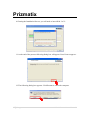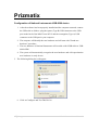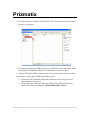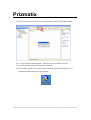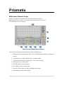
1
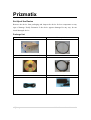
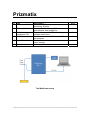
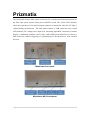
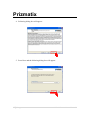
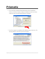
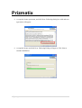
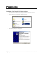
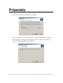
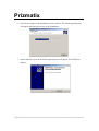
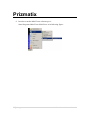
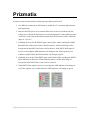
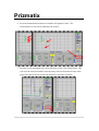
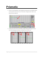
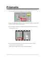
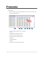
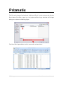
MitoViewer MV-2
User Manual
Software version: 1.1
Main Office
European Sales Office
North America Sales Office
Phone: +972-8-929-7844 Phone: +44 (0)77-9172-9592
Phone: +1-(248)-436-8085
Fax: +972-8-929-8772
Fax: +44 (0)20-7681-2977
Fax: +1-(248)-281-5236
[email protected]
[email protected]
[email protected]
P.O. B. 4234 M od iin- Ilite 7 191 9, I sr ael
Prizmatix
Table of Contents MitoViewer MV‐2 System Description ............................................................................................ 3 MitoViewer Principle of NADH Monitoring ..................................................................................... 3 Main Features of the Mito‐Viewer ................................................................................................... 4 The MitoViewer is suitable for the testing of ................................................................................... 4 Health and Safety ............................................................................................................................. 5 Set Up of the Device ......................................................................................................................... 6 Package List ......................................................................................................................... 6 System overview ................................................................................................................. 8 Additional equipment not provided with the MitoViewer ............................................... 11 Software Installation ...................................................................................................................... 12 Installation of the National Instruments software ........................................................... 12 Installation of the Prizmatix MitoViewer software ........................................................... 20 MitoViewer Software Usage .......................................................................................................... 24 Appendix A: Real‐time evaluation of mitochondrial function by monitoring NADH redox state using the MitoViewer ..................................................................................................................... 31 Appendix B: Typical Results while monitoring the Brain in vivo ................................................... 34 2 | P a g e
MitoViewer User Manual
Prizmatix
MitoViewer MV-2 System Description
The MitoViewer MV-2 is a unique optical monitoring device enabling real time
evaluation of mitochondrial function under in vitro and in vivo conditions. The
MitoViewer MV-2 provides significant and critical information originating from the
intracellular "Nano" organelles – the mitochondria which provide energy for cellular
functions. The measurement is based on the auto fluorescence of the intramitochondrial
NADH which serves as a control marker in energy generation processes by the
respiratory chain in the mitochondria. NADH redox state is sensitive to the intracellular
oxygen level and to energy consumption by the tissue.
MitoViewer Principle of NADH Monitoring
MitoViewer measurement is based on the auto fluorescence of the intramitochondrial
NADH. The Mitochondrial NADH absorption band ranges from 300nm to 400nm, with
the NADH fluorescence emission peaking at around 450nm. Due to historical reasons
most NADH measurements were performed with excitation light provided by Mercury
lamps at 365nm and not at the NADH absorption peak at 340nm. The MitoViewer was
designed according to this tradition.
The MitoViewer device uses 365nm UV LED for excitation of the NADH fluorescence
(Fluor signal). This excitation light is also utilized for the correction of hemodynamic
artifacts by measuring the reflection light intensity at same excitation wavelength of
365nm (the Refl signal). The light is transmitted from and to the device by means of a
flexible fiber optic probe.
The software displays the Refl, Fluor, NADH corrected fluorescence signal and the
photodiode PD signal.
3 | P a g e
MitoViewer User Manual
Prizmatix
Main Features of the Mito-Viewer
•
Suitable for various experimental animal models and variety of organs under in
vitro / in vivo conditions.
•
LabVIEW user friendly interface.
•
Calculates corrected NADH (independent of hemodynamic changes in the tissue).
•
Measurements taken via flexible fiberoptic probe.
•
Custom made probes can be supplied to suit experimental set-ups.
•
Possible integration of the MitoViewer probe with other probes in existing setups.
The MitoViewer is suitable for the testing of:
•
Ischemia, hypoxia, anoxia, heperoxia or hypercapnia in various organs, i.e brain,
kidney, liver or muscle tissue.
•
Activation of the brain by cortical spreading depression or seizures.
•
Changing of heart metabolism by isotropic and chronotropic drugs.
•
Drug safety and efficacy monitored in vivo in various organs.
•
Organ transplants for on mitochondrial function during the postoperative period.
4 | P a g e
MitoViewer User Manual
Prizmatix
Health and Safety
Before applying power to the power adaptor of the system, ensure that the protective
conductor of the three conductor mains power cord is correctly connected to the
protective earth contact of the socket outlet! Improper grounding can cause electric shock
with implications for your health or even death!
Prizmatix products are NOT authorized for use as components in life support devices or
systems.
The MitoViewer MV‐2 is for use as laboratory equipment only. It is not cleared or authorized for clinical use. The MitoViewer must not be operated in explosion endangered environments!
Any maintenance shall ONLY be performed by a technician authorized by Prizmatix.
Cellular phones or other radio transmitters should not be used within the vicinity of 3-4
meters of the unit.
The MitoViewer MV-2 emits UV and other intense light. Intense UV light can be emitted
by the system and from the probe tip during operation. Precautions must be taken to
prevent looking directly at the UV light with unprotected eyes or illuminating the UV
light on skin. Do not look directly into the UV light, through the optical system, or the
probe tip during operation of the device. This can cause permanent damage to the eyes
even if only exposed for brief periods.
Keep the metal caps of optical units closed unless the fiberoptic probe is connected.
5 | P a g e
MitoViewer User Manual
Prizmatix
Set Up of the Device
Remove the device from packaging and inspect the device for lose components or any
sign of damage. Notify Prizmatix if the device appears damaged in any way: do not
install damaged device.
Package List
MitoViewer MV‐2 Fiberoptic Probe Software Installation CDs (x3)
USB‐A to USB‐B Cable Mains Power Cord Power Adaptor 6 | P a g e
MitoViewer User Manual
Prizmatix
#
1
Item
MitoViewer MV-2
2
Fiberoptic Probe
3
4
Software
Installation CDs
USB Cable
5
Mains Power Cord
6
Power Adaptor
Description
Mitochondrial Function
Monitoring System
Standard Probe: Y-shaped fiberoptic cable
with stainless steel straight tip
Three self-executable CDs containing
software and drivers.
Cable to connect the MitoViewer MV-2 to
the computer
Cord to connect the power adaptor to
mains voltage
Universal power adaptor
QTY
1
1
3
1
1
1
The MitoViewer setup
7 | P a g e
MitoViewer User Manual
Prizmatix
System overview
The MitoViewer system comprises the following main subunits:
Light Source Unit – provides UV light at 365nm for NADH fluorescence
excitation and tissue reflection measurements. Also included is a reference
photodiode that enables correction of signals in cases of intensity changes of the
LED during measurement.
Fiber Optic Probe – transmits the UV light from the Light Source Unit to the
measured tissue site and transmits the collected light (the reflection (Refl) and the
fluorescence (Fluor) from the tissue, to the Detection Unit.
Detection Unit – provides detection to transform the Refl and Fluor light into
electrical signals which are transmitted to the electronics board for amplification.
Electronics Unit – provides amplification for the Refl and Fluor signals as well
as control of the detector gain and driver for the UV LED. The amplified signals
are transmitted to Analog to Digital convertor module.
USB-6009 module – provides Analog to Digital (A/D) conversion for the Refl
and Fluor signals and Digital to Analog (D/A) conversion for control signals sent
from a PC to control the function of the MitoViewer.
PC – Personal Computer enables control of the MitoViewer operation through the
MitoViewer software.
Power Adaptor – provides the DC voltage for operation of the MitoViewer.
MitoViewer subunits diagram
8 | P a g e
MitoViewer User Manual
Prizmatix
The front panel features SMA optical connectors for excitation and collection branches of
the fiber optic probe and the main power ON/OFF switch. The yellow LED indicator
above the Light Source Unit optical output connector is turned ON when the UV light is
emitted during measurement. The back panel features: a USB connector with a small
LED indicator, DC voltage power input jack, Ain analog input BNC connector (to enable
display of additional parameter side by side with NADH on the MitoViewer software),
DIO connector (enables triggering or synchronizing of the MitoViewer with external
devices).
MitoViewer front panel
MitoViewer MV-2 back panel
9 | P a g e
MitoViewer User Manual
Prizmatix
Standard MitoViewer MV-2 fiber optic probe
The standard fiber optic probe features a needle-type distal end designed for
measurements on exposed tissue by means of a micromanipulator or within a cannula.
The probe contains seven fibers which are arranged in a honeycomb structure in the distal
end. The central fiber is the excitation fiber which delivers the UV LED light to the
tissue. The six satellite fibers collect the light from the tissue and deliver it to the
detection unit in the MitoViewer. The proximal end of the probe features two branches
connectorized with optical SMA connectors. The thin branch contains the single
excitation fiber and should be connected to the Light Source Unit port on the front panel
of the MitoViewer. The thick branch contains the six collection fibers and should be
connected to the Detection Unit port in the front panel of the MitoViewer.
General Specifications:
Total length:
Common length:
Fibers:
Number of excitation fibers:
Number of detection fibers:
Connectors:
Tip:
Maximum temperature:
Sterilization method:
Biocompatibility:
10 | P a g e
100 cm (+10cm / -0)
80 cm (+5cm / -0)
Plastic optical fibers, ∅750µm, NA=0.5
1
6
SMA 905 (x2)
Stainless steel Tube, 12G (0.109" OD), length 20mm.
70°C
Ethylene Oxide gas. Do not apply Autoclave sterilization!
Materials in contact with the tissue are known to be inert to
the tissues. However ISO 10993 or USP VI tests were not
performed on the probe to establish this claim.
MitoViewer User Manual
Prizmatix
Additional equipment not provided with the MitoViewer:
1. PC with Windows XP / Vista / 7 operating system.
2. Methods and means for stabilizing the probe on the monitored tissue may require
special mechanisms or procedures. Consult Prizmatix regarding your specific
application.
11 | P a g e
MitoViewer User Manual
Prizmatix
Software Installation
The software installation is performed in two steps:
1. Installation of the National Instruments software
• NI-DAQmx Driver
• Measurement and Automation Explorer
2. Setup of Prizmatix MitoViewer software.
Installation of the National Instruments software
1. Insert the National Instruments installation CD 1 of 2 of NI-DAQmx into the CD
drive.
2. The auto run will start the setup software, please follow the instructions that
appear on the screen.
3. Click "Install Software" in the following dialog box:
12 | P a g e
MitoViewer User Manual
Prizmatix
4. Following dialog box will appear:
5. Press Next and the following dialog box will appear:
13 | P a g e
MitoViewer User Manual
Prizmatix
6. If you would like to install the National Instruments software into different
location, use the Browse button to change the installation location. Press Next to
continue the setup. Next the following dialog box will appear:
7. Press Next to approve the installation of the additional features of the driver. The
following dialog box with license agreement will appear:
14 | P a g e
MitoViewer User Manual
Prizmatix
8. Accept the license agreement, and click Next. Following dialog box with end-user
agreement will appear:
9. Accept the license and click Next. Following Dialog will appear. Click Next to
start the installation:
15 | P a g e
MitoViewer User Manual
Prizmatix
10. During the Installation Process you will need to insert Disk 2 of 2:
11. At the end of the process following dialog box will appear. Press Next to approve.
12. The following dialog box appears. Click Restart to reboot the computer.
16 | P a g e
MitoViewer User Manual
Prizmatix
Configuration of National Instruments USB-6009 device
1. After the software has been properly installed and the computer restarted, connect
the USB cable as follows: plug the square (Type B) USB connector to the USB
port on the back of the MitoViewer MV-2 and the rectangular (Type A) USB
connector to the USB port in your computer.
2. The computer will identify the new hardware and will start with "Found new
hardware" procedure.
3. The new hardware is National Instruments A/D module called USB-6009 or USB6009-OEM.
4. The system will automatically recognize the new hardware and will report that the
new hardware is ready for use.
5. The following dialog box will appear:
6. Click on Configure and Test This Device.
17 | P a g e
MitoViewer User Manual
Prizmatix
7. The following screen, titled " NI USB 6009 "Dev1" Measurement & Automation
Explorer" will appear:
8. The National Instruments USB-6009 device will be listed in the side menu. Make
sure that the NI USB-6009-OEM (or NI USB-6009) is marked as Dev1.
9. If the NI USB-6009-OEM is named as Dev2 or by other name, rename it to Dev1.
10. In order to rename the NI USB-6009-OEM as Dev1:
•
•
Click on the NI USB-6009-OEM with left button of the mouse and select
option Rename from the list.
Change the name of the NI USB-6009-OEM to Dev1 and click anywhere
outside. The name will change to NI USB-6009-OEM: "Dev1"
18 | P a g e
MitoViewer User Manual
Prizmatix
11. Click on Self-test icon to check the functionality of the NI USB-6009-OEM.
12. A report message should appear: "The device has passed the self-test"
13. Close the Measurement & Automation Explorer.
14. Tip: another option to access the Measurement & Automation Explorer is by
clicking the following icon on the desktop:
19 | P a g e
MitoViewer User Manual
Prizmatix
Installation of the Prizmatix MitoViewer software
1. Insert the Prizmatix MitoViewer software installation CD into the CD drive.
2. Run the setup.exe to initialize the installation process.
3. The following dialog box will appear:
20 | P a g e
MitoViewer User Manual
Prizmatix
4. Press Next, the following dialog box will appear:
5. If you would like to install the MitoViewer software into different location, use
Browse button to change the installation location. Press Next to continue the
setup. The following dialog box will appear:
21 | P a g e
MitoViewer User Manual
Prizmatix
6. Press Next to approve the installation of the software. The following dialog box
will appear showing the process of the installation:
7. At the end of the process the following dialog box will appear. Press Finish to
approve.
22 | P a g e
MitoViewer User Manual
Prizmatix
8. In order to run the MitoViewer software go to:
Start>Programs>MitoViwer>MitoViwer as in following figure:
23 | P a g e
MitoViewer User Manual
Prizmatix
MitoViewer Software Usage
Connect all cables as described in the Set Up of the Device section.
Run the MitoViewer software as described at the end of previous section.
The following screen will appear:
Main screen of MitoViewer software
The main screen comprises the following controls and indicators:
1. Main graph area displaying the Refl, Fluor, NADH and PD parameters as function
of time.
2. Current numerical values of Refl, Fluor, NADH and PD.
3. Control buttons Marker, Normalization, Clear Chart and Stop.
4. LED power level control panel.
5. Refl PMT detector control panel.
6. Fluor PMT detector control panel.
7. Detectors Values indicator and smoothing control panel.
8. Auto Ranging feature control panel.
24 | P a g e
MitoViewer User Manual
Prizmatix
In order to start measurement the following steps shall be performed:
1. At LED Power panel press ON button to enable the UV excitation light and start
data acquisition.
2. Increase the LED power level control slider close to the level indicated by the
yellow arrow. Watch the LED power level at the Detectors Values indicator panel
- LED PD. At the start of the measurements the LED PD value will be within the
range of 3.5 to 4.5.
3. Gradually increase the Refl PMT1 gain control slider, while watching the PMT1
Refl indicator (at Detectors Values indicator panel). At this initial stage of the
measurements the PMT1 Refl value will be about 4. If the PMT1 Refl signal is
too low or too high the LED indicator will change to red. If the signal level is
within acceptable limits the LED indicator will change to green.
4. Gradually increase the Fluor PMT2 gain control slider while watching the PMT2
Fluor indicator (at Detectors Values indicator panel). At this initial stage of
measurement the PMT2 Fluor value will be around 4.
5. If the PMT2 Fluor signal is too low or too high the LED indicator will change to
red. If the signal level is within limits the LED indicator will change to green.
25 | P a g e
MitoViewer User Manual
Prizmatix
6. Press the Normalization button to normalize all signals to 100%. The
Normalization event will be marked by the marker.
7. In order to clear unwanted traces from the chart use the Clear Chart button. This
will clear the chart area and the chart drawing will start from the left side of the
graph. The data in the file will not be influenced from this operation.
26 | P a g e
MitoViewer User Manual
Prizmatix
8. In order to mark important events during the measurements use the Marker button
on the right side of the main screen. The marker will appear at the chart screen
and on the stored data file.
9. Scaling of the chart can be performed by simply clicking on the upper or lower
scale and changing the maximum or minimum value of the scale.
27 | P a g e
MitoViewer User Manual
Prizmatix
10. In order to enable continuous measurements without detector saturations or too
low S/N conditions the auto-ranging feature may be engaged.
11. During Auto-Ranging the LED Power will automatically change to bring the PMT
detectors within reasonable operation gain.
12. The Smoothing constant may be changed by clicking on the current value and
choosing different value.
13. At the end of the measurement, press the stop button to end the UV LED
excitation and to stop data acquisition.
Note: Always stop the software by only using the Stop button.
28 | P a g e
MitoViewer User Manual
Prizmatix
14. Data storage
The data is stored in an ASCII Tab delimited text file *.TXT file. The file can be
reviewed by simple editor.exe
Following is the description of the file columns:
(1) Time
(2) Point number from start of the file
(3) Refl Value (not normalized to PD)
(4) Fluor value (not normalized to PD)
(5) NADH
(6) LED Photodiode
(7) Marker
The Red arrow shows the Marker signature.
29 | P a g e
MitoViewer User Manual
Prizmatix
The file can be imported and analyzed in Microsoft Excel. In order to import the data into
Excel choose Text Files (*.prn; *txt, *crv) option at Files of type selection at File Open
dialog box as shown in following figure:
The file will be imported into excel in correct order as shown here:
30 | P a g e
MitoViewer User Manual
Prizmatix
Appendix A:
Real-time evaluation of mitochondrial function by monitoring
NADH redox state using the MitoViewer
The MitoViewer MV-2 is based on principles and technologies that have emerged during
the last five decades, and the user is strongly advised to read a few publications before
starting to use device routinely. The broad spectrum of publications on NADH
monitoring was reviewed most recently by Mayevsky and Rogatsky (2007). Several other
articles appear in the list of publications attached to this document. Accordingly, only
few significant points will be discuss in the following paragraphs.
Introduction
Only the reduced form of NAD+, i.e. NADH, absorbs UV light at nearly the entire UVA
spectrum area (315-400 nm), and fluoresces at 420-480 nm. A small part of the excitation
light delivered to the tissue by the excitation optical fiber will be absorbed and re-emitted
as fluorescence by NADH. Any change in the redox status of NAD+/NADH will be
reflected in the intensity of the emitted fluorescence, while the more reduced state
(NADH) shows grater intensity of fluorescence than the oxidized state (NAD+).
The redox state of NADH - and hence the fluorescence - depends on the availability of
oxygen as well as on the rate of ATP turnover (ADP availability). When ATP
consumption is constant, a decrease in intracellular O2 shifts the redox potential to a
more reduced state, thereby increasing [NADH] and fluorescence intensity. When the O2
level is 0, [NADH] accumulates to its maximal level. On the other hand, when [O2] is not
a limiting factor any increase in ATP turnover (ADP availability) induces a shift in the
mitochondrial NADH redox potential to a more oxidized state, thus decreasing the
fluorescence signal intensity. Due to the dual dependence of the NADH redox state on
O2 availability and ATP turnover, the cause for a change in fluorescent signal is not
unambiguous. However, combined with the information provided by other technologies
31 | P a g e
MitoViewer User Manual
Prizmatix
and parameters such as microcirculatory blood flow, a more accurate interpretation of the
NADH fluorescence changes can be accomplished.
There is general agreement on the characteristics of the NADH spectra and their
biochemical significance when measured in vivo and in vitro. The absorption and
fluorescence spectra of NADH (the reduced form) have been well characterized at
different levels of organizational complexity: in solution, cell suspension, tissue slices,
perfused organs and under in vivo monitoring conditions.
The conceptual foundations for tissue mitochondrial NADH fluorometry were established
in the early 50's by Britton Chance. He defined various metabolic states of activity or rest
for in vitro mitochondria preparation. These metabolic states depend on the availability of
oxygen, substrate and phosphate acceptor (ADP), and are associated with different redox
states of NAD+/NADH.
In the current paper, the brain is given as an in vivo example due to the relatively large
amount of published material on this organ, yet the same technology and data
interpretation could be presented for other organs in vivo and in vitro. As in most optical
monitoring systems, the main critical factor in achieving good NADH results is the stable
contact between the fiber optic probe and the analyzed tissue. This goal is more easily
achieved in brain monitoring and therefore the typical results of this organ will be
presented.
Total backscattered light-reflectance signal
Due to the scattering of excitation light by the tissue, some small part of the excitation
light is reflected back to the tissue surface, collected by the collecting fibers and
transmitted to the photodetector. This light intensity is referred to as backscattered or
reflectance signal.
A certain amount of light is absorbed by the tissue (primarily due to hemoglobin). The
reflectance signals are inversely correlated to the level of light absorption by the tissue.
Since hemoglobin is the main absorption element in the tissue, upon blood elimination
from the tissue the Reflectance signal increases dramatically. Conversely, when the tissue
becomes more perfused with blood, Reflectance decreases. Accordingly, Reflectance
32 | P a g e
MitoViewer User Manual
Prizmatix
provides information regarding blood volume in the tissue. This feature of the
Reflectance signal also helps to correct "artifacts" in the NADH fluorescence signal due
to changes in tissue blood volume. When the blood volume increases, the Reflectance
and the Fluorescence signals decrease even when the oxygen levels remain the same and
vice versa. Various algorithms, including F/R and F-R, have been used for correction. It
is recommended to study the best correction technique for the specific monitored tissue,
although the two approaches can be applied in most cases. For details see Mayevsky and
Rogatsky 2007.
33 | P a g e
MitoViewer User Manual
Prizmatix
Appendix B:
Typical Results while monitoring the Brain in vivo
The fiber optic probe of the MitoViewer was attached to the surface of the brain via an
appropriate holder cemented to the skull with dental acrylic cement. We used rats (200250 grams) anesthetized and operated as discussed in the various papers appears in the
attached list.
Fig. B1 presents typical responses of the brain to complete oxygen depletion by exposing
the rat to 100% nitrogen. As can be seen the Fluor signal (blue) is elevated due to
inhibition of the respiratory chain activity. The Refl signal (green) is decreasing, as
expected under this condition, due to the elevation in blood volume in the monitored
tissue. The corrected NADH signal (black) shows a symmetrical increase and decrease
during the anoxic cycle.
Fig. B1: Responses to Anoxia (100% nitrogen)
Fig. B2 shows the responses to 2 events affecting the oxygen levels, in the brain, in
opposite directions. In the left side the rat was exposed to hypoxia (mixture of 6% oxygen
and 94% nitrogen) while in the right side of the figure to hyperoxia (100% oxygen). As
expected, under hypoxia the Fluor signal and NADH were moderately elevated compared
to anoxia and the Refl signal shows the same direction as in anoxia. When the rat was
34 | P a g e
MitoViewer User Manual
Prizmatix
exposed to hyperoxia the NADH became more oxidized (decrease signal) and the Refl
signal was elevated due to decrease in blood volume in the tissue
Fig. B2: Responses to Anoxia (100% nitrogen)
In Fig. B3, the effects of partial ischemia (occlusion of right and left carotid arteries) are
presented. As expected, the blood volume decreased momentarily due to the occlusion as
seen in the increase of the Refl signal. The Fluor and the NADH signals were initially
elevated to high levels followed by a spontaneous partial recovery. Under this situation
the ischemia is not complete due to the compensation of blood via the circle of Willis.
Only after reopening two of the carotid arteries the signals recovered to the baseline
levels.
35 | P a g e
MitoViewer User Manual
Prizmatix
Fig. B3: Effect of Ischemia (Occlusion of 2 carotid arteries)
The effects of deep anesthesia are presented in Fig. B4, compared to a ten minute control
period shown in Fig. B5. When the preparation of the monitored tissue is good it is
possible to have very stable signals. After injection of the Urethane the Fluor and the
NADH signals was elevated slightly to a higher level, probably due to decrease in oxygen
consumption.
Fig. B4: Effect of deep anaesthesia induced by IP injection of Urethane.
36 | P a g e
MitoViewer User Manual
Prizmatix
Fig. B5: Stability of signals during 10 minutes
The effects of the increase in energy consumption, shown in Fig. B6, were induced by
exposure of the brain to Cortical Spreading Depression (by high level of potassium.). In
the normoxic brain the oxygen supply is not limited, and accordinly the Fluor (blue) and
the NADH (red) decreased due to the oxidation of NADH. Under this condition the ATP
turnover was dramatically increased and the extra oxygen supply was provided by an
increase in microcirculatory blood flow.
Fig. B6: Effect of Cortical Spreading Depression (SD) induced by topical application
0.5 M KCl.
37 | P a g e
MitoViewer User Manual
Prizmatix
References
Chance, B. et al. Science 137: 499-508 (1962).
Mayevsky, A. and Chance, B. Brain Res. 98: 149-165 (1975).
Mayevsky, A. and Chance, B. Science 217: 537-540 (1982).
Mayevsky, A. Brain Res. Rev. 7: 49-68 (1984).
Mayevsky, A. Mitochondrion 9:165-179 (2009).
Mayevsky, A. and Chance, B. Mitochondrion 7: 330-339 (2007).
Mayevsky, A. and Rogatsky, G. G. Am. J. Physiol. Cell Physiol: 292:
C615-C640 (2007).
38 | P a g e
MitoViewer User Manual