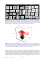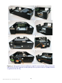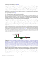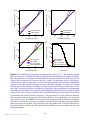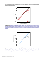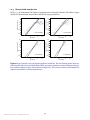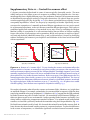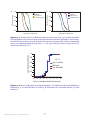
1
Supplementary Information
The C. elegans Lifespan Machine
Nicholas Stroustrup1 , Bryne E. Ulmschneider, Zachary M. Nash, Isaac F. López Moyado,
Javier Apfeld∗,1 and Walter Fontana∗,1
Department of Systems Biology, Harvard Medical School
These authors are co-last authors 1 These authors are corresponding authors
[email protected] — [email protected] — [email protected]
∗
Table of Contents
List of Figures . . . . . . . . . . . . . . . . . . . . . . . . . . . . . . . . . . . . . . . . . . . . . . . . . . .. . . . . . . . . . . . . . . . . . . . . . . . . . . . . . . . . . . . . . . .
Supplementary Note 1 — Lifespan Machine hardware . . . . . . . . . . . . . . . . . . . . . . . . . . . . . . . . . . . . . . . . . . . . .
1.1 Image acquisition . . . . . . . . . . . . . . . . . . . . . . . . . . . . . . . . . . . . .
1.2 Temperature control during image acquisition . . . . . . . . . . . . . . . . . . . . .
Supplementary Note 2 — Image processing. . . . . . . . . . . . . . . . . . . . . . . . . . . . . . . . . . . . . . . . . . . . . . . . . . .. . . . . .
2.1 Masking, background subtraction, and segmentation . . . . . . . . . . . . . . . . .
2.2 Registration of consecutive scanner images . . . . . . . . . . . . . . . . . . . . . . .
2.3 Worm recognition . . . . . . . . . . . . . . . . . . . . . . . . . . . . . . . . . . . . .
Supplementary Note 3 — Assembly . . . . . . . . . . . . . . . . . . . . . . . . . . . . . . . . . . . . . . . . . . . . . . . . . . .. . . . . . . . . . . . . . .
3.1 Introduction . . . . . . . . . . . . . . . . . . . . . . . . . . . . . . . . . . . . . . . .
3.2 Complete parts list . . . . . . . . . . . . . . . . . . . . . . . . . . . . . . . . . . . . .
3.3 Recommended scanner modifications . . . . . . . . . . . . . . . . . . . . . . . . . .
3.3.1 Overview of the fan mounting procedure . . . . . . . . . . . . . . . . . . . .
3.3.2 Modifying the scanner chassis . . . . . . . . . . . . . . . . . . . . . . . . . .
3.3.3 Modifying the transparency unit . . . . . . . . . . . . . . . . . . . . . . . . .
3.3.4 Accessing and preparing the fixed scanner lens . . . . . . . . . . . . . . . . .
3.4 Instructions for focusing a scanner . . . . . . . . . . . . . . . . . . . . . . . . . . . .
Supplementary Note 4 — Software toolset . . . . . . . . . . . . . . . . . . . . . . . . . . . . . . . . . . . . . . . . . . . . . . . . . . .. . . . . . .
4.1 Software components . . . . . . . . . . . . . . . . . . . . . . . . . . . . . . . . . . .
4.2 The Worm Browser GUI client . . . . . . . . . . . . . . . . . . . . . . . . . . . . . .
Supplementary Note 5 — Movement analysis. . . . . . . . . . . . . . . . . . . . . . . . . . . . . . . . . . . . . . . . . . . . . . . . . . .. . . .
5.1 Identification of stationary worms . . . . . . . . . . . . . . . . . . . . . . . . . . . .
5.2 Posture analysis of stationary worms . . . . . . . . . . . . . . . . . . . . . . . . . .
Supplementary Note 6 — Statistics of worm movement . . . . . . . . . . . . . . . . . . . . . . . . . . . . . . . . . . . . . . . . . . .
Supplementary Note 7 — A “death phenotype” . . . . . . . . . . . . . . . . . . . . . . . . . . . . . . . . . . . . . . . . . . . . . . . . . . .. .
Supplementary Note 8 — Censoring . . . . . . . . . . . . . . . . . . . . . . . . . . . . . . . . . . . . . . . . . . . . . . . . . . .. . . . . . . . . . . . . .
8.1 Right censoring and interval censoring . . . . . . . . . . . . . . . . . . . . . . . . .
8.2 Tracking population size and right censoring strategies . . . . . . . . . . . . . . . .
8.3 Worm clusters and interval censoring . . . . . . . . . . . . . . . . . . . . . . . . . .
8.4 Censoring strategies for worm clusters . . . . . . . . . . . . . . . . . . . . . . . . . .
8.5 Determining total population size . . . . . . . . . . . . . . . . . . . . . . . . . . . .
Supplementary Note 9 — Statistical power. . . . . . . . . . . . . . . . . . . . . . . . . . . . . . . . . . . . . . . . . . . . . . . . . . .. . . . . . .
Supplementary Note 10 — Aggregation of survival curves . . . . . . . . . . . . . . . . . . . . . . . . . . . . . . . . . . . . . . . .
10.1 Device-specific environmental effects on lifespan . . . . . . . . . . . . . . . . . . . .
10.2 The device-effect regression model . . . . . . . . . . . . . . . . . . . . . . . . . . . .
Nature Methods: doi: 10.1038/nmeth.2475
1
2
4
4
5
7
7
7
8
13
13
13
16
16
16
17
21
22
25
25
25
27
27
29
31
33
36
36
37
40
41
42
43
45
45
45
Supplementary Note 11 — Hazard . . . . . . . . . . . . . . . . . . . . . . . . . . . . . . . . . . . . . . . . . . . . . . . . . . .. . . . . . . . . . . . . . . .
11.1 Hazard estimation and rendering . . . . . . . . . . . . . . . . . . . . . . . . . . . .
11.2 Hazard of wildtype . . . . . . . . . . . . . . . . . . . . . . . . . . . . . . . . . . . . .
11.3 Hazard and sample size . . . . . . . . . . . . . . . . . . . . . . . . . . . . . . . . . .
Supplementary Note 12 — Control for scanner effect . . . . . . . . . . . . . . . . . . . . . . . . . . . . . . . . . . . . . . . . . . . . . .
Supplementary Note 13 — Additional survival curves, controls. . . . . . . . . . . . . . . . . . . . . . . . . . . . . . . . . . .
Supplementary Note 14 — Limitations . . . . . . . . . . . . . . . . . . . . . . . . . . . . . . . . . . . . . . . . . . . . . . . . . . .. . . . . . . . . . .
Supplementary Note 15 — Resolution and scalability . . . . . . . . . . . . . . . . . . . . . . . . . . . . . . . . . . . . . . . . . . . . . .
Supplementary Note 16 — Use cases . . . . . . . . . . . . . . . . . . . . . . . . . . . . . . . . . . . . . . . . . . . . . . . . . . .. . . . . . . . . . . . .
Acknowledgments . . . . . . . . . . . . . . . . . . . . . . . . . . . . . . . . . . . . . . . . . . . . . . . . . . .. . . . . . . . . . . . . . . . . . . . . . . . . . . . . . . . . . .
References . . . . . . . . . . . . . . . . . . . . . . . . . . . . . . . . . . . . . . . . . . . . . . . . . . .. . . . . . . . . . . . . . . . . . . . . . . . . . . . . . . . . . . . . . . . . . . .
List of Figures
1-1
1-2
1-3
2-1
2-2
2-3
2-4
2-5
3-1
3-2
3-3
3-4
3-5
3-6
4-1
5-1
6-1
6-2
7-1
7-2
7-3
8-1
8-2
8-3
8-4
8-5
9-1
11-1
11-2
11-3
11-4
11-5
12-1
13-1
Lifespan Machine hardware . . . . . . . . . . . . . . . . . . . . . . . . . . . . . . . . . . . . . . . . . . . . . . . . . . .. . . . . . . . . . . . . .
The effect of scanner temperature on lifespan . . . . . . . . . . . . . . . . . . . . . . . . . . . . . . . . . . . . . . . . . . . . .
Consistency of scanner surface temperature . . . . . . . . . . . . . . . . . . . . . . . . . . . . . . . . . . . . . . . . . . . . . . .
Registration of consecutive scanner images . . . . . . . . . . . . . . . . . . . . . . . . . . . . . . . . . . . . . . . . . . . . . . . .
Feature palette for worm recognition . . . . . . . . . . . . . . . . . . . . . . . . . . . . . . . . . . . . . . . . . . . . . . . . . . .. . . .
Worm and non-worm objects . . . . . . . . . . . . . . . . . . . . . . . . . . . . . . . . . . . . . . . . . . . . . . . . . . .. . . . . . . . . . . .
Support Vector Machine for worm recognition . . . . . . . . . . . . . . . . . . . . . . . . . . . . . . . . . . . . . . . . . . . .
Image segmentation examples . . . . . . . . . . . . . . . . . . . . . . . . . . . . . . . . . . . . . . . . . . . . . . . . . . .. . . . . . . . . . .
Scanner modification diagram . . . . . . . . . . . . . . . . . . . . . . . . . . . . . . . . . . . . . . . . . . . . . . . . . . .. . . . . . . . . . .
Photographs of chassis modifications . . . . . . . . . . . . . . . . . . . . . . . . . . . . . . . . . . . . . . . . . . . . . . . . . . .. . .
Photographs of mounted scanner fan . . . . . . . . . . . . . . . . . . . . . . . . . . . . . . . . . . . . . . . . . . . . . . . . . . .. . . .
Fan wiring diagram . . . . . . . . . . . . . . . . . . . . . . . . . . . . . . . . . . . . . . . . . . . . . . . . . . .. . . . . . . . . . . . . . . . . . . . . . . .
Inside the scanner chassis . . . . . . . . . . . . . . . . . . . . . . . . . . . . . . . . . . . . . . . . . . . . . . . . . . .. . . . . . . . . . . . . . . .
Reference images demonstrating sufficient focus . . . . . . . . . . . . . . . . . . . . . . . . . . . . . . . . . . . . . . . . .
Worm Browser screen shot . . . . . . . . . . . . . . . . . . . . . . . . . . . . . . . . . . . . . . . . . . . . . . . . . . .. . . . . . . . . . . . . . .
Parameter robustness of posture analysis . . . . . . . . . . . . . . . . . . . . . . . . . . . . . . . . . . . . . . . . . . . . . . . . . .
Population statistics of movement classes . . . . . . . . . . . . . . . . . . . . . . . . . . . . . . . . . . . . . . . . . . . . . . . . .
Stationarity (posture) span . . . . . . . . . . . . . . . . . . . . . . . . . . . . . . . . . . . . . . . . . . . . . . . . . . .. . . . . . . . . . . . . . .
The volume of a worm dips at or near death . . . . . . . . . . . . . . . . . . . . . . . . . . . . . . . . . . . . . . . . . . . . . . .
Coincidence of volume dip and cessation of motion. . . . . . . . . . . . . . . . . . . . . . . . . . . . . . . . . . . . . . .
Generality of the volume dip. . . . . . . . . . . . . . . . . . . . . . . . . . . . . . . . . . . . . . . . . . . . . . . . . . .. . . . . . . . . . . . . .
Interval midpoint vs Turnbull estimator . . . . . . . . . . . . . . . . . . . . . . . . . . . . . . . . . . . . . . . . . . . . . . . . . . .
Going missing and getting lost (schematic) . . . . . . . . . . . . . . . . . . . . . . . . . . . . . . . . . . . . . . . . . . . . . . . .
Going missing and getting lost (data) . . . . . . . . . . . . . . . . . . . . . . . . . . . . . . . . . . . . . . . . . . . . . . . . . . .. . . .
Interval censoring for worm clusters (schematic) . . . . . . . . . . . . . . . . . . . . . . . . . . . . . . . . . . . . . . . . .
Censoring strategies for terminal worm clusters . . . . . . . . . . . . . . . . . . . . . . . . . . . . . . . . . . . . . . . . . .
Statistical power . . . . . . . . . . . . . . . . . . . . . . . . . . . . . . . . . . . . . . . . . . . . . . . . . . .. . . . . . . . . . . . . . . . . . . . . . . . . . .
Wildtype high-resolution hazard functions at 25 ◦ C . . . . . . . . . . . . . . . . . . . . . . . . . . . . . . . . . . . . . .
Wildtype high-resolution quantile-quantile plots at 25 ◦ C . . . . . . . . . . . . . . . . . . . . . . . . . . . . . . .
Wildtype hazard at 25 ◦ C scored manually . . . . . . . . . . . . . . . . . . . . . . . . . . . . . . . . . . . . . . . . . . . . . . . . .
Wildtype hazard at 20 ◦ C on HT115 . . . . . . . . . . . . . . . . . . . . . . . . . . . . . . . . . . . . . . . . . . . . . . . . . . .. . . . . .
Population size and hazard parameter estimates . . . . . . . . . . . . . . . . . . . . . . . . . . . . . . . . . . . . . . . . . .
Absence of a “scanner effect” . . . . . . . . . . . . . . . . . . . . . . . . . . . . . . . . . . . . . . . . . . . . . . . . . . .. . . . . . . . . . . . .
Wildtype at 25◦ , device-corrected . . . . . . . . . . . . . . . . . . . . . . . . . . . . . . . . . . . . . . . . . . . . . . . . . . .. . . . . . . .
Nature Methods: doi: 10.1038/nmeth.2475
2
47
47
48
51
52
54
58
60
61
62
62
4
5
5
7
9
10
10
11
18
19
20
21
23
24
26
30
31
32
33
34
35
36
38
39
40
41
43
48
49
50
50
51
52
54
13-2
13-3
13-4
13-5
13-6
13-7
13-8
RNAi lifespans at 20 ◦ C . . . . . . . . . . . . . . . . . . . . . . . . . . . . . . . . . . . . . . . . . . . . . . . . . . .. . . . . . . . . . . . . . . . . . .
daf-16(RNAi) at 25 ◦ C . . . . . . . . . . . . . . . . . . . . . . . . . . . . . . . . . . . . . . . . . . . . . . . . . . .. . . . . . . . . . . . . . . . . . . . .
Automated lifespan assay of a severely uncoordinated mutant . . . . . . . . . . . . . . . . . . . . . . . . . .
Survival curves of unc-50(e306) and unc-4(e120);rol-6(su1006) at 25 ◦ C . . . . . . . . . . . . .
Lifespans of of temperature-sensitive self-sterile mutants . . . . . . . . . . . . . . . . . . . . . . . . . . . . . . .
Visual controls of additional mutant survival curves . . . . . . . . . . . . . . . . . . . . . . . . . . . . . . . . . . . . . .
Error of LM operation at high-temperature . . . . . . . . . . . . . . . . . . . . . . . . . . . . . . . . . . . . . . . . . . . . . . . .
Nature Methods: doi: 10.1038/nmeth.2475
3
54
55
55
56
56
57
57
Supplementary Note 1 — Lifespan Machine hardware
1.1
1.1
1.2
Image acquisition . . . . . . . . . . . . . . . . . . . . . . . . . . . . . . . . . . . . . . . . . . . . . . . . . . . . .
Temperature control during image acquisition . . . . . . . . . . . . . . . . . . . . . . . . . . . . .
Image acquisition
4
5
Our reference installation consists of 50 scanners allocated to 5 incubators. A single scanner is
sufficient to obtain controlled survival curves of high accuracy, while a fleet of scanners enables
high throughput. In Note 16, we provide a description of use cases for varying numbers of scanners. Instructions for the modification of a commercially available scanner model (Epson v700)
are provided as a separate supplementary document. At www.lifespanmachine.org, we have set up
pointers to a public repository for the software components of the Lifespan Machine to facilitate
user-driven development and improvement.
a
b
c
d
Figure 1-1: Lifespan Machine hardware. (a) A Thermo Forma incubator provides temperature
control for ten flatbed scanners. (b) A scanner unit with open lid and plates loaded, showing fans
mounted on the lid to create a flow of air between lid and scanner surface when the lid is closed. (c)
A scanner unit with closed lid, as during operation, shown from rear to highlight fans mounted into
the chassis. (d) The typical plate configuration consists of 16 plates inserted face down through
holes punched into a rubber mat. The mat seals the plates to the glass surface of the scanner,
stabilizing their position and preventing dessication. Note the light source located in the lid (transillumination mode).
Nature Methods: doi: 10.1038/nmeth.2475
4
1.2
Temperature control during image acquisition
An important aspect of our method is the control of temperature within a tight range, reducing the
large excursions that are sometimes unavoidable in the manual method where plates are moved in
and out of an incubator for scoring. The notorious temperature sensitivity of C. elegans is highlighted by its variation between scanners (Fig. 1-2).
Mean worm lifespan (days)
14.4
R::3
14.2
14
13.8
R::2
13.6
L::1
R::1
13.4
R::5
R::4
13.2
L::2
13
L::5
L::4
L::3
12.8
24.8
25
25.2
25.4
Mean scanner temperature (°C)
25.6
Figure 1-2: The effect of scanner temperature on lifespan. The lifespans of wildtype animals were
acquired as described for Fig. 2 of the main text, and loaded onto 160 plates distributed across 10
scanners. The average lifespan of all animals on a given scanner was compared to the temperature
measured on that scanner (Online Methods). A linear regression was run with all scanners (r2 =
0.163, red dashed line) and with the R::3 outlier excluded (r2 = 0.746, red solid line).
34
fans off fans on
33
incubator layout
Temperature (°C)
32
b
25.4
25.3
L2
R2
L3
R3
30
L4
R4
29
L5
R5
28
27
b
25.1
24.9
24.8
24.7
24.6
24
24.5
3
4
R2
3
Time (h)
c
L1
R4
R5
R1
25.0
25
2
L3
R3
25.2
26
1
L5
L4
L2
25.5
R1
31
0
3.5
Scanner surface temperature (°C)
25.5
25.4
25.3
25.2
25.1
25
24.9
24.8
24.7
24.6
4
Time (h)
d
25.6
Scanner surface temperature (°C)
25.6
L1
Temperature (°C)
a
25.4
L1
25.2
L2
L4
L5
25
L3
24.8
R4
R3
24.6
R1
R2
24.4
R5
24.2
24
24.5
L1
L2
L3
L4
L5
R1
R2
R3
R4
R5
24
Position inside incubator (Row::Column)
24.2
24.4
24.6
24.8
25
25.2
25.4
Scanner surface temperature (°C)
Figure 1-3: Consistency of scanner surface temperature. Caption on next page.
Nature Methods: doi: 10.1038/nmeth.2475
5
Figure 1-3 (Cont’d): (a) Local temperature differences between and temperature excursions of
10 scanners operating at a scan frequency of 15 minutes inside an incubator (Online Methods). On
the left, the fans are turned off; on the right, they are turned on. The inset diagrams the shelving of
scanners for reference in panels (b)-(d). (b) Detail of the rectangular area indicated in panel (a).
Fans reduce local temperature differences due to incubator airflow to within 1◦ C and temperature
excursions from scanning to within 0.2◦ C. (c) Data shown in panel (b) are reported against the
location of the scanners in an incubator adjusted to maintain 25◦ C, with 5 shelves and 2 scanners
(Left and Right) per shelf; see inset of panel (a). Time series are summarized by the mean with
error bars indicating one standard deviation. The left side of the incubator has a slightly higher
average temperature compared to the right side due to incubator airflow. (d) In a separate experiment, the scanner temperatures of a single incubator were measured at one time (x-axis) and again
two months later (y-axis). The temperature measures correlate (R2 = 0.85), indicating stability of
operating conditions.
Temperature control is especially important when measuring lifespans outside the “room temperature” range, as with thermotolerance assays (e.g. Fig. 5 of the main text). To control temperature
gradients that result from a scanner’s electronics and light source, cooling fans are mounted into
the chassis of the scanner, as shown in Fig. 1-1b and schematically in Fig. 1 of the main text. Fig.
1-3 below shows the efficacy of these modifications.
Nature Methods: doi: 10.1038/nmeth.2475
6
Supplementary Note 2 — Image processing
2.1
2.1
2.2
2.3
Masking, background subtraction, and segmentation . . . . . . . . . . . . . . . . . . . . . . .
Registration of consecutive scanner images . . . . . . . . . . . . . . . . . . . . . . . . . . . . . . . .
Worm recognition . . . . . . . . . . . . . . . . . . . . . . . . . . . . . . . . . . . . . . . . . . . . . . . . . . . . .
7
7
8
Masking, background subtraction, and segmentation
In images captured by a scanner, only areas corresponding to the agar surface of each plate need
to be analyzed. “Masking” is the process of separating multiple plates and removing their edges.
The Worm Browser (Note 4 section 4.2 and Supplementary Video 6) generates a low-resolution
composite of each image captured. The user can then draw on top of this image to produce an
overlay (the mask) that specifies the location of each plate. This drawing can be done quickly in
programs such as Photoshop or GIMP [1]. (This task is a candidate for future automation.) The
image-processing server uses the resulting mask to extract individual plate images and stores them
in a stereotyped folder hierarchy associated with each experiment.
Prior to analysis, all images X (t) , t = 1, . . . , T , undergo background subtraction. Each pixel xi ∈
X (t) is reduced by the median value in a surrounding square of 25×25 pixels 198 µm×198 µm. This
median filtering removes variation at spatial scales larger than roughly twice the average width
of an adult worm (198 µm) suppressing background effects including shadows, smooth bacterial
lawns, and uneven agar shading.
(t)
Median filtering sets the stage for image segmentation—the separation of worms from their agar
and bacterial surface background—using a global threshold. Two global thresholds are applied to
images, one restrictive threshold that identifies only dark objects such as worms, but tends to break
worms into multiple objects, and a second more permissive threshold that tends to pick up extraneous features of the bacterial lawn. The two are compared to produce a final image segmentation.
See also Fig. 2-5.
Registration of consecutive scanner images
2.2
The panels in Fig. 2-1 show the magnitude of two types of jitter in the acquisition of images by our
scanners (and presumably consumer electronics scanners in general). Displacements like these
must be taken into account when processing images leading up to the movement analysis (sections
5.1 and 5.2).
b
c
3
2
1
0
∆Y worm position (pixels)
3
∆Y worm position (pixels)
∆Y plate position (pixels)
a
2
1
0
-1
-1
-2
-3
-2
0.5
1
1.5
Time (d)
2
2.5
1
0
-1
-2
-3
-4
-4
0
2
0
0.5
1
1.5
Time (d)
2
2.5
-2
-1
0
1
∆Y plate position (pixels)
2
Figure 2-1: Registration of consecutive scanner images. (a): The mechanism advancing the scanning bar causes offsets of a few pixels relative to world coordinates between subsequent images of
the same plate. (b): Slight distortions in plate images cause displacements of a few pixels relative
to plate coordinates for stationary worms. (c): The two types of displacements shown in panels
(a) and (b) are uncorrelated.
Nature Methods: doi: 10.1038/nmeth.2475
7
2.3
Worm recognition
The image-processing pipeline computes for each foreground object a set of features that are subsequently used by a Support Vector Machine (SVM) [2], Fig. 2-4, to discriminate between worm
and non-worm objects, Fig. 2-3. The following is a list of 65 features used by SVM-based worm
classifier. Their distribution of values is shown in Fig. 2-2.
• Pixel Area
• Rectangular Diagonal
• Max Width
• Width at Center
• Spine (SP) Length / Pixel Area
• SP Length / Rect Diagonal
• SP Length / Width at Front
• Average Curvature
• Curvature X Intercepts
• Absolute Intensity (AI)
• AI Roughness (Entropy)
• AI Average Along SP
• AI darkest 20% area
• AI of Region
• AI Normalized Avg Along SP
• RI Skew
• RI Max
• RI darkest 20% of pixels
• RI Variance Along Spine
• RI Normalized Max
• Edge Area / Object Area
• Intensity Profile Max
• Rectangular Width
• Spine Length
• Min Width
• Width at Front
• SP Length / Rect Width
• SP Length / Max Width
• SP Length / Width at Rear
• Max Curvature
• Curvature Variance
• AI Variance
• AI Roughness
• AI Average in Nbrhd
• AI Dist from Nbrhd
• AI Normalized Average
• Relative Intensity (RI)
• RI Roughness (Entropy)
• RI Average Along SP
• RI darkest 20% area
• RI of Region
• RI Normalized Avg Along SP
• Intensity Profile at Edge
• Intensity Profile Variance
• Rectangular Height
• Average Width
• Variance in Width
• Width at Rear
• SP Length / Rect Height
• SP Length / Average Width
• Ratio of end widths
• Total Curvature
• Distance between ends
• AI Skew
• AI Maximum
• AI darkest 20% of pixels
• AI Variance Along SP
• AI Normalized Max
• RI Variance
• RI Roughness
• RI Average in Nbrhd
• RI Dist from Nbrhd
• RI Normalized Average
• Edge Area
• Intensity Profile at Center
Because no single feature is sufficient to accurately discriminate between worm and non-worm objects, information from multiple features must be integrated. To assess whether accurate discrimination is feasible, we hand-annotated ∼4,000 images as depicting either a worm or a non-worm
object. We divided this set into two parts, a training set (1489 worms, 945 non-worms) and a test
set (992 worms, 631 non-worms). Using the libsvm library, we built an SVM model of the training
data, and evaluated its performance on the test set. The confusion matrix is shown below, demonstrating a discrimination accuracy of 97.7%. The Worm Browser (Note 4 section 4.2) allows rapid
inspection of all objects predicted by the machine to be worms. Mistakes can therefore be annotated, producing a final false positive rate of nearly zero.
SVM prediction
by-hand
worm
non-worm
worm
991
36
non-worm
1
595
To visualize the feasibility of discrimination, we computed the eigenvectors of the feature correlation matrix of the gold standard and represented each animal as a dot projected onto the subspace
spanned by the eigenvectors with the three largest eigenvalues (principal components PCA 1-3), as
shown in Fig. 2-4.
Nature Methods: doi: 10.1038/nmeth.2475
8
0.000
0
5000
40
probability density
30
20
10
0
0
0.1
0.2
0.3
ratio of medial axis length to area
(pixel widths per pixel)
probability density
0.04
0.03
0.02
0.01
0.00
probability density
0.25
0.20
0.15
0.10
0.005
0.000
1.0
0.00
0.0
0.0045
0.004
0.0035
0.003
0.0025
0.002
0.0015
0.001
0.0005
0
0.0015
0.001
0.0005
0.8
0.6
0.4
0.2
0.0
0
10 20 30 40 50 60 70
Maxiumum Curvature Along Medial Axis
(1/pixel width)
curvature average (1/pixel width)
1.1
1.0
0.9
0.8
0.7
0.6
0.5
0.4
0.3
0.2
0.1
0.0
3.5
3.0
2.5
2.0
1.5
1.0
0.5
0.0
-1
0
1
2
3
2
skew of median-filtered object intensity
(8 bit pixel intensity)
1.1
1.0
0.9
0.8
0.7
0.6
0.5
0.4
0.3
0.2
0.1
0.0
0.020
0.015
0.010
0.005
0.000
-1 0 1 2 3
4 5 6
skew of absolute object intensity
(8 bit pixel intensity)
0 20 40 60 80 100 130 160 190
maximum absolute object intensity
(8 bit pixel intensity)
0.035
0.012
3.0
0.030
0.010
1.5
1.0
0.025
0.020
0.015
0.010
0.008
0.006
0.004
0.002
0.5
0.005
0.0
0.000
0.000
-130 -100-80 -60 -40 -20 0 20 40 60
100 200 300 400 500 600 700 800
difference between average absolute object intensity
and average intensity of object neighborhood
perimeter length (pixel widths)
(8 bit pixel intensity)
30000
1
2
3
4
5
entropy of absolute object intensity
(bits)
0.040
0.007
0.06
0.035
0.006
15
10
5
probability density
0.07
25
probability density
30
20
0.05
0.04
0.03
0.02
0.01
0
0.2
0.3
0.4
0.5
0.6
0.7
0.8
ratio of perimiter length and medial
axis length (unitless)
0.030
0.025
0.020
0.015
0.010
0.000
0
10
20
30
40
50
60
70
0.004
0.003
0.002
0.000
0 10
average median-filtered intensity
along perimeter (8 bit pixel intensity)
0.005
0.001
0.005
0.00
0.1
4
0.025
3.5
2.0
3
entropy of median-filtered object intensity
(bits)
0.030
-2
2.5
10
20
30
width average (pixel widths)
1.0
0 1 2 3 4 5 6 7 8 9 11 13 15 17 19
200 400 600 800 100012001400
0.002
variance in absolute object intensity along medial axis
(square 8 bit pixel intensity)
probability density
1.5
0
1000
2000
3000
variance of absolute object intensity
(square 8 bit pixel intensity)
probability density
probability density
0.00045
0.0004
0.00035
0.0003
0.00025
0.0002
0.00015
0.0001
0.00005
0
20000
2.0
0.5
100
200
average absolute object intensity
(8 bit pixel intensity)
10000
2.5
0.05
0
0
3.0
probability density
probability density
0.010
probability density
1.2
0.30
0.0025
0.015
0
1.4
variance of median-filtered object intensity
(square 8 bit pixel intensity)
0.020
0.00
3.5
0
0.025
0.05
4.0
20 30 40 50 60 70 80 90
average median-filtered object intensity
(8 bit pixel intensity)
0.10
0.35
probability density
probability density
0.05
0.15
0.40
0
10
20
30
40
50
ratio of medial axis length to maximum animal width
(pixel widths per pixel)
0.06
0
0.005
0.000
0
100
200
300
distance between head and tail (pixel widths)
100
200
300
length of medial axis (pixel widths)
probability density
probability density
50
0.010
probability density
2000 3000 4000
animal area (pixels)
0.005
probability density
1000
0.010
probability density
0
0.015
0.20
0.015
probability density
0.0002
0.020
probability density
0.0004
0.020
probability density
0.0006
0.025
probability density
worm
non-worm
0.001
0.0008
probability density
probability density
0.0012
30
50 60
80
100 120 140
absolute object intensity at eroded
animal center (8 bit pixel intensity)
0
1000
2000
variance in absolute object intensity
around perimiter after repeated erosions
(squared 8 bit pixel intensity)
Figure 2-2: Feature palette for worm recognition. The panels show the frequency distribution of
the values for each feature in the case of worms (blue) and non-worms (red). Because feature value
distributions of worm and non-worm objects overlap in all cases, no single feature is sufficient for
accurate discrimination. 1 pixel width corresponds to 8µm.
Nature Methods: doi: 10.1038/nmeth.2475
9
non-worms
worms
Figure 2-3: Worm and non-worm objects. The images inside the red rectangle exemplify worm
objects; the images on the left, outside the rectangle, illustrate various non-worm objects ranging
from fibers, to dust, to lot numbers stamped on plates. The two classes of objects are well-separated
by the Support Vector Machine, Fig. 2-4, based on the feature set shown in Fig. 2-2.
PCA 2
-5
0
-10
-10
-5
PCA 1 0
5
10
5
0
PCA 3
-5
Figure 2-4: Support Vector Machine for worm recognition. Black: worms, Red: Non-worms.
Each object of a hand-annotated “gold standard” is represented as a dot in a 65-dimensional feature
space. The figure shows the projection of this point cloud onto the subspace spanned by the three
principal components that explain the largest share of total variation (60.4%).
The previous table was based solely on wildtype. The following table illustrates the performance of
the SVM when trained on a mix of strains and then applied to each separately (rather than trained
on each specifically). For this analysis, we (1) lumped a number of genotypes into one big set; (2)
assigned randomly 3/5 of animals to the training set and the remainder to the test set; (3) trained
an SVM on the training set; (4) tested it on the test set; (5) calculated the confusion matrix for each
Nature Methods: doi: 10.1038/nmeth.2475
10
genotype. FP=false positives, FN=false negatives:
FP
all
daf-16 (mu86)
73/2426 = 3.0%
11/336 = 3.3%
84/1550 = 5.4%
FN
23/207 = 11.1%
wildtype
21/562 = 3.7%
19/777 = 2.4%
glp-1
age-1(hx546)
31/883 = 3.5%
12/430 = 2.8%
32/453 = 7.1%
8/328 = 2.4%
All strains have a false negative rate between 2.4% and 3.5%. The false positive rate is more varied,
but ultimately immaterial, since we catch the non-worm objects during visual inspection in the
quality control phase with the Worm Browser.
Fig. 2-5 provides examples of image segmentation and object recognition using the SVM, illustrating the difficulties of disentangling worm clusters. Refer to sections 8.2 and 8.3, as well as Figs.
8-3 and 8-5, for more detail on the accounting of multi-worm clusters. Supplementary Videos 1–5
provide time-lapse movies of plates with wildtype, age-1(hx546), and unc-64(e246) mutants for
the entire duration of an experiment, both with and without metadata overlay.
a
c
unprocessed
Image
segmented
image
b
Figure 2-5: Image segmentation examples. Caption on next page.
Nature Methods: doi: 10.1038/nmeth.2475
11
classified
objects
Figure 2-5 (Cont’d): (a) A scanner raw image of an agar plate with worms on patches of bacterial
lawn (E. coli OP50). Scale bar on the left corresponds to 5mm; the scale bar on the right to 1mm.
(b) The same image after background subtraction (using a median filter), with foreground objects
resulting from image segmentation highlighted in color. Objects classified as worms by the SVM
are colored red; non-worms are colored blue. (c) Enlarged images of worms indicated in panel
(b), as they appear in consecutive image processing steps. The scale bar marks 1mm. The first,
third, and fourth rows are examples of correctly processed animals. The second row illustrates
the difficulties of parsing multiple worm clusters. Note how several of the worms are incorrectly
identified. Such errors, when they occur in the context of dead worms, can be rapidly identified in
the Worm Browser (Supplementary Video 6).
Nature Methods: doi: 10.1038/nmeth.2475
12
Supplementary Note 3 — Assembly
3.1
3.2
3.3
3.1
Introduction . . . . . . . . . . . . . . . . . . . . . . . . . . . . . . . . . . . . . . . . . . . . . . . . . . . . . . . . .
Complete parts list . . . . . . . . . . . . . . . . . . . . . . . . . . . . . . . . . . . . . . . . . . . . . . . . . . . .
Recommended scanner modifications . . . . . . . . . . . . . . . . . . . . . . . . . . . . . . . . . . . .
3.3.1 Overview of the fan mounting procedure . . . . . . . . . . . . . . . . . . . . . . . . . . . . .
3.3.2 Modifying the scanner chassis . . . . . . . . . . . . . . . . . . . . . . . . . . . . . . . . . . . . . .
3.3.3 Modifying the transparency unit . . . . . . . . . . . . . . . . . . . . . . . . . . . . . . . . . . . .
3.3.4 Accessing and preparing the fixed scanner lens . . . . . . . . . . . . . . . . . . . . . . . .
3.4
Instructions for focusing a scanner . . . . . . . . . . . . . . . . . . . . . . . . . . . . . . . . . . . . . . .
Introduction
13
13
16
16
16
17
21
22
The lifespan machine identifies worm death times through the combination of four interacting
components:
(i)
An image acquisition system based on modified flatbed document scanners;
(ii) An image acquisition control server running under Linux on a PC;
(iii) An image analysis server that can run in the background of any x86 Windows, Apple, or Linux
computer;
(iv) A data annotation and aggregation client program that allows rapid inspection and annotation of automated results.
This document contains specifications for a reference implementation of the image acquisition
system. An up-to-date, detailed specification of the software is provided as part of the source code
documentation.
These instructions are lengthy, representing the authors’ attempt at assembling a comprehensive,
self-sufficient document. The modifications themselves are actually quite straightforward. A research assistant (RA) with a life science undergraduate degree and no engineering background
learned how to perform all modifications over the course of several hour-long training sessions.
The modifications required to complete two scanners, capable of monitoring over a thousand individuals, should take a novice two or three half days. All modifications are easily performed on a
standard lab bench.
The authors do not currently possess good estimates for the operating lifespan of a modified scanner. Existing devices appear to operate correctly after nearly two years of continuous use. Several
have been in operation for nearly five years. Across a fifty-scanner installation, the authors have
retired only three scanners as a result of malfunction, usually after only a few weeks of operation.
Presumably, scanner fluorescent bulbs will eventually require replacement.
3.2
Complete parts list
The following list details the equipment required for a reference, 10 scanner installation capable of
monitoring 5600 nematodes simultaneously. Smaller or larger clusters can be easily assembled at
proportional cost. All prices are from circa 2012 and approximate.
10x Epson v700 Perfection V700 Photo Scanners.
Available from Amazon.com or Epson.com for $540. In the past, we have also used the discontinued Epson 4990 Photo scanner. Other scanner models that possess appropriate transillumination capabilities are likely to be adaptable.
70x 80x80x25mm DC fans
This is a standardized computer chassis component, and as such is widely available from
Nature Methods: doi: 10.1038/nmeth.2475
13
many suppliers. For example, the Thermaltake DuraMax 8 AF0058 80mm Case Fan available from NewEgg.com for $8.20.
10x 120x120x25mm DC fans
This is a standardized computer chassis component, and as such is widely available from
many suppliers. For example, the Thermaltake A2492 120mm Case Fan is available from
NewEgg.com for $9.
1x A 300+Watt source of 12V DC Power
We use standard Power Supply Units (PSU), which are mass-produced components of PC
computer chasses, and as such are widely available at low prices from many suppliers. For
example, the CORSAIR Builder Series CX430 V2 (CMPSU-430CXV2) 430W is available from
NewEgg.com for $45
1x Temperature-controlled environment for equipment
This might be an incubator or a temperature-controlled room. For example, the Thermo
Forma Environmental Chamber is a multi-use incubator which many labs already possess.
This incubator, in 2010, cost approximately $13,000 new, and as such represents more than
half the price of an incubator-based Lifespan Machine installation.
50x Machine Screws
For example, Stainless Steel Machine Screw and Hex Nuts 11/32x1/8” 8-32 available from
McMaster-Carr; a 100-unit box for $4.25
50x Machine Screw Nuts
For example, Stainless Steel Machine Screw and Hex Nuts 11/32x1/8” 8-32 available from
McMaster-Carr; a 100-unit box for $4.25
80x Nylon twine or string, cut into 16.5” lengths
Used for mounting fans. Simpson’s Individual Stringettes; available from McMaster-Carr;
1800 feet for $8.00
1x Reasonably modern (circa 2012) x86 PC or laptop capable of running Linux
More details provided under “Software Components” (section 1.3.1 of the scientific Supplementary Information); $650
10x 9x11.5” inch glass sheets
Standard window pane glass, available from most local hardware stores; $5 per sheet.
10x 8.7x11.3” inch rubber mats
Cut from rubber stock, for example High-Strength EPDM Rubber Sheet, 40A,Black, 36”x36”x1/16”
available from McMaster-Carr at $20 per foot.
40x Plastic spacers to support the scanner lid
1” segments cut from 8’ Polyethylene U-Shaped Channels. Leg Length - 3/4”, Base Length 5/4”. Available for McMaster-Carr; $18.8 for eight feet.
2x Gaffer’s tape
This is similar to duct tape, but better in every way. Available from McMaster-Carr; $10 a
roll. Black looks sleek; pink looks totally rad.
10x Wire to power fans
20 AWG gauge or lower.
Nature Methods: doi: 10.1038/nmeth.2475
14
10x Rubber mats
Plates should be sealed to prevent desiccation. Many methods for doing this are possible, we
use a design cut into High-Strength EPDM Rubber Sheet, 40A,Black, available from McMasterCarr at $22 per 3 feet. A reference design, appropriate for laser-cutting, is provided.
1x Round Hole Arch Punch 2” Diameter —Optional tool for construction
If a laser-cut design is not available, rubber mats can be manufactured by hand using this
punch. Available from McMaster-Carr for $70.
160x Small Petri Dishes —Consumed by each 5600 worm experiment
BD Falcon Petri Dishes, 50x9mm, Tight-Fit Lid, Sterile, BD Biosciences. Available from multiple suppliers; $215 per box of 500.
40x Butt-splices or wire nuts —Optional
To avoid soldering joints, butt-splices or wire nuts can be used to splice fan wiring. Available
from McMaster-Carr in large packs for a few dollars. Yes, these have a silly name.
1x Mini screw driver set for scanner disassembly —Tool for Construction
Already present in most labs. For example, a 5-Piece Phillips Screwdriver Set available from
McMaster-Carr for $22.18.
1x Spiral saw for cutting holes in scanner chassis —Tool for construction
Other types of cutting devices may work well, but a spiral saw cuts quickly and will impress
your friends. For example, Rotozip Spiral Saw Electric, 5.5 Amp, 15000 - 30000 rpm, 9-1/4”
L, available from McMaster-Carr for $106.25.
1x Hot-Melt Glue Gun —Tool for construction
Standard Duty Hot-Melt Glue Gun available from McMaster-Carr for $23. Packs of Hot melt
glue sticks available from McMaster-Carr for $10.
1x Soldering Iron or Gun —Optional tool for construction
For example, a Soldering Gun available from McMaster-Carr for $58
1x A power drill / screw driver —Optional tool for construction
Unless you have wrists of steel, it is easier to use a power drill to add and remove machine
screws. Available from McMaster-Carr for $150, or for less at a home improvement store like
Lowes.
1x Canned air —Optional tool for construction
A source of compressed air, either from a can (often sold to clean computer keyboards, sold
at any office supply store), or from a lab air outlet.
10x Additional 80x 80x 25mm DC fans —Optional
Used to circulate air throughout incubator to achieve best-possible temperature calibration.
Only required for certain experimental designs.
10x Metal ductwork —Optional
Used to circulate air throughout incubator to achieve best-possible temperature calibration.
Only required for certain experimental designs. For example, 3” Aluminum Bend-and-Stay
Duct Hose, available from McMaster-Carr for $5.25 Each.
2x Plastic bin for precise drying of plates —Useful ancillary equipment
Exact dimensions are not crucial, but long and flat boxes work best. For example, the 35-5/8”
x 18-1/4” x 6-1/4” h Long Underbed Box with Wheels, available from The Constainer Store .
com for $20.
Nature Methods: doi: 10.1038/nmeth.2475
15
2x Sheet of Wire Mesh, fitted to plastic bin (i.e. 18”x36”) —Useful ancillary equipment
Mounted in plastic bins for precise drying of plates. Available at local hardwarde stores.
Corrosion-Resistant Mesh Fencing is available from McMaster-Carr for $4.70 per square
foot.
1x 25lbs Regular (non-indicating) 8-mesh Drierite —Useful ancillary equipment
Available from drierite.com for $83.38.
3.3
Recommended scanner modifications
In this section we present the steps required for modifying stock Epson v700 Perfection Photo
Scanners into a device capable of producing data of the quality presented in our manuscript. The
modifications accomplish two goals: (1) install fans as a means for regulating scanner surface temperatures and (2) raise the focal plane of the scanner to optimize its use in scanning agar plates.
We see great potential for the future development of simpler, more effective strategies to accomplish these goals. The authors consider the current design primarily as a starting point from which
the technology can be adapted according to each individual researcher’s needs.
3.3.1
Overview of the fan mounting procedure
The Perfection v700 Photo Scanner has two parts—a bottom chassis, which contains the CCD image sensor, and a detachable lid (also called the Transparency Unit or “TPU”), which contains the
transilluminating light source. To prevent the build-up of heat within the chassis, we mount three
8cm computer fans into the sides of the bottom chassis (labelled F1 ,F2, and F3 in the figures).
These serve to move the heat produced during each scan out of the scanner body. To prevent the
build-up of heat within the TPU, we hang four fans (F4, F5, F6, and F7), which serve both to remove
heat from inside the TPU and to circulate cold air across the plates. A single 12cm fan (F8) is hung
from the rear of the TPU, which serves to remove heat from the scanner TPU, the scanner chassis,
and moves cold air across the plates.
Four cuts are made into the scanner chassis, and three into the TPU, to permit cold air to cycle
through both compartments. These cuts are easily made using a spiral saw or other cutting device.
Our experience is that most Dremel tools aren’t quite up to the task of cutting through the hard
chassis plastic. When working with any cutting tool, wear safety goggles and always pay attention.
Clean, uncluttered work spaces promote safe practices.
Three fans—F1, F2, F3—are sealed in place using a glue gun. Four fans are hung from the scanner
TPU are suspended by a loop of nylon twine, hanging from machine screws drilled into the TPU at
positions S1, S2, S3 and S4. The holes can be cut with a power drill if one is available, or else using
the spiral saw.
The exact location and dimension of each modification are presented in Figure 3-1. Photographs
of the cut holes and glued fans are provided in several consecutive figures.
3.3.2
Modifying the scanner chassis
(1) Connect an assembled scanner to a computer and scan a piece of paper, to confirm that the
hardware functions. Scan in “transparency” mode to confirm that the light source in the TPU
functions properly.
(2) Disconnect the scanner and remove the TPU.
(3) The top surface of the scanner chassis is secured by several screws, located under small plastic
covers in each corner. Pop out the plastic caps and release the screws. Pop off the top surface
Nature Methods: doi: 10.1038/nmeth.2475
16
(4)
(5)
(6)
(7)
(8)
(9)
(10)
3.3.3
of the scanner and place it somewhere safe. Be careful not to get finger prints on the bottom
of the scanner glass.
Using your hand, gently push the scanner bar 3/4 of the way toward the front of the scanner,
to move it out of harm’s way.
Place a piece of tape over the long slit opening of the scanner bar, to prevent plastic chips
from entering the bar.
Using a spiral saw or your favorite cutting device, cut holes H1, H2, H3 and H4. Cuts H1 and
H3 run somewhat close to internal scanner wiring. The cuts shouldn’t come extremely close,
but be careful and fold any nearby cables out of harm’s way. Any time you use a spiral saw,
wear safety goggles and stay aware. The spiral saw tends to throw little chips of plastic.
Take an 8cm fan, and use it to confirm that a fan will fit snug into each hole. Enlarge the
holes where necessary. However, don’t remove more plastic than required, as doing so will
make it harder to hot-glue the fans in place.
Using compressed air, either from a can or from a hose hooked up to house air, blow away all
the plastic chips produced by the cuts. If ignored, these plastic chips have the nasty habit of
working their way into moving parts, in particular the scanner’s power button. If this button
ever stops working, there’s probably a chip in it somewhere.
Place a 8cm fan into hole H1. Make sure the power cable is facing towards the front of
the scanner, preferably from the upper right hand corner of the fan. Fan F1 should face
outwards—the airflow should be heading into of the scanner. Glue it in place along all four
sides. You may need to hold the fan in place with one hand and glue with another. In the
same way, glue fans F2 and F3 into holes H2 and H3. Fan F2 and F3 should both be facing
outwards.
Remove the protective tape added earlier to the scanner bar.
Modifying the transparency unit
(1) Put the scanner chassis aside, and consider the scanner TPU. The bottom of the TPU is secured to its the cop cover by eight screws. Release them and pop off the bottom cover, and
place it somewhere safe
(2) Make cuts H5–H7. Note that the TPU connector is located at the center of H7. Remove this
cable from its notch, and make cut H7. You might consider cutting a new notch somewhere
next to cut H7, so that this cable can be again secured.
(3) Using a power drill or the spiral saw, drill holes S1 through S5. Make sure to use a bit appropriate for the machine screws you are using, as you’ll want the screws to fit snugly.
(4) Using compressed air, either from a can or from a hose hooked up to house air, blow away
all the plastic chips produced by the cuts.
(5) Insert machine screws into each hole S1–S5. The head of the screw should be inside the TPU.
Secure the screw by tightening a nut on the reverse side (the outside top of the TPU).
(6) Grab the scanner TPU bottom, and snap it back into place. Screw in all screws.
(7) Cut five 16.5” inch segments of nylon twine. Put the TPU of a scanner on a flat surface, with
the machine screws facing upward. Thread one end of a piece of thread through the two holes
at the top of a 8cm fan. Tie a knot at the appropriate piece of the thread such that the fan
hangs snug at the TPU’s side when hung from machine screw S1. Repeat three more times,
hanging fans from machine screws S2–S4. Remember to orient the fans correctly; Fans F4
and F5 face inward; fans F6 and F7 face outward.
(8) In the same way, tie a 12cm fan F8 to machine screw S5. It should face inward.
(9) Finished!
Nature Methods: doi: 10.1038/nmeth.2475
17
Figure 3-1: Scanner modification diagram. The location and dimensions for each cut are illustrated, for both the scanner chassis and the Transparency Unit (TPU).
Nature Methods: doi: 10.1038/nmeth.2475
18
Figure 3-2: Photographs of chassis modifications. (a): The scanner chassis with its lid removed,
and four cuts, H1–H4, made into its walls. (b–e): Close up views of each cut. (f–h): 8cm fans
are glued into holes H1–H3.
Nature Methods: doi: 10.1038/nmeth.2475
19
Figure 3-3: Photographs of mounted scanner fan. (a): The Transparency Unit (TPU) is shown
with its top and bottom separated. (b–c): Three cuts, H5–H7, are made into the bottom of the
TPU. (d): Four holes are drilled into the TPU lid, and (e) filled with machine screws. (f): The
holes are shown from the top of the lid.
Nature Methods: doi: 10.1038/nmeth.2475
20
Figure 3-4: Fan wiring diagram. The wiring that supplies the fans is shown. “MX” indicates
a standard Molex connector. The connection between fans F4–F8 can be made by clipping off
the Molex connectors and splicing the wires together using solder, or by using a butt-clamp or a
wire nut. Be careful to add appropriate insulation. Be careful to plug the Molex connectors in the
correct order, otherwise excess current may be passed through each connector. Be careful to use an
appropriate of gauge wire for the indicated current. The eight fans attached to each scanner draw
about 1.6 amps at 12V DC. To prevent heat build-up along the wire, a voltage drop of less than
3% along the wire is widely considered safe. If the power source is 10 feet away from the scanner,
this suggests that an AWG gauge no larger than 20 should be used. Incorrect wiring can generate
excess heat and represents a fire hazard.
3.3.4
Accessing and preparing the fixed scanner lens
The Epson Perfection v700 contains a single fixed lens, whose position sets the focal plane of the
device. Worms on agar plates are out of focus on stock scanners, because they are raised above the
standard focal plane. To optimize image quality, we move the lens position very slightly.
(1) The scanner lens is located inside the moving scanner bar, situated inside the bottom scanner
chassis. Remove the lid of the scanner chassis (as described in Recommended Mounting
Procedure Step 3), and identify the scanner bar. Refer to Figure 3-5.
(2) There is a removable plastic flap in the top of the bar. Remove it with a screwdriver. Put it
aside in a safe place. If you look into this slot, you should see, a few inches down, a black
tube. This is the top of the lens. Unfortunately, scanners ship with this lens glued in place by
a piece of hard resin. We need to remove this resin before we can adjusting the lens position.
Removing the resin, in turn, requires we access the lens directly through a port on the bottom
of the scanner bar. This, in turn, requires we disconnect the scanner bar from the scanner
chassis, to make it possible to access the bottom port.
(3) Identify the small metal clip that attaches the scanner bar to the drive belt. The drive belt is
the long rubber belt running the length of the scanner chassis. Take a look at this clip and
remember how it is attached. Pop it off by pushing down on the exposed lever.
(4) Locate the nut at the front of the scanner that holds the support bar in place. The support
bar is the long metal pole that running the length of the scanner chassis. Remove the nut.
Notice that the bar is still held securely by a friction connection at the back of the scanner.
Nature Methods: doi: 10.1038/nmeth.2475
21
(5) When handling the support bar, wear gloves to avoid exposing it to skin oils. Rotate the
support bar in place to release the friction connection. Sometimes, pliers may be necessary.
Only use pliers on the rectangular end of the bar, never on the cylindrical part. The scanner
bar and support bar should now be loose
(6) Remove the support bar from the scanner bar, and put it in a safe, clean place.
(7) Flip the scanner bar so that rests upside down inside the chassis. Be careful not to catch any
wires or to crease the ribbon cable.
(8) You should see several screws on the bottom of the scanner bar, securing the bottom lid.
Remove them, and remove the black bottom lid.
(9) You should now be able to see the fixed lens, as shown in Figure 3-5. It is black, often with
a white dot. There is a second lens, used for IR measurements, which you can safely ignore.
You should see a little piece of gum melted onto the lens. Remove it with a screwdriver or
a razor blade. This should take about a minute. You’ll need to work around the edges, but
usually chunks can be pried off in big pieces. Make sure to clean around the edges of the lens.
(10) You have freed the lens! Replace scanner bar’s bottoms cover, and secure all the screws.
Make sure the ribbon cable running into the bar enters through the correct slot.
(11) Slide the support bar back into the scanner bar, and fit everything back into its original place.
(12) Rotate the support bar in situ such the friction joint holds it secure.
3.4
Instructions for focusing a scanner
(1) Obtain a few reference plates with which to calibrate the focal plane. The authors’ prefer
to optimize focus using live worms on agar plates, but can imagine a more stable reference
might be ideal. If live worms are used, let the plates dry enough so that they will absorb
some liquid. Seed with 125–250 microliters bacteria and let the liquid absorb, providing the
animals with a comfortable, dry, lawn.
(2) Pick 20–40 animals onto each plate.
(3) Very Important! Let the animals calm down for an hour or two at room temperature after
plating, to ensure that they have stopped moving quickly. If calibration is attempted using
startled worms, all images will appear blurry regardless of focus, due to animal motion. It is
easy to waste an hour attempting, in vain, to focus fast-moving worms.
(4) If the scanner chassis lid is currently removed, place it back on. Do not tighten any screws,
because it will be necessary to enter the chassis in each step of the focusing process. Do make
sure that the lid is seated flush. Sometimes, the lid can catch on one side, which will raise it
slightly and affect the focus.
(5) If the TPU is currently unattached, put it on and plug it in.
(6) If the scanner isn’t plugged in to a computer and a power outlet, do so.
(7) If the chassis and TPU fans aren’t running, plug them in and start them running. Scanning
produces heat, and without operating fans, plates will begin to fog over during calibration.
(8) Place the calibration reference plates into the scanner. If plates are to be placed on glass
sheets during experiments (as is recommended), remember to include this glass sheet into
the calibration process.
(9) On the computer, run a preview scan that captures the entire scanner surface.
(10) Select a small location of one plate on which to focus. Smaller areas will be captured more
rapidly, speeding the calibration process. If worms are being used, try to select a small area
that contains several worms. The calibration process is fastest considering a single plate, but
this is somewhat risky as plates may vary slightly in height. Ideally, the focal plane should be
adjusted to a consensus height that works well for most plates.
(11) On the computer, capture an image.
(12) Inspect the captured image at 100% zoom. The quality of the current scanner focus can be
Nature Methods: doi: 10.1038/nmeth.2475
22
Figure 3-5: Inside the scanner chassis. (a): A top-down view of the scanner chassis with its lid
removed. (b): A close-up of the clip that connects the scanner bar to the drive belt. (c): The
slot in the scanner bar through which the lens can be adjusted. (d): The freed scanner bar can be
overturned to reveal a bottom access panel. (e): With the panel removed, the bottom of the lens
can be seen, glued in place. (f): The gum can be removed with a screw driver. (g): The lens freed
from gum.
evaluated through comparison to a collection of gold-standard images, available in Figure
3-6.
(13) If optimal focus has not been achieved, remove the lid of the scanner chassis, along with the
Nature Methods: doi: 10.1038/nmeth.2475
23
entire TPU, as one piece. With proper organization of your workspace, it should be possible
to do this without detaching any cables.
(14) Look through the top access port of the scanner bar, and identify the fixed lens. With a small
flat head screw driver, nudge the lens slightly in the desired direction. The lens should be adjusted in tiny, nearly imperceptible increments. If you think you’ve made a large adjustment,
in all likelihood the scanner is now extremely out of focus. In this case, it will be a long job
hunting your way back. One strategy is to scratch notches into the paint of fixed lens housing.
These scratches can then be used as a reference to mark your position.
(15) Reattach the chassis lid and TPU, making sure that the lid is seated flush.
(16) Repeat steps 11–15 until a scanner is in focus. It is the authors’ experience for half the scanners they have adjusted, optimal focus is achieved within two or three adjustments. In this
case, focus is achieved in less than five minutes. For the other half of scanners, optimal focus
is more elusive, requiring up to half an hour of iterations. Be patient, this process only needs
to be done once in the lifespan of the scanner.
Figure 3-6: Reference images demonstrating sufficient focus. These images were collected across
several scanners, and represent the end-point of scanner focal-plane optimization for the devices.
In the authors’ experience, attempts at acquiring images sharper than those presented are generally
counterproductive.
Nature Methods: doi: 10.1038/nmeth.2475
24
Supplementary Note 4 — Software toolset
4.1
4.2
Software components . . . . . . . . . . . . . . . . . . . . . . . . . . . . . . . . . . . . . . . . . . . . . . . . . . 25
The Worm Browser GUI client . . . . . . . . . . . . . . . . . . . . . . . . . . . . . . . . . . . . . . . . . . 25
Software components
4.1
1. Statistical software: To assemble survival curves, we used JMP [3], a commercially available
statistical package, and R [4], an excellent GPL-licensed statistical software environment. In
R, we used the surv, rms, flexsurv, and muhaz packages.
2. Worm Browser: See section 4.2.
3. Image acquisition server software: Custom software controls experiments by communicating with the scanners via USB. Scanning schedules are stored in the MySQL database and take
roughly the form “At time t, run a scan on device D between positions (x1 , y1 ) and (x2 , y2 )”.
Typically, four plates are imaged in one scan, representing a column of plates on the scanner.
The server monitors the schedule database and executes scans as they become due. Various
telemetry is collected, including the start and stop times of each scan, various gross image
properties such as average image intensity, and a description of delays or errors that occurred. Since a Lifespan Machine may run for three weeks or more, it is likely that errors
will crop up in some system component, such as a scanner, the network connection, a power
supply, etc. The image server software has several layers of redundancy and error handling to
mitigate such problems, including the ability to send emails or text messages reporting new
problems.
4. Image-processing software: The image processing pipeline and associated job scheduling
software is written in C++. The Worm Browser is written in C++ using the library FLTK
(GNU GPL V2) to provide cross-platform GUI functionality.
5. Availability: All software is currently (2013) accessible at www.lifespanmachine.org.
4.2
The Worm Browser GUI client
The Worm Browser is designed as a desktop client that complements the web interface to the lifespan machine. The Worm Browser performs five separate tasks:
• Processing and uploading image capture schedules. The Worm Browser accepts and processes a high-level description of an experimental schedule, written as an XML file. This
schedule file contains a description of the image acquisitions that should be performed, in
the style of “scan all plates on 10 scanners, executing a scan every 15 minutes. Do this for 28
days”. The worm browser compiles this file into a list of specific scan times, which it uploads
to the central database.
• Generating and processing mask files. Individual plates must be identified in images of
scans. The Worm Browser creates the mask files and saves them to local storage for annotation. The annotated mask file is then loaded back into the Worm Browser and the results
submitted to the central database.
• Inspecting and annotating worm image data. The Worm Browser enables a user to browse
through various representations of processed image data, including “storyboards” that contain images and videos of each identified animal as it goes through the death transition (Supplementary Video 6).
Nature Methods: doi: 10.1038/nmeth.2475
25
• Output of statistical files. The Worm Browser can produce a variety of statistical data germane to both the operation and the results of analysis.
• Debugging image processing pipeline steps. The Worm Browser acts as a platform for experimenting with new image processing techniques. This is often easier in a client than working
with the image processing server directly.
Figure 4-1: Worm Browser screen shot. See also Supplementary Video 6.
Nature Methods: doi: 10.1038/nmeth.2475
26
Supplementary Note 5 — Movement analysis
5.1
5.1
5.2
Identification of stationary worms . . . . . . . . . . . . . . . . . . . . . . . . . . . . . . . . . . . . . . . 27
Posture analysis of stationary worms . . . . . . . . . . . . . . . . . . . . . . . . . . . . . . . . . . . . . 29
Identification of stationary worms
Once foreground objects have been classified into worms and non-worms (Figs. 2-2 and 2-5), the
task is to identify when individual worm-objects have permanently stopped moving. The Lifespan
Machine distinguishes two kinds of motion: (i) spontaneous locomotion across the agar surface
and (ii) subtle posture changes that do not involve whole body displacement (also referred to as
local motion). Accordingly, the identification of death events occurs in two phases. First, we identify worms that have stopped locomotion. Such worms are termed stationary. Second, images of
stationary worms are further analyzed to identify cessation of spontaneous posture changes. The
retrospectively identified cessation of all motion is then recorded as a death event.
A trained Support Vector Machine (SVM), see Fig. 2-2 and Fig. 1d of the main text, identifies the
position (x, y) of worm objects in each image X (t) . Let
P (t) = {(x, y) | object at position (x, y) in X (t) is an SVM-identified worm}
denote the set of these positions. Early in a lifespan experiment, when most worms move around
rapidly, the scanning rate of two images per two hour period does not allow determination of which
worm in P (t) corresponds to which worm in P (t+1) . To identify death events, however, we do not
require such early-stage detail. It suffices that worms become trivially trackable once they have
ceased locomotion. These worms are then monitored for subtle head and tail displacements, as
described in Note 5 section 5.2. Identifying the onset of a worm’s stationary phase is a retrospective
analysis. It results in a straight “time-track” (Fig. 1d of the main text) with an associated series
of images that is then subject to posture analysis. While this is straightforward in principle, the
intermittent locomotion of worms, their spatial interactions, and the noisiness of image-capture
(Fig. 2-1) and worm recognition create challenges.
We construct time-tracks of stationary worms in a stepwise fashion and clean them up prior to
posture analysis. The time-track of a stationary worm wi is defined as a sequence of locations:
wi = [p1 p2 · · · pn ]i with p1 ∈ P (t1 ) , p2 ∈ P (t2 ) , . . . , pn ∈ P (tn ) , t1 < t2 < · · · < tn
(1)
The time-ordered series of stationary positions pk ∈ P (tk ) are meant to correspond to the same
spatial location across images X (t) . The time points need not necessarily be consecutive scans,
i.e. the track may contain gaps. Such gaps can arise for several reasons, such as the temporary
misidentification of the object at that position (a second worm might transiently cluster or cross,
creating a structure not recognized as a worm) or the behavior of the tracking algorithm (below).
These time-tracks are built up sequentially proceeding backwards from the last scan in the experiment toward earlier scans. Stationary tracks of worms that died early may not remain identifiable
throughout the experiment, since a dead animal may dissolve over time and become unrecognizable by the SVM. Thus, despite proceeding backwards from a state in which all worms are dead,
we might nonetheless have to initiate new stationary tracks as we encounter their ending.
Outlining the procedure requires a bit more notation. Since a worm’s time-track is built over time,
(t)
we decorate wi , defined above, with a scan time t, i.e.wi = [p1 p2 · · · pn ]i , to indicate the most
up-to-date time-track as of time t ≤ t1 . (Remember we proceed backward in time; at the next
addition of a time point, the current p1 becomes p2 and the current t1 becomes t2 .) To denote the set,
(t)
rather than the sequence, of positions belonging to worm i, we write Set(wi ) ≡ {p1 , p2 , · · · , pn }i .
Nature Methods: doi: 10.1038/nmeth.2475
27
This allows us to express that no location can belong to more than one time-track: for all i ̸= j ,
(t)
(t)
Set(wi ) ∩ Set(wj ) = ∅. We refer to the most recently added component (youngest animal age)
of the time-track of worm i as wi1 , that is, wi1 = p1 of wi . Throughout the process we maintain a
(t)
set of time-tracks W (t) = {wi }, each track tentatively belonging to a single worm. Corresponding
(t)
(t)
to W (t) , there is a set of anchor points W1 = {wi1 } containing the most recently identified points
of each track. Finally, we maintain a set of positions that have not been assigned to a time-track
(t)
retrospectively up to time t, F (t) = {p | p ∈ P (τ ) , τ ≥ t and ∀i p ∈
/ Set(wi )}.
(t)
(t)
(t)
Given sets W (t) and F (t) , we need to identify which positions P (t−1) , derived from scan t−1, extend
(backwards in time) existing time-tracks in W (t) , or initiate a new time-track. This is accomplished
(t)
by finding an assignment f : W1 ∪ F (t) 7→ P (t−1) that minimizes the total Euclidean distance || · ||
(t)
between positions pk in W1 ∪ F (t) and their images under f :
∑
||pk − f (pk )|| = minimum
(2)
!
k
where as many elements as possible must be assigned. If either W1 ∪ F (t) has fewer elements
than P (t−1) or vice versa, some positions must be left unmatched (i.e. they are matched to nil).
This combinatorial optimization problem is of a well-known form that can be solved [5].
(t)
The new set F (t−1) (recall that we proceed from later towards earlier times) is formed from all
elements of F (t) that were not matched to a p ∈ P (t−1) plus all p ∈ P (t−1) that have no match in
(t)
W1 ∪ F (t) . Each element in F (t) paired with an element in P (t−1) initiates a new time-track (and
thus identifies a new worm) that is added to W (t) in producing W (t−1) . A p ∈ P (t−1) that pairs
(t)
(t)
(t−1)
(t)
up with wi1 extends the time-track of worm wi , i.e. wi
= pwi . In effect, p becomes the new
(t−1)
(t)
wi1 . Gaps occur when a freshly paired wi1 is several scans old. The whole process begins with
(T )
t = T (the last scan), setting F (T ) = P (T ) , W (T ) = ∅ and, consequently, W1 = ∅.
The mechanism advancing the scanning bar causes jitters of a few hundred microns that make it
difficult to exactly map position (x, y) in one image of a plate to the same position in a subsequent
image (Fig. 2-1). Thus, for two positions to correspond to each other, we require them to be within
distance r. In optimizing the assignment f , we therefore exclude (penalize) pairings between a
(t)
position in W1 ∪ F (t) and a position in P (t−1) whose Euclidean distance is larger than r. This
speeds up the optimization process and tunes it to detect stationarity, since pairings between more
distant positions are unlikely to refer to the same animal in our time-lapse context.
Various sources of noise and measurement error may produce non-optimal behavior of the loco(t)
motion analysis procedure. For example, wi may drift perceptively over time, as plates dry slightly
or the scanner bar drifts. The r-criterion may allow two time-tracks to drift close to each other and
“cross over”, or failure of the SVM model to detect worms may produce time-tracks with excessive
intermittency or very long gaps. Spurious events may be introduced by fast moving worms that
are coincidentally caught close to the anchor point of a worm time-track. To handle all of these
cases, paths are discarded or merged (“cleaned up”) according to a panel of criteria, prior to being
submitted to local movement analysis.
• A wi is discarded if it is too short (total duration of less than 6 hours), too sparse (i.e.it contains points at only half of all its observation times), or it drifts quickly (the average distance
between consecutive scans is greater than 5 pixels).
• If two or more wi are located very close to each other (less than 50 pixels) near the beginning
Nature Methods: doi: 10.1038/nmeth.2475
28
of one wi and near the end of the other, it is likely that a single stationary animal was subjected
to a one-time displacement, causing it to be incorrectly split into two wi . If the corresponding
ends of the two wi s do not not overlap for a very long time (more than two hours) and are not
separated by a large gap in time (more than twelve hours) the two wi s are merged.
The end result of this analysis is a set of time-tracks, as shown in Fig. 1d of the main text. Each
straight track corresponds to a stationary worm with an associated stack of cropped images.
5.2
Posture analysis of stationary worms
The input to local movement analysis is a series of cropped images Xi , t = 1, . . . , T of a stationary
worm i, with t = 1 indicating the image from the first scan and t = T the image from the last scan.
For the sake of less clutter, we shall omit the index i with the understanding that the following
analysis pertains to a single time-track.
(t)
All images have undergone background subtraction as described in Note 2 section 2.1. Furthermore, a scanner does not produce perfectly aligned images in consecutive measurements of the
same plate (Fig. 2-1). Each image X (t) is therefore aligned computationally with the consecutive
image X (t+1) to remove machine jitter. An image is a set of pixel intensities on an (x, y)-grid and
represents a discrete sampling of the continuously varying intensities being measured. Although
pixels are located at integer offsets, the scanner alignments errors resulting from the advancing
scanner bar can vary continuously. To account for such sub-pixel errors in alignment, we subsample images to allow registration offsets of distances less than a single pixel.
A typical stationary worm series with posture changes is shown in Fig. 1e of the main text. The
objective of local movement analysis is to detect these changes by comparing consecutive images of
the same stationary worm. Since animals may take a variety of postures, and various mutants may
present novel postures, we do not attempt to determine the exact form of these changes. Instead,
we simply determine the overall change ∆t in pixel intensity inside the alignment frame between
X (t) and X (t+1) . This strategy also has the advantage of not relying on accurate segmentation,
allowing subtle head movements to be detected even in the common situation in which heads are
not fully retained during segmentation.
Consecutive images of the same plate region may vary in overall intensity simply because of variations in the output of the scanner light source or stray light. To compensate for differences in
average brightness between subsequent images, we use as the local movement score
∆t =
N
∑
(t)
|(xi
−
X (t) )
−
(t+1)
(xi
−
X (t+1) )|
=
N
∑
i=1
i=1
(t)
|xi
−
(t+1)
xi
|
−|
N
∑
i=1
(t)
xi
−
N
∑
(t+1)
xi
|
(3)
i=1
where X (t) denotes the mean pixel value in image X (t) and the summation runs over all N pixels
in the alignment of images X (t) and X (t+1) . The above expression yields a time series of local
movement scores ∆t , t = 1, . . . , T − 1 for a stationary worm (see Fig. 1e in the main text and Figs.
7-1a, 7-1b, 7-3c).
Additional forms of scanner noise may cause ∆t to deviate from zero even for dead worms (Fig. 1e
in the main text and Fig. 7-2b). This necessitates a one-time calibration step to define a threshold value ∆max below which we consider an animal as non-moving. However, dead worms might
appear to sporadically move as live animals push them around. We therefore define an animal as
permanently non-moving, i.e. dead, when it is observed below ∆max for longer than a threshold
value τ (which exceeds the maximum resting time of live animals). To obtain these two threshold
values, ∆max and τ , we assemble a “gold standard” time series for several hundred worms whose
Nature Methods: doi: 10.1038/nmeth.2475
29
a
b
10h 14h
18h
1
4h
0.5
0.25
14h
18h
1.25
1.25
0.75
10h
6h
6h
Standard deviation
Mean squared difference
1.5
1.5
4h
0.75
0.5
2h
0.25
2h
0
0.0005
1
0.001
0.003
0.006
0.01
0.02
0.04 0.06
0.1
0.2
0.3 0.4
0.6
Movement threshold
0
0.0005
0.001
0.003
0.006
0.01
0.02
0.04 0.06
0.1
0.2
0.3 0.4
0.6
Movement threshold
Figure 5-1: Parameter robustness of posture analysis. To determine an optimal combination of
the parameters ∆max (movement threshold) and τ (hold time), image strips of several hundred
animals were inspected using the Worm Browser and their death times were annotated. Many
combinations of threshold and hold time values were systematically tested. Each combination was
used to estimate a death time for each animal using our posture analysis algorithm, resulting in an
estimation error when compared to the by-hand annotation. The panels show the mean squared
error (a) and the standard deviation of the squared errors (b). The algorithm produces accurate
and robust results for threshold values between 0.01 and 0.02, and for hold time values between 6
and 14 hours. The chosen parameter combination is shown as a green dot (arrow).
death times are annotated by visual inspection of the image stack in the Worm Browser (Note 4
section 4.2 and Supplementary Video 6). The thresholds are chosen such as to minimize the deviation between computed and certified death times of the gold standard set, Fig. 5-1. Fig. 5-1
also indicates that the death calls are fairly robust with regard to the so-chosen parameters. Using
these parameters, the automated analysis of posture changes returns the first t at which ∆t last falls
below threshold for the required duration of time. The discrete time interval associated with the
death event is [t, t + 1], and corresponds to the wall-clock time elapsed between onset of adulthood
and scans t and t + 1.
We examined several image transforms, such as equalization or normalization, prior to pairwise
comparison of images that might amplify and clarify local motion. Yet, none yielded a distinctive
advantage over the simple brightness adjustment, equation (3).
Nature Methods: doi: 10.1038/nmeth.2475
30
Supplementary Note 6 — Statistics of worm movement
The movement analysis described in sections 5.1 and 5.2 can be used to partition the population
of worms into movement classes, similar to those defined in [19]. Stationarity (or posture) span
refers to the time between cessation of translatory locomotion and death, when the only motion
consists in subtle changes of head and tail position without affecting overall location. For wildtype
and age-1(hx546) animals shown in Fig. 3 of the main text, we calculated the stationarity span and
find that wildtype animals spent 1.27 ± 0.17 days, whereas age-1(hx546) mutants spent 3.0 ± 0.15
days in this state, see Fig. 6-2.
b
a
1
3000
2500
Fraction of animals
Number of animals
3500
4
2000
2
1500
1000
3
500
0
0
5
10
15
20
25
30
Time (d)
1
3
total observed
posture change
2
4
fast moving
cumulative deaths
1
0.8
0.6
0.4
0.2
0
1
0.8
0.6
0.4
0.2
0
1
0.8
0.6
0.4
0.2
0
1
0.8
0.6
0.4
0.2
0
wildtype; empty
wildtype; daf-2(RNAi)
daf-16(mu86); empty
daf-16(mu86); daf-2(RNAi)
0
5
10
15
20
25
30
Time (d)
Figure 6-1: Population statistics of movement classes. (a): The red curve (curve #1) reports the
total number of wildtype worms observed by the machine at any time in this experiment. (This
number changes for reasons detailed in Note 8 section 8.2 and Fig. 8-3). The green curve (#2) is
the number of individuals that crawl across the agar, i.e. are not stationary in the sense of Note 5
section 5.1. The orange curve (#3) shows the number of worms that are stationary, but still change
posture in the sense of Note 5 section 5.2. The blue curve (#4) reports the number of animals that
have died. The overall plot exhibits a population dynamics of movement categories reminiscent of
an irreversible chemical reaction chain in which A → B → C. (b): As in panel (a), but for wildtype
and daf-16 backgrounds with daf-2 RNAi and empty vectors.
Nature Methods: doi: 10.1038/nmeth.2475
31
b
10
9
8
7
6
5
4
3
2
1
0
Cumulative probability
By-hand posture span (Days)
a
1.00
0.75
0.50
wildtype
age-1(hx546)
0.25
0.00
0
wildtype
age-1(hx546)
1
2
3
4
5
6
7
8
9 10
Posture span (days)
Figure 6-2: Stationarity (posture) span. (a) The time spans between cessation of translatory
motion and death (”stationarity spans”) are shown with overlaid averages (wide bar) and standard
deviations (narrow bar). (b) Comparison of the cumulative stationarity span distributions. 1.0
minus this curve would be a “survival curve” whose terminal event is death and whose initial time
point t = 0 corresponds to cessation of translatory motion.
Nature Methods: doi: 10.1038/nmeth.2475
32
Supplementary Note 7 — A “death phenotype”
After an animal ceases to crawl, the image analysis of the Lifespan Machine produces a time series
of registered images of that animal. This image stack, typically used to quantify local movement and
identify death times (Note 5 section 5.2), can also be used to longitudinally monitor morphological
b
1.15
wildtype 25°C
1.15
Worm area
(fraction of area at death)
Worm area
(fraction of area at death)
a
1.1
1.05
1
0.95
wildtype 35°C
1.1
1.05
1
0.95
0.9
0.9
-2
-1
0
1
2
3
0
4
Time relative to death (d)
d
1.25
Worm area
(fraction of area at death)
Worm area
(fraction of area at death)
c
25°C
1.2
wildtype
daf-16(m86)
age-1(hx546)
1.15
1.1
1.05
1
0.95
f
1.0
wildtype
0.0
1.0
0.5
daf-16(mu86)
0.0
1.0
0.5
age-1(hx546)
0.0
35°C
1.2
wildtype
daf-16(m86)
age-1(hx546)
1.15
1.1
1.05
1
0.95
0
0.1
0.2
0.3
0.4
0.5
1.0
0.5
wildtype
0.0
1.0
0.5
daf-16(mu86)
0.0
1.0
0.5
age-1(hx546)
0.0
0.9
0.9
1
1.1
1.2
Worm area (fraction of area at death)
Time interval
relative to death
1.5
Time relative to death (d)
Cumulative Probability Density
Cumulative Probability Density
-2 -1 0 1 2 3 4 5 6 7 8 9 10
Time relative to death (d)
0.5
1
1.25
0.9
-0.1
0.9
e
0.5
Time relative to death (d)
1
1.1
1.2
Worm area (fraction of area at death)
[-.75 d , -0.25 d )
[ -0.25 d, 0.25 d)
[ 1.75 d, 2.25 d)
Time
relative to death
-2 h
0h
+3 h
Figure 7-1: The volume of a worm dips at or near death. (a) We followed the total area (in pixels)
of a single wildtype animal dying on OP50 at 25 ◦ C. This area is plotted against time before and
after the animal’s death (t = 0) as determined by visual inspection. Dots represent scores; solid
lines are cubic spline fits [3, 18]. The size of the animal decreases starting about a day before its
apparent death, after which the trend sharply reverses and size increases. The volume of a worm
dips at or near death. (Figure on previous page.) (b) A single wildtype animal dying at 35 ◦ C
exhibits a consistent increase in size after its death. Caption continued on next page.
Nature Methods: doi: 10.1038/nmeth.2475
33
Figure 7-1 (Cont’d): Data prior to death is scant, because the stationary movement phase at
this temperature is very short. (c) To evaluate whether death-related contraction and expansion
of worm area is typical, we determined individual time-series of areas for 44 wildtype, 90 daf16(mu86), and 143 age-1(hx546) animals whose death time on OP50 at 25 ◦ C was determined by
visual inspection of the image stack using the Worm Browser. A cubic spline was fit to minimize
the mean squared error across all individuals at each measurement. This reveals a conspicuous
contraction before death and expansion thereafter. (d) We proceeded as in (c) for 189 wildtype,
304 daf-16(mu86), and 194 age-1(hx546) animals on OP50 at 35 ◦ C. Animals dying from acute heat
stress were not observed to show a decrease in size immediately before death. This either represents a true difference in late-life morphological changes compared to animals dying at 25 ◦ C, or the
shrinking close to death occurs while animals are still crawling and can, therefore, not be measured
with this technique. (e) The average behavior of worms shown in (c) and (d) may mask individual
variation. To estimate the effect of such variation, we grouped all measurements made within three
half-day intervals—the first between 0.75 and 0.25 days before death, the second within ±0.25 days
of death, and the third between 1.75 and 2.25 days after death. The cumulative distribution of each
group is shown for each genotype. In all cases, the middle interval (±0.25 days surrounding death)
showed the smallest median and mean size, indicating that animals reach their smallest area just
as they cease to move. (f) At 35 ◦ C, animal area was considered at three single time points, two
hours before apparent death, at apparent death, and three hours after apparent death. Animals
appear to begin increasing in size before their last movement, and the size continues to increase
afterwards.
features of worms nearing death. This led us to discover that most animals exhibit a stereotyped
morphological change at or near the time of their final posture change. They first shrink by 10% or
more and subsequently expand by more than that amount. The phenotype is established in Fig. 71, its coincidence with the cessation of all spontaneous motion (the death criterion of our method)
is shown in Fig. 7-2, and Fig. 7-3 indicates its generality.
b
Worm area
(fraction of area at death)
1.25
wildtype
daf-16(m86)
age-1(hx546)
1.2
1.15
Movement score
a
1.1
1.05
1
wildtype
daf-16(m86)
age-1(hx546)
0.2
0.1
0.95
0
0.9
-2 -1 0 1 2 3 4 5 6 7 8 9 10
Time relative to death (d)
1.25
1.15
Time relative to death (d)
d
wildtype
daf-16(m86)
age-1(hx546)
1.2
-2 -1 0 1 2 3 4 5 6 7 8 9 10
Average Iintensity of worm
(fraction of intensity at death)
c
Total region intensity
(fraction of region intensity at death)
0.3
1.1
1.05
1
0.95
1.25
1.2
1.15
wildtype
daf-16(m86)
age-1(hx546)
1.1
1.05
1
0.95
0.9
0.9
-2 -1 0 1 2 3 4 5 6 7 8 9 10
-2 -1 0 1 2 3 4 5 6 7 8 9 10
Time relative to death (d)
Time relative to death (d)
Figure 7-2: Coincidence of volume dip and cessation of motion. Caption on next page.
Nature Methods: doi: 10.1038/nmeth.2475
34
Figure 7-2 (Cont’d): Coincidence of volume dip and cessation of motion. (a) This is the same
panel as in Fig. 7-1c. (b) To corroborate that these volumetric changes occur contemporaneously
with the cessation of movement, we plotted spline fits (as in Fig. 7-1) of the corresponding movement score time-series. Death time identification by movement score is seen to agree with visual
determination of death. On average, animals indeed cease to contract and begin to expand approximately at the time of their last movement. (c) On visual inspection, animals appeared to darken
slightly with size. Pixel intensity of animals and animal area might be related, since area is calculated from a segmented image, whose segmentation depends on pixel intensity. To demonstrate
that worms exhibited a net decrease in pixel intensity independently of segmentation, we measured the total pixel intensity within a box of constant size bounding the animal across its image
time series. This metric shows the same trend as worm area, providing further evidence that morphological changes surrounding the death time are independent of our segmentation algorithm.
(d) We determined the average intensity of pixels inside worms over time. This metric, too, shows
the same trend as worm area, suggesting that the animals both shrink and diffract less nearing
death, while getting larger and darker post mortem.
1.25
1.2
1.15
1.1
b
25˚C
Worm area
(fraction of area at death)
Worm area
(fraction of area at death)
a
wildtype; empty
wildtype; daf-2(RNAi)
daf-16(mu86); empty
daf-16(mu86); daf-2(RNAi)
1.05
1
0.95
0.9
1.25
1.2
20˚C
same legend as in (a)
1.15
1.1
1.05
1
0.95
0.9
-2 -1 0 1 2 3 4 5 6 7 8 9 10
-2 -1 0 1 2 3 4 5 6 7 8 9 10
Time relative to death (d)
Time relative to death (d)
Figure 7-3: Generality of the volume dip. To assess the generality of the late-life characteristics
described in Fig. 7-1, we performed the same analysis on animals grown on a different bacterial
food source, HT115. (a) Area vs time relative to death on HT115 at 25 ◦ C. (b) Area vs time relative
to death on HT115 at 20 ◦ C.
Nature Methods: doi: 10.1038/nmeth.2475
35
Supplementary Note 8 — Censoring
8.1
8.1
8.2
8.3
8.4
8.5
Right censoring and interval censoring . . . . . . . . . . . . . . . . . . . . . . . . . . . . . . . . . . .
Tracking population size and right censoring strategies . . . . . . . . . . . . . . . . . . . . . .
Worm clusters and interval censoring . . . . . . . . . . . . . . . . . . . . . . . . . . . . . . . . . . . .
Censoring strategies for worm clusters . . . . . . . . . . . . . . . . . . . . . . . . . . . . . . . . . . . .
Determining total population size . . . . . . . . . . . . . . . . . . . . . . . . . . . . . . . . . . . . . . . .
Right censoring and interval censoring
36
37
40
41
42
Individuals are “lost” from a lifespan experiment when supervening events preclude the observation of the age-induced death of these individuals. For example, in both manual and automated
lifespan experiments, worms are occasionally found dehydrated on the plate wall. These individuals are lost from further observation, but cannot simply be ignored during statistical analyses, as
doing so would involve consideration of a biased sample. Kaplan and Meier proposed a nonparametric method for estimating survival curves that takes into account the contribution of individuals
to the population at risk up until the moment they are lost [7]. We compile our survival curves according to Kaplan-Meier (KM).
Many statistical packages, like JMP, require time-to-event data to assemble a survival curve.
Fraction surviving
1.0
interval censoring
at mean of interval
0.5
1.0
Turnbull estimator
0.5
0.0
0
5
10
15
Age (days of adulthood)
20
25
Figure 8-1: Interval midpoint vs Turnbull estimator. See text for details. The two curves are
effectively indistinguishable if drawn on top of each other.
Our scans, however, happen at discrete times. Death or loss events are therefore only known to
occur within a time interval defined by successive scans. Based on our scanning schedule, these
intervals are typically either short (15 minutes) or long (1 hour and 45 minutes), but they can be
longer if the system fails to detect a worm in scans immediately preceding its death. Quite generally,
when events are only known to have occurred within an interval, a number of approaches exist to
handle what is generally referred to as “interval censoring”. One interval censoring technique is
the Turnbull estimator [8], provided by statistical packages such as JMP [3] or STATA [9]. When
scan intervals are small compared to even the shortest lifespan in the population, a reasonable
and computationally much faster alternative consists in simply choosing the interval midpoint as
the time of death for all events known to have occurred in that interval, and then apply standard
time-to-event KM.
Nature Methods: doi: 10.1038/nmeth.2475
36
We compared the Turnbull estimator with the simple midpoint estimation (Fig. 8-1). Using Turnbull estimation, the median of survival fell between 12 days 3.53 hours, and 12 days 3.60 hours.
Using the interval midpoint, the median of survival occurred at 12 days 3 hours. This 36 minute
difference is overwhelmed by several sources of experimental error that can produce much larger
differences, for example temperature. In view of this negligible difference, the substantially faster
speed, and the more useful analytics provided by many statistical packages when using time-toevent KM, we chose the midpoint estimate as our standard practice. All our data, however, are
optionally supplied in an interval format suitable for Turnbull estimation.
8.2
Tracking population size and right censoring strategies
As in the manual lifespan procedure, worms whose lifespans are acquired automatically can be
lost before their death is observed. Such animals must be right censored when their observation
ends. A classic loss occurs when a worm crawls up the vertical sidewalls of a plate. In the Lifespan
Machine, this causes a decrease in the total count of worms observed on that plate. Other events
that require censoring are of biological nature, such as the “rupture” of a worm that fails to release
fertilized eggs. These events are currently not detected automatically, and are counted as deaths,
unless explicitly censored by visual inspection with the Worm Browser during the quality control
phase that follows data acquisition and image processing.
One source of censoring events is specific to the automated method. Individuals can aggregate
into clusters or clumps of two or more animals, where they are usually either misidentified during
image processing as a single worm or ignored entirely (Fig. 2-5). Yet, for living animals, the resultant loss is often only temporary, as these individuals become again observable once they leave
a clump. Dead animals inside clumps, as well as animals that leave the field of observation will
remain missing permanently. We thus refer to an animal that is temporarily unaccounted for as
missing, to distinguish it from being lost (i.e. “permanently missing”). For the purpose of adjudicating (right) censoring events, a worm inside the Lifespan Machine can therefore be in three
states—missing, lost, or dead. A significant number of animals may be missing at times. In the
manual procedure, observers can search for missing animals and physically separate clumps, but
the Lifespan Machine has no such options. This difference requires additional processing to handle censoring. First, we must track the total number of animals on a plate from which we can infer
missing animals. Second, we must determine how many among the missing animals were lost (i.e.
never returned from the missing state) and at what times these losses occurred.
Because young worms move large distances between consecutive observations, we cannot directly
determine when a particular animal goes missing nor, in case of its return, the time when it becomes
observable again. Instead, we apply a tunable heuristic, explained below, to estimate the timing of
both events. To estimate the statistical effects of this heuristic on our survival curves, we compare
two extreme cases that delimit whatever unknowable histories have occurred in an experiment.
The curves derived from these extreme censoring protocols are compared to each other in Fig. 8-5.
Let t index scans. Let d(t) denote the number of deaths identified
∑tat time t and D(t) the number
of deaths that have occurred up until (and including) t, D(t) = i=0 d(i). Further, let a(t) be the
number of worms alive at t and N the total number of worms present initially. At any given time a
worm is either alive or it has died sometime in the past (including now) or it is missing:
a(t) + D(t) + m(t) = N
(4)
with m(t) denoting the number of worms that are unaccounted for (missing) at time t. Automated
analysis of the image stack acquired during the lifespan experiment yields a(t) and D(t), providing
us with m(t) = N − a(t) − D(t). Worms mostly go missing in three ways: (i) A scan does not image
Nature Methods: doi: 10.1038/nmeth.2475
37
a worm properly. (For example, it may have crawled near or up the vertical plate wall.) (ii) A worm
is misidentified as a non-worm by the classifier. (iii) A worm has disappeared into a clump of
worms where it goes undetected. In all of these cases, a worm may return from the missing and be
accounted for at a later time. Therefore, a plot of m(t) versus t typically results in a non-monotonic
step function, a schematic of which is depicted in Fig. 8-2 (green curve) and an actual example is
shown in Fig. 8-3b.
a
b
D4
m(t)
m(t)
D3
D2
D3
D2
D1
t1
D1
t5
t1
t8
Time
t5 t6
t8
Time
Figure 8-2: Going missing and getting lost (schematic). In both panels, the green curve represents a schematic m(t) of equation (4), the number of animals detected missing over time. Each
up-step (in the direction of increasing time) indicates the number of animals that have joined the
ranks of the unaccounted for. Each down-step indicates the number of those that have become
observable again. The orange curve is the censoring curve. Each up-step indicates the number of
animals lost from further observation and that must therefore be right censored at that time interval. (a): In this scenario, the orange curve is constructed right-to-left, matching the green curve at
first. Each green down-step of size 1 is matched against the closest up-step of size 1 and the orange
curve is extended to the green level at that last match. (b): In this scenario, each green downstep is matched at random with a previous green up-step. When all down-steps have been paired,
the orange curve is built left-to-right, tracking the number of unpaired green up-steps. Although
the green curve m(t) is the same in cases (a) and (b), the two strategies result in quite different
censoring curves.
When m(t) decreases, some worms that went missing before t are again accounted for. Yet, we
have no information to deduce when these recaptured animals went missing. In other words, we
cannot determine unambiguously how many of the worms that went missing at a given time were
actually lost. Consequently, we cannot know exactly how many worms must be right censored at
what time. The many possibilities are bounded by two extreme scenarios.
In one extreme scenario, we hypothesize that the recaptured animals are those that went missing
most recently. This scenario matches each downslope of m(t) with the most recent upslope, see
Fig. 8-2a. Each unmatched upslope step then provides the number of animals that were lost to
the experiment at that time and must be right censored in the final assembly of the survival curve.
This strategy can be formalized as a retrospective (right to left) propagation of the smallest value
Nature Methods: doi: 10.1038/nmeth.2475
38
of m(t):
z(ti ) = min(m(ti ), z(ti+1 )), i = n − 1, . . . , 1 with z(tn ) = m(tn )
(5)
The series of z(ti ) is a step function that increases monotonically with increasing i. A step at time ti
triggers a censoring annotation in the lifespan log, asserting that ∆(ti ) = z(ti+1 ) − z(ti ) individuals
are permanently not accounted for and should thus be right censored at time ti .
b
30
Number of animals
Number of animals
a
1
20
3
10
4
2
0
3500
3000
2
1
2500
2000
1500
3
1000
500
4
0
0
5
10
15
0
5
10
Time (d)
c
total observed
cumulative deaths
missing
permanently lost
cumulative deaths of worms outside clusters
cumulative observed deaths of worms inside clusters
cumulative estimated deaths of worms inside clusters
Number of animals
1
2
3
4
5
6
7
15
20
25
30
Time (d)
3500
3000
2
1
2500
5
2000
1500
1000
4
500
6
7
0
0
5
10
15
20
25
30
Time (d)
Figure 8-3: Going missing and getting lost (data). In all panels, the red curve #1 shows the total
number of observed animals obs(t) at time t, i.e. obs(t) = a(t) + D(t) in equation (4). The green
curve #3 is m(t), and is obtained by subtracting the red curve from the total population size N at
the beginning of the experiment. Determination of N is discussed in section 8.5. The red curve
shows change at long time scales, presumably reflecting the overall clustering behavior of worms
in the population. Early in the experiment, obs(t) declines because a fraction of worms cluster.
Worms then leave clusters, perhaps because food becomes locally scarce. As aging animals become
progressively paralyzed, a subset co-locate into juxtaposed pairs or tuples. Close to death, these
tuples are misidentified by our image processing as single worms, see section 8.3. The green curve
m(t) mirrors obs(t) by construction. The orange curve #4 corresponds to the orange curve in the
schematic of Fig. 8-2. (a) An example of missing worm detection is shown for a single plate. This
plate was chosen because an unusually large fraction of animals was lost. (b) For each of 160 plates,
the equation (5) is applied to individual plates, and the results are aggregated. (c) The same data in
(b) is replotted with deaths separated into singletons, two-worm clusters, and three-worm clusters.
In the other scenario, starting from t = 0, each down-step (recapture event) at time t is randomly
matched with an available up-step event (“gone missing”) that occurred before t, whereupon that
up-step is no longer available for future recaptures. As in the first scenario, unpaired up-steps
Nature Methods: doi: 10.1038/nmeth.2475
39
correspond to lost animals (see Fig. 8-2b).
In general, we can tune the time horizon within which a recapture event is matched up with a “gone
missing” event. Specifically, given a time threshold τ , we attempt to pair a down-step that occurred
at time t with an up-step that occurred within [t − τ, t]. If all up steps in that interval have been
paired, we choose an up step within [t − 2τ, t − τ ] and so on. The first scenario corresponds to
setting τ = 1 (one scan interval) and the second scenario sets τ = T (the whole experiment).
In all of this, we need to know the total number of worms N at the beginning of the experiment.
This is the subject of section 8.5.
8.3
Worm clusters and interval censoring
The most frequent reason for animals to remain unaccounted for in the Lifespan Machine is the
irreversible clumping (clustering) of some worms near the end of their life. In the manual setting,
clumping presents no problem, since the experimenter can physically separate individuals that
have aggregated. There is no such intervention in the Lifespan Machine.
Early in life, when individuals are highly mobile, aggregation is dynamic and transient, yielding
small clusters with odd shapes (such as a Y or T) that our image analysis software can occasionally
recognize as multi-worm objects and disentangle computationally (Fig. 2-5). Later in life, when
worms become stationary, clusters tend to be more persistent and worms appear to adhere to one
another, acquiring an overall linear shape that our image analysis almost always misidentifies as a
single worm.
Future developments in image analysis might be able to detect and disambiguate worm clusters.
However, at present, the fastest and most reliable alternative is to identify and annotate clusters by
visual inspection using the Worm Browser (Note 4 section 4.2). This task can be performed easily
and quickly. Processing an experiment with three thousand animals takes less than an hour and
is part of the data validation recommended for any novel data set to confirm that all steps of the
image analysis pipeline are functioning as expected.
second worm enters cluster
second death
first worm enters cluster
first death
time
Figure 8-4: Interval censoring of a terminal worm pair (schematic). The first worm becomes
stationary at t1 . A cluster is nucleated once a second worm joins. A terminal pair is created if the
latter becomes stationary in the cluster. The last of the two animals dies at t2 (a valid individual
death time). The first worm to die unnoticed within the terminal pair is then assigned (upon visual
inspection of machine death calls) a death time corresponding to the midpoint of the time interval
between t1 and t2 . To the Lifespan Machine the whole episode appears as a single worm becoming
stationary at t1 and dying at t2 .
Although image processing typically misclassifies a persistent cluster of n worms as a single individual, the death event of a cluster is a valid event associated with the last worm dying. Once a
cluster is identified, our software finds the first stationary worm that nucleated the cluster, thus
defining a time interval within which the remaining n − 1 worms must have died, Fig. 8-4. This,
then, is handled by interval censoring, either using the Turnbull estimator or the interval midpoint,
see section 8.1.
Nature Methods: doi: 10.1038/nmeth.2475
40
8.4
Censoring strategies for worm clusters
Our current image-processing software requires manual intervention to distinguish single animals
from pairs or triplets lying in close contact to each at death. To evaluate the statistical effects of
such annotations, we compare 6 different strategies for handling multi-worm clusters, Fig. 8-5. All
death times were acquired as discussed in Fig. 2 of the main text.
0.6
Fraction surviving
1.0
Fraction surviving
0.8
0.6
0.5
0.4
12
13
Age (days of adulthood)
0.4
Best-Practice Method
Interval censor clusters
“Fully Automatic” Method
exclude all but last death in cluster
0.2
0.0
0
5
10
15
20
25
Age (days of adulthood)
Alternative Methods
include all at cluster death time
right censor at cluster nucleation
exclude all animals in cluster;
minimize missing duration
exclude cluster members as missing;
random missing duration
Figure 8-5: Censoring strategies for terminal worm clusters. Curve 1 (orange): Our best practice
method, in which multiple-worm clusters are manually annotated using the Worm Browser. The
image-processing pipeline determines an interval bounded by the nucleation time of a cluster and
its death time. (The cluster is typically misidentified as a single worm.) One individual is assigned
the cluster death time, all others are assigned the interval midpoint. This is the curve shown in Fig.
2b of the main text. Curve 2 (blue): No manual annotations are considered, and only a single death
time is de facto recorded for all animals in a cluster. This death occurs at the death time of the last
animal to cease movement within the cluster. Curve 3(red): Using manual annotations of multiworm clusters, all members of the cluster are assigned the same death time, which is the cluster
death time. Curve 4 (purple): Using manual annotations of multi-worm clusters, worms entering
into clusters are declared lost at the time of cluster nucleation and right censored. Curve 5 (green):
Using manual annotations of multi-worm clusters, such clusters are completely eliminated from
the death record. As a consequence, all animals that disappeared into terminal clusters are handled
according to the “missing worm procedure” (which also handles transient clusters), as described
in section 8.2. Curve 5 is obtained when the procedure operates according to equation (5), which
minimizes the time a worm is assumed to have been missing. Curve 6 (cyan): As for curve 5, but
the procedure pairs missing events and recapture events at random, see section 8.2.
Nature Methods: doi: 10.1038/nmeth.2475
41
8.5
Determining total population size
Sometimes the total number of worms N at t = 0 may be unknown; for example when animals
are loaded onto plates without being counted. In this situation, we estimate the total population
size by determining and comparing two quantities. First, we take the maximum of a(t) + D(t)
for t = 1, . . . , T (see section 8.2 and Fig. 8-3). This is the largest number of worm objects ever
identified during the course of the experiment. Let this number be N0 . The visual identification of
final clusters with the Worm Browser provides another estimation of the total number of worms,
call it Nc . Nc is the number of worms found in clusters at the end of the experiment plus the number
of singleton death events Ds (t), as determined by the machine. We take N to be the maximum of
N0 and Nc .
Nature Methods: doi: 10.1038/nmeth.2475
42
Supplementary Note 9 — Statistical power
●
●
●
●
●
●
●
●
●
●
●
●
●
●
●
●
●
●
10−08
●
●
●
●
●
●
●
●
●
●
●
●
●
●
●
●
●
●
●
●
●
●
●
●
●
●
●
●
●
●
●
●
●
●
●
●
●
●
●
●
●
●
●
●
●
●
●
●
●
●
●
●
●
●
●
●
●
●
●
●
●
●
●
●
●
●
●
●
●
●
●
●
●
●
●
●
●
●
●
●
●
●
●
●
●
●
●
●
●
●
●
●
●
●
●
●
●
●
●
●
●
●
●
●
●
●
●
●
●
●
●
●
●
●
●
●
●
●
●
●
●
●
●
●
●
●
●
●
●
●
●
●
●
●
●
●
●
●
●
●
●
●
●
●
●
●
●
●
●
●
●
●
●
●
●
●
●
●
●
●
●
●
●
●
●
●
●
●
●
●
●
●
●
●
●
●
●
●
●
●
●
●
●
●
●
●
●
●
●
●
●
●
●
●
●
●
●
●
●
●
●
●
●
●
●
●
●
●
●
●
●
●
●
●
●
●
●
●
●
●
●
●
●
●
●
●
●
●
●
●
●
●
●
●
●
●
●
●
●
●
●
●
●
●
●
●
●
●
●
●
●
●
●
●
●
●
●
●
●
●
●
●
●
●
●
●
●
●
●
●
●
●
●
●
●
●
●
●
●
●
●
●
●
10−16
●
●
●
●
1.0
●
●
●
●
●
●
●
●
●
●
●
●
●
●
●
●
●
●
●
●
●
●
●
●
●
●
●
●
●
●
●
●
●
●
●
●
●
●
●
●
●
●
●
●
●
●
●
●
●
●
●
●
●
●
●
●
●
●
●
●
●
●
●
●
●
●
●
●
●
●
●
●
●
●
●
●
●
●
●
●
●
●
●
●
●
●
●
●
●
●
●
●
●
●
●
●
●
●
●
●
●
●
●
●
●
●
●
●
●
●
●
●
●
●
●
●
●
●
●
●
●
●
●
●
●
●
●
●
●
●
●
●
●
●
●
●
●
●
●
●
●
●
●
●
●
●
●
●
●
●
●
●
●
●
●
●
●
●
●
●
●
●
●
●
●
●
●
●
●
●
●
●
●
●
●
●
●
●
●
●
●
●
●
●
●
●
●
●
●
●
●
●
●
●
●
●
●
●
●
●
●
●
●
●
●
●
●
●
●
●
●
●
●
●
●
●
●
●
●
●
●
●
●
●
●
●
●
●
●
●
●
●
●
●
●
●
●
●
●
●
●
●
●
●
●
●
●
●
●
●
●
●
●
●
●
●
●
●
●
●
●
●
●
●
●
●
●
●
●
●
●
●
●
●
●
●
●
●
●
●
●
●
●
●
●
●
●
●
●
●
●
●
●
●
●
●
●
●
●
●
●
●
●
●
●
●
●
●
●
●
●
●
●
●
●
●
●
●
●
●
●
●
●
●
●
●
●
●
●
●
●
●
●
●
●
●
●
●
●
●
●
●
●
●
●●
●
●
●
●
●
●
●
●
0.001
●
●
●
2
100
200
300
sample size
d
400
x10
500
-2
●
2.5
●
●
●
●
●
●
●
●●
●
●
●
●
●
●
●●
2.0
●
●
●
●
●
●
●
●
●●
●
●
1.5
●
●
●
●
●
●
●
●
●
●●
●
●
●
●
●
●
●
●
●
●
●
●
●
●
●
●
●
●
●
●
●
●
●
●
●
●
●
2
sample size
●
●
●●
1.0
●
●
●
●
●
●
●
●
●
●
●
●
●
●
●
0.5
●
●
●
●
●
●
●
●
●
●
●
●● ●
●
●
●
●
●
●
●
0
●
●
●
● ●
● ● ●
● ●
● ●
● ●
● ● ●
● ● ●
● ●
● ● ●
●
●
●
●
●
●
● ●
●
●
●
●
●
● ● ● ● ●
●
●
●
●
●
●
● ● ●
●
●
●
● ●
●
●
2 20 60 100 140 180 220 260 300 340
Fraction of tests with significant p-value
0.050
● ●●
●●●
●●●
●
●●●●
●●
●●● ●
●●
●●●●
●●
●
●
●●●●●
●●●
●●●
●●●●●
●●
●●
● ●
●●
●
● ●●
●●
●●●● ●
●●●
●● ●
●●
●● ●
●●●
●●● ●●
●●
●●●●
●●●● ●
●●●
● ●
●●
●●●
●●
● ● ●●
●●
●●●●
●
●
●
● ●●
●●●●
●●
●● ●●
● ● ●
●●
●● ●● ●●●
●
●● ●
●●● ●●●
●
●
● ●
●
●
●● ● ● ●
●
●●
●
●
●●●
●
● ●
●
●
●●
● ●
●● ●●
●●
●
● ●
●
● ●● ●
●
●
● ●
●
●
0.005
logrank test p−value
0.500
c
●
●
2 20 60 100 140 180 220 260 300 340
sample size
●
●
●
●
0.8
●
●
●
●
●
●
●
●
●
●
●
●
●
●
●
●
0.6
●
●
0.4
●
●
●
●
●
●
0.2
b
●
●
●
●
0.0
●
●
●
●
●
●
●
●
●
●
●
●
●
●
●
●
●
●
●
●
Fraction of tests with significant p-value
10−04
● ●●
●●
●●
●●
●
●●
●●●
●
●
●
●
●●
●
●● ●
●●
●
●●
●●
●●
●
●●
●●
●
●●
●
●●
●●●●
●
●●
●
● ●
●
●●
●●●
●●
●●
●●
●
●●
●
●●●
●
●●●●
●●●
●
● ●
●●
●●
●●
●●
●
●●●
●●●●●
●
●●
●●
●●● ●
●●●●●● ●
●
●
●●
●●
●●
●●
●
●
●●
●●●
● ●● ● ●
●
●
● ●●●
●
●●
●●●
●●●
●●●
● ●●●
●●●●●
●
● ●●
●
●
● ●●
● ● ●
●
●●●
●●
●●
●●●●● ● ● ●
●● ● ●
●
●
● ●
●●
●●
●
● ●● ●
●●
●
●
●
● ● ●
●
10−12
logrank test p−value
a
1
The effect of a perturbation on lifespan is established through comparison of the perturbed population against an unperturbed control, using a statistical test. In the case where the perturbation
does have an effect, we would like the test to reject the null hypothesis that the two populations are
identical not only with a sufficient level of confidence 1 − a (where a is the rate of false positives,
type I error) but also with a sufficiently high probability 1 − b (where b is the rate of false negatives
or type II errors). a is called the statistical significance (typically chosen as 0.05) and 1−b is known
as the power, typically chosen to be 0.8 or 0.9. We used our actual lifespan data to reason about
the sample size required to reach a desired power.
100
200
300
sample size
400
500
Figure 9-1: Statistical power. The top panels, (a) and (b), illustrate the sample size dependence
of the Log-rank test. In 800 replicates, we drew two sub-population of the specified size (abscissa)
at random from a wildtype and daf-16 mutant population observed at high temperature (34.5 ◦ C) by
the Lifespan Machine. At this temperature the daf-16 mutation appears to produce a 10% reduction
in average lifespan (Fig. 5a). For each pair of random subsets, we used the Log-rank test to decide
whether or not to reject the null hypothesis that the samples are statistically indistinguishable.
Panel (a) shows the scatter of p-values found in each replicate at each sample size. (To allow
fast rendering of the figure, only a random subset of all 800 replicates at each size are shown)
Note that the ordinate of panel (a) is logarithmic. The red line indicates 99% confidence (i.e. 0.01
significance). Caption continued on next page.
Nature Methods: doi: 10.1038/nmeth.2475
43
Figure 9-1 (Cont’d): Each dot above the red line is a test with non-significant outcome, incorrectly failing to reject the null hypothesis. The green line marks the median of the p-value distributions. Panel (b) shows the frequency with which the (wrong) null hypothesis is correctly rejected
as a function of sample size. This is known as the power of the test. (The complement of this frequency is the type II error rate or the rate of false negatives.) It can be readily seen how the power
increases with sample size. The bottom panels, (c) and (d), are controls for a case in which the null
hypothesis is true—in this case the samples being compared are drawn from the same (wildtype)
population (at 25 ◦ C). Note that in both cases, the abscissa is linear in scale, but tick marks were
placed at each size sampled. As for panel (a), the ordinate of panel (c) is logarithmic. See text for
details.
In one case we compare two samples, one drawn from the wildtype population and one from the
daf-16 population presented in Fig. 4b of the main text (both at 35 ◦ C). In that case the null hypothesis is false by construction. In another case we compare two samples both drawn from the
same wildtype population of 3578 individuals (25 ◦ C) presented in Fig. 2b of the main text, illustrating the situation in which the null hypothesis is true. The red line indicates 0.01 significance
(99% confidence). Each dot above the red line represents a test correctly not rejecting the true
null hypothesis, whereas each dot below the red line corresponds to a test that turned out significant, thus erroneously rejecting the null hypothesis (type I error or false positive). In Fig. 9-1d we
plotted the fraction of tests, as a function of sample size, that reject the true null hypothesis. As
expected, this fraction is very low even at small sample sizes and approaches zero for large samples.
In contrast, Fig. 9-1a reports the case in which the null hypothesis is wrong, as high-resolution data
shows daf-16 mutant populations to differ in mean lifespan from wildtype by about 10% at 34.5 ◦ C.
In the simulation, we observed sample size to have a substantial effect on power, the probability
of rejecting the wrong null hypothesis at a given level of confidence, Fig. 9-1b. With a population
of 110 animals every 5th test will get it wrong (a power of 80% or 20% false negatives). At around
200 animals, power has increased to 99%.
In summary, our arguments in this section indicate that sample sizes of sufficient power to detect
with high confidence a difference in survival prospects of 15% or more are comfortably within reach
of the manual method. Detecting 10% differences at high confidence and power becomes more tedious (on the order of 200 animals) and quickly becomes a bottleneck for determining the mortality
statistics associated with a larger number of environmental or genetic conditions. The detection of
small effect sizes also places more stringent requirements on the reproducibility of environmental
conditions, hardly achievable with the routine manual approach. Finally, biological insights often
depend on showing the absence of an effect, in which case very small effect sizes need to be determined with high confidence, requiring population sizes of several hundred animals. These sizes
are routinely achievable with the Lifespan Machine at high throughput.
Nature Methods: doi: 10.1038/nmeth.2475
44
Supplementary Note 10 — Aggregation of survival curves
10.1
10.2
10.1
Device-specific environmental effects on lifespan . . . . . . . . . . . . . . . . . . . . . . . . . . . 45
The device-effect regression model . . . . . . . . . . . . . . . . . . . . . . . . . . . . . . . . . . . . . . . 45
Device-specific environmental effects on lifespan
The natural units of aggregation in the Lifespan Machine are its “units of confinement”: plate,
scanner, incubator. The procedures described in the previous sections apply to a single plate and
yield a survival curve for that plate. Each plate produces data with high temporal but low statistical
resolution, as a plate contains about 30-50 worms. Better statistics can be produced by aggregating
multiple plates containing animals of the same strain, either directly with our software or using a
statistical package like JMP [3]. Building curves for individual plates provides a highly valuable
sanity check, as it affords an opportunity for statistical tests of equivalence and for recognizing
when entire plates may have to be censored (for example, due to fungal invasion, desiccation, or
fogging). Data from individual plates are aggregated to generate survival curves at the level of individual scanners. Before pooling scanner-aggregated curves into an overall curve at the incubator
level, they may have to be registered to account for small device-specific environmental differences,
especially local temperature.
C. elegans lifespan is sensitive to environmental conditions and lifespan assays can detect the effects on survival that result from small differences in these conditions. It is therefore important to
maintain a constant environment across the different plates in an experiment. This is technically
difficult using the manual assay, and the problem is compounded when using a large, distributed
apparatus like the Lifespan Machine. Factors such as temperature (Fig. 1-3b) or the composition of the bacterial lawn may vary slightly between plates and scanners, creating distinct microenvironments that can impose “extrinsic” lifespan variation on the “intrinsic” variation of interest.
Scanners, for example, can differ in temperature because they produce and dissipate their own heat
within a large confined space (incubator) whose airflow they obstruct. We see temperature differences of up to 1◦ C (Figs. 1-3b and 1-3c) between scanners located inside the same incubator, and
identify statistically significant differences between the lifespans of animals housed on different
scanners (Fig. 2c of the main text and Fig. 1-2). Since the populations on each scanner are drawn
from the same homogeneous population, the differences between survival curves most likely reflect
temperature differences rather than biological variation.
We deploy fans (Fig. 1-1) and optimize scanning frequencies (Figs. 1-3b) to minimize temperature
differences between scanners. Ideally, these modifications would reduce environmental variation
to the point where its effects fall below the detection limits of our method. This, however, is a high
standard for our equipment, since the Lifespan Machine can monitor large populations at short
intervals, enabling it to detect very small effects from environmental variation. We therefore pursue a different strategy: detecting and correcting such environmental variation computationally
by using a linear regression model in which scanners are treated as values of a categorical variable (“device”). This model makes a few assumptions: that each scanner has the same effect on
the lifespan of all individuals it measures and that it alters lifespan linearly, either shifting it by a
constant offset or scaling it by a constant factor. The latter case is known as the Accelerated Failure
Time (AFT) model.
10.2
The device-effect regression model
Let tij be the lifespan of the j th individual on scanner i, i = 1, . . . , s. The effect of environment
on lifespan can be modelled in two simple ways. First, additive effects might result in time shifts
between survival curves, such that tij is ϵij + βi with βi a constant offset specific to scanner i and
Nature Methods: doi: 10.1038/nmeth.2475
45
ϵij (a “residual”) the corresponding lifespan absent the “treatment” by scanner i. Second, multiplicative effects might result in a proportional scaling of lifespans, such that tij becomes eij βi (the
AFT model). For the sake of brevity, we describe additive and proportional effects using the same
(additive) model, with the understanding that in the proportional case the tij denote the logarithm
of lifespan.
The model can be written as
tij = α + βi + ϵij
tsj = α −
s−1
∑
if i ̸= s
βi + ϵsj
(6)
(7)
i=1
In these equations, α is a landmark against which the device-dependent effects are determined. A
possible choice is the grand mean µ of all N lifespans (or their log) measured in the same incubator
or the average lifespan of a population on a particular scanner. The grand mean is a good choice
when the environment varies evenly between scanners, and worms are distributed evenly across the
scanners. When α is the average lifespan on a specific scanner with temperature T , we effectively
correct the lifespans on all other scanners as if they
∑ were at temperature T . The βi , i = 1, . . . , s − 1
are the deviations of the mean lifespan µi =
Ni individuals on scanner i from
j tij /Ni of the∑
s
α and ϵ∑
is
the
deviation
of
t
from
µ
.
Since
we
must
have
ij
ij
i
i=1 βi = 0, βs is determined by
s−1
βs = − i=1 βi .
⃗ j whose
The relation with linear regression is made explicit by introducing a categorical variable X
(vector) value encodes the scanner hosting individual j :
⃗ j β⃗ + ϵj
tj = α + X
(8)
⃗ the vector of scanner effects to be determined. Estimates of the parameters of these types
with β
of equations are handled routinely with statistical packages such as R [4] or JMP [3].
In our case, we also must account for the fact that we place animals on scanners several days after
onset of adulthood. The time that individual j is exposed to the environmental impact of a scanner
is therefore not its lifespan tj , but tj − t or log(tj − t), where t is the age at which animals were
placed on scanners.
The Cox model constitutes an additional possibility to account for a scanner effects. In Cox regression, the scanner environment is assumed to have an additive effect on the log of the hazard
function. We refrain from running a Cox model, which requires fitting a baseline hazard, for the
purpose of the present paper.
Any of the AFT-class of regression models can be applied to censored data using the Buckley-James
estimator based on modified least-squares estimating equations [10]. This method replaces each
censored observation by the conditional expectation given the observed data and the covariates.
Because the conditional expectation itself depends on β , the estimating equations must be solved
iteratively.
Nature Methods: doi: 10.1038/nmeth.2475
46
Supplementary Note 11 — Hazard
11.1
11.1
11.2
11.3
Hazard estimation and rendering . . . . . . . . . . . . . . . . . . . . . . . . . . . . . . . . . . . . . . . . 47
Hazard of wildtype . . . . . . . . . . . . . . . . . . . . . . . . . . . . . . . . . . . . . . . . . . . . . . . . . . . . 48
Hazard and sample size . . . . . . . . . . . . . . . . . . . . . . . . . . . . . . . . . . . . . . . . . . . . . . . . 51
Hazard estimation and rendering
The hazard rate is a fundamental statistical observable in aging. It is the instantaneous rate at
which individuals that survived to time (age) t die in the next instant. The role played by this rate
in a non-reproducing population, like a plate of sterile worms, is analogous to that of a rate constant in a first order chemical reaction that thins out an initial population of molecules by degradation. However, the hazard rate h(t) is typically not a constant, but a time-dependent quantity
that may (and often does) increase with time reflecting the functional decline associated with aging: ds(t)/dt = −h(t)s(t), where s(t) is the survival function. h(t) can be observed directly by
counting the frequency of death events relative to the survivors (the population at risk) at the beginning t of intervals of fixed duration ∆t (“period hazard”): h(t) = [s(t) − s(t + ∆t)]/∆ts(t). To do
this accurately, however, requires population sizes that are larger and more frequently observed
than is typically the case in manual lifespan experiments. The Lifespan Machine makes this fairly
straightforward, and the machine can be scaled up, if desired, to test many conditions.
We determined three kinds of parametric hazard functions for our data: Gompertz, Weibull and
Log-logistic. Gompertz and Weibull hazards represent exponential and polynomial increases in
the risk of failure, respectively, and are widely used to describe mortality data in biological and
technological systems. The Log-logistic is a Weibull distribution with a deceleration term. We used
it here as a reference to compare the deceleration of mortality rates observed in most cases. Such
deceleration has been described anecdotally before in C. elegans [20–22] and flies [23, 24] (and
humans, wasps and automobiles [20]), although never in stress regimes (e.g. high temperature)
nor systematically for a large number of mutants.
The parameters of model hazard functions—Gompertz, Weibull, and Log-logistic—were obtained
by maximum likelihood estimation (MLE) using the lifespan data (death times) of the wildtype
population shown in Fig. 2 of the main text. Gompertz and Weibull hazards do not decelerate and
MLE estimates of the parameters of these distributions will yield a poor fit. The origins of the deceleration phenomenon deserve an empirical study in their own right, which we leave to future work
(but see [25–27] for computational and theoretical approaches). In the present data analysis we
focus on the functional form of early mortality, which is a natural and easier first task in organizing
our volume of data. To this end—and unless otherwise noted—when obtaining MLE estimates of
model parameters, we only use lifespan data up to the median, right-censoring the remainder and
thus eliminating the late phase in which hazard decelerates. This is what we mean in legends or
captions when referring to, for example, “50% Gompertz”. The phrase “100% Weibull” then means
that the full range of lifespan data was used, except for cropping 1% at the noisy tails. The maximum likelihood estimation of model parameters was performed using the flexsurv package of
R [4].
A quantile-quantile (Q-Q) plot is a diagnostic for assessing the extent to which survival data conform with the distributions whose parameters we determined. In a Q-Q plot we graph the time
it takes a functional form (Gompertz, Weibull, or Log-logistic) to reach a certain quantile against
the time it takes the empirical survival function to reach the same value. In our figures we place
the former on the abscissa and the latter on the ordinate. The values (plotting positions) are determined by the steps in the Kaplan-Meier survival curve obtained from our data (Note 8 section
8.1). The 95% confidence intervals in the Q-Q plot derive from the confidence intervals generated
Nature Methods: doi: 10.1038/nmeth.2475
47
in the survival curve assembly from data (Greendwood’s formula) using the survfit function of
R’s survival package. The extent to which empirical data and (MLE) fitted functions coincide can
be assessed by how closely the points hug the identity line. It is worth pointing out that while we
determine parameters based on the data up to the median lifespan, we extend the Q-Q plot to include the entire range of data. This makes the onset of deceleration neatly visible as an upward
departure from the diagonal for Gompertz and Weibull hazard functions.
11.2
Hazard of wildtype
The next two figures evidence that high-resolution lifespan data for wildtype conform with a Weibull
distribution better than a Gompertz distribution up to times slightly beyond median lifespan when
deceleration becomes noticeable.
●●
1
●●
●● ●
●
●●●
Hazard rate (d-1)
●●
1
●●
●
● ●●
● ● ●
●
●
● ●
● ●
●●●●● ●
●●
●●●
●
●●
●
●
●
●
●
●●
●●
●
●
●
●
●
●
0.01
●
●
●
●
●
●
●
●
●
●
●
●
●
●
●●
0.1
●
●
●
●
●
●
●
0.01
●
●
●
●
●
●
● ●●
●
●
●
●
●
●
●
●
●
●
0.1
●●● ● ●
●●
●●
●
●
●
●
●● ●
●
●
●●
50% Gompertz
50% Weibull
0.001
5
10
15
●
50% Gompertz
50% Weibull
0.001
20
5
10
15
20
Age (days)
Age (days)
Figure 11-1: Wildtype high-resolution hazard functions at 25 ◦ C. In this figure we graph, for
the purpose of visual comparison, our hazard rate data (black dots) and the Gompertz (red) and
Weibull (blue) model hazard functions as determined by MLE from our lifespan data. The black
dots are produced by subdividing the duration of the experiment into equal intervals and reporting the average risk of death (the number of deaths as a proportion of the population initially at
risk) within each interval. The comparison with model hazard functions suggests that a power-law
(Weibull) describes early mortality better than an exponential (Gompertz). The left panel reproduces Fig. 2d of the main text and is plotted using a log-log scale (in which the Weibull hazard is a
straight line), while the right panel uses a log-lin scale (in which the Gompertz hazard is a straight
line). The hazard fits are drawn solid over the data range used in the fits (i.e. up to median lifespan)
and dotted elsewhere.
Nature Methods: doi: 10.1038/nmeth.2475
48
100% Gompertz
100% Weibull
5
10
15
Predicted Age (Days)
Fraction Surviving
20
15
10
10
15
Predicted Age (Days)
20
15
10
5
50% Gompertz
50% Weibull
50% Log-Logistic
100% Generalized F
5
50% Gompertz
50% Weibull
20
d
5
Observed Age (Days)
c
5
10
15
Observed Age (Days)
20
b
5
Observed Age (Days)
a
10
15
Predicted Age (Days)
20
1.0
0.8
0.6
0.4
0.2
50% Gompertz
50% Weibull
50% Log-Logistic
100% Generalized F
0
5
20
10
15
20
Age (Days)
Figure 11-2: Wildtype high-resolution quantile-quantile plots at 25 ◦ C. The quantile-quantile
plots (see section 11.1) visualize the extent of (dis)agreement between data and a variety of models.
(a): The parameters of Gompertz and Weibull hazards were determined by MLE using the full
range of data (except for the 1% crops at the tails). Clearly, the agreement is poor, since the hazard deceleration present in the data is at odds with functions that cannot decelerate. (b): Model
parameters were determined using data only up to the median lifespan. This panel is a striking
indication that early mortality in wildtype C. elegans follows a Weibull more closely than a Gompertz. (c): Two further models are considered: a Log-logistic, whose parameters were determined
using data up to the median, and a generalized F distribution [28] that takes into account the whole
range of data. The Log-logistic coincides with the Weibull distribution at early times, but evidently
overestimates the extent of hazard deceleration. The generalized F is a four-parameter umbrella
that encompasses both Log-logistic and Weibull distributions. Not surprisingly, it can be tailored
to the data very well. Panel (d) shows the survival curves implied by the fitted model parameters.
The black curve with the narrow 95% confidence interval represents the non-parametric KaplanMeier curve based on our data. The generalized F sits snug underneath. The main differences to
the other models are chiefly in the tail. Again, the blue Weibull curve is seen to match the data well
beyond the median.
Nature Methods: doi: 10.1038/nmeth.2475
49
The hazard analysis of the manually scored results also indicates a better fit to the Weibull than
the Gompertz distributuion.
5
●●
●●
●
● ●●
● ●● ●
●
●
●
● ●
● ●
●●● ● ●
●● ●
●●
●
● ●●
Hazard rate (1/days)
0.5
●
●
●●●
●
●
●
●
●
●
●
●
●
●●
●
●
●
●
.05
● ●
●
●
●●
●
●
●
●
●
.005
●
●
●
●
●
●
●
●
●
machine
by hand
●
.0005
●
5
10
15
Age (days of adulthood)
20
Figure 11-3: Wildtype hazard at 25 ◦ C scored manually. This figure compares the empirical hazard of the manually scored wildtype population (Fig. 2 in the main text) with that of the LM-scored
population. The Weibull hazard is seen as the better fit for early mortality data obtained with either
method.
5
Hazard rate (1/days)
0.5
●
●
●
●
●
●
●
●
●
●
●
●
●
●
.05
●
●
●
●
●
0.005
●
0.0005
10
15
20
25
Age (days of adulthood)
30
Figure 11-4: Wildtype hazard at 20 ◦ C on HT115. This figure depicts a hazard rate plot with
Weibull fit obtained from a wildtype population (220 animals) feeding on HT115 bacteria at 20 ◦ C.
It shows that the general features of the mortality kinetics at 25 ◦ C persist at 20 ◦ C.
Nature Methods: doi: 10.1038/nmeth.2475
50
11.3
Hazard and sample size
In Fig. 11-5 we demonstrate the impact of population size on hazard estimates. The effects of population size demonstrates the precision afforded by large populations.
2.0
2.0
0.5
0.2
0.05
0.5
0.2
0.05
10
Age (Days)
15
●
●
● ●●
●●
●
● ●
●
●
●
300 Individuals
●
●
● ● ● ●
●
●
0.01
●
●
●
20
10
Age (Days)
15
20
2.0
0.5
●●
0.2
●
● ● ●
●
●●
● ●
● ● ● ●
● ●
●
●
●
●
●
●●
●
●
●
●
●
●
●
● ●
●
●
●
●
●
●
●
●
●
●
0.05
●
●
●
0.5
●
●
●
●
●
● ●
●
● ●
●
●
0.2
●
●
●
●
●
●
●
●
0.05
●
●
●
●
●
●
●
●
Hazard rate (1/days)
Hazard rate (1/days)
●
●
● ● ●
●
2.0
0.01
●
●
●●
●
●
● ●●
●
●
●
●●
●
●
1000 Individuals
0.01
●
●
●
●
●
●●
● ●
●
● ●
●●
●
●
●
●
●●●
●
●
● ●
● ●●
●
●●● ● ●●
● ●
●● ●
●●●● ●
● ● ●● ●
●
●
●●
●
●
●● ●
●●●● ●
●
●
●
●
● ●
●
●
●● ● ●●
●
●
●
●
●
●● ●●
●
●
●
● ●●
●
●
●
● ●
● ●●
●
● ●
●
●
● ●●● ●
●
Hazard rate (1/days)
Hazard rate (1/days)
●●
●
●
●●
●
●
●
●
●●
●
● ● ● ●●
● ●
●●●
● ●●
● ●●
●
●
●● ●
●
●● ●
●●
●●●
●
●
●
●
●
●● ● ● ● ●●● ● ●●
●●
●●
●
●●●
● ●
●
●●
●●
●●●●
●
●● ● ●● ●
●
●
●
●
●
●
●
●
●
●
●●
● ●● ● ●●●●● ● ●● ●
●
●
● ●
●
●● ●●●●
● ● ●●
●
●
●
●
●
● ● ●
●●●● ●
●
●
●●
●
● ●●
●
●
●
●●
●●
● ●●
●●
●
●
●
●
●
●
●●
●●
●●
●
●
●
●
● ●●
●
● ●
●●
●
●
●
●●
●●
●● ●
●
●
●
● ●
●
● ●●
●● ● ●
● ●
● ●●
●●
●
● ●● ●●
●●
● ●●
●● ● ●
●
●
●
● ●●
●● ●
●
●
●
●
●●
● ●● ● ●
●
●●
●
●●
●
●
●
● ●
●
●
● ●
●
● ●●
●
●
●
●
●
●
●● ●
● ●
●
●
100 Individuals
0.01
●
10
Age (Days)
15
20
●
●
50 Individuals
10
15
Age (Days)
20
Figure 11-5: Population size and hazard parameter estimates. The four hazard panels show empirical hazard data (dots) and Weibull fits (MLE parameter estimates to original lifespan data) for
four random samples (colors) at the indicated sample size. This conveys a sense of how sample size
impacts the estimation of hazard parameters.
Nature Methods: doi: 10.1038/nmeth.2475
51
Supplementary Note 12 — Control for scanner effect
C. elegans researchers define death in terms of macroscopically observable proxies. The most
widely used proxy is the failure to move in response to prodding with a platinum wire. Our automated approach uses a slightly different proxy—the permanent cessation of all spontaneous motion
as determined by retrospective analysis of long-term observations. We observe these two proxies
correlate surprisingly well (Fig. 2b and Fig. 12-1). Yet, absent a principled theory of death, it seems
conceptually impossible to “control” for one proxy by comparing it to another. Specifically, we cannot expect the comparison of a manually performed lifespan experiment to act as a strict control
for an automated experiment, as the two measure somewhat different phenotypes. This is why we
established quality and consistency of our method in two ways: First, by evaluating the Lifespan
Machine’s ability to recapitulate in a self-consistent fashion known effects on lifespan resulting
from a wide variety of interventions, such as mutations, RNAi, and exposure to stressors of physical (heat) and chemical (t-BuOOH) nature (Figs. 3 and 4 of the main text). Second, by developing
software facilitating the human ex-post inspection and validation of the visual record associated
with an experiment.
0.8
0.6
0.4
0.2
by hand / scanner
by hand / box
1.0
machine / scanner
by hand / scanner
by hand / box
0.8
15
0.6
14.5
Mean lifespan
b
1.0
Fraction surviving
Fraction surviving
a
0.4
14
13.5
machine / scanner
by hand / scanner
by hand / box
13
0.2
24.5 24.6 24.7 24.8 24.9
25
25.1 25.2
Temperature (ºC)
0.0
0.0
0
2
4
6 8 10 12 14 16 18 20 22
Age (days of adulthood)
0
2
4
6 8 10 12 14 16 18 20 22
Age (days of adulthood)
Figure 12-1: Absence of a “scanner effect”. To test whether the scanner environment affects the
lifespan statistics of worms, we manually scored plates residing on scanners, thus exposed to the
same environment as plates processed by the Lifespan Machine. Panel (a) compares a set of such
manually acquired survival curves with curves assembled from the traditional manual scoring of
plates stacked in a box inside the same incubator. Both curves agree. Panel (b) compares the two
manual approaches (plates inside a scanner or in a box) with the Lifespan Machine. The inset of
panel (b) shows the temperature differences between animals on individual scanners, monitored
by various techniques, inside the incubator set at 25 ◦ C (see also Fig. 1-2). All plates were seeded
with HT115(DE3), since the data were acquired in conjunction with Fig. 13-3.
We sought to determine what effects the scanner environment (light, vibrations, etc.) might have
on nematode lifespan. For example, a stimulation of worm movement in response to light has been
noted using standard microscopy techniques [11]. Mathew et al. [12] suggest that the light produced
by flatbed scanners may elicit a similar response. We transferred several thousand wildtype animals onto plates stored in two environments—inside operating flatbed scanners and inside a box
sitting inside a neighboring, temperature-matched incubator. For animals stored inside flatbed
scanners, we observed a portion by hand and the remainder using the Lifespan Machine, Fig. 12-1.
We first discuss animals scored by hand. We observed that animals located inside scanners lived 18
hours shorter than those housed outside scanners in a separate incubator. To place this difference
in the context of temperature, we identified a significant correlation between measured scanner
Nature Methods: doi: 10.1038/nmeth.2475
52
surface temperature and mean lifespan, both through linear regression on plate means (R2 = 0.65;
p = 0.027) and in Cox regression with temperature as a continuous covariate (p = 0.002). Using our
linear regression as a predictor for lifespan we found that the difference between scanner and box
groups was reduced to 15.6 hours, or 4.4% of lifespan, representing our best estimate of the effect of
the scanner micro-environment on nematode lifespan. We suspect that effects of this magnitude,
despite our attempts at accounting for the effect of temperature, might nevertheless result entirely
from limitations in our ability to accurately measure temperature as it varies between scanner and
box environments.
We then considered animals housed on scanners, scored either by hand or the Lifespan Machine.
We found that animals observed by hand appeared to live 6.2 hours shorter than those observed by
machine, or 1.8% of total lifespan. This close correspondence between our method and the manual
procedure suggests that our proxy for death, cessation of movement closely matches that of the
manual, and that any worm that is capable of spontaneous movement does eventually move.
Nature Methods: doi: 10.1038/nmeth.2475
53
Supplementary Note 13 — Additional survival curves, controls
Fraction surviving
1.0
machine
by hand
machine
(device-corrected)
0.8
0.6
0.4
0.2
0.0
0
5
10
15
20
Age (days of adulthood)
25
Figure 13-1: Wildtype at 25◦ , device-corrected. In Fig. 2 of the main text, panel 2b shows the
wildtype survival data aggregated from the entire experiment and panel 2c shows survival data
from individual scanners with their device corrected versions using the categorical regression of
Note 10 section 10.2. This figure shows, for completeness sake, the aggregated device-corrected
curve alongside the curves of panel 3b for comparison. At the aggregate level, device correction is
seen to have only a minor impact in this case, suggesting that the small variations in temperature
across scanners and their effects nearly cancel out.
1.0
20°C
Fraction survivng
0.8
wildtype; empty vector
wildtype; daf-2(RNAi)
daf-16(mu86); empty vector
daf-16(mu86); daf-2(RNAi)
0.6
0.4
automated acquisition
by-hand acquisition
0.2
0.0
0
5
10
15
20
25
30
35
40
45
50
55
Age (days of adulthood)
Figure 13-2: RNAi Lifespans at 20 ◦ C. To test the performance of the Lifespan Machine at 20 ◦ C,
we placed a subset of animals prepared for Fig. 3a in the main text on scanners in an incubator calibrated at 20 ◦ C. The by-hand control for wildtype on daf-2(RNAi) was truncated as experimenters
left for winter holidays; the Lifespan Machine continued unaffected.
Nature Methods: doi: 10.1038/nmeth.2475
54
1.0
b
25°C
0.8
Fraction surviving
Fraction surviving
a
0.6
0.4
0.2
wildtype; empty vector
wildtype; daf-16 (RNAi)
0.0
1.0
25°C
0.8
0.6
0.4
0.2
wildtype; empty vector
wildtype; daf-16 (RNAi)
0.0
0
2
4
6 8 10 12 14 16 18 20 22
Age (days of adulthood)
0
2
4
6 8 10 12 14 16 18 20 22
Age (days of adulthood)
Figure 13-3: daf-16(RNAi) at 25 ◦ C. RNAi of daf-16 has been reported to shorten lifespan [13,14].
Here we note that the RNAi knockdown is noticeably weaker than the mutant shown in Fig. 3c of
the main text. Panel (a): survival curves by plate. Panel (b): aggregated survival curves.
1.0
25°C
Fraction survivng
0.8
unc-119(ed3); automated
unc-119(ed3); visual inspection
wildtype; automated
wildtype; visual inspection
0.6
0.4
0.2
0.0
0
5
10
15
Age (days of adulthood)
20
Figure 13-4: Automated lifespan assay of a severely uncoordinated mutant. We used unc119(ed3) animals to assess the performance of the Lifespan Machine on worms with unusual movement phenotype. These animals show abnormal development of the nervous system and exhibit
both slow and uncoordinated movement [15]. Using the posture analysis parameters optimized
for wildtype animals, our image analysis consistently identifies death times of unc-119 as being,
on average, only 1.5 days shorter than wildtype. The death times of unc-119 were slightly underestimated by 0.3 days in mean lifespan compared to the visual inspection. This difference is not
statistically significant (p = .09). Movement parameters can be optimized for specific behavioral
mutants, if necessary.
Nature Methods: doi: 10.1038/nmeth.2475
55
b
1.0
25°C
0.8
Fraction surviving
Fraction surviving
a
0.6
0.4
visual inspection
wild type
unc-50(e306)
0.2
0.0
0
2
4
1.0
25°C
0.8
0.6
0.4
visual inspection
wild type
rol-6(su1006); unc-4(e120)
0.2
0.0
6
8 10 12 14
Age (days of adulthood)
16
18
0
2
4
6
8 10 12 14
Age (days of adulthood)
16
18
Figure 13-5: Survival curves of unc-50(e306) and unc-4(e120);rol-6(su1006) at 25 ◦ C. Panel (a):
Hermaphrodites possessing the unc-4(e120) allele have been previously characterized [16] as having a significantly shorter lifespan than animals with the wildtype allele. Pairwise comparisons between automated analysis (colored curves) and visual inspection of the image record (black curves)
are provided as in Fig. 3d of the main text. Panel (b): The inclusion of a strain exhibiting a roller
phenotype further demonstrates the broad applicability of our approach.
1.0
25°C
Fraction survivng
0.8
spe-9(hc88)I; fer-15(b26)
age-1(hx542)
0.6
spe-9(hc88)I; fer-15(b26)
0.4
0.2
0.0
0
5
10
15
20
25
30
35
40
Age (days of adulthood)
Figure 13-6: Lifespans of temperature-sensitive self-sterile mutants. To avoid the potential for
cohort confusion, lifespan experiments with self-fertilizing hermaphrodites on the same plate must
prevent the accumulation of live progeny. This is often achieved with 5-fluoro-2′ -deoxyuridine
(FUdR) [17]. To assess the performance of the Lifespan Machine without the use of FUdR, we
assayed two strains containing temperature-dependent sterile mutations in the spe-9 and fer-15
genes. Placing the eggs of such mutants at 25 ◦ C results in sterile populations.
Nature Methods: doi: 10.1038/nmeth.2475
56
b
1.00
glp-1(e2141)
wildtype
hsf-1(sy441)
Visual Inspection
0.75
Fraction surviving
Fraction surviving
a
0.50
0.25
0.00
1.00
unc-64(e246)
wildtype
ced-1(e1735)
visual Inspection
0.75
0.50
0.25
0.00
0
5
10
15
20
25
Age (days of adulthood)
30
35
0
5
10
15
20
25
Age (days of adulthood)
30
35
Figure 13-7: Visual controls of additional mutant survival curves. Fig. 3 in the main text misses
the juxtaposition of the survival curves acquired by machine with those obtained by visual inspection with the Worm Browser for the mutants shown here in panels a and b. Visual and automated
curves were indistinguishable by Log-rank (p > 0.04). glp-1 and hsf-1 strains were grown at 15∗ C
before being shifted to 25∗ C.
Cumulative probability
1.0
0.8
35 °C
0.6
age-1(hx546)
Wild type
daf-16(mu86)
0.4
0.2
0
–8 –6 –4 –2 0 2 4 6 8
Error in machine death time (hours)
Figure 13-8: Error of LM operation at high-temperature. To complement the visual validation of
the LM at 35 ◦ C, we plot the LM error (as in Fig. 2f of the main text), associated with Fig. 4c of the
main text.
Nature Methods: doi: 10.1038/nmeth.2475
57
Supplementary Note 14 — Limitations
As a system, the Lifespan Machine is best evaluated in terms of the quality of the survival statistics
it produces. Yet, components such as imaging hardware, worm classifiers, and movement detection algorithms, can also be evaluated in isolation. For example, adoption of imaging devices other
than flatbed scanners (e.g. cell phone cameras) may decrease both the physical bulk of the Lifespan Machine and the potential for temperature variation between plates. Furthermore, we find
that our SVM classifiers produce the best results on younger worms, strains with larger bodies,
and plates with thinner agar lawns, as these situations yield more distinct images of worms. Our
posture analysis algorithm works best on animals that do not exhibit long pauses in late-life movement, as our use of a pause threshold to remove noise works less well in these cases. Occasional
imperfect performance in all these components can be seen by scrutinizing Supplementary Videos
1–5. We expect that improvements will be made in many components of the Lifespan Machine.
For example, movement time-series analysis might use Hidden-Markov models, or worm detection strategies might integrate image data from consecutive frames. The consequences of such local
improvements, however, should be evaluated with regard to both the independent behavior of the
component and its integrated effect on the overall quality of the survival curve.
Beyond transient limitations that are more reflective of the current state of development of the
Lifespan Machine, our approach is restricted in some more fundamental aspects.
• Worm density on a single plate
Our software can analyze 100 or more worms on each plate. In practice, however, worm behavior introduces complexities that limit the usefulness of our analysis of high-density populations. At all densities, we see animals often die closely as pairs, juxtaposed head-to-head
and tail-to-tail. This circumstance makes it hard for the image analysis to automatically
distinguish a single dead animal from a pair of dead animals stuck together (Note 8 section
8.3) These cases we resolve through rapid visual inspection using the Worm Browser (Note
4 section 4.2). For many users, it may be permissible to instruct the survival curve assembly
software to discard such pairs entirely, as doing so does not appear to cause problems of misestimation, see Fig. 8-5, presumably because such “paired dying” occurs randomly. At high
densities, however, we see an increasing number of animals dying in groups of four or more,
at which point even careful human inspection cannot disentangle the varoius death times.
Ultimately, we prefer to minimize the potential scope of such problems by maintaining low
animal densities. We find that with 35 animals per plate the frequency of worms dying in close
juxtaposition is very tolerable. Lowering worm density can further reduce issues associated
with worm aggregation, if desired.
• Deaths from causes unrelated to aging
The current version of the Lifespan Machine cannot discern causes of death that are unrelated
to aging, such as bagging. The risk of bagging (and perhaps of non-aging deaths in general)
decreases as egg production slows with age. As a result, bagging occurs almost exclusively
during a nematode’s reproductive window. In our by-hand data sets the frequency of such
events is less than 5% and in some cases 0%, agreeing with previous characterization [6].
Bagging and vulval ruptures can be identified in the visual data validation step using the
Worm Browser, where they can be annotated for censoring. In Fig. 8-5, we demonstrate how
various strategies for handling a much more frequent censoring event—the disappearance of
worms—have measurable but ultimately negligible effects on survival curves. We therefore
do not expect typical bagging rates to have much of an impact on the quality of survival data
produced by the Lifespan Machine. However, certain environments and specific mutations
may increase the rate of bagging, rendering automated analysis infeasible. In such cases we
Nature Methods: doi: 10.1038/nmeth.2475
58
recommend the following strategy for recognizing (and removing, if desired) bagged animals.
Because bagging occurs primarily early in adulthood when deaths are very infrequent, animals need not be placed onto scanners until late in their reproductive span. Populations can
be manually inspected for bagging (or any other visible phenotype) for several days of adulthood, after which they are transferred into the automated imaging apparatus for survival
analysis.
• Inaccessibility of plates and animals during an experiment
A clear limitation of our approach is that animals cannot be handled during automated observation. It follows that nematodes must be sterile during observation, so that progeny do not
obscure the analysis of parents. One common means for ensuring sterility is to add FUdR
to media. If FUdR is problematic (affecting, for example, the robustness of specific RNAi
effects), there are several options available to the experimenter, such as the use self-sterile
mutants (see Fig. 13-6). If the experimenter can tolerate some loss of precision during the
first few days of death, animals may be passaged by hand as young adults and then loaded
onto scanners after they cease laying eggs during mid-life (as in the bagging case described
above). If the research community as a whole decides to move away from FUdR, new methods will need to be devised to prevent live progeny. Because of our adherence to standard
culturing techniques, these methods are likely to be applicable in the context of the Lifespan
Machine as well. Another is the use of temperature-sensitive sterile strains. However, these
solutions may not always be appropriate, and in certain cases the reproductive span of individuals may persist into late ages, precluding the simple solution of transferring animals
onto scanner only after they have ceased reproducing. The Lifespan Machine would not be
very useful in such instances.
In the main text we demonstrate the use of the Lifespan Machine as an assay for survival
under exposure to the oxidant t-BuOOH (Fig. 4 of main). Quite generally, our method can
be employed to assess the impact of chemicals on lifespan. The inaccessibility of plates, however, requires that chemicals must persist at defined initial concentrations throughout an
experiment, as they cannot be replenished.
• Traits that may affect the acquisition and interpretation of lifespan data
There may be mutants whose behaviors makes automated analysis impractical; for example
mutants that show no movement late in life, or mutants that crawl off the plates at a high frequency, or mutants that bag cryptically late in life. The possibility of these cases underscores
the importance of quality control with the Worm Browser to detect them.
Nature Methods: doi: 10.1038/nmeth.2475
59
Supplementary Note 15 — Resolution and scalability
The apparent smoothness of a survival curve depends not only on population size but also on sampling (scanning) frequency. A large population that is observed infrequently will yield a survival
curve with large steps, as many deaths accrue between successive measurements. An example is
the wildtype manual control presented in Fig. 2b of the main text, where a population of ≈ 500
animals is observed at a frequency of once a day, producing large steps near the mean lifespan.
At the other extreme, frequent observation of small populations also yields survival curves with
pronounced steps, as in that case each individual death accounts for a sizeable fraction of the population. Evidently, some advantages of large populations are only fully realized when paired with
frequent measurements. Therein lies one advantage of an automated method like the Lifespan
Machine. Another advantage is scalability. When the overall lifespan of a population is short—as
it is, for example, at 35◦ C (Fig. 4 of the main text)—even our baseline observation frequency of 24
measurements per day permits relatively few observations, potentially eliminating the advantage
of large population sizes. In such a case, however, we can increase the effective temporal resolution
by using multiple scanners and staggering their scan times to effectively monitor at all times some
sub-population of the total population residing in the apparatus. We use this technique to improve
the temporal resolution of 35 ◦ C survival assays, resulting in smoother aggregated survival curves
(Fig. 4b of main).
Nature Methods: doi: 10.1038/nmeth.2475
60
Supplementary Note 16 — Use cases
The lifespan machine captures images of worms using flatbed scanners, whose data are analyzed
and integrated by software. Scanners can be easily added and removed, allowing the technique to
be applied at small scale in installations including one or two scanners (as in Fig. 3c, e, f of the
main text, where four mutants were monitored in two rounds on two scanners), or at large scale
(as in Fig. 2b, where one mutant was monitored across ten scanners) As such, the method reduces
labor in a variety of experimental contexts; adding scientific value by enabling new or old (and
recalcitrant) questions to be approached with commensurate data.
• One or two scanners
A single scanner holds 16 plates with 35 worms each for a total of 560 individuals. As indicated
in Note 9, this capacity is sufficient to detect 10% lifespan differences between two genotypes
at 99% confidence and 99%power. Therefore, a single scanner enables standardized, highresolution survival assays, appropriate for detecting subtle genetic effects. A single scanner
installation involves three components—an incubator (for temperature control), a scanner
(for image capture), and a computer (to control the scanner and store data). The incubator is
standard equipment in most labs working with C. elegans, and small installations may simply
use one shelf of a pre-exisiting incubator.
The use of single-scanner installations constrains the number of different populations that
can be simultaneously monitored and the statistical power at which those populations can be
characterized, limiting the number of possible applications. For example, the characterization of mortality-rate deceleration may not be feasible using a single scanner, since hazard
deceleration becomes prominent only after the median lifespan (at which point the dwindling
population size attainable with single scanners may provide insufficient statistical power).
Moreover, we find several instances where the proportional hazards assumption of certain
statistical tests (e.g. Fig. 3b) are not appropriate, necessitating the use of less powerful statistical approaches requiring larger populations to be monitored. All of these limitations are
compounded by the loss of worms (Note 8 section 8.1) from plates, as well as the loss of entire
plates due to fogging or desiccation, which often lowers the population sizes captured on a
single scanner from their theoretical maximum by about 10
• Five or Ten Scanners
The limitations of single-scanner installations are easily alleviated by using multiple scanners. The lifespan machine is designed to allow multiple scanners to be deployed with little
additional difficulty or complexity, compared to setting up a single scanner. If a laboratory
acquires a dedicated computer and incubator for automated lifespan analysis, it is not very
laborious to fill the incubator with scanners. A large incubator with 5 shelves will hold 10
scanners, forming a natural “medium” sized installation. Scaling up from a single scanner
to ten scanners is merely a matter of purchasing, modifying, and plugging in the additional
scanners; our image server software is designed to accept new scanners even while experiments are running.
A ten-scanner installation affords population sizes that provide greater statistical power not
only for standard analyses such as the Log-rank test or Cox regression, but also for the application of somewhat more exotic analyses, such as non-proportional-hazards approaches
or techniques that directly estimate the time-dependent hazard function. Our intended purpose of providing high statistical power is not to simply permit detection of increasingly small
lifespan effects, as there will be a point when statistical significance ceases to correlate with
biological significance. Instead, the machine is meant to enable investigations into the shape
Nature Methods: doi: 10.1038/nmeth.2475
61
and functional form of the hazard rate, which often requires the evaluation of subtle effects
that occur late in life, at which time effective population numbers have dwindled, as well as
early in life where only a fraction of deaths in each population occur. Installations of five to
ten scanners allow the experimenter to perform multiple replicates of these types of analyses.
Installations of five to ten scanners also permit experimenters to screen through libraries
of mutants and RNAi constructs. When population sizes of 100 animals are chosen, three
strains can be reasonably tested per scanner, allowing thirty strains to be measured in a single
incubator. Consider that a wildtype population should be run on every scanner along with
the target populations. This will necessarily curtail real estate for mutants that can be colocated on the same scanner. One should also consider that this use captures entire survival
curves for each population, increasing the quality but necessarily decreasing the throughput
compared to the single time-point assays currently used in many high-throughput screening
contexts.
Stress resistance assays, such as the thermotolerance experiments shown in Fig. 4 of the main
text, also benefit from an incubator full of scanners. Being able to spread animals across multiple scanners allows observation times to be staggered, thereby permitting a finer temporal
resolution for capturing the time-dependent hazard function.
• Twenty or more scanners
Construction of a scanner farm requires the dedicated use of several incubators or a temperature controlled room. At this scale, any number of projects becomes possible, including
further development and testing of the apparatus itself in parallel with running experiments.
The tunable statistical resolution makes our method ideally suited for large rapid reversegenetic and chemical screens for effects on survival at intermediate or higher statistical resolution.
Acknowledgments
We would like to thank Joy Alcedo for providing the hsf-1 and glp-1 mutant strains, Becky Ward
and Debora Marks for critical reading of our manuscript and Catalina Romero, Debora Marks and
members of the Fontana lab for helpful discussions and encouragement throughout this project.
We would like to thank Tom Kolokotrones, Eric Smith, and Lee Jen Wei for discussions and statistical advice, and Mason Miranda our departmental IT specialist, for patiently meeting our unbounded needs for data storage. This work was funded by NIH grants R03 AG032481, R03 AG032481S1, and R01 AG034994-01.
References
[1] Jones J (2008) GIMP User’s Manual.
[2] Boser BE, Guyon I, Vapnik V (1992) A training algorithm for optimal margin classifiers. In:
Proceedings of the Fifth Annual Workshop on Computational Learning Theory. ACM Press,
pp. 144–152.
[3] Sall J, Lehman A, Stephens M (2012) JMP Start Statistics: a Guide to Statistics and Data
Analysis Using Jmp, Fifth Edition. SAS Institute.
[4] R Development Core Team (2009) R: A Language and Environment for Statistical Computing.
R Foundation for Statistical Computing, Vienna, Austria. URL http://www.R-project.org.
ISBN 3-900051-07-0.
Nature Methods: doi: 10.1038/nmeth.2475
62
[5] Kuhn H (1955) The Hungarian method for the assignment problem. Naval Research Logistics
Quarterly 53.
[6] Apfeld J, O’Connor G, McDonagh T, DiStefano PS, R C (2004) The AMP-activated protein
kinase AAK-2 links energy levels and insulin-like signals to lifespan in C. elegans. Genes Dev
18: 3004–3009.
[7] Kaplan EL, Meier P (1958) Nonparametric estimation from incomplete observations. Journal
of the American Statistical Association : 457–481.
[8] Turnbull BW (1976) The empirical distribution function with arbitrarily grouped, censored
and truncated data. Journal of the Royal Statistical Society Series B 38: 290–295.
[9] Stata Corporation (2007) Stata Base Reference Manual : Release 10. College Station, TX :
Stata Press.
[10] Buckley J, James I (1979) Linear regression with censored data. Biometrika 66: 429–436.
[11] Ward A, Liu J, Feng Z, Xu XZS (2008) Light-sensitive neurons and channels mediate phototaxis in C. elegans. Nature Neuroscience 11: 916–922.
[12] Mathew M, Mathew N, Ebert P (2012) WormScan: A Technique for High-Throughput Phenotypic Analysis of Caenorhabditis elegans. PLoS ONE 7: e33483.
[13] Larsen PL, Albert PS, Riddle DL (1995) Genes that regulate both development and longevity
in Caenorhabditis elegans. Genetics 139: 1567–1583.
[14] Morley JF, Morimoto RI (2004) Regulation of longevity in Caenorhabditis elegans by heat
shock factor and molecular chaperones. Mol Biol Cell 15: 657–664.
[15] Maduro MF, Gordon M, Jacobs R, Pilgrim DB (2000) The UNC-119 family of neural proteins
is functionally conserved between humans, Drosophila and C. elegans. Journal of Neurogenetics 13: 191–212.
[16] Gems D, Riddle DL (2000) Genetic, behavioral and environmental determinants of male
longevity in Caenorhabditis elegans. Genetics 154: 1597–1610.
[17] Hosono R (1978) Sterilization and growth inhibition of Caenorhabditis elegans by 5fluorodeoxyuridine. Experimental Gerontology 13: 369–374.
[18] Reinsch CH (1967) Smoothing by Spline Functions. Numerische Mathematik 10: 177.
[19] Herndon LA, Schmeissner PJ, Dudaronek JM, Brown PA, Listner KM, et al. (2002) Stochastic
and genetic factors influence tissue-specific decline in ageing C. elegans. Nature 419: 808–
814.
[20] Vaupel JW, Carey JR, Christensen K, Johnson TE, Yashin AI, et al. (1998) Biodemographic
trajectories of longevity. Science 280: 855–860.
[21] Johnson TE, Wu D, Tedesco P, Dames S, Vaupel JW (2001) Age-specific demographic profiles
of longevity mutants in Caenorhabditis elegans show segmental effects. J Gerontol A (Biol Sci
Med Sci) 56: B331–339.
[22] Baeriswyl S, Diard M, Mosser T, Leroy M, Manière X, et al. (2009) Modulation of aging profiles in isogenic populations of Caenorhabditis elegans by bacteria causing different extrinsic
mortality rates. Biogerontology 11: 53–65.
Nature Methods: doi: 10.1038/nmeth.2475
63
[23] Curtsinger JW, Fukui HH, Townsend DR, Vaupel JW (1992) Demography of genotypes: failure of the limited life-span paradigm in Drosophila melanogaster. Science 258: 461–463.
[24] Carey JR, Liedo P, Vaupel JW (1995) Mortality dynamics of density in the Mediterranean
fruit fly. Exp Gerontol 30: 605–629.
[25] Weitz J, Fraser H (2001) Explaining mortality rate plateaus. Proc Natl Acad Sci USA 98:
15383–15386.
[26] Steinsaltz D, Evans SN (2004) Markov mortality models: implications of quasistationarity
and varying initial distributions. Theoretical Population Biology 65: 319–337.
[27] Steinsaltz D, Evans SN (2007) Quasistationary distributions for one-dimensional diffusions
with killing. Transactions of the American Mathematical Society 359: 1285–1324.
[28] Cox C (2008) The generalized F distribution: an umbrella for parametric survival analysis.
Statist Med 27: 4301–4312.
Nature Methods: doi: 10.1038/nmeth.2475
64