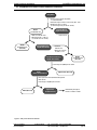
1
CLONTECH Yeast Protocols Handbook (PT3024-1) Protocol Supplement for: Catalog # Product* K1605-1 MATCHMAKER Two-Hybrid System K1604-1 MATCHMAKER Two-Hybrid System 2 K1603-1 MATCHMAKER One-Hybrid System (many) MATCHMAKER Libraries NL4000AA MATCHMAKER Random Peptide Library K1609-1 MATCHMAKER LexA Two-Hybrid System (many) MATCHMAKER LexA Libraries 5398-1 GAL4 AD Monoclonal Antibody 5399-1 GAL4 DNA-BD Monoclonal Antibody * Please refer to the User Manual for system-specific information and procedures. (PR7X265) FOR RESEARCH USE ONLY Yeast Protocols Handbook CLONTECH Laboratories, Inc. Table of Contents I. Introduction 4 II. Introduction to Yeast Promoters 5 III. Culturing and Handling Yeast 10 IV. Preparation of Yeast Protein Extracts A. General Information B. Preparation of Yeast Cultures for Protein Extraction C. Preparation of Protein Extracts: Urea/SDS Method D. Preparation of Protein Extracts: TCA Method E. Troubleshooting 12 12 12 14 16 17 V. Yeast Transformation Procedures A. General Information B. Reagents and Materials Required C. Tips for a Successful Transformation D. Integrating Plasmids into the Yeast Genome E. Small-scale LiAc Yeast Transformation Procedure F. Troubleshooting Yeast Transformation 18 18 19 20 20 20 22 VI. β-Galactosidase Assays A. General Information B. In vivo Plate Assay Using X-gal in the Medium C. Colony-lift Filter Assay D. Liquid Culture Assay Using ONPG as Substrate E. Liquid Culture Assay Using CPRG as Substrate F. Liquid Culture Assay Using a Chemiluminescent Substrate 23 23 25 25 26 27 28 VII. Working with Yeast Plasmids A. General Information B. Plasmid Isolation From Yeast C. Transforming E. coli with Yeast Plasmids 31 31 31 33 VIII. Analysis of Yeast Plasmid Inserts by PCR A. General Information B. Tips for Successful PCR of Yeast Plasmid Templates 36 36 36 IX. 39 39 40 41 Additional Useful Protocols A. Yeast Colony Hybridization B. Generating Yeast Plasmid Segregants C. Yeast Mating X. References 43 XI. MATCHMAKER and Related Products 46 APPENDICES A. Glossary of Technical Terms B. Yeast Genetic Markers Used in the MATCHMAKER Systems C. Media Recipes A. Yeast Media B. E. coli Media D. Solution Formulations E. Plasmid Information F. Yeast Host Strain Information Technical Support page 2 TEL: 650-424-8222 or 800-662-2566(CLON ) FAX: 650-424-1064 or 800-424-1350 47 49 50 50 53 54 58 61 Protocol # PT3024-1 Version # PR7X265 Yeast Protocols Handbook CLONTECH Laboratories, Inc. Table of Contents continued List of Tables Table I. Yeast Promoter Constructs Used to Regulate Reporter Gene Expression in MATCHMAKER Plasmids and Host Strains 6 Table II. Yeast Promoter Constructs in the MATCHMAKER Cloning Vectors 9 Table III. Comparison of β-galactosidase Assays 24 Table IV. Selected Yeast Genes and Their Associated Phenotypes 49 Table V. MATCHMAKER Reporter Genes and Their Phenotypes 49 Table VI. MATCHMAKER Two-Hybrid System Cloning Vectors 58 Table VII. MATCHMAKER One-Hybrid System Cloning/Reporter Plasmids 59 Table VIII. MATCHMAKER Reporter and Control Plasmids 60 Table IX. Yeast Reporter Strains in the MATCHMAKER One- and Two-Hybrid Systems 61 List of Figures Figure 1. Sequence of GAL4 DNA-BD recognition sites in the GAL1 and GAL2 UASs and the UASG 17-mer Figure 2. Urea/SDS protein extraction method 13 Figure 3. TCA protein extraction method 15 6 Notice to Purchaser Practice of the two-hybrid system is covered by US Patents #5,283,173 and #5,468,614 assigned to the Research Foundation of the State University of New York. Purchase of any CLONTECH two-hybrid reagent does not imply or convey a license to practice the two-hybrid system covered by these patents. Commercial entities purchasing these reagents must obtain a license from the Research Foundation of the State University of New York before using them. CLONTECH is required by its licensing agreement to submit a report of all purchasers of two-hybrid reagents to SUNY Stony Brook. Please contact Carol Dempster, Ph.D., at the Long Island Research Institute for license information (Tel: 516-361-6800; Fax: 516-361-6840). All plasmids (except for pACT2 and pAS2-1) are licensed from The Research Foundation of the State University of New York. pACT2, pAS2-1, and yeast strains Y187 and Y190 are licensed from Baylor University. CLONTECH encourages researchers not to redistribute the plasmids or yeast strains without prior written consent. CG-1945 is the property of CLONTECH Laboratories, Inc., and should not be redistributed without prior written consent. PJ69-2A is the property of the University of Wisconsin Research Foundation (WARF) and may not be redistributed without their prior written consent. The Polymerase Chain Reaction (PCR) process is covered by patents owned by Hoffman-LaRoche and F. Hoffmann-La Roche, Ltd. Protocol # PT3024-1 Version # PR7X265 Technical Support TEL: 650-424-8222 or 800-662-2566(CLON) FAX: 650-424-1064 or 800-424-1350 page 3 Yeast Protocols Handbook CLONTECH Laboratories, Inc. I. Introduction The Yeast Protocols Handbook provides background information and general yeast protocols that complement our system-specific User Manuals. The protocols in this Handbook have been optimized with our yeast-based MATCHMAKER Two-Hybrid and One-Hybrid Systems, and MATCHMAKER Libraries. The Yeast Protocols Handbook is especially useful for researchers who wish to use yeast as a vehicle for their molecular biology experiments, but have little or no prior experience working with yeast. For novice and experienced users alike, the Yeast Protocols Handbook will help you obtain the best possible results with your MATCHMAKER and other yeast-related products from CLONTECH. This Handbook includes: • detailed information on culturing and handling yeast • information on the yeast promoters used in the MATCHMAKER Systems • two protocols for preparing protein extracts from yeast • quantitative and qualitative β-galactosidase assays (for use with lacZ yeast reporter strains) • a simple, optimized protocol for isolating plasmids from yeast • PCR amplification and yeast colony hybridization protocols for the rapid analysis of positive clones obtained in a library screening • a small-scale, lithium acetate yeast transformation protocol • additional protocols for working with certain yeast plasmids and host strains The special application of yeast transformation for one- and two-hybrid library screening is covered in detail in each product-specific User Manual. The special application of yeast mating for library screening is covered in the Pretransformed MATCHMAKER Libraries User Manual. About our yeast-based products The MATCHMAKER GAL4 Two-Hybrid Systems (#K1604-1, K1605-1) and LexA Two-Hybrid System (#K1609-1) are complete kits for identifying and investigating protein-protein interactions in vivo using the yeast two-hybrid assay. The MATCHMAKER One-Hybrid System (#K1603-1) provides the basic tools for identifying novel proteins in vivo that bind to a target DNA sequence such as a cis-acting regulatory element. The MATCHMAKER Libraries are constructed in vectors that express inserts as fusions to a transcriptional activation domain, and are thus a convenient resource for researchers wishing to screen a library using the one- or two-hybrid assays. Pretransformed MATCHMAKER Libraries provide an even greater level of convenience for those wishing to perform a two-hybrid library screening without using large- or library-scale yeast transformations. CLONTECH offers an extensive line of kits and reagents that support and complement the MATCHMAKER Systems and Libraries. The YEASTMAKERTM Yeast Transformation Kit (#K1606-1) includes all the necessary reagents and protocols for efficient transformation using the lithium acetate method. Also available from CLONTECH: a selection of GAL4 DNA-binding domain (DNA-BD) and activation domain (AD) hybrid cloning vectors; the pGilda Vector for use with LexA-based two-hybrid systems; monoclonal antibodies and sequencing primers; and yeast media, including Minimal SD Base and many different formulations of Dropout (DO) Supplement. Our YEXpressTM Yeast Expression Systems are ideal for expressing and characterizing positive clones identified in a one-or two-hybrid library screening. For example, pYEX-BX (#6199-1) has the copper-inducible CUP1 promoter driving expression of cloned proteins. pYEX 4T-1, -2, and -3 (#6196–1, 6197-1, and 6198-1, respectively) have CUP1 promoters driving expression of GST fusion proteins. pYEX-S1 (#6200-1) is used for expression and secretion of cloned proteins, including GST-tagged proteins. For ordering information on these products, please see Chapter XI of this Handbook or the CLONTECH Catalog. Technical Support page 4 TEL: 650-424-8222 or 800-662-2566(CLON ) FAX: 650-424-1064 or 800-424-1350 Protocol # PT3024-1 Version # PR7X265 Yeast Protocols Handbook CLONTECH Laboratories, Inc. II. Introduction to Yeast Promoters Yeast promoters and other cis-acting regulatory elements play a crucial role in yeast-based expression systems and transcriptional assays such as the MATCHMAKER One- and Two-Hybrid Systems. Differences in the promoter region of reporter gene constructs can significantly affect their ability to respond to the DNA-binding domain of specific transcriptional activators; promoter constructs also affect the level of background (or leakiness) of gene expression and the level of induced expression. Furthermore, differences in cloning vector promoters determine the level of protein expression and, in some cases, confer the ability to be regulated by a nutrient (such as galactose in the case of the GAL1 promoter). This chapter provides a brief introduction to several commonly used yeast promoters and cis-regulatory elements. For further information on the regulation of gene expression in yeast, we recommend the Guide to Yeast Genetics and Molecular Biology by Guthrie & Fink (1991; #V2010-1); Molecular Biology and Genetic Engineering of Yeasts, edited by Heslot & Gaillardin (1992); Stargell & Struhl (1996); and Pringle et al. (1997; #V2365-1). UAS and TATA regions are basic building blocks of yeast promoters The initiation of gene transcription in yeast, as in other organisms, is achieved by several molecular mechanisms working in concert. All yeast structural genes (i.e., those transcribed by RNA polymerase II) are preceded by a region containing a loosely conserved sequence (TATA box) that determines the transcription start site and is also a primary determinant of the basal transcription level. Many genes are also associated with cis-acting elements—DNA sequences to which transcription factors and other trans-acting regulatory proteins bind and affect transcription levels. The term “promoter” usually refers to both the TATA box and the associated cis-regulatory elements. This usage is especially common when speaking of yeast gene regulation because the cis regulatory elements are relatively closely associated with the TATA box (Yoccum, 1987). This is in contrast to multicellular eukaryotes, where cisregulatory elements (such as enhancers) can be found very far upstream or downstream from the promoters they regulate. In this text, "minimal promoter" will refer specifically to the TATA region, exclusive of other cis-acting elements. The minimal promoter (or TATA box) in yeast is typically approximately 25 bp upstream of the transcription start site. Yeast TATA boxes are functionally similar to prokaryotic Pribnow boxes, but are not as tightly conserved. Furthermore, some yeast transcription units are preceded by more than one TATA box. The yeast HIS3 gene, for example, is preceded by two different TATA boxes: TR, which is regulated, and TC, which is constitutive (Mahadevan & Struhl, 1990). Yeast TATA boxes can be moved to a new location, adjacent to other cis-regulatory elements, and still retain their transcriptional function. One type of cis-acting transcription element in yeast is upstream activating sequences (UAS), which are recognized by specific transcriptional activators and enhance transcription from adjacent downstream TATA regions. The enhancing function of yeast UASs is generally independent of orientation; however, it is sensitive to distance effects if moved more than a few hundred base pairs from the TATA region. There may be multiple copies of a UAS upstream of a yeast coding region. In addition, UASs can be eliminated or switched to change the regulation of target genes. UAS and TATA regions can be switched to create novel promoters The "mix and match" nature of yeast TATA boxes and UASs has been used to great advantage in yeast two-hybrid systems to create novel promoters for the reporter genes. (For general references on yeast two-hybrid systems, see Chapter X.) In most cases, the lacZ, HIS3, and LEU2 reporter genes are under control of artificial promoter constructs comprised of a TATA and UAS (or operator) sequence derived from another gene (Table I). In some cases, the TATA sequence and the UAS are derived from different genes; indeed, the LexA operator is a cis-acting regulatory element derived from E. coli. For GAL4-based systems, either a native GAL UAS or a synthetic UASG 17-mer consensus sequence (Heslot & Gaillardin, 1992) provides the binding site for the GAL4 DNA-BD. For LexA-based systems, multiple copies of the LexA operator provide the binding site for the LexA protein. If you are putting together your own one- or two-hybrid system, you must make sure that the reporter gene's promoter will be recognized by the DNA-BD moiety encoded in your DNA-BD fusion-generating vector. Protocol # PT3024-1 Version # PR7X265 Technical Support TEL: 650-424-8222 or 800-662-2566(CLON) FAX: 650-424-1064 or 800-424-1350 page 5 Yeast Protocols Handbook CLONTECH Laboratories, Inc. II. Introduction to Yeast Promoters continued TABLE I . YEAST PROMOTER CONSTRUCTS USED TO REGULATE REPORTER GENE EXPRESSION IN MATCHMAKER PLASMIDS AND HOST STRAINS Plasmid or host straina Reporter gene Origin of UAS UAS regulated by Origin of Expression levelb TATA sequence Induced (uninduced) CG-1945 lacZ HIS3 UASG 17-mer (x3)c GAL1 GAL4 GAL4 CYC1 GAL1 low high (slightly leaky) HF7c lacZ HIS3 UASG 17-mer (x3)c GAL1 GAL4 GAL4 CYC1 GAL1 low high (tight) Y190 lacZ HIS3 GAL1 GAL1 GAL4 GAL4 GAL1 HIS3 (TC+TR) high high (leaky) Y187 lacZd GAL1 GAL4 GAL1 high SFY526 lacZ GAL1 GAL4 GAL1 high PJ69-2A HIS3 ADE2 GAL1 GAL2 GAL4 GAL4 GAL1 GAL2 high (tight) high (tight) EGY48 LEU2 LexA op(x6) LexA LEU2 high high p8op-lacZ lacZ LexA op(x8) LexA GAL1e pHISi HIS3 (none)f (n.a.) HIS3 (TC+TR) n.a. f (leaky) pHISi-1 HIS3 (none)f (n.a.) HIS3 (TC+TR) n.a. f (leaky) pLacZi lacZ (none)f (n.a.) HIS3 (TC+TR) n.a. f (tight) a b c d e f See Appendices E & F for references. When induced by a positive two-hybrid interaction; "leaky" and "tight" refer to expression levels in the absence of induction. Conserved 17-bp palindromic sequence to which the GAL4 protein binds (Guthrie & Fink, 1991). Y187 probably contains two copies of the lacZ gene, judging by the strength of the signal in this strain and in the strains from which it was derived (Durfee et al., 1993; Harper et al., 1993). This is the minimal TATA region of the GAL1 promoter; it does not include the GAL1 UAS and therefore is not responsive to regulation by GAL4 protein. The MATCHMAKER One-Hybrid System vectors do not contain a UAS because they are used to experimentally test target elements inserted upstream of the minimal promoter for their ability to bind specific transcriptional activators. In the absence of inserted target elements, reporter gene expression is not induced; however, expression levels may be leaky, depending on the nature of the minimal promoter used in that vector. GAL1 UAS GAL1-bs1 GAL1-bs2 GAL1-bs3 GAL1-bs4 TAGAAGCCGCCGAGCGG GACAGCCCTCCGAAGGA GACTCTCCTCCGTGCGT CGCACTGCTCCGAACAA GAL2 UAS GAL2-bs1 GAL2-bs2 GAL2-bs3 GAL2-bs4 GAL2-bs5 CGGAAAGCTTCCTTCCG CGGCGGTCTTTCGTCCG CGGAGATATCTGCGCCG CGGGGCGGATCACTCCG CGGATCACTCCGAACCG UASG 17-mer CGGAAGACTCTCCTCCG Figure 1. Sequence of the GAL4 DNA-BD recognition sites in the GAL1 and GAL2 UASs and the UASG consensus sequence (Giniger & Ptashne, 1988). Technical Support page 6 TEL: 650-424-8222 or 800-662-2566(CLON ) FAX: 650-424-1064 or 800-424-1350 17-mer Protocol # PT3024-1 Version # PR7X265 Yeast Protocols Handbook CLONTECH Laboratories, Inc. II. Introduction to Yeast Promoters continued Reporter genes under the control of GAL4-responsive elements In yeast, the genes required for galactose metabolism are controlled by two regulatory proteins, GAL4 and GAL80, as well as by the carbon source in the medium (Guthrie & Fink, 1991; Heslot & Gaillardin, 1992). When galactose is present, the GAL4 protein binds to GAL4-responsive elements within the UAS upstream of several genes involved in galactose metabolism and activates transcription. In the absence of galactose, GAL80 binds to GAL4 and blocks transcriptional activation. Furthermore, in the presence of glucose, transcription of the galactose genes is immediately repressed (Johnston et al., 1994). The UASs of the 20 known galactose-responsive genes all contain one or more conserved palindromic sequences to which the GAL4 protein binds (Guthrie & Fink, 1991; Giniger et al. 1985; reviewed in Heslot & Gaillardin, 1992). The 17-mer consensus sequence, referred to here as UASG 17-mer, functions in an additive fashion, i.e., multiple sites lead to higher transcription levels than a single site (Giniger & Ptashne, 1988). The protein binding sites of the GAL1 and GAL2 UASs, and the UASG 17-mer consensus sequence, are shown in Figure 1. The tight regulation of the GAL UASs by GAL4 makes it a valuable tool for manipulating expression of reporter genes in two-hybrid systems that are dependent on the GAL4 DNA-BD. However, in such systems, the yeast host strains must carry deletions of the gal4 and gal80 genes to avoid interference by endogenous GAL4 and GAL80 proteins; thus, no significant glucose repression is observed in these strains and no induction is observed unless a two-hybrid interaction is occurring. Therefore, nutritional regulation of GAL UASs is not a feature of GAL4-based two-hybrid systems. However, the host strain used in the LexA system does support galactose induction, as it is wild type for GAL4 and GAL80 functions. In the GAL4-based MATCHMAKER Two-Hybrid Systems, either an intact GAL UAS (which contains four GAL4-binding sites) or an artifically constructed UAS consisting of three copies of the 17-mer consensus binding sequence, is used to confer regulated expression on the reporter genes (Table I). The HIS3 reporter of PJ69-2A, HF7c, and CG-1945, and the lacZ reporter of Y190, Y187, and SFY526 are all tightly regulated by the intact GAL1 promoter (including the GAL1 UAS and GAL1 minimal promoter). In HF7c and CG1945, lacZ expression is under control of UASG 17-mer (x3) and the extremely weak minimal promoter of the yeast cytochrome C1 (CYC1) gene. lacZ under the control of the intact GAL1 promoter can be expressed at ~10X the level obtained with the UASG 17-mer (x3)/CYC1 minimal promoter construct under similar induction conditons (CLONTECH Laboratories; unpublished data). Therefore, some weak or transient two-hybrid interactions may not be detectable in HF7c or CG1945 unless you use a highly sensitive β-galactosidase assay (such a liquid culture assay using a chemiluminescent substrate; Chapter VI.F). The ADE2 reporter of PJ69-2A is tightly regulated by the intact GAL2 promoter, whose induction properties are similar to those of the GAL1 promoter. Reporter genes under the control of a minimal HIS3 promoter The native yeast HIS3 promoter contains a UAS site recognized by the transcriptional activator GCN4, and two TATA boxes. GCN4 regulates one of the TATA boxes (TR), while the other TATA box (TC) drives low-level constitutive expression of HIS3 (Iyer & Struhl, 1995). TC is not regulated by the native GCN4binding UAS, the GAL1 UAS, or artificial UASG constructs (Mahadevan & Struhl, 1990; Hope & Struhl, 1986). The HIS3 reporter gene in yeast strain Y190 is unusual among the GAL4 two-hybrid reporter gene constructs in that it is under the control of the GAL1 UAS and a minimal promoter containing both HIS3 TATA boxes (Flick & Johnston, 1990). The result is high-level expression (due to the GAL1 UAS) when induced by a positive two-hybrid interaction; this construct also exhibits a significant level of constitutive leaky expression (due to the HIS3 TC). In contrast, in HF7c and CG-1945, the entire HIS3 promoter (including both TATA boxes) was replaced by the entire GAL1 promoter, leading to tight regulation of the HIS3 reporter in those strains (Feilotter et al., 1994). The HIS3 reporter plasmids pHISi and pHISi-1 used in the MATCHMAKER One Hybrid System also have both of the HIS3 TATA boxes present in the minimal promoter. By inserting a cis-acting element in the MCS, the regulated TATA box (TR) can be affected, but there is still a significant amount of constitutive, leaky expression due to the HIS3 TC. The leaky HIS3 expression of these one-hybrid plasmids is first Protocol # PT3024-1 Version # PR7X265 Technical Support TEL: 650-424-8222 or 800-662-2566(CLON) FAX: 650-424-1064 or 800-424-1350 page 7 Yeast Protocols Handbook CLONTECH Laboratories, Inc. II. Introduction to Yeast Promoters continued used to help construct HIS3 reporter strains, and later is controlled by including 3-aminotriazole in the medium to suppress background growth. Reporter genes under the control of LexA operators In LexA-based two-hybrid systems, the DNA-BD is provided by the entire prokaryotic LexA protein, which normally functions as a repressor of SOS genes in E. coli when it binds to LexA operators, which are an integral part of the promoter (Ebina et al., 1983). When used in the yeast two-hybrid system, the LexA protein does not act as a repressor because the LexA operators are integrated upstream of the minimal promoter and coding region of the reporter genes. LEU2 reporter expression in yeast strain EGY48 is under the control of six copies of the LexA operator (op) sequence and the minimal LEU2 promoter. In the lacZ reporter plasmids, lacZ expression is under control of 1–8 copies of the LexA op (Estojak et al., 1995) and the minimal GAL1 promoter. Because all of the GAL1 UAS sequences have been removed from the lacZ reporter plasmids (West et al ., 1984), this promoter is not regulated by glucose or galactose. Promoters used to drive fusion protein expression in two-hybrid cloning vectors The ADH1 promoter (or a truncated version of it) is the promoter used to drive expression of the fusion proteins in most of the MATCHMAKER cloning vectors. The 1500-bp full-length ADH1 promoter (Ammerer, 1983; Vainio, GenBank accession number: Z25479) leads to high-level expression of sequences under its control in pGAD GH, pLexA, and pAS2-1 during logarithmic growth of the yeast host cells. Transcription is repressed in late log phase by the ethanol that accumulates in the medium as a by-product of yeast metabolism. Several MATCHMAKER cloning vectors contain a truncated 410-bp ADH1 promoter (Table II). At one point, it was believed that only this portion was necessary for high-level expression (Beier & Young, 1982). In most vector constructs, however, this truncated promoter leads to low or very low levels of fusion protein expression (Ruohonen et al., 1991; Ruohonen et al. , 1995; Tornow & Santangelo, 1990). This observation has been confirmed at CLONTECH by quantitative Western blots (unpublished data). The high-level expression reported by Beier & Young (1982) was apparently due to a segment of DNA derived from pBR322, which was later found to coincidentally enhance transcriptional activity in yeast (Tornow & Santangelo, 1990). In the MATCHMAKER vector pACT2, strong constitutive fusion protein expression is driven by the 410-bp truncated ADH1 promoter adjacent to this enhancing pBR322 segment. The AD cloning vector pB42AD and the alternative DNA-BD vector pGilda used in the MATCHMAKER LexA Two-Hybrid System utilize the full-length GAL1 promoter to drive fusion protein expression. Because the LexA system host strain is wild-type for GAL4 and GAL80, fusion protein expression is regulated by glucose and galactose. Technical Support page 8 TEL: 650-424-8222 or 800-662-2566(CLON ) FAX: 650-424-1064 or 800-424-1350 Protocol # PT3024-1 Version # PR7X265 Yeast Protocols Handbook CLONTECH Laboratories, Inc. II. Introduction to Yeast Promoters continued TABLE II. YEAST PROMOTER CONSTRUCTS IN THE MATCHMAKER CLONING VECTORS Vectorsa Promoter Regulation/ Relative Protein Expression Level p LexA, pGAD GH, pAS2-1, pAS2 ADH1 (full-length) Ethanol-repressed/High +++ pACT2, pACT ADH1 (truncated+)c Constitutive/medium ++ pGAD GL pGAD424, pGAD10 pGBT9 ADH1 (truncated) Constitutive/low Constitutive/ very low pB42AD, pGilda GAL1 (full-length) Repressed by glucose; (not detectable) d induced (high-level) by galactose +++d p8op-lacZ GAL1 (minimal) Not regulated by glucose or galactose a b c d Signal Strength on Western blot b +/– (weak) (not detectable) (no data) See Appendix E for vector references. Unpublished data obtained at CLONTECH Laboratories using the appropriate GAL4 domain-specific mAb (#5398-1 or #5399-1). Soluble protein extracts were prepared from CG-1945 transformed with the indicated plasmid. Samples equivalent to ~1 OD600 unit of cells were electrophoresed and then blotted to nitrocellulose filters. The blots were probed with either GAL4 DNA-BD mAb (0.5 µg/ml) or GAL4 AD mAb (0.4 µg/ml) using 1 ml of diluted mAb per 10 cm 2 of blot, followed by HRP-conjugated polyclonal Goat Anti-Mouse IgG (Jackson Immunological Research; diluted 1:15,000 in TBST). Signals were detected using a chemiluminescent detection assay and a 2.5-min exposure of x-ray film. Signal intensities were compared to that of known amounts of purified GAL4 DNA-BD (a.a. 1–147) or GAL4 AD (a.a. 768–881). The truncated ADH1 promoter in pACT2 is adjacent to a section of pBR322 which acts as a transcriptional enhancer in yeast. Data obtained using EGY48[p8op-lacZ] transformed with pGilda and grown in the presence of glucose or galactose, respectively (April 1997 CLONTECHniques); no data available for pB42AD. Protocol # PT3024-1 Version # PR7X265 Technical Support TEL: 650-424-8222 or 800-662-2566(CLON) FAX: 650-424-1064 or 800-424-1350 page 9 Yeast Protocols Handbook CLONTECH Laboratories, Inc. III. Culturing and Handling Yeast For additional information on yeast, we recommend Guthrie and Fink (1991) Guide to Yeast Genetics and Molecular Biology (#V2010-1). A. Yeast Strain Maintenance, Recovery from Frozen Stocks, and Routine Culturing 1. Long-term storage • Yeast strains can be stored indefinitely in YPD medium with 25% glycerol at –70°C. For storage >1 year, the temperature must be maintained below –55°C. • Transformed yeast strains are best stored in the appropriate SD dropout medium to keep selective pressure on the plasmid. (See Appendix C.A for recipes and Appendix E for plasmid information.) To prepare new glycerol stock cultures of yeast: a. Use a sterile inoculation loop to scrape an isolated colony from the agar plate. b. Resuspend the cells in 200–500 µl of YPD medium (or the appropriate SD medium) in a 1.5-ml microcentrifuge tube. Vortex tube vigorously to thoroughly disperse the cells. Add sterile 50% glycerol to a final concentration of 25%. c. Tightly close the cap. Shake the vial before freezing at –70°C. 2. To recover frozen strains and prepare working stock plates: a. Streak a small portion of the frozen glycerol stock onto a YPD (or appropriate SD) agar plate. b. Incubate the plate at 30°C until yeast colonies reach ~2 mm in diameter (this takes 3–5 days). Use these colonies as your working stock. c. Seal plates with Parafilm and store at 4°C for up to two months. Streak a fresh working stock plate from the frozen stock at 1–2-month intervals. d. If you cannot recover the strain, the cells may have settled ; in this case, thaw the culture on ice, vortex vigorously, and restreak. The glycerol stock tube may be refrozen a few times without damaging the cells. 3. To prepare liquid overnight cultures: a. Use only fresh (<2-months old) colonies from the working stock plate. Use one large (2–3-mm diameter) colony per 5 ml of medium. If colonies are small, or if you are inoculating a larger volume, use several colonies. Important: Vigorously vortex the medium for ~1 min to thoroughly disperse the cells. Notes: • Liquid cultures will grow slower than expected if clumps are present in the inoculum; cells in the interior of the clumps do not have access to the nutrients in the medium. • If you are inoculating a volume greater than 1 ml, it is easier to disperse the clumps if the colonies are first placed in 1 ml of medium in a microcentrifuge tube, vortexed, and then transferred to the desired volume. • When growing overnight cultures of yeast transformants, use the appropriate SD minimal medium to keep selective pressure on extrachromosomal plasmid(s). • The growth in YPD of yeast strains carrying the ade2-101 mutation will be enhanced by adding adenine hemisulfate (0.003% final concentration) to the medium (Appendix C.A). All of the host strains (except EGY48) used in the MATCHMAKER Systems carry this auxotrophic mutation. • The growth of transformed PJ69-2A cells in SD/–Trp may also be enhanced by adding excess adenine to the medium (Appendix C.A). b. Incubate at 30°C for 16–18 hr with shaking at 230–270 rpm. With most strains, this will yield a stationary phase culture (OD600 > 1.5). Note: Different yeast strains grow at different rates. Growth rates may also be affected by the presence of fusion proteins in certain transformants. In addition, the doubling time of most strains growing in SD minimal medium is twice as long as in YPD. c. If you need a mid-log phase culture, transfer enough of the overnight culture into fresh medium to produce an OD600 = 0.2–0.3. Incubate at 30°C for 3–5 hr with shaking (230–250 rpm). This will, with most strains, produce a culture with an OD600 ~0.4–0.6. Note: Generally, YPD may be used in this incubation. Because of the shorter incubation time, plasmid loss will not be significant. However, do not use YPD if you want to induce protein expression from the yeast GAL1 promoter of a LexA system plasmid, e.g., pB42AD or pGilda; YPD contains glucose, which represses transcription from the GAL1 promoter. Technical Support page 10 TEL: 650-424-8222 or 800-662-2566(CLON ) FAX: 650-424-1064 or 800-424-1350 Protocol # PT3024-1 Version # PR7X265 Yeast Protocols Handbook CLONTECH Laboratories, Inc. III. Culturing and Handling Yeast continued B. Growth Selection for Transformation Markers and Reporter Gene Expression Most yeast cloning vectors and control plasmids (including those provided in our MATCHMAKER Systems) carry at least one nutritional marker to allow for selection of yeast transformants plated on SD minimal medium lacking that specific nutrient. Furthermore, if you are cotransforming yeast with two or more different plasmids bearing different nutritional markers, the plasmids can be independently selected. Thus, the SD selection medium you choose for plating transformants depends generally on the purpose of the selection. Specific factors to consider in choosing the appropriate SD selection medium are: • • • • • the plasmid(s) used and whether you are selecting for one or more plasmids whether you are selecting for colonies in which two hybrid proteins are interacting whether—and to what extent—the host strain is leaky for reporter gene expression whether you want to induce protein expression from the regulated GAL1 promoter whether you intend to perform in-vivo, agar-plate β-galactosidase assays (for lacZ reporter expression in the LexA Two-Hybrid System). Please refer to your system-specific User Manual for further information on choosing the appropriate SD selection media for particular plasmids, host strains, and applications. Protocol # PT3024-1 Version # PR7X265 Technical Support TEL: 650-424-8222 or 800-662-2566(CLON) FAX: 650-424-1064 or 800-424-1350 page 11 Yeast Protocols Handbook CLONTECH Laboratories, Inc. IV. Preparation of Yeast Protein Extracts A. General Information We provide two alternative protocols for the preparation of protein extracts from yeast. The results (i.e., protein yield and quality) will vary depending on the protein and may be more successful with one protocol than with the other. Because it is difficult to predict which procedure will give better results, we provide two protocols for comparison. The cell culture preparation method (Section B) is the same for both protein extraction procedures. Both extraction procedures address the two most challenging aspects of isolating proteins from yeast: 1) disrupting yeast cell walls; and 2) inhibiting the many endogenous yeast proteases. Yeast cell walls are tough and must be disrupted by a combination of physical and chemical means; methods that utilize glycolytic enzymes are not recommended for this application because they are often contaminated with proteases. Endogenous proteases must be counteracted with a cocktail of strong protease inhibitors (recipe in Appendix D.A). If you know your protein of interest is susceptible to a protease not inhibited by the recommended cocktail, add the appropriate inhibitor before using the mixture. You may also wish to add other inhibitors such as sodium fluoride to prevent dephosphorylation, if that is appropriate for your protein. B. Preparation of Yeast Cultures for Protein Extraction Reagents and Materials Required: • YPD and appropriate SD liquid medium (Recipes in Appendix C.A) • 20- and 50-ml culture tubes • Ice-cold H2O • Dry ice or liquid nitrogen 1. For each transformed yeast strain you wish to assay in a Western blot, prepare a 5-ml overnight culture in SD selection medium as described in Section III.A, except use a single isolated colony (1–2 mm in diameter, no older than 4 days). Use the SD medium appropriate for your system and plasmids (Appendix E). Also prepare a 10-ml culture of an untransformed yeast colony in YPD or (if possible) appropriate SD medium as a negative control. 2. Vortex the overnight cultures for 0.5–1 min to disperse cell clumps. For each clone to be assayed (and the negative control), separately inoculate 50-ml aliquots of YPD medium with the entire overnight culture. 3. Incubate at 30°C with shaking (220–250 rpm) until the OD600 reaches 0.4–0.6. (Depending on the fusion protein, this will take 4–8 hr.) Multiply the OD600 (of a 1-ml sample) by the culture volume (i.e., 55 ml) to obtain the total number of OD600 units; this number will be used in Sections C & D. (For example, 0.6 x 55 ml = 33 total OD600 units.) Note: During late log phase the ADH1 promoter shuts down and the level of endogenous yeast proteases increases. 4. Quickly chill the culture by pouring it into a prechilled 100-ml centrifuge tube halfway filled with ice. 5. Immediately place tube in a prechilled rotor and centrifuge at 1000 x g for 5 min at 4°C. 6. Pour off supernatant and resuspend the cell pellet in 50 ml of ice-cold H2O. (Any unmelted ice pours off with the supernatant.) 7. Recover the pellet by centrifugation at 1,000 x g for 5 min at 4°C. 8. Immediately freeze the cell pellet by placing the tube on dry ice or in liquid nitrogen. Store cells at –70°C until you are ready to proceed with the experiment. Technical Support page 12 TEL: 650-424-8222 or 800-662-2566(CLON ) FAX: 650-424-1064 or 800-424-1350 Protocol # PT3024-1 Version # PR7X265 Yeast Protocols Handbook CLONTECH Laboratories, Inc. IV. Preparation of Yeast Protein Extracts continued Cell pellets • Thaw and resuspend cell pellets in prewarmed Cracking buffer • Add cells to glass beads • Heat at 70°C for 10 min • Vortex vigorously for 1 min • Centrifuge at 14,000 rpm for 5 min First supernatant Pellet • Boil for 3–5 min • Vortex vigorously for 1 min • Centrifuge at 14,000 rpm for for 5 min Pellet (discard) Second supernatant • Place on ice • Combine with second supernatant • Place on ice Combined supernatants • Immediately load gel or freeze at –70°C or colder Figure 2. Urea/SDS protein extraction method. Protocol # PT3024-1 Version # PR7X265 Technical Support TEL: 650-424-8222 or 800-662-2566(CLON) FAX: 650-424-1064 or 800-424-1350 page 13 Yeast Protocols Handbook CLONTECH Laboratories, Inc. IV. Preparation of Yeast Protein Extracts continued C. Preparation of Protein Extracts: Urea/SDS Method (Figure 2; Printen & Sprague, 1994) Reagents and Materials Required: • 1.5-ml screw-cap microcentrifuge tubes • Glass beads (425–600 µm; Sigma #G-8772) • Protease inhibitor solution (Appendix D.A) • PMSF stock solution (Appendix D.A) • Cracking buffer stock solution (Appendix D.A) • Cracking buffer, complete (Appendix D.A) Note: Unless otherwise stated, keep protein samples on ice. 1. Prepare complete cracking buffer (Appendix D.A) and prewarm it to 60°C. Because the PMSF degrades quickly, prepare only the amount of cracking buffer you will need immediately. Use 100 µl of cracking buffer per 7.5 OD600 units of cells. (For example, for 33 total OD600 units of cells, use 0.44 ml of cracking buffer.) 2. Quickly thaw cell pellets by separately resuspending each one in the prewarmed cracking buffer. • If cell pellets are not immediately thawed by the prewarmed cracking buffer, place the tubes briefly at 60°C to hasten melting. To avoid risk of proteolysis, do not leave them longer than 2 min at 60°C. • Because the initial excess PMSF in the cracking buffer degrades quickly, add an additional aliquot of the 100X PMSF stock solution to the samples after 15 min and approximately every 7 min thereafter until Step 9, when they are placed on dry ice or are safely stored at –70°C or colder. (Use 1 µl of 100X PMSF per 100 µl of cracking buffer.) 3. Transfer each cell suspension to a 1.5-ml screw-cap microcentrifuge tube containing 80 µl of glass beads per 7.5 OD600 units of cells. Note: The volume of the glass beads can be measured using a graduated 1.5-ml microcentrifuge tube. 4. Heat samples at 70°C for 10 min. Note: This initial incubation at 70°C frees membrane-associated proteins. Thus, if you skip this step, membraneassociated proteins will be removed from the sample at Step 6 (high-speed centrifugation). 5. Vortex vigorously for 1 min. 6. Pellet debris and unbroken cells in a microcentrifuge at 14,000 rpm for 5 min, preferably at 4°C, otherwise at room temperature (20–22°C). 7. Transfer the supernatants to fresh 1.5-ml screw-cap tubes and place on ice (first supernatants). 8. Treat the pellets as follows: a. Place tubes in a 100°C (boiling) water bath for 3–5 min. b. Vortex vigorously for 1 min. c. Pellet debris and unbroken cells in a microcentrifuge at 14,000 rpm for 5 min, preferably at 4°C, otherwise at room temperature. d. Combine each supernatant (second supernatant) with the corresponding first supernatant (from Step 7). Note: If no supernatant is obtained, add more cracking buffer (50–100 µl) and repeat Steps 8.b & c. 9. Boil the samples briefly. Immediately load them on a gel. Alternatively, samples may be stored on dry ice or in a –70°C freezer until you are ready to run them on a gel. Technical Support page 14 TEL: 650-424-8222 or 800-662-2566(CLON ) FAX: 650-424-1064 or 800-424-1350 Protocol # PT3024-1 Version # PR7X265 Yeast Protocols Handbook CLONTECH Laboratories, Inc. IV. Preparation of Yeast Protein Extracts continued Cell pellets • Thaw and resuspend cell pellets in cold TCA buffer • Add cells to glass beads and ice-cold, 20% TCA • Bead-beat cells 2 x 30 sec (or vortex vigorously for 10 min at 4°C) First Cell Extract (liquid above beads) Beads and unbroken cells • Add ice-cold 20% TCA • Bead-beat cells 1 x 30 sec (or vortex for 5 min at 4°C) Beads and unbroken cells (discard) Second Cell Extract (liquid above beads) • Place on ice • Combine Cell Extracts • Allow glass beads to settle ~1 min Beads Combined Cell Extracts (liquid above beads) and unbroken cells (discard) • Centrifuge at 14,000 rpm for 10 min Pellet Supernatant (Discard) (Protein and contaminants) • Resuspend in TCA-Laemmli loading buffer • Boil 10 min • Centrifuge at 14,000 rpm for 10 min Supernatant (Protein extract) Pellet (discard) Immediately load gel or freeze at –70°C or colder Figure 3. TCA protein extraction method. Protocol # PT3024-1 Version # PR7X265 Technical Support TEL: 650-424-8222 or 800-662-2566(CLON) FAX: 650-424-1064 or 800-424-1350 page 15 Yeast Protocols Handbook CLONTECH Laboratories, Inc. IV. Preparation of Yeast Protein Extracts continued D. Preparation of Protein Extracts: TCA Method (Figure 3; Horecka, J., personal communication) Reagents and Materials Required: • 1.5-ml screw-cap microcentrifuge tubes • Glass beads (425–600 µm; Sigma #G-8772) • Protease inhibitor solution (Appendix D.A) • PMSF Stock solution (Appendix D.A; Add as necessary throughout the protocol.) • [Recommended] Bead Beater (BioSpec, Bartlesville, OK) Note: If you do not have access to a Bead Beater, a high-speed vortexer can be used instead. However, vortexing is not as effective as bead-beating at disrupting the cells. • TCA buffer (Appendix D.A) • Ice-cold 20% w/v TCA in H2O (see Sambrook et al. [1989] for tips on preparing TCA solutions) • TCA-Laemmli loading buffer (Appendix D.A) Note: Unless otherwise stated, keep protein samples on ice. 1. Thaw cell pellets on ice (10–20 min). 2. Resuspend each cell pellet in 100 µl of ice-cold TCA buffer per 7.5 OD 600 units of cells. (For example, for 33 total OD600 units of cells, use 0.44 ml of TCA buffer.) Place tubes on ice. 3. Transfer each cell suspension to a 1.5-ml screw-cap microcentrifuge tube containing glass beads and ice-cold 20% TCA. Use 100 µl of glass beads and 100 µl of ice-cold 20% TCA per 7.5 OD600 units of cells. Note: The volume of the glass beads can be measured using a graduated 1.5-ml microcentrifuge tube. 4. To disrupt cells, place tubes in a Bead-Beater and set speed at highest setting. Bead-beat the cells for 2 X 30 sec, placing tubes on ice for 30 sec in between the two bead-beatings. Place tubes on ice. Note: If you do not have access to a Bead-Beater, you can vortex the tubes vigorously at 4°C for 10 min; alternatively, you can vortex at room temperature for shorter periods (of 1 min each) at least 4 times, placing tubes on ice for 30 sec in between each vortexing. Place tubes on ice. 5. Transfer the supernatant above the settled glass beads to fresh 1.5-ml screw-cap tubes and place tubes on ice. This is the first cell extract. Note: The glass beads settle quickly, so there is no need to centrifuge tubes at this point. 6. Wash the glass beads as follows: a. Add 500 µl of an ice-cold, 1:1 mixture of 20% TCA and TCA buffer. b. Place tubes in Beat Beater and beat for another 30 sec at the highest setting. (Alternatively, vortex for 5 min at 4°C, or vortex 2 X 1 min at room temperature, placing the tube on ice for 30 sec in between the two vortexings.) c. Transfer the liquid above the glass beads (second cell extract) to the corresponding first cell extract from Step 5. 7. Allow any carryover glass beads to settle in the combined cell extracts ~1 min, then transfer the liquid above the glass beads to a fresh, prechilled 1.5-ml screw-cap tube. 8. Pellet the proteins in a microcentrifuge at 14,000 rpm for 10 min at 4°C. 9. Carefully remove supernatant and discard. 10. Quickly spin tubes to bring down remaining liquid. Remove and discard liquid using a pipette tip. 11. Resuspend each pellet in TCA-Laemmli loading buffer. Use 10 µl of loading buffer per OD600 unit of cells. Note: If too much acid remains in the sample, the bromophenol blue in the buffer will turn yellow. Generally, this will not affect the results of the electrophoresis. 12. Place tubes in a 100°C (boiling) water bath for 10 min. 13. Centrifuge samples at 14,000 rpm for 10 min at room temperature (20–22°C). 14. Transfer supernatant to fresh 1.5-ml screw-cap tube. 15. Load the samples immediately on a gel. Alternatively, samples may be stored on dry ice or in a –70°C freezer until you are ready to run them on a gel. Technical Support page 16 TEL: 650-424-8222 or 800-662-2566(CLON ) FAX: 650-424-1064 or 800-424-1350 Protocol # PT3024-1 Version # PR7X265 Yeast Protocols Handbook CLONTECH Laboratories, Inc. IV. Preparation of Yeast Protein Extracts continued E. Troubleshooting Optimal electrophoretic separation of proteins depends largely on the quality of the equipment and reagents used in the gel system, the manner in which the protein samples are prepared prior to electrophoresis, the amount of protein loaded on the gel, and the voltage conditions used during electrophoresis. These same considerations are important for the subsequent transfer of proteins to the nitrocellulose membrane where transfer buffer composition, temperature, duration of transfer, and the assembly of the blotting apparatus can all have profound effects on the quality of the resultant protein blot. The following troubleshooting tips pertain to the isolation of protein from yeast. Information on running polyacrylamide protein gels and performing Western blots is available in published laboratory manuals (e.g., Sambrook et al., 1989, or Ausubel et al., 1987–96). 1. Few or no immunostained protein bands on the blot • The transfer of protein bands to the blot may be confirmed by staining the blot with Ponceau S. • The presence of protein bands in the gel (before transfer) may be confirmed by staining a parallel lane of the gel with Coomassie blue. (Note that once a gel has been stained with Coomassie blue, the protein bands will not transfer to a blot.) • The extent of cell wall disruption can be determined by examining a sample of treated cells under the microscope. Incomplete cell lysis will lower the protein yield. 2. Several bands appear on the blot where a single protein species is expected • Protein degradation and/or proteolysis may have occurred during sample preparation. Additional protease inhibitors may be used as desired. Also, make sure that in Steps C.8.a and D.12 (boiling the protein extracts), the samples are placed into a water bath that is already boiling. If samples are placed in the water before it has reached boiling temperature, a major yeast protease (Proteinase B) will be activated. (Proteinase B is a serine protease of the subtilisin family.) • Dephosphorylation of a normally phosphoryated fusion protein may have occurred during sample preparation. Sodium fluoride (NaF) may be added to the protease inhibitor stock solution to help prevent dephosphorylation (Sadowski et al ., 1991). 3. If you are running a reducing gel, make sure that the protein sample has been completely reduced with either dithiothreitol or 2-mercaptoethanol prior to loading the gel. Protocol # PT3024-1 Version # PR7X265 Technical Support TEL: 650-424-8222 or 800-662-2566(CLON) FAX: 650-424-1064 or 800-424-1350 page 17 Yeast Protocols Handbook CLONTECH Laboratories, Inc. V. Yeast Transformation Procedures A. General Information LiAc-mediated yeast transformation There are several methods commonly used to introduce DNA into yeast, including the spheroplast method, electroporation, and the lithium acetate (LiAc)-mediated method (reviewed in Guthrie & Fink, 1991). At CLONTECH, we have found the LiAc method (Ito et al., 1983), as modified by Schiestl & Gietz (1989), Hill et al. (1991), and Gietz et al. (1992), to be simple and highly reproducible. This chapter provides detailed protocols for using the LiAc procedure in a standard plasmid transformation and in a modified transformation to integrate linear DNA into the yeast genome. In the LiAc transformation method, yeast competent cells are prepared and suspended in a LiAc solution with the plasmid DNA to be transformed, along with excess carrier DNA. Polyethylene glycol (PEG) with the appropriate amount of LiAc is then added and the mixture of DNA and yeast is incubated at 30°C. After the incubations, DMSO is added and the cells are heat shocked, which allows the DNA to enter the cells. The cells are then plated on the appropriate medium to select for transformants containing the introduced plasmid(s). Because, in yeast, this selection is usually nutritional, an appropriate synthetic dropout (SD) medium is used. Simultaneous vs. sequential transformations The LiAc method for preparing yeast competent cells typically results in transformation efficiencies of 105 per µg of DNA when using a single type of plasmid. When the yeast is simultaneously cotransformed with two plasmids having different selection markers, the efficiency is usually an order of magnitude lower due to the lower probability that a particular yeast cell will take up both plasmids. (Yeast, unlike bacteria, can support the propagation of more than one plasmid having the same replication origin, i.e., there is no plasmid incompatibility issue in yeast.) Thus, in a cotransformation experiment, the efficiency of transforming each type of plasmid should remain at ~105 per µg of DNA, as determined by the number of colonies growing on SD medium that selects for only one of the plasmids. The cotransformation efficiency is determined by the number of colonies growing on SD medium that selects for both plasmids and should be ~104 cfu/µg DNA. Simultaneous cotransformation is generally preferred because it is simpler than sequential transformation—and because of the risk that expression of proteins encoded by the first plasmid may be toxic to the cells. If the expressed protein is toxic, clones arising from spontaneous deletions in the first plasmid will have a growth advantage and will accumulate at the expense of clones containing intact plasmids. However, if there is no selective disadvantage to cells expressing the first cloned protein, sequential transformation may be preferred because it uses significantly less plasmid DNA than simultaneous cotransformation. In some cases, such as when one of the two plasmids is the same for several different cotransformations, sequential transformations may be more convenient. Scaling up or down The small-scale yeast transformation procedure described here can be used for up to 15 parallel transformations, and uses 0.1 µg of each type of plasmid. Depending on the application, the basic yeast transformation method can be scaled up without a decrease in transformation efficiency. If you plan to perform a two-hybrid library screening, you will need a large or library-scale transformation procedure, which will require significantly more plasmid DNA. Please refer to your MATCHMAKER system-specific User Manual for further information on library screening strategies and specific protocols. Integration vs. nonintegration of yeast plasmids For most yeast transformations performed while using the MATCHMAKER Systems, it is not necessary or desirable to have the plasmid integrate into the yeast genome. (In fact, yeast plasmids do not efficiently integrate if they carry a yeast origin of replication and are used uncut.) However, there are two exceptions to this general rule, as explained in the respective system-specific User Manuals: (a) In the MATCHMAKER One-Hybrid System, the researcher must construct their own custom reporter plasmid and then integrate it into the yeast host strain before performing the onehybrid assay. (b) In the MATCHMAKER LexA Two-Hybrid System, the p8op-lacZ reporter plasmid Technical Support page 18 TEL: 650-424-8222 or 800-662-2566(CLON ) FAX: 650-424-1064 or 800-424-1350 Protocol # PT3024-1 Version # PR7X265 Yeast Protocols Handbook CLONTECH Laboratories, Inc. V. Yeast Transformation Procedures continued can be used either as an autonomously replicating plasmid or as an integrated plasmid, depending on the desired level of reporter gene expression. The primary reason for integrating a plasmid in some MATCHMAKER applications is to generate a stable yeast reporter strain in which only one copy of the reporter gene is present per cell, and thereby control the level of background expression. If you have an application that requires integration of a plasmid into the yeast genome, please see Section V.D. Transformation controls When setting up any type of transformation experiment, be sure to include proper controls for transformation efficiencies. In the case of simultaneous cotransformation, it is important to determine the transformation efficiencies of both plasmids together, as well as of each type of plasmid independently. That way, if the cotransformation efficiency is low, you may be able to determine whether one of the plasmid types was responsible (see Troubleshooting Guide, Section F). Therefore, be sure to plate an aliquot of the transformation mixture on the appropriate SD media that will select for only one type of plasmid. Example calculations are shown in Section V.E. When screening a library or performing a one- or two-hybrid assay, you will need additional controls, as explained your system-specific User Manual. B. Reagents and Materials Required Note: The YEASTMAKER Yeast Transformation System (#K1606-1) contains all the solutions (except media, H2O, and DMSO) required for yeast transformation. YEASTMAKER reagents have been optimized for use in the MATCHMAKER One- and Two-Hybrid Systems. • YPD or the appropriate SD liquid medium • Sterile 1X TE/1X LiAc (Prepare immediately prior to use from 10X stocks; stock recipes in Appendix D.B) • Sterile 1.5-ml microcentrifuge tubes for the transformation • Appropriate SD agar plates (100-mm diameter) Notes: • Prepare the selection media and pour the required number of agar plates in advance. (See your system-specific User Manual or Appendix E for media recommendations.) Be sure to plan for enough plates for the control transformations and platings. • Allow SD agar plates to dry (unsleeved) at room temperature for 2–3 days or at 30°C for 3 hr prior to plating any transformation mixtures. Excess moisture on the agar surface can lead to inaccurate results due to uneven spreading of cells or localized variations in additive concentrations. • Appropriate plasmid DNA in solution (check amounts required) • Appropriate yeast reporter strain for making competent cells (check volume of competent cells required; Steps 1–11 of Section V.E will give you 1.5 ml, enough for 14–15 small-scale transformations) • Herring testes carrier DNA (Appendix D.B) • Sterile PEG/LiAc solution (Prepare only the volume needed, immediately prior to use, from 10X stocks; Appendix D.B) • 100% DMSO (Dimethyl sulfoxide; Sigma #D-8779) • Sterile 1X TE buffer (Prepare from 10X TE buffer; Appendix D.B) • Sterile glass rod, bent Pasteur pipette, or 5-mm glass beads for spreading cells on plates. Protocol # PT3024-1 Version # PR7X265 Technical Support TEL: 650-424-8222 or 800-662-2566(CLON) FAX: 650-424-1064 or 800-424-1350 page 19 Yeast Protocols Handbook CLONTECH Laboratories, Inc. V. Yeast Transformation Procedures continued C. Tips for a Successful Transformation • Fresh (one- to three-week-old) colonies will give best results for liquid culture inoculation. A single colony may be used for the inoculum if it is 2–3 mm in diameter. Scrape the entire colony into the medium. If colonies on the stock plate are smaller than 2 mm, scrape several colonies into the medium. See Chapter III.A for further information on starting liquid cultures from colonies and from a liquid culture inoculum. • Vigorously vortex liquid cultures to disperse the clumps before using them in the next step. • The health and growth phase of the cells at the time they are harvested for making competent cells is critical for the success of the transformation. The expansion culture (Step E.6) should be in log-phase growth (i.e., OD600 between 0.4 and 0.6) at the time the cells are harvested. If they are not, see the Troubleshooting guide (Section V.F). • When collecting cells by centrifugation, a swinging bucket rotor results in better recovery of the cell pellet. • For the highest transformation efficiency (as is necessary for library screening), use competent cells within 1 hr of their preparation. If necessary, competent cells can be stored (after Step E.11) at room temperature for several hours with a minor reduction in competency. • To obtain an even growth of colonies on the plates, continue to spread the transformation mixtures over the agar surface until all liquid has been absorbed. Alternatively, use 5-mm sterile glass beads (5–7 beads per 100-mm plate) to promote even spreading of the cells. D. Integrating Plasmids into the Yeast Genome Important: Please read Section V.A for guidelines on when it is appropriate to use this procedure. To promote integration of yeast plasmids, follow the small-scale LiAc transformation procedure (Section V.E below) with the following exceptions: • Before transformation, linearize 1–4 µg of the reporter vector by digesting it with an appropriate restriction enzyme in a total volume of 40 µl at 37°C for 2 hr. Electrophorese a 2-µl sample of the digest on a 1% agarose gel to confirm that the plasmid has been efficiently linearized. Notes: • If the vector contains a yeast origin of replication (i.e., 2 µ ori), it will be necessary to remove it before you attempt to integrate the vector. • The vector should be linearized within the gene encoding the transformation (i.e., nutritional selection) marker. However, if the digestion site is within a region that is deleted in the host strain, the plasmid will not be able to integrate. Please refer to your product-specific User Manual for recommended linearization sites. • At Step 12, add 1–4 µg of the linearized reporter plasmid + 100 µg of carrier DNA; for each reporter plasmid, also set up a control transformation with undigested plasmid (+ 100 µg carrier DNA). • At Step 20, resuspend cells in 150 µl of TE buffer. • Plate the entire transformation mixture on one plate of the appropriate SD medium to select for colonies with an integrated reporter gene. E. Small-scale LiAc Yeast Transformation Procedure 1. Inoculate 1 ml of YPD or SD with several colonies, 2–3 mm in diameter. Note: For host strains previously transformed with another autonomously replicating plasmid, use the appropriate SD selection medium to maintain the plasmid (Appendix E). 2. 3. 4. 5. Vortex vigorously for 5 min to disperse any clumps. Transfer this into a flask containing 50 ml of YPD or the appropriate SD medium. Incubate at 30°C for 16–18 hr with shaking at 250 rpm to stationary phase (OD 600>1.5). Transfer 30 ml of overnight culture to a flask containing 300 ml of YPD. Check the OD600 of the diluted culture and, if necessary, add more of the overnight culture to bring the OD600 up to 0.2–0.3. 6. Incubate at 30°C for 3 hr with shaking (230 rpm). At this point, the OD600 should be 0.4–0.6. Note: If the OD600 is <0.4, something is wrong with the culture (see Troubleshooting Section F.6). 7. Place cells in 50-ml tubes and centrifuge at 1,000 x g for 5 min at room temperature (20–21°C). Technical Support page 20 TEL: 650-424-8222 or 800-662-2566(CLON ) FAX: 650-424-1064 or 800-424-1350 Protocol # PT3024-1 Version # PR7X265 Yeast Protocols Handbook CLONTECH Laboratories, Inc. V. Yeast Transformation Procedures continued 8. Discard the supernatant and add 25–50 ml of sterile TE or distilled H2O to the tube. Thoroughly resuspend the cell pellets by vortexing. 9. Pool cells in one tube and centrifuge at 1,000 x g for 5 min at room temperature. 10. Decant the supernatant. 11. Resuspend the cell pellet in 1.5 ml of freshly prepared, sterile 1X TE/1X LiAc. 12. Add 0.1 µg of plasmid DNA and 0.1 mg of herring testes carrier DNA to a fresh 1.5-ml tube and mix. Notes: • For simultaneous cotransformation (using two different plasmids), use 0.1 µg of each plasmid (an approximately equal molar ratio), in addition to the 0.1 mg of carrier DNA. • For transformations to integrate a reporter vector, use at least 1 µg of linearized plasmid DNA in addition to the carrier DNA. 13. 14. 15. 16. 17. 18. 19. 20. 21. Add 0.1 ml of yeast competent cells to each tube and mix well by vortexing. Add 0.6 ml of sterile PEG/LiAc solution to each tube and vortex at high speed for 10 sec to mix. Incubate at 30°C for 30 min with shaking at 200 rpm. Add 70 µl of DMSO. Mix well by gentle inversion. Do not vortex. Heat shock for 15 min in a 42°C water bath. Chill cells on ice for 1–2 min. Centrifuge cells for 5 sec at 14,000 rpm at room temperature. Remove the supernatant. Resuspend cells in 0.5 ml of sterile 1X TE buffer. Plate 100 µl on each SD agar plate that will select for the desired transformants. To ensure that you will obtain a plate with well-separated colonies, also spread 100 µl of a 1:1000, 1:100, and 1:10 dilution on 100-mm SD agar plates. These will also provide controls for (co)transformation efficiency. Note: If you are performing a cotransformation, plate controls to check transformation efficiency and markers of each plasmid. On separate 100-mm plates, spread 1 µl (diluted in 100 µl H2 O) on medium that will select for a single type of plasmid. 22. Incubate plates, up-side-down, at 30°C until colonies appear (generally, 2–4 days). 23. To calculate the cotransformation efficiency, count the colonies (cfu) growing on the dilution plate from Step 22 above that has 30–300 cfu. cfu x total suspension vol. (µl) = cfu/µg DNA Vol. plated (µl) x dilution factor x amt. DNA used (µg)* * In a cotransformation, this is the amount of one of the plasmid types, not the sum of them. If you have used unequal amounts of two plasmids, use the amount of the lesser of the two. Sample calculation: • 100 colonies grew on the 1:100 dilution plate (dilution factor = 0.01) • plating volume: 100 µl • resuspension volume = 0.5 ml • amount of limiting plasmid = 0.1 µg 100 cfu x 0.5 ml x 103 µl/ml = 5 x 10⁵ cfu/µg DNA 100 µl x 0.01 x 0.1 µg 24. Pick the largest colonies and restreak them on the same selection medium for master plates. Seal plates with Parafilm and store at 4°C for 3–4 weeks. Protocol # PT3024-1 Version # PR7X265 Technical Support TEL: 650-424-8222 or 800-662-2566(CLON) FAX: 650-424-1064 or 800-424-1350 page 21 Yeast Protocols Handbook CLONTECH Laboratories, Inc. V. Yeast Transformation Procedures continued F. Troubleshooting Yeast Transformation The overall transformation efficiency should be at least 104 cfu/µg for transformation with a single type of plasmid, and 103 cfu/µg for simultaneous cotransformation with two types of plasmids. If your cotransformation efficiency is lower than expected, calculate the transformation efficiency of the single plasmids from the number of transformants growing on the appropriate control plates. If the two types of plasmids separately gave transformation efficiencies >105 cfu/µg, switch to sequential transformation. If the transformation efficiency for one or both of the separate plasmids is <105 cfu/µg, several causes are possible. 1. Suboptimal plasmid preparation • Repeat the transformation using more (up to 0.5 µg) of the plasmid DNA that had the low transformation efficiency. • Check the purity of the DNA and, if necessary, repurify it by ethanol precipitation before using it again. 2. Suboptimal carrier DNA • If you are not already doing so, use YEASTMAKER Carrier DNA, which is available separately (#K1606-A) or as part of the YEASTMAKER Yeast Transformation System (#K1606-1), and has been optimized for high transformation efficiencies in this system. • If transformation efficiencies are declining in successive experiments, the carrier DNA may be renaturing. Reboil the carrier DNA for 20 min, and then chill it quickly in an ice-water bath. 3. Suboptimal yeast competent cells • Make sure that the expansion culture (Step E.6) was in log-phase growth at the time the cells were harvested for making competent cells. If the overnight culture (Step E.4) or expansion culture (Step E.6) grew slower than expected (or not at all), start over at Step E.1 by preparing a fresh overnight culture. Failure to thoroughly disperse the colony used for the inoculum will result in slow growth; see Section III.A.3. If you still have problems obtaining a healthy liquid culture, streak a fresh working stock plate (from the frozen glycerol stock) and inoculate with a fresh colony. • Check the liquid medium to make sure it was made correctly. If you suspect that the medium or carbon source stock solutions have been over-autoclaved, remake fresh solutions and either filter sterilize them or adjust the autoclave settings appropriately before autoclaving. • The addition of adenine hemisulfate to YPD (in Steps E.3 and E.5) will enhance the growth of yeast strains that contain the ade2-101 mutation. All of our MATCHMAKER host strains (except EGY48) carry this mutation. • Check the concentration of the resuspended competent cells (after Step E.11) using a hemocytometer. If the cell concentration is <1 x 10⁹/ml, spin the cells down again (at 1,000 x g for 5 min) and resuspend them in a smaller volume of 1X TE/LiAc buffer. • Occasionally, there is a contaminant in the water that can affect transformation efficiency and/ or cell growth. Prepare all reagents using sterile, deionized, distilled water such as Milli-QTMfiltered. Confirm that your water purification system is functioning properly. Technical Support page 22 TEL: 650-424-8222 or 800-662-2566(CLON ) FAX: 650-424-1064 or 800-424-1350 Protocol # PT3024-1 Version # PR7X265 Yeast Protocols Handbook CLONTECH Laboratories, Inc. VI. β-Galactosidase Assays A. General Information • X-gal must be used as the β-galactosidase substrate for solid-support assays because of its high degree of sensitivity. (X-gal is ~10 6-fold more sensitive than ONPG.) Although more sensitive than X-gal, Galacton-StarTM is not recommended for agar plate and filter assays because it gives troublesome background. • The filter assay and all of the liquid assays described here use at least one freeze/thaw cycle in liquid nitrogen to lyse the yeast cell walls. Freeze-thaw cycles are a rapid and effective cell lysis method which permits accurate quantification of β-galactosidase activity (Schneider et al., 1996). • The colony-lift filter assay (Breeden & Nasmyth, 1985) is primarily used to screen large numbers of cotransformants that survive the HIS3 growth selection in a GAL4 two-hybrid or one-hybrid library screeening. It can also be used to assay for an interaction between two known proteins in a GAL4 two-hybrid system. • The in vivo, agar plate assay is primarily used to screen large numbers of cotransformants for the expression of the lacZ reporter gene in a LexA two-hybrid library screening when the reporter gene is maintained on an autonomously replicating plasmid. The in vivo assay works for LexA transformants because of the lacZ reporter plasmid's high copy number and because of the preamplification step that normally precedes the β-galactosidase assay in this system. (Please refer to the MATCHMAKER LexA Two-Hybrid User Manual for more information on library screening.) Because of its relatively low sensitivity, the in vivo, agar plate assay is not suitable for screening transformants in a GAL4-based two-hybrid assay, or in a LexA-based two-hybrid assay when the reporter gene has been integrated into the host genome. • Liquid cultures are assayed for β-galactosidase to verify and quantify two-hybrid interactions. Because of their quantitative nature, liquid assays can be used to compare the relative strength of the protein-protein interactions observed in selected transformants. However, there is no direct correlation between β-galactosidase activity and the K d of an interaction (Estojak et al., 1995). Furthermore, quantitative data cannot be compared between different host strains having different lacZ reporter constructs. In fact, due to promoter strength differences, it may be possible to quantitate the relative strength of interactions in some yeast strains (e.g., Y190, Y187), but not in others (e.g., CG-1945 or HF7c). (See Chapter II for a discussion of the promoters.) • The liquid assays described here use one of three substrates: ONPG, CPRG, or a chemiluminescent substrate (Galacton-Star). The three substrates differ in their relative cost, sensitivity, and reproducibility. See Table III. • To reduce variability in liquid β-galactosidase assays, assay five separate transformant colonies, and perform each assay in triplicate. • It is important that the colonies to be assayed for β-galactosidase activity are growing on the appropriate SD minimal medium. SD (dropout) medium is used to keep selective pressure on the hybrid plasmids and, in the case of the MATCHMAKER LexA Two-Hybrid System, the lacZ reporter plasmid up to the time the cells are lysed for the assay.The type of SD medium needed depends on the plasmids and host strains used. Furthermore, when working with a lacZ reporter under the control of the inducible GAL1 promoter (such as in the LexA System), the SD medium must contain galactose (not glucose) as the carbon source. See the systemspecific User Manual for media recommendations. Protocol # PT3024-1 Version # PR7X265 Technical Support TEL: 650-424-8222 or 800-662-2566(CLON) FAX: 650-424-1064 or 800-424-1350 page 23 Yeast Protocols Handbook CLONTECH Laboratories, Inc. VI. β-Galactosidase Assays continued TABLE III. COMPARISON OF β-GALACTOSIDASE ASSAYS Type of assay Substrate Protocol Section In vivo, agar plate X-gal in medium VI.B • Less sensitive than colony-lift assays; recommended only when the cells to be assayed contain many copies of the lacZ reporter gene (such as on a highcopy-number plasmid) • Convenient for large-scale experiments; screen many plates and colonies at the same time • Potential drawbacks: – Qualitative results only – Expensive if assaying many plates – Need to check for blue color development at several time intervals between 24 and 96 hr. – Background can be troublesome Colony-lift, X-gal on filter VI.C • Relatively sensitive; recommended when the cells filter to be assayed contain one or only a few copies of the lacZ reporter gene • Convenient for large-scale experiments; screen many plates and colonies at the same time • Relatively inexpensive to screen many plates • Get results quickly (in most cases, within a few hours) • Potential drawbacks: – Qualitative results only – More manipulations required than for in vivo assay Liquid culture ONPG VI.D • For assaying a small number of selected transformants • Less expensive than CPRG or Galacton-StarTM • Potential drawbacks: – May not be sensitive enough to quantify weak or transient two-hybrid interactions Liquid culture CPRG VI.E • For assaying a small number of selected transformants • 10-times more sensitive than ONPG • Potential drawbacks: – Less reproducible than ONPG for strong positive colonies because of CPRG's fast reaction rate Liquid culture Chemiluminescent VI.F (Galacton-StarTM ) • For assaying a small number of selected transformants • The most sensitive β-gal substrate • Potential drawbacks: – Relatively expensive – Requires luminometer or scintillation counter – Can give high background Applications/Comments Summary: Relative sensitivity of the five types of β-galactosidase assays: [Least sensitive] X-gal (in agar plates) > [Most sensitive] ONPG (liquid assay) CPRG (liquid assay) Technical Support page 24 X-gal (filter assay) Galacton Star (liquid assay) TEL: 650-424-8222 or 800-662-2566(CLON ) FAX: 650-424-1064 or 800-424-1350 Protocol # PT3024-1 Version # PR7X265 Yeast Protocols Handbook CLONTECH Laboratories, Inc. VI. β-Galactosidase Assays continued B. In vivo Plate Assay Using X-gal in the Medium Reagents and Materials Required: • Appropriate SD agar plates containing X-gal (80 mg/L) and 1X BU salts (Appendix C.A). Notes: • BU salts are included in the medium to maintain the optimum pH for b-galactosidase and to provide the phosphate needed for the assay. • The X-gal should be incorporated into the medium before the plates are poured. If the X-gal is spread over the surface of the agar plates, it can result in uneven distribution and thus localized variations in X-gal concentration. Also, the extra liquid on the plate surface (from spreading the X-gal) may lead to uneven spreading of the cell suspension and will delay absorption of the liquid. • X-gal is heat-labile and will be destroyed if added to hot (i.e.>55°C) medium. • Prepare the required number of plates in advance. Allow plates to dry (unsleeved) at room temperature for 2–3 days or at 30°C for 3 hr prior to spreading or streaking the cells. Excess moisture on the agar surface can lead to uneven spreading of cells. 1. Streak, replica plate, or spread the transformants to be assayed on selection medium containing X-gal and BU salts. • When performing a two-hybrid library screening where very few of the cotransformants are expected to be positive for lacZ expression (or where it is difficult to predict the number of interactors), plate the cells at a high density. We recommend plating at two different densities to cover a range; e.g., 0.5 x 10⁶ cfu on some (150-mm plates) and 2 x 10⁶ on others. • When performing a two-hybrid assay where most or all of the individual colonies may be LacZ+, spread 200–400 cfu per 100-mm plate. 2. Incubate plates at 30°C for 4–6 days. 3. Check plates every 12 hr (up to 96 hr) for development of blue color. Notes: • If you are performing a two-hybrid library screening using the MATCHMAKER LexA System, please see the User Manual for further information on identifying and storing LacZ+ colonies. • Colonies grown on X-gal-containing medium will be somewhat smaller than those grown without X-gal. C. Colony-lift Filter Assay Reagents and Materials Required: • Whatman #5 or VWR Grade 410 paper filters, sterile Notes: • 75-mm filters (e.g., VWR #28321-055) can be used with 100-mm plates; 125-mm filters (e.g., VWR #28321-113) can be used with 150-mm plates • Alternatively, 85- and 135-mm filters can be specially ordered from Whatman. • Nitrocellulose filters also can be used, but they are prone to crack when frozen. • • • • • Forceps for handling the filters Z buffer (Appendix D) Z buffer/X-gal solution (Appendix D) X-gal stock solution (Appendix D) Liquid nitrogen 1. For best results use fresh colonies (i.e., grown at 30°C for 2–4 days), 1–3 mm in diameter. Notes: • If only a few colonies are to be assayed, streak them (or spread them in small patches) directly onto master SD selection agar plates. Incubate the plates at 30°C for an additional 1–2 days, and then proceed with the β-galactosidase assay below. • Use the SD selection medium appropriate for your system and plasmids. When testing LexA transformants, be sure to use gal/raff induction medium. 2. Prepare Z buffer/X-gal solution as described in Appendix D. 3. For each plate of transformants to be assayed, presoak a sterile Whatman #5 or VWR grade 410 filter by placing it in 2.5–5 ml of Z buffer/X-gal solution in a clean 100- or 150-mm plate. 4. Using forceps, place a clean, dry filter over the surface of the plate of colonies to be assayed. Gently rub the filter with the side of the forceps to help colonies cling to the filter. Protocol # PT3024-1 Version # PR7X265 Technical Support TEL: 650-424-8222 or 800-662-2566(CLON) FAX: 650-424-1064 or 800-424-1350 page 25 Yeast Protocols Handbook CLONTECH Laboratories, Inc. VI. β-Galactosidase Assays continued 5. Poke holes through the filter into the agar in three or more asymmetric locations to orient the filter to the agar. 6. When the filter has been evenly wetted, carefully lift it off the agar plate with forceps and transfer it (colonies facing up) to a pool of liquid nitrogen. Using the forceps, completely submerge the filters for 10 sec. Note: Liquid nitrogen should be handled with care; always wear thick gloves and goggles. 7. After the filter has frozen completely (~10 sec), remove it from the liquid nitrogen and allow it to thaw at room temperature. (This freeze/thaw treatment is to permeabilizes the cells.) 8. Carefully place the filter, colony side up, on the presoaked filter (from Step C.3). Avoid trapping air bubbles under or between the filters. 9. Incubate the filters at 30°C (or room temperature) and check periodically for the appearance of blue colonies. Notes: • The time it takes colonies producing β-galactosidase to turn blue varies, typically from 30 min to 8 hr in a library screening. Prolonged incubation (>8 hr) may give false positives. • Yeast transformed with the β-galactosidase positive control plasmid will turn blue within 20–30 min. Most yeast reporter strains cotransformed with the positive controls for a two-hybrid interaction give a positive blue signal within 60 min. CG-1945 cotransformed with the control plasmids may take an additional 30 min to develop. If the controls do not behave as expected, check the reagents and repeat the assay. 10. Identify the β-galactosidase-producing colonies by aligning the filter to the agar plate using the orienting marks. Pick the corresponding positive colonies from the original plates to fresh medium. If the entire colony was lifted onto the filter, incubate the original plate for 1–2 days to regrow the colony. D. Liquid Culture Assay Using ONPG as Substrate Reagents and Materials Required: • Appropriate liquid medium (Appendix C.A) • 50-ml culture tubes • Z buffer (Appendix D) • Z buffer + β-mercaptoethanol (Appendix D) • ONPG (Appendix D) • 1 M Na2CO3 • Liquid nitrogen 1. Prepare 5-ml overnight cultures in liquid SD selection medium as described in Chapter III.A.3. Use the SD medium appropriate for your system and plasmids. Note: Be sure to use SD medium that will maintain selection on the plasmids used. 2. On the day of the experiment, dissolve ONPG at 4 mg/ml in Z buffer (Appendix D) with shaking for 1–2 hr. 3. Vortex the overnight culture tube for 0.5–1 min to disperse cell clumps. Immediately transfer 2 ml of the overnight culture to 8 ml of YPD (except for the LexA System). Note: For the LexA System, use the appropriate SD/Gal/Raff induction medium for the strains being assayed. 4. Incubate the fresh culture at 30°C for 3–5 hr with shaking (230–250 rpm) until the cells are in mid-log phase (OD600 of 1 ml = 0.5–0.8). Record the exact OD600 when you harvest the cells. Note: Before checking the OD, vortex the culture tube for 0.5–1 min to disperse cell clumps. 5. Place 1.5 ml of culture into each of three 1.5-ml microcentrifuge tubes. Centrifuge at 14,000 rpm (10,000 x g) for 30 sec. 6. Carefully remove supernatants. Add 1.5 ml of Z buffer to each tube and vortex until cells are resuspended. 7. Centrifuge cells again and remove supernatants. Resuspend each pellet in 300 µl of Z buffer. (Thus, the concentration factor is 1.5 /0.3 = 5-fold). Note: Differences in cell recoveries after this wash step can be corrected for by re-reading the OD600 of the resuspended cells. 8. Transfer 0.1 ml of the cell suspension to a fresh microcentrifuge tube. Technical Support page 26 TEL: 650-424-8222 or 800-662-2566(CLON ) FAX: 650-424-1064 or 800-424-1350 Protocol # PT3024-1 Version # PR7X265 Yeast Protocols Handbook CLONTECH Laboratories, Inc. VI. β-Galactosidase Assays continued 9. Place tubes in liquid nitrogen until the cells are frozen (0.5–1 min). 10. Place frozen tubes in a 37°C water bath for 0.5–1 min to thaw. 11. Repeat the freeze/thaw cycle (Steps 9 & 10) two more times to ensure that the cells have broken open. 12. Set up a blank tube with 100 µl of Z buffer. 13. Add 0.7 ml of Z buffer + β-mercaptoethanol to the reaction and blank tubes. Do not add Z buffer prior to freezing samples. 14. Start timer. Immediately add 160 µl of ONPG in Z buffer to the reaction and blank tubes. 15. Place tubes in a 30°C incubator. 16. After the yellow color develops, add 0.4 ml of 1 M Na2CO3 to the reaction and blank tubes. Record elapsed time in minutes. Notes: • The time needed will vary (3–15 min for the single-plasmid, β-gal-positive control; ~30 min for a two-hybrid positive control; and up to 24 hr for weaker interactions). • The yellow color is not stable and will become more intense with time. You will need to run a new blank tube with every batch. 17. Centrifuge reaction tubes for 10 min at 14,000 rpm to pellet cell debris. 18. Carefully transfer supernatants to clean cuvettes. Note: The cellular debris, if transferred with the supernatant, will strongly interfere with the accuracy of this test. 19. Calibrate the spectrophotometer against the blank at A420 and measure the OD 420 of the samples relative to the blank. The ODs should be between 0.02–1.0 to be within the linear range of the assay. 20. Calculate β-galactosidase units. 1 unit of β-galactosidase is defined as the amount which hydrolyzes 1 µmol of ONPG to o-nitrophenol and D-galactose per min per cell (Miller, 1972; Miller, 1992): β-galactosidase units = 1,000 x OD420 /(t x V x OD600) where: t V OD 600 = = = elapsed time (in min) of incubation 0.1 ml x concentration factor* A600 of 1 ml of culture * The concentration factor (from Step D.7) is 5. However, it may be necessary to try several dilutions of cells at this step (hence different concentration factors) to remain within the linear range of the assay. E. Liquid Culture Assay Using CPRG as Substrate Reagents and Materials Required: • Appropriate liquid medium (Appendix C.A) • 50-ml culture tubes • Buffer 1 (Appendix D) • Buffer 2 (Appendix D) • CPRG (chlorophenol red-β-D-galactopyranoside; BMC#884 308) • 3 mM ZnCl2 (Filter sterilized to preserve for ~3 months) • Liquid nitrogen 1. Prepare 5-ml overnight cultures in liquid SD medium as described in Chapter III.A.3. Use the SD selection medium appropriate for your system and plasmids. Note: Be sure to use SD medium that will maintain selection on the plasmids used. 2. Vortex the overnight culture tube for 0.5–1 min to disperse cell clumps. Immediately transfer 2 ml of the overnight culture to 8 ml of YPD (except for LexA System). Note: For the LexA System, use the appropriate SD/Gal/Raff induction medium for the strains being assayed. 3. Incubate fresh culture at 30°C for 3–5 hr with shaking (230–250 rpm) until the cells are in midlog phase (OD600 of 1 ml = 0.5–0.8). Record the exact OD600 when you harvest the cells. Note: Before checking the OD, vortex the culture tube for 0.5–1 min to disperse cell clumps. Protocol # PT3024-1 Version # PR7X265 Technical Support TEL: 650-424-8222 or 800-662-2566(CLON) FAX: 650-424-1064 or 800-424-1350 page 27 Yeast Protocols Handbook CLONTECH Laboratories, Inc. VI. β-Galactosidase Assays continued 4. Place 1.5 ml of culture into each of three 1.5-ml microcentrifuge tubes. Centrifuge at 14,000 rpm (16,000 x g) for 30 sec to pellet the cells. 5. Carefully remove the supernatant, add 1.0 ml of Buffer 1, and vortex until cells are thoroughly resuspended. 6. Centrifuge at 14,000 rpm (16,000 x g) for 30 sec to pellet the cells. 7. Carefully remove the supernatant and resuspend the cells in 300 µl of Buffer 1. (The concentration factor is 1.5 /0.3 = 5-fold.) Note: Differences in cell recoveries after this wash step can be corrected for by re-reading the OD600 of the resuspended cells. 8. Transfer 0.1 ml of the cell suspension to a fresh microcentrifuge tube. 9. Place tubes in liquid nitrogen until the cells are frozen (0.5–1 min). 10. Place frozen tubes in a 37°C water bath for 0.5–1 min to thaw. 11. Repeat the freeze/thaw cycle (Steps 9 and 10) two times to ensure that all cells are broken open. 12. Add 0.7 ml of Buffer 2 to each sample and mix by vortexing. Thorough mixing is critical to the assay. 13. Record the time when Buffer 2 was added. This is the starting time. 14. Add 1 ml of Buffer 2 to a separate tube (this will be the buffer blank). 15. When the color of the samples is yellow/grey to red, add 0.5 ml of 3.0 mM ZnCl2 to each sample and the buffer blank to stop color development. Record the stop time. (For very strong β-galactosidase-positive colonies, color development occurs within seconds; weak-to-moderate reactions take several hours to develop). 16. Centrifuge samples at 14,000 rpm for 1 min to pellet cell debris. 17. Transfer samples to fresh tubes. 18. Zero the spectrophotometer using the buffer blank and measure the OD578 of the samples. (An OD578 between 0.25 and 1.8 is within the linear range of the assay.) 19. Calculate β-galactosidase units. 1 unit of β-galactosidase is defined as the amount which hydrolyzes 1 µmol of CPRG to chlorophenol red and D-galactose per min per cell (Miller, 1972; Miller, 1992): β-galactosidase units = 1000 x OD578 /(t x V x OD600) where: t V OD600 = = = elapsed time (in min) of incubation 0.1 x concentration factor* A600 of 1 ml of culture * The concentration factor (from Step E.7) is 5. However, it may be necessary to try several dilutions of cells at this step (hence different concentration factors) to remain within the linear range of the assay. F. Liquid Culture Assay Using a Chemiluminescent Substrate Reagents and Materials Required: • Appropriate liquid medium (Appendix C.A) • 50-ml culture tubes • Z buffer (Appendix D) • Galacton-Star reaction mixture (Provided with the Luminescent β-galactosidase Detection Kit II) • Liquid nitrogen • Luminometer [or scintillation counter with single-photon-counting program] • Optional: 96-well, opaque white, flat-bottom microtiter plates [Xenopore #WBP005] • Optional: Purified β-galactosidase (for a standard curve) Note: For best results, we recommend using the Luminescent β-galactosidase Detection Kit II (#K2048-1), which includes a reaction buffer containing the Galacton-Star substrate and the Sapphire IITM accelerator, positive control bacterial β-galactosidase, and a complete User Manual. Technical Support page 28 TEL: 650-424-8222 or 800-662-2566(CLON ) FAX: 650-424-1064 or 800-424-1350 Protocol # PT3024-1 Version # PR7X265 Yeast Protocols Handbook CLONTECH Laboratories, Inc. VI. β-Galactosidase Assays continued Chemiluminescent detection of β-galactosidase It is important to stay within the linear range of the assay. High-intensity light signals can saturate the photomultiplier tube in luminometers, resulting in false low readings. In addition, low intensity signals that are near background levels may be outside the linear range of the assay. If in doubt, determine the linear range of the assay and, if necessary, adjust the amount of lysate used to bring the signal within the linear range. See Campbell et al. (1995) for a chemiluminescent β-galactosidase assay used in a yeast two-hybrid experiment. 1. Prepare 5-ml overnight cultures in liquid SD medium as described in Chapter III.A.3. Use the SD medium appropriate for your system and plasmids. Note: For qualitative data, a whole colony, resuspended in Z buffer, may be used for the assay directly. See instructions following this section. 2. On the day of the experiment, prepare the Galacton-Star reaction mixture. Keep buffer on ice until you are ready to use it. 3. Vortex the overnight culture tube for 0.5–1 min to disperse cell clumps. Immediately transfer at least 2 ml of the overnight culture to no more than 8 ml of YPD (except for the LexA System). Note: For the LexA System, use the appropriate SD/Gal/Raff induction medium for the strains being assayed. 4. Incubate the fresh culture at 30°C for 3–5 hr with shaking (230–250 rpm) until the cells are in mid-log phase (OD 600 of 1 ml = 0.4–0.6). 5. Vigorously vortex the culture tube for 0.5–1 min to disperse cell clumps. Record the exact OD 600 when you harvest the cells. 6. Place 1.5 ml of culture into each of three 1.5-ml microcentrifuge tubes. Centrifuge at 14,000 rpm (10,000 x g) for 30 sec. 7. Carefully remove supernatants. Add 1.5 ml of Z buffer to each tube and thoroughly resuspend the pellet. 8. Centrifuge at 14,000 rpm (10,000 x g) for 30 sec. 9. Remove the supernatants. Resuspend each pellet in 300 µl of Z buffer. (Thus, the concentration factor is 1.5 /0.3 = 5-fold.) 10. Read the OD600 of the resuspended cells. The OD600 should be ~2.5. If the cell density is lower, repeat Steps 5–9 , except resuspend the cells in <300 µl of Z buffer. 11. Vortex each cell suspension and transfer 100 µl to a fresh tube. Note: The remaining cell suspension can be stored at –70°C to –80°C. 12. 13. 14. 15. 16. 17. Place tubes in liquid nitrogen for 0.5–1 min to freeze the cells. Place frozen tubes in a 37°C water bath for 0.5–1 min to thaw. Repeat freeze/thaw cycle (Steps 12 & 13) once to ensure that cells have been cracked open. Warm to room temperature enough reaction buffer for the entire experiment. Set up a blank tube with 25 µl of Z buffer. [Optional] If you wish to obtain absolute as well as relative data, set up a series of β-galactosidase standard tubes containing 0.0005, 0.001, 0.003, 0.010, and 0.020 unit of β-galactosidase in 25 µl of Z-buffer. 18. Place 20–30 µl of each cell lysate in a separate sample tube (or into wells of an opaque 96-well, flat-bottom microtiter plate suitable for plate luminometers). If you are using a sample tube, the tube should hold at least 0.5 ml. Note: The amount of yeast extract required may vary depending upon the level of β-gal expression and the detection device used. Use 10–30 µl of extract for positive controls and 20–30 µl for experimental samples with potentially low levels of enzyme activity. It is important to vary the amount of extract to keep the signal within the linear range of the assay. 19. Add 200 µl of Galacton-Star reaction mixture to each sample tube or well and mix gently. 20. Incubate at room temperature (20°–25°C) for 60 min. Note: Light signals produced during this incubation are stable for >1 hr; therefore, detection can be performed 1–2 hr after the incubation. 21. Centrifuge tubes at 14,000 rpm (16,000 x g) for 1 min at 4°C. (If you are using microtiter plates, centrifuge plates at 1,000 x g for 5 min in a specially adapted rotor.) Proceed directly to the appropriate detection steps for your assay: Step 22, 23, 24, or 27. Protocol # PT3024-1 Version # PR7X265 Technical Support TEL: 650-424-8222 or 800-662-2566(CLON) FAX: 650-424-1064 or 800-424-1350 page 29 Yeast Protocols Handbook CLONTECH Laboratories, Inc. VI. β-Galactosidase Assays continued 22. Detection using a tube luminometer a. Turn on the tube luminometer. Set the integration time for 5 sec. b. Calibrate the luminometer according to the manufacturer's instructions. c. If the sample is not already in a tube suitable for luminometer readings, transfer the entire solution from (Step 21) to an appropriate tube. Do not disturb the pellet. d. Place one sample at a time in the luminometer compartment and record the light emission (RLU) as a 5-sec integral. Use your blank sample as a reference when interpreting the data. 23. Detection using a plate luminometer After Step 21, simply record light signals as 5-sec integrals. 24. Detection using a scintillation counter a. Transfer the entire solution from Step 21 to a 0.5-ml microcentrifuge tube. Note: Plan to use scintillation counter adaptors that keep the tubes upright. b. Place the tube in the washer of the scintillation counter adaptor and place the adaptor in the machine's counting rack. Set the integration time for at least 15 sec. Note: Integration times <15 sec may not produce accurate results. c. To detect chemiluminescent signals, use a single-photon-count program. Consult your scintillation counter's manufacturer for further information about this software. 25. For detection methods described in Steps 22–24: Calculate the β-galactosidase activity in terms of RLU/OD600 unit of cell culture. (Note that Miller unit calculations are not possible using these methods.) 26. [Optional] If you have set up β-galactosidase standards, prepare a standard curve of RLU vs. the amount of β-galactosidase. Estimate the quantity of β-galactosidase in the unknown samples using the standard curve. Determine the amount of enzyme per OD600 unit of cell culture. The final OD600 units of cells assayed per sample is calculated as follows: OD600 (from Step 5) x vol (from Step 18) x conc. factor (from Step 9) 27. Detection by exposure of x-ray film Light emission can also be recorded by exposure of x-ray film to reaction samples in opaque 96-well flat-bottom microtiter plates. The relative intensity of the resulting spots on the film can be estimated by comparison to positive and negative controls. Note that x-ray film is several orders of magnitude less senstive than a luminometer or scintillation counter. Overlay the microtiter plate with x-ray film, cover the film with plastic wrap, and place a heavy object such as a book on top to hold the film in place. Expose the film at room temperature for 5–30 min. Note: To compare samples accurately, they must be within the linear response capability of the x-ray film. We therefore recommend that you obtain several different exposures. Qualitative Liquid Assay Using Galacton-Star as the Substrate This alternative cell preparation method directly detects β-galactosidase activity in resuspended yeast colonies. It is recommended for detecting extremely weak lacZ transcriptional signals that cannot be detected by X-gal filter assays. For a +/– result, it is more labor-intensive than a filter assay. However, because of its greater sensitivity, it is less likely to give a false-negative result. 1. Grow colonies on the appropriate SD selection medium. 2. Transfer an entire large (2–3 mm), fresh (2–4-day-old) colony to a 0.5-ml tube containing 50 µl of Z buffer. If colonies are small, use several. At the same time, prepare a master or reference plate of the colonies to be assayed. 3. Completely resuspend the colony in the Z buffer by repeatedly pipetting up and down. 4. Place tubes in liquid nitrogen for 0.5–1 min to freeze the cells. 5. Continue with Step 13 of the main procedure (VI.F) above. 6. Compare results with those of the negative control. Technical Support page 30 TEL: 650-424-8222 or 800-662-2566(CLON ) FAX: 650-424-1064 or 800-424-1350 Protocol # PT3024-1 Version # PR7X265 Yeast Protocols Handbook CLONTECH Laboratories, Inc. VII. Working With Yeast Plasmids A. General Information Isolating plasmid DNA from yeast is not trivial, primarily because of the tough cell wall. Furthermore, the relatively large size (>6 kb) and low copy number (~50/cell) of some yeast plasmids results in very low DNA yields, regardless of the plasmid isolation method used. In addition, plasmid DNA isolated from yeast is often contaminated by genomic DNA because yeast contain ~3X as much genomic DNA as E. coli, and the isolation method breaks the yeast chromosomes and releases them from cellular material. There are several yeast plasmid isolation procedures currently in use. The various protocols differ primarily in the method used to break the cell walls. Here we provide the protocol that we optimized for our YEASTMAKER Yeast Plasmid Isolation Kit (#K1611-1). This procedure, which was modified from the method of Ling et al. (1995), uses extensive digestion with lyticase to weaken the cell walls and SDS to burst the resulting spheroplasts. The DNA preps can be cleaned up using either CHROMA SPINTM Columns or phenol:chloroform extraction followed by ethanol precipitation. If CHROMA SPINTM Columns are used, this method takes <2 hr from cell pellets to purified plasmid, and is simple enough to be easily adapted for processing many samples simultaneously. This purification method yields sufficient DNA of sufficient purity for use as a PCR template (Chapter VIII) or for transforming E. coli (Chapter VII.C). However, if you need a large quantity of plasmid, or very pure plasmid DNA, such as for sequencing or restriction enzyme digestion, you will have to transform E. coli and prepare plasmid using standard methods (Sambrook et al., 1989). Plasmid rescue via complementation of E. coli mutations Plasmid isolation from yeast cotransformants is complicated by the presence of two (or more) types of plasmids in a single yeast colony. Nutritional selection of E. coli transformants bearing the yeast plasmid of interest can be an efficient way to "rescue" one type of plasmid from a mixture of plasmids bearing different nutritional transformation markers. For more information on plasmid rescue via transformation of E. coli, see Section VII.C. B. Plasmid Isolation from Yeast Reagents and Materials Required The YEASTMAKER Yeast Plasmid Isolation Kit (#K1611-1) provides the SDS and lyticase solutions, CHROMA SPIN1000 DEPC-H2 O Columns, and 2-ml centrifuge tubes for use with the columns. • Appropriate SD liquid or agar medium to keep selection on the plasmids (Appendix C.A; Appendix E). • Sterile, 1.5-ml microcentrifuge tubes (or a 96-tube microtiter array, multichannel pipettors, and centrifuge adaptor for multiwell plates). • 20% SDS • Lyticase Solution (5 units/µl in TE buffer; store at 4°C for up to 2 months or at –20°C for up to 6 months. If colloidal material precipitates, mix the solution by inversion before using.) • Recommended: CHROMA SPIN-1000 DEPC-H2O Columns (#K1334-1) and 2-ml centrifuge tubes for use with the columns • If you do not use CHROMA SPIN Columns, you will need materials to perform phenol:chloroform extraction and ethanol precipitation: • Phenol:chloroform:isoamyl alcohol (25:24:1; See Sambrook et al., 1989, for information on preparing neutralized phenol solutions) • 10 M ammonium acetate • 95–100% ethanol Protocol # PT3024-1 Version # PR7X265 Technical Support TEL: 650-424-8222 or 800-662-2566(CLON) FAX: 650-424-1064 or 800-424-1350 page 31 Yeast Protocols Handbook CLONTECH Laboratories, Inc. VII. Working With Yeast Plasmids continued 1.Prepare yeast cultures for lysis (Step a, b, or c below) a. From a solid patch of growth: i. Spread a thin film of yeast cells (~2-cm2 patch) onto the appropriate SD agar medium. ii. Incubate plate at 30°C for 3–4 days. (The patch should show abundant yeast growth.) iii. Scrape up a portion of the patch (~10 mm2) and resuspend the cells in 50 µl of sterile H2O or TE in a 1.5-ml microcentrifuge tube. b. From a liquid culture: i. Inoculate a large (2–4-mm), fresh (2–4-day-old) yeast colony into 0.5 ml of the appropriate SD liquid medium. Vortex tube vigorously to completely break up the colony and resuspend the cells. ii. Incubate at 30°C overnight with shaking at 230–250 rpm. iii. Spin down the cells by centrifuging at 14,000 rpm for 5 min. iv. Carefully pour off the supernatant and resuspend pellets in the residual liquid (total volume ~50 µl). c. For semi-automated handling of a large number of samples: i. Place a large (2–4-mm), fresh (2–4-day-old) yeast colony into 0.5 ml of the appropriate SD liquid medium in separate wells of a 96-tube microtiter array. Vortex each tube vigorously to resuspend the cells. (Alternatively, use 0.5 ml of an overnight SD liquid culture instead of a yeast colony.) ii. Using a centrifuge adapted for multiwell plates, centrifuge the entire array at 1,000 x g for 5 min to pellet the cells. iii. Carefully pour (or draw) off supernatants and resuspend pellets in the residual medium (~50 µl) by vortexing or pipetting up and down. 2. Add 10 µl of lyticase solution to each tube. Thoroughly resuspend the cells by vortexing or repeatedly pipetting up and down. 3. Incubate tubes at 37°C for 30–60 min with shaking at 200-250 rpm. [Optional] Check a drop of the cell suspension under a phase contrast microscope (400X) for the progress of cell lysis by adding a drop of 20% SDS to the side of the coverslip. As they come into contact with the SDS, most cells should lose their refractile appearance and appear as "ghost-like" spheroplasts. If there are still many intact cells present, incubate the samples for another 30 min. 4. Add 10 µl of 20% SDS to each tube and vortex vigorously for 1 min to mix. 5. Put the samples through one freeze/thaw cycle (at –20°C) and vortex again to ensure complete lysis of the cells. 6. If necessary, samples can be stored frozen at –20°C. If samples have been frozen, vortex them again before using them. 7. Pour the entire contents of the tube from Step 5 above onto a prespun CHROMA SPIN1000 Column and purify the plasmid DNA according to the CHROMA SPIN User Manual. Purified plasmid DNA will elute from the column. If you do not use CHROMA SPIN Columns, clean up the prep as follows: a. Bring the volume of the sample up to 200 µl in TE buffer (pH 7.0). b. Add 200 µl of phenol:chloroform:isoamyl alchol (25:24:1). c. Vortex at highest speed for 5 min. d. Centrifuge at 14,000 rpm for 10 min. e. Transfer the aqueous (upper) phase to a fresh tube. f. Add 8 µl of 10 M ammonium acetate and 500 µl of 95–100% Ethanol. h. Place at –70°C or in a dry-ice/ethanol bath for 1 hr. i. Centrifuge at 14,000 rpm for 10 min. j. Discard supernatant and dry the pellet. k. Resuspend pellet in 20 µl of H2O. Note: The amount of plasmid DNA recovered is small relative to the contaminating genomic DNA; therefore, it cannot be measured by A 260 or seen on an agarose gel. Technical Support page 32 TEL: 650-424-8222 or 800-662-2566(CLON ) FAX: 650-424-1064 or 800-424-1350 Protocol # PT3024-1 Version # PR7X265 Yeast Protocols Handbook CLONTECH Laboratories, Inc. VII. Working With Yeast Plasmids continued C. Transforming E. coli with Yeast Plasmids We recommend using electroporation (Section C.1) when transforming E. coli with plasmids isolated from yeast because of the relatively high transformation efficiency that can be obtained. This is important because of the yeast genomic DNA that is present in yeast-isolated plasmids; the presence of genomic DNA reduces the transformation efficiency of the plasmids. However, if you choose to use chemically competent cells (Section C.2), it is essential that the cells be able to yield a transformation efficiency of at least 107 cfu/µg (of pUC19 DNA). Nutritional selection of E. coli transformants In the MATCHMAKER two-hybrid systems, cloning vectors carrying HIS3, LEU2 , or TRP1 markers can be selectively rescued by complementation of the E. coli hisB, leuB, or trpC mutations, respectively. (The yeast HIS3, LEU2 , and TRP1 genes are expressed well enough in E. coli to allow this complementation.) Furthermore, due to incompatibility of the E. coli plasmid replication origins used on the different vectors, only one plasmid construct will propagate in a given E. coli transformant plated on selection medium. Thus, there is no need to screen every E. coli transformant for the presence of the other (unwanted) plasmids. If you plan to perform a nutritional selection for plasmid rescue, we recommend using E. coli srain KC8, which carries the hisB, leuB, and trpC mutations (K. Struhl, personal communication). KC8 Chemically Competent (#C2004-1) and Electrocompetent (#C2023-1) Cells are available from CLONTECH. HB101, which carries the leuB mutation (Bolivar & Backman, 1979), may be used to select for yeast plasmids bearing the LEU2 marker only. For nutritional selection of KC8 and HB101 transformants on M9 minimal medium, add a 1X mixture of amino acids (i.e., dropout [DO] supplement) lacking the specific nutrient that will allow selection of the desired plasmid (Appendix E). (The same DO supplements used for yeast SD medium can be used to supplement M9 minimal medium; see Appendix C for recipes). • Because of its auxotrophic mutations, KC8 requires His, Leu, Trp, and thiamine for growth on minimal medium, unless one of these nutrients is specifically omitted for the selection. • HB101 requires Leu, proline, and thiamine for growth on minimal medium, unless one of these nutrients is specifically omitted for the selection; note that HB101 is streptomycin resistant. • Although optional, we recommend including ampicillin (50 µg/ml) in the medium to reduce background growth. Any of the common E. coli host strains (e.g., DH5α; JM109) may be used if you prefer to select transformants by resistance to ampicillin rather than using a nutrional selection. However, because both the DNA-BD and AD plasmids will be represented in the E. coli transformant population (and not necessarily in equal proportions), many transformant colonies will need to be screened for the presence of the desired plasmid(s). The plasmids can be distinguished by restriction enzyme digestion or PCR amplification using AD vector-specific insert-screening primers. Protocol # PT3024-1 Version # PR7X265 Technical Support TEL: 650-424-8222 or 800-662-2566(CLON) FAX: 650-424-1064 or 800-424-1350 page 33 Yeast Protocols Handbook CLONTECH Laboratories, Inc. VII. Working With Yeast Plasmids continued Reagents and materials required • E. coli competent cells (chemically competent or electrocompetent) Notes: • For methods to prepare electrocompetent E. coli cells, see Kaiser & Auer (1993), Dower et al . (1988), Chuang et al. (1995), and Sambrook et al. (1989). Alternatively, purchase premade chemically competent or electrocompetent E. coli cells from CLONTECH. • If you use the direct electroporation method of Marcil & Higgins (1992), the E. coli competent cells must be transformed at an efficiency of 109 cfu/µg (of pUC19 DNA) to work satisfactorily with yeast plasmids. • For transformation of electrocompetent cells, you need an electroporator and a cuvette with a 0.1-cm gap. • Yeast plasmid DNA (from Section B above) • Sterile, 14-ml polypropylene conical tubes (e.g., FalconTM #2059) • Hanahan's SOC medium or LB broth (Sambrook et al., 1989) • LB/amp (50 µg/ml) agar plates for antibiotic selection or appropriately supplemented M9/amp plates for nutritional selection (Appendix C.B) • Materials for isolating plasmid DNA from E. coli. 1. Procedure for transforming electrocompetent E. coli KC8 a. Prepare or thaw electrocompetent E. coli cells. b. Add 1–2 µl of yeast plasmid solution to 40 µl of electrocompetent cells on ice. c. Transfer samples to a prechilled cuvette having a 0.1-cm gap. Perform the electroporation according to the manufacturer's instructions. d. Add 1 ml of LB or (preferably) SOC medium with no antibiotic to the cuvette. Transfer the cell suspension to a 14-ml conical Falcon tube. e. Incubate at 37°C for 1 hr with vigorous shaking (250 rpm). e. Pellet cells by centrifuging at 2,500 rpm for 5 min in a tabletop centrifuge. f. Discard supernatant and resuspend pellet in residual liquid. g. Plate cells on supplemented M9/amp agar medium. h. Incubate plates at 37°C for 24 hr (LB/amp selection only), or for 36–48 hr (for nutritional selection on M9 medium). If you do not recover any colonies, see the Troubleshooting tips below. i. See Section C.3 for tips on plasmid isolation. 2. Procedure for transforming chemically competent E. coli KC8 Transformation efficiency is significantly affected by temperature. Therefore, prechill the 14-ml Falcon tubes and pipette tips to 4°C before using them. a. Prepare the chemically competent cells or thaw them on ice. b. Add 10 µl of yeast plasmid solution to a prechilled Falcon tube. c. Add 100 µl of competent cells to the tube and mix well by gently tapping the tube. d. Incubate on ice for 30 min. e. Heat shock by transferring the tube to a 42°C water bath and incubating for 45–50 sec. f. Chill on ice for 2 min. g. Add 1 ml of LB broth or (preferably) SOC medium with no antibiotic. h. Incubate at 37°C for 1 hr with vigorous shaking (250 rpm). i. Pellet cells by centrifuging at 2,500 rpm for 5 min in a table-top centrifuge. j. Discard supernatant and resuspend pellet in residual liquid. k. Plate cells on appropriate medium (LB/amp or supplemented M9). l. Incubate plates at 37°C for 24 hr (LB/amp selection only), or for 36–48 hr (for nutritional selection on M9 medium). Typically, 10–100 colonies will be seen on the plate for a successful transformation using plasmid isolated from yeast. If you do not recover any colonies, see the Troubleshooting tips below. Technical Support page 34 TEL: 650-424-8222 or 800-662-2566(CLON ) FAX: 650-424-1064 or 800-424-1350 Protocol # PT3024-1 Version # PR7X265 Yeast Protocols Handbook CLONTECH Laboratories, Inc. VII. Working With Yeast Plasmids continued m. If you performed a parallel transformation using the control pUC19 DNA, calculate the transformation efficiency. (The competent cells should have been transformed with an efficiency of ≥1 x 10⁷ cfu/µg. See Section V.E.23 for a sample calculation.) n. See Section C.3 for tips on plasmid isolation. 3. Tips on Isolating plasmid DNA from the E. coli transformants a. Use a standard plasmid mini-prep procedure to isolate plasmid DNA from the E. coli transformants (Sambrook et al., 1989). Notes: • If you are using an endA+ bacterial strain such as KC8 or HB101 as the host strain, extra care must be taken when preparing plasmid DNA because of the presence of endonuclease A. (See Sambrook et al. [1989]1:1.22–1.23.) • Boiling lysis is not recommended for isolation of plasmids from endA+ bacteria. • If you are using a commercial plasmid preparation kit, follow the manufacturer's directions for host strains that are endA+. • If you plan to use the plasmid for sequencing or other applications requiring highly purified DNA, the plasmid should be extracted with phenol:chloroform:isoamyl alcohol and precipitated with ethanol before use. Alternatively, CHROMA SPINTM +TE-400 Columns (#K1323-1) may be used to purify the plasmid. b. To verify that you have obtained the correct plasmid, amplify the insert by PCR, digest it with Alu I or Hae III, and run a small sample on an agarose/EtBr gel. Compare the restriction digestion pattern with that of the original clone isolated from yeast. 4. Troubleshooting tips a. If you do not obtain any transformants, you may need to improve the transformation effiency of the cells. • If you performed a nutritional selection on M9 minimal medium, repeat the transformation, but plate the cells on LB/amp instead. (The recovery of new transformants is generally better on on LB/amp than on M9 medium.) Then replica plate the Ampr transformants to the appropriate M9 minimal medium for selection of the desired plasmid and to verify that the undesired plasmid(s) have been lost. Note that it takes somewhat longer to see colonies on M9 medium than on LB. • If you are not already doing so, use electrotransformation rather than chemical transformation; higher transformation efficiencies are usually obtained with electroporation. • Use competent cells that are known to be transformed with a very high efficiency. (Both chemically competent and electrocompetent cells are available from CLONTECH.) b. If you try the measures recommended in Section 4.a above and still do not recover any E. coli transformants the problem may be the plasmid preparation or the plasmid itself. • The yeast plasmid preparation may have no plasmid DNA in it. Check the medium you used for the overnight cultures. It is important to use a medium that maintains selection on the desired plasmid. The working stock plate used as your inoculum source should also keep selection on the plasmid. When you repeat the plasmid isolation procedure, be sure to include the freeze/thaw cycle at Step VII.B.5 to ensure complete cell lysis. • Check the concentration of total DNA in your plasmid prep using absorbance at 260 nm or by running a small sample (10 µl) on a gel. Although plasmid DNA makes up only a small fraction of the total DNA, you can at least confirm that you have DNA in your prep. The larger chromosomal DNA fragments should be visible on a 1% agarose/EtBr gel. (The limit of detection with EtBr staining is ~4 ng [Sambrook et al., 1989, Appendix E.5].) • Even if you have a substantial amount of DNA in your prep, there is a remote possibility that the plasmid of interest has integrated into the yeast chromosome and therefore cannot replicate autonomously when introduced into E. coli. If the plasmid's insert can be amplified by PCR (Chapter VIII), it may be possible to recover the insert by subcloning from the PCR product. • The plasmid may encode a protein that is toxic to E. coli. Again, it may be possible to recover the insert by subcloning the PCR-amplified fragment. Protocol # PT3024-1 Version # PR7X265 Technical Support TEL: 650-424-8222 or 800-662-2566(CLON) FAX: 650-424-1064 or 800-424-1350 page 35 Yeast Protocols Handbook CLONTECH Laboratories, Inc. VIII. Analysis of Yeast Plasmid Inserts by PCR A. General Information Sometimes a two-hybrid library screening results in many, even hundreds, of positive candidate clones. However, a few abundant insert sequences may account for the majority. Sorting colonies into groups will eliminate duplicates bearing the same plasmid insert and will save time in the subsequent analysis. The cDNA inserts from all plasmids encoding candidate interacting proteins can be amplified by PCR and sorted into groups based on restriction digestion patterns. After colonies have been sorted, a representative clone from each group can be transferred to a new master plate for further analysis. To ensure efficient amplification of all inserts, regardless of size, we strongly recommend the use of long-distance (LD) PCR (Barnes, 1994; Cheng et al., 1994) with the Advantage® cDNA Polymerase Mix (#8417-1). The Advantage® cDNA PCR Kit (#K1905-1) provides a KlenTaq DNA Polymerase Mix (which includes TaqStartTM Antibody), a 10X KlenTaq PCR reaction buffer, dNTPs, a positive control template, a mix of positive control primers, and a complete User Manual. CLONTECH offers PCR primers designed to amplify inserts cloned into MATCHMAKER TwoHybrid System vectors. The insert-screening amplimers hybridize to sequences flanking the multiple cloning site (MCS) of the respective vectors. If you purchase MATCHMAKER LD-Insert Screening Amplimers, we recommend that you use the LD-PCR protocol that accompanies that product. However, LD-Insert Screening Amplimers can also be used in conventional PCR using a single DNA polymerase to amplify inserts up to 3 kb (e.g., Ausubel et al., [1995] Chapters 15.1 & 15.3). • MATCHMAKER AD LD-Insert Screening Amplimers (#9103-1) are for amplifying inserts in the GAL4 AD cloning vectors pGAD10, pGAD424, pGAD GL, pGAD GH, pACT, and pACT2. • MATCHMAKER pB42AD LD-Insert Screening Amplimers (#9108-1) are for amplifying inserts in the LexA system AD cloning vector pB42AD. • MATCHMAKER DNA-BD Vector Insert Screening Amplimers (#5417-1) are for conventional PCR amplification of inserts in the GAL4 DNA-BD cloning vectors pGBT9, pAS2, and pAS2-1. • MATCHMAKER LexA DNA-BD Insert Screening Amplimers (#9109-1) are for conventional PCR amplification of inserts in pLexA and pGilda. B. Tips For Successful PCR of Yeast Plasmid Templates 1. Optimization of thermal cycling parameters The optimal cycling parameters will vary with different templates, primers, experimental protocols, tubes, and thermal cyclers. Refer to the LD-Insert Screening Amplimers User Manual, Ausubel et al . (1995), or Roux (1995) for suggestions on optimizing PCR conditions. In some cases, “touchdown” PCR may be needed. We have found that touchdown PCR significantly improves the specificity of many PCR reactions in a wide variety of applications (Don et al., 1991; Roux, 1995). Briefly, touchdown PCR involves using an annealing/extension temperature that is several degrees (typically 3–10°C) higher than the Tm of the primers during the initial PCR cycles (typically 5–10). The annealing/extension temperature is then reduced to the primer Tm for the remaining PCR cycles. The change can be performed either in a single step or in increments over several cycles; for example, use 72°C for the first five cycles, 70°C for the next 5 cycles, and 68°C for the remaining cycles. 2. Primer design Primer design is the single largest variable in PCR applications and the single most critical factor in determining the success or failure of PCR reactions. For best results, we recommend that you use LD-Insert-Screening Amplimers from CLONTECH. However, if you design your own primers, be sure to use sequences flanking the MCS. Always check and recheck your primer design before constructing or ordering primers. Length and G/C content: In general, primers should have a Tm of at least 70°C to achieve optimal results in a two-step cycling program with a 68°C annealing/extension step. Therefore, whenever possible, primers should be at least 22 nucleotides (nt) long (25–30-mers are preferred) and should have a GC content of 45–60%. Technical Support page 36 TEL: 650-424-8222 or 800-662-2566(CLON ) FAX: 650-424-1064 or 800-424-1350 Protocol # PT3024-1 Version # PR7X265 Yeast Protocols Handbook CLONTECH Laboratories, Inc. VIII. Analysis of Yeast Plasmid Inserts by PCR continued 3. Thermostable polymerase The Advantage ® Polymerase Mixes are designed for LD PCR—i.e., they contain both primary and proofreading polymerases to permit amplification of virtually any insert, regardless of size. If you do not use an Advantage Polymerase Mix, you will need to prepare your own polymerase mix from commercially available, LD PCR-licensed DNA polymerases, such as Taq or AmpliTaq® . We also strongly recommend that you include TaqStart Antibody in the polymerase mix, for automatic hot start (see Section B.4 below). (TaqStart Antibody is premixed in the Advantage cDNA Polymerase Mix.) 4. Use of antibody-mediated or conventional hot start To minimize nonspecific amplification, we strongly recommend that you perform hot start PCR. There are several methods available for hot start PCR, including those using wax beads (Chou et al., 1992) or a manual hot start (D'aquila et al., 1991). TaqStart Antibody (#5400-1, -2) provides an automatic hot start when used with Taq or KlenTaq DNA Polymerase (Kellogg et al., 1994). Antibody-mediated hot start with TaqStart Antibody is more convenient than manual hot start or wax-bead-mediated hot start, and has been proven to be at least as effective as the conventional methods. 5. Template quality a. Because of the exponential nature of PCR amplification, many conventional PCR applications such as screening cDNA inserts work well with templates of average or even low quality, including plasmid DNA isolated from yeast. Use 1-2 µl of yeast plasmid DNA preparation (from Section VII.B) per PCR. b. Be sure to use a single, well-isolated yeast colony when inoculating liquid cultures for preparation of plasmid from yeast (Chapter VII.B). c. If the yeast transformant contains more than one plasmid insert sequence, you may see multiple PCR bands. Restreak the yeast transformant on the appropriate SD medium that maintains selection on the desired plasmid(s) but not on their interactions (Appendix E). The extra generations of growth will allow segregation (i.e., loss) of some of the plasmids. After reconfirming the presence of positive plasmids using a β-gal colony-lift assay, repeat the plasmid isolation and PCR analysis on well-isolated colonies. In some cases, it may be necessary to transform E. coli with the yeast plasmid prep, and isolate plasmid from E. coli transformants to ensure a homogeneous plasmid preparation (Chapter VII.C). 6. Tips for characterizing PCR products a. Electrophorese 10 µl samples of the PCR product on an EtBr/0.8% agarose gel to confirm that the PCR worked and to determine if the plasmid prep contains multiple (nonhomogeneous) plasmids. b. Digest another 10-µl sample of each amplified insert with a frequent-cutter restriction enzyme, such as Alu I or Hae III, in a 20-µl volume reaction. Run these samples on an EtBr/ 1.8% agarose gel in parallel with DNA size markers for comparison. 7. Good PCR practices a. Prepare reactions with dedicated pipettors in a dedicated work space Due to the tremendous amplification power of PCR, minute amounts of contaminating DNA can produce nonspecific amplification; in some instances, contaminants can cause DNA bands even in the absence of added template DNA. We recommend that you set up your PCR reactions in a dedicated lab area or noncirculating containment hood and use dedicated pipettors, PCR pipette tips with hydrophobic filters, and dedicated solutions. Perform post-PCR analysis in a separate area with a separate set of pipettors. b. Pipetting Because of the small volumes used in PCR experiments and the potential for tube-to-tube variation, careful pipetting technique is extremely important. Always be sure that no extra solution is on the outside of the pipette tip before transfer. When adding solution to a tube, immerse the tip into the reaction mixture, deliver the solution, and rinse the pipette tip by pipetting up and down several times. Protocol # PT3024-1 Version # PR7X265 Technical Support TEL: 650-424-8222 or 800-662-2566(CLON) FAX: 650-424-1064 or 800-424-1350 page 37 Yeast Protocols Handbook CLONTECH Laboratories, Inc. VIII. Analysis of Yeast Plasmid Inserts by PCR continued c. Use a Master Mix To reduce tube-to-tube variation, use a master mix whenever you set up multiple PCR reactions. If you wish, include the primers in the master mix also. If you are setting up several sets of parallel samples, assemble multiple master mixes (e.g., each with a different set of primers). The master mix should be thoroughly mixed before use (i.e., vortexed without bubbling). d. Always include positive and negative controls (i.e., H2O instead of DNA template). Positive controls are provided with all of CLONTECH's Insert Screening Amplimer Sets. Technical Support page 38 TEL: 650-424-8222 or 800-662-2566(CLON ) FAX: 650-424-1064 or 800-424-1350 Protocol # PT3024-1 Version # PR7X265 Yeast Protocols Handbook CLONTECH Laboratories, Inc. IX. Additional Useful Protocols A. Yeast Colony Hybridization Yeast colony hybridization is an efficient way to screen a large collection of library transformants for the presence of an abundant cDNA insert. Duplicate colonies bearing the same library plasmid can then be eliminated from further analysis. We have had success with this modification of the classic protocol of Grunstein and Hogness (1975; Kaiser, et al., 1994; Ausubel et al., 1994). In this procedure, colonies are directly lifted onto a nylon membrane. β-glucuronidase is used to break cell walls. Reagents and Materials Required • Appropriate SD agar plates that will keep selection on the plasmid(s) of interest (Appendix E or the system-specific User Manual) • Labeled cDNA probe complementary to previously isolated cDNAs Note: Oligonucleotides, random-primed cDNAs, or PCR-generated fragments can be used as probes. Oligonucleotide probes may be advantageous if the cDNA is a member of a protein family to avoid inadvertently excluding related genes that are not identical to those initially obtained. • • • • • • • • • • 1 M sorbitol/20mM EDTA/50 mM DTT (prepare fresh) 1 M sorbitol/20 mM EDTA 0.5 M NaOH 0.5 M Tris-HCl (pH 7.5)/6X SSC (Ausubel et al ., 1994) 2X SSC (Ausubel et al., 1994) 100,000 units/ml β-glucuronidase (type HP-2 crude solution from Helix pomatia; Sigma #G-7017) 82-mm circular nylon membrane, sterile Whatman 3 MM paper 80°C vacuum oven or UV cross-linker Additional reagents and equipment for bacterial filter hybridization (Ausubel et al., 1994) 1. If you have not done so already, collect the colonies to be screened onto a master plate in a grid pattern to facilitate future identification of the colonies. Include a positive and negative control on each plate. Since this will be your master plate, it is important to use the appropriate SD agar medium to maintain selection on all plasmids (including any reporter plasmid). Incubate plate at 30°C for 2–4 days until colonies appear. 2. Prepare sorbitol/EDTA/DTT solution. 3. For each plate of colonies to be screened, presoak a Whatman 3 MM paper in the sorbitol/ EDTA/DTT solution. 4. Using forceps, place a sterile, prelabeled, dry nylon membrane over the surface of the plate of colonies to be assayed. Gently rub the membrane with the side of the forceps to help colonies cling to the membrane. 5. Poke holes through the membrane into the agar in three or more assymetric locations to orient the membrane to the master plate. 6. When the membrane has been evenly wetted, carefully lift it off the agar plate with forceps and allow it to air dry briefly (~5 min). Place membrane, colony-side-up, on a presoaked sheet of Whatman 3 MM paper (from Step 3 above) and incubate for ~30 min. 7. Optional: Place membranes at –70°C for 5 min, then thaw at room temperature for one or more cycles to facilitate the disruption of the cell walls. 8. Dilute the β-glucuronidase 1:500 in sorbitol/EDTA. Use 2 µl (of the 100,000 units/ml β− glucuronidase stock) per ml of sorbitol/EDTA to give a final concentration of 200 units/ml). Allow 3–5 ml of diluted β-glucuronidase per filter to be screened. 9. For each membrane to be screened, cut another piece of Whatman 3 MM paper to fit inside a 100-mm petri dish. Place the paper disc in the dish containing the diluted β-glucuronidase to saturate the paper. Remove excess liquid. Protocol # PT3024-1 Version # PR7X265 Technical Support TEL: 650-424-8222 or 800-662-2566(CLON) FAX: 650-424-1064 or 800-424-1350 page 39 Yeast Protocols Handbook CLONTECH Laboratories, Inc. IX. Additional Useful Protocols continued 10. Carefully layer the nylon membrane, colony side up, on top of the β-glucuronidase-soaked filter. Avoid trapping air bubbles in between the two layers. Cover the dish. Incubate the membrane on the filter for up to 6 hr at 37°C until >80% of the cells lack a cell wall. Note: The extent of cell wall removal can be determined by removing a small quantity of cells from the filter to a drop of sorbitol/EDTA on a microscope slide, and observing directly with a phase-contrast microscope at ≥60X magnification. Cells lacking a cell wall are nonrefractile. 11. Place membrane on Whatman 3 MM paper saturated with 0.5 M NaOH for 8–10 min. 12. Place membrane on Whatman 3 MM paper saturated with 0.5 M Tris-HCl (pH 7.5)/6X SSC for 5 min. Repeat step 12 with a second sheet of presoaked Whatman 3 MM paper. 13. Place membrane on Whatman 3 MM paper saturated with 2X SSC for 5 min. Then place membrane on dry Whatman paper to air dry for 10 min. 14. Bake membrane at 80°C for 90 min in a vacuum oven or UV cross-link. 15. Proceed as for bacterial filter hybridization (Ausubel et al., 1994). B. Generating Yeast Plasmid Segregants For some applications, it is useful to generate a segregant strain that has only a single type of plasmid from yeast cotransformants containing more than one kind of plasmid. There are several ways this can be accomplished. The most reliable but also most time-consuming way is to isolate the mixed plasmid DNA from yeast, use it to transform E. coli, isolate the desired plasmid from E. coli transformants, and transform the desired yeast host strain with the isolated plasmid DNA. Alternatively, the yeast cotransformant strain can be grown for several generations on SD medium that maintains selection on the desired plasmid only, as described in Section B.1 below. The search for yeast segregants can be significantly accelerated if you are working with a cycloheximideresistant yeast host strain and the unwanted plasmid confers sensitivity to cycloheximide, as described in Section B.2 below. Cycloheximide counterselection is an option with the MATCHMAKER Two-Hybrid System 2 (#K1604-1), but cannot be used with the host strains provided with Pretransformed MATCHMAKER Libraries or the original MATCHMAKER System (#K1605-1). 1. Segregation by natural loss of an unselected plasmid a. Culture individual cotransformant colonies (separately) in 3 ml of the appropriate SD liquid selection medium for 1–2 days at 30°C with shaking (230–250 rpm). The medium must maintain selection on the plasmid of interest, but not on the plasmid you wish to lose. Under these conditions, the plasmids that are not selected for are lost at a rate of 10–20% per generation. Refer to Appendix E for information on yeast plasmid transformation/selection markers. b. Spread a diluted sample of this liquid culture on agar plates that will select for the desired plasmid. c. Incubate the plate at 30°C for 2–3 days or until colonies appear. d. Using sterile toothpicks or pipette tips, transfer 20–30 individual colonies (in an orderly grid fashion) to appropriate SD selection plates to verify that they have lost the unwanted plasmid and retained the plasmid of interest. Note: Store the yeast segregants on the appropriate SD selection plates wrapped in Parafilm at 4°C for up to two weeks. 2. Cycloheximide counterselection of yeast segregants Some yeast host strains, such as CG-1945 and Y190, carry the cyhr2 mutant allele and are cycloheximide resistant (CyhR; C. Giroux, personal communication, for CG-1945, and Harper et al., 1993, for Y190). The wild-type CYHs2 gene is dominant to the cyhr2 mutant allele. Thus, when transformed with a plasmid such as pAS2-1 that contains the wild-type CYHs2 gene, the host strain will become sensitive to cycloheximide; this holds true for a CyhR host strain cotransformed with a CYHs2-bearing plasmid and another plasmid that does not carry the CYHs 2. gene. Therefore, one can effectively select for yeast cells that have spontaneously lost the CYH s2-bearing plasmid while retaining the other plasmid, simply by plating the cotransformants on the appropiate SD medium containing cycloheximide. Note: The CYH2 gene encodes the L29 protein of the yeast ribosome. Cycloheximide, a drug which blocks polypeptide elongation during translation, prevents the growth of cells that contain the wild-type CYH2 gene. Cycloheximide resistance results from a single amino acid change in the CYH2 protein. Cells containing both the Technical Support page 40 TEL: 650-424-8222 or 800-662-2566(CLON ) FAX: 650-424-1064 or 800-424-1350 Protocol # PT3024-1 Version # PR7X265 Yeast Protocols Handbook CLONTECH Laboratories, Inc. IX. Additional Useful Protocols continued sensitive (wild-type) and the resistant (mutant) CYH2 alleles fail to grow on medium containing cycloheximide. Therefore, the loss of a CYH2-containing plasmid can be selected for directly if the host carries the resistant allele chromosomally (Guthrie & Fink [1991], pp 306–307). a. From each of the restreaked (CyhS ) cotransformants of interest, pick a colony, 1–3 mm in diameter, and resuspend it in 200 µl of sterile H 2O. Vortex thoroughly to disperse the cells. Note: Do not patch or streak cells from the colony over to the cycloheximide-containing medium. Cells transferred in this way are at too high a density for the cycloheximide selection to work. b. Spread 100 µl of the cell suspension onto an SD/–Leu/+cycloheximide plate. Also spread 100 µl of a 1:100 dilution. Note: The concentration of cycloheximide to use in the medium depends on the host strain. For example, use 1.0 µg/ml for CG-1945; 10.0 µg/ml for Y190. c. Incubate the plate at 30°C until individual CyhR colonies appear. (This usually takes 3–5 days.) d. Transfer the CyhR colonies to appropriate SD selection plates to verify that they have lost the CYHs2-bearing plasmid and retained the plasmid of interest. Refer to Appendix E for information on yeast plasmid transformation/selection markers. Note: These yeast clones are referred to as CyhR segregants. Store them on the appropriate SD selection plates wrapped in Parafilm at 4°C for up to two weeks. C. Yeast Mating Yeast mating is a convenient method of introducing two different plasmids into the same host cells, and, in some applications, can be used as a convenient alternative to yeast cotransformations (Bendixen et al., 1994; Harper et al., 1993; Finley & Brent, 1994). See Guthrie & Fink (1991) or Pringle et al. (1997) for information on the biology of yeast mating. The following small-scale protocol works well for creating diploids by yeast mating. If you wish to screen a Pretransformed MATCHMAKER Libary using yeast mating, please refer to the User Manual provided with those libraries for an optimized, library-scale mating protocol. 1. Preparation for yeast mating a. If you have not done so already, generate an appropriate yeast strain containing the plasmid of interest. b. Transform the chosen mating partner separately with the plasmids you wish to test in combination with the plasmid of interest. Be sure to include transformations with the appropriate negative and positive control plasmids, if applicable. c. Select for transformants on the appropriate SD dropout medium. d. For each plasmid of interest to be tested, set up pairwise yeast matings with transformants containing control plasmids. Use either the standard procedure (Section C.2) or the procedure adapted for microtiter (96-well) plates (Section C.3). 2. Yeast mating procedure (standard) a. Pick one colony of each type to use in the mating. Use only large (2–3-mm), fresh (<2-months old) colonies from the working stock plates. b. Place both colonies in one 1.5-ml microcentrifuge tube containing 0.5 ml of YPD medium. Vortex tubes to completely resuspend the cells. c. Incubate at 30°C overnight (20–24 hr) with shaking at 200 rpm. d. Spread 100-µl aliquots of the mating culture on the appropriate SD minimal media. Use double dropout to select for both plasmids and triple dropout to select for diploids in which a positive two-hybrid interaction is occurring. Proceed to step 4 below. 3. Yeast mating procedure (microtiter plate version) If you have many plasmids of interest to mate to several control strains, it may be more efficient to set up the matings in separate wells of a sterile, flat-bottom microtiter plate. In between steps, keep plate covered with a sterile lid. a. Aliquot 160 µl of YPD medium to each well. b. For each plasmid of interest to be tested, place a single transformant colony in a 1.5-ml microcentrifuge tube containing 1 ml of YPD. Vigorously vortex the tube to disperse the cells. Protocol # PT3024-1 Version # PR7X265 Technical Support TEL: 650-424-8222 or 800-662-2566(CLON) FAX: 650-424-1064 or 800-424-1350 page 41 Yeast Protocols Handbook CLONTECH Laboratories, Inc. IX. Additional Useful Protocols continued c. For each type of control plasmid to be used, place several transformant colonies in 3 ml of YPD in a sterile, 10-ml conical tube. Vigorously vortex the tube to disperse the cells. d. Aliquot 20 µl of the cell suspension from Step 3.b into each well of a vertical column. Use a separate column for each plasmid of interest to be tested. e. Aliquot 20 µl of the cell suspension from Step 3.c into each well of a horizontal row. Use a separate row for each type of control plasmid. f. Place plate on a rotating platform shaker and incubate at 30°C for 6–18 hr at 200 rpm. Note: Do not rotate at a higher speed or the medium will spill out of the wells. g. Spread 100 µl of each mating culture on 100-mm plates containing the appropriate SD minimal medium and proceed to next step. 4. Incubate plates at 30°C for 3–5 days to allow diploid cells to form visible colonies. 5. Score for growth on the SD agar plates. 6. Confirm nutritional and reporter phenotypes of diploids To detect (or reconfirm) protein-protein interactions, assay the fresh diploid colonies from the SD selection plates (Step 4) above for β-gal activity using the colony-lift filter assay (Section VI.C). Discard any β-gal-positive colonies that contain the candidate library plasmid alone. Technical Support page 42 TEL: 650-424-8222 or 800-662-2566(CLON ) FAX: 650-424-1064 or 800-424-1350 Protocol # PT3024-1 Version # PR7X265 Yeast Protocols Handbook CLONTECH Laboratories, Inc. X. References Yeast Two-Hybrid System References: Bartel, P. L., Chien, C.-T., Sternglanz, R. & Fields, S. (1993a) Using the two-hybrid system to detect protein-protein interactions. In Cellular Interactions in Development: A Practical Approach., ed. Hartley, D.A. (Oxford University Press, Oxford) pp 153–179. Bartel, P. L, Chien, C.-T., Sternglanz, R. & Fields, S. (1993b) Elimination of false positives that arise in using the two-hybrid system. BioTechniques 14:920–924. Brent, R. & Ptashne, M. (1985) A eukaryotic transcriptional activator bearing the DNA specificity of a prokaryotic repressor. Cell 43:729–736. Chien, C. T., Bartel, P. L., Sternglanz, R. & Fields, S. (1991) The two-hybrid system: A method to identify and clone genes for proteins that interact with a protein of interest. Proc. Nat. Acad. Sci. USA 88:9578–9582. Fields, S. & Song, O. (1989) A novel genetic system to detect protein-protein interactions. Nature 340:245–247. Fields, S. (1993) The two-hybrid system to detect protein-protein interactions. METHODS: A Companion to Meth. Enzymol . 5:116–124. Fields, S. & Sternglanz, R. (1994) The two-hybrid system: an assay for protein-protein interactions. Trends Genet. 10: 286–292. Fritz, C. C. & Green, M. R. (1992) Fishing for partners. Current Biol. 2:403–405. Golemis, E. A., Gyuris, J. & Brent, R. (1996) Analysis of protein interactions; and Interaction trap/two-hybrid systems to identify interacting proteins. In Current Protocols in Molecular Biology (John Wiley & Sons, Inc.), Chapters 20.0 and 20.1. Golemis, E. A., Gyuris, J. & Brent, R. (1994) Interaction trap/two-hybrid systems to identify interacting proteins. In Current Protocols in Molecular Biology (John Wiley & Sons, Inc.), Ch. 13. 14. Guarente, L. (1993) Strategies for the identification of interacting proteins. Proc. Natl. Acad. Sci. USA 90:1639–1641. Gyuris, J., Golemis, E., Chertkov, H. & Brent, R. (1993) Cdi1, a human G1 and S phase protein phosphatase that associates with Cdk2. Cell 75:791–803. Luban, J. & Goff, S. P. (1995) The yeast two-hybrid system for studying protein-protein interactions. Curr. Opinion in Biotechnol . 6:59–64. Mendelsohn, A. R. & Brent, R. (1994) Biotechnology applications of interaction traps/two-hybrid systems. Curr. Opinion in Biotechnology 5:482–486. Other References: Alexandre, C., Grueneberg, D. A. & Gilman, M. Z.(1993) Studying Heterologous Transcription Factors in Yeast. METHODS: A Companion to Methods in Enzymology 5:147–155. Ammerer, G. (1983) Expression of Genes in Yeast Using the ADCI Promoter. Methods in Enzymology 101:192–201. Ausubel, F. M., Brent, R., Kingston, R. E., Moore, D. D., Seidman, J. G., Smith, J. A., Struhl, K. (1994) Current Protocols in Molecular Biology (John Wiley & Sons, Inc.) Vol. 1, Chap. 5. Barnes, W. M. (1994) PCR amplification of up to 35-kb DNA with high fidelity and high yield from λ bacteriophage templates. Proc. Natl. Acad. Sci. USA 91:2216–2220. Beier, D. R. & Young, E. T. (1982) Characterization of a regulatory region upstream of the ADR2 locus of S. cerevisiae., Nature 300:724–728. Bendixen, C., Gangloff, S. & Rothstein, R. (1994) A yeast mating-selection scheme for detection of protein-protein interactions. Nucleic Acids Res. 22:1778–1779. Bolivar, F. & Backman, K. (1979) Plasmids of Escherichia coli as cloning vectors. Methods Enzymol. 68:245–267. Breeden, L. & Nasmyth, K. (1985) Regulation of the Yeast HO Gene. Cold Spring Harbor Symposium Quant. Biol. 50:643–650. Campbell, K. S., Buder, A. & Deuschle, U. (1995) Interactions between the amino-terminal domain of p56lck and cytoplasmic domains of CD4 and CD8-alpha in yeast. Eur. J. Immunol. 25:2408-2412. Campbell, K.S., Buder, A., Deuschle, U. (1995) Interaction of p56lck with CD4 in the yeast two-hybrid system. Ann NY Acad Sci. 766:89-92. Cheng, S., Fockler, C., Barnes, W. M. & Higuchi, R. (1994) Effective amplification of long targets from cloned inserts and human genomic DNA. Proc. Natl. Acad. USA 91:5695–5699. Chien, C. T., Bartel, P. L., Sternglanz, R. & Fields, S. (1991) The two-hybrid system: A method to identify and clone genes for proteins that interact with a protein of interest. Proc. Nat. Acad. Sci. USA, 88:9578–9582. Chou, Q., Russell, M., Birch, D., Raymond, J. & Bloch, W. (1992) Prevention of pre-PCR mispriming and primer dimerization improves low-copy-number amplifications. Nucleic Acid Res. 20:1717–1723. Chuang, S.-E., Chen, A.-L. & Chao, C.-C. (1995) Growth of E. coli at low temperature dramatically increases the transformation frequency by electroporation. Nucleic Acids Res. 23:1641. Protocol # PT3024-1 Version # PR7X265 Technical Support TEL: 650-424-8222 or 800-662-2566(CLON) FAX: 650-424-1064 or 800-424-1350 page 43 Yeast Protocols Handbook CLONTECH Laboratories, Inc. X. References continued D'aquila, R. T., Bechtel, L. J., Videler, J. A., Eron, J. J., Gorczyca, P. & Kaplan, J. C. (1991) Maximizing sensitivity and specificity of PCR by preamplificiation heating. Nucleic Acids Res. 19:3749. Don, R. H., Cox, P. T., Wainwright, B. J., Baker, K. & Mattick, J. S. (1991) 'Touchdown' PCR to circumvent spurious priming during gene amplification. Nucleic Acids Res. 19:4008. Dower, W. J., Miller, J. F. & Ragsdale, W. W. (1988) High efficiency transformation of E. coli by high voltage electroporation. Nucleic Acids Res. 16:6127–6145. Durfee, T., Becherer, K., Chen, P. L., Yeh, S. H., Yang, Y., Kilbburn, A. E., Lee, W. H. & Elledge, S. J. (1993) The retinoblastoma protein associates with the protein phosphatase type 1 catalytic subunit. Genes Devel. 7:555–569. Ebina, Y., Takahara, Y., Kishi, F. & Nakazawa, A. (1983) LexA protein is a repressor of the colicin E1 gene. J. Biol. Chem. 258:13258–13261. Estojak, J., Brent, R. & Golemis, E. A. (1995) Correlation of two-hybrid affinity data with in vitro measurements. Mol. Cell Biol . 15:5820–5829. Feilotter, H. E., Hannon, G. J., Ruddel, C. J. & Beach, D. (1994) Construction of an improved host strain for two hybrid screening. Nucleic Acids Res. 22:1502–1503. Finley, Jr., R. L. & Brent, R. (1994) Interaction mating reveals binary and ternary connections between Drosophila cell cycle regulators. Proc. Natl. Acad. Sci. USA 91:12980–12984. Flick, J. S. & Johnston, M. (1990) Two systems of glucose repression of the GAL1 promoter in Saccharomyces cerevisiae. Mol. Cell. Biol.10(9):4757–4769. Gietz, D., St. Jean, A., Woods, R. A., & Schiestl, R. H. (1992) Improved method for high efficiency transformation of intact yeast cells. Nucleic Acids Res. 20:1425. Gimeno, R. E., Espenshade, P., & Kaiser, C. A. (1996) COPII coat subunit interactions: Sec24p and Sec23p bind to adjacent regions of Sec16p. Mol. Biol. Cell 7:1815-23. Giniger, E., Varnum, S. M. & Ptashne, M. (1985) Specific DNA binding GAL4, a positive regulatory protein of yeast. Cell 40: 767–74. Giniger, E. & Ptashne, M. (1988) Cooperative DNA binding of the yeast transcriptional activator GAL4. Proc. Natl. Acad. Sci. USA 85:382–386. Golemis, E. A., Gyuris, J. & Brent, R. (1996) Analysis of protein interactions; and interaction trap/two-hybrid systems to identify interacting proteins. In Current Protocols in Molecular Biology (John Wiley & Sons, Inc.), Ch. 20.0 and 20.1. Guthrie, C. & Fink, G. R. (1991) Guide to yeast genetics and molecular biology. In Methods in Enzymology (Academic Press, San Diego)194:1–932. Harper, J. W., Adami, G. R., Wei, N., Keyomarsi, K. & Elledge, S. J. (1993) The p21 Cdk-interacting protein Cip1 is a potent inhibitor of G1 cyclin-dependent kinases. Cell 75:805–816. Heslot, H. & Gaillardin, C., eds. (1992) Molecular Biology and Genetic Engineering of Yeasts, CRC Press, Inc. Hill, J., Donald, K. A. & Griffiths, D. E. (1991) DMSO-enhanced whole cell yeast transformation. Nucleic Acids Res. 19:5791. Hope, I. A. & Struhl, K. (1986) Functional dissection of a eukaryotic transcriptional protein, GNN4 of yeast. Cell 46:885–894. Ito, H., Fukada, Y., Murata, K. & Kimura, A. (1983) Transformation of intact yeast cells treated with alkali cations. J. Bacteriol. 153:163–168. Iwabuchi, K., Li, B., Bartel, P. & Fields, S. (1993) Use of the two-hybrid system to identify the domain of p53 involved in oligomerization. Oncogene 8:1693–1696. Iyer, V. & Struhl, K. (1995) Mechanism of differential utilization of the his3 TR and TC TATA elements. Mol. Cell. Biol. 15:70597066. James, P., Halladay, J. & Craig, E. A. (1996) Genomic libraries and a host strain designed for highly efficient two-hybrid selection in yeast. Genetics 144:1425–1436. Johnston, M., Flick, J. S. & Pexton, T. (1994) Multiple mechanisms provide rapid and stringent glucose repression of GAL gene expression in Saccharomyces cerevisiae . Mol. Cell. Biol. 14:3834–3841. Kaiser, P. & Auer, B. (1993) Rapid shuttle plasmid preparation from yeast cells by transfer to E. coli . BioTechniques 14:552. Kellogg, D. E., Rybalkin, I., Chen, S., Mukhamedova, N., Vlasik, T., Seibert, P. & Chencik, A. (1994) TaqStart Antibody: Hot start PCR facilitated by a neutrualizing monoclonal antibody directed against Taq DNA polymerase. BioTechniques 16:1134–1137. Li, B. & Fields, S. (1993) Identification of mutations in p53 that affect its binding to SV40 T antigen by using the yeast two-hybrid system. FASEB J. 7:957–963. Li, L., Elledge, S. J., Peterson, C.A., Bales, E. S. & Legerski, R. J. (1994) Specific association between the human DNA repair proteins XPA and ERCC1. Proc. Natl. Acad. Sci. USA 91:5012–5016. Ling, M., Merante, F. & Robinson, B. H. (1995) A rapid and reliable DNA preparation method for screening a large number of yeast clones by polymerase chain reaction. Nucleic Acids Res . 23:4924–4925. Technical Support page 44 TEL: 650-424-8222 or 800-662-2566(CLON ) FAX: 650-424-1064 or 800-424-1350 Protocol # PT3024-1 Version # PR7X265 Yeast Protocols Handbook CLONTECH Laboratories, Inc. X. References continued Liu, J., Wilson, T.E., Milbrandt, J. & Johnston, M. (1993) Identifying DNA-binding sites and analyzing DNA-binding domains using a yeast selection system. In: Methods: A Companion to Methods in Enzymology 5:125–137. Luo, Y., Vijaychander, S., Stile, J. & Zhu, L. (1996) Cloning and analysis of DNA-binding proteins by yeast one-hybrid and onetwo-hybrid systems. BioTechniques 20:564–568. Mahadevan, S. & Struhl, K. (1990) Tc, an unusual promoter element required for constitutive transcription of the yeast HIS3 gene. Mol. Cell. Biol. 10:4447–55. Marcil, R. & Higgins, D. R. (1992) Direct transfer of plasmid DNA from yeast to E. coli by electroporation. Nucleic Acids Res. 20:917. Miller, J. H. (1972) Experiments in Molecular Genetics (Cold Spring Harbor Laboratory, Cold Spring Harbor, NY). Miller, J. H. (1992) In A Short Course in Bacterial Genetics. Cold Spring Harbor Laboratory Press, Cold Spring Harbor; p. 74. Pringle, J. R., Roach, J. R. & Jones, E. W., eds. (1997) The Molecular and Cellular Biology of the Yeast Saccharomyces: Cell Cycle and Cell Biology (Cold Spring Harbor Laboratory Press, Cold Spring Harbor, NY). Printen, J. A., & Sprague, G. F. Jr. (1994) Protein interactions in the yeast pheromone response pathway: Ste5p interacts with all members of the MAP kinase cascade. Genetics 138:609-619. Roux, K. H. (1995) Optimization and troubleshooting in PCR. PCR Methos Appl. 4:5185–5194. Ruohonen, L. Aalto, M. K. & Keränen, S. (1995) Modifications to the ADH1 promoter of Saccharomyces cerevisiae for efficient production of heterologous proteins. Journal of Biotechnology 39:193–203. Ruohonen, L., Penttila, M. & Keränen, S. (1991) Optimization of Bacillus α-amylase Production by Saccharomyces cerevisiae . Yeast 7:337–346. Sambrook, J., Fritsch, E. F. & Maniatis, T. (1989) Molecular Cloning: A Laboratory Manual (Cold Spring Harbor Laboratory, Cold Spring Harbor, NY) Sadowski, I., Niedbala, D., Wood, K. & Ptashne, M. (1991) GAL4 is phosphorylated as a consequence of transcriptional activation. Proc. Natl. Acad. Sci. USA 88:10510-10514. Schiestl, R. H. & Gietz, R. D. (1989) High efficiency transformation of intact cells using single stranded nucleic acids as a carrier. Curr. Genet. 16:339–346. Schneider, S., Buchert, M. & Hovens, C. M. (1996) An in vitro assay of β-galactosidase from yeast. BioTechniques 20:960–962. Smirnov, M.N., Smirnov, V.N., Budowsky, E. I., Inge-Vechtomov, S.G. & Serebrjakov, N. G. (1967) Red pigment of adeninedeficient yeast Saccharomyces cerevisiae. Biochem. Biophys. Res. Commun. 27:299–304. Stargell, L. A. & Struhl, K. (1996) Mechanisms of transcriptional activation in vivo: two steps forward. Trends Genet . 12(8):311-315. Tornow, J. & Santangelo, G. G. (1990) Efficient expression of Saccharomyces cerevisiae glycolytic gene ADH1 is dependent upon a cis-acting regulatory element (UAS PRG) found initially in genes encoding ribosomal proteins. Gene 90:79–85. van Aelst, L., Barr, M., Marcus, S., Polverino, A. & Wigler, M. (1993) Complex formation between RAS and RAF and other protein kinases. Proc. Natl. Acad. Sci. USA 90:6213–6217. Weisman, L. S., Bacallao, R. & Wickner, W. (1987) Multiple methods of visualizing the yeast vacuole permit evaluation of its morphology and inheritance during the cell cycle. J. Cell Biol. 105:1539–1547. West, R. W. Jr., Yoccum, R. R. & Ptashne, M. (1984) Saccharomyces cerevisiae GAL1-GAL10 Divergent Promoter Region: Location and Function of the Upstream Activating Sequence UASG Molecular and Cellular Biology 4:2467–2478. Yoccum, R. R. (1987) The GAL1, 7 and 10 upstream activator sequences are not enhancers, in Biological Research on Industrial Yeasts. Vol. 3, Stewart, G., Russell, I., Klein, R. & Hiebsch, R., Ed., CRC Press, Boca Raton, Fl; page 61. Protocol # PT3024-1 Version # PR7X265 Technical Support TEL: 650-424-8222 or 800-662-2566(CLON) FAX: 650-424-1064 or 800-424-1350 page 45 Yeast Protocols Handbook CLONTECH Laboratories, Inc. XI. MATCHMAKER and Related Products Product Cat. # General reagents for work with yeast: • YEASTMAKERTM Yeast Transformation Kit • YEASTMAKER Carrier DNA • YEASTMAKER Yeast Plasmid Isolation Kit • YPD Medium • YPD Agar Medium • Minimal SD Base (contains glucose) • Minimal SD Agar Base (contains glucose) • Minimal SD Base/Gal/Raf (contains galactose & raffinose) • Minimal SD Agar Base/Gal/Raf (contains galactose & raffinose) • Dropout (DO) Supplements for use with any SD Base • YEXpressTM Yeast Inducible Expression Systems • YEXpress Secretion Yeast Expression System GAL4-based one- and two-hybrid systems and related products: • Mammalian MATCHMAKER Two-Hybrid Assay Kit • MATCHMAKER One-Hybrid System • MATCHMAKER Two-Hybrid System • MATCHMAKER Two-Hybrid System 2 • Two-Hybrid cDNA Library Construction Kit • MATCHMAKER cDNA Libraries • MATCHMAKER Pretransformed Libraries • MATCHMAKER DNA-BD Insert Screening Amplimer Set • MATCHMAKER AD LD-Insert Screening Amplimer Set • GAL4 Activation Domain Sequencing Primer • GAL4 Binding Domain Sequencing Primer • GAL4 AD mAb • GAL4 DNA-BD mAb • GAL4 DNA-BD Vectors • GAL4 AD Vectors LexA-based two-hybrid system and related products • MATCHMAKER LexA Two-Hybrid System • MATCHMAKER LexA Libraries • LexA Monoclonal Antibody • MATCHMAKER pB42AD LD-Insert Screening Amplimer Set • MATCHMAKER LexA DNA-BD Insert Screening Amplimer Set • pGilda LexA Vector General cloning reagents • QUICK-CloneTM cDNA • Genomic DNA • E. coli KC8 Electrocompetent Cells • E. coli KC8 Chemically Competent Cells • Ligation ExpressTM Kit Miscellaneous related reagents: • X-Gal (5-Bromo-4-chloro-3-indolyl, β-D-galactopyranoside) • Luminescent β-gal Detection Kit II • Advantage® cDNA PCR Kit • Advantage® cDNA Polymerase Mix • CHROMA SPINTM TE-400 Columns Technical Support page 46 TEL: 650-424-8222 or 800-662-2566(CLON ) FAX: 650-424-1064 or 800-424-1350 K1606-1 K1606-A K1611-1 8600-1 8601-1 8602-1 8603-1 8611-1 8612-1 many many 6200-1 K1602-1 K1603-1 K1605-1 K1604-1 K1607-1 many many 5417-1 9103-1 6473-1 6474-1 5398-1 5399-1 K1605-A, K1604-B many K1609-1 many 5397-1 9108-1 9109-1 6183-1 many many C2023-1 C2004-1 K1049-1 8060-1, -2 K2048-1 K1905-1, -y 8417-1 K1323-1, -2 Protocol # PT3024-1 Version # PR7X265 Yeast Protocols Handbook CLONTECH Laboratories, Inc. APPENDIX A. Glossary of Technical Terms Note: Many of these terms have other meanings in different contexts. For brevity, we have included only definitions relevant to this Yeast Protocols Handbook. allele: One of two or more forms that can exist at a given genetic locus (e.g., his3-200 is a mutant allele and HIS3 is a wild-type allele at the his3 locus). In standard yeast nomenclature, mutant alleles are written in lower case italics, while wild-type alleles are written in upper case italics. auxotroph: A strain of yeast or other microorganisms that will proliferate only when the medium is supplemented with some specific nutrient not normally required by the organism. For example, Trp– yeast strains are auxotrophic for tryptophan (Trp); they require Trp in the medium. cis-acting element (or cis-acting locus): A DNA sequence that affects the transcriptional activity of genes located on the same DNA molecule, often via binding of regulatory proteins or factors. confluent: When yeast or bacterial colonies growing on an agar plate are so numerous that the edges of the colonies touch each other. clone: (a) A group of genetically identical cells or individuals derived by asexual division from a common ancestor. (b) A heterologous cDNA fragment inserted into a vector; also refers to copies of that original cDNA. colony: A visible clone of cells growing on solid medium. diploid: In yeast, a cell having two complete chromosome sets as a result of mating of haploid a and α strains. A cell can also be diploid for one particular gene or several genes, due to the presence of plasmids, or as a result of gene duplication. dropout (DO) supplement: A mixture of several amino acids and nucleosides that must be added to minimal synthetic medium to support the growth of yeast strains that have defined nutritional requirements; typically, one or more specific nutrients is left (or "dropped") out of the DO supplement so that the resulting synthetic dropout (SD) medium will only support the growth of yeast that are able to synthesize that nutrient. gene: (a) The fundamental physical unit of heredity, recognized through its variant alleles; (b) a DNA sequence that regulates and encodes a functional product, e.g., a polypeptide chain or an RNA molecule. genetic complementation: The production of a wild-type phenotype when (a) two different mutations are combined in a diploid cell; or (b) when a wild-type allele on a plasmid is introduced into a cell bearing a defective chromosomal allele via yeast mating or transformation. genome: The entire complement of genetic material in a cell excluding autonomously replicating plasmids and mitochondrial DNA. genotype: Generally, a list of mutant alleles and exogenous genetic elements. Wild-type alleles are sometimes listed as well for clarity in a specific experimental context. haploid: A cell having one chromosome set. A diploid cell or organism can also be haploid for a given gene due to chromosomal deletions. hybridization probe: A defined nucleic acid segment which can be labeled and used to identify specific DNA clones bearing the complementary sequence via hybridization. leaky mutant: A mutant that represents a partial rather than a complete inactivation of the wild-type function; leaky phenotypes can result from a mutation in the coding region or in the promoter region. In yeast one- and two-hybrid systems, some of the host strains are leaky for expression of certain auxotrophic markers (for example, HIS3 expression in Y190). mating types: A genetically haploid state of unicellular organisms that can reproduce sexually by cellular and nuclear fusion to produce a diploid organism. In S. cerevisiae, there are two mating types, a and α, which differ only physiologically and not in physical form. mutant: An organism or cell carrying a mutation. mutant allele: An allele differing from the allele found in the standard or wild type. Protocol # PT3024-1 Version # PR7X265 Technical Support TEL: 650-424-8222 or 800-662-2566(CLON) FAX: 650-424-1064 or 800-424-1350 page 47 Yeast Protocols Handbook CLONTECH Laboratories, Inc. APPENDIX A. Glossary of Technical Terms continued mutation: (a) The process that produces a gene or a chromosome differing from the wild type. (b) The DNA or amino acid change resulting from such a process. operator: In bacteria, a DNA region that acts a binding site for a specific repressor protein and thereby exerts control over transcription of the adjacent structural gene or operon. operon: In bacteria, a set of adjacent structural genes that are transcribed into a single mRNA molecule, plus the adjacent regulatory genes that affect transcription of the structural genes. PCR: Polymerase chain reaction; a process by which a defined segment of DNA is exponentially replicated in vitro by the action of a thermostable DNA polymerase during repeated cycles of heating and cooling. phenotype: The observable properties of an organism determined by the organism's genetic constitution (genotype) and the effects of the environment. plasmid: A genetic element in bacteria or yeast that can replicate autonomously in the host cell. Some plasmids can also be inserted into the host's genome in defined natural or experimental situations, e.g., via transformation of linearized plasmid DNA. promoter: A DNA sequence to which RNA polymerase complex binds and initiates transcription of an adjacent structural gene or gene cluster. In yeast, the promoter is typically comprised of at least one TATA box and other closely associated cis-regulatory elements (e.g., UASs). prototroph: A strain of yeast or other microorganisms that will proliferate even if a particular nutrient is not supplied in the medium. For example, Trp– yeast strains are protototrophic for Trp; they can synthesize their own Trp from other biomolecules and do not require it in the medium. A prototrophic transformation marker or reporter gene can be used to complement the corresponding auxotrophic allele in another strain. segregation: Genetically, the production from a single cell of two daughter cells having distinct genotypes and phenotypes due to the separation of two alleles of a gene. In yeast, this can occur during sporulation or in transformant clones as a result of loss of a plasmid. trans-acting element: A gene that controls transcriptional activity of another gene through a diffusable gene product (protein) such as a repressor or activator. transformation: The process of introducing foreign DNA into a cell. transformation markers: Genetic alleles whose phenotypes identify the presence of a plasmid introduced into a cell; typically, such markers are genes that complement a nutritional requirement or confer resistance to an antibiotic. UAS: Upstream Activating Sequence; yeast DNA sequences that control the initiation of transcription of adjacent structural genes via binding of specific regulatory proteins. An example is the binding of the yeast GAL4 transcriptional activator (or DNA-BD) to the UASG of the GAL1 promoter. wild type: The genotype or phenotype of an organism as it is found in nature or in a standard laboratory strain. Technical Support page 48 TEL: 650-424-8222 or 800-662-2566(CLON ) FAX: 650-424-1064 or 800-424-1350 Protocol # PT3024-1 Version # PR7X265 Yeast Protocols Handbook CLONTECH Laboratories, Inc. APPENDIX B. Yeast Genetic Markers Used in the MATCHMAKER Systems TABLE IV. SELECTED YEAST GENES AND THEIR ASSOCIATED PHENOTYPES Allele Wild type Mutant Phenotype of mutant TRP1 trp1-901 Trp– Requires tryptophan (Trp) in the medium to grow, i.e., is a Trp auxotroph LEU2 leu2-3, 112 Leu– Requires leucine (Leu) to grow, i.e., is a Leu auxotroph HIS3 his3-200 His– Requires histidine (His) to grow, i.e., is a His auxotroph URA3 ura3-52 Ura– Requires uracil (Ura) to grow, i.e., is a Ura auxotroph LYS2 lys2-801 Lys– Requires lysine (Lys) to grow, i.e., is a Lys auxotroph ADE2 ade2-101 Ade– Requires adenine (Ade) to grow; i.e., is an Ade auxotroph; in addition, confers a pink or red colony color to colonies growing on media low in adenine. The red pigment is apparently an oxidized, polymerized derivative of 5-aminoimidazole ribotide which accumulates in vacuoles (Smirnov et al., 1967; Weisman et al., 1987). GAL4 gal4-542 Gal– Deficient in regulation of galactose-metabolizing genes (Flick & Johnston, 1990; Johnston et al., 1994) (or gal4 ∆) GAL80 gal80-538 Gal– Deficient in regulation of galactose-metabolizing genes (GAL genes are constitutively expressed) CYHs2 cyhr2 Cyhr Resistant to cycloheximide TABLE V. MATCHMAKER REPORTER GENES AND THEIR PHENOTYPES Reporter Gene Gene Description Positive Phenotypea Negative Phenotypea lacZ Encodes β-galactosidase LacZ+ • Blue colony • β-gal activity above background LacZ– • White colony • Undetectable or background level of β-gal activity HIS3 Confers His prototrophy His+ • Grows on SD/–His His– • Does not grow on SD/–Hisb LEU2 Confers Leu prototrophy Leu+ • Grows on SD/–Leu Leu– • Does not grow on SD/–Leu ADE2 Confers Ade prototrophy Ade+ • Grows on SD/–Ade Ade– • Does not grow on SD/–Ade • Pink or red colony color when grown on medium (such as YPD) low in Ade a Relative levels of background expression and reporter gene induction are dependent on the promoter constructs controlling them. See Chapter II for information on the promoters. b 5–60 mM 3-AT may be required to suppress leaky HIS3 expression in certain host strains and transformants and to obtain an accurate His– phenotype. Protocol # PT3024-1 Version # PR7X265 Technical Support TEL: 650-424-8222 or 800-662-2566(CLON) FAX: 650-424-1064 or 800-424-1350 page 49 Yeast Protocols Handbook CLONTECH Laboratories, Inc. APPENDIX C. Media Recipes A. YEAST MEDIA • YPD medium YPD Medium (#8600-1) and YPD Agar Medium (#8601-1) are available in convenient powder form from CLONTECH. Our YPD Medium is a blend of peptone, yeast extract, and dextrose in optimal proportions for growth of most strains of Saccharomyces cerevisiae . See Chapter XI for ordering information. If you purchase CLONTECH's YPD media, prepare the medium according to the instructions provided. If you prefer, you can prepare your own YPD mixture as follows: 20 g/L Difco peptone 10 g/L Yeast extract 20 g/L Agar (for plates only) Add H2O to 950 ml. Adjust pH to 5.8 if necessary, and autoclave. Allow medium to cool to ~ 55°C and then add dextrose (glucose) to 2% (50 ml of a sterile 40% stock solution). Note: If you add the sugar solution before autoclaving, autoclave at 121°C for 15 min; autoclaving at a higher temperature, for a longer period of time, or repeatedly may cause the sugar solution to darken and will decrease the performance of the medium. Note that YPD from CLONTECH already contains glucose. • [Optional] For adenine-supplemented YPD (YPDA), prepare YPD medium as above. After autoclaved medium has cooled to 55°C, add 15 ml of a 0.2% adenine hemisulfate solution per liter of medium. (final concentration is 0.003%, in addition to the trace amount of Ade that is naturally present in YPD). • [Optional] For kanamycin-containing medium, prepare YPD as above. After autoclaved medium has cooled to 55°C, add 0.2–0.3 ml of 50 mg/ml kanamycin (final concentration 10–15 mg/L). • SD medium Synthetic dropout (SD) is a minimal medium used in yeast transformations to select and test for specific phenotypes. SD medium is generally prepared by combining a minimal SD base (providing a nitrogen base, a carbon source, and in some cases, ammonium sulfate) with a stock of “dropout” solution that contains a specific mixture of amino acids and nucleosides. Minimal SD Base and Minimal SD Agar Base, either with dextrose (glucose), or galactose + raffinose, are available from CLONTECH in convenient powder form. (See Chapter XI for ordering information.) If you purchase CLONTECH's Minimal SD Base, prepare the medium according to the instructions provided. If you prefer, you can purchase yeast nitrogen base from another supplier (e.g., Difco #0919-15-3) and prepare SD medium as follows: 6.7 g Yeast nitrogen base without amino acids 20 g Agar (for plates only) 850 ml H2O 100 ml of the appropriate sterile 10X dropout solution or the required amount of DO Supplement powder (see package instructions). • Adjust pH to 5.8 if necessary, and autoclave. Allow medium to cool to ~ 55°C before adding 3-AT, cycloheximide, additional adenine, or X-gal (see below). • Add the appropriate sterile carbon source, usually dextrose (glucose) to 2%, unless specified otherwise for your application. Adjust final volume to 1L if necessary. Notes: • If you add the sugar solution before autoclaving, autoclave at 121°C for 15 min; autoclaving at a higher temperature, for a longer period of time, or repeatedly may cause the sugar solution to darken and will decrease the performance of the medium. Note that SD Minimal Base from CLONTECH already contains a carbon source. • If you purchase galactose separately, it must be highly purified and contain <0.01% glucose. • [Optional] For 3-AT-containing medium, add the appropriate amount of 1 M 3-AT stock solution and swirl to mix well. The concentration of 3-AT used in the medium depends on the yeast strain and, to some extent, on the presence of transforming plasmid(s). See your system-specific User Manual for further information. Notes: • 3-AT is heat-labile and will be destroyed if added to medium hotter than 55°C. • 3-AT, a competitive inhibitor of the yeast HIS3 protein (His3p), is used to inhibit low levels of His3p expressed in a leaky manner in some reporter strains (Fields, 1993; Durfee et al. , 1993). • [Optional] For cycloheximide-containing medium, add the appropriate amount of 1 mg/ml cycloheximide stock solution and swirl to mix well. The concentration of cycloheximide used in the medium depends on the yeast strain. See your system-specific User Manual for further information. Technical Support page 50 TEL: 650-424-8222 or 800-662-2566(CLON ) FAX: 650-424-1064 or 800-424-1350 Protocol # PT3024-1 Version # PR7X265 Yeast Protocols Handbook CLONTECH Laboratories, Inc. APPENDIX C. Media Recipes continued Notes: • Cycloheximide is heat-labile and will be destroyed if added to medium hotter than 55°C. • Cycloheximide-containing medium is used for selection of yeast strains, such as Y190 and CG-1945, carrying the cyhr 2 allele. • [Optional] If you wish to add excess adenine to SD medium, add 15 ml of 0.2% adenine hemisulfate solution per liter of medium. • Pour plates and allow medium to harden at room temperature. Store plates inverted, in a plastic sleeve at 4°C. • SD/Gal/Raf/X-gal plates Prepare SD medium as described above except use 725 ml of H2O and do not adjust the pH. Autoclave, and cool to ~ 55°C. Then add: Final concentration To prepare 1 L of medium Galactose 2% 50 ml of 40% stock Raffinose 1% 25 ml of 40% stock 10X BU salts 1X 100 ml of 10X stock X-Gal 80 mg/L 4 ml of 20 mg/ml Pour plates and allow medium to harden at room temperature. Store plates inverted, in a plastic sleeve, in the dark, at 4°C for up to two months. Adjust final volume to 1L if necessary. Notes: • Galactose must be highly purified and contain <0.01% glucose. • If the medium is too hot (i.e., >55°C) when the salt solution is added, the salts will precipitate. Also, X-Gal is heat labile and will be destroyed if added to hot medium. • BU salts must be included in the medium to adjust the pH to ~7, which is closer to the optimal pH for β-galactosidase activity, and to provide the phosphate necessary for the β-gal assay to work. • As the plates age, salt crystals will form in the medium. These do not affect the performance of the medium or the results of the β-galactosidase assay. • If you are assaying for expression of a lacZ reporter gene in a system that requires expression of a protein from an intact yeast GAL1 promoter (such as in the MATCHMAKER LexA Two-Hybrid System), you must use 2% galactose + 1% raffinose as the carbon sources instead of glucose. If you are not using CLONTECH's SD/Gal/Raf Minimal Base, be sure to obtain high-quality galactose that is not contaminated by glucose. L-Tryptophan L-Tyrosine L-Uracil 200 mg/L 300 mg/L 200 mg/L T-0254 T-3754 U-0750 Stock solutions for use with SD Media • 1 M 3-AT (3-amino-1,2,4-triazole; Sigma #A-8056); prepare in deionized H2O and filter sterilize. Store at 4°C. Store plates containing 3-AT sleeved at 4°C for up to 2 months. • 10X BU Salts Dissolve the following components in 1 L (total) of H2O: 70 g Na2HPO4• 7H2O 30 g NaH2PO4 Adjust to pH 7, then autoclave and store at room temperature. • Carbon sources, filter sterilized or autoclaved: Note: Autoclave at 121°C for 15 min; autoclaving at a higher temperature, for a longer period of time, or repeatedly may cause the sugar solution to darken and will decrease the performance of the medium. • 40% Dextrose (glucose) • 40% Galactose (for LexA Two-Hybrid System; D(+) Galactose, e.g., Sigma #G-0750) • 40% Raffinose (for LexA Two-Hybrid System) • 1 mg/ml (1000X) CHX (Cycloheximide; Sigma #C-7698); prepare in deionized H2O and filter sterilize. Store at 4°C for up to 2 months. Store plates containing CHX sleeved at 4°C for up to 1 month. • 50 mg/ml kan (kanamycin); prepare in deionized H2O and filter sterilize. Store at –20°C indefinitely. Store plates containing kan sleeved at 4°C for up to 1 month. Protocol # PT3024-1 Version # PR7X265 Technical Support TEL: 650-424-8222 or 800-662-2566(CLON) FAX: 650-424-1064 or 800-424-1350 page 51 Yeast Protocols Handbook CLONTECH Laboratories, Inc. APPENDIX C. Media Recipes continued • X-gal (20 mg/ml in DMF) Dissolve 5-bromo-4-chloro-3-indolyl-β-D-galactopyranoside (X-GAL; #8060-1) in N,N-dimethylformamide. Store in the dark at –20°C. • 10X Dropout (DO) supplements 10X dropout solutions contain all but one or more of the following components. A combination of a minimal SD base and a DO supplement will produce a synthetic, defined minimal medium lacking one or more specific nutrients. The specific components omitted depends on the selection medium desired. To prepare SD/–Trp/–Leu, for example, use a 10X dropout supplement lacking Trp and Leu. If a component is not indicated as missing, then it is assumed to be present in the medium. Many of the commonly used 10X dropout supplements can be purchased from CLONTECH. If you prefer, you can prepare your own DO supplements as described below. 10X dropout supplements may be autoclaved and stored at 4°C for up to 1 year. Note: Serine, aspartic acid, and glutamic acid have been left out of this recipe because they make the media too acidic. The yeast can synthesize these amino acids endogenously. L-Isoleucine L-Valine L-Adenine hemisulfate salt L-Arginine HCl L-Histidine HCl monohydrate L-Leucine L-Lysine HCl L-Methionine L-Phenylalanine L-Threonine L-Tryptophan L-Tyrosine L-Uracil Technical Support page 52 10X Concentration 300 mg/L 1500 mg/L 200 mg/L 200 mg/L 200 mg/L 1000 mg/L 300 mg/L 200 mg/L 500 mg/L 2000 mg/L 200 mg/L 300 mg/L 200 mg/L TEL: 650-424-8222 or 800-662-2566(CLON ) FAX: 650-424-1064 or 800-424-1350 Sigma Cat. # I-7383 V-0500 A-9126 A-5131 H-9511 L-1512 L-1262 M-9625 P-5030 T-8625 T-0254 T-3754 U-0750 Protocol # PT3024-1 Version # PR7X265 Yeast Protocols Handbook CLONTECH Laboratories, Inc. APPENDIX C. Media Recipes continued B. E. coli MEDIA • Hanahan's SOC Medium Final concentration Bactotryptone 2% Yeast extract 0.5% NaCl 10 mM KCl 2.5 mM MgCl2* 10 mM MgSO4* 10 mM Glucose* 20 mM Deionized H2O to 1 L To Prepare One Liter 20 g 5 g 10 ml of 1 M NaCl 2.5 ml of 1 M KCl 10 ml of 1 M MgCl2• 6 H2O 10 ml of 1 M MgSO4 • 7H2O 20 ml of 1 M glucose * Before adding MgCl2 , MgSO4, and glucose stock solutions, separately filter sterilize them using a 0.2-µm filter. Add the bactotryptone, yeast extract, and NaCl to 900 ml of deionized H2O; stir or shake until solutes have dissolved. Add the KCl. Adjust the pH to 7 with 5 N NaOH (~0.2 ml). Adjust the volume to 960 ml with deionized H2O and autoclave. Just before use, add filter-sterilized MgCl2, MgSO4, and glucose. • LB broth Bacto-tryptone 10 g/L Bacto-yeast extract 5 g/L NaCl 5 g/L Adjust pH to 7.0 with 5 N NaOH. Autoclave. Store broth at 22°C. • LB/amp agar plates Prepare LB broth (Sambrook et al., 1989) as above. Add agar (18 g/L), autoclave, and cool to 50°C. Add ampicillin to 50 µg/ml. Pour plates and store at 4°C. • M9 minimal medium for nutritional selection of E. coli transformants complemented by the wildtype yeast gene. For optimal recovery of KC8 and HB101 transformants, add a 1X mixture of amino acids (i.e., dropout [DO] supplement) lacking the specific nutrient that will allow selection of the desired plasmid. (The same DO supplements used for yeast SD medium can be used to supplement M9 minimal medium; see Appendix C.A for dropout recipe or purchase premixed DO Supplements from CLONTECH.) In addition, KC8 requires thiamine, and HB101 requires thiamine and proline, for growth on minimal medium. Prepare 900 ml of M9 medium as directed in Sambrook et al. (1989). To prepare agar plates, add agar (20 g/L) prior to autoclaving. After autoclaving, allow medium to cool to 55°C. Then add the following: • 1 ml of 50 mg/ml ampicillin stock • 1 ml of 1.0 M thiamine-HCl stock • 100 ml of an appropriate sterile 10X DO stock solution In addition, for HB101 cells only: • 4 ml of a 10 mg/ml stock of proline • Stock solutions for use with M9 or LB media Ampicillin (50 mg/ml in H 2O). Store at 4°C no longer than 1 month. Thiamine-HCl (1 M, filter-sterilized) Proline (10 mg/ml, filter sterilized) 10X DO stock solution (Appendix C.A) Protocol # PT3024-1 Version # PR7X265 Technical Support TEL: 650-424-8222 or 800-662-2566(CLON) FAX: 650-424-1064 or 800-424-1350 page 53 Yeast Protocols Handbook CLONTECH Laboratories, Inc. APPENDIX D. Solution Formulations A. For Preparation of Protein Extracts • Protease Inhibitor Solution (concentrated) Always prepare solution fresh just before using. Place on ice to prechill. To prepare 688 µl: 66 µl of a 1 mg/ml stock solution* in DMSO 2 µl of a 10.5 mM stock solution* Pepstatin A 0.1 mg/ml (Sigma #P4265) Leupeptin 0.03 mM (Sigma #L2884) Benzamidine 145 mM (Sigma #B6506) Aprotinin 0.37 mg/ml (Sigma #A6279) 500 µl of a 200 mM stock solution* 120 µl of a 2.1 mg/ml stock solution* Type of protease(s) inhibited:________ Carboxyl proteases Some thiol and serine proteases Trypsin, plasmin, and thrombin Some serine proteases * Store the individual stock solutions as directed on the labels and follow label precautions. • PMSF (phenylmethyl-sulfonyl fluoride) stock solution [100X*] Dissolve 0.1742 g PMSF (Sigma #P7626) in 10 ml isopropanol. Wrap tube in foil and store at room temperature. PMSF primarily inhibits serine proteases. * Although this is a 100X stock solution, the final concentration of PMSF is greater than 1X in some mixtures, i.e., PMSF is used in excess. Caution: PMSF is hazardous. Wear gloves. Handle with care and read label precautions. • Glass Beads (425–600 µm; Sigma #G-8772) For Urea/SDS Protein Extraction Method: • Cracking buffer stock solution Urea SDS Tris-HCl [pH6.8] EDTA* Bromophenol blue Deionized H2O 8 5% 40 0.1 0.4 M w/v mM mM mg/ml To prepare 100 ml: 48 g 5g 4 ml of a 1 M stock solution 20 µl of a 0.5 M stock solution 40 mg To a final volume of 100 ml * EDTA primarily inhibits metalloproteases. • Cracking buffer (complete): The following recipe is sufficient for one protein extract. Scaleup recipe as required. Prepare only the volume you need just before use. Because PMSF has a short half-life (~7 min) in aqueous solutions, you may need to add additional aliquots of PMSF during the course of the procedure. The initial excess PMSF in the Cracking buffer quickly degrades. To prepare 1.13 ml of complete Cracking buffer: Cracking buffer stock solution 1 ml (recipe above) β-mercaptoethanol 10 µl Protease inhibitor solution 70 µl, prechilled (recipe above) PMSF 50 µl of 100X stock solution Technical Support page 54 TEL: 650-424-8222 or 800-662-2566(CLON ) FAX: 650-424-1064 or 800-424-1350 Protocol # PT3024-1 Version # PR7X265 Yeast Protocols Handbook CLONTECH Laboratories, Inc. APPENDIX D. Solution Formulations continued For TCA Protein Extraction Method: • 20% w/v TCA in H2O (Store at 4°C; See Sambrook et al. [1989] for tips on preparing TCA solutions.) • TCA Buffer Place on ice to prechill before use. Add the protease inhibitor solution and pMSF immediately prior to use. To prepare 10 ml of TCA buffer: Tris-HCl (pH 8) 20 mM 200 µl of a 1 M stock solution Ammonium acetate 50 mM 66.6 µl of a 7.5 M stock solution EDTA 2 mM 40 µl of a 0.5 M stock solution Deionized H2O 9.7 ml Protease inhibitor solution 50 µl/ml 500 µl, prechilled (recipe above) PMSF 100 µl of 100X stock solution • SDS/glycerol stock solution SDS 7.3% w/v Glycerol 29.1% v/v Tris-base 83.3 mM Bromophenol blue Deionized H2O To prepare 12 ml: 3.5 ml of a 25% stock solution 3.5 ml of 100% 1.0 ml of a 1 M stock solution, not pH-adjusted Spatula tip-full To a final volume of 12 ml • Tris/EDTA solution Tris-base EDTA Deionized H2O To prepare 10 ml: 2.0 ml of 1 M stock solution, not pH-adjusted 0.4 ml of a 0.5 M stock solution 7.6 ml 200 mM 20 mM • TCA-Laemmli loading buffer Prepare fresh just prior to use. SDS/glycerol stock solution Tris/EDTA solution β-mercaptoethanol PMSF Protease inhibitor solution Deionized H2O Protocol # PT3024-1 Version # PR7X265 Technical Support To prepare 1 ml: 480 µl (Stock solution may need to be warmed to 60°C to reliquefy) 400 µl (Recipe above) 50 µl 20 µl PMSF stock solution (100X) 20 µl Prechilled (recipe above) 30 µl TEL: 650-424-8222 or 800-662-2566(CLON) FAX: 650-424-1064 or 800-424-1350 page 55 Yeast Protocols Handbook CLONTECH Laboratories, Inc. APPENDIX D. Solution Formulations continued B. For Transformation of Yeast • Herring testes carrier DNA (10 mg/ml) Sonicated, herring testes carrier DNA in solution can be purchased separately (#K1606-A; see Chapter XI for ordering information), or can be prepared using a standard method (Sambrook et al., 1989). Just prior to use, denature the carrier DNA by placing it in a boiling water bath for 20 min and immediately cooling it on ice. Use only high-quality carrier DNA; nicked calf thymus DNA is not recommended. • PEG/LiAc solution (polyethylene glycol/lithium acetate) Prepare fresh just prior to use. Final Conc. To prepare 10 ml of solution PEG 4000 40% 8 ml of 50% PEG TE buffer 1X 1 ml of 10X TE LiAc 1X 1 ml of 10X LiAc • Stock solutions 50% PEG 3350 (Polyethylene glycol, avg. mol. wt. = 3,350; Sigma #P-3640) prepare with sterile deionized H2O; if necessary, warm solution to 50°C to help the PEG go into solution. 100% DMSO (Dimethyl sulfoxide; Sigma #D-8779) 10X TE buffer: 0.1 M Tris-HCl, 10 mM EDTA, pH 7.5. Autoclave. 10X LiAc: 1 M lithium acetate (Sigma #L-6883) Adjust to pH 7.5 with dilute acetic acid and autoclave. C. For β-galactosidase Filter Assays • Z buffer Na2HPO4 • 7H2O 16.1 g/L NaH2PO4 • H2O 5.50 g/L KCl 0.75 g/L MgSO4 • 7H2O 0.246 g/L Adjust to pH 7.0 and autoclave. Can be stored at room temperature for up to 1 year. • X-gal stock solution Dissolve 5-bromo-4-chloro-3-indolyl-β-D-galactopyranoside (X-GAL; #8060-1) in N,N-dimethylformamide (DMF) at a concentration of 20 mg/ml. Store in the dark at –20°C. • Z buffer/X-gal solution 100 ml Z buffer 0.27 ml β-mercaptoethanol (β-ME; Sigma #M-6250) 1.67 ml X-gal stock solution D. For Liquid β-galactosidase Assays with ONPG as Substrate • Z buffer (see preceding section for recipe) • Z buffer with β-mercaptoethanol To 100 ml of Z buffer, add 0.27 ml of β-mercaptoethanol. • ONPG (o-nitrophenyl β-D-galactopyranoside; Sigma #N-1127) 4 mg/ml in Z buffer. Adjust to pH 7.0 and mix well. Notes: • ONPG requires 1–2 hr to dissolve. • Prepare solution fresh before each use. Technical Support page 56 TEL: 650-424-8222 or 800-662-2566(CLON ) FAX: 650-424-1064 or 800-424-1350 Protocol # PT3024-1 Version # PR7X265 Yeast Protocols Handbook CLONTECH Laboratories, Inc. APPENDIX D. Solution Formulations continued E. For Liquid β-galactosidase Assays with CPRG as Substrate • Buffer 1 To prepare 100 ml of solution HEPES 2.38 g NaCl 0.9 g L-Aspartate [hemi-Mg salt; Sigma #A-9506] 0.065 g BSA 1.0 g Tween 20 50.0 µl Dissolve the above components in 75 ml of deionized H2O. Adjust pH to 7.25–7.30, then bring volume to 100 ml. Filter sterilize. Store at 4°C for up to 3 months. • Buffer 2 (20 ml) Dissolve 27.1 mg of CPRG in 20 ml of Buffer 1 (final concentration of CPRG is 2.23 mM). Filter sterilize. Store at 4°C in the dark for up to 3 months. Protocol # PT3024-1 Version # PR7X265 Technical Support TEL: 650-424-8222 or 800-662-2566(CLON) FAX: 650-424-1064 or 800-424-1350 page 57 page 58 Technical Support TEL: 650-424-8222 or 800-662-2566(CLON ) FAX: 650-424-1064 or 800-424-1350 LexA 2H (K1609-1) LexA 2H (K1609-1) pGilda pLexA d c b –Leu –Leu –Leu –Trp –His –His GAL4(768–881) AD, LEU2, ampr GAL4(768–881) AD, LEU2, ampr GAL4(1–147) DNA-BD, TRP1 , ampr LexA(1–202), HIS3 , ampr LexA(1–202), –Leu GAL4(768–881) AD, LEU2, ampr GAL4(768–881) AD, LEU2, ampr –Trp acidic activator B42, TRP1 , ampr , HA epitope tag –Trp –Leu GAL4(768–881) AD, LEU2, ampr , HA epitope tag GAL4(1–147) DNA-BD, TRP1 , ampr , CYHs 2 –Leu Selection on SD Medium GAL4(768–881) AD, LEU2, ampr , HA epitope tag Description b 10.2 6.6 5.5 6.9 7.9 6.6 6.6 6.45 8.4 8.1 7.65 Size (kb) U30497 U29899 not available GenBank Accession # Hin d III (5.2, 4.8, 0.2) Hin d III (0.2, 6.3) Hin d III (4.6, 0.9) Hin d III (6.1, 0.5, 0.3) Hin d III (7.1, 0.5, 0.3) Hin d III (5.9, 0.7) d Hin d III (5.9, 0.7) d not available U07646 not available not available U07647 U13188 Hin d III not available (3.4, 2.1, 0.6, 0.35) Hin d III (4.6, 2.2, 0.9, 0.7) Hin d III (7.3, 0.8) Eco R I (3.0, 3.05, 1.6) Diagnostic R.E. Sites (kb) Gyuris et al., 1993 (pEG202) Golemis et al., 1996 Gimeno et al., 1996 Bartel et al., 1993a van Aelst et al., 1993 van Aelst et al., 1993 Bartel et al., 1993a Bartel et al., 1993a Gyuris et al., 1993 (pJG4-5) Harper et al., 1993 Li et al., 1994; Elledge, pers. comm. Durfee et al., 1993; Elledge, pers. comm. References (Plasmid name in reference) Key to system abbreviations: GAL4 2H-2 = MATCHMAKER Two-Hybrid System 2 (#1604-1); LexA 2H = MATCHMAKER LexA Two-Hybrid System (#K1609-1); GAL4 2H = MATCHMAKER Two-Hybrid System (#K1605-1). MM 1H = MATCHMAKER One-Hybrid System (#K1603-1). Some plasmids are also available separately. Additional vector information, restriction maps, and multiple cloning site (MCS) sequences are provided in the MATCHMAKER GAL4 Two-Hybrid System Vectors Handbook, the LexA Two-Hybrid System User Manual, and Vector Information Packets (provided with regular and Pretransformed MATCHMAKER Libraries). pAS2-1 is a derivative of the plasmid described in this reference; the plasmid was modified at CLONTECH. The EcoR I site is unique in pAS2-1. pGAD424 is linearized by digestion with Sal I; pGAD10 does not contain a Sal I site. GAL4 2H (K1605-1) pGBT9 a MM Libraries MM Libraries pGAD10 pGAD GL LexA 2H (K1609-1) pB42AD MM Libraries GAL4 2H-2 (K1604-1) pAS2-1c pGAD GH GAL4 2H-2 (K1604-1) & MM Libraries pACT2 GAL4 2H (K1605-1) MM Libraries pACT pGAD424 Systema Vector TABLE VI. MATCHMAKER TWO- HYBRID SYSTEM CLONING VECTORS Yeast Protocols Handbook CLONTECH Laboratories, Inc. APPENDIX E. Plasmid Information Protocol # PT3024-1 Version # PR7X265 Protocol # PT3024-1 Version # PR7X265 MM 1H (K1603-1) pHISi-1 Technical Support d c b a lacZ under control of cloned target element, URA3, ampr 6.4 5.4 –Hisc HIS3 under control of cloned target element, ampr –Ura 6.7 –Ura, –Hisc HIS3 under control of cloned target element, URA3, ampr Descriptionb EcoR I/Xho I (6.4, 0.04)d EcoR I/Xho I (4.4, 1.0) EcoR I/Xho I (5.7, 1.0)d Diagnostic R.E. Sites (kb) not available not available not available GenBank Accession # Luo et al., 1996 Alexandre et al., 1993 Alexandre et al., 1993 Reference In the one-hybrid system, a putative recognition sequence (the target DNA sequence) must be cloned into the MCS of one of the reporter plasmids. The construct is then used to generate the necessary yeast reporter strain for detecting specific DNA-protein interactions. Additional vector information, restriction maps, and multiple cloning site (MCS) sequences are provided in the MATCHMAKER One-Hybrid System User Manual. Leaky HIS3 expression in these plasmids permits its use as a selectable marker on SD/–His (without 3-AT). In addition, pHISi and pLacZi have a single Sma I site, which makes it possible to distinguish them from p53HIS and p53Blue, which are not cut by Sma I. MM 1H (K1603-1) MM 1H (K1603-1) pHISi pLacZi System Vectora Size (kb) Selection on SD Medium TABLE VII. MATCHMAKER ONE- HYBRID SYSTEM CLONING /REPORTER PLASMIDS Yeast Protocols Handbook CLONTECH Laboratories, Inc. APPENDIX E: Plasmid Information continued TEL: 650-424-8222 or 800-662-2566(CLON) FAX: 650-424-1064 or 800-424-1350 page 59 page 60 –Trp –Leu SV40 large T-antigen (87–708) in pB42AD, TRP1, amp r LexA 2H (K1609-1) GAL4 2H & 2H-2 wild-type full-length GAL4 (K1605-1 & gene in a YCp50 derivative, LEU2, amp K1604-1) MM 1H (K1603-1) GAL4 2H (K1605-1) GAL4 2H-2 (K1604-1) LexA 2H (K1609-1) LexA 2H (K1609-1) LexA 2H (K1609-1) GAL4 2H (K1605-1) GAL4 2H-2 (K1604-1) GAL4 2H (K1605-1) GAL4 2H-2 (K1604-1) pB42AD-T pCL1 pGAD53m Technical Support pLAM5' pLAM5'-1 pLexA-53 pLexA-Lam pLexA-Pos pTD1 pTD1-1 pVA3 TEL: 650-424-8222 or 800-662-2566(CLON ) FAX: 650-424-1064 or 800-424-1350 pVA3-1 11.1 10.6 ~13.5 ~15.0 ~10.0 –His –His –His –Leu –Leu –Trp –Trp murine p53 (72–390) in pLexA HIS3, amp r Human lamin C(66–230) in pLexA, HIS3, amp r LexA/GAL4 fusion gene, HIS3, amp r SV40 large T-antigen (84–708) in pACT2, LEU2, ampr murine p53 (72–390) in pGBT9 TRP1, ampr murine p53 (72–390) in pAS2-1 TRP1, ampr Iwabuchi et al., 1993 Iwabuchi et al., 1993 Chien et al., 1991 Hind III (4.6, 1.8) Hind III (4.6, 2.2, 1.7, 0.9) b pGAD53m is not cut by Xho I. Li & Fields, 1993; Hind III (7.3, 1.2, 1.0, 0.5) c Li & Fields, 1993; Chien et al., 1991 Bartel et al., 1993a Hind III (5.2, 4.3, 0.9, 0.2) Hind III (12, 1.3, 1.2, 0.5) Iwabuchi et al., 1993 Hind III (5.7, 5.2, 0.2) Golemis et al., 1994 (pSH17-4) Bartel et al., 1993a Hind III (4.6, 2.2, 0.9, 0.85, 0.4) Hind III (6.0, 4.5, 3.0) Bartel et al., 1993a Luo et al., 1996 EcoR Ic (7.6) Hind III (4.7, 0.8, 0.6) Fields & Song, 1989 Hind III (~11.2, 2.8, 1.8) pB42AD-T, pLAM5', pLAM5'-1, pLexA-53, pLexA-Lam, pTD1-1, and pVA3-1 are derivatives of the plasmids described in the indicated references; plasmids were modified at CLONTECH. p53HIS and p53Blue are not cut by Sma I, which makes it possible to distinguish them from pHISi and pLacZi, each of which have a single Sma I site. 9.4 Luo et al., 1996 EcoR I/Xho Ib (6.3, 0.1) Li & Fields, 1993; Chien et al., 1991 Luo et al., 1996 EcoR I/Xho Ib (5.7, 1.0) Hind III (3.4, 2.1, 1.0, 0.9, 0.6, 0.5) Estojak et al., 1995 (pSH18-34) References (Plasmid name in reference) Hind III (6.3, 2.1, 1.9) Diagnostic R.E. Sites (kb) a SV40 large T-antigen (84–708) in pGAD3F, LEU2, amp r 6.4 ~9.0 –Trp Human lamin C(66–230) in pGBT9, TRP1, ampr Human lamin C(66–230) in pAS2-1 TRP1, ampr 7.6 6.0 –Leu ~15.3 8.5 6.4 6.7 ~10.3 Size (kb) –Trp murine p53(72–300) fused to GAL4(768–881) AD, LEU2, amp r –Ura lacZ under control of p53 binding sites in pLacZi, URA3, ampr MM 1H (K1603-1) p53BLUE –Ura, –His HIS3 under control of p53 binding sites in pHISi, HIS3, URA3, ampr MM 1H (K1603-1) p53HIS –Ura lacZ under control of lexAop(x8), URA3, ampr LexA 2H (K1609-1) p8op-lacZ Selection on SD Medium System Vectora Description TABLE VIII . MATCHMAKER REPORTER AND CONTROL PLASMIDS Yeast Protocols Handbook CLONTECH Laboratories, Inc. APPENDIX E: Plasmid Information continued Protocol # PT3024-1 Version # PR7X265 Protocol # PT3024-1 Version # PR7X265 Technical Support TEL: 650-424-8222 or 800-662-2566(CLON) FAX: 650-424-1064 or 800-424-1350 LexA 2H & MM 1H (K1609-1 & K1603-1) PT Libraries YM4271 PJ69-2Af f e d c b MATa, trp1-901, leu2-3, 112, ura3-52, his3-200, gal4∆, gal80∆, LYS2 : : GAL1UAS -GAL1TATA-HIS3, GAL2 UAS-GAL2TATA-ADE2 HIS3, ADE2 LEU2 MATα, ura3, his3, trp1, LexAop (x6) -LEU2 MATa, ura3- 52, his3-200, lys2-801, ade2-101, ade5, trp1-901, leu2-3, 112, tyr1-501, gal4∆, gal80∆, ade5 : : hisG HIS3, lacZ HIS3, lacZ MATa, ura3 - 52, his3-200, ade2 -101 , lys2-801, trp1- 901, leu2-3, 112, gal4∆, gal80∆, cyh r2, LYS2 : : GAL1UAS -HIS3TATA-HIS3, URA3 : : GAL1UAS-GAL1TATA-lacZ MATa, ura3 - 52, his3-200, ade2 -101 , lys2-801, trp1- 901, leu2-3, 112, gal4-542 , gal80-538, cyhr2, LYS2 : : GAL1UAS -GAL1TATA-HIS3, URA3 : : GAL4 17-mers(x3) -CYC1TATA-lacZ lacZ HIS3, lacZ MATa, ura3- 52, his3-200, ade2 -101 , lys2-801, trp1- 901, leu2-3, 112, gal4-542 , gal80-538, LYS2 : : GAL1UAS -GAL1TATA-HIS3, URA3 : : GAL4 17-mers(x3) -CYC1TATA-lacZ MATα, ura3- 52, his3- 200, ade 2-101 , trp 1-901, leu 2- 3, 112, gal4∆, met– , gal80∆, URA3 : : GAL1UAS-GAL1TATA-lacZ lacZ MATa, ura3 - 52, his3-200, ade2 -101 , lys2-801, trp 1-901, leu2 -3, 112, canr, gal4-542, gal80-538, URA3 : : GAL1UAS-GAL1TATA-lacZ trp1, ura3, leu2 his3, trp1 his3, trp1, ura3 trp1, leu2, cyhr2 trp1, leu2, cyhr2 trp1, leu2 trp1, leu2, cyhr2 trp1, leu2 Transformation Markersc James et al., 1996 Liu et al., 1993 Estojak et al., 1995 Harper et al., 1993; Flick & Johnston, 1990 Feilotter et al., 1994; C. Giroux, personal communication Harper et al., 1993 Feilotter et al., 1994; C. Giroux, personal communication Harper et al., 1993 References The trp1, his3, gal4, and gal80 mutations are all deletions; leu2–3, 112 is a double mutation. The LYS2 gene is nonfunctional in the HF7c and CG-1945. See Chapter II for more information on the promoters of the reporter genes. See Table V for more information on reporter genes and their phenotypes. Genes that are used as selection markers in this system. PT Libraries = Pretransformed MATCHMAKER Libraries. CG-1945 is a derivative of HF7c (Feilotter et al., 1994). The ade2–101 gene of the precursor strain was replaced (by recombination) with the GAL2-ADE2 reporter construct. LexA 2H (K1609-1) EGY48 a GAL4 2H-2 (K1604-1) Y190 GAL4 2H-2, (K1604-1) CG-1945e GAL4 2H (K1605-1) HF7c GAL4 2H-2 (K1604-1 & PT Libraries) d GAL4 2H (K1605-1) SFY526 Y187 System Strain Reporter(s)b Genotypea TABLE IX. YEAST REPORTER STRAINS IN THE MATCHMAKER ONE- AND TWO- HYBRID SYSTEMS Yeast Protocols Handbook CLONTECH Laboratories, Inc. APPENDIX F: Yeast Host Strain Information page 61 Yeast Protocols Handbook CLONTECH Laboratories, Inc. Advantage® is a registered trademark of CLONTECH Laboratories, Inc. CHROMA SPINTM, Ligation Express TM, QUICK-CloneTM , TaqStartTM, YEASTMAKER TM, and YEXpress TM are trademarks of CLONTECH Laboratories, Inc. AmpliTaq® and GeneAmp ® are registered trademarks of Hoffmann-LaRoche, Inc., and F. Hoffmann LaRoche, Ltd. FalconTM is a trademark of Becton Dickinson & Co. Galacton-StarTM and Sapphire II TM are trademarks of Tropix, Inc. GenBankTM is a trademark of the U.S. Department of Health & Human Services. Parafilm® is a registered trademark of the American Can Co. 1997, CLONTECH Laboratories, Inc. All rights reserved. Technical Support page 62 TEL: 650-424-8222 or 800-662-2566(CLON ) FAX: 650-424-1064 or 800-424-1350 Protocol # PT3024-1 Version # PR7X265