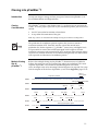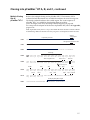
1
Bac-to-Bac® Baculovirus Expression System An efficient site-specific transposition system to generate baculovirus for high-level expression of recombinant proteins Catalog nos. 10359-016, 10360-014, 10584-027, 10712-024 Version E 19 January 2009 10359 Corporate Headquarters Invitrogen Corporation 1600 Faraday Avenue Carlsbad, CA 92008 T: 1 760 603 7200 F: 1 760 602 6500 E: [email protected] For country-specific contact information visit our web site at www.invitrogen.com User Manual ii Table of Contents Kit Contents and Storage ........................................................................................................................................... v Accessory Products ..................................................................................................................................................vii Introduction ....................................................................................................................... 1 Overview...................................................................................................................................................................... 1 The Bac-to-Bac® Baculovirus Expression System ................................................................................................... 3 Experimental Outline ................................................................................................................................................. 7 Culturing Insect Cells........................................................................................................ 8 General Guidelines ..................................................................................................................................................... 8 Generating the Recombinant pFastBac™ Vector .......................................................... 10 General Information ................................................................................................................................................. 10 Cloning into pFastBac™1.......................................................................................................................................... 11 Cloning into pFastBac™HT A, B, and C ................................................................................................................. 12 Cloning into pFastBac™ Dual .................................................................................................................................. 16 Transformation and Analysis.................................................................................................................................. 18 Generating the Recombinant Bacmid............................................................................ 20 Transforming DH10Bac™ E. coli.............................................................................................................................. 20 Isolating Recombinant Bacmid DNA..................................................................................................................... 24 Analyzing Recombinant Bacmid DNA by PCR ................................................................................................... 26 Producing Recombinant Baculovirus............................................................................ 29 Transfecting Insect Cells .......................................................................................................................................... 29 Isolating P1 Viral Stock ............................................................................................................................................ 32 Amplifying Your Baculoviral Stock ....................................................................................................................... 34 Performing a Viral Plaque Assay ........................................................................................................................... 36 Expressing Your Recombinant Protein.................................................................................................................. 43 Troubleshooting .............................................................................................................. 46 Appendix .......................................................................................................................... 52 Recipes........................................................................................................................................................................ 52 Bacmid DNA Isolation Using PureLink™ HiPure Maxiprep Kit........................................................................ 54 Map and Features of pFastBac™1............................................................................................................................ 57 Map and Features of pFastBac™HT........................................................................................................................ 59 Map and Features of pFastBac™ Dual .................................................................................................................... 61 Map of pFastBac™-Gus............................................................................................................................................. 63 Map of pFastBac™HT-CAT...................................................................................................................................... 64 Map of pFastBac™ Dual-Gus/CAT......................................................................................................................... 65 Technical Support..................................................................................................................................................... 66 Purchaser Notification ............................................................................................................................................. 67 References .................................................................................................................................................................. 69 iii iv Kit Contents and Storage Types of Products This manual is supplied with the products listed below. For a list of the reagents supplied with each catalog number, see below and the next page. Quantity Cat. no. ® Product Bac-to-Bac Baculovirus Expression System 1 kit 10359-016 Bac-to-Bac® Vector Kit 1 kit 10360-014 Bac-to-Bac® HT Vector Kit 1 kit 10584-027 1 kit 10712-024 ™ pFastBac Dual Vector Kit Kit Components Each catalog number contains the components listed below. Important: Note that catalog numbers 10360-014, 10584-027, and 10712-024 contain pFastBac™ vectors only. See the next page for a detailed description about the specific pFastBac™ vector and other reagents supplied with each catalog number. Component Cat. no. 10359-016 Cat. no. 10360-014 Cat. no. 10584-027 Cat. no. 10712-024 pFastBac™ Vectors MAX Efficiency DH10Bac Competent E. coli Cellfectin® II Reagent ® Shipping/Storage ™ The Bac-to-Bac® Baculovirus Expression System is shipped in three boxes as described below. Upon receipt, store each box as detailed below. All reagents are guaranteed for six months if stored properly. Box 1 2 3 Vector Kits Item ™ pFastBac Vectors ® ™ MAX Efficiency DH10Bac Competent E. coli ® Cellfectin II Reagent Shipping Storage Blue ice 4°C Dry ice –80°C Blue ice 4°C Cat. nos. 10360-014, 10584-027, and 10712-024 are shipped on blue ice. Upon receipt, store the vectors at 4°C. Continued on next page v Kit Contents and Storage, continued pFastBac™ Vectors Each catalog number includes a specific pFastBac™ vector(s) and a corresponding expression control, and are supplied as detailed below. Store at 4°C. Product pFastBac™ Vector Cat. no. ® Bac-to-Bac Baculovirus Expression System 10359-016 Bac-to-Bac® Vector Kit 10360-014 Bac-to-Bac® HT Vector Kit 10584-027 ™ Expression Control pFastBac 1 pFastBac™-Gus Supplied: 20 l at 0.5 g/l in TE, pH 8.0* (10 g total) Supplied: 20 l at 0.2 ng/l in TE, pH 8.0 (4 ng total) pFastBac™1 pFastBac™-Gus Supplied: 20 l at 0.5 g/l in TE, pH 8.0 (10 g total) Supplied: 20 l at 0.2 ng/l in TE, pH 8.0 (4 ng total) pFastBac™HT A pFastBac™HT-CAT pFastBac™HT B Supplied: 15 l at 1 ng/l in TE, pH 8.0 (15 ng total) ™ pFastBac HT C Supplied: 20 l each at 0.5 g/l in TE, pH 8.0 (10 g total of each vector) pFastBac™ Dual 10712-024 pFastBac™ Dual pFastBac™ Dual-Gus/CAT Supplied: 20 l at 0.5 g/l in TE, pH 8.0 (10 g total) *TE buffer, pH 8.0: 10 mM Tris-HCl, 1 mM EDTA, pH 8.0. MAX Efficiency® DH10Bac™ Competent E. coli Reagents Supplied: 20 l at 0.2 ng/l in TE, pH 8.0 (4 ng total) MAX Efficiency® DH10Bac™ Chemically Competent E. coli are supplied with the Bac-to-Bac® Baculovirus Expression System only, and include the following items. Transformation efficiency is 1 108 cfu/μg DNA. Store at –80°C. Item Composition Amount MAX Efficiency® Chemically Competent DH10Bac™ -- 5 100 μl pUC19 Control DNA 10 pg/μl in 5 mM Tris-HCl, 0.5 mM EDTA, pH 8 100 μl Genotype of DH10Bac™ F– mcrA (mrr-hsdRMS-mcrBC) 80lacZM15 lacX74 recA1 endA1 araD139 (ara, leu)7697 galU galK – rpsL nupG/bMON14272/pMON7124 Cellfectin® II Transfection Reagent Cellfectin® II Reagent is supplied with the Bac-to-Bac® Baculovirus Expression System only. Amount supplied: 1 ml Composition: 1 mg/ml transfection reagent in membrane-filtered water Storage conditions: 4°C vi Accessory Products Additional Products All of the reagents supplied in the Bac-to-Bac® Baculovirus Expression System as well as other products suitable for use with the Bac-to-Bac® System are available separately from Invitrogen. Ordering information for these reagents is provided below. Item Quantity Cat. no. ® Bac-to-Bac Vector Kit 1 kit 10360-014 ® 1 kit 10584-027 pFastBac Dual Vector Kit 1 kit 10712-024 Gateway® pDEST™8 Vector 6 g 11804-010 Gateway® pDEST™10 Vector 6 g 11806-015 Bac-to-Bac HT Vector Kit ™ 6 g 11807-013 ® ® 1 kit A11100 ® ® Bac-to-Bac C-His TOPO Cloning Kit 1 kit A11098 Bac-to-Bac® N-His TOPO® Expression System 1 kit A11101 Bac-to-Bac® N-His TOPO® Cloning Kit 1 kit A11099 5 100 l 10361-012 20 50 l C4040-03 One Shot MAX Efficiency DH10B -T1 Chemically Competent E. coli 20 50 l 12331-013 MAX Efficiency® Stbl2™ Competent Cells 1 ml 10268-019 Cellfectin® II Reagent 1 ml 10362-100 Ampicillin Sodium Salt, irradiated 200 mg 11593-027 Kanamycin Sulfate (100X), liquid 100 ml 15160-054 Gentamicin Reagent Solution, liquid (50 mg/ml) 10 ml 15750-060 Bluo-gal 1g 15519-028 Neutral Red, high purity 25 mg N-3246 Isopropylthio--galactoside (IPTG) 1g 15529-019 S.O.C. Medium 10 10 ml 15544-034 AcTEV™ Protease 1,000 Units 12575-015 Platinum® Taq DNA Polymerase 100 reactions 10966-018 Platinum Taq DNA Polymerase High Fidelity 100 reactions 11304-011 PCR SuperMix High Fidelity 100 reactions 10790-020 4% Agarose Gel ® ™ Gateway pDEST 20 Vector Bac-to-Bac C-His TOPO Expression System ® ™ MAX Efficiency DH10Bac Competent E. coli ® One Shot TOP10 Chemically Competent E. coli ® ® ™ R ® 40 ml 18300-012 ™ 1 kit K2100-12 ™ PureLink HiPure Plasmid Miniprep Kit 25 preps 100 preps K2100-02 K2100-03 PureLink™ HiPure Plasmid Maxiprep Kit 10 preps 25 preps K2100-06 K2100-07 PureLink Quick Gel Extraction System Continued on next page vii Accessory Products, continued Insect Cell Culture Products A variety of insect cell lines and GIBCO™ cell culture products are available from Invitrogen to facilitate baculovirus-mediated expression of your recombinant protein in insect cells. For more information about the insect cell lines and GIBCO™ cell culture products, refer to our website (www.invitrogen.com) or contact Technical Support (see page 66). Note: Reagents are also available in other sizes. Item Quantity Cat. no. Sf9 Cells, SFM Adapted 1.5 10 cells 11496-015 Sf21 Cells, SFM Adapted 1.5 107 cells 11497-013 High Five™ Cells 3 106 cells B855-02 Mimic Sf9 Insect Cells 7 1 10 cells 12552-014 Sf-900 II SFM 500 ml 10902-096 ™ Sf-900 III SFM 500 ml 12658-019 Sf-900 Medium (1.3X) 100 ml 10967-032 Express Five SFM 1000 ml 10486-025 Grace’s Insect Cell Culture Medium, Unsupplemented 500 ml 11595-030 Grace’s Insect Cell Culture Medium, Supplemented 500 ml 11605-094 Grace’s Insect Cell Culture Medium (2X) 100 ml 11667-037 Penicillin-Streptomycin 100 ml 15070-063 100 ml 24040-032 ™ ® ® PLURONIC F-68, 10% (100X) 7 PLURONIC® is a registered trademark of BASF Corporation Purifying Recombinant Fusion Proteins If you use the pFastBac™ HT A, B, or C vector to express your gene of interest as a fusion with the 6His tag, you may use Invitrogen’s ProBond™ or Ni-NTA resins to purify your recombinant fusion protein. See the table below for ordering information. Item ™ viii Quantity Cat. no. ProBond Nickel-chelating Resin 50 ml 150 ml R801-01 R801-15 ProBond™ Purification System 6 purifications K850-01 Ni-NTA Agarose 10 ml 25 ml 100 ml R901-01 R901-15 R901-10 Ni-NTA Purification System 6 purifications K950-01 Introduction Overview Introduction Advantages of the Bac-to-Bac® Baculovirus Expression System The Bac-to-Bac® Baculovirus Expression System provides a rapid and efficient method to generate recombinant baculoviruses (Ciccarone et al., 1997). This method was developed by researchers at Monsanto, and is based on site-specific transposition of an expression cassette into a baculovirus shuttle vector (bacmid) propagated in E. coli (Luckow et al., 1993). The major components of the Bac-to-Bac® Baculovirus Expression System include: A choice of pFastBac™ donor plasmids that allow generation of an expression construct containing the gene of interest where expression of the gene of interest is controlled by a baculovirus-specific promoter. An E. coli host strain, DH10Bac™, that contains a baculovirus shuttle vector (bacmid) and a helper plasmid, and allows generation of a recombinant bacmid following transposition of the pFastBac™ expression construct. A control expression plasmid containing the Gus and/or CAT gene that allows production of a recombinant baculovirus which, when used to infect insect cells, expresses -glucuronidase and/or chloramphenicol acetyltransferase. Using the Bac-to-Bac® Baculovirus Expression System to generate a recombinant baculovirus provides the following advantages over the traditional method using homologous recombination: Requires less than 2 weeks to identify and purify a recombinant baculovirus as compared to the 4–6 weeks required to generate a recombinant baculovirus using homologous recombination Reduces the need for multiple rounds of plaque purification as the recombinant virus DNA isolated from selected colonies is not mixed with parental, non-recombinant virus Permits rapid and simultaneous isolation of multiple recombinant baculoviruses, and is suited for the expression of protein variants for structure/function studies Continued on next page 1 Overview, continued Choosing a pFastBac™ Vector A number of pFastBac™ vectors are available for use with the Bac-to-Bac® Baculovirus Expression System (see table below). Choose the vector that best suits your needs. Vector ™ pFastBac 1 pFastBac™HT pFastBac™ Dual Features Reference Strong AcMNPV polyhedrin (PH) promoter for highlevel protein expression (Anderson et al., 1996) Large multiple cloning site for simplified cloning Strong polyhedrin (PH) promoter for high-level protein expression N-terminal 6His tag for purification of recombinant fusion proteins using metal-chelating resin and a TEV protease cleavage site for removal of the 6His tag following protein purification Vector supplied in 3 reading frames for simplified cloning Two strong baculovirus promoters (PH and p10) to allow simultaneous expression of two proteins Two large multiple cloning sites for simplified cloning (Polayes et al., 1996) (Harris & Polayes, 1997) The Bac-to-Bac® TOPO® Expression System provides a rapid and highly effective Bac-to-Bac® ® TOPO Expression method to generate recombinant baculoviruses by combining the ease of blunt-end TOPO® cloning with the efficiency of site-specific transposition System technology of the Bac-to-Bac® System. The Bac-to-Bac® TOPO® Expression System is available separately from Invitrogen with a choice of pFastBac™/CT-TOPO® or pFastBac™/NT-TOPO® donor plasmids, which are also available separately as part of Bac-to-Bac® C-His TOPO® or Bac-to-Bac® N-His TOPO® Cloning Kits (see page vii for ordering information). Continued on next page 2 Overview, continued Purpose of This Manual Important This manual provides an overview of the Bac-to-Bac® Baculovirus Expression System, and provides instructions and guidelines to: 1. Clone your gene of interest into the pFastBac™ donor plasmid of choice. 2. Transform the pFastBac™ construct into MAX Efficiency® DH10Bac™ competent E. coli to generate a recombinant bacmid. 3. Transfect the recombinant bacmid DNA into the insect cell line of choice to generate a recombinant baculovirus. 4. Amplify and titer the baculoviral stock, and use this stock to infect insect cells to express your recombinant protein. The Bac-to-Bac® Baculovirus Expression System is designed to help you create a recombinant baculovirus for high-level expression of your gene of interest in insect cells. Although the system has been designed to help you easily generate a baculovirus and express your recombinant protein of interest, use of the system is geared towards those users who are familiar with baculovirus biology and insect cell culture. We highly recommend that users possess a working knowledge of viral and tissue culture techniques. For more information about baculovirus biology, refer to published reference sources (King & Possee, 1992; Luckow, 1991; O'Reilly et al., 1992). For more information about insect cell culture, refer to the Guide to Baculovirus Expression Vector Systems (BEVS) and Insect Cell Culture Techniques for downloading on our website at www.invitrogen.com or by contacting Technical Support (see page 66). 3 The Bac-to-Bac® Baculovirus Expression System Components of the Bac-to-Bac® Baculovirus Expression System The Bac-to-Bac® Baculovirus Expression System facilitates rapid and efficient generation of recombinant baculoviruses (Ciccarone et al., 1997). Based on a method developed by Luckow et al., 1993, the Bac-to-Bac® Baculovirus Expression System takes advantage of the site-specific transposition properties of the Tn7 transposon to simplify and enhance the process of generating recombinant bacmid DNA. The first major component of the System is a pFastBac™ vector into which the gene(s) of interest will be cloned. Depending on the pFastBac™ vector selected, expression of the gene(s) of interest is controlled by the Autographa californica multiple nuclear polyhedrosis virus (AcMNPV) polyhedrin (PH) or p10 promoter for high-level expression in insect cells. This expression cassette is flanked by the left and right arms of Tn7, and also contains a gentamicin resistance gene and an SV40 polyadenylation signal to form a mini Tn7. The second major component of the System is the DH10Bac™ E. coli strain that is used as the host for your pFastBac™ vector. DH10Bac™ cells contain a baculovirus shuttle vector (bacmid) with a mini-attTn7 target site and a helper plasmid (see the next page for details). Once the pFastBac™ expression plasmid is transformed into DH10Bac™ cells, transposition occurs between the mini-Tn7 element on the pFastBac™ vector and the mini-attTn7 target site on the bacmid to generate a recombinant bacmid. This transposition reaction occurs in the presence of transposition proteins supplied by the helper plasmid. Once you have performed the transposition reaction, you will isolate the high molecular weight recombinant bacmid DNA and transfect the bacmid DNA into insect cells to generate a recombinant baculovirus that can be used for preliminary expression experiments. After the baculoviral stock is amplified and titered, this high-titer stock can be used to infect insect cells for large-scale expression of the recombinant protein of interest. For a schematic representation of the Bac-to-Bac® Baculovirus Expression System, see the diagram on page 6. Continued on next page 4 The Bac-to-Bac® Baculovirus Expression System, continued Baculovirus Shuttle Vector The baculovirus shuttle vector (bacmid), bMON14272 (136 kb), present in DH10Bac™ E. coli contains: A low-copy number mini-F replicon Kanamycin resistance marker A segment of DNA encoding the LacZ peptide from a pUC-based cloning vector into which the attachment site for the bacterial transposon, Tn7 (miniattTn7) has been inserted. Insertion of the mini-attTn7 does not disrupt the reading frame of the LacZ peptide. The bacmid propagates in E. coli DH10Bac™ as a large plasmid that confers resistance to kanamycin and can complement a lacZ deletion present on the chromosome to form colonies that are blue (Lac+) in the presence of a chromogenic substrate such as Bluo-gal or X-gal and the inducer, IPTG. Recombinant bacmids (composite bacmids) are generated by transposing a miniTn7 element from a pFastBac™ donor plasmid to the mini-attTn7 attachment site on the bacmid. The Tn7 transposition functions are provided by a helper plasmid (see below). Helper Plasmid DH10Bac™ E. coli also contain the helper plasmid, pMON7124 (13.2 kb), which encodes the transposase and confers resistance to tetracycline. The helper plasmid provides the Tn7 transposition function in trans (Barry, 1988). Continued on next page 5 The Bac-to-Bac® Baculovirus Expression System, continued The figure below depicts the generation of recombinant baculovirus and the expression of your gene of interest using the Bac-to-Bac® Baculovirus Expression System. Diagram of the Bac-to-Bac® System pFastBac donor plasmid Clone Gene of Interest Gene of Interest Tn7L Tn7R Transformation Helper Helper mid Bac Donor Transposition Antibiotic Selection lacZ Donor mini-attTn7 P PH Foreign Gene PPH Recombinant Donor Plasmid E. coli (LacZ - ) Containing Recombinant Bacmid Competent DH10Bac E.coli Cells Mini-prep of High Molecular Weight DNA or Determine Viral Titer by Plaque Assay Recombinant Baculovirus Particles Transfection of Insect Cells with Cellfectin® II Reagent Infection of Insect Cells Recombinant Gene Expression or Viral Amplification 6 Recombinant Bacmid DNA Experimental Outline Flow Chart The figure below illustrates the general steps required to express your gene of interest using the Bac-to-Bac® Baculovirus Expression System. pFastBac donor plasmid Clone gene of interest pFastBac Recombinant Transform into MAX Efficiency® DH10Bac Cells (containing bacmid and helper) E. coli Colonies with Recombinant Bacmid Restreak Verified E. coli Colonies with Recombinant Bacmid Grow overnight culture and isolate recombinant bacmid DNA Recombinant Bacmid DNA Transfect insect cells using Cellfectin® II Reagent P1 Recombinant Baculovirus Stock (>106 pfu/ml) Infect insect cells to amplify virus P2 Recombinant Baculovirus Stock (>107 pfu/ml) Titer and infect insect cells Protein Expression 7 Culturing Insect Cells General Guidelines Introduction We recommend using Spodoptera frugiperda Sf9 or Sf21 insect cells as the host for your baculovirus transfer vector. Before you start your transfection and expression experiments, be sure to have cultures of Sf9 or Sf21 cells growing and have frozen master stocks available. Sf9 and Sf21 cells and cell culture reagents are available separately from Invitrogen (see page viii for ordering information). Note: High Five™ and Mimic™ Sf9 insect cells are suitable for use for expression only. Using Serum-Free Medium Insect cells may be cultured under serum-free conditions. We recommend using Sf-900 II SFM or Sf-900™ III SFM available from Invitrogen (see page viii for ordering information). Both Sf-900 II SFM and Sf-900™ III SFM are protein-free media optimized for the growth and maintenance of Sf9 and Sf21 cells, and for large-scale production of recombinant proteins expressed using the Bac-to-Bac® System. For more information, see our website (www.invitrogen.com) or call Technical Support (see page 66). Insect Cell Culture Reference Guide For guidelines and detailed information on insect cell culture, refer to the Guide to Baculovirus Expression Vector Systems (BEVS) and Insect Cell Culture Techniques. This guide is available on our website at www.invitrogen.com or by contacting Technical Support (see page 66), and contains information on: Maintaining and passaging insect cells in adherent and suspension culture Freezing cells Using serum-free medium (includes protocols to adapt cells to serum-free medium) Scaling up cell culture Continued on next page 8 General Guidelines, continued General Guidelines Insect cells are very sensitive to environmental factors. In addition to chemical and nutritional culture factors, physical factors can also affect insect cell growth, and optimization is required to maximize cell growth. Consider the following when culturing insect cells: Temperature: The optimal range to grow and infect cultured insect cells is 27C to 28C. pH: A range of 6.1 to 6.4 works well for most culture systems. Sf-900 II SFM will maintain a pH in this range under conditions of normal air and opencapped culture systems. Osmolality: The optimal osmolality of medium for use with lepidopteran cell lines is 345 to 380 mOsm/kg. Aeration: Insect cells require passive oxygen diffusion for optimal growth and recombinant protein expression. Active or controlled oxygenated systems require dissolved oxygen at 10% to 50% of air saturation. Shear Forces: Suspension culture generates mechanical shear forces. Growing insect cells in serum-containing media (10% to 20% FBS) generally provides adequate protection from cellular shear forces. If you are growing insect cells in serum-free conditions, supplementation with a shear force protectant such as PLURONIC® F-68 may be required. Note: Growing cells in Sf-900 II SFM does not require addition of shear force protectants. Cells for Transfection You will need log-phase cells with >95% viability to perform a successful transfection. Refer to page 30 to determine how many cells you will need for transfection. 9 Generating the Recombinant pFastBac™ Vector General Information Introduction To generate a recombinant plasmid containing your gene(s) of interest for use in the Bac-to-Bac® Baculovirus Expression System, you will use restriction enzyme digestion and ligation to clone your gene(s) into one of the pFastBac™ vectors. For recommendations and guidelines to help you design your cloning strategy, refer to the appropriate section on pages 11–17 depending on the pFastBac™ vector you are using. General Molecular Biology Techniques For help with restriction enzyme digestion, ligation, DNA sequencing, and other general molecular biology techniques, refer to Molecular Cloning: A Laboratory Manual (Sambrook et al., 1989) or Current Protocols in Molecular Biology (Ausubel et al., 1994). Propagation and Maintenance of Plasmids The pFastBac™ vectors and their corresponding expression control plasmids contain the ampicillin resistance gene to allow for selection in E. coli using ampicillin. To propagate and maintain the pFastBac™ vectors and the pFastBac™ control plasmids, use the following procedure: 10 1. Use the stock solution of vector provided to transform a recA, endA E. coli strain such as TOP10, DH10B™, or DH5™ (see page 18 for more information). 2. Select transformants on LB agar plates containing 100 g/ml ampicillin. 3. Prepare a glycerol stock from each transformant containing plasmid for longterm storage (see page 19). Cloning into pFastBac™1 Introduction To help you design a strategy to clone your gene of interest into pFastBac™1, see the recommendations and diagram below. Cloning Considerations The pFastBac™1 vector is a non-fusion vector (i.e., no fusion tags are present in the vector). To ensure proper expression of your recombinant protein, your insert must contain: An ATG start codon for initiation of translation A stop codon for termination of the gene Note: Stop codons are included in the multiple cloning site in all three reading frames. The production of recombinant proteins requires that your insert contain a translation initiation ATG. Generally, transfer vectors that contain intact polyhedrin (PH) leader sequences (e.g., pFastBac™ vectors) may yield higher levels of expression than vectors that contain interrupted leader sequences. Protein translation can initiate at the mutated ATG (ATT) upstream of the multiple cloning site; however, initiation from this site is inefficient and generally does not interfere with expression and detection of recombinant protein. Multiple Cloning Site of pFastBac™1 Below is the multiple cloning site for pFastBac™1. Restriction sites are labeled to indicate the actual cleavage site. Potential stop codons are underlined. The vector sequence of pFastBac™1 is available for downloading from our website (www.invitrogen.com) or by contacting Technical Support (see page 66). For a map and a description of the features of pFastBac™1, refer to the Appendix, pages 57–58. Start of Transcription Polyhedrin promoter 3901 TAGATCATGG AGATAATTAA AATGATAACC ATCTCGCAAA TAAATAAGTA wild-type ATG mutated to ATT 3951 TTTTACTGTT TTCGTAACAG TTTTGTAATA AAAAAACCTA TAAATATTCC Bam H I 4001 Stu I Sal I Sst I Spe I Not I Nsp V GCGGAATTCA AAGGCCTACG TCGACGAGCT CACTAGTCGC GGCCGCTTTC Xba I 4101 BssH II GGATTATTCA TACCGTCCCA CCATCGGGCG CGGATCCCGG TCCGAAGCGC EcoR I 4051 Rsr II Pst I Xho I Sph I Kpn I Hind III GAATCTAGAG CCTGCAGTCT CGAGGCATGC GGTACCAAGC TTGTCGAGAA SV40 polyadenylation signal 4151 GTACTAGAGG ATCATAATCA GCCATACCAC ATTTGTAGAG GTTTTACTTG 11 Cloning into pFastBac™HT A, B, and C Introduction The pFastBac™HT vector is supplied with the multiple cloning site in three reading frames (A, B, and C) to facilitate cloning your gene of interest in frame with the N-terminal 6His tag. See the recommendations below and the diagrams on pages 13–15 to help you design a cloning strategy. Cloning Considerations The pFastBac™HT vectors are fusion vectors. To ensure proper expression of your recombinant protein, you must: Clone your gene in frame with the initiation ATG at base pairs 4050–4052. This will create a fusion with the N-terminal 6His tag and a cleavage site for the AcTEV™ Protease. Include a stop codon with your insert Generally, transfer vectors that contain intact polyhedrin (PH) leader sequences (e.g., pFastBac™ vectors) may yield higher levels of expression than vectors that contain interrupted leader sequences. Protein translation can initiate at the mutated ATG (ATT) upstream of the multiple cloning site; however, initiation from this site is inefficient and generally does not interfere with expression and detection of recombinant protein. Continued on next page 12 Cloning into pFastBac™HT A, B, and C, continued Multiple Cloning Site of pFastBac™HT A Below is the multiple cloning site for pFastBac™HT A. The initiation ATG is indicated in bold. Restriction sites are labeled to indicate the actual cleavage site. The vector sequence of pFastBac™HT A is available for downloading from our website (www.invitrogen.com) or by contacting Technical Support (see page 66). For a map and a description of the features of pFastBac™HT, refer to the Appendix, pages 59–60. Start of Transcription Polyhedrin promoter 3901 TAGATCATGG AGATAATTAA AATGATAACC ATCTCGCAAA TAAATAAGTA wild-type ATG mutated to ATT 3951 TTTTACTGTT TTCGTAACAG TTTTGTAATA AAAAAACCTA TAAATATTCC 4001 GGATTATTCA TACCGTCCCA CCATCGGGCG CGGATCTCGG TCCGAAACC 6xHis tag 4050 ATG TCG TAC TAC CAT CAC CAT CAC CAT CAC GAT TAC GAT ATC Met Ser Tyr Tyr His His His His His His Asp Tyr Asp Ile TEV recognition site 4092 Ehe I Nco I Bam H I CCA ACG ACC GAA AAC CTG TAT TTT CAG GGC GCC ATG GAT CCG Pro Thr Thr Glu Asn Leu Tyr Phe Gln Gly Ala Met Asp Pro TEV cleavage site EcoR I 4134 Sal I Sst I Spe I Not I GAA TTC AAA GGC CTA CGT CGA CGA GCT CAA CTA GTG CGG CCG Glu Phe Lys Gly Leu Arg Arg Arg Ala Glu Leu Val Arg Pro Nsp V 4176 Stu I Xba I Pst I Xho I Sph I Kpn I Hind III CTT TCG AAT CTA GAG CCT GCA GTC TCG AGG CAT GCG GTA CCA Leu Ser Asp Leu Glu Pro Ala Val Ser Arg His Ala Val Pro SV40 polyadenylation signal 4218 AGC TTG TCG AGA AGT ACT AGA GGA TCA TAA TCA GCCATACCAC ... Ser Leu Ser Arg Ser Thr Arg Gly Ser *** Continued on next page 13 Cloning into pFastBac™HT A, B, and C, continued Multiple Cloning Site of pFastBac™HT B Below is the multiple cloning site for pFastBac™HT B. The initiation ATG is indicated in bold. Restriction sites are labeled to indicate the actual cleavage site. The boxed nucleotide indicates the variable region. The vector sequence of pFastBac™HT B is available for downloading from our website (www.invitrogen.com) or by contacting Technical Support (see page 66). For a map and a description of the features of pFastBac™HT, refer to the Appendix, pages 59–60. Start of Transcription Polyhedrin promoter 3901 TAGATCATGG AGATAATTAA AATGATAACC ATCTCGCAAA TAAATAAGTA wild-type ATG mutated to ATT 3951 TTTTACTGTT TTCGTAACAG TTTTGTAATA AAAAAACCTA TAAATATTCC 4001 GGATTATTCA TACCGTCCCA CCATCGGGCG CGGATCTCGG TCCGAAACC 6xHis tag 4050 ATG TCG TAC TAC CAT CAC CAT CAC CAT CAC GAT TAC GAT ATC Met Ser Tyr Tyr His His His His His His Asp Tyr Asp Ile Ehe I Nco I TEV recognition site 4092 Bam H I CCA ACG ACC GAA AAC CTG TAT TTT CAG GGC GCC ATG GGA TCC Pro Thr Thr Glu Asn Leu Tyr Phe Gln Gly Ala Met Gly Ser TEV cleavage site EcoR I 4134 Sst I Spe I Not I Xba I Pst I Xho I Sph I Kpn I GCT TTC GAA TCT AGA GCC TGC AGT CTC GAG GCA TGC GGT ACC Ala Phe Glu Ser Arg Ala Cys Ser Leu Glu Ala Cys Gly Thr Hind III 4218 Sal I GGA ATT CAA AGG CCT ACG TCG ACG AGC TCA CTA GTC GCG GCC Gly Ile Glu Arg Pro Thr Ser Thr Ser Ser Leu Val Ala Ala Nsp V 4176 Stu I SV40 polyadenylation signal AAG CTT GTC GAG AAG TAC TAG AG GATCATAATC AGCCATACCA ... Lys Leu Val Glu Lys Tyr *** Continued on next page 14 Cloning into pFastBac™HT A, B, and C, continued Multiple Cloning Site of pFastBac™HT C Below is the multiple cloning site for pFastBac™HT C. The initiation ATG is indicated in bold. Restriction sites are labeled to indicate the actual cleavage site. The boxed nucleotide indicates the variable region. The vector sequence of pFastBac™HT C is available for downloading from our website (www.invitrogen.com) or by contacting Technical Support (see page 66). For a map and a description of the features of pFastBac™HT, refer to the Appendix, pages 61–62. Note: In pFastBac™HT C, there is a stop codon within the Xba I site that is in frame with the N-terminal tag. Make sure that the 5 end of your gene is cloned upstream of the Xba I site. Start of Transcription Polyhedrin promoter 3901 TAGATCATGG AGATAATTAA AATGATAACC ATCTCGCAAA TAAATAAGTA wild-type ATG mutated to ATT 3951 TTTTACTGTT TTCGTAACAG TTTTGTAATA AAAAAACCTA TAAATATTCC 4001 GGATTATTCA TACCGTCCCA CCATCGGGCG CGGATCTCGG TCCGAAACC 6xHis tag 4050 ATG TCG TAC TAC CAT CAC CAT CAC CAT CAC GAT TAC GAT ATC Met Ser Tyr Tyr His His His His His His Asp Tyr Asp Ile TEV recognition site 4092 Ehe I Nco I Bam H I CCA ACG ACC GAA AAC CTG TAT TTT CAG GGC GCC ATG GGG ATC Pro Thr Thr Glu Asn Leu Tyr Phe Gln Gly Ala Met Gly Ile TEV cleavage site EcoR I 4134 Sal I Sst I Spe I Not I CGG AAT TCA AAG GCC TAC GTC GAC GAG CTC ACT AGT CGC GGC Arg Asn Ser Lys Ala Tyr Val Asp Glu Leu Thr Ser Arg Gly Nsp V 4176 Stu I Xba I Pst I Xho I Sph I Kpn I Hind III CGC TTT CGA ATC TAG AGCCTGCAGT CTCGAGGCAT GCGGTACCAA Arg Phe Arg Ile *** SV40 polyadenylation signal 4221 GCTTGTCGAG AAGTACTAGA GGATCATAAT CAGCCATACC ... 15 Cloning into pFastBac™ Dual Introduction The pFastBac™ Dual vector contains two multiple cloning sites to allow expression of two heterologous genes; one controlled by the polyhedrin (PH) promoter and one by the p10 promoter. To help you design a strategy to clone your genes of interest into pFastBac™ Dual, see the recommendations and the diagram below. Cloning Considerations The pFastBac™ Dual vector is a non-fusion vector. To ensure proper expression of your recombinant proteins, both of your inserts must contain: An ATG start codon for initiation of translation A stop codon for termination of the gene if you don’t use one of the stop codons provided in the multiple cloning site The production of recombinant proteins requires that your insert contain a translation initiation ATG. Generally, transfer vectors that contain intact polyhedrin leader sequences (e.g., pFastBac™ vectors) may yield higher levels of expression than vectors that contain interrupted leader sequences. For inserts cloned downstream of the polyhedrin promoter, note that protein translation can initiate at the mutated ATG (ATT); however, initiation from this site is inefficient and generally does not interfere with expression and detection of recombinant protein. Multiple Cloning Site Downstream of the PH Promoter Below is the multiple cloning site located downstream of the PH promoter in pFastBac™ Dual. Restriction sites are labeled to indicate the actual cleavage site. Potential stop codons are underlined. The vector sequence of pFastBac™ Dual is available for downloading from our website (www.invitrogen.com) or by contacting Technical Support (see page 66). For a map and a description of the features of pFastBac™ Dual, refer to the Appendix, pages 61–62. Start of Transcription Polyhedrin promoter 4481 ATGGAGATAA TTAAAATGAT AACCATCTCG CAAATAAATA AGTATTTTAC wild-type ATG mutated to ATT 4531 TGTTTTCGTA ACAGTTTTGT AATAAAAAAA CCTATAAATA TTCCGGATTA Bam H I 4581 EcoR I TTCATACCGT CCCACCATCG GGCGCGGATC CCGGTCCGAA GCGCGCGGAA Stu I 4631 BssH II Rsr II Sal I Sst I Spe I Not I Nsp V Xba I TTCAAAGGCC TACGTCGACG AGCTCACTAG TCGCGGCCGC TTTCGAATCT Pst I Hind III 4681 AGAGCCTGCA GTCTCGACAA GCTTGTCGAG AAGTACTAGA GGATCATAAT 4731 CAGCCATACC ACATTTGTAG AGGTTTTACT TGCTTTAAAA AACCTCCCAC SV40 polyadenylation signal Continued on next page 16 Cloning into pFastBac™ Dual, continued Multiple Cloning Site Downstream of the p10 Promoter Below is the multiple cloning site located downstream of the AcMNPV p10 promoter in pFastBac™ Dual. Restriction sites are labeled to indicate the actual cleavage site. Potential stop codons are underlined. The vector sequence of pFastBac™ Dual is available for downloading from our website (www.invitrogen.com) or by contacting Technical Support (see page 66). For a map and a description of the features of pFastBac™ Dual, refer to the Appendix, pages 61–62. Start of Transcription p10 promoter 4460 TATACGGACC TTTAATTCAA CCCAACACAA TATATTATAG TTAAATAAGA 4410 ATTATTATCA AATCATTTGT ATATTAATTA AAATACTATA CTGTAAATTA Bbs II 4360 Xho I CATTTTATTT ACAATCACTC GACGAAGACT TGATCACCCG GGATCTCGAG Nco I 4310 Sma I Nhe I Pvu II Nsi I Sph I Kpn I CCATGGTGCT AGCAGCTGAT GCATAGCATG CGGTACCGGG AGATGGGGGA HSV tk polyadenylation signal 4260 GGCTAACTGA AACACGGAAG GAGACAATAC CGGAAGGAAC CCGCGCTATG 17 Transformation and Analysis Introduction Once you have completed your ligation reactions, you are ready to transform your pFastBac™ construct into E. coli. Many E. coli host strains and transformation procedures are suitable. General recommendations to transform E. coli and analyze transformants are provided in this section. E. coli Host Once you have cloned your insert into one of the pFastBac™ vectors, you will transform the ligation reaction into E. coli and select for ampicillin-resistant transformants. You may use any recA, endA E. coli strain including TOP10, DH10B™, or DH5™ for transformation. Do not transform the ligation reaction into DH10Bac™ cells. Note: Chemically competent TOP10, DH10B™, and DH5™ E. coli are available from Invitrogen in a convenient One Shot® format (see table below). Item Quantity Cat. no. One Shot TOP10 Chemically Competent E. coli 20 50 l C4040-03 One Shot® MAX Efficiency® DH10B™-T1R Chemically Competent E. coli 20 50 l 12331-013 One Shot® MAX Efficiency® DH5™-T1R Chemically Competent E. coli 20 50 l 12297-016 ® Transformation Method You may use any method of choice to transform E. coli. Chemical transformation is the most convenient method, while electroporation is the most efficient and method of choice for large plasmids. To select for transformants, use LB agar plates containing 100 g/ml ampicillin. Analyzing Transformants Once you have obtained ampicillin-resistant transformants, we recommend the following: 18 1. Pick 10 transformants and culture them overnight in LB or S.O.B. containing 100 g/ml ampicillin. 2. Isolate the plasmid DNA using your method of choice. We recommend using the PureLink™ HiPure Plasmid DNA Miniprep Kit to purify high quality plasmid DNA from your E. coli transformants (see page vii for ordering information). 3. Analyze the plasmids by restriction analysis to confirm the presence and correct orientation of the insert. Use a restriction enzyme or a combination of enzymes that cut once in the vector and once in the insert. Transformation and Analysis, continued Analyzing Transformants by PCR You may also analyze positive transformants using PCR. Use the appropriate PCR primers and amplification conditions for your insert. If you are using this technique for the first time, you may want to perform restriction analysis in parallel. Artifacts may be obtained because of mispriming or contaminating template. The protocol below is provided for your convenience. Other protocols are suitable. Materials Needed: PCR SuperMix High Fidelity (see page vii for ordering information) Appropriate forward and reverse PCR primers (20 μM each) Procedure: 1. For each sample, aliquot 48 μl of PCR SuperMix High Fidelity into a 0.5 ml microcentrifuge tube. Add 1 μl each of the forward and reverse PCR primer. 2. Pick 10 colonies and resuspend them individually in 50 μl of the PCR SuperMix containing primers (remember to make a patch plate to preserve the colonies for further analysis). 3. Incubate reaction for 10 minutes at 94C to lyse cells and inactivate nucleases. 4. Amplify for 20 to 30 cycles. 5. For the final extension, incubate at 72C for 10 minutes. Store at 4C. 6. Visualize by agarose gel electrophoresis. Sequencing You may sequence your construct to confirm that your gene of interest is in the correct orientation for expression. If you have cloned your gene into one of the pFastBac™HT vectors, verify that your gene is cloned in frame with the N-terminal tag. Long-Term Storage Once you have identified the correct clone, be sure to purify the colony and make a glycerol stock for long-term storage. We recommend that you store a stock of plasmid DNA at –20°C. 1. Streak the original colony out for single colony on LB plates containing 100 g/ml ampicillin. 2. Isolate a single colony and inoculate into 1–2 ml of LB containing 100 g/ml ampicillin. 3. Grow until culture reaches stationary phase. 4. Mix 0.85 ml of culture with 0.15 ml of sterile glycerol and transfer to a cryovial. 5. Store at –80°C. 19 Generating the Recombinant Bacmid Transforming DH10Bac™ E. coli Introduction Once you have generated your pFastBac™ construct, you are ready to transform purified plasmid DNA into DH10Bac™ E. coli for transposition into the bacmid. You will use blue/white selection to identify colonies containing the recombinant bacmid. MAX Efficiency® DH10Bac™ chemically competent cells are supplied with the Bac-to-Bac® Baculovirus Expression System, but are also available separately from Invitrogen (see page vii). Guidelines and instructions to transform DH10Bac™ cells are provided in this section. Positive Control Each pFastBac™ plasmid is supplied with a corresponding control plasmid for use as a positive transfection and expression control (see table below). Depending on the pFastBac™ vector you are using, we recommend including the corresponding control plasmid in your DH10Bac™ transformation experiment (see table below). For maps and a description of the features of each control plasmid, see the Appendix, pages 63–65. pFastBac™ Vector Control Plasmid ™ pFastBac -Gus ™ pFastBac HT pFastBac™HT-CAT pFastBac™ Dual pFastBac™ Dual-Gus/CAT pFastBac 1 ™ Continued on next page 20 Transforming DH10Bac™ E. coli, continued MEND ION AT RECOM Materials Needed Have the following materials on hand before beginning: Your purified pFastBac™ construct (200 pg/l in TE, pH 8) Positive expression control (i.e., pFastBac™-Gus, pFastBac™HT-CAT, or pFastBac™ Dual-Gus/CAT; use as a control for transposition) MAX Efficiency® DH10Bac™ chemically competent cells (supplied with the Bac-to-Bac® Baculovirus Expression System; use 1 tube of competent cells for every transformation) pUC19 (supplied with the MAX Efficiency® DH10Bac™ E. coli; use as a control for transformation, if desired) LB agar plates containing kanamycin, gentamicin, tetracycline, Bluo-gal, and IPTG (3 plates for each transformation; use freshly prepared plates; see recommendation below) LB agar plate containing 100 g/ml ampicillin (for plating pUC19 transformation control) S.O.C. Medium (see page vii) 15 ml round-bottom polypropylene tubes 42C water bath 37C shaking and non-shaking incubator You will need to prepare LB agar plates containing 50 g/ml kanamycin, 7 g/ml gentamicin, 10 g/ml tetracycline, 100 g/ml Bluo-gal, and 40 g/ml IPTG to select for DH10Bac™ transformants. See page vii to order antibiotics, Bluo-gal, and IPTG, and page 53 for instructions to prepare plates. If you are preparing LB plates using a pre-mixed formulation, we recommend using Luria Broth Base instead of Lennox L (LB). Using Lennox L plates will reduce the color intensity and may reduce the number of colonies obtained. Note: Use Bluo-gal instead of X-gal for blue/white selection. Bluo-gal generally produces a darker blue color than X-gal. Preparing for Transformation For each transformation, you will need one vial of competent cells and three selective plates. Equilibrate a water bath to 42C. Warm selective plates at 37C for 30 minutes. Warm the S.O.C. Medium to room temperature. Pre-chill one 15 ml round-bottom polypropylene tube for each transformation. Continued on next page 21 Transforming DH10Bac™ E. coli, continued Transformation Procedure Follow the procedure below to transform MAX Efficiency® DH10Bac™ chemically competent cells with your pFastBac™ construct. We recommend including positive controls for transposition (i.e., pFastBac™ expression plasmid) and transformation (i.e., pUC19) in your experiment to help you evaluate your results. 1. Thaw on ice one vial of MAX Efficiency® DH10Bac™ competent cells for each transformation. 2. For each transformation, gently mix and transfer 100 l of the DH10Bac™ cells into a pre-chilled, 15 ml round-bottom polypropylene tube. 3. Add the appropriate amount of plasmid DNA to the cells and mix gently. Do not pipet up and down to mix. Your pFastBac™ construct: 1 ng (5 l) pFastBac™ control plasmid: 1 ng pUC19 control: 50 pg (5 l) 4. Incubate cells on ice for 30 minutes. 5. Heat-shock the cells for 45 seconds at 42C without shaking. 6. Immediately transfer the tubes to ice and chill for 2 minutes. 7. Add 900 l of room temperature S.O.C. Medium. 8. For pFastBac™ transformations: Shake tubes at 37C at 225 rpm for 4 hours. For pUC19 transformation: Shake tube at 37C at 225 rpm for 1 hour. For each pFastBac™ transformation: Prepare 10-fold serial dilutions of the cells (10–1, 10–2, 10–3) with S.O.C. Medium. Plate 100 l of each dilution on an LB agar plate containing 50 g/ml kanamycin, 7 g/ml gentamicin, 10 g/ml tetracycline, 100 g/ml Bluo-gal, and 40 g/ml IPTG. For the pUC19 transformation: Dilute the cells 1:100 with S.O.C. Medium. Plate 100 l of the dilution on an LB agar plate containing 100 g/ml ampicillin. 10. Incubate plates for 48 hours at 37C. Pick white colonies for analysis (see the next page for recommendations). 9. Note: We do not recommend picking colonies earlier than 48 hours as it may be difficult to distinguish between white and blue colonies. Important Insertions of the mini-Tn7 into the mini-attTn7 attachment site on the bacmid disrupt the expression of the LacZ peptide, so colonies containing the recombinant bacmid are white in a background of blue colonies that harbor the unaltered bacmid. Select white colonies for analysis. True white colonies tend to be large; therefore, to avoid selecting false positives, choose the largest, most isolated white colonies. Avoid picking colonies that appear gray or are darker in the center as they can contain a mixture of cells with empty bacmid and recombinant bacmid. Continued on next page 22 Transforming DH10Bac™ E. coli, continued Verifying the Phenotype 1. Pick 10 white colonies and restreak them on fresh LB agar plates containing 50 g/ml kanamycin, 7 g/ml gentamicin, 10 g/ml tetracycline, 100 g/ml Bluo-gal, and 40 g/ml IPTG. Incubate the plates overnight at 37C. 2. From a single colony confirmed to have a white phenotype on restreaked plates containing Bluo-gal and IPTG, inoculate a liquid culture containing 50 g/ml kanamycin, 7 g/ml gentamicin, and 10 g/ml tetracycline. 3. Isolate recombinant bacmid DNA using the procedure provided on the next page for analysis. You may also use the procedure for the PureLink™ HiPure Plasmid Maxiprep Kit provided in the Appendix, page 54, for increased recombinant bacmid yield. 4. Analyze the recombinant bacmid DNA to verify successful transposition to the bacmid. We recommend using PCR to analyze your bacmid DNA (see Analyzing Recombinant Bacmid DNA by PCR, page 26, for details). Note: It is possible to verify successful transposition to the bacmid by using agarose gel electrophoresis to look for the presence of high molecular weight DNA. This method is less reliable than performing PCR analysis as high molecular weight DNA can be difficult to visualize. 23 Isolating Recombinant Bacmid DNA Introduction The PureLink™ HiPure Plasmid DNA Miniprep Kit allows you to purify high quality Bacmid DNA from DH10Bac™ E. coli (see page vii for ordering information). The isolated bacmid DNA is suitable for use in insect cell transfections. Note: We do not recommend the PureLink™ HiPure Precipitator Module or the PureLink™ HiPure Plasmid Filter Mini/Midi/Maxiprep Kits for isolating bacmid DNA. Before Starting Inoculate a single white bacterial colony into 2 ml LB medium with 50 g/ml kanamycin, 7 g/ml gentamicin, and 10 g/ml tetracycline. Incubate the culture at 37°C in a shaking water bath at 250 rpm overnight. Verify that RNase A is added to the Resuspension Buffer (R3) and that the Lysis Buffer (L7) contains no precipitates. Equilibrating the Column Place the PureLink™ HiPure Mini column on the PureLink™ Nucleic Acid Purification Rack (see the manual supplied with the rack for more details). Apply 2 ml Equilibration Buffer (EQ1) to the column. Allow the solution in the column to drain by gravity flow. Preparing the Cell Lysate 1. Harvest 1.5 ml bacterial cells by centrifuging at 9,000 g for 15 minutes. Remove all medium. 2. Add 0.4 ml Resuspension Buffer (R3) containing RNase A to the pellet and resuspend the cells until homogeneous. Transfer cell suspension to a centrifuge tube. 3. Add 0.4 ml Lysis Buffer (L7). Mix gently by inverting the capped tube five times. Do not vortex. Incubate at room temperature for 5 minutes. 4. Add 0.4 ml Precipitation Buffer (N3) and mix immediately by inverting the capped tube until the mixture is homogeneous. Do not vortex. 5. Centrifuge the mixture at >15,000 g at room temperature for 10 minutes. Note: If the pellet does not adhere to the bottom of the tube, incubate the tube at room temperature for 5 minutes to allow the separation of the lysate and gelatinous pellet. Pipette the clear lysate into a sterile tube and centrifuge at >15,000 g for 5 minutes at room temperature to remove any remaining cellular debris. Binding and Washing the DNA 1. Load the supernatant from Step 5 (see above) onto the equilibrated column. Allow the solution in the column to drain by gravity flow. 2. Wash the column twice with 2.5 ml Wash Buffer (W8). Allow the solution in the column to drain by gravity flow after each wash. Discard the flow-through. Continued on next page 24 Isolating Recombinant Bacmid DNA, continued Eluting and Precipitating DNA 1. Place a sterile centrifuge tube (elution tube) under the column. 2. Add 0.9 ml Elution Buffer (E4) to the column to elute DNA. Allow the solution to drain by gravity flow. Do not force out any remaining solution. 3. The elution tube contains the purified DNA. Discard the column. 4. Add 0.63 ml isopropanol to the elution tube. Mix and place on ice for 10 minutes. 5. Centrifuge the mixture at >15,000 g at 4°C for 20 minutes. Carefully remove and discard the supernatant. 6. Resuspend the DNA pellet in 1 ml 70% ethanol. 7. Centrifuge at >15,000 g at 4°C for 5 minutes. Carefully remove and discard the supernatant. 8. Air-dry the pellet for 10 minutes. 9. Resuspend the DNA pellet in 40 μl TE Buffer (TE). Allow pellet to dissolve for at least 10 minutes on ice. To avoid shearing the DNA, pipette only 1–2 times to resuspend. 10. Store the bacmid DNA at 4°C (see Important below). Important You may store your bacmid DNA at –20°C if you avoid frequent freeze/thaw cycles as it decreases the transfection efficiency. To store your purified bacmid DNA at –20°C, aliquot into separate tubes in TE Buffer, pH 8.0 to avoid more than one freeze/thaw cycle and do not store in a frost-free freezer. You may also store the purified bacmid DNA for up to 2 weeks at 4°C in TE Buffer, pH 8.0. You may prepare glycerol stocks of DH10Bac™ E. coli containing the bacmid DNA from mid-logarithmic phase culture grown from white colonies picked during the blue-white screening, and store at –80°C for future bacmid DNA isolation. You may also use the procedure for PureLink™ HiPure Plasmid Maxiprep Kit provided in the Appendix, page 54, for increased recombinant bacmid yield. The PureLink™ HiPure Plasmid Prep Kits, available separately from Invitrogen, allow the purification of all types and sizes of plasmid DNA, including BAC, bacmids, and ssM13 DNAs, and are ideally suited for bacmid purification (see page vii for ordering information). 25 Analyzing Recombinant Bacmid DNA by PCR Introduction Recombinant bacmid DNA is greater than 135 kb in size. Since restriction analysis is difficult to perform with DNA of this size, we recommend using PCR analysis to verify the presence of your gene of interest in the recombinant bacmid. Use the pUC/M13 Forward and Reverse primers (sequences given below) that hybridize to sites flanking the mini-attTn7 site within the lacZ-complementation region to facilitate PCR analysis (see figure below). Guidelines and instructions are provided in this section to perform PCR using the pUC/M13 Forward and Reverse primers. Transposed pFastBac sequence Tn7R Bacmid DNA Gene of interest mini-attTn7 pUC/M13 Forward PCR Analysis with pUC/M13 Primers Tn7L pUC/M13 Reverse To verify the presence of your gene of interest in the recombinant bacmid using PCR, you may: Use the pUC/M13 Forward and Reverse primers (see sequences below). Use a combination of the pUC/M13 Forward or Reverse primer and a primer that hybridizes within your insert. Invitrogen does not supply the pUC/M13 Forward and Reverse primers; you must have these primers custom synthesized. DNA Polymerase Primer Sequence pUC/M13 Forward 5-CCCAGTCACGACGTTGTAAAACG-3 pUC/M13 Reverse 5-AGCGGATAACAATTTCACACAGG-3 You may use any DNA polymerase of your choice for PCR including Platinum® Taq DNA Polymerase. If the expected PCR product is > 4 kb, we recommend using a polymerase mixture such as Platinum® Taq DNA Polymerase High Fidelity for best results (see page vii for ordering information). Continued on next page 26 Analyzing Recombinant Bacmid DNA by PCR, continued Producing the PCR Product Use the procedure below to amplify your recombinant bacmid DNA using the pUC/M13 Forward and Reverse primers and Platinum® Taq polymerase. If you are using a combination of the pUC/M13 Forward or Reverse primers primer and a primer specific for your gene, you will need to determine the amplification conditions to use. If you are using another polymerase, follow the manufacturer’s recommendations for the polymerase you are using. Note: Amplification conditions may need to be optimized if your insert is > 4 kb. 1. For each sample, set up the following 50 μl PCR reaction in a 0.5 ml microcentrifuge tube: Recombinant bacmid DNA (100 ng) 1 l 10X PCR Buffer (appropriate for enzyme) 5 l 10 mM dNTP Mix 1 l 50 mM MgCl2 1.5 l PCR Primers (1.25 l each 10 M stock) 2.5 l 38.5 l Sterile Water Platinum Taq polymerase (5 units/μl) 0.5 l Total Volume 50 μl ® 2. Amplify using the following cycling parameters: Step 3. Time Temperature Initial Denaturation 3 minutes 93°C Denaturation 45 seconds 94°C Annealing 45 seconds 55°C Extension 5 minutes 72°C Final Extension 7 minutes 72°C Cycles 1X 25–35X 1X Remove 5–10 μl from the reaction and analyze by agarose gel electrophoresis. Continued on next page 27 Analyzing Recombinant Bacmid DNA by PCR, continued What You Should See If transposition has occurred and you have used the pUC/M13 Forward and Reverse primers for amplification, you should see a PCR product of the following size on the agarose gel: Sample Size of PCR Product Bacmid alone ~300 bp Bacmid transposed with pFastBac™1 ~2300 bp + size of your insert Bacmid transposed with pFastBac™-Gus ~4200 bp ™ ~2430 bp + size of your insert ™ ~3075 bp ™ ~2560 bp + size of your insert ™ ~5340 bp Bacmid transposed with pFastBac HT Bacmid transposed with pFastBac HT-CAT Bacmid transposed with pFastBac Dual Bacmid transposed with pFastBac Dual-Gus/CAT If you have used a combination of the pUC/M13 Forward or Reverse primer and a gene-specific primer for amplification, you will need to determine the expected size of your PCR product. Refer to the diagram on page 26 to help you calculate the expected size of your PCR product. 28 Producing Recombinant Baculovirus Transfecting Insect Cells Introduction Once you have confirmed that your recombinant bacmid contains the gene of interest, you are ready to transfect insect cells to produce recombinant baculovirus. Guidelines and instructions to transfect insect cells are provided in this section. Plasmid Preparation You may use any method to prepare purified recombinant bacmid DNA for transfection. Bacmid DNA must be clean and free from phenol and sodium chloride as contaminants may kill the cells, and salt will interfere with lipid complexing, decreasing transfection efficiency. We recommend isolating bacmid DNA using the PureLink™ HiPure Plasmid Miniprep Kit (see page vii for ordering information) or the procedure provided in the Appendix, page 54. Transfection Method We recommend using a cationic lipid such as Cellfectin® II Reagent for transfection. Cellfectin® II Reagent is supplied with the Bac-to-Bac® Baculovirus Expression System and is available separately from Invitrogen (see page vii for ordering information). Cellfectin® II Reagent Cellfectin® II Reagent is a proprietary cationic lipid formulation that offers the highest transfection efficiencies and protein expression levels on the widest variety of adherent and suspension insect cell lines, including Sf9 and Sf21 cells. Insect Cell Lines We recommend using Sf9 or Sf21 cells for transfection and identification of recombinant plaques. High Five™ and Mimic™ Sf9 cells are not recommended because they generally transfect less efficiently. However, once you have generated your baculovirus stock, you may use High Five™ or Mimic™ Sf9 cells for expression studies. Media for Transfection For the highest transfection efficiency, we recommend performing the transfection in Grace’s Insect Cell Culture Medium, Unsupplemented (see page viii). Note that the Grace’s Insect Cell Culture Medium should not contain supplements or fetal bovine serum (FBS) as the supplements and the proteins in the FBS will interfere with the Cellfectin® II Reagent, inhibiting the transfection. Note: If you are culturing Sf9 or Sf21 cells in Sf-900 II SFM or Sf-900™ III SFM, you can perform the transfection in unsupplemented Grace’s Medium, then easily switch back to Sf-900 II SFM or Sf-900™ III SFM after transfection. Continued on next page 29 Transfecting Insect Cells, continued If you have generated a recombinant bacmid from one of the pFastBac™ control plasmids (i.e., pFastBac™-Gus, pFastBac™HT-CAT, or pFastBac™ Dual-Gus/CAT), we recommend including this positive control in your transfection and expression experiments to help you evaluate your results. In these bacmids, the gene encoding -glucuronidase (Gus) and/or chloramphenicol acetyltransferase (CAT) will be expressed under the control of the polyhedrin (PH) or p10 promoter. After transfection, expression of -glucuronidase or CAT may be assayed, as appropriate. Materials Needed Purified recombinant bacmid DNA from your pFastBac™ construct (500 ng/l in TE Buffer, pH 8.0) Purified recombinant bacmid DNA from the appropriate pFastBac™ control construct (if desired, 500 ng/l in TE Buffer, pH 8.0) Sf9 or Sf21 cells cultured in the appropriate medium Cellfectin® II Reagent (store at 4°C until use) Grace’s Insect Cell Medium, Unsupplemented (see page viii), media should not contain supplements, FBS, or antibiotics 6-well tissue culture plates and other tissue culture supplies 1.5 ml sterile microcentrifuge tubes Complete growth medium for culturing insect cells (e.g., Sf-900 II SFM, Sf-900™ III SFM, TNM-FH, Grace’s Supplemented Insect Cell Culture Medium, or other suitable medium) MEND ION AT RECOM Positive Control Transfection Conditions Calculate the number of Sf9 or Sf21 cells that you will need for your transfection experiment and expand cells accordingly. Make sure your cells are healthy with greater than 95% viability and are growing in the logarithmic phase with a density of 1.5 106–2.5 106 cells/ml before proceeding to transfection. We generally produce baculoviral stocks in Sf9 or Sf21 cells using the following transfection conditions. Note that these conditions should be used as a starting point for your transfection. To obtain the highest transfection efficiency and low non-specific effects, you may optimize transfection conditions by varying DNA and Cellfectin® II Reagent concentrations, and cell density. Condition Amount Tissue culture plate size 6-well (35 mm) plate (one well/bacmid) Number of Sf9 or Sf21 cells to transfect 8 105 cells Amount of bacmid DNA 1 g (can vary from 1 to 2 g) Amount of Cellfectin® II Reagent 8 l (can vary from 1.5 to 9 l) Note: This procedure is for insect cells in a 6-well format. All amounts and volumes are given on a per well basis. Continued on next page 30 Transfecting Insect Cells, continued Important Guidelines for Transfection Transfection Procedure Use Grace’s Insect Cell Culture Medium, Unsupplemented to seed all cells in plate for Sf9 and Sf21 cells grown in Grace’s Insect Cell Culture Medium, Supplemented (with 10% FBS). With Cellfectin® II, you do not have to remove the medium from cells and wash cells prior to adding the DNA-lipid complex to cells. The DNA-lipid complex formation time is shorter (~15–30 minutes) when using Cellfectin® II as compared to Cellfectin® reagent. Do not add antibiotics during transfection as this causes cell death. For Sf9 or Sf21 insect cells cultured in Supplemented Grace’s Insect Medium containing 10%FBS, use the following protocol to prepare your cells for transfection in a 6-well format. All amounts and volumes are given on a per well basis. If you wish to transfect cells in other tissue culture formats, you will need to determine the optimal conditions to use. 1. Verify that the Sf9 or Sf21 cells are in the log phase (1.5–2.5 106 cells/ml) with greater than 95% viability. 2. If the cell density is in range of 1.5 106–2.5 106 cells/ml and the culture is without antibiotics, proceed to step 2a. If the cell density is not in this range or the cell culture contains antibiotics, follow steps 2b–2c: a. Add 2 ml of Grace’s Insect Medium, Unsupplemented (without antibiotics and serum) in each well. Seed 8 × 10⁵ Sf9 or Sf21 cells from Step 1 per well. Do not change medium or wash the cells. The medium carried over will enhance the transfection efficiency. Allow cells to attach for 15 minutes at room temperature in the hood. Proceed to step 3. b. Prepare 10ml plating medium by mixing 1.5 ml Supplemented Grace’s Insect Medium containing 10%FBS (without antibiotics) and 8.5 ml Grace’s Insect Medium, Unsupplemented (without FBS and antibiotics). c. Plate 8 × 10⁵ Sf9 or Sf21 cells from Step 1 per well. Allow cells to attach for 15 minutes at room temperature in the hood. Remove the medium. Add 2.5 ml plating medium from step 2b per well. Proceed to step 3. 3. For each transfection sample, prepare complexes as follows: a. Mix Cellfectin® II before use, and dilute 8 μl in 100 l Grace’s Medium, Unsupplemented (without antibiotics and serum). Vortex briefly to mix. Note: You may leave this mixture at room temperature for up to 30 minutes. b. Dilute 1 μl baculovirus DNA in 100 μl Grace’s Medium, Unsupplemented (without antibiotics and serum). Mix gently. c. Combine the diluted DNA with diluted Cellfectin® II (total volume ~210 μl). Mix gently and incubate for 15–30 minutes at room temperature. 4. Add ~210 μl DNA-lipid mixture or transfection mixture (Step 3c) dropwise onto the cells from Step 2. Incubate cells at 27°C for 3–5 hours. 5. Remove the transfection mixture and replace with 2 ml of complete growth medium (e.g., Grace’s Insect Medium, Supplemented and 10% FBS). Using antibiotics is optional. 6. Incubate cells at 27°C for 72 hours or until you see signs of viral infection. 31 Isolating P1 Viral Stock Introduction Budded virus should be released into the medium 72 hours after transfection. However, if your transfection efficiency was not optimal, cells may not show all of the signs of viral infection until 4 or 5 days post-transfection. Beginning at 72 hours after transfection, you should visually inspect the cells daily for signs of infection (see below). Once the cells appear infected (i.e., demonstrate chacteristics typical of late to very late infection), harvest the virus from the cell culture medium using the procedure below. Characteristics of Infected Cells Virally-infected insect cells typically display the following characteristics as observed from visual inspection using an inverted phase microscope at 250–400X magnification. The time points provided below assume that the transfection was successful (i.e., transfection efficiency was high). Signs of Infection Early (first 24 hours) Late (24–72 hours) Very Late (>72 hours) Preparing the P1 Viral Stock Phenotype Description Increased cell diameter A 25–50% increase in cell diameter may be seen. Increased size of cell nuclei Nuclei may appear to "fill" the cells. Cessation of cell growth Cells appear to stop growing when compared to a cell-only control. Granular appearance Signs of viral budding; vesicular appearance to cells. Detachment Cells release from the plate or flask. Cell lysis Cells appear lysed, and show signs of clearing in the monolayer. 1. Once the transfected cells from Step 6, previous page demonstrate signs of late stage infection (e.g., 72 hours post-transfection), collect the medium containing virus from each well (~2 ml) and transfer to sterile 15 ml snap-cap tubes. Centrifuge the tubes at 500 g for 5 minutes to remove cells and large debris. 2. Transfer the clarified supernatant to fresh 15 ml snap-cap tubes. This is the P1 viral stock. Store at 4ºC, protected from light. See the next page for additional storage information. Note: If you wish to concentrate your viral stock to obtain a higher titer, you may filter your viral supernatant through a 0.2 m, low protein binding filter after the low-speed centrifugation step, if desired. Continued on next page 32 Isolating P1 Viral Stock, continued Storing Viral Stocks The Next Step Store viral stocks as follows: Store viral stock at 4ºC, protected from light. If medium is serum-free (e.g., Sf-900 II SFM, Sf-900™ III SFM), add fetal bovine serum to a final concentration of 2%. Serum proteins act as substrates for proteases. For long-term storage, store an aliquot of the viral stock at –80ºC for later reamplification. Do not store routinely used viral stocks at temperatures below 4ºC. Repeated freeze/thaw cycles can result in a 10- to 100-fold decrease in virus titer. Once you have obtained your clarified P1 baculoviral stock, you may: Amplify the viral stock (see the next section for details). This procedure is recommended to obtain the highest viral titers and optimal results in your expression studies. Determine the titer of your viral stock (see Performing a Viral Plaque Assay, page 36). Plaque purify your recombinant baculovirus, if desired (see Performing a Viral Plaque Assay, page 36). Use the P1 viral stock to infect Sf9 or Sf21 cells for preliminary expression experiments (see below). If you wish to perform small-scale or preliminary expression experiments, it is possible to proceed directly to expression studies by using the P1 viral stock to infect your Sf9 or Sf21 cells. Note that the amount of viral stock is limited and expression conditions may not be reproducible (i.e., MOI is unknown if titer is not determined). 33 Amplifying Your Baculoviral Stock Introduction The P1 viral stock is a small-scale, low-titer stock. You may use this stock to infect cells to generate a high-titer P2 stock. The titer of the initial viral stock obtained from transfecting Sf9 or Sf21 cells generally ranges from 1 106 to 1 107 plaque forming units (pfu)/ml. Amplification allows production of a P2 viral stock with a titer ranging from 1 107 to 1 108 pfu/ml and is generally recommended. Guidelines and protocols are provided in this section to amplify the recombinant baculovirus to prepare a P2 viral stock. Materials Needed You should have the following materials on hand before beginning: Important Multiplicity of Infection (MOI) Sf9 or Sf21 cells cultured in the appropriate growth medium P1 baculoviral stock Any appropriate tissue culture vessel (see Important Note below) Tissue culture reagents 27ºC humidified incubator To amplify your P1 viral stock, you may infect Sf9 or Sf21 cells growing in suspension or monolayer culture. Depending on your needs, you may amplify your P1 viral stock at any scale, but remember that you may be limited by the amount of P1 viral stock available. We generally amplify our P1 viral stock in a 10 ml suspension culture at 2 106 cells/ml or in 6-well tissue culture plates at 2 106 cells/well. Calculate the number of Sf9 or Sf21 cells that you will need for infection and expand cells accordingly. Make sure that the cells are healthy, of low passage (5–20), and have >95% viability before proceeding to infection. To amplify your viral stock, infect cells at a multiplicity of infection (MOI) ranging from 0.05 to 0.1. MOI is defined as the number of virus particles per cell. Use the following formula to calculate how much viral stock to add to obtain a specific MOI: Inoculum required (ml) ( MOI (pfu/cell) number of cells titer of viral stock (pfu/ml) ) Note: If you have not determined the titer of your P1 viral stock, you may assume that the titer ranges from 1 106 to 1 107 pfu/ml. Example We wish to infect a 10 ml culture at 2 106 cells/ml using an MOI = 0.1. We assume that the titer of our P1 viral stock is 5 106 pfu/ml. Inoculum required (ml) ( 0.1 pfu/cell 2 10 7 cells 5 10 6 pfu/ml ) Inoculum required (ml) 0.4 ml Continued on next page 34 Amplifying Your Baculoviral Stock, continued Important considerations Amplification Procedure For successful amplification of your baculovirus, you should pay attention to several key points: Use Sf9 or Sf21 cells that are in excellent health, low passage (5–20), log-phase growth, and have >95% viability Use sterile P1 baculoviral stock that is free of contaminants Use a low MOI between 0.05–0.1 as higher MOI will reduce baculovirus quality Harvest the virus when 70–80% of cells are dead You cannot amplify the baculovirus indefinitely, as they acquire deleterious mutations with each passage. Usually, P3 is highest usable passage. Follow the guidelines below to amplify your P1 viral stock in a 6-well plate. 1. On the day of infection, prepare your Sf9 or Sf21 cell suspension and plate cells at 2 106 cells/well. Incubate cells at room temperature for 1 hour to allow attachment. 2. After 1 hour, inspect cells under an inverted microscope to verify attachment. 3. Add the appropriate amount of P1 viral stock to each well. 4. Incubate the cells for 48 hours in a 27ºC humidified incubator. 5. 48 hours post-infection, collect 2 ml of medium containing virus from each well and transfer to sterile 15 ml snap-cap tubes. Centrifuge the tubes at 500 g for 5 minutes to remove cells and large debris and to obtain clarified baculoviral stock. Note: It is possible to harvest virus at later times after infection (e.g., 72 hours). Optimal harvest times can vary and should be determined for each baculoviral construct. Remember that culture viability will decrease over time as cells lyse. 6. Transfer the supernatant to fresh 15 ml snap-cap tubes. This is the P2 viral stock. Store at 4ºC, protected from light. For long-term storage, you may store an aliquot of the P2 stock at –80ºC, protected from light. See page 33 for storage guidelines. 7. Proceed to the next section to determine the titer of your P2 viral stock. Scaling Up the Amplification Procedure Once you have generated a high-titer P2 baculoviral stock, you may scale-up the amplification procedure to any volume of your choice. To produce this high-titer P3 stock, scale up the amount of cells and volume of virus used appropriately, and follow the guidelines and procedure outlined in this section. Generating HighTiter Stocks From Frozen Master Stock If you have stored your viral master stock at –80ºC, we recommend amplifying this stock to generate another high-titer stock for use in expression experiments. Viral titers generally decrease over time when virus is stored at –80ºC. Follow the guidelines and amplification procedure detailed in this section. 35 Performing a Viral Plaque Assay Introduction We recommend using the BaculoTiter™ Assay kit, available separately from Invitrogen, to determine the titer of your baculoviral stock. The BaculoTiter™ Assay kit provides accurate results for your baculovirus titer in two days as compared to 10-day dilution assays. For more information, refer to our website at www.invitrogen.com or contact Technical Support (page 66). Alternatively, you may follow the guidelines and instructions provided below to: Experimental Outline Factors Affecting Viral Titer Determine the titer of your baculoviral stock Plaque purify the virus (optional) To determine the titer of a baculoviral stock, you will: 1. Plate Sf9 or Sf21 cells in 6-well plates. 2. Prepare 10-fold serial dilutions of your baculoviral stock. 3. Add the different dilutions of baculovirus to Sf9 or Sf21 cells and infect cells for 1 hour. 4. Remove the virus and overlay the cell monolayer with Plaquing Medium. 5. Incubate the cells for 7–10 days, stain (if desired), and count the number of plaques in each dilution. A number of factors can influence viral titers including: The size of your gene of interest. Titers will generally decrease as the size of the insert increases. The transfection efficiency. For the highest transfection efficiency, we recommend transfecting Sf9 or Sf21 cells using Cellfectin® II Reagent. Prepare DNA:lipid complexes in Grace’s Insect Medium, Unsupplemented (see pages 29–31 for details). The age of your baculoviral stock. Viral titers may decrease with long-term storage at 4ºC or –80ºC. If your baculoviral stock has been stored for 6 months to 1 year, we recommend titering or re-titering your baculoviral stock prior to use in an expression experiment. Number of freeze/thaw cycles. If you are storing your viral stock at –80ºC, viral titers can decrease as much as 10% with each freeze/thaw cycle. Improper storage of your baculoviral stock. For routine use, baculoviral stocks should be aliquotted and stored at 4ºC, protected from light. Continued on next page 36 Performing a Viral Plaque Assay, continued Materials Needed Your clarified baculoviral stock (store at 4ºC until use) Sf9 or Sf21 cells cultured in the appropriate medium (30 ml of log-phase cells at 5 105 cells/ml for each baculoviral stock to be titered) Sf-900 II SFM, Sf-900™ III SFM or other appropriate complete growth medium (see Note below) Sf-900 Medium (1.3X) (100 ml) or other appropriate plaquing medium (see Note below) 4% Agarose Gel (specifically formulated for optimal insect cell growth, see page vii) Sterile, cell-culture grade, distilled water 100 ml sterile, glass bottle 6-well tissue-culture plates (2 plates for each viral stock to be titered) Sterile hood Waters baths at 40ºC and 70ºC Microwave oven (optional) 27ºC humidified incubator Neutral Red, high purity (see page vii) See page viii for ordering information. If you are culturing your Sf9 or Sf21 cells in serum-supplemented media (i.e., complete TNM-FH), you should have the following reagents on hand: Grace’s Insect Cell Culture Medium, Supplemented Grace’s Insect Cell Culture Medium (2X) Fetal Bovine Serum (FBS), Qualified, Heat-Inactivated See page viii for ordering information. Continued on next page 37 Performing a Viral Plaque Assay, continued Preparing the Plaquing Medium Plaquing medium consists of a mixture of culture medium and agarose, and will be used to immobilize the infected cells for the plaque assay. Prepare plaquing medium immediately before use following the procedure below. If you are culturing the Sf9 cells in Sf-900 II SFM, prepare Sf-900 Plaquing Medium. If you are culturing cells in TNM-FH, prepare Grace’s Plaquing Medium. Note: Other Plaquing Media are suitable. 1. Melt the 4% Agarose Gel by placing the bottle in a 70ºC water bath for 20 to 30 minutes or heating the agarose in a microwave oven. While the 4% agarose gel is melting, place the following in the 40ºC water bath: Empty, sterile 100 ml bottle Sf-900 Medium (1.3X) or Grace’s Insect Cell Culture Medium (2X), as appropriate 2. Once the 4% agarose gel has liquefied, move the agarose gel, medium, and empty 100 ml bottle to a sterile hood. 3. Working quickly, prepare the plaquing medium as follows: Sf-900 Plaquing Medium: Combine 30 ml of Sf-900 Medium (1.3X) and 10 ml of the melted 4% Agarose Gel in the empty 100 ml bottle and mix gently. Grace’s Plaquing Medium: Add 20 ml of heat-inactivated FBS to the 100 ml bottle of Grace’s Insect Medium (2X) and mix. Combine 25 ml of the Grace’s Insect Medium (2X) containing serum with 12.5 ml of cell-culture grade, sterile, distilled water and 12.5 ml of the melted 4% Agarose Gel in the empty 100 ml bottle and mix gently. 4. Return the bottle of plaquing medium to the 40ºC water bath until use. Continued on next page 38 Performing a Viral Plaque Assay, continued Plaque Assay Procedure Use the procedure below to perform a plaque assay in 6-well plate format to determine the titer of your pFastBac™ baculoviral stock. If you have generated a baculoviral stock of the pFastBac™ expression control pFastBac™-Gus, we recommend titering this stock as well. Remember to include a negative control (no virus) in your experiment. Note: The amounts provided in this procedure are suitable to titer one baculoviral stock (two 6-well plates per viral stock). If you wish to titer more than one baculoviral stock, scale up the reagent quantities accordingly. 1. On the day of infection, harvest Sf9 or Sf21 cells and prepare a 30 ml cell suspension at 5 105 cells/ml in Sf-900 II SFM (or other complete growth medium). Aliquot 2 ml of cell suspension into each well of two 6-well plates. If you are including a negative control, you will need another 6-well plate. 2. Allow the cells to settle to the bottom of the plate and incubate, covered, at room temperature for 1 hour. 3. Following the 1 hour incubation, observe the cell monolayers using an inverted microscope. Sf9 cells should be attached and at 50% confluence. 4. Prepare an 8-log serial dilution (10–1 to 10–8) of the clarified baculoviral stock in Sf-900 II SFM or Grace’s Insect Cell Culture Medium, Supplemented, without FBS, as appropriate. To do this, sequentially dilute 0.5 ml of the baculoviral stock or previous dilution in 4.5 ml of medium in 12 ml disposable tubes. You should finish with 8 tubes of diluted viral stock (i.e., 10–1, 10–2, 10–3, 10–4, 10–5, 10–6, 10–7, 10–8). You will use the dilutions 10–4 to 10–8 in your assay. 5. Move the 6-well plates containing Sf9 cells and the tubes of diluted virus to the sterile hood. Label the plates, in columns of 2 (1 sample well plus 1 duplicate) as follows: no virus (negative control), 10–4, 10–5, 10–6, 10–7, 10–8. 6. Remove the medium from each well, discard, and immediately replace with 1 ml of the appropriate virus dilution. As a negative control, add the appropriate medium without virus. 7. Incubate cells with virus for 1 hour at room temperature. 8. Following the 1 hour incubation, move the cells and the bottle of plaquing medium from the 40ºC water bath (Step 4, previous page) to a sterile hood. 9. Sequentially starting from the highest dilution (10–8) to the lowest dilution (10–4), remove the medium containing virus from the wells and replace with 2 ml of plaquing medium. Work quickly to avoid dessication of the cell monolayer. 10. Allow agarose overlay to harden for 1 hour at room temperature before moving the plates. 11. Incubate the cells in a 27ºC humidied incubator for 7–10 days until plaques are visible and ready to count. If you wish to stain plaques to facilitate counting, see the next page. To calculate the titer, see page 41. Continued on next page 39 Performing a Viral Plaque Assay, continued To improve the visualization of plaques, stain the plates using Neutral Red. Crystalline Blue and other plaque staining dyes containing organic solvents are not recommended because they kill the host cells. To stain plaques, you may do one of the following: Prepare an agarose solution containing neutral red and overlay this solution on the plates 4 days post-infection. Count plaques 7–10 days post-infection. or Prepare a neutral red solution and add to plates for 1–2 hours just prior to counting plaques (7–10 days post-infection). Important: If you plan to plaque purify your baculovirus, you should not stain plaques as neutral red is a known mutagen that can alter your recombinant virus. Materials Needed Neutral Red Staining Procedure Neutral Red, high purity (see page vii for ordering information) Cell-culture grade, distilled water Sf-900 II SFM or other appropriate complete growth medium (if preparing the agarose solution; see page vii for ordering information) 4% Agarose Gel (if preparing the agarose solution; see page vii for ordering information) 40ºC water bath (if preparing the agarose solution) Preparing a Neutral Red Agarose Overlay (for use on Day 4) 1. Prepare a 1 mg/ml Neutral Red solution in Sf-900 II SFM (or other appropriate complete growth medium). Filter-sterilize. 2. Combine the reagents below in a 50 ml tube and place in a 40ºC water bath. 1 mg/ml Neutral Red solution Sf-900 II SFM 1.5 ml 16.5 ml 3. Microwave 4% Agarose Gel until melted, then place in a 40ºC water bath for 5 minutes. 4. Move the 50 ml tube of Neutral Red solution and the 4% agarose gel to a sterile hood. Add 6 ml of 4% agarose gel to the Neutral Red solution. 5. Add 1 ml of the Neutral Red overlay to each well containing plaquing overlay. Once the agarose has hardened, return plates to a 27ºC humidified incubator until plaques are ready to count. Plaques will appear as clear spots on a red monolayer. Procedure continued on next page Continued on next page 40 Performing a Viral Plaque Assay, continued Neutral Red Staining Procedure, continued Calculating the Titer Procedure continued from previous page Preparing a Neutral Red Stain (for use on Day 7–10 prior to counting plaques) 6. Prepare a 1 mg/ml Neutral Red solution in cell-culture grade, distilled water. 7. Add 0.5 ml of Neutral Red solution to each well containing plaquing overlay. Incubate for 1 to 2 hours at room temperature. 8. Gently remove excess stain with a pipet or blotter and count the plaques. Plaques will appear as clear spots in a nearly clear gel against a red background. Count the number of plaques present in each dilution, then use the following formula to calculate the titer (plaque forming units (pfu)/ml) of your viral stock. Note that the optimal range to count is 3 to 20 plaques per well of a 6-well plate. titer (pfu/ml) number of plaques dilution factor Example 1 ml of inoculum/w ell In this example, we add 1 ml of inoculum and observe 20 plaques in the well containing the 10–6 viral dilution. Using the formula above, the titer of this viral stock is: 1 titer (pfu/ml) 20 plaques 10 6 ( ) 1 ml of inoculum/w ell titer (pfu/ml) = 2 107 pfu/ml What You Should See When titering pFastBac™ baculoviral stocks, we generally obtain titers ranging from: 1 106 to 1 107 pfu/ml for P1 viral stocks 1 107 to 1 108 pfu/ml for P2 viral stocks Note: If the titer of your baculoviral stock is less than 1 106 pfu/ml or 1 107 pfu/ml for a P1 or P2 viral stock, respectively, we recommend producing a new baculoviral stock. See page 36 and the Troubleshooting section, page 50 for more tips and guidelines to optimize your viral yield. Continued on next page 41 Performing a Viral Plaque Assay, continued Plaque Purification You may generate a viral stock from a single viral clone by plaque purifying your baculovirus, if desired. Use a protocol of your choice or the procedure below. Materials Needed Plate containing well-spaced viral plaques (from Plaque Assay Procedure, Step 11, page 39; do not stain plates with Neutral Red) Log phase Sf9 or Sf21 cells at greater than 95% viability Sterile Pasteur pipette and bulb Procedure 42 1. Follow Steps 1–3 in the Plaque Assay Procedure, page 39 to seed Sf9 or Sf21 cells. 2. Using a sterile Pasteur pipette and bulb, carefully pick a clear plaque and transfer the agarose plug (containing virus) to a 1.5 ml microcentrifuge tube containing 500 l of complete growth medium. Mix well by vortexing. 3. Add 100 l of the agarose plug solution to each well. 4. Incubate the cells in a 27ºC humidified incubator for 72 hours. 5. Collect the medium containing virus from each well (~2 ml) and transfer to sterile 15 ml snap-cap tubes. Centrifuge the tubes at 500 g for 5 minutes to remove cells and large debris. 6. Transfer the clarified supernatant to fresh 15 ml snap-cap tubes. This is your plaque-purified viral stock. 7. Proceed to Amplifying Your Baculoviral Stock, page 34. Expressing Your Recombinant Protein Introduction Once you have generated a pFastBac™ baculoviral stock with a suitable titer (e.g., 1 108 pfu/ml), you are ready to use the baculoviral stock to infect insect cells and assay for expression of your recombinant protein. Guidelines for infection and expression are provided below. Positive Control If you have generated a high-titer viral stock from the pFastBac™ control baculoviral construct (i.e., pFastBac-Gus, pFastBac™HT-CAT, pFastBac™ DualGus/CAT), you may want to include this recombinant baculovirus in your experiments for use as an expression control. Once you have infected cells with the FastBac™ control virus, the gene encoding -glucuronidase (Gus) and/or chloramphenicol acetyltransferase (CAT) will be constitutively expressed and can be easily assayed (see page 45). Guidelines for Expression General guidelines are provided below to infect insect cells with the recombinant baculovirus to express your protein of interest. Cell line: Depending on your application and gene of interest, you may use any insect cell line including Sf9, Sf21, High Five™, or Mimic™ Sf9 for expression. Cells may be grown in adherent or suspension culture in the culture vessel of choice. Note: If you are expressing a secreted protein, you may improve expression by using High Five™ cells. Culture Conditions: We generally culture cells in serum-free conditions using Sf-900 II SFM, Sf-900™ III SFM, or Express Five® SFM as appropriate (see page viii). Depending on your application and the protein of interest, note that it may be necessary to supplement the culture post-infection with 0.1% to 0.5% FBS or BSA to protect the recombinant protein from proteolysis. Proteinbased protease inhibitors are generally less expensive and more effective than many synthetic protease inhibitors. Infection Conditions: We recommend infecting cultures while cells are in the mid-logarithmic phase of growth at a density of 1 106 to 2 106 cells/ml. Make sure that the culture is not rate-limited by nutritional (i.e., amino acid or carbohydrate utilization) or environmental factors (i.e., pH, dissolved O2, or temperature) during infection. MOI: Optimal MOI will vary between cell lines, and the relative infection kinetics of the virus isolate or clone used. A dose response should be established for each virus, medium, reactor, and cell line employed to determine the optimal infection parameters to use for protein expression. As a starting point, infect cells using an MOI of 1 to 5. Time course: We recommend performing a time course to determine the expression kinetics for your recombinant protein as many proteins may be degraded by cellular proteases released in cell culture. Note: Maximum expression of secreted proteins is generally observed between 30 and 72 hours and non-secreted proteins between 48 and 96 hours post-infection. Continued on next page 43 Expressing Your Recombinant Protein, continued Optimizing Expression Harvesting Baculovirus Infected Insect Cells Detecting Recombinant Protein A number of factors can influence determination of optimal expression conditions including the cell line, MOI, your application of interest, and the nature of your gene of interest. You may perform the following to determine the optimal conditions to use to express your recombinant protein of interest: Cell line: Infect Sf9, Sf21, High Five™, or Mimic™ Sf9 cells at a constant MOI. Assay for recombinant protein expression at different times post-infection (e.g., 24, 48, 72, 96 hours post-infection). Choose the cell line that provides the optimal level of recombinant protein expression. MOI: Infect a population of cells at varying MOIs (e.g., 1, 2, 5, 10, 20) and assay for protein expression. Use the MOI that provides the optimal level of recombinant protein expression. Time course: Infect cells at a constant MOI and assay for recombinant protein expression at different times post-infection (e.g., 24, 48, 72, 96 hours postinfection). Choose the time point at which optimal recombinant protein expression is obtained. Use the following procedure for harvesting recombinant baculovirus infected insect cells to analyze expression of your recombinant protein of interest. This procedure is adapted from Luckow and Summers and is designed to allow expression analysis in a 24-well format from cells harvested 24 to 96 hours postinfection. Other protocols are also suitable. 1. Seed 6 105 Sf9 or Sf21 cells per well in a 24-well plate. Let cells attach for at least 30 minutes. 2. Remove the media and rinse the cells once with fresh growth media. Replace with 300 l of fresh media. 3. Add the pFastBac™ baculoviral stock to each well at the desired MOI. Include the appropriate controls (e.g., mock-infected (uninfected) cells, pFastBac™ positive control baculovirus, previously characterized recombinant baculoviruses). 4. Incubate cells in a 27ºC humidified incubator. 5. Harvest cells (or media, if the recombinant protein is secreted) at the appropriate time (i.e., 24, 48, 72, 96 hours post-infection). If harvesting cells, remove the media and rinse the cells once with serum-free medium. 6. Lyse the cells with 400 l of 1X SDS-PAGE Buffer (62.5 mM Tris-HCl, pH 6.8, 2% SDS). 7. Freeze samples at –20ºC or boil samples for at least 3 minutes and separate proteins by SDS-PAGE. You may use any method of choice to detect your recombinant protein of interest including functional analysis or western blot. If you perform western blot analysis, you will need to have an antibody to your protein of interest. Continued on next page 44 Expressing Your Recombinant Protein, continued If you have cloned your gene of interest in frame with the 6His tag in pFastBac™HT, the presence of the N-terminal 6His tag and the recognition site for the AcTEV™ Protease will increase the size of your protein by at least 3 kDa. Assay for -glucuronidase If you include the baculoviral control created using the pFastBac™-Gus or pFastBac™ Dual-Gus/CAT control constructs in your expression experiment, you may assay for -glucuronidase expression using the following methods. Other methods are suitable. Identify blue plaques on agarose plates containing the chromogenic indicator, X-glucuronide. To assess -glucuronidase expression in a rapid but qualitative manner, mix a small amount of media from the infected cells with X-glucuronide and observe development of blue color. Briefly, mix 5 l of a 20 mg/ml X-glucuronide solution (in DMSO or dimethylformamide) with 50 l of cellfree medium. Monitor for development of blue color within 2 hours. Assay for CAT Protein If you include the baculoviral control created using the pFastBac™HT-CAT or pFastBac™ Dual-Gus/CAT baculoviral construct in your expression experiment, you may assay for CAT expression using your method of choice. There are commercial kits available for assaying CAT expression as well as a rapid radioactive assay (Neumann et al., 1987). Purifying Recombinant Protein You may use any method of choice to purify your recombinant protein of interest. Refer to published references (Deutscher, 1990; Janson & Ryden, 1989) for general guidelines on protein purification methods. Note: If you have cloned your gene of interest in frame with the 6His tag in pFastBac™HT, you may purify your recombinant protein using a metal-chelating resin such as ProBond™ or Ni-NTA available from Invitrogen (see page viii for ordering information). Refer to the manual included with each product for guidelines to purify your fusion protein. These manuals are available for downloading from our website (www.invitrogen.com) or by contacting Technical Support (see page 66). Removing the N-Terminal Fusion Tag Using TEV Protease pFastBac™HT vector contains a Tobacco Etch Virus (TEV) recognition site that allows the removal of the 6His tag from your recombinant fusion protein using the AcTEV™ Protease (available separately from Invitrogen, see page vii). Instructions for digestion are included with the product. For more information, contact Technical Support (see page 66). Note: Depending on which restriction enzymes are used for cloning, additional amino acids may be present at the N-terminus of your protein (refer to the diagrams on pages 13–15 for more help). 45 Troubleshooting Cloning into the pFastBac™ Vectors The table below lists some potential problems that you may encounter when generating your pFastBac™ construct. Possible solutions that may help you troubleshoot your cloning are provided. Problem Reason ™ Recombinant pFastBac construct lacks insert Solution Incomplete digestion of pFastBac™ plasmid or insert DNA Use additional restriction enzyme for digestion. Purify insert DNA. Incomplete or excessive phosphatase treatment of pFastBac™ plasmid Optimize dephosphorylation conditions according to the manufacturer’s recommendations for the phosphatase you are using. Poor recovery of pFastBac™ plasmid or insert DNA from agarose gel Use PureLink™ Quick Gel Extraction System to purify high quality plasmid DNA from your agarose gel (see page vii). Incomplete ligation reactions Follow ligation conditions according to the manufacturer’s recommendations for the ligase you are using. Optimize ligation reaction by varying vector:insert molar ratios (e.g., 1:3, 1:1, 3:1). Insert contains unstable DNA sequences such as LTR sequences and inverted repeats No or few colonies obtained after transformation Low transformation efficiency of competent E. coli Impurities in DNA Grow transformed cells at lower temperatures (30C). Use MAX Efficiency® Stbl2™ Competent Cells available from Invitrogen (see page vii) for transformation. Stbl2™ E. coli are specifically designed for cloning unstable inserts. If stored incorrectly, prepare or obtain new competent cells. Use Invitrogen’s One Shot® TOP10 or One Shot® MAX Efficiency® DH10B™-T1R Chemically Competent E. coli for transformation (see page vii). Purify insert DNA. Make sure to remove excess phenol, proteins, detergents, and ethanol from the DNA solution. Continued on next page 46 Troubleshooting, continued Cloning into the pFastBac™ Vectors, continued Problem No or few colonies obtained after transformation, continued Reason Too much DNA transformed Incomplete ligation reaction Solution For chemically competent cells, add 1 to 10 ng of DNA in a volume of 5 l or less per 100 l of cells. For electrocompetent cells, add 10 to 50 ng of DNA in a volume of 1 l or less per 20 l of cells. If you have purchased competent cells, follow the manufacturer’s instructions. Optimize the ligation reaction. Include a ligation control (i.e., digested pFastBac™ vector + ligase; no insert). Check the ligation reaction on a gel. Note: Ligated products and linear DNA transform 10X and 100–100X less efficiently, respectively than super-coiled DNA (Hanahan, 1983). Ligation reaction mix inhibits Reduce the amount of ligation reaction transformation of competent cells transformed. Dilute ligation reaction 5X with TE Buffer prior to transformation. Problem with antibiotic Confirm use of the correct antibiotic; confirm antibiotic concentration. Check that the antibiotic is not degraded (i.e., change in color of solution or the appearance of precipitate). Use fresh antibiotic. Competent cells stored improperly Store competent cells at –80C. Competent cells handled improperly Thaw cells on ice; use immediately after thawing; do not vortex. Cells not heat-shocked or incubated properly during transformation Follow the recommended transformation procedure for the cells you are using. Continued on next page 47 Troubleshooting, continued Generating Recombinant Bacmid DNA The table below lists some potential problems that you may encounter when generating the recombinant bacmid following transformation into DH10Bac™ E. coli. Possible solutions that may help you troubleshoot the transposition reaction are provided. Problem No blue (non-recombinant) colonies obtained (i.e., all colonies are white) Note: Although you will pick white colonies, you should expect to see some blue colonies. Blue colonies contain nonrecombinant bacmids. Reason Insufficient time for color development Wait at least 48 hours before identifying colony phenotypes. Used X-gal instead of Bluo-gal in agar plates Use Bluo-gal in selective plates to increase the contrast between blue and white colonies. Insufficient growth after transposition Grow transformed cells in S.O.C. Medium for a minimum of 4 hours before plating. Bluo-gal and IPTG omitted from plates Prepare fresh selective plates containing 50 g/ml kanamycin, 7 g/ml gentamicin, 10 g/ml tetracycline, 100 g/ml Bluo-gal, and 40 g/ml IPTG. Too many colonies on the plate Serially dilute the transformation mixture and plate to give wellseparated colonies. Adjust the serial dilutions of cells (10–2 to 10–4) to obtain well-spaced colonies. Do not use plates that are more than 4 weeks old. Store plates protected from light. Plates too old or stored in light All colonies are blue Solution Incubation period too short or temperature too low Wait at least 48 hours before picking colonies. Incubate plates at 37C. pFastBac™ DNA used for transformation was of poor quality Use purified plasmid DNA for transformation. Check the quality of your plasmid DNA; make sure that the DNA is not degraded. Gentamicin omitted from plates Prepare fresh selective plates containing 50 g/ml kanamycin, 7 g/ml gentamicin, 10 g/ml tetracycline, 100 g/ml Bluo-gal, and 40 g/ml IPTG. Continued on next page 48 Troubleshooting, continued Generating Recombinant Bacmid DNA, continued Problem Reason Few colonies obtained Poor blue/white colony differentiation Used LB medium for recovery/expression period Use S.O.C. Medium for the 4 hours growth time. Recovery/expression time too short Increase the recovery time to > 4 hours at 37C or 6 hours at 30C. Agar not at correct pH Adjust pH of LB agar to 7.0. Intensity of the blue color too weak Use Bluo-gal, not X-gal. Increase the concentration of Bluogal to 300 g/ml. Use dark and light backgrounds to view plates. Too many or too few colonies on plate Adjust the serial dilutions of cells to obtain an optimal number of colonies. Incubation period too short or temperature too low Do not pick colonies until 48 hours after plating. Incubate plates at 37C. IPTG concentration not optimal Isolating Bacmid DNA Problem Bacmid DNA is degraded Solution Optimize the IPTG concentration. A range of 20–60 g/ml IPTG generally gives optimal color development. The table below lists some potential problems and possible solutions to help you troubleshoot recombinant bacmid DNA isolation. Reason DNA stored improperly High molecular weight bacmid DNA handled improperly Solution Store purified bacmid DNA in aliquots at –20C. Do not freeze/thaw repeatedly. When isolating bacmid DNA, do not vortex the DNA solution. Do not resuspend DNA pellets mechanically; allow the solution to sit in the tube with occasional gentle tapping of the bottom of the tube. Continued on next page 49 Troubleshooting, continued Isolating Bacmid DNA, continued Problem Poor yield Reason Used incorrect antibiotic concentrations Bacmid DNA contains a Picked a colony that was gray or mixture of recombinant dark in the center bacmid and empty bacmid Transfecting Insect Cells Problem Low yield of virus Solution Grow transformed DH10Bac™ cells in LB medium containing 50 g/ml kanamycin, 7 g/ml gentamicin, and 10 g/ml tetracycline. Analyze more white DH10Bac™ transformants and choose one that contains recombinant bacmid DNA only. The table below lists some potential problems and possible solutions that may help you troubleshoot insect cell transfection. Reason Low transfection efficiency Solution Use Invitrogen’s Cellfectin® II Reagent for transfection. Perform transfection in Grace’s Medium, Unsupplemented; make sure that no supplements, FBS, or antibiotics are present during transfection. Harvest viral supernatant when signs of infection are visible (i.e., >96 hours post-transfection). Cells plated too sparsely Plate insect cells at the recommended cell density. Used too much or too little Cellfectin® II or other lipid reagent Optimize the amount of Cellfectin® II or other lipid reagent used. Time of incubation with DNA:lipid complexes too short or too long Optimize the incubation time (e.g., 3 to 8 hours). Recombinant bacmid DNA is degraded Check the quality of your recombinant DNA by agarose gel electrophoresis prior to transfection. Prepare bacmid DNA using Invitrogen’s PureLink™ HiPure Plasmid DNA Miniprep or Maxiprep Kit (see page vii for ordering information) or use the procedure provided on page 54. Continued on next page 50 Troubleshooting, continued Transfecting Insect Cells, continued Problem Low yield of virus, continued Expressing Your Protein Problem Low protein yield Reason Bacmid DNA is not pure (i.e., contains recombinant bacmid and empty bacmid) Solution Screen other DH10Bac™ transformants and choose one that contains only recombinant bacmid. Perform plaque purification to isolate recombinant baculovirus. The table below lists some potential problems and possible solutions that may help you troubleshoot your expression experiments. Reason Solution Viral stock contains a mixture of recombinant and nonrecombinant baculovirus Perform plaque purification to isolate recombinant baculovirus. Baculovirus not recombinant Verify transposition by PCR analysis of bacmid DNA using the pUC/M13 Forward and Reverse primers. Re-transfect insect cells with new recombinant bacmid DNA. Used too low or too high viral titer Optimize infection conditions by varying the MOI. Time of cell harvest not optimal Perform a time course of expression to determine the optimal time to obtain maximal protein expression. Cell growth conditions and medium not optimal Optimize culture conditions based on the size of your culture vessel and expression conditions. Culture cells in Sf-900 II SFM or Sf-900™ III SFM for optimal cell growth and protein expression. Cell line not optimal Try other insect cell lines. 51 Appendix Recipes Antibiotic Stock Solutions Antibiotics can be ordered in either dry powdered form or as a stabilized, sterile, premixed solution. Store these solutions according to the manufacturer’s recommendations. For the antibiotics below, prepare and store the stock solutions as directed: Antibiotic IPTG Bluo-gal Stock Solution Concentration Storage Ampicillin 50 mg/ml in water; filter-sterilize –20°C, protected from light Kanamycin 10 mg/ml in water; filter-sterilize –20°C, protected from light Tetracycline* 10 mg/ml in 100% ethanol; filtersterilize –20°C, protected from light Gentamicin 7 mg/ml in water; filter-sterilize –20°C, protected from light Follow the procedure below to prepare a 200 mg/ml stock solution of IPTG. 1. Dissolve 2 g of IPTG in 8 ml of sterile water. 2. Adjust the volume of the solution to 10 ml with sterile water. 3. Filter-sterilize through a 0.22 micron filter. 4. Dispense the stock solution into 1 ml aliquots. 5. Store at –20°C. Follow the guidelines below to prepare a 20 mg/ml stock solution of Bluo-gal. Dissolve the Bluo-gal in dimethylformamide or dimethyl sulfoxide (DMSO) to make a 20 mg/ml stock solution. Use a glass or polypropylene tube. Important: Exercise caution when working with dimethylformamide. Dispense solutions in a vented chemical hood only. Do not filter the stock solution. Store at –20°C protected from light. Continued on next page 52 Recipes, continued LB (Luria-Bertani) Medium Composition: 1.0% Tryptone (casein peptone) 0.5% Yeast Extract 1.0% NaCl pH 7.0 1. For 1 liter, dissolve 10 g tryptone, 5 g yeast extract, and 10 g NaCl in 950 ml deionized water. 2. Adjust the pH of the solution to 7.0 with NaOH and bring the volume up to 1 liter. 3. Autoclave on liquid cycle for 20 minutes. Allow solution to cool to ~55°C and add antibiotic if needed. 4. Store at room temperature or at 4°C. LB (Luria-Bertani) Plates Follow the procedure below to prepare LB agar plates. 1. Prepare LB medium as above, but add 15 g/liter agar before autoclaving. 2. Autoclave on liquid cycle for 20 minutes. 3. After autoclaving, cool to ~55°C, add antibiotic(s) and pour into 10 cm plates. 4. Let harden, then invert and store at 4°C, in the dark. Plates containing antibiotics are stable for up to 4 weeks. LB agar selective plates for DH10Bac™ transformation 1. Follow Steps 1-2 in the procedure above. 2. After autoclaving, cool to ~55°C, and add the following: 3. 50 g/ml kanamycin 7 g/ml gentamicin 10 g/ml tetracycline 100 g/ml Bluo-gal 40 g/ml IPTG Let harden, then invert and store at 4°C, in the dark. Tetracycline and Bluo-gal are light sensitive, so make sure that plates are stored protected from light. 53 Bacmid DNA Isolation Using PureLink™ HiPure Maxiprep Kit Introduction After you have transformed your pFastBac™ construct containing your gene of interest into the appropriate competent E. coli and performed the transposition reaction, use the PureLink™ HiPure Plasmid Maxiprep Kit to purify recombinant bacmid DNA from the transformed E. coli (see page vii for ordering information). Bacmid DNA purified by this method is suitable for use in PCR analysis or insect cell transfections. Note: We do not recommend the PureLink™ HiPure Precipitator Module or the PureLink™ HiPure Plasmid Filter Mini/Midi/Maxiprep Kits for isolating bacmid DNA. For more information on PureLink™ HiPure purification products, visit our website at www.invitrogen.com or contact Technical Support (see page 66). Growing Bacmid DNA Stock Growing bacmid DNA stock from E. coli transformants in LB medium requires three days. Day 1: Pick a single white bacterial colony from among the transformants (see page 18) and inoculate 4 ml of LB medium containing 50 g/ml kanamycin, 7 g/ml gentamicin and 10 g/ml tetracycline. Alternatively, you can thaw glycerol stocks of DH10Bac™ cells harboring your verified recombinant bacmid and use 100 l to inoculation. Incubate the culture at 37°C in a shaking water bath at 250 rpm overnight. Day 2: Transfer the entire 4 ml of overnight culture into 50 ml of fresh LB medium with antibiotics (as above) and incubate at 37°C in a shaking water bath at 250 rpm overnight. Day 3: Transfer the entire 50 ml of overnight culture into 500 ml of fresh LB medium with antibiotics (as above) and incubate at 37°C in a shaking water bath at 250 rpm overnight. On Day 4, proceed with the PureLink™ HiPure bacmid DNA isolation procedure as described on the next page. Continued on next page 54 Bacmid DNA Isolation Using PureLink™ HiPure Maxiprep Kit, continued Before Starting Before beginning, verify that RNase A has been added to the Resuspension Buffer (R3) and that no precipitate has formed in the Lysis Buffer (L7). Equilibrating the Column Place the PureLink™ HiPure Maxi column on the PureLink™ Nucleic Acid Purification Rack (see the manual supplied with the rack for more details). Apply 30 ml Equilibration Buffer (EQ1) to the column. Allow the solution in the column to drain by gravity flow. Proceed to Preparing the Cell Lysate (next page) while the column is equilibrating. Preparing the Cell Lysate 1. Harvest 250–500 ml of the overnight culture by centrifuging at 4,000 g for 10 minutes in a bucket. Remove all medium. 2. Add 20 ml Resuspension Buffer (R3) with RNase A to the pellet and resuspend the cells until homogeneous. Transfer cell suspension to a 50-ml centrifuge tube. 3. Add 20 ml Lysis Buffer (L7). Mix gently by inverting the capped tube five times. Do not vortex. Incubate at room temperature for 5 minutes. Note: Do not allow lysis to proceed for more than 5 minutes. 4. Add 20 ml Precipitation Buffer (N3) and mix immediately by inverting the capped tube until the mixture is homogeneous. Do not vortex. 5. Centrifuge the mixture at >12,000 g at room temperature for 10 minutes. Note: If the pellet does not adhere to the bottom of the tube, incubate the tube at room temperature for 5 minutes to allow the separation of the lysate and gelatinous pellet. Pipette the clear lysate into another tube and centrifuge at >15,000 g for 5 minutes at room temperature to remove any remaining cellular debris. Binding and Washing the DNA 1. Load the supernatant from Step 5 (see above) onto the equilibrated column. Allow the solution in the column to drain by gravity flow. 2. Wash the column with 60 ml Wash Buffer (W8). Allow the solution in the column to drain by gravity flow after each wash. Discard the flow-through. Continued on next page 55 Bacmid DNA Isolation Using PureLink™ HiPure Maxiprep Kit, continued Eluting and Precipitating the DNA 1. Place a sterile 30 ml centrifuge tube (elution tube) under the column. 2. Add 15 ml Elution Buffer (E4) to the column to elute DNA. Allow the solution to drain by gravity flow. Do not force out any remaining solution. The elution tube contains the purified DNA. Discard the column. 3. Add 10.5 ml isopropanol to the elution tube. Mix well. 4. Centrifuge the mixture at >15,000 g at 4°C for 30 minutes. Carefully remove and discard the supernatant. 5. Add 1 ml 70% ethanol to the pellet in the 30 ml elution tube, displace the pellet from the side of the tube, and transfer all the pellet fragments into a 1.5 ml microcentrifuge tube. 6. Centrifuge at >15,000 g at 4°C for 10 minutes. Carefully remove and discard the supernatant. 7. Add another 1 ml fresh 70% ethanol to the pellet in the microcentrifuge tube, and centrifuge at >15,000 g at 4°C for another 10 minutes (second wash). Carefully remove and discard the supernatant. 8. Air-dry the pellet at room temperature until the appearance of the pellet changes from white-opaque to translucent and crystalline. 9. Resuspend the DNA pellet in 200–500 l TE Buffer, pH 8.0 by vortexing. 10. Measure the concentration of the purified bacmid DNA. The concentration should be in range of 150–300 ng/ml 11. Store the tube at 4°C Important We do not recommend storing the purified bacmid DNA by freezing at –20°C as it decreases the transfection efficiency. You can store the purified bacmid DNA for up to 2 weeks at 4°C in TE Buffer, pH 8.0. You can prepare glycerol stocks of DH10Bac™ E. coli containing the bacmid DNA from mid-logarithmic phase culture grown from white colonies picked during the blue-white screening and store at –80°C for future bacmid DNA isolation. 56 Map and Features of pFastBac™1 The map below shows the elements of pFastBac™1. The vector sequence of pFastBac™1 is available from our website (www.invitrogen.com) or by contacting Technical Support (see page 66). BamH I Rsr II BssH II EcoR I Stu I Sal I Sst I Spe I Not I Nsp V Xba I Pst I Xho I Sph I Kpn I Hind III pFastBac™1 Map PPH SV4 0 pA n ici m f 1 or i pFastBacTM1 4775 bp lli n Tn7R 7L Ge nt a Tn pi ci Comments for pFastBacTM1 4775 nucleotides pU Co ri Am f1 origin: bases 2-457 Ampicillin resistance gene: bases 589-1449 pUC origin: bases 1594-2267 Tn7R: bases 2511-2735 Gentamicin resistance gene: bases 2802-3335 (complementary strand) Polyhedrin promoter (PPH): bases 3904-4032 Multiple cloning site: bases 4037-4142 SV40 polyadenylation signal: bases 4160-4400 Tn7L: bases 4429-4594 Continued on next page 57 Map and Features of pFastBac™1, continued Features of the Vector pFastBac™1 (4775 bp) contains the following elements. All features have been functionally tested. Feature 58 Benefit Polyhedrin promoter (PPH) Allows efficient, high-level expression of your recombinant protein in insect cells (O'Reilly et al., 1992). Multiple cloning site Allows restriction enzyme-mediated cloning of your gene of interest. SV40 polyadenylation signal Permits efficient transcription termination and polyadenylation of mRNA (Westwood et al., 1993). Tn7L and Tn7R Mini Tn7 elements that permit site-specific transposition of the gene of interest into the baculovirus genome (i.e., bmon14272 bacmid) (Luckow et al., 1993). f1 origin Allows rescue of single-stranded DNA. Ampicillin resistance gene Allows selection of the plasmid in E. coli. pUC origin Permits high-copy replication and maintenance in E. coli. Gentamicin resistance gene Permits selection of the recombinant bacmid in DH10Bac™ E. coli. Map and Features of pFastBac™HT The map below shows the elements of pFastBac™HT A. The vector sequences of the pFastBac™HT A, B, and C vectors are available from our website (www.invitrogen.com) or by contacting Technical Support (see page 66). PPH SV4 0 pA n ici m pFastBacTMHT A 4856 bp lli n Tn7R 7L Ge nt a Tn f 1 or i ATG 6xHis TEV Ehe I Nco I BamH I* EcoR I Stu I Sal I Sst I Spe I Not I Nsp V Xba I Pst I Xho I Sph I Kpn I Hind III pFastBac™HT A Map pi ci Comments for pFastBac HT A 4856 nucleotides TM pU Co ri f1 origin: bases 2-457 Ampicillin resistance gene: bases 589-1449 pUC origin: bases 1594-2267 Tn7R: bases 2511-2735 Gentamicin resistance gene: bases 2802-3335 (complementary strand) Polyhedrin promoter (PPH): bases 3904-4032 Initiation ATG: bases 4050-4052 6xHis tag: bases 4062-4079 TEV recognition site: bases 4101-4121 Multiple cloning site: bases 4119-4222 SV40 polyadenylation signal: bases 4240-4480 Tn7L: bases 4509-4674 Am *Frameshift occurs at the BamH I site in each vector Continued on next page 59 Map and Features of pFastBac™HT, continued Features of the Vector The pFastBac™HT A (4856 bp), B (4857 bp), and C (4858 bp) vectors contain the following elements. All features have been functionally tested. Feature 60 Benefit Polyhedrin promoter (PPH) Allows efficient, high-level expression of your recombinant protein in insect cells (O'Reilly et al., 1992). 6His tag Allows purification of your recombinant protein using a metal-chelating resin such as ProBond™ or Ni-NTA (see page viii). TEV recognition site Permits removal of the N-terminal tag from your recombinant protein using AcTEV™ Protease (Carrington & Dougherty, 1988; Dougherty et al., 1988). Multiple cloning site Allows restriction enzyme-mediated cloning of your gene of interest. SV40 polyadenylation signal Permits efficient transcription termination and polyadenylation of mRNA (Westwood et al., 1993). Tn7L and Tn7R Mini Tn7 elements that permit site-specific transposition of the gene of interest into the baculovirus genome (i.e., bmon14272 bacmid) (Luckow et al., 1993). f1 origin Allows rescue of single-stranded DNA. Ampicillin resistance gene Allows selection of the plasmid in E. coli. pUC origin Permits high-copy replication and maintenance in E. coli. Gentamicin resistance gene Permits selection of the recombinant bacmid in DH10Bac™ E. coli. Map and Features of pFastBac™ Dual BamH I Rsr II BssH II EcoR I Stu I Sal I Sst I Spe I Not I Nsp V Xba I Pst I Hind III The map below shows the elements of pFastBac™ Dual. The vector sequence of pFastBac™ Dual is available from our website (www.invitrogen.com) or by contacting Technical Support (see page 66). Kpn I Sph I Nsi I Pvu II Nhe I Nco I Xho I Sma I Bbs I pFastBac™ Dual Map PPH pA SV 4 0 p 7L Tn HS V A tk Pp10 pi ci Tn R 7 Comments for pFastBacTM Dual 5238 nucleotides lli n 5238 bp f1 ori Gentimicin pFastBacTM Dual p U C ori Am f1 origin: bases 102-557 Ampicillin resistance gene: bases 689-1549 pUC origin: bases 1694-2367 Tn7R: bases 2611-2835 Gentamicin resistance gene: bases 2902-3435 (complementary strand) HSV tk polyadenylation signal: bases 3992-4274 (complementary strand) Multiple cloning site: bases 4274-4337 (complementary strand) p10 promoter (Pp10): bases 4338-4459 (complementary strand) Polyhedrin promoter (PPH): bases 4478-4606 Multiple cloning site: bases 4606-4704 SV40 polyadenylation signal: bases 4722-4962 Tn7L: bases 4991-5156 Continued on next page 61 Map and Features of pFastBac™ Dual, continued Features of the Vector pFastBac™ Dual (5238 bp) contains the following elements. All features have been functionally tested. Feature 62 Benefit Polyhedrin promoter (PPH) Allows efficient, high-level expression of your recombinant protein in insect cells (O'Reilly et al., 1992). Multiple cloning site Allows restriction enzyme-mediated cloning of your gene of interest. SV40 polyadenylation signal Permits efficient transcription termination and polyadenylation of mRNA (Westwood et al., 1993). Tn7L and Tn7R Mini Tn7 elements that permit site-specific transposition of the gene of interest into the baculovirus genome (i.e., bmon14272 bacmid) (Luckow et al., 1993). f1 origin Allows rescue of single-stranded DNA. Ampicillin resistance gene Allows selection of the plasmid in E. coli. pUC origin Permits high-copy replication and maintenance in E. coli. Gentamicin resistance gene Permits selection of the recombinant bacmid in DH10Bac™ E. coli. Herpes Simplex Virus (HSV) thymidine kinase (tk) polyadenylation signal Allows efficient transcription termination and polyadenylation of mRNA (Cole & Stacy, 1985). Multiple cloning site (Pp10) Allows restriction enzyme-mediated cloning of your gene of interest. p10 promoter (Pp10) Allows efficient, high-level expression of your recombinant protein in insect cells (O'Reilly et al., 1992). Map of pFastBac™-Gus pFastBac™-Gus Map The figure below summarizes the features of the pFastBac™-Gus vector. The vector sequence of pFastBac™-Gus is available from our website (www.invitrogen.com) or by contacting Technical Support (see page 66). EcoR I Stu I Spe I Not I Xba I Pst I Xho I Sph I Kpn I Hind III pFastBac™-Gus is a 6661 bp control vector containing the Arabidopsis thaliana gene for -glucuronidase (Gus) (Kertbundit et al., 1991), and was generated by restriction cloning of the Gus gene into pFastBac™1. The molecular weight of -glucuronidase is 68.5 kDa. GUS PPH SV4 0 pA n ici m pFastBacTM1-Gus 6661 bp lli n Tn7R 7L Ge nt a Tn f 1 or i BamH I Description pi ci Comments for pFastBacTM1-Gus 6661 nucleotides pU Co ri Am f1 origin: bases 2-457 Ampicillin resistance gene: bases 589-1449 pUC origin: bases 1594-2267 Tn7R: bases 2511-2735 Gentamicin resistance gene: bases 2802-3335 (complementary strand) Polyhedrin promoter (PPH): bases 3904-4032 GUS ORF: bases 4081-5892 SV40 polyadenylation signal: bases 6047-6287 Tn7L: bases 6315-6480 63 Map of pFastBac™HT-CAT Description pFastBac™HT-CAT is a 5500 bp control vector containing the gene for chloramphenicol acetyltransferase (CAT), and was generated by restriction cloning of the CAT gene into pFastBac™HT. The CAT gene is expressed as a fusion to the N-terminal 6His tag. The molecular weight of the fusion protein is 28 kDa. pFastBac™HT-CAT The figure below summarizes the features of the pFastBac™HT-CAT vector. The vector sequence of pFastBac™HT-CAT is available from our website Map ATG 6xHis TEV CAT PPH SV4 0 pA n ici m Ge nt a Tn pFastBacTMHT-CAT lli n 5500 bp pi ci Comments for pFastBac HT-CAT 5500 nucleotides TM pU Co ri f1 origin: bases 2-457 Ampicillin resistance gene: bases 589-1449 pUC origin: bases 1594-2267 Tn7R: bases 2511-2735 Gentamicin resistance gene: bases 2802-3335 (complementary strand) Polyhedrin promoter (PPH): bases 3904-4032 Initiation ATG: bases 4050-4052 6xHis tag: bases 4062-4079 TEV recognition site: bases 4101-4121 CAT ORF: bases 4131-4790 SV40 polyadenylation signal: bases 4884-5124 Tn7L: bases 5153-5318 64 Am f 1 or i 7L Tn7R Xba I Pst I Sph I Hind III (www.invitrogen.com) or by contacting Technical Support (see page 66). Map of pFastBac™ Dual-Gus/CAT GUS CAT PPH pA SV 4 0 p 7L Tn HS V A tk Pp10 Pst I Hind III The figure below summarizes the features of the pFastBac™ Dual-Gus/CAT vector. The vector sequence of pFastBac™ Dual-Gus/CAT is available from our website (www.invitrogen.com) or by contacting Technical Support (see page 66). BamH I pFastBac™ DualGus/CAT Map Xho I Sma I Bbs I pFastBac™ Dual-Gus/CAT is a 7843 bp control vector containing the Arabidopsis thaliana gene for -glucuronidase (Gus) (Kertbundit et al., 1991) and the chloramphenicol acetyltransferase (CAT) gene. The vector was generated by restriction cloning of the Gus and CAT genes into pFastBac™ Dual. Expression of CAT and Gus are controlled by the polyhedrin (PH) and p10 promoters, respectively. The molecular weight of -glucuronidase and CAT are 68.5 kDa and 26 kDa, respectively. Kpn I Sph I Description TM f1 ori 7843 bp pi ci Tn R 7 Comments for pFastBacTMDual 5238 nucleotides lli n Gentimicin pFastBac DualGus/CAT p U C ori Am Polyhedrin promoter (PPH): bases 16-144 CAT ORF: bases 181-840 SV40 polyadenylation signal: bases 964-1204 Tn7L: bases 4991-5156 f1 origin: bases 1582-2037 Ampicillin resistance gene: bases 2169-3029 pUC origin: bases 3174-3847 Tn7R: bases 4091-4315 Gentamicin resistance gene: bases 4382-4915 (complementary strand) HSV tk polyadenylation signal: bases 5472-5754 (complementary strand) GUS ORF: bases 5878-7689 (complementary strand) p10 promoter (Pp10): bases 7719-7840 (complementary strand) 65 Technical Support World Wide Web Visit the Invitrogen website at www.invitrogen.com for: Technical resources, including manuals, vector maps and sequences, application notes, MSDSs, FAQs, formulations, citations, handbooks, etc. Complete technical support contact information Access to the Invitrogen Online Catalog Additional product information and special offers Contact Us For more information or technical assistance, call, write, fax, or email. Additional international offices are listed on our Web page (www.invitrogen.com). Corporate Headquarters: Invitrogen Corporation 5791 Van Allen Way Carlsbad, CA 92008 USA Tel: 1 760 603 7200 Tel (Toll Free): 1 800 955 6288 Fax: 1 760 602 6500 E-mail: [email protected] Japanese Headquarters: Invitrogen Japan LOOP-X Bldg. 6F 3-9-15, Kaigan Minato-ku, Tokyo 108-0022 Tel: 81 3 5730 6509 Fax: 81 3 5730 6519 E-mail: [email protected] European Headquarters: Invitrogen Ltd Inchinnan Business Park 3 Fountain Drive Paisley PA4 9RF, UK Tel: 44 (0) 141 814 6100 Tech Fax: 44 (0) 141 814 6117 E-mail: [email protected] MSDS MSDSs (Material Safety Data Sheets) are available at www.invitrogen.com/msds. Certificate of Analysis The Certificate of Analysis (CofA) provides detailed quality control information for each product and is searchable by product lot number, which is printed on each box. CofAs are .available on our website at www.invitrogen.com/support. Limited Warranty Invitrogen is committed to providing our customers with high-quality goods and services. Our goal is to ensure that every customer is 100% satisfied with our products and our service. If you should have any questions or concerns about an Invitrogen product or service, contact our Technical Support Representatives. Invitrogen warrants that all of its products will perform according to specifications stated on the certificate of analysis. The company will replace, free of charge, any product that does not meet those specifications. This warranty limits Invitrogen Corporation’s liability only to the cost of the product. No warranty is granted for products beyond their listed expiration date. No warranty is applicable unless all product components are stored in accordance with instructions. Invitrogen reserves the right to select the method(s) used to analyze a product unless Invitrogen agrees to a specified method in writing prior to acceptance of the order. Invitrogen makes every effort to ensure the accuracy of its publications, but realizes that the occasional typographical or other error is inevitable. Therefore Invitrogen makes no warranty of any kind regarding the contents of any publications or documentation. If you discover an error in any of our publications, please report it to our Technical Support Representatives. Invitrogen assumes no responsibility or liability for any special, incidental, indirect or consequential loss or damage whatsoever. The above limited warranty is sole and exclusive. No other warranty is made, whether expressed or implied, including any warranty of merchantability or fitness for a particular purpose. 66 Purchaser Notification Introduction Use of the Bac-to-Bac® Baculovirus Expression System and the Bac-to-Bac® vectors is covered under the licenses detailed below. Information for European Customers The DH10Bac™ strain is genetically modified and carries the pBR322-derived plasmid, pMON7124 (bom+, tra–, mob–). As a condition of sale, this product must be in accordance with all applicable local legislation and guidelines including EC Directive 90/219/EEC on the contained use of genetically modified organisms. Limited Use Label License No. 5: Invitrogen Technology The purchase of this product conveys to the buyer the non-transferable right to use the purchased amount of the product and components of the product in research conducted by the buyer (whether the buyer is an academic or for-profit entity). The buyer cannot sell or otherwise transfer (a) this product (b) its components or (c) materials made using this product or its components to a third party or otherwise use this product or its components or materials made using this product or its components for Commercial Purposes. The buyer may transfer information or materials made through the use of this product to a scientific collaborator, provided that such transfer is not for any Commercial Purpose, and that such collaborator agrees in writing (a) not to transfer such materials to any third party, and (b) to use such transferred materials and/or information solely for research and not for Commercial Purposes. Commercial Purposes means any activity by a party for consideration and may include, but is not limited to: (1) use of the product or its components in manufacturing; (2) use of the product or its components to provide a service, information, or data; (3) use of the product or its components for therapeutic, diagnostic or prophylactic purposes; or (4) resale of the product or its components, whether or not such product or its components are resold for use in research. Invitrogen Corporation will not assert a claim against the buyer of infringement of patents owned or controlled by Invitrogen Corporation which cover this product based upon the manufacture, use or sale of a therapeutic, clinical diagnostic, vaccine or prophylactic product developed in research by the buyer in which this product or its components was employed, provided that neither this product nor any of its components was used in the manufacture of such product. If the purchaser is not willing to accept the limitations of this limited use statement, Invitrogen is willing to accept return of the product with a full refund. For information on purchasing a license to this product for purposes other than research, contact Licensing Department, Invitrogen Corporation, 5791 Van Allen Way, Carlsbad, California 92008. Phone (760) 603-7200. Fax (760) 602-6500. Email: [email protected] Limited Use Label License No. 21: Bac-to-Bac® and Bac-to-Bac® HT This product is the subject of U.S. Patent No. 5,348,886. This product is sold under patent license from Monsanto for research purposes only and no license for commercial use is included. Requests for licenses for commercial manufacture or use should be directed to Director, Monsanto Corporate Research, 800 N. Lindbergh, St. Louis, Missouri 63167. Continued on next page 67 Purchaser Notification, continued Limited Use Label License No. 22: Vectors & Clones Encoding Histidine Hexamer This product is licensed under U.S. Patent Nos. 5,284,933 and 5,310,663 and foreign equivalents from Hoffmann-LaRoche, Inc., Nutley, NJ and/or HoffmannLaRoche Ltd., Basel, Switzerland and is provided only for use in research. Information about licenses for commercial use is available from QIAGEN GmbH, Max-Volmer-Str. 4, D-40724 Hilden, Germany. Limited Use Label License No. 326: Cellfectin® II Transfection Reagent This product is subject to U.S. Patents 4,897,355 and 5,550,289. This product is licensed for use in in vitro transfection or in vitro delivery of proteins, peptides, or other macromolecules only. For a license to use the product in any other field-ofuse, including in vivo uses, contact: Roche Palo Alto LLC., 3431 Hillview Avenue, Palo Alto, CA 94304-1397. Limited Use Label License No. 69: Baculovirus Vectors and Reagents This recombinant baculovirus expression system is the subject of one ore more of US patents 4,745,051; 4,879,236; 5,155,037; and 5,278,050 and corresponding foreign applications licensed to Invitrogen Corporation and sold for research purposes only. Utilization of this product or system for the expression of gene products for commercial product development, manufacturing, or sale requires a license under the rights of The Texas A&M University System. Please contact: Technology Licensing Manager, Agriculture and Life Sciences, Technology Licensing Office, The Texas A&M University System, 310 Wisenbaker, College Station, TX 778433369. Phone: (409) 847-8682; Fax: (409) 845-1402. You may not distribute the System or the vectors or host strains contained in it to others. You may not transfer modified, altered, or original material from the System to a third party without written notification to, and written approval from Invitrogen. You may not assign, sub-license, rent, lease or otherwise transfer any of the rights or obligations set forth herein, except as expressly permitted by Invitrogen. 68 References Anderson, D., Harris, R., Polayes, D., Ciccarone, V., Donahue, R., Gerard, G., and Jessee, J. (1996) Rapid Generation of Recombinant Baculoviruses and Expression of Foreign Genes Using the Bac-ToBac® Baculovirus Expression System. Focus 17, 53-58 Ausubel, F. M., Brent, R., Kingston, R. E., Moore, D. D., Seidman, J. G., Smith, J. A., and Struhl, K. (1994) Current Protocols in Molecular Biology, Greene Publishing Associates and Wiley-Interscience, New York Barry, G. F. (1988) A Broad Host-Range Shuttle System for Gene Insertion into the Chromosomes of Gram-negative Bacteria. Gene 71, 75-84 Carrington, J. C., and Dougherty, W. G. (1988) A Viral Cleavage Site Cassette: Identification of Amino Acid Sequences Required for Tobacco Etch Virus Polyprotein Processing. Proc. Natl. Acad. Sci. USA 85, 3391-3395 Ciccarone, V. C., Polayes, D., and Luckow, V. A. (1997) Generation of Recombinant Baculovirus DNA in E. coli Using Baculovirus Shuttle Vector. Methods in Molecular Medicine (Reischt, U., Ed.), 13, Humana Press Inc., Totowa, NJ Cole, C. N., and Stacy, T. P. (1985) Identification of Sequences in the Herpes Simplex Virus Thymidine Kinase Gene Required for Efficient Processing and Polyadenylation. Mol. Cell. Biol. 5, 2104-2113 Deutscher, M. P. (ed) (1990) Guide to Protein Purification Vol. 182. Methods in Enzymology. Edited by Abelson, J. N., and Simon, M. I., Academic Press, San Diego, CA. Dougherty, W. G., Carrington, J. C., Cary, S. M., and Parks, T. D. (1988) Biochemical and Mutational Analysis of a Plant Virus Polyprotein Cleavage Site. EMBO J. 7, 1281-1287 Hanahan, D. (1983) Studies on Transformation of Escherichia coli with Plasmids. J. Mol. Biol. 166, 557-580 Harris, R., and Polayes, D. (1997) A New Baculovirus Expression Vector for the Simultaneous Expression of Two Heterologous Proteins in the Same Insect Cell. Focus 19, 6-8 Janson, J. C., and Ryden, L. (1989) in Protein Purification: Principles, High Resolution Methods, and Applications, VCH Publishers, New York Kertbundit, S., Greve, H. d., Deboeck, F., Montagu, M. V., and Hernalsteens, J. P. (1991) In vivo Random bglucuronidase Gene Fusions in Arabidopsis thaliana. Proc. Natl. Acad. Sci. USA 88, 5212-5216 King, L. A., and Possee, R. D. (1992) The Baculovirus Expression System: A Laboratory Guide, Chapman and Hall, New York, NY Luckow, V. A. (1991) in Recombinant DNA Technology and Applications (Prokop, A., Bajpai, R. K., and Ho, C., eds), McGraw-Hill, New York Luckow, V. A., Lee, C. S., Barry, G. F., and Olins, P. O. (1993) Efficient Generation of Infectious Recombinant Baculoviruses by Site-Specific Transposon-Mediated Insertion of Foreign Genes into a Baculovirus Genome Propagated in Escherichia coli. J. Virol. 67, 4566-4579 Luckow, V. A., and Summers, M. D. (1988) Signals Important for High-Level Expression of Foreign Genes in Autographa californica Nuclear Polyhedrosis Virus Expression Vectors. Virology 167, 56-71 Neumann, J. R., Morency, C. A., and Russian, K. O. (1987) A Novel Rapid Assay for Chloramphenicol Acetyltransferase Gene Expression. BioTechniques 5, 444-447 Continued on next page 69 References, continued O'Reilly, D. R., Miller, L. K., and Luckow, V. A. (1992) Baculovirus Expression Vectors: A Laboratory Manual, W. H. Freeman and Company, New York, N. Y. Polayes, D., Harris, R., Anderson, D., and Ciccarone, V. (1996) New Baculovirus Expression Vectors for the Purification of Recombinant Proteins from Insect Cells. Focus 18, 10-13 Sambrook, J., Fritsch, E. F., and Maniatis, T. (1989) Molecular Cloning: A Laboratory Manual, Second Ed., Cold Spring Harbor Laboratory Press, Plainview, New York Westwood, J. A., Jones, I. M., and Bishop, D. H. L. (1993) Analyses of Alternative Poly(A) Signals for Use in Baculovirus Expression Vectors. Virology 195, 90-93 ©1998-2009 Invitrogen Corporation. All rights reserved. For research use only. Not intended for any animal or human therapeutic or diagnostic use. 70 Corporate Headquarters Invitrogen Corporation 5791 Van Allen Way Carlsbad, CA 92008 T: 1 760 603 7200 F: 1 760 602 6500 E: [email protected] For country-specific contact information, visit our web site at www.invitrogen.com User Manual