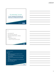
Download Infection Prevention and Control Guidelines for Anesthesia Care
Transcript
American Association of Nurse Anesthetists 222 South Prospect Avenue Park Ridge, IL 60068 www.aana.com Infection Prevention and Control Guidelines for Anesthesia Care Table of Contents Introduction ................................................................................................................................................. 2 Standard Precautions ................................................................................................................................. 3 Hand Hygiene .............................................................................................................................................. 3 Personal Protective Equipment ................................................................................................................. 4 Transmission-Based Precautions ............................................................................................................... 8 Respiratory Hygiene ................................................................................................................................... 9 Skin Preparation ......................................................................................................................................... 9 Aseptic Technique .................................................................................................................................... 10 Airway Management: Considerations Specific to Anesthesia Professionals ...................................... 10 Safe Injection Practices ........................................................................................................................... 11 Drug Preparation and Administration: USP Chapter <797> Compounding ................................. 11 Needle and Syringe Use ................................................................................................................. 12 Gels, Lubricants, and Ointments .................................................................................................... 13 Equipment and Environmental Cleaning, Disinfection, and Sterilization ......................................... 13 The Spaulding Disinfection and Sterilization Classification Scheme ........................................... 13 Single-Use Devices and Reprocessed Disposable Equipment ....................................................... 15 The Anesthesia Machine and Breathing System............................................................................ 15 Equipment Considerations for Special Patient Populations ........................................................... 17 Environmental Surfaces ................................................................................................................. 17 Linens and Disposable Drapes ....................................................................................................... 18 Biohazardous Waste Management ................................................................................................. 18 Invasive Procedure Technique................................................................................................................. 18 Considerations for Ultrasound-Guided Procedures ...................................................................... 19 Considerations for Epidural Catheters and Continuous Peripheral Nerve Block Catheters ......... 19 Considerations for Central Venous Catheter Maintenance and Procedures .................................. 19 Considerations for Implanted Ports .............................................................................................. 22 Considerations for Arterial Catheters and Pressure Monitoring Devices ...................................... 22 Vaccinations, Post Exposure Prophylaxis, and Screening .................................................................... 22 Reducing the Risk of Adverse Events ..................................................................................................... 23 Conclusion ................................................................................................................................................. 25 Infection Prevention and Control Glossary ............................................................................................ 26 References .................................................................................................................................................. 29 1 Introduction Effective infection control and prevention protocols reduce the transmission of communicable diseases in all healthcare settings. A major cause of healthcare-associated infections (HAIs) is the lack of consistent compliance by healthcare workers with basic prevention techniques such as hand hygiene.1-3 Failure to follow the principles of aseptic technique as well as ineffective equipment decontamination and surgical site preparation, have contributed to increased rates of surgical site infections (SSIs), catheter-associated urinary tract infections (CAUTIs), ventilator associated pneumonia, and other HAIs.4 Unsafe injection practices and improper reuse of needles, syringes, and single-use devices, as well as the increase in multiple-drug resistant organisms (MDROs) have also contributed to a rise in emerging infections.1 In 2011, there were over 721,000 cases of infections attributed to improper infection control practices in healthcare facilities, accounting for about 75,000 deaths.2 These rates of morbidity and mortality have serious health implications for patients and cost healthcare facilities millions of dollars annually, adding urgency to the adherence to universal infection control practices.5 Healthcare providers have an ethical duty to protect patients and prevent unnecessary harm. In lifethreatening emergencies requiring immediate action, healthcare providers should weigh the relative risk to patient life and determine the most appropriate infection control practice under those circumstances. Following emergency care, review all actions taken and intervene as appropriate to assure that all appropriate infection control guidelines and standards are addressed as soon as possible. Healthcare providers shall document any deviations from these standards (e.g., emergency cases for which informed consent cannot be obtained, surgical interventions or procedures that invalidate application of a monitoring standard) and state the reason for the deviation on the patient’s record. The American Association of Nurse Anesthetists (AANA) supports patient safety through the use of evidence-based infection prevention and control practices. The purpose of these guidelines is to describe infection prevention and control best practices to increase awareness and reduce the risk of patients, Certified Registered Nurse Anesthetists (CRNAs), and other healthcare providers transmitting and acquiring an HAI. These guidelines do not supersede federal, state or local statutes or regulations or facility policy but constitute minimum practice recommendations and considerations. The Centers for Medicare and Medicaid Services (CMS) has developed a comprehensive worksheet to determine facility compliance with the Infection Control Condition of Participation.6,7 2 Standard Precautions Standard precautions are the basic level of infection control protocols that reduce the risk of disease transmission when providing patient care.1,8 Basic standard precautions include, but are not limited to: • • • • • Hand Hygiene Personal Protective Equipment Respiratory Hygiene Safe Injection Practices Equipment and Environmental Cleaning, Disinfection, and Sterilization Anesthesia and other healthcare providers should always refer to their facility’s policy on infection control standard precautions. Hand Hygiene Hand hygiene is the practice of removing microorganisms from hands.9,10 Performing proper hand hygiene significantly reduces the incidence of infection.1,3,9 Table 1 describes when hand hygiene is indicated and Table 2 describes specific hand hygiene definitions and protocols. Table 1. Indications for hand hygiene. Before • Patient contact. • Donning protective equipment. • Performing invasive procedures (e.g., catheter insertion, epidurals, surgery). • • • • • Table 2. Hand hygiene definitions and instructions. Term Definition Washing hands with water and an Antiseptic Handwashing antiseptic agent, (e.g., soap, hand rub). Rubbing non-visibly soiled hands Alcoholwith a product that contains alcohol Based Handrubbing to decontaminate hands. • • • • Surgical Hand Antisepsis • Washing hands with an antiseptic agent before a surgical procedure.2,7,8 • • • • After Contact with patient’s skin and immediate surroundings (e.g., bedside area). Contamination. Contact with body fluids and wounds. Removing protective equipment. Using the restroom. Protocol Wet hands with water, apply antiseptic soap and rub hands together for at least 20 seconds.9 Apply manufacturer recommended amount to palm. Rub hands together covering all surfaces and fingernails until dry. Refrain from contact until hands are completely dry. Remove jewelry (e.g., rings, bracelets, wristwatches) prior to performing surgical hand hygiene.11 Follow manufacturer guidelines for scrub time. Clean under fingernails using a nail cleaner. Keep natural nail length to less than ¼ inch.2,7 Do not wear artificial nails or nail extenders.2 Performing adequate hand hygiene while providing anesthesia care can be challenging due to the nature and intensity of care anesthesia professionals provide.12 Observational studies of anesthesia professionals in the operating room indicate that there are a high number of missed hand hygiene opportunities during patient care.12,13 Given the demands of anesthesia care and proportion of missed hand hygiene opportunities, aggressive strategies are needed to improve hand hygiene among anesthesia professionals. The considered use of single and double exam gloves that may be removed after contamination, the availability of alcohol-based sanitizer in the anesthetizing area, targeted environmental cleaning of the anesthetizing area after each case, and ongoing research to design new methods are each important to control bacterial transmission in the anesthetizing area.12,13 3 Personal Protective Equipment Personal protective equipment (PPE) is specialized clothing or equipment worn for protection against contamination. PPE protects the patient and the healthcare provider from transmitting and contracting infection.1,9,10 Always perform hand hygiene prior to applying PPE, after removing all PPE (except for respirators), and prior to exiting the operating or patient room. While donning PPE, providers should refrain from touching surfaces and their face when possible to prevent the further spread of infection. Table 3 offers examples of PPE and information on how to properly wear, remove, and dispose of the gear. Table 3. PPE examples and guidelines. PPE Indications Disposable • Routine patient Gloves care. (Non• Shared patientSterile)9 provider use of a difficult-to-clean device (e.g., computer keyboards). • • • • Guidelines Remove and replace gloves promptly when contaminated or damaged. This is an important practice to keep anesthetizing locations and patient care areas clean.9 Remove gloves and perform hand hygiene after caring for a patient and between patients.3 Do not use the same pair of gloves for more than one patient. Special considerations, such as pore size and glove composition (e.g., latex), may apply based on patient, provider, or procedure. • • • • • • Disposable Gloves (Sterile)9 • • • • • Surgical procedures.9 Vaginal deliveries. Invasive radiological procedures. Performing vascular access and procedures. Preparing total parental nutrition and chemotherapeutic agents. • • • • Remove and replace gloves promptly when contaminated or damaged. This is an important practice to keep anesthetizing locations and patient care areas clean.9 Remove gloves and perform hand hygiene after caring for a patient and between patients.3 Do not use the same pair of gloves for more than one patient. Special considerations, such as pore size and glove composition (e.g., latex), may apply based on patient, provider, or procedure. • • • • • 4 Removal Protocol Grasp outer edge of glove near wrist.1,9 Peel away from hand turning inside out. Hold removed glove in opposite gloved hand. Slide ungloved finger under wrist of gloved hand so finger is inside gloved area. Peel off the glove from inside creating a ‘bag’ for both gloves. Dispose of gloves in proper waste receptacle. Partially remove the first glove by peeling it back with fingers of the opposite hand (all five fingers should still be covered with the glove).9 Remove the other glove completely, turning it inside out, only touching the outside of the glove with the covered fingers of the partially gloved hand. Remove the glove on the partially gloved hand completely, using the inside out removed glove.9 Skin is only contacted by the inner surface of the glove.9 Dispose of gloves in proper waste PPE Double Gloves Indications • Regional neuraxial techniques.14 • Airway manipulation.15 • Increased risk of complications from needle stick injuries (e.g., HIV, Hepatitis C contamination).16,1 7 Guidelines • • • • • • Gowns (nonsterile) • Risk of limb contamination.1,12 • • • After performing the planned intervention, immediately remove and safely dispose of the outer gloves. Remove and replace gloves promptly when contaminated or damaged. This is an important practice to keep anesthetizing locations and patient care areas clean.9 Remove gloves and perform hand hygiene after caring for a patient and between patients.1,3 Do not use the same pair of gloves for more than one patient. Resume urgent patient care activities (e.g., patient ventilation) with sterile, inner gloved hands. Special considerations, such as pore size and glove composition (e.g., latex), may apply based on patient, provider, or procedure. Wear a gown that provides appropriate coverage.1,18 Secure gown in the back of the neck and waist. Discard after each use. Removal Protocol receptacle. • • • • • • • • Gowns (sterile) • • Eye Protection • Insertion of pulmonary artery catheters and central venous catheters. Invasive procedures (e.g., surgery). Potential for • • • Wear a gown that provides appropriate coverage.1,18 Secure gown in the back of the neck and waist. Discard after each use. • • • • Select appropriate eye protection based on the type of 5 • First remove the outer glove by following the protocols for sterile glove removal. Remove other PPE equipment. Remove inner glove following the protocols for sterile glove removal. Perform hand hygiene. Unfasten ties in back of neck and waist. Remove the gown touching only the inside of the gown. Roll or fold gown inside out. Dispose of gown in proper waste receptacle. If donning double gloves, dispose of outer glove following sterile glove removal protocol prior to removing gown. Follow removal protocol for nonsterile gowns. Dispose of gown in proper waste receptacle. If donning double gloves, dispose of PPE Surgical Masks Indications contact with infectious material. • Splash or spray hazards. • • • Hair Coverings • • Shoe Coverings • Invasive procedures (e.g., arterial and central venous access, regional anesthesia). Regional neuraxial technique.14,19 Potential for contact with infectious material. Upon entry to semi-restricted and restricted areas. Regional neuraxial technique.14,19 Risk of splash contamination. • • • • • • • • Guidelines hazard exposure, the duration of exposure, and the availability of other PPE. Pretest selected eye protection for suitability and appropriate fit. Clean and disinfect nondisposable eyewear prior to use (e.g., laser glasses, goggles, N95 respirator, face shields). • • • Wear to cover facial hair. The surgical mask should cover the mouth and nose and be secured in a manner that prevents venting at the sides of the mask.20 Remove and discard when wet or soiled, and at the end of a case or procedure.1,21,22 Perform hand hygiene immediately following mask removal and disposal. • Cover hair, facial hair, sideburns and the back of the neck using a clean covering.23,24 Launder reusable cloth caps daily and when visibly soiled. • • • • • • • Slip coverings over shoes prior to donning gloves and other PPE. Shoe coverings must be changed each time a worker exits the area. 6 • • Removal Protocol outer glove following sterile glove removal protocol prior to removing eye protection. Lift head band or ear piece. Refrain from touching the face shield. Dispose of eye protection in proper receptacle for reprocessing or disposal. If donning double gloves, dispose of outer glove following sterile glove removal protocol prior to removing surgical mask. Undo the ties or grasp the elastics at the top and bottom of the mask and remove without touching the front of the mask. Dispose of mask in proper waste receptacle. If donning double gloves, dispose of outer glove following sterile glove removal protocol prior to removing surgical cap. Remove cap using gloves, refraining from contacting inner part of cap. Dispose of cap in proper waste receptacle. If donning double gloves, dispose of outer glove following sterile glove removal protocol prior to removing shoe covers. With already donned gloves, remove PPE Indications Guidelines • • Removal Protocol shoe coverings. Dispose of coverings in proper waste receptacle. Spray shoes with disinfectant if necessary. Scrubs • Follow facility policy regarding donning scrubs prior to entering restricted and semi-restricted areas. • Wear a clean set of scrubs each day and change into clean scrubs if contaminated. o Home-laundering scrubs is acceptable if they have not been contaminated with blood or infectious material.25 o Launder in hot water with sodium hypochlorite and detergent. Dry using high heat.26,27 • Follow your facility policy regarding removal of scrubs upon exiting restricted and semi-restricted areas. Cover Apparel (e.g., lab coats) • Follow facility policy regarding use of cover apparel. • • Cover apparel should be clean or single-use.23 Lab coats are not recommended in the operating room, as they have the potential to become contaminated.23,28 • Follow your facility policy regarding removal of lab coats upon entering and exiting restricted and semirestricted areas. Launder cover apparel after each daily usage and when contaminated.23 • 7 Transmission-Based Precautions In addition to standard precautions, transmission-based precautions should always be followed once a patient develops symptoms of an infection to reduce opportunities for disease transmission.1 The three categories of transmission-based precautions include contact, droplet, and airborne precautions. Because diagnostic tests are often required to confirm an infection and generally require a few days for conclusive results, precautionary measures should be taken until the presence or absence of infection is confirmed.1 Table 4 describes protocols and examples of transmission-based precautions. Table 4. Transmission-based precautions.1,29 Precaution Description Contact Prevents • transmission of • infectious agents spread by contact • with the patient or environment. • Droplet Prevents transmission of infectious agents spread by close contact with respiratory secretions. Protocol Use single-patient rooms when possible. Maintain ≥ three feet spatial separation between beds in rooms with more than one patient. Wear a gown and gloves for all contact with the patient or the patient’s environment. Wear PPE before entering the patient’s room and discard it before exiting the patient’s room. Examples Include, but not limited to: • Clostridium difficile* • Norovirus* • Scabies • • Include, but not limited Use single-patient rooms when possible. to: Maintain ≥ three feet spatial separation between • Influenza beds in rooms with more than one patient. • Pertussis • Wear a gown, gloves and mask for all contact • Mumps with the patient or the patient’s environment. • Rubella • Wear PPE before entering the patient’s room and discard it before exiting the patient’s room. • Place a facemask on the patient during transport. Airborne Prevents Include, but not limited • Place patients in an airborne infection isolation transmission of room designed with monitored negative pressure, to: infectious agents • M. tuberculosis 12 air exchanges per hour, and air exhausted suspended in the directly to the outside or recirculated through • Measles air. high-efficiency particulate air filtration. • Varicella • Facilities should establish a respiratory protection program. • Isolate N95 or higher level masked patients in a private room when airborne precautions cannot be achieved. • Healthcare workers should don gloves, gowns, and N95 mask upon entering an infectious patient’s room. • Immune healthcare workers are the preferred providers for infectious patients with airborne diseases. *Facilities should consider use of a hypochlorite solution for environmental cleaning as an additional contact precaution. During heightened periods of virulent and highly contagious infectious outbreaks (e.g. Ebola virus disease (EVD), Enterovirus), healthcare providers are encouraged to refer to the following resources for supplemental information regarding transmission-based precautions: • Local and/or state health departments. • Centers for Disease Control and Prevention (CDC) (http://www.cdc.gov/). • Society for Healthcare Epidemiology of America (http://www.shea-online.org/). • Association for Professionals in Infection Control and Epidemiology (http://www.apic.org/). • AANA Practice Committee (www.aana.com/). 8 Respiratory Hygiene Respiratory hygiene includes cough etiquette and the appropriate use of isolation precautions to prevent the spread of infection.30 Perform the following measures for cough etiquette when afflicted with a respiratory disease:30,31 • Cover mouth and nose with a tissue when coughing or sneezing. • Dispose of tissue after use in the waste bin. • Perform hand hygiene following contact with respiratory secretions. • Do not perform patient care when infected or ill. During periods of elevated respiratory infection incidence, facilities may offer facemasks to patients and healthcare providers who are coughing and take additional transmission-based precautions as necessary.30,31 Skin Preparation Preparing the patient’s skin prior to performing clinical procedures significantly reduces the risk of infection. Individuals should always follow manufacturer recommendations and their facility policy for the proper use of skin prep agents. An ideal skin prep agent should decrease microorganism count, inhibit rebound and regrowth of microorganisms, activate quickly, and be effective against a variety of microorganisms.32 Each prep agent has a specific mechanism of action along with specific advantages and disadvantages that should be weighed in all clinical situations.32 The patient’s allergies, skin condition, and other contraindications as well as the site of the procedure should be considered prior to applying the agent. Table 5 provides examples of skin prep agents as well as advantages and disadvantages to use. Table 5. Skin prep agent examples, descriptions and recommendations. Agent Description and Recommendations Chlorhexidine • Preferred skin prep agent due to immediate action, residual activity, and gluconate persistent effectiveness against a wide range of microorganisms.32,33 • Strong tendency to bind to tissue, contributing to extended anti-microbial action.33 • Highly effective in the presence of blood and organic material.32 • Addition of alcohol to the disinfectant provides more rapid and effective germicidal activity.32,33 • Limited sporicidal activity.33 • Not recommended for use on eyes, ears, brain and spinal tissues, mucus membranes, or genitalia.32 • Concentrations > 0.5 percent not recommended for procedures such as epidurals and other neuraxial procedures due to neurotoxicity.34 Povidone-iodine • Suitable alternative when Chlorhexidine is contraindicated.33 • Highly effective against a broad range of microorganisms and acts immediately.32,33 • Safe to use on face, head, mucous membranes, vaginal area and during other neuraxial procedures.33 • Minimally persistent compared to Chlorhexidine.33 • Limited residual activity.32 • Decreased effectiveness in the presence of blood and organic material.32 Parachoroxylenol • Less effective than chlorhexidine gluconate and povidone-iodine at eliminating microorganisms.32 • Moderately effective against a broad range of mircoorganisms.33 9 Agent • • • Iodine-base with alcohol • • • Description and Recommendations Moderate persistent/residual activity. Nontoxic with no tissue contraindications.32 Remains effective in the presence of blood and organic material and in the presence of saline solution.32 Highly effective against a broad range of microorganisms.33 Acts immediately.32,33 Highly flammable.32 Fire Risk: Agents that are alcohol-based or have flammable properties have the potential to increase the risk of surgical fires. Aseptic Technique Aseptic technique requires multiple methods to prevent the transmission of microorganisms from the environment, healthcare provider, and patient.35 Table 6 refers to recommendations for aseptic procedure. Table 6. Guidelines for aseptic technique.35 Precaution Guidelines May include some or all of the following items depending on the Equipment (Maximal sterile procedure: barriers) • Sterile gloves • Sterile gowns • Surgical masks • Sterile drapes Preparation • Antiseptic skin preparation of patient prior to procedure. o Consult manufacturer product instructions for directions and warnings regarding the proper use and application of specific skin antiseptics such as chlorhexidine-alcohol or povidone-iodine. • Ensure that all instruments, equipment, and devices are sterile. Environmental • Close doors during operative procedures. Controls • Minimize unnecessary staff and traffic in/out of operating room. Contact • Precautions should be taken to mitigate contact with non-sterile surfaces and objects. Airway Management: Considerations Specific to Anesthesia Professionals Airway management poses unique challenges to anesthesia practitioners in limiting or preventing environmental contamination. In order to mitigate disease transmission while ensuring the standard of care for proper airway management, the following practices are recommended: • • • • Maintenance of oxygenation takes priority over all issues.36 Ventilate the patient immediately upon airway manipulation. o CDC guidelines indicate the need to remove gloves, wash hands, and don new gloves, which would conflict with the standard of clinical care for airway instrumentation and maintenance.37 Immediately following maneuvers undertaken to establish a patent airway, the patient should be ventilated manually, the breath sounds auscultated, and the expired breath examined for presence of expired carbon dioxide. It is recommended that anesthesia practitioners consider double gloving prior to airway manipulation.15 10 Following tube or device insertion, remove contaminated outer gloves and perform necessary actions to assure airway security and patency. When the situation is stable, remove the inner gloves, perform hand hygiene, and don clean gloves to continue with patient care. Targeted environmental cleaning of the anesthetizing area after each case, and ongoing research to design new methods are each important to control bacterial transmission in the anesthetizing area.12,13 o • • Safe Injection Practices Improper injection practices put patients and healthcare providers at risk of infection from bloodborne pathogens, which can lead to the spread of HAIs.1,38-41 Following safe injection practices can prevent the spread of disease. These measures can also protect providers from disciplinary action and legal recourse.40,42,43 Drug Preparation and Administration - USP Chapter <797> Sterile Compounding The U.S. Pharmacopeia Convention (USP) is a scientific nonprofit organization responsible for defining standards for medicines and other products using a system of standards and quality control along with a national drug formulary. USP Chapter <797> is not law, but is an accepted guideline for best practices for compounding sterile preparations (CSPs).44 USP General Chapter <797>, Pharmaceutical Compounding – Sterile Preparations,45 describes conditions and practices for preparing CSPs.46 These guidelines apply to all healthcare providers administering CSPs within an institution when that institution has adopted use of Chapter <797>. Federal, state, and local statutes and regulations and accreditation standards may also require compliance with USP <797> guidelines. Anesthesia professionals should ensure compliance with applicable statutes, regulations, accreditation requirements, and facility policies in the preparation of CSPs. The following summarizes USP Chapter <797> as it applies to anesthesia professionals: • • • All CSPs must be compounded with aseptic manipulations entirely within an ISO Class 5 (using a containment hood or compounding aseptic isolator) or better air quality environment.44,45,47,48 o The only exception to this is the “immediate-use provision” designed for the following situations: Cardiopulmonary resuscitation Emergency room treatment Preparation of diagnostic agents Critical therapy where normal CSP preparation would cause more harm to the patient due to delay Chapter <797> categorizes CSPs into three risk levels (low, medium, and high) and sets preparation standards for each level. o Risk levels are defined according to the probability of CSP contamination.45 Anesthesia medications may meet the “immediate use provision” if the delay from preparation of CSPs following the preparation standards of a low-risk level drug would render additional risk to the patient.44 o Medium- and high-risk CSPs cannot be prepared under the immediate-use provision. 45,47,48 o CSPs prepared in accordance with the immediate-use exception may not be stored or prepared by batch compounding. Daily anesthesia workflow makes the immediate-use provision challenging to meet as providers are prohibited from batch medication preparation.44 11 o The following criteria for low-risk CSPs must be met to qualify for the immediate-use provision: The CSP should have no more than three commercially manufactured packages of sterile nonhazardous products from the manufacturer’s original container, and no more than two entries into a sterile administration container/device or sterile infusion solution.45 The compounding procedure is continuous and does not exceed one hour.44,45 Aseptic technique is followed and the prepared CSP is under continuous supervision until administered. Administration begins no later than one hour following the start of the CSP preparation.44,45 The CSP must be labeled with patient identification information, the names and amounts of all ingredients, the name or initials of the CSP preparer, and the exact beyond-use date and time, unless the CSP is immediately and completely administered by the CSP preparer or unless immediate and complete administration of the CSP is overseen by another preparer.44,45 If the prepared CSP administration has not started within one hour following the start of preparation, the CSP must be promptly, properly, and safely discarded.44,45 All personnel involved in compounding should understand how they may contribute to the risk of CSP contamination during preparation. To decrease the risk of contamination, many hospital pharmacies commonly prepare medications used in delivery (e.g., phenylephrine) or buy ready-to-use, prefilled medications (e.g., fentanyl, sufentanil). Anesthesia professionals should prepare CSPs using proper aseptic technique.44,45 Needle and Syringe Use • Avoid recapping of needles and discard used needles and syringes into a puncture-resistant sharps container.39,40,49 • Consult the AANA Safe Injection Guidelines for Needle and Syringe Use41 and the CDC recommendations for safe injection practice39,49 for more complete guidance. Syringes, Needles, and Needleless Access Devices • Use syringes, needles, and needleless access devices only once.43,49 • Do not refill a syringe once used, even for the same patient.43,49 • Efforts should be made to keep syringes prepared for single patient use under direct observation, or locked securely, with a patient identification label attached. Infusion Sets, Bags, and Pumps • Use infusion, pump syringe, and intravenous administration sets only once.39 • Do not use bags or bottles of intravenous solution as a common source of diluent for multiple patients.39,50 • Clean and process intravenous infusion and syringe pumps according to manufacturer recommendations between patients. Medication Vials and Ampules • Prevent coring and particulate contamination by applying in-line final filtration using a 45µ rater.51 • Use 70 percent alcohol to clean the access diaphragm of medication vial or to clean the outside of an ampule prior to insertion of a device or needle into the vial.1,50 • Use 70 percent alcohol to clean the diaphragm prior to access when removing the cap from a new vial.50 12 • Handle and discard medications according to facility policy and manufacturer guidelines. Single-dose Vials • Use single-dose vials for medications when possible.39,40,50 • Do not combine or save leftover medications from single-dose vials/ampules for later use.39,40 • Discard single-dose medication vials, ampules, and intravenous infusion bags safely after use on a single patient.40,50 Multi-dose Vials • Dedicate multi-dose vials to a single patient when possible.40,52 • Use a syringe or needle only once to withdraw medication from a multi-dose vial. o Label the date on the multi-dose vial once opened.52 • Do not keep multi-dose vials in the immediate patient treatment area (e.g., patient rooms or bays, operating rooms, anesthesia carts).39,52 o If a multi-dose medication vial enters a patient treatment area, it should be treated as a single-use vial and discarded at the end of the individual case.38,39 • Discard multi-use medication vials if sterility is compromised or questionable.38,52 • Discard multi-use medication vials within 28 days of opening.38,52 o If the manufacturer-labelled expiration date falls within 28 days of opening, discard the vial prior to the manufacturer expiration date.38,52,53 Gels, Lubricants, and Ointments • Dedicate ointments, gels, and lubricants to a single patient when possible. • Use sterile skin prep agents when indicated. Equipment and Environmental Cleaning, Disinfection, and Sterilization The following information regarding equipment and environmental cleaning, disinfection, and sterilization is not intended to be comprehensive. Review the CDC Guideline for Disinfection and Sterilization in Healthcare Facilities 2008,54 federal, state or local statutes and regulations, equipment manufacturer recommendations, and facility policy and procedures as the best sources for current evidence-based practice guidelines.54 The following are general considerations for equipment and environmental cleaning and should not substitute review and adherence to previous referenced resources: • Facilities should develop an infection control policy and a method for monitoring compliance that specifies appropriate disinfection and sterilization protocols for anesthesia equipment.54-56 • Facilities should select disinfectants or detergents registered with the U.S. Environmental Protection Agency (EPA) and follow manufacturer recommendations regarding use, exposure time, and disposal.57 • Anesthesia equipment should be adequately cleaned prior to disinfection and sterilization.57 • The amount of personal equipment (e.g., stethoscopes) and belongings (e.g., jackets, backpacks, bags, purses, personal electronic devices) brought into the operating room and/or patient care areas should be minimized. The Spaulding Disinfection and Sterilization Classification Scheme The Spaulding scheme classifies disinfection and sterilization methods for medical equipment by the risk of infection involved.54,55 View the details of the classification scheme in Table 7. 13 Table 7. Spaulding Disinfection and Sterilization Classification Scheme. Device Device Example(s) Process Classification Surgical instruments, Sterilization Critical • Contact sterile angiocatheters tissue or the vascular system. • • • Semi-critical Contact mucous membranes or non-intact skin. Anesthesia and respiratory therapy equipment, breathing circuits, endotracheal tubes, endoscopes, laryngoscopes, fiberoptic scopes, Magill forceps, cystoscopes Laryngoscope blades High- level disinfection • • • • • • • • Laryngoscope handles Non-critical Contact intact skin. Patient Care Items: Electronic devices, stethoscopes, blood pressure cuffs, arm board, nametags, pulse oximeter sensors, head straps, monitor cables, blood warmers, medication Intermediate or low-level disinfection • • 14 Recommendation Sterilize devices with sterilants that destroy all vegetative bacteria, nonlipid viruses and bacterial spores. Rinse with sterile water.56 Medical devices can be sterilized using chemical or physical properties depending on degree of contact with the patient.57 Chemical germicides should be used rationally and in accordance with manufacturer recommendations and facility policy.57 Clean and disinfect devices with high-level disinfectants to destroy all vegetative bacteria and nonlipid viruses. Rinse with sterile water.56 Dry all equipment surfaces to prevent humidity from encouraging microorganism growth.57 Wrap laryngoscope blades individually.58,59 If high-level disinfection is used, a closed plastic bag may be used for storage. If steam sterilized, a peel pack may be used for storage.59 Partially remove the blade from the package, attach to light source, and test, or keep the blade covered - manipulation of the blade onto the light source/handle can be tested without actually removing the blade from the bag or pack without touching the blade itself.59 Following testing, insert the blade back into the package and return to a clean storage location. This protocol applies to disposable blades as well.59 At a minimum, wipe the handle with an intermediate--level disinfectant after use. This protocol applies to disposable handles as well.58,59 Clean all equipment between patients and when visibly soiled in accordance with manufacturer recommendations and facility policy. o Low and intermediate-level disinfection differs by disinfectant type, concentration, and exposure to pathogen.54,60 Stethoscopes may be washed with water and Device Classification Device Example(s) Process administration pumps, carts, beds and monitors Recommendation • • Environmental Surfaces: Bed rails, food utensils, bedside furniture, computer keyboards, floors, mobile devices Low-level disinfection (unless otherwise noted) • • wiped with alcohol.56 Use protective covering for non-critical surfaces that are difficult to clean (e.g., keyboard covers).25 Hydrogen peroxide gas decontamination is an effective sterilization method for reusable items that are difficult to clean.61 Clean all equipment between patients and when visibly soiled in accordance with manufacturer recommendations and facility policy. Use protective covering for non-critical surfaces that are difficult to clean (e.g., keyboard covers).25 Single-Use Devices and Reprocessed Disposable Equipment • A single-use device is a medical device that is only to be used on one patient for a single procedure.62 Numerous studies have linked outbreaks of infection to the use of improperly reprocessed single-use devices.54,63 • Reuse of single-use devices may expose healthcare providers and facilities to additional liability.64 • Refer to the FDA for guidance and information on reprocessed single-use devices.54,65 To mitigate incidence of outbreaks, it is recommended that healthcare facilities: • Establish a policy to verify the cleanliness and functionality of reprocessed disposable equipment prior to use.56 • Disassemble, clean, dry, reassemble, repackage, and disinfect or sterilize reprocessed, disposable equipment prior to use as appropriate.56 The Anesthesia Machine and Breathing System Although there is no direct contact between anesthesia machine controls and the patient, microorganisms can be transferred between the machine and patient by the healthcare provider.66 Refer to federal, state or local statutes and regulations and facility policies as well as specific manufacturer instructions for guidance concerning: • • • Cleaning and disinfecting the anesthesia machine.57 Pasteurizing or autoclaving of valves.56 Disassembling and disinfecting adjustable pressure-limiting valves.57 Anesthesia Machine Surfaces and Carts • Clean, then spray or wipe anesthesia machine surfaces and knobs with an appropriate germicide between cases and at the end of each day.55,56,66 • Take protective measures to prevent materials stored on the anesthesia machine from becoming inadvertently contaminated by airborne debris (e.g., blood). • Remove equipment from drawers, clean and disinfect drawers regularly.56 • Place a clean covering on the top of the anesthesia cart at the beginning of each case.56 • Wipe small surfaces with 70 percent isopropyl alcohol to reduce bacterial contamination.67 • Clean carbon dioxide and soda lime absorbers when the absorber is changed and remove debris from the screens. 15 Anesthesia Breathing System Review the user manual to determine manufacturer cleaning recommendations for the breathing system. Filters Breathing system filters are single-use items that are assessed according to their bacterial filtration efficiency (BFE) and viral filtration efficiency (VFE).57,68 The efficacy of filtration for bacterial contaminants is higher than for viral particles.56,57 Filters may prove problematic during spontaneous respiration due to increased resistance to air flow.56 Aside from patients with an active Myobacterium. tuberculosis infection, no recommendation is made for the routine use of breathing system filters due to inconclusive data demonstrating their efficacy in reducing the risk of patient infection.25 However, when a patient with a respiratory infection must be given inhalational anesthesia, a filter should be used.56 • Practitioners may choose to place a high-efficiency filter on the inspiratory limb of the breathing circuit to protect the patient from the anesthesia machine, and to place a highefficiency filter in the expiratory limb to protect the anesthesia machine from the patient. • Filters may be interposed between the endotracheal tube and the Y-piece.57 • Use circuit filters and follow-up with post-anesthesia machine disinfection after caring for patients with known pulmonary infection or trauma.57,69 Carbon Dioxide Absorbers • Follow the manufacturer instructions for disassembly, cleaning, and sterilization of carbon dioxide absorbers. • Clean canisters when the absorbent is changed and carefully remove debris from the screens.56 • Discard disposable plastic canisters. • Bellows, unidirectional valves, and carbon dioxide absorbers should be cleaned and disinfected periodically.57,69 Circuits Anesthesia circuits may be manufactured as either single patient use items or multiple patient use items (provided that a new breathing system filter is placed between the Y-piece and endotracheal tube after sterilization or high level disinfection).57 Anesthesia professionals should pay close attention to anesthesia circuit product labeling.56,57 • At a minimum, provide high-level disinfection for multiple-patient use breathing circuits.54 o If available, ultrasonic cleaning is effective.57 • The outer surface of the circuit can become easily contaminated when the system is not changed between patients and therefore should be disinfected between each use.54 • End- tidal carbon dioxide tubing should be changed between patients.70,71 • Following anesthesia care of a patient with pulmonary infection or trauma, disinfection of the internal and respiratory system anesthesia machine components is mandatory. Heat and Moisture Exchangers • Heat and moisture exchangers alone are not effective in decreasing the transmission of microorganisms to the anesthesia breathing system.68,72 Supraglottic Airway Devices • If possible, use disposable single- use device laryngeal mask airways (LMAs) due to the extreme difficulty in completely eradicating protein deposits from reusable LMAs.73-77 16 • • Reusable LMAs should be rinsed and soaked in enzymatic detergent prior to autoclaving to remove occult blood. o Numerous studies have demonstrated that protein deposits are extremely difficult to eradicate completely from reusable LMAs.73-77 Consult manufacturer directions for cleaning and sterilizing supraglottic airway devices. Equipment Considerations for Special Patient Populations Creutzfeldt-Jakob Disease56,78-81 Multiple-use devices used on patients with Creutzfeldt-Jakob Disease (CJD) may transmit the disease. To properly disinfect equipment, consult the following recommendations: • Use disposable equipment when possible for patients with CJD; incinerate equipment after use.78,79 • Destroy laryngoscopes and supraglottic devices used on patients with CJD. • Safely discard devices that are difficult or impossible to clean. • Clean and perform steam sterilization of instruments for 30 to 60 minutes at 132° C.56 • Perform steam sterilization for 18 minutes at 134° C-138° C when using a prevacuum sterilizer.56 o Immerse instruments in 1N sodium hydroxide solution for one hour at room temperature followed by steam sterilization for 30 minutes at 121° C as an alternative to the prevacuum sterilizer.56 • Disinfect noncritical items and environmental surfaces with bleach or 1N sodium hydroxide for 15 minutes at room temperature.56 • Consult the CDC recommendations for best infection control practices when working with patients with CJD.78,82 Tuberculosis • Place a high-efficiency particulate air (HEPA) filter between the breathing system and the patient.56 • Sterilize or perform high-level disinfection on equipment used on patients with cases of suspected or confirmed Tuberculosis.56 • Culturing anesthesia equipment is not required.56 Environmental Surfaces Facilities should establish a routine disinfection policy for environmental surfaces and a program for monitoring compliance and performance improvement. The policy should include the frequency and level (i.e., high-level, low-level) of disinfection and a list of the facility-approved EPA-registered disinfectants or detergents.55 • Thoroughly clean environmental surfaces to reduce transmission of HAIs from surfaces to providers and patients.83 • Clean anesthetizing locations and equipment surfaces (e.g., intravenous and epidural pumps, blood glucose meters and other point-of-care devices, stand-alone monitors, blood and fluid warmers, forced air warmers) between cases and at the end of each day in accordance with facility-specific policies.55 • Follow manufacturer recommendations regarding use, exposure time, and disposal of disinfectants and sterilants. • Place items that may be used during the next case on clean surfaces. • Consult the CDC recommendations for standard precautions and transmission-based precautions for additional guidance.1,27 17 Linens and Disposable Drapes Handle linens and other disposable drapes in a manner that limits the transfer of blood and microorganisms.84 • Handle contaminated laundry as little as possible. • Place and transport the laundry in labeled or color-coded bags or containers. • Do not sort or rinse contaminated laundry. Avoid body contact with soiled items. • When standard precautions are applied to the handling of soiled laundry, alternative labeling or • color-coding is sufficient if it permits all personnel to recognize the container(s) as meeting compliance. • Store laundered items in a clean, dry area to prevent contamination by dust or other particles. Biohazardous Waste Management Biohazardous waste refers to any item that is contaminated with infectious or potentially infectious materials. Sharps disposal is of particular concern due to the potential for injury when handling (e.g., needles, scalpel blades, drill bits, glass items). • Dispose of all regulated waste in specified biohazard waste receptacles following federal, state and local statutes and regulations. • If a biohazardous waste container becomes contaminated, place the container inside of another biohazardous waste container.85 • Consult relevant EPA documents for specific guidance.86 Single-Use Items Discard disposable single-use devices in a biohazardous bag/container (e.g., breathing circuits, airway devices, orogastric tubes) immediately after use. Reprocessed Items • Place items that will be reprocessed in a plastic bag or container immediately after use. • Close containers prior to removing from the anesthetizing location. Sharps Sharps include any device that may puncture skin (e.g., needles, syringes, scalpels, lancets, blades, glass). • Use safety devices when possible. • Do not bend or recap contaminated needles. If a needle must be bent, use the one-handed technique.1 • Discard sharps immediately in a closeable sharps container. Drug Disposal Follow facility policy and applicable federal, state, and local statutes and regulations regarding the appropriate method for disposal of partially remaining drugs in vials, ampules, syringes, and IV bags. Invasive Procedure Technique Invasive procedures such as catheter insertion often expose patients and healthcare providers to heightened risk of exposure and infection.1 Ensuring that the proper measures are taken prior to performing invasive procedures will help prevent adverse events such as surgical site infections, central line-associated bloodstream infections, and catheter-associated urinary tract infections. Healthcare providers should perform hand hygiene before assembling equipment as well as before and after performing the procedure. All invasive procedures should be performed using aseptic technique and in accordance with facility policy. 18 Considerations for Ultrasound-Guided Procedures Ultrasound guidance for procedures such as vascular access and catheter placement has been shown to reduce infection rates and improve patient satisfaction.87 • Site selection should consider factors such as vessel size, depth, course, surrounding structures, and adjacent pathology prior to access.87 • Prepare patient skin with appropriate agent.87 o Use of single-use containers/sachets as multi-use bottles can result in bacterial contamination.88 • Use a sterile sheath, sterile probe covers, and sterile ultrasound gel to mitigate the risk of contamination. • Disinfect ultrasound probes between each procedure and patient.89 o Direct application of non-manufacturer-approved cleaning solutions to the transducer may result in damage. Considerations for Epidural Catheters and Continuous Peripheral Nerve Block Catheters • Adhere to strict aseptic technique and use single-use sterile gel to prevent contamination during catheter placement.90 • Don maximal sterile barriers, especially surgical masks, during procedure.19,91 • Prepare patient skin with an appropriate agent.34,92-96 • Dress the insertion site with a sterile transparent, occlusive dressing.90,93 o Use chlorhexidine-impregnated dressings at insertion sites to reduce epidural skin entrypoint colonization.97,98 • Check the insertion site and overall patient status at least daily for early identification of superficial infection (e.g., erythema, tenderness, itching at the site), deep infection (e.g., fever, back pain, lower limb weakness, headache), and sensory motor status.90,99 • Remove once no longer clinically indicated. Disconnected Catheters The use of an epidural catheter for a prolonged period of time increases the risk of becoming disconnected from the insertion site, which heightens the risk of infection.100 The choice to reconnect or remove the catheter is at the discretion of the anesthesia professional if not addressed in facility policy. Factors to be considered include the potential of contamination and patient-specific riskbenefit ratios.100,101 When a disconnected catheter is discovered and static fluid has moved more than five inches from the disconnected end, the catheter should be removed.102 Considerations for Central Venous Catheter Maintenance and Procedures Central venous catheters (CVCs), also known as central lines, are used to administer medications, provide fluids for nutrition, and conduct medical tests.103 Manufacturer recommendations and facility policies should be followed for specific care and maintenance of CVCs. Table 8 describes the different types of CVCs. Table 8. Examples and descriptions of Central Venous Catheters (CVCs).103 Catheter Description Tunneled catheter (e.g., Hickman, • Surgically inserted for extended use (months to Groshong®) years). • Catheter and attachments emerge from underneath the skin. Non-tunneled catheter (e.g., Quinton) • Percutanesously inserted for shorter use (1-2 weeks). • Catheter attachments protrude directly. Peripherally-Inserted Central Catheter • Inserted into a peripheral vein in the arm. 19 (PICC ) Implanted Port • • Inserted entirely under the skin. Medications administered through blunt needle (e.g., Huber needle) placed through the skin to the catheter. Central Venous Catheter Insertion In order to reduce the incidence of infections such as central line-associated bloodstream infections, the following is recommended for the proper insertion of a central line: • Consider the risks and benefits of placing a central line at various sites (e.g., subclavian, peripheral, jugular, femoral) before insertion.104 • Perform hand hygiene and don sterile gloves, sterile gown, surgical cap, and surgical mask, and cover the patient’s entire body with a large sterile drape prior to insertion.105 • Prepare patient skin using appropriate agent.105 • Use antibiotic-impregnated catheter if the catheter is to remain in place for longer than five days.106,107 • Replace catheter when adherence to aseptic technique cannot be ensured (e.g., catheters inserted during a medical emergency). Otherwise, do not routinely replace CVCs.104 • Remove any intravascular catheter once it is no longer indicated.106 • For complete guidance, refer to the CDC Guidelines for the Prevention of Intravascular CatheterRelated Infections.104 Central Venous Catheter Access When accessing central venous catheters, closed access systems are preferred in addition to the following recommendations: • Scrub the injection cap (e.g., needleless connector) with an appropriate antiseptic agent and allow to dry according to manufacturer recommendation.108,34 o Povidone-iodine is the recommended agent for children < two months old.109 • Access the injection port with the syringe or intravenous tubing.108 o If necessary, open the clamp.108 Flushing Technique Refer to the manufacturer instructions for the catheter and the needleless connector for the appropriate technique to use; unless otherwise specified, perform the following: • The type of flush (e.g., saline, heparin, dilute heparin), concentration, volume, and frequency of flushing should be determined in accordance with manufacturer indications for use and facility policy and per the treating clinician’s orders. Individualized patient needs should also be considered.108,110 • Use a single-use flushing system (e.g., single-dose vials, prefilled syringes).108 o At a minimum, use a 10 mL syringe.111 • Flush the catheter vigorously using a positive pressure technique by maintaining pressure at the end of the flush to prevent reflux.108 Positive Pressure Technique108 This technique may not apply to neutral-displacement or positive-displacement needleless connectors: • Flush the catheter, continue to hold the plunger of the syringe while closing the clamp on the catheter, and then disconnect the syringe.108 • Withdraw the syringe as the last 0.5-1 mL of fluid is flushed when using catheters without a clamp.108 20 Heparin Flushes • Flushing CVCs with heparin solutions is a recommended practice in many guidelines despite the lack of conclusive evidence of efficacy and safety compared with 0.9 percent normal saline flushing.110 • Heparin flushes are appropriate for maintaining patency of CVCs for dialysis.112 o Higher concentrations of heparin should be used for patients who have evidence of occlusion or thrombosis.112 o The injected volume of the heparin flush should not exceed the internal volume of the catheter.112 Assessing Placement and Patency • Aspirate catheter for blood return to identify correct placement of the catheter within the vein, indicated by blood return in syringe.113 • Clear line of hemoglobin to prevent clotting in catheter. • Flush immediately with saline after aspirating to assess for patency and detect resistance.113 Specimen Collection • Access the catheter as outlined above, maintaining aseptic technique. • Draw the first 3-5 mL of blood, dispose in an appropriate biohazardous waste receptacle, or return to the patient in accordance with the procedure or as indicated by the patient.108,114 • Before specimen is collected, flush catheter in accordance with facility policy and per the treating clinician’s orders.111 • Discard 1.5-2 times the volume of the internal catheter lumen before drawing the specimen.111 • Collect the specimen.108,114 • Flush the catheter as directed by the procedure and facility policy and per treating clinician’s orders.114 o Clamp the catheter as flushing is completed and promptly dispose of used syringe(s). Changing the Injection Cap (e.g., needleless connector) • When there are signs of contamination (e.g., blood, precipitate), damage (e.g., leaks, septum destruction) change immediately. Unless otherwise indicated by manufacturer recommendation, change injection port cap weekly.108 • Scrub the injection cap and catheter hub with appropriate agent (e.g., chlorhexidine, isopropyl alcohol); clamp the catheter if necessary as cap is removed.108 • Attach a new cap to catheter hub using aseptic technique. Site Dressing • Supplies for site cleansing and dressing are single-use items.108 o Refer to manufacturer recommendations to ensure compatibility with catheter material.108 • Wear clean gloves.108 • Prepare patient skin with appropriate agent.87 o If replacing dressing, remove existing dressing, inspect the site visually, and document prior to skin prep.108 • Except for dialysis patients, do not apply topical antibiotic ointment or cream to catheter site.115,108 • Cover site with either sterile gauze or sterile, transparent, semipermeable dressing.108 • Replace or change dressing when indicated.108,111 21 Considerations for Implanted Ports In addition to the following recommendations, always discuss with the patient the best approach or technique for accessing and deaccessing the patient’s port. Port Access Procedure • Don clean gloves.108 • Examine the port site for complications to look for any swelling, erythema, drainage or leakage, or assess for presence of pain, discomfort, or tenderness.108 • Palpate the outline of the port to identify insertion diaphragm.116 o Mark location on patient skin for blunt needle insertion. • Remove gloves , perform hand hygiene, and don new sterile gloves.108 • Cleanse port site with appropriate agent prior to entry.116 • Stabilize port with one hand, and insert blunt, non-coring needle (e.g., Huber needle) until port backing is felt.108 • Aspirate blood to ensure patency by return.116 • Stabilize needle/port with tape, securement device, or stabilization device. o Apply gauze and tape for short-term use (e.g., outpatient treatment).116 Port Deaccess Procedure • Don clean gloves.108,116 • Flush device in accordance with facility policy and per the treating clinician’s orders.116 • Stabilize port with one hand, and remove needle with the other hand.108,116 • Maintain positive pressure technique on the syringe while deaccessing by flushing the catheter while withdrawing the needle from the septum. • Apply dressing. Port Maintenance and Care • For short-term use in outpatient settings, a light dressing may be used in place of an occlusive dressing during the infusion; ensure the needle is secure in the portal septum as described above.108 • When not in use, implanted ports should be flushed every four to eight weeks to maintain patency.108 Considerations for Arterial Catheters and Pressure Monitoring Devices • Catheters that need to be in place for > five days should not be routinely changed if no evidence of infection is observed.106 • Maintain sterility of stopcocks: cap when not in use: apply 70 percent alcohol prior to access.106 • Maintain sterility of pressure monitoring devices. • Minimize the number of manipulations and entries into the pressure monitoring device. • When the pressure monitoring system is accessed through a diaphragm rather than a stopcock, scrub the diaphragm with an appropriate antiseptic agent before accessing the system. Vaccinations, Post Exposure Prophylaxis, and Screening Preventative measures such as vaccination, prophylaxis, and screening can help protect healthcare workers from contracting and spreading disease. Below are recommendations for vaccination, prophylaxis, and screening. Seasonal Influenza (Flu) Vaccination The CDC recommends that all healthcare workers receive an annual influenza vaccine.117 The nasal-spray flu vaccine is not recommended for healthcare workers who may work with severely 22 immunocompromised patients.117 If unable to obtain the influenza vaccine, consult facility policy regarding patient care. Hepatitis B Vaccination Healthcare providers who perform tasks that may involve exposure to blood or body fluids should receive a three--dose series of hepatitis B vaccine at 0-, 1-, and 5-month intervals.118 Test for hepatitis B surface antibody (anti-HBs) to document immunity 1–2 months after the third dose.118 • A recombinant vaccine indicated for active immunization against disease caused by hepatitis A virus and infection caused by all known subtypes of hepatitis B virus has been approved by the FDA and is available for use.119 Post-Exposure Prophylaxis Immediately review and follow facility policy for recommendations regarding a high-risk exposure event to hepatitis B, hepatitis C, human immunodeficiency virus, or M. tuberculosis. Tuberculosis (TB) Screening • Healthcare providers who may be occupationally exposed should receive TB skin testing annually and post exposure. o A positive TB skin test (Mantoux tuberculin skin test) or TB blood test only indicates that a person has been infected with TB bacteria. It does not tell whether the person has latent TB infection (LTBI) or has progressed to TB disease.120,121 o Other tests, such as a chest x-ray and a sample of sputum, determine the presence of active TB disease, in accordance with symptoms such as fever, weight loss, and night sweats.121 • Review your facility policy for specific guidelines for identification, reporting, and management of an active TB case. o Facility policies should be implemented in accordance with Occupational Safety and Health Administration (OSHA) and state health department standards.122 o Refer to Equipment Considerations for Special Patient Populations for information regarding the use of filters and appropriate cleaning procedures for the anesthesia machine following a suspected case of active TB. Reducing the Risk of Adverse Events Anesthesia professionals should take precautions to mitigate adverse events such as ventilator-associated pneumonia and surgical site infections (SSIs), which can potentially be encountered within their practice. Recommendations to mitigate these adverse events are listed below. Ventilator-Associated Pneumonia123 • Practice hand hygiene prior to and following care. • Use noninvasive ventilation when possible. • Extubate as early as possible. • Prevent aspiration: o Maintain patients in semirecumbent position, 30° - 40° if possible. o Avoid gastric overdistention. o Avoid unplanned extubation and reintubation. o Use cuffed endotracheal tube with in-line subglottic suctioning. o Maintain cuff pressure of at least 20cm H2O. • Avoid nasotracheal intubation. • Avoid histamine H2-blocking agents and proton pump inhibitors if possible due to risk of acid suppressive therapy enhancing bacterial colonization of aerodigestive tract. • Perform regular oral care with an antiseptic solution. • Eliminate potential contamination risk to equipment: 23 o o o o Use sterile water rinse. Remove condensate from ventilatory circuit. Change circuit only when visibly soiled. Use sterile sheathe-enclosed suction catheters. Surgical Site Infection • Perform enhanced SSI surveillance to determine the source, extent, and potential solutions to the problem.4 • Use proper hair removal methods to ensure the preservation of skin integrity (e.g., avoid the use of razors or depilatories).4 • Monitor the blood glucose level during the immediate postoperative period. • Maintain perioperative normothermia for patients undergoing colorectal surgery.4 Perioperative Antibiotic Therapy • Administer antimicrobial prophylaxis within one hour before surgical incision.4,124 o Select appropriate agent based upon the type of surgical procedure.125,126 • Deliver intravenous antimicrobial prophylaxis within one hour prior to incision, recognizing that two hours may be allowed for the administration of vancomycin and fluoroquinolones. • Discontinue prophylaxis within 24 hours of surgery and 48 hours of cardiac procedures. • Use antimicrobial prophylactic agents in accordance with published guidelines.124,127 24 Conclusion This document presents current evidence-based infection prevention practices, safety considerations, and guidelines for healthcare providers, facilities, and patients. The science and practice of infection prevention and management continues to evolve. Healthcare teams must maintain their familiarity with infection prevention and control practices as they are updated in federal, state and local statutes and regulations as well as nationally recognized infection prevention and control practices and guidelines. Examples of organizations that promulgate such recognized guidelines include the CDC, APIC, and SHEA. As the breadth and depth of infection control and prevention science continues to grow, CRNAs have the opportunity to contribute to this burgeoning field through research, education, and practice improvement. Excellence in infection prevention will lead to improved patient outcomes and spur excellence throughout clinical practice. 25 Infection Prevention and Control Glossary Airborne Precautions: measures taken to prevent the transmission of infectious agents suspended in the air, which can remain infectious over long distances. Considered to be the highest-level of transmissionbased precautions.1,9,29,108 Antiseptic: an agent that is used on skin or tissue for inhibiting the growth of and destroying microorganisms.128,129 Antiseptic Handrubbing: the process of rubbing hands with an alcohol-based antiseptic agent until dry to reduce and/or eliminate the presence of microorganisms. Not indicated for visibly soiled hands.9,11 Antiseptic Handwashing: the process of washing hands with water and soap or detergent containing an antiseptic agent for at least 20 seconds to reduce and/or eliminate the presence of microorganisms. Indicated for visibly soiled hands.9,11,128 Asepsis: a condition free from microorganism contamination.9,128 Contact Precaution: measures taken to prevent the transmission of infectious agents spread through direct or indirect contact with the patient or the patient’s immediate environment. Considered to be the lowest level of transmission-based precautions.1,9,29,108 Contamination: direct contact with microorganisms, often resulting in increased risk of infection.128 Creutzfeldt-Jakob disease (CJD): a degenerative neurological disorder transmitted by abnormal isoforms of neural proteins called prions. CJD is also known as transmissible spongiform encephalopathy (TSE).78-81,128 Critical Device: an infection risk category of medical equipment that directly contacts sterile areas of the human body (e.g., bloodstream, tissue, vascular system). There is a substantial risk of acquiring an infection if the item is contaminated at the time of use.1,54,57,128 Decontamination: a process or treatment that removes, inactivates, or destroys pathogens to the point where they are no longer capable of transmitting infection.128 Disinfectant: a chemical agent used on inanimate objects to destroy pathogenic microorganisms, but not necessarily all microbial forms (e.g., bacterial endospores). Refer to disinfectant label to determine whether the agent is a "limited-," "general-" or "hospital-" grade disinfectant.128 Disinfection: the destruction of pathogenic and other kinds of microorganisms by physical or chemical means. Destroys most recognized pathogenic microorganisms, but not necessarily all microbial forms, such as bacterial spores.128 Droplet Precaution: measures taken to prevent the transmission of infectious agents spread through close respiratory or mucus membrane contact with patients. Considered to be the intermediate level of transmission-based precautions.1,9,29,108 Droplets: small moisture particles typically generated when a person coughs or sneezes or when water is converted to a fine mist. These particles may include infectious pathogens, which tend to quickly settle out from the air so that any risk of disease transmission is generally limited to persons in close proximity to the droplet source.1,9,29,108,128 Hand hygiene: a general term for removing microorganisms from hands.1,9,128 26 Healthcare-associated infection (HAI): an infection that develops in a patient as a result of receiving care in a healthcare facility.1,9,128 High-level disinfection: an advanced disinfection method that disinfects bacteria, fungi, and viruses but not necessarily high numbers of bacterial spores. Used for disinfection of semi-critical devices.1,54,57,128 Immunocompromised patients: patients whose immune systems are deficient because of congenital or acquired immunologic disorders. Examples include, but are not limited to, human immunodeficiency virus (HIV), cancer, and organ transplant recipients.1 Immunity: protection against a specific disease indicated by the presence of antibodies in the blood that protect against a specific antigen (pathogen).128 Immunization: the process by which a person becomes immune, or protected, against a disease, typically through vaccination. This process is not always effective at preventing disease.128 Infection: transmission of microorganisms into a host after evading immune system defenses, resulting in the organism’s proliferation and invasion within the host. Usually triggers an immune response (e.g., fever, nausea, aches).1 Intermediate-Level Disinfection: a disinfection method that inactivates bacteria, most fungi, and most viruses but not bacterial spores. Typically used for disinfection of non-critical devices.1,54,57,128 Low-Level Disinfection: a process that will inactivate most bacteria, fungi, and viruses but cannot be relied on to inactivate resistant microorganisms. Used for disinfection of some non-critical devices and environmental surfaces.1,54,57,128 Multidrug-Resistant Organisms (MDROs): bacteria that are resistant to multiple classes of antimicrobial agents.1 Non-Critical Device: an infection risk category of medical devices or surfaces that carry the least risk of disease transmission. This category also includes environmental surfaces.2,5,12 Nosocomial Infection: refers to any infection that develops during or as a result of an admission to an acute-care facility (hospital).1,58,130 Personal Protective Equipment (PPE): a variety of barriers used alone or in combination to protect mucous membranes, skin, and clothing from contact with infectious agents. PPE includes, but is not limited to, gloves, masks, respirators, goggles, face shields, and gowns.1,9 Respiratory Hygiene/Cough Etiquette: a combination of preventative measures designed to minimize the transmission of respiratory pathogens via contact, droplet, or airborne transmission in healthcare settings.1,29,30 Semi-Critical Device: an infection risk category of medical devices or instruments that come into contact with mucous membranes and do not ordinarily penetrate body surfaces.2,5,12 Spaulding Classification: a classification system of medical devices and environmental surfaces based upon the degree of infection risk involved in their use. System includes critical, semi-critical, and noncritical devices. The system also establishes three levels of germicidal activity for disinfection (high, intermediate, and low).1,54,57,128 27 Standard Precautions: a group of infection prevention practices that apply to all patients, regardless of suspected or confirmed diagnosis or presumed infection status.1,29,128 Sterilization: the use of chemical agents or physical method to destroy all microorganisms including large numbers of resistant bacterial spores.128 Used for sterilizing critical devices. Transmission-Based Precautions: a set of practices that apply to patients with a documented or suspected transmissible and/or virulent infection. Provisions beyond the standard precautions are needed to interrupt transmission in healthcare settings. Degrees of transmission-based precautions vary based upon risk of transmission and virulence of infection and include: contact, droplet, and airborne precautions.1,11,25,29,128 Tuberculosis Infection (Latent): a condition in which living Mycobacterium tuberculosis is present in the body but the disease is not clinically active. Infected persons usually have positive tuberculin skin test, but they have no symptoms related to the infection and are not infectious.25,122,128 Tuberculosis Infection (Active): a condition in which living Mycobacterium tuberculosis is present in the body and the disease is clinically active. Infected persons usually have positive tuberculin skin tests and symptoms related to the infection and are contagious.25,122,128 Vaccine: an agent that produces immunity and protects the body from the disease. Vaccines are typically administered through injections, by mouth, by aerosol, or through skin absorption.128 28 References 1. Siegel JD, Rhinehart E, Jackson M, Chiarello L. 2007 Guideline for Isolation Precautions: Preventing Transmission of Infectious Agents in Health Care Settings. Am J Infect Control. Dec 2007;35(10 Suppl 2):S65-164. 2. Centers for Disease Control and Prevention. Data and Statistics. Healthcare-associated Infections (HAIs). 2014. http://www.cdc.gov/HAI/surveillance/. Accessed November 26, 2014. 3. Petty WC. Closing the hand hygiene gap in the postanesthesia care unit: a body-worn alcoholbased dispenser. J Perianesth Nurs. Apr 2013;28(2):87-93; quiz 94-87. 4. Anderson DJ, Kaye KS, Classen D, et al. Strategies to prevent surgical site infections in acute care hospitals. Infect Control Hosp Epidemiol. Oct 2008;29 Suppl 1:S51-61. 5. Centers for Disease and Control Prevention. Antibiotic Resistance Threats in the United States, 2013. 2013. 6. Centers for Medicare and Medicaid Services. Hospital Infection Control Worksheet. 2014. http://www.cms.gov/Medicare/Provider-Enrollment-andCertification/SurveyCertificationGenInfo/Downloads/Survey-and-Cert-Letter-15-12Attachment-1.pdf. Accessed January 7, 2015. 7. Centers for Medicare and Medicare Services. Section 482.42 - Condition of participation: Infection control. 2009. http://www.gpo.gov/fdsys/pkg/CFR-2009-title42-vol5/xml/CFR-2009title42-vol5-sec482-42.xml. Accessed January 7, 2014. 8. Boyce JM, Pittet D. Guideline for Hand Hygiene in Health-Care Settings. Recommendations of the Healthcare Infection Control Practices Advisory Committee and the HIPAC/SHEA/APIC/IDSA Hand Hygiene Task Force. Am J Infect Control. Dec 2002;30(8):S146. 9. World Health Organization. WHO guidelines on hand hygiene in health care. 2009. 10. Pittet D, Allegranzi B, Boyce J. The World Health Organization Guidelines on Hand Hygiene in Health Care and their consensus recommendations. Infect Control Hosp Epidemiol. Jul 2009;30(7):611-622. 11. Boyce JM, Pittet D. Guideline for Hand Hygiene in Health-Care Settings. Recommendations of the Healthcare Infection Control Practices Advisory Committee and the HICPAC/SHEA/APIC/IDSA Hand Hygiene Task Force. Society for Healthcare Epidemiology of America/Association for Professionals in Infection Control/Infectious Diseases Society of America. MMWR Recomm Rep. Oct 25 2002;51(RR-16):1-45, quiz CE41-44. 12. Biddle C, Shah J. Quantification of anesthesia providers' hand hygiene in a busy metropolitan operating room: what would Semmelweis think? Am J Infect Control. Oct 2012;40(8):756-759. 13. Rowlands J, Yeager MP, Beach M, Patel HM, Huysman BC, Loftus RW. Video observation to map hand contact and bacterial transmission in operating rooms. Am J Infect Control. Jul 2014;42(7):698-701. 14. American Society of Anesthesiologists Task Force on infectious complications associated with neuraxial techniques. Practice advisory for the prevention, diagnosis, and management of infectious complications associated with neuraxial techniques: a report by the American Society of Anesthesiologists Task Force on infectious complications associated with neuraxial techniques. Anesthesiology. Mar 2010;112(3):530-545. 15. Karlet M, Gold M, Grace Ford M, Manju M, Griffis C. Infection Control: It's Everyone's Business. http://www.aana.com/meetings/meetingmaterials/assemblyschoolfaculty/Documents/Griffis_Infection%20Control%20Lecture.pdf. Accessed December 23, 2014. 16. Twomey C. Does Double Gloving Double the Protection? A Look at the Issues. 2000. http://www.infectioncontroltoday.com/articles/2000/05/infection-control-today-does-doublegloving-doubl.aspx. Accessed December 2, 2014. 17. National Institute for Occupational Safety and Health. How to Prevent Needlestick and Sharps Injuries. NIOSH Fast Facts. 2012. http://www.cdc.gov/niosh/docs/2012-123/pdfs/2012-123.pdf. Accessed December 2, 2014. 29 18. Roxburgh M, Gall P, Lee K. A cover up? Potential risks of wearing theatre clothing outside theatre. J Perioper Pract. Jan 2006;16(1):30-33, 35-41. 19. Koscielniak-Nielsen ZJ, Dahl JB. Ultrasound-guided peripheral nerve blockade of the upper extremity. Curr Opin Anaesthesiol. Apr 2012;25(2):253-259. 20. Attire. 2014. http://www.aorn.org/Secondary.aspx?id=20970&terms=cover%20apparel. Accessed December 19, 2014. 21. Philips BJ, Fergusson S, Armstrong P, Anderson FM, Wildsmith JA. Surgical face masks are effective in reducing bacterial contamination caused by dispersal from the upper airway. Br J Anaesth. Oct 1992;69(4):407-408. 22. Centers for Disease Control and Prevention. Interim Recommendations for Facemask and Respirator Use to Reduce 2009 Influenza A (H1N1) Virus Transmission. 2009. http://www.cdc.gov/h1n1flu/masks.htm. Accessed July, 7, 2012. 23. Bourdon L. RP First Look: New recommended practices for surgical attire. AORN Connections. 2014;100(5):C9-C10. 24. Braswell ML, Spruce L. Implementing AORN recommended practices for surgical attire. AORN J. Jan 2012;95(1):122-137; quiz 138-140. 25. Sehulster L, Chinn RY. Guidelines for environmental infection control in health-care facilities. Recommendations of CDC and the Healthcare Infection Control Practices Advisory Committee (HICPAC). MMWR Recomm Rep. Jun 6 2003;52(RR-10):1-42. 26. Wilson JA, Loveday HP, Hoffman PN, Pratt RJ. Uniform: an evidence review of the microbiological significance of uniforms and uniform policy in the prevention and control of healthcare-associated infections. Report to the Department of Health (England). J Hosp Infect. Aug 2007;66(4):301-307. 27. Gerba CP, Kennedy D. Enteric virus survival during household laundering and impact of disinfection with sodium hypochlorite. Appl Environ Microbiol. Jul 2007;73(14):4425-4428. 28. Wiener-Well Y, Galuty M, Rudensky B, Schlesinger Y, Attias D, Yinnon AM. Nursing and physician attire as possible source of nosocomial infections. Am J Infect Control. Sep 2011;39(7):555-559. 29. Virginia Department of Public Health. Standard Precautions and Transmission-Based Precautions. 2012. http://www.vdh.virginia.gov/epidemiology/surveillance/hai/StandardPrecautions.htm. Accessed November 14, 2014. 30. Respiratory Hygiene/Cough Etiquette in Healthcare Settings. 2012. http://www.cdc.gov/flu/professionals/infectioncontrol/resphygiene.htm. Accessed September 8, 2014. 31. Respiratory Hygiene/Cough Etiquette in Healthcare Settings. Centers for Disease Control and Prevention; 2004. 32. Zinn J, Jenkins J, Swofford V, Harrelson B, McCarter S. Intraoperative Patient Skin Prep Agents: Is There a Difference? AORN J. 2010;92(6):662-674. 33. Digison MB. A review of anti-septic agents for pre-operative skin preparation. Plast Surg Nurs. Oct-Dec 2007;27(4):185-189; quiz 190-181. 34. Checketts MR. Wash & go--but with what? Skin antiseptic solutions for central neuraxial block. Anaesthesia. Aug 2012;67(8):819-822. 35. Preventing Central-Line Associated Bloodstream Infections: Useful Tools. 2013. http://www.jointcommission.org/assets/1/6/CLABSI_Toolkit_Tool_38_Aseptic_versus_Clean_Technique.pdf. Accessed November 14, 2014. 36. Miller DM, Eriksson LI, Fleisher LA, Wiener-Kronish JP, Young WL. Airway Management in the Adult. In: Miller DM, ed. Miller's Anesthesia. Vol 2. Philadelphia, PA: Churchill Livingstone Elsevier; 2010:1573-1610. 37. Pedersen T, Nicholson A, Hovhannisyan K, Moller AM, Smith AF, Lewis SR. Pulse oximetry for perioperative monitoring. Cochrane Database Syst Rev. 2014;3:CD002013. 30 38. One and Only Campaign. Frequently Asked Questions (FAQs) Regarding Safe Practices for Medical Injections. http://www.oneandonlycampaign.org/sites/default/files/upload/pdf/Injection%20Safety%20FAQ s_7pages_FINAL.pdf. Accessed January 23, 2015. 39. Centers for Diseaes Control and Prevention. Safe Injection Practices to Prevent Transmission of Infections to Patients. 2011. http://www.cdc.gov/injectionsafety/IP07_standardPrecaution.html. Accessed November 20, 2014. 40. Centers for Disease Control and Prevention. Protect Patients Against Preventable Harm from Improper Use of Single–Dose/Single–Use Vials. 2012. http://www.cdc.gov/injectionsafety/CDCposition-SingleUseVial.html. Accessed November 20, 2014. 41. Safe Injection Guidelines for Needle and Syringe Use. Park Ridge, IL: American Association of Nurse Anesthetists; 2014. 42. Safe Injection Practices and the Criminalization of Reuse http://www.aana.com/myaana/Advocacy/stategovtaffairs/Pages/Safe-Injection-Practices-andthe-Criminalization-of-Reuse.aspx. Accessed November 24, 2014. 43. Information for Providers. Injection Safety. 2011. http://www.cdc.gov/injectionsafety/providers.html. Accessed November 14, 2014. 44. USP Chapter <797> and Anesthesia Practice. Park Ridge, IL: American Association of Nurse Anesthetists;2011. 45. <797> Pharmaceutical Compounding - Sterile Preparations. USP <797> Guidebook to Pharmaceutical Compounding - Sterile Preparations Rockville, MD: 2008. 46. U.S. Pharmacopeial Convention. USP–NF General Chapters for Compounding. 2015. http://www.usp.org/usp-healthcare-professionals/compounding/compounding-general-chapters. Accessed January 27, 2015. 47. Kastango ES. Compounding USP <797>: inspection, regulation, and oversight of sterile compounding pharmacies. JPEN J Parenter Enteral Nutr. Mar 2012;36(2 Suppl):38S-39S. 48. Kastango ES, Bradshaw BD. USP chapter 797: establishing a practice standard for compounding sterile preparations in pharmacy. Am J Health Syst Pharm. Sep 15 2004;61(18):1928-1938. 49. Injection Safety. 2014. http://www.cdc.gov/injectionsafety/. Accessed November 14, 2014. 50. Dolan SA, Felizardo G, Barnes S, et al. APIC position paper: safe injection, infusion, and medication vial practices in health care. Am J Infect Control. Apr 2010;38(3):167-172. 51. Singhal SK. Particulate contamination in intravenous drugs: coring from syringe plunger. J Anaesthesiol Clin Pharmacol. Oct 2010;26(4):564-565. 52. Centers for Disease Control and Prevention. Questions about Multi-dose vials. 2010. http://www.cdc.gov/injectionsafety/providers/provider_faqs_multivials.html. Accessed January 23, 2015. 53. The Joint Commission. Multi-dose Vials. 2010. http://www.jointcommission.org/mobile/standards_information/jcfaqdetails.aspx?StandardsFAQ Id=143&StandardsFAQChapterId=76. Accessed January 30, 2015. 54. Rutala, WA, Weber, DJ, Healthcare Infection Control Practices Advisory Committee. Guideline for disinfection and sterilization in healthcare facilities. Atlanta, GA: Centers for Disease Control and Prevention; 2008. 55. Centers for Disease Control and Prevention. Guide to infection prevention for outpatient settings: Minimum expectations of safe care. 2011. http://www.cdc.gov/HAI/pdfs/guidelines/Outpatient-Care-Guide-withChecklist.pdf 56. Dorsch J, Dorsch S. Cleaning and Sterilization. In: Brown B, ed. Understanding Anesthesia Equipment. 5th ed. Philadelphia, PA: Lippincott Williams and Wilkins; 2008:955-1000. 57. Juwarkar CS. Cleaning and Sterilization of Anaesthetic Equipment. Indian J Anaesth. 2013;57(5):541-550. 58. Call TR, Auerbach FJ, Riddell SW, et al. Nosocomial contamination of laryngoscope handles: challenging current guidelines. Anesth Analg. Aug 2009;109(2):479-483. 31 59. The Joint Commission. Laryngoscopes - Blades and Handles - How to clean, disinfect and store these devices. 2012. http://www.jointcommission.org/mobile/standards_information/jcfaqdetails.aspx?StandardsFAQ Id=508&StandardsFAQChapterId=69. Accessed December 2, 2014. 60. Petersson LP, Albrecht UV, Sedlacek L, Gemein S, Gebel J, Vonberg RP. Portable UV light as an alternative for decontamination. Am J Infect Control. Dec 2014;42(12):1334-1336. 61. Andersen BM, K. H, J. D. Cleaning and Decontamination of Reusable Medical Equipments, Including the use of Hydrogen peroxide Gas Decontamination. J Microbial Biochem. 2012;4(2):57-62. 62. U.S. Food and Drug Administration. Reusing Disposable Medical Devices. 2014. http://www.fda.gov/MedicalDevices/DeviceRegulationandGuidance/ReprocessingofSingleUseDevices/ucm121465.htm. Accessed December 8, 2014. 63. Shuman EK, Chenoweth CE. Reuse of medical devices: implications for infection control. Infect Dis Clin North Am. Mar 2012;26(1):165-172. 64. Feigal D. Reuse of Single-use Devices. 2000. http://www.fda.gov/NewsEvents/Testimony/ucm115002.htm. Accessed December 2, 2014. 65. U.S. Food and Drug Administration. CPG Sec. 300.500 *Reprocessing of Single Use* Devices. 2005. http://www.fda.gov/iceci/compliancemanuals/compliancepolicyguidancemanual/ucm073887.ht m. Accessed December 11, 2014. 66. Baillie JK, Sultan P, Graveling E, Forrest C, Lafong C. Contamination of anaesthetic machines with pathogenic organisms. Anaesthesia. Dec 2007;62(12):1257-1261. 67. Rothwell M, Pearson D, Wright K, Barlow D. Bacterial contamination of PCA and epidural infusion devices. Anaesthesia. Jul 2009;64(7):751-753. 68. Wilkes AR. Heat and moisture exchangers and breathing system filters: their use in anaesthesia and intensive care. Part 1 - history, principles and efficiency. Anaesthesia. Jan 2011;66(1):31-39. 69. Spertini V, Borsoi L, Berger J, Blacky A, Dieb-Elschahawi M, Assadian O. Bacterial contamination of anesthesia machines' internal breathing-circuit-systems. GMS Krankenhhyg Interdiszip. 2011;6(1):Doc14. 70. U.S. Food and Drug Administration. List of Single-Use Devices Known To Be Reprocessed or Considered for Reprocessing (Attachment 1). 2014. http://www.fda.gov/MedicalDevices/DeviceRegulationandGuidance/ReprocessingofSingleUseDevices/ucm121218.htm. Accessed January 8, 2015. 71. Medtronic. Monitoring End-Tidal Carbon Dioxide (EtCO2). 2003. http://www.physiocontrol.com/uploadedfiles/products/defibrillators/product_data/operator_checklists/lp12_etco2_c hecklist_3200569-001.pdf. 72. Neft MW, Goodman JR, Hlavnicka JP, Veit BC. To reuse your circuit: the HME debate. AANA J. Oct 1999;67(5):433-439. 73. Brimacombe J, Stone T, Keller C. Supplementary cleaning does not remove protein deposits from re-usable laryngeal mask devices. Can J Anaesth. Mar 2004;51(3):254-257. 74. Clery G, Brimacombe J, Stone T, Keller C, Curtis S. Routine cleaning and autoclaving does not remove protein deposits from reusable laryngeal mask devices. Anesth Analg. Oct 2003;97(4):1189-1191, table of contents. 75. Miller DM, Youkhana I, Karunaratne WU, Pearce A. Presence of protein deposits on 'cleaned' re-usable anaesthetic equipment. Anaesthesia. Nov 2001;56(11):1069-1072. 76. Coetzee GJ. Eliminating protein from reusable laryngeal mask airways. A study comparing routinely cleaned masks with three alternative cleaning methods. Anaesthesia. Apr 2003;58(4):346-353. 77. Greenwood J, Green N, Power G. Protein contamination of the Laryngeal Mask Airway and its relationship to re-use. Anaesth Intensive Care. Jun 2006;34(3):343-346. 32 78. Centers for Disease Control and Prevention. Infection Control Practices, Creutzfeldt-Jakob Disease. 2010. http://www.cdc.gov/ncidod/dvrd/cjd/qa_cjd_infection_control.htm. Accessed September 4, 2014. 79. Rutala WA, Weber DJ. Creutzfeldt-Jakob disease: recommendations for disinfection and sterilization. Clin Infect Dis. May 1 2001;32(9):1348-1356. 80. Weber DJ, Rutala WA. Managing the risk of nosocomial transmission of prion diseases. Curr Opin Infect Dis. Aug 2002;15(4):421-425. 81. Rutala, W. A., Weber, D. J., Society for Healthcare Epidemiology of America. Guideline for disinfection and sterilization of prion-contaminated medical instruments. Infect Control Hosp Epidemiol. Feb 2010;31(2):107-117. 82. Sehulster L, Chinn RY. Guidelines for Environmental Control in Health-Care Facilities. Centers for Disease Control and Prevention;2003. 83. Weber DJ, Anderson D, Rutala WA. The role of the surface environment in healthcareassociated infections. Curr Opin Infect Dis. Aug 2013;26(4):338-344. 84. Centers for Disease Control and Prevention. Laundry: Washing Infected Material. 2011. http://www.cdc.gov/HAI/prevent/laundry.html. Accessed December 22, 2014. 85. Occupational Safety & Health Administration. Bloodborne Pathogens 1910.1030. United States Department of Labor; 2011. 86. Environmental Protection Agency. Wastes - Hazardous Waste. http://www.epa.gov/epawaste/hazard/index.htm. Accessed November 24, 2014. 87. American Institute of Ultrasound in Medicine. AIUM Practice Guideline for the Performance of Selected Ultrasound-Guided Procedures. 2014. http://www.aium.org/resources/guidelines/usGuidedProcedures.pdf. Accessed December 14, 2014. 88. Birnbach DJ, Stein DJ, Murray O, Thys DM, Sordillo EM. Povidone iodine and skin disinfection before initiation of epidural anesthesia. Anesthesiology. Mar 1998;88(3):668-672. 89. Mirza WA, Imam SH, Kharal MS, et al. Cleaning methods for ultrasound probes. J Coll Physicians Surg Pak. May 2008;18(5):286-289. 90. Dawson S. Epidural catheter infections. J Hosp Infect. Jan 2001;47(1):3-8. 91. American Society of Anesthesiologists Task Force on infectious complications associated with neuraxial t. Practice advisory for the prevention, diagnosis, and management of infectious complications associated with neuraxial techniques: a report by the American Society of Anesthesiologists Task Force on infectious complications associated with neuraxial techniques. Anesthesiology. Mar 2010;112(3):530-545. 92. Sato S, Sakuragi T, Dan K. Human skin flora as a potential source of epidural abscess. Anesthesiology. Dec 1996;85(6):1276-1282. 93. Grewal S, Hocking G, Wildsmith JA. Epidural abscesses. Br J Anaesth. Mar 2006;96(3):292302. 94. Birnbach DJ, Meadows W, Stein DJ, Murray O, Thys DM, Sordillo EM. Comparison of povidone iodine and DuraPrep, an iodophor-in-isopropyl alcohol solution, for skin disinfection prior to epidural catheter insertion in parturients. Anesthesiology. Jan 2003;98(1):164-169. 95. Kinirons B, Mimoz O, Lafendi L, Naas T, Meunier J, Nordmann P. Chlorhexidine versus povidone iodine in preventing colonization of continuous epidural catheters in children: a randomized, controlled trial. Anesthesiology. Feb 2001;94(2):239-244. 96. Shibata S, Shibata I, Tsuda A, Nagatani A, Sumikawa K. Comparative effects of disinfectants on the epidural needle / catheter contamination with indigenous skin bacterial flora. Anesthesiology. 2004;101. 97. Shapiro JM, Bond EL, Garman JK. Use of a chlorhexidine dressing to reduce microbial colonization of epidural catheters. Anesthesiology. Oct 1990;73(4):625-631. 98. Mann TJ, Orlikowski CE, Gurrin LC, Keil AD. The effect of the biopatch, a chlorhexidine impregnated dressing, on bacterial colonization of epidural catheter exit sites. Anaesth Intensive Care. Dec 2001;29(6):600-603. 33 99. Holt HM, Andersen SS, Andersen O, Gahrn-Hansen B, Siboni K. Infections following epidural catheterization. J Hosp Infect. Aug 1995;30(4):253-260. 100. Hebl JR. The importance and implications of aseptic techniques during regional anesthesia. Reg Anesth Pain Med. Jul-Aug 2006;31(4):311-323. 101. Paton L, Jefferson P, Ball DR. The disconnected epidural catheter: a survey of current practice in Scotland. Eur J Anaesthesiol. Sep 2012;29(9):453-455. 102. Langevin PB, Gravenstein N, Langevin SO, Gulig PA. Epidural catheter reconnection. Safe and unsafe practice. Anesthesiology. Oct 1996;85(4):883-888. 103. Centers for Disease Control and Prevention. Frequently Asked Questions about Catheters. 2010. http://www.cdc.gov/HAI/bsi/catheter_faqs.html#a1. Accessed January 21, 2015. 104. Centers for Disease Control and Prevention, HICPAC. 2011 Guidelines for the Prevention of Intravascular Catheter-Related Infections 2011. http://www.cdc.gov/hicpac/BSI/03-bsisummary-of-recommendations-2011.html. Accessed January 20, 2015. 105. Centers for Disease Control and Prevention. Central Line Insertion Practices (CLIP) Adherence Monitoring. 2015. http://www.cdc.gov/nhsn/PDFs/pscManual/5psc_CLIPcurrent.pdf. Accessed December 19, 2014. 106. O'Grady NP, Alexander M, Burns LA, et al. Guidelines for the prevention of intravascular catheter-related infections. Am J Infect Control. May 2011;39(4 Suppl 1):S1-34. 107. National Helathcare Safety Network. Central Line Insertion Practices (CLIP) Training Course. 2008. 108. Centers for Disease Control and Prevention. Basic Infection Control and Prevention Plan for Outpatient Oncology Settings 2011. http://www.cdc.gov/HAI/settings/outpatient/basic-infectioncontrol-prevention-plan-2011/central-venous-catheters.html. Accessed September 4, 2014. 109. Central line procedures. http://www.anesthesiology.uci.edu/clinical_centralline.shtml. Accessed September 4, 2014. 110. Lopez-Briz E, Ruiz Garcia V, Cabello JB, Bort-Marti S, Carbonell Sanchis R, Burls A. Heparin versus 0.9% sodium chloride intermittent flushing for prevention of occlusion in central venous catheters in adults. Cochrane Database Syst Rev. 2014;10:CD008462. 111. National Guideline C. Standardizing central venous catheter care: hospital to home. http://www.guideline.gov/content.aspx?id=38459. Accessed 1/27/2015. 112. Moran JE, Ash SR, Committee ACP. Locking solutions for hemodialysis catheters; heparin and citrate--a position paper by ASDIN. Semin Dial. Sep-Oct 2008;21(5):490-492. 113. Infusion Nurses Society. Aspirating a blood return from a catheter. http://www.ins1.org/files/public/QA_Session_1_Webinar.pdf. Accessed January 7, 2015. 114. UCDavis Health System. Central Line Blood Draw. . http://www.ucdmc.ucdavis.edu/cppn/resources/clinical_skills_refresher/central_line_blood_dra w/Central%20Line%20Blood%20Draw.pdf. Accessed January 23, 2015. 115. Centers for Disease Control and Prevention. CDC Approach to BSI Prevention in Dialysis Facilities (i.e., the Core Interventions for Dialysis Bloodstream Infection (BSI) Prevention). 2014. http://www.cdc.gov/dialysis/prevention-tools/core-interventions.html. Accessed January 26, 2015. 116. The Johns Hopkins Hospital Interdisciplnary Clinical Practice Manual, Infection Control. Vascular Access Device Policy, Adult. 2008. http://www.hopkinsmedicine.org/armstrong_institute/_files/clabsi_toolkit/vad_appx/HL_Implan ted_Central_Venous_Access_Port.pdf. Accessed January 24, 2015. 117. Centers for Disease Control and Prevention. Prevention Strategies for Seasonal Influenza in Healthcare Settings 2011. http://www.cdc.gov/flu/professionals/infectioncontrol/healthcaresettings.htm. Accessed November 25, 2014. 118. Centers for Disease Control and Prevention. Recommended Vaccines for Healthcare Workers. 2014. http://www.cdc.gov/vaccines/adults/rec-vac/hcw.html. Accessed December 23, 2014. 34 119. Claborn KR, Meier E, Miller MB, Leffingwell TR. A systematic review of treatment fatigue among HIV-infected patients prescribed antiretroviral therapy. Psychol Health Med. Aug 11 2014:1-11. 120. Centers for Disease Control and Prevention. Tuberculin Skin Testing. 2012. http://www.cdc.gov/tb/publications/factsheets/testing/skintesting.htm. Accessed December 22, 2014. 121. Centers for Disease Control and Prevention. Latent Tuberculosis Infection: A Guide for Primary Health Care Providers 2013. http://www.cdc.gov/tb/publications/LTBI/diagnosis.htm#1. Accessed December 22, 2014. 122. Bujedo BM. Current evidence for spinal opioid selection in postoperative pain. Korean J Pain. Jul 2014;27(3):200-209. 123. Coffin SE, Klompas M, Classen D, et al. Strategies to prevent ventilator-associated pneumonia in acute care hospitals. Infect Control Hosp Epidemiol. Oct 2008;29 Suppl 1:S31-40. 124. Bratzler DW, Dellinger EP, Olsen KM, et al. Clinical practice guidelines for antimicrobial prophylaxis in surgery. Surg Infect (Larchmt). Feb 2013;14(1):73-156. 125. National Guideline C. Clinical practice guidelines for antimicrobial prophylaxis in surgery. http://www.guideline.gov/content.aspx?id=39533. Accessed 1/27/2015. 126. Bratzler DW, Dellinger EP, Olsen KM, et al. Clinical practice guidelines for antimicrobial prophylaxis in surgery. Am J Health Syst Pharm. Feb 1 2013;70(3):195-283. 127. American Society of Health-System Pharmacists. ASHP Therapeutic Guidelines on Antimicrobial Prophylaxis in Surgery. Am J Health Syst Pharm. Sep 15 1999;56(18):1839-1888. 128. Prevention CfDCa. Infection Control Glossary. 2013. http://www.cdc.gov/OralHealth/infectioncontrol/glossary.htm. Accessed November 20, 2014. 129. Rutala WA, Weber DJ. Sterilization, high-level disinfection, and environmental cleaning. Infect Dis Clin North Am. Mar 2011;25(1):45-76. 130. Weber DJ, Raasch R, Rutala WA. Nosocomial infections in the ICU: the growing importance of antibiotic-resistant pathogens. Chest. Mar 1999;115(3 Suppl):34S-41S. _____________________________________________________________________________________ The Infection Control Guide for Certified Registered Nurse Anesthetists was adopted by the AANA Board of Directors in 1992 and revised in 1993, 1997, November 2012. In February 2015, the AANA Board of Directors archived the guide and adopted the Infection Prevention and Control Guidelines for Anesthesia Care. © Copyright 2015 35


