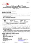
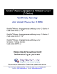
1
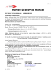
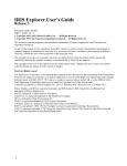
ZenBio, Inc. Human Skeletal Muscle Myoblast Care Manual: Maintenance and Differentiation from Myoblasts to Myocytes INSTRUCTION MANUAL ZBM0044.04 SHIPPING CONDITIONS Human Skeletal Muscle Myoblast Cells Orders are delivered via Federal Express courier. All US and Canada orders are shipped via Federal Express Priority service and are usually received the next day. International orders are usually received in 3-4 days. Must be processed upon shipment receipt. STORAGE CONDITIONS Media: Short Term 4°C 6 months -20°C All Zen-Bio Inc products are for research use only. Not approved for human or veterinary use or for use in diagnostic or clinical procedures. LIMITED PRODUCT WARRANTY This warranty limits our liability to replacement of this product. No other warranties of any kind, expressed or implied, including without limitation implied warranties of merchantability or fitness for a particular purpose, are provided by Zen-Bio, Inc. Zen-Bio, Inc. shall have no liability for any direct, indirect, consequential, or incidental damages arising out of the use, the results of use, or the inability to use this product. Zen-Bio, Inc warrants its cells only if Zen-Bio media are used and the recommended protocols are followed. Cryopreserved myoblasts are assured to be viable when thawed and maintained according to Zen-Bio protocols. ORDERING INFORMATION AND TECHNICAL SERVICES ZenBio, Inc. 3200 East NC Highway 54, Suite 100 PO Box 13888 Research Triangle Park, NC 27709 U.S.A. Telephone (919) 547-0692 Facsimile (FAX) (919) 547-0693 Toll free (continental US only) 1-866-ADIPOSE 1-(866)-234-7673 Electronic mail (e-mail) [email protected] World Wide Web http://www.zenbio.com Rev OCT 2012 Page 1 of 10 ZenBio, Inc. CONTENTS PAGE # Introduction 3 Materials Provided for Each Catalog Item 3 Media Compositions 4 Plating Procedure for Cryopreserved Human skeletal muscle Myoblasts 5 Differentiation of Myoblasts into Myocytes 6 Expansion Procedure for Cryopreserved Human skeletal muscle Myoblasts 8 Troubleshooting 9 Frequently Asked Questions 9 Pathogen Testing 10 Rev OCT 2012 Page 2 of 10 ZenBio, Inc. INTRODUCTION Cultured human skeletal muscle myoblasts Skeletal muscle is an important site of insulin-stimulated glucose disposal and often the site of insulin resistance in obesity. Human primary cultured skeletal muscle cells can directly reflect a patient’s metabolic phenotype, because many of the signaling pathways are maintained intact. Zen-Bio offers human primary skeletal muscle cells from a variety of donors, including obese donors with Type 2 diabetes. Skeletal muscle satellite cells are isolated from the rectus abdominus and propagated in culture as myoblasts. Each lot is analyzed for myotube formation and the expression of myocyte specific markers. The myoblasts are cryopreserved and guaranteed for use with ZenBio support media. PRECAUTIONS This product is for research use only. It is not intended for human, veterinary, or in vitro diagnostic use. Proper precautions and biological containment should be taken when handling cells of human origin, due to their potential biohazardous nature. Always wear gloves and work behind a protective screen when handling primary human cells. All media, supplements, and tissue cultureware used in this protocol should be sterile. Human myoblast viability depends greatly on the use of suitable media, reagents, and sterile plastic wear. If these parameters are not carefully observed, limited differentiation may occur and cell growth may be slow. MATERIALS PROVIDED FOR EACH CATALOG ITEM Note: Zen-Bio recommends that the Human skeletal muscle myoblasts be processed immediately upon receipt. Cryopreserved Human Skeletal Muscle Myoblasts (catalog # SKB-F) Frozen vial containing at least 0.5 x10⁶ myoblasts (store in liquid nitrogen upon receipt) 50 ml Skeletal Muscle Cell Growth medium (cat# SKM-M) Rev OCT 2012 Page 3 of 10 ZenBio, Inc. MEDIA COMPOSTIONS Skeletal Muscle Cell Growth Medium cat # SKM-M DMEM Fetal bovine serum Bovine Serum Albumin Fetuin Human Epidermal Growth Factor Dexamethasone Human Insulin Penicillin Streptomycin Amphotericin B Skeletal Muscle Cell Differentiation Medium cat # SKM-D DMEM Horse serum Bovine Serum Albumin Fetuin Penicillin Streptomycin Amphotericin B Skeletal Myoblast Cryopreservation Medium Cat# SKM-100 DMSO SKM-M medium All media contain 1.0 g/L D-glucose. MEDIA EXPIRATION DATES: If placed at 4C upon arrival, the media is stable until the expiration date on the bottle label. If stored at -20C upon arrival, it is stable for 6 months. Add fresh antibiotics when you are ready to use. Rev OCT 2012 Page 4 of 10 ZenBio, Inc. Plating Procedure for Cryopreserved Human Skeletal Muscle Myoblasts Cryopreserved Human Skeletal Muscle Myoblasts (Catalog # SKB-F) 1. Remove cells from liquid nitrogen and place immediately into a 37 C water bath and agitate while in bath. Be careful not to submerge the cap of the vial into water. Do not leave the vials in water bath after most of the content has thawed. Rinse the vials with 70% ethanol before taking them to the culture hood. 2. Upon thawing, transfer the cells to a sterile conical bottom centrifuge tube containing 10 ml of Skeletal Muscle Growth Medium (cat # SKM-M). Centrifuge: 1,200 rpm (282 X g) / 20C / 5 minutes. Aspirate the supernatant. TAKE CARE TO NOT ASPIRATE ANY OF THE CELL PELLET. 3. The cell vial contains a minimum of 0.5 x 10⁶ viable cells; however, we recommend performing a cell count to determine a more exact number of cells. Resuspend the cell pellet in 0.5 ml Skeletal Muscle Growth Medium, dilute an aliquot in 0.4% trypan blue solution. We suggest withdrawing an aliquot of 50 l of cells and mixing with 100 l of the trypan blue solution, resulting in a dilution factor of 3. Count live (unstained) cells on a hemacytometer. 4. Plate approximately 5,000-15,000 cells / cm2 using the media volumes from the table below. Refer to the manufacturer’s specifications for the specific cultureware brand you are using. FORMAT VOLUME PER WELL 96 well plate 150 l TOTAL VOLUME PER FORMAT* 14.4 ml 48 well plate 24 well plate 12 well plate 6 well plate 10 cm dish T-75 flask T25 flask 500 l 1 ml 2 ml 3 ml 15 ml 20 ml 7 ml 24.0 ml 24.0 ml 24.0 ml 18.0 ml 15.0 ml 20.0 ml 7.0 ml *We recommend preparing slightly larger volumes to allow for loss due to foam and pipet error. 5. Plate cells in desired format and place in a humidified 37oC incubator with 5% CO2. Do not agitate the plate, as cells will not plate evenly. To differentiate the cells please see the protocol on page 6 starting at step 1. Rev OCT 2012 Page 5 of 10 ZenBio, Inc. DIFFERENTIATION OF MYOBLASTS INTO MYOCYTES 1. Plated myoblasts in Skeletal Muscle Growth Medium (cat # SKM-M) can undergo differentiation using Skeletal Muscle Cell Differentiation Medium (cat # SKM-D). Differentiation should be initiated when the plated myoblasts reach 80-90% confluence. The exact number of days necessary to reach 80-90% confluence will depend on your initial seeding density (typically 1-3 days). 2. To start the process, aspirate the entire volume of Skeletal Muscle Growth Medium from all wells. Add the appropriate volume of Skeletal Muscle Cell Differentiation Medium (catalog # SKM-D) to the wells (see Table 1. Feeding Volumes). Incubate the plate at 370C and 5% CO2. Fresh Differentiation Medium will need to be added every 2-3 days. Remove all of the medium and replace with fresh medium. 3. After 6 days the cells should have fused to form myotubes. These are elongated, multinucleated cells. They will appear to be lined up when viewed under a microscope. 4. The myocytes may be used for assays 6-8 days after the initiation of differentiation and are suitable for most assays. Table 1. Feeding Volumes Format Plating Change SKM-M to Change SKM-D to Change SKM-D to SKM-D SKM-D SKM-D IN OUT IN OUT IN OUT IN 96 well plate 48 well plate 24 well plate 12 well plate 6 well plate 150 l/ well 150 l/ well 150l/ well 150l/ well 150l/ well 150l/ well 150l/ well 500 l/ well 500l/ well 500l/ well 500l/ well 500l/ well 500l/ well 500l/ well 1.0 ml/well 1.0 ml/well 1.0 ml/well 1.0ml/well 1.0ml/well 2.0 ml/well 2.0 ml/well 2.0 ml/well 2.0 ml/well 2.0 ml/well 2.0 ml/well 2.0 ml/well 3.0 ml/well 3.0 ml/well 3.0 ml/well 3.0 ml/well 3.0 ml/well 3.0 ml/well 3.0 ml/well T-75 flask 20 ml/flask 20 ml/flask 20 ml/flask 20 ml/flask 20 ml/flask 20 ml/flask 20 ml/flask T-25 flask 7 ml/flask 7 ml/flask 7 ml/flask 7 ml/flask 7 ml/flask 7 ml/flask 7 ml/flask Rev OCT 2012 1.0 ml/well Page 6 of 10 1.0ml/well ZenBio, Inc. A. 80% Confluent myoblasts B. 3 day old myocytes (3 days post-differentiation) C. 1-week-old myocytes (1 wk post-differentiation) MYOBLAST MATURE SKELETAL MYOCYTE Figure 1: Photographs of 80% confluent Myoblasts (A), 3 day-old (post-differentiation) cultured myocytes (B) and mature (1 week post-differentiation) cultured Myocytes (C). These are unstained photographs of human myocyte morphology (20X). The cells should appear comparable in appearance to these pictures. The myocytes should be 80% confluent after plating for differentiation. If they are not 80% confluent, the cells will not differentiate well. Please see the Troubleshooting guide for any problems. EXPANSION PROCEDURE Cryopreserved Human Skeletal Muscle Myoblasts (Catalog # SKB-F) 1. Remove cells from liquid nitrogen and place immediately into a 37C water bath with agitation. Be careful not to submerge the cap of the vial into water. Do not leave the vials in water bath after most of the content has thawed. Rinse the vials with 70% ethanol before taking them to the culture hood. 2. Upon the thawing, add the cells to a sterile conical bottom centrifuge tube, containing 10 ml of Skeletal Muscle Growth Medium (SKM-M). 3. Centrifuge at 280 x g, 20C, 5 minutes. Aspirate the medium and resuspend cells in a volume of SKM-M appropriate for counting the cells. Count using a hemacytometer. 6 4. Place approximately 0.5 X 10 cells in T-75 culture flasks using Skeletal Muscle Growth Medium. 5. Incubate cells until they are 70% confluent (in about 3-5 days). Do not let the cells become 100% confluent. Cells will need to be fed every 2-3 days with SKM-M. Replace all medium with fresh SKM-M. 6. Aspirate medium and wash myoblasts 4-5 times using sterile Phosphate Buffered Saline (PBS) to remove all traces of serum (until there is no foaming of the medium). Remove the PBS and release the cells from the flask bottom by adding 2 mL/T-75 flask of 0.25% trypsin/ 2.21mM EDTA solution. Allow cells to trypsinize for 5 minutes at 37C. Tap the flask gently to loosen the cells. Rev OCT 2012 Page 7 of 10 ZenBio, Inc. 7. Neutralize the trypsin using 7 ml Skeletal Muscle Cell Growth Medium (cat# SKM-M) per T-75 flask. Check the flask under a microscope to ensure all cells are free of the flask bottom. 8. Count the cells and plate in desired format (see page 5 for plating protocol). Ensure cells are evenly suspended when plating large numbers of plates or flasks. Do not agitate plates and flasks after plating. Place in a humidified incubator at 37C and 5% CO2, making sure the surface is level for even cell distribution. 9. Follow the differentiation protocol as outlined on page 6 or split the cells 1:4 for further expansion. 10. We DO NOT recommend differentiating myoblasts that are older than Passage 6. Cells will arrive at Passage 3 or 4. TROUBLESHOOTING GUIDE Observation Myoblasts do not differentiate Possible causes 1. Cells have been passaged too many times 1. Use cells of a lower passage number 2. Differentiation conditions not optimal 2. Use our defined differentiation media. Make sure that wells are 80% confluent BEFORE initiating differentiation. 3. Cells were plated at a low density Myoblasts do not grow Edge effects Rev OCT 2012 Suggestions 3. Use the cell density recommended in our manual 4. Cultureware used not optimal for human primary myoblasts 1. Cells have been passaged too many times 4. Verify the surface area for the cultureware brand you are using. 2. Cells expanded too high 1. Medium in outside wells evaporated 2. Do not exceed 1:4 expansion ratio 1. Ensure a saturated humidity in the incubator and feed the cells no less than every 3 days. Make sure multiple plates are stacked no more than 3 plates high. Page 8 of 10 1. Use cells of a lower passage number ZenBio, Inc. FREQUENTLY ASKED QUESTIONS When do the cells differentiate? Cells should begin to fuse and line up within 3 days after differentiation is induced. [See Figure 1] Can I pass the cells? Myocytes cannot be passed because they are terminally differentiated. Myoblasts can be trypsinized and replated several times. Myoblasts grow slower with each passage and differentiate poorly after passage 6. Cells are shipped at Passage 3-4. How fast do the cells replicate? The average doubling time is 24-36 hours. However, keep in mind that the replication rate for human myoblasts varies slightly from patient to patient. How long do the cells last in culture? Myocytes retain similar morphology and express myocyte specific genes for at least 1 week after induction of differentiation. Should antibiotics be included in the medium? Yes. Antibiotics and anti-fungal agents are always recommended since the cells are primary cells. All Zen-Bio media contain antibiotics and anti-fungal agents. Where are the cells from? The myoblasts are isolated from human rectus abdominus muscle. How are the cells shipped? Frozen cells will be packaged on dry ice and shipped to customers via Federal Express overnight delivery. How long do I have to wait before receiving the cells? We do not ship cells to domestic locations on Fridays. In general, myoblasts can be shipped the second day after the purchase order is confirmed. Do you test for pathogens? Which ones? Yes. Samples from each donor are tested via PCR to confirm non-reactivity for HIV-1, HIV-2, HTLV I, HTLV II, hepatitis B and hepatitis C. However, since we cannot test all pathogens, please treat the culture as a potentially infectious agent. What donor information do I receive? The donor’s gender, age, and BMI will be provided. Are the cells from one donor? Yes. We can also provide lot numbers containing cells mixed donors to get average responses. Please inquire about availability of single donor and mixed donor (called a superlot) lots at time order is placed. PATHOGEN TESTING Rev OCT 2012 Page 9 of 10 ZenBio, Inc. Samples from each donor are tested via PCR to confirm non-reactivity for HIV-1, HIV-2, HTLV I, HTLV II, hepatitis B and hepatitis C. However, no known test can offer complete assurance that the cells are pathogen free. Our products are tested and are free from mycoplasma contamination. Proper precautions and biological containment should be taken when handling cells of human origin, due to their potential biohazardous nature. All human based products should be handled at a BSL-2 (Biosafety Level 2) or higher. Always wear gloves and work behind a protective screen when handling primary human cells. REFERENCES Lists of articles using ZenBio, Inc cultured human cultured products may be found at our website (http://www.zenbio.com) under the COMPANY button. zenbio, inc. e-mail: [email protected] http:// www.zenbio.com p. o. box 13888 3200 east nc highway 54 suite 100 research triangle park north carolina 27709 phone: (919) 547-0692 fax: (919) 547-0693 Toll free: 1-866-ADIPOSE (234-7673) Rev OCT 2012 Page 10 of 10