1
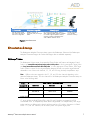
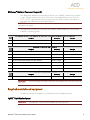
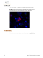
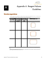
USER MANUAL
RNAscope® Fluorescent Multiplex Kit User
Manual
PART 2
Document Number 320293
For Part 1 Sample Preparation and Pretreatment Guide, see Document Number 320513 for
Fresh Frozen Tissue or Document Number 322452-USM for FFPE Tissue.
Research Use Only. Not for diagnostic use.
Rev. Date 11052015
For Research Use Only (RUO). Not for diagnostic use.
Tr ademarks
RNAscope® and HybEZ™ are trademarks of Advanced Cell Diagnostics, Inc. All other trademarks belong to their respective
owners.
C iting RNAscope® in Pu blications
When describing a procedure for publication using this product, please refer to it as the RNAscope ® 2.0 Assay and cite:
Wang F, Flanagan J, Su N, Wang L-C, Bui S, Nielson A, Wu X, Vo H-T, Ma X-J and Luo Y. RNAscope®: A Novel In Situ
RNA Analysis Platform for Formalin-Fixed Paraffin-Embedded Tissues. J. Mol. Diagnostics, 2012, 14:22–29.
D isclaimer s
Advanced Cell Diagnostics, Inc. reserves the right to change its products and services at any time to incorporate technological
developments. This manual is subject to change without notice.
Although this manual has been prepared with every precaution to ensure accuracy, Advanced Cell Diagnostics, Inc. assumes
no liability for any errors, omissions, or for any damages resulting from the use of this information.
C opyright
© 2015, Advanced Cell Diagnostics, Inc. All rights reserved.
Contents
Chapter 1. Product Information ..................................................5
About this guide.........................................................................................5
Product description .....................................................................................5
Background ...................................................................................... 5
Overview.......................................................................................... 5
Compatible sample types .................................................................... 6
Kit contents and storage ..............................................................................7
RNAscope ® Probes ............................................................................ 7
RNAscope ® Multiplex Fluorescent Reagent Kit........................................ 9
Requir ed materials and equ ipment ...............................................................9
HybEZ™ Hybridization System ............................................................. 9
User-supplied materials .................................................................... 10
Chapter 2. Before You Begin ...................................................11
Important procedural guidelines .................................................................11
Chapter 3. RNAscope® Fluorescent Multiplex Assay .......................12
Workflow ................................................................................................12
Materials required for the assay .................................................................13
Prepare the materials ................................................................................13
Prepare 1X Wash Buffer ................................................................... 13
Prepare probes................................................................................ 13
Equilibr ate reagents ......................................................................... 14
Run the assay ..........................................................................................14
Hybridize probe .............................................................................. 14
Hybridize Amp 1-FL ......................................................................... 14
Hybridize Amp 2-FL ......................................................................... 15
Hybridize Amp 3-FL ......................................................................... 15
Hybridize Amp 4-FL ......................................................................... 15
Counterstain and mount the slides ...................................................... 16
Evaluate the samples ................................................................................16
Fluorescent Imaging Recommendations ............................................... 17
Control examples............................................................................. 18
Troubleshooting .......................................................................................18
Appendix A. Reagent Volume Guidelines ....................................19
Determine reagent volume .........................................................................19
RNAscope® Fluorescent Multiplex Kit User Manual
3
Appendix B. Safety ...............................................................20
Chemical safety........................................................................................20
Biological hazard safety............................................................................20
Documentation and support.....................................................22
Obtaining MSDSs ....................................................................................22
Obtaining su pport ....................................................................................22
Contact information ..................................................................................22
Limited product warranty...........................................................................22
4
RNAscope® Fluorescent Multiplex Kit User Manual
Chapter 1. Product Information
1
Before using this product, read and understand the information in Appendix B. Safety on
page 20 in this document.
IMPORTANT!
We recommend reading the entire user manual before beginning any protocols.
About this guide
This user manual provides guidelines and protocols to use the RNAscope ® Fluorescent Multiplex
Reagent Kit (Cat. No.320850). You must use both an RNAscope® Reagent kit user manual and a
sample preparation and pretreatment user guide to perform the entire assay.
IMPORTANT!
For Part 1 Sample Preparation and Pretreatment Guide , see Document No. 320513
for Fresh Frozen Tissue or Document No. 322452-USM for FFPE Tissue.
Visit www.acdbio.com/technical-support/user-manuals to download a sample preparation user guide.
Product description
Background
The RNAscope® Multiplex Fluorescent Assay uses a novel and proprietary method of in situ
hybridization (ISH) to simultaneously visualize up to three different RNA targets per cell in samples
mounted on slides. Simultaneous detection of four different RNA targets is possible, and requires a
custom kit order. The assay is based on ACD’s patented signal amplification and background
suppression technology and incorporates multiplexed signal amplification systems, which enable
users to investigate expression as well as positional relationship between multiple genes within a
cellular context.
Overview
The RNAscope® Multiplex Fluorescent Assay procedure is illustrated in Figure 1 on page 6 and can
be completed in 6 hours. Most of the RNAscope® Assay reagents are available in convenient ReadyTo-Use (RTU) dropper bottles and provide a simple, nearly pipette-free workflow. Starting with
properly prepared samples, sections are first pretreated, and then RNA-specific probes designed for
different fluorescent detection channels are hybridized to multiple target RNAs. After a series of
highly effective and specific signal amplifications, single RNA transcripts for two or more target
genes appear as punctate dots in two or more distinctly fluorescent channels. These dots are visible
using a common fluorescent microscope with the appropriate filters.
RNAscope® Fluorescent Multiplex Kit User Manual
5
Compatible sample types
The RNAscope® Multiplex Fluorescent Assay is compatible with fresh frozen (FF) tissue, cultured
adherent cells on chamber slides, formalin-fixed, paraffin-embedded (FFPE) tissue, fixed frozen tissue,
and peripheral blood mononuclear cells (PBMC).
Use the guide below to determine the appropriate pretreatment reagent from the Universal
Pretreament Reagents Kit (Cat No. 322380) or RNAscope® Protease III and IV Reagents (Cat. No.
332340).
F l uores cent Detection Pretrea tment Guid e
Tis s ue Typ e
Pretrea tment Kit
Pretrea tment Ca t. No.
FFPE
RNAscope Target Retrieval*
322335
RNAscope® Protease III
320045
Cultured Adherent Cells
®
RNAscope Protease III
320045
Fresh Frozen
RNAscope® Protease IV
322336
Fixed Frozen
RNAscope Target Retrieval*
322335
RNAscope® Protease III
320045
RNAscope® Protease III
320045
Peripheral Blood Mononuclear Cells
(PBMC)
®
®
* RNAscope® Target Retrieval is not included in the RNAscope® Fluorescent Multiplex Reagent Kit. Please order it separately
(Cat. No.320043).
Note:
For FFPE tissue preparation, follow the sample preparation guide (Document No. 322452USM), except for the following differences:
•
RNAscope® Hydrogen Peroxide is not required.
•
RNAscope® Target Retrieval before creating the hydrophobic barrier.
•
Replace RNAscope® Protease III with RNAscope® Protease IV—this will reduce potential issues
with autofluorescence during imaging.
Please contact technical support at [email protected] if you have any questions.
6
RNAscope® Fluorescent Multiplex Kit User Manual
1: Tissue section
2:Hybridize to target RNA
Start with properly prepared
tissue sections and pretreat to
allow access to target RNA.
Hybridize multiple sets of
gene-specific probe pairs to target
mRNAs.
3: Amplify signal
Use up to four signal amplification systems to
detect multiple target RNAs. Probes are hybridized
to a cascade of signal amplification molecules,
culminating in binding of dye-labeled probes
visible in different fluorescent chanels.
4: I mage
Visualize target RNA
using a standard
fluorescent microscope.
F igure 1. Procedure overview
Kit contents and storage
The RNAscope® Multiplex Fluorescent Assay requires the RNAscope® Probes and the RNAscope®
Multiplex Fluorescent Reagent Kit. Probes and Reagent Kits are available separately.
RNAscope® Probes
The RNAscope® Probes consist of user-specified Target Probes and Positive and Negative Control
Probes. Visit www.acdbio.com/products/target-probes/search-product to find a gene-specific Target Probe.
Visit http://www.acdbio.com/control-slides-and-probes to order appropriate Control Probes. Each Target
Probe contains a mixture of short oligonucleotides designed to bind to a specific target RNA and
detectable in one of three color channels, C1, C2, and C3 using the Amp 4 amplification step.
Note:
Different colors are assigned to the C1, C2, and C3 color channels depending on the
particular RNAscope ® Assay. The color channels for the RNAscope® Multiplex Fluorescent Assay are
shown in the following table:
Amp 4 Al t A F l uores cent L a b el
Prob e
Cha nnel I D
Excita tion
Emis s ion
C1*
Alexa 488 nm
540 ± 10 nm
GREEN
C2
Atto 550 nm
580 ± 10 nm
ORANGE
C3
Atto 647 nm
690 ± 10 nm
Col or
FAR RED
* Default channel
C1 target probes are Ready-To-Use (RTU), while C2 and C3 probes are shipped as a 50X
concentrated stock. To independently detect different target RNAs in a multiplex assay, each target
probe must be in a different color channel and there must be a C1 probe in the mixture. A “Blank
Probe – C1” (Cat. No. 300041) can be used in place of a specific target probe.
RNAscope® Fluorescent Multiplex Kit User Manual
7
IMPORTANT!
C1 and C2 probes can be used for either fluorescent or chromogenic detection.
However, 3-plex or higher multiplexing capability is only possible with the fluorescent kit.
There are 3 options for alternate fluorescent color modules. Any fluorescent label combinations (Amp
4 Alt A, B, or C) can be selected based on your experiment design.
Col or Mod ul e Op tions
Probe Channel ID
Amp 4 Al t A-F L
Amp 4 Al t B-F L
Amp 4 Al t C-F L
C1
Alexa 488
Atto 550
Atto 550
C2
Atto 550
Alexa 488
Atto 647
C3
Atto 647
Atto 647
Alexa 488
Each probe is sufficient for staining ~20 sections, each with an area of approximately 20 mm x
20 mm (0.75” x 0.75”). Larger tissue sections will result in fewer tests. The probes have a shelf life
of two years from the date of bulk manufacturing when stored as indicated in the following table:
Ta rg et Prob es
R ea g ent
Ca t. No.
Content
Qua ntity
Stora g e
Target Probe – [species] – [gene]
Various
Ready-To-Use (RTU) probe for color
channel 1
3 mL x 1 bottle
4°C
Target Probe – [species] – [gene] –
C2
Various
50X probe for color channel 2
60 μL x 1 tube
4°C
Target Probe – [species] – [gene] –
C3
Various
50X probe for color channel 3
60 μL x 1 tube
4°C
Target Probe – [species] – [gene] –
C4†
Various
50X probe for color channel 4
60 μL x 1 tube
4°C
Control Prob es
R ea g ent
Ca t. No.
Content
Qua ntity
Stora g e
Positive Control Probe
Various
RTU probe targeting a common
housekeeping gene. Each detection
channel has its own positive control
probe.
3 mL x 1 bottle
4°C
3-Plex Positive Control Probe
320861
RTU mixture of three probes targeting
POLR2A in channel C1, PPIB in channel
C2, and UBC in channel C3.
3 mL x 1 bottle
4°C
Negative Control Probe – dapB
Various
RTU probe targeting a bacterial gene.
Each detection channel has its own
negative control probe.
3 mL x 1 bottle
4°C
Blank Probe-C1
300041
RTU Target Probe diluent
3 mL x 1 bottle
4°C
† Available only for custom orders.
8
RNAscope® Fluorescent Multiplex Kit User Manual
RNAscope® Multiplex Fluorescent Reagent Kit
Each RNAscope® Multiplex Fluorescent Reagent Kit (Cat. No. 320850) provides enough reagents
to stain ~20 tissue sections ~20 sections, each with an area of approximately 20 mm x 20 mm
(0.75” x 0.75”). Larger tissue sections will result in fewer tests. Each kit contains three sub-kits: a
Pretreatment Kit, a Detection Kit, and a Wash Buffer Kit.
IMPORTANT!
Directions to use the Pretreatment Kit are included in separate sample preparation and
pretreatment user guides.
The reagents have a shelf life of nine months from the date of bulk manufacturing when stored as
indicated in the following table:
Pretrea tment Kit ( Ca t. No. 322340) or Univers a l Pretrea tment R ea g ents ( Ca t. No. 322380)
R ea g ent
Qua ntity
Stora g e
1X RNAscope® Protease III
4.5 mL x 1 bottle
4°C
2X RNAscope Protease IV
4.5 mL x 2 bottles
4°C
®
Detection–F L Kit ( Ca t. No. 320851)
R ea g ent
Qua ntity
Stora g e
Amp 1-FL
3 mL x 1 bottle
4°C
Amp 2-FL
4.5 mL x 1 bottle
4°C
Amp 3-FL
3 mL x 1 bottle
4°C
Amp 4-FL–Alt A Display module (Cat. No. 320855)
4.5 mL x 1 bottle
4°C
Amp 4-FL–Alt B Display module (Cat. No. 320856)
4.5 mL x 1 bottle
4°C
Amp 4-FL–Alt C Display module (Cat. No. 320857)
4.5 mL x 1 bottle
4°C
DAPI
3 mL x 1 bottle
4°C
Wa s h Buffer Kit ( Ca t. No. 310091)
R ea g ent
50X Wash Buffer
IMPORTANT!
same name.
Qua ntity
60 mL x 4 bottles
Stora g e
Room temperature (20–25°C)
Do not interchange the reagent components of the Reagent Kits, even those having the
Required materials and equipment
The following materials and equipment are needed to perform the RNAscope ® Assay.
HybEZ™ Hybridization System
IMPORTANT!
The RNAscope® Assay has been validated using this system only.
RNAscope® Fluorescent Multiplex Kit User Manual
9
The HybEZ™ Hybridization System (110 VAC, Cat. No. 310010; 220 VAC, Cat. No. 310013) is
designed for the hybridization and incubation steps in the RNAscope ® Assays. Incubation steps in
the RNAscope® Assay require humid conditions to prevent sections from drying out.
For instructions on how to use the HybEZ™ Hybridization System, refer to the HybEZ™
Hybridization System User Manual available at www.acdbio.com/technical-support/user-manuals and
view the training video at www.acdbio.com/technical-support/learn-more. The system contains the
following components:
Comp onent
Qua ntity
Ca t. No.
HybEZ Oven (110 or 220 VAC)
1 oven
310010 or 310013
HybEZ ™ Humidity Control Tray (with lid)
1 tray
310012
HybEZ ™ Slide Rack (20 slide capacity)
1 rack
310014
HybEZ ™ Humidifying Paper
2 sheets
—
HybEZ ™ Humidifying Paper Pack
15 sheets
310015
™
User-supplied materials
Des crip tion
Sup p l ier
Ca t. No.
Fluorescent mounting medium
Invitrogen/MLS*
P36930
Tissue-Tek ® Vertical 24 Slide Rack
American Master Tech Scientific/MLS
LWSRA24
Tissue-Tek ® Staining Dish
American Master Tech Scientific/MLS
LWT4457EA
Tissue-Tek ® Clearing Agent Dish, xylene
resistant
American Master Tech Scientific/MLS
Cover glass 24 x 50 mm
Fisher Scientific/MLS
12-545-F
Carboy (>3L)
MLS
—
Water bath or incubator, capable of
holding temperature at 40 +/- 1°C
MLS
—
Distilled water
MLS
—
Tubes (various sizes)
MLS
—
Paper towel or absorbent paper
MLS
—
Fluorescent microscope with filter set:
MLS
—
LWT4456EA
Ex 358 nm/Em 461 nm (DAPI)
Ex 501 nm/Em 523 nm (FITC)
Ex 554 nm/Em 576 nm (Cy3)
Ex 644 nm/Em 669 nm (Cy5)
Ex 740 nm/Em 764 nm (Cy7) †
* Major Laboratory Supplier in North America. For other regions, please check Catalog Numbers with your local lab supplier.
† For custom 4-plex assays only.
10
RNAscope® Fluorescent Multiplex Kit User Manual
Chapter 2. Before You Begin
2
IMPORTANT!
For Part 1 Sample Preparation and Pretreatment Guide , see Document No. 320513
for Fresh Frozen Tissue or Document No. 322452-USM for FFPE Tissue.
Prior to running the RNAscope® Assay on your samples for the first time, we recommend that you:
•
View the video demonstrations available at www.acdbio.com/technical-support/learn-more.
•
Use an RNAscope® Multiplex Fluorescent Channel Assessment Slide (Cat. No. 310022) to
ensure that the fluorescent microscope is properly equipped with the correct excitation and
emission filter set.
Important procedural guidelines
•
Start with properly prepared sections. Refer to our sample preparation and pretreatment user
guides available at www.acdbio.com/technical-support/user-manuals. Use only samples mounted
on SuperFrost Plus® Slides (Fisher Scientific; Cat. No. 12-550-15).
•
Follow the recommended pretreatment conditions for your sample. Refer to our sample
preparation and pretreatment user guides available at www.acdbio.com/technical-support/usermanuals.
•
Always run positive and negative control probes on your sample to assess sample RNA quality
and optimal permeabilization.
•
Do not substitute required materials. Assay has been validated with these materials only.
•
Follow the protocol exactly for best results.
•
Do not let your sections dry out during the procedure.
•
Use good laboratory practices and follow all necessary safety procedures. Refer to Appendix B.
Safety on page 20 for more information.
RNAscope® Fluorescent Multiplex Kit User Manual
11
3
Chapter 3. RNAscope® Fluorescent
Multiplex Assay
IMPORTANT!
For Part 1 Sample Preparation and Pretreatment Guide , see Document No. 320513
for Fresh Frozen Tissue or Document No. 322452-USM for FFPE Tissue.
This procedure flows directly from sample preparation and pretreatment. Refer to the appropriate
sample preparation and pretreatment user guide for your specific sample type.
Workflow
Prep a re the ma teria l s ~30 MI N
R un the a s s a y ~5.5 HR S
Hybridize probe ~2 HRS
Hybridize Amp 1-FL~30 MIN
Hybridize Amp 2 ~15 MIN
Hybridize Amp 3 ~30 MIN
Hybridize Amp 4 ~15 MIN
Counterstain the slides ~2 MIN
Eva l ua te the s a mp l es
12
RNAscope® Fluorescent Multiplex Kit User Manual
Materials required for the assay
Ma terials p rovid ed b y the R NAs cop e ® Ma terials provided by RNAscope®
F l uores cent Mul tip l ex Kit
Other ma teria l s a nd eq uip ment
Prob es
• 50X Wash Buffer
• C1 Target Probe
• Prepared sections
• Amp 1-FL
• 50X C2 Target Probe
• Distilled water
• Amp 2-FL
• 50X C3 Target Probe
• Carboy (>3L)
• Amp 3-FL
• 3-Plex Positive Control Probe
• Tissue-Tek ® Staining Dish
• Amp 4-FL–Alt A, Amp 4-FL–Alt B, or Amp 4- • Negative Control Probe
• Tissue-Tek ® Clearing Agent Dish,
xylene-resistant
• DAPI
• HybEZ ™ Humidifying System
FL–Alt C
• Water bath or incubator
• Tissue-Tek ® Vertical 24 Slide Rack
• Tubes (various sizes)
• Paper towel or absorbent paper
• Fluorescent mounting medium
• Cover Glass, 24 mm x 50 mm
Prepare the materials
You may prepare the reagents at the same time you prepare pretreatment reagents. Refer to a
sample preparation and pretreatment user guide available at www.acdbio.com/technical-support/usermanuals.
Some of the materials may be prepared in advance and stored at room temperature.
Prepare 1X Wash Buffer
•
Prepare 3 L of 1X Wash Buffer by adding 2.94 L distilled water to 1 bottle (60 mL) in a large
carboy. Mix well.
Note: If precipitation occurs in 50X Wash Buffer, warm it up at 40°C for 10–20 MIN before
making 1X Wash Buffer. 1X Wash Buffer may be prepared ahead of time and stored at room
temperature for up to one month.
Prepare probes
1. Warm probes for 10 MIN at 40°C in a water bath or incubator, then cool to ROOM
TEMPERATURE (RT).
1. Briefly spin the C2 and C3 probes to collect the liquid at the bottom of the tubes.
2. Mix 1:1:50 ratios of C2, C3, and C1 probes by pipetting 1 volume of C2 and 1 volume
of C3 probes to 50 volumes of C1 probe into a tube. Invert the tube several times.
Note: Do not mix probes of the same channel. The mixed Target Probes can be stored at 4°C
for up to 6 months.
RNAscope® Fluorescent Multiplex Kit User Manual
13
Equilibrate reagents
•
Place Amp 1–4 FL reagents at RT.
•
Ensure HybEZ™ Oven and prepared Humidity Control Tray are at 40°C.
Run the assay
IMPORTANT!
solutions.
IMPORTANT!
proceeding.
Do NOT let sections dry out between incubation steps. Work quickly and fill barrier with
View the wash step video at www.acdbio.com/technical-support/learn-more before
Note:
We recommend running control probe on your sample before running any of your specific
target probes to optimize the protocol.
Hybridize probe
IMPORTANT!
Prior to this step, ensure you have pretreated your samples. See Catalog No. 320513
for Fresh Frozen Tissue or Catalog No. 322452-USM for FFPE Tissue.
IMPORTANT!
Ensure probes are prewarmed and cooled to RT prior to use.
1. Tap and/or flick to remove excess liquid from slides and place in the HybEZ™ Slide Rack.
Add ~4 drops of the appropriate probe to entirely cover each section.
Note: Refer to Appendix A. Reagent Volume Guidelines on page 19 to determine the recommended
number of drops needed per slide. For example, for a 0.75” x 0.75” barrier add 4 drops of the
appropriate probe.
2. Place the HybEZ™ Slide Rack in the HybEZ™ Humidity Control Tray removed from the
HybEZ™ Oven. Close tray and insert back into the oven for 2 HRS at 40°C.
IMPORTANT!
To prevent evaporation, make sure the turn nob is completely turned to lock position.
3. Remove the HybEZ™ Control Tray from the oven and remove HybEZ™ Slide Rack.
4. One slide at a time, quickly remove excess liquid by decanting and place slide in a TissueTek ® Slide Rack submerged in the Tissue-Tek ® Staining Dish filled with 1X Wash Buffer.
5. Wash slides in 1X Wash Buffer for 2 MIN at RT. Agitate slides by moving the slide rack up
and down in the dish.
6. Repeat Step 5 with fresh 1X Wash Buffer.
Hybridize Amp 1-FL
1. Take each slide one at a time from the Tissue-Tek ® Slide Rack and tap/and or flick to
remove the excess liquid before placing in the HybEZ™ Slide Rack. Add ~4 drops of
Amp 1-FL to entirely cover each section.
2. Place the HybEZ™ Slide Rack in the HybEZ™ Humidity Control Tray removed from the
HybEZ™ Oven. Seal tray and insert back into the oven for 30 MIN at 40°C.
14
RNAscope® Fluorescent Multiplex Kit User Manual
3. Remove the HybEZ™ Control Tray from the oven and remove HybEZ™ Slide Rack.
4. One slide at a time, quickly remove excess liquid and place slide in a Tissue-Tek ® Slide
Rack submerged in the Tissue-Tek ® Staining Dish filled with 1X Wash Buffer.
5. Wash slides in 1X Wash Buffer for 2 MIN at RT with occasional agitation.
6. Repeat Step 5 with fresh 1X Wash Buffer.
Hybridize Amp 2-FL
1. Take each slide one at a time from the Tissue-Tek ® Slide Rack and tap and/or flick to
remove the excess liquid before placing in the HybEZ™ Slide Rack. Add ~4 drops of
Amp 2-FL to entirely cover each section.
2. Place the HybEZ™ Slide Rack in the HybEZ™ Humidity Control Tray. Close tray and insert
into the oven for 15 MIN at 40°C.
3. Remove the HybEZ™ Control Tray from the oven and remove HybEZ™ Slide Rack.
4. One slide at a time, quickly remove excess liquid and place slide in a Tissue-Tek ® Slide
Rack submerged in the Tissue-Tek ® Staining Dish filled with 1X Wash Buffer.
5. Wash slides in 1X Wash Buffer for 2 MIN at RT with occasional agitation.
6. Repeat Step 5 with fresh 1X Wash Buffer.
Hybridize Amp 3-FL
1. Take each slide one at a time from the Tissue-Tek ® Slide Rack and tap and/or flick to
remove the excess liquid before placing in the HybEZ™ Slide Rack. Add ~4 drops of Amp
3-FL to entirely cover each section.
2. Place the HybEZ™ Slide Rack in the HybEZ™ Humidity Control Tray. Close tray and insert
into the oven for 30 MIN at 40°C.
3. Remove the HybEZ™ Control Tray from the oven and remove HybEZ™ Slide Rack.
4. One slide at a time, quickly, remove excess liquid and place slide in a Tissue-Tek ® Slide
Rack submerged in the Tissue-Tek ® Staining Dish filled with 1X Wash Buffer.
5. Wash slides in 1X Wash Buffer for 2 MIN at RT with occasional agitation.
6. Repeat Step 5 with fresh 1X Wash Buffer.
Hybridize Amp 4-FL
1. Take each slide one at a time from the Tissue-Tek ® Slide Rack and tap and/or flick to
remove the excess liquid before placing in the HybEZ™ Slide Rack. Add ~4 drops of
Amp 4-FL to entirely cover each section.
Note: There are 3 options for alternate fluorescent color modules. Any fluorescent label
combination (Amp 4-FL– Alt A, B, or C) can be selected. For tissue, the default recommended
module is Amp 4-FL– Alt B.
2. Place the HybEZ™ Slide Rack in the HybEZ™ Humidity Control Tray. Close tray and insert
into the oven for 15 MIN at 40°C.
3. Remove the HybEZ™ Control Tray from the oven and remove HybEZ™ Slide Rack.
RNAscope® Fluorescent Multiplex Kit User Manual
15
4. One slide at a time, quickly remove excess liquid and place slide in a Tissue-Tek ® Slide
Rack submerged in the Tissue-Tek ® Staining Dish filled with 1X Wash Buffer.
5. Wash slides in 1X Wash Buffer for 2 MIN at RT with occasional agitation.
6. Repeat step 5 with fresh 1X Wash buffer.
Counterstain and mount the slides
IMPORTANT!
Do this procedure with no more than 5 slides at a time.
1. Remove excess liquid from the slides and add ~4 drops of DAPI to each section.
2. Incubate for 30 SEC at RT.
3. Remove DAPI from slides and immediately place 1–2 drops of the fluorescent mounting
medium onto each section.
4. Carefully place a 24 mm x 50 mm coverslip over the tissue section. Avoid trapping air
bubbles. Store slides in the dark at 4°C.
IMPORTANT!
Image the slides after 8 hours or within a few days.
Evaluate the samples
For an example of successful staining, see Figure 2 on page 18. Examine tissue sections under a
standard fluorescent microscope at 20–40X magnification. A confocal microscope may also be
used:
16
•
Assess tissue and cell morphology.
•
Assess positive control signal strength. Positive control signal should be visible as punctuate
dots within cell with 20X magnification.
•
Assess negative control background. Five dots in every10 cells displaying background staining
per microscope field is acceptable with 20X magnification.
•
Evaluate target probe signal using the scoring guidelines in the next section.
RNAscope® Fluorescent Multiplex Kit User Manual
Fluorescent Imaging Recommendations
Here are a few fluorescent imaging recommendations:
V iew ing
Image capture is the
recommended digital
capturing option
Fluorescence viewing is
the recommended
viewing option
Detection
Microscope with camera and
fluorescence options.
Multispectrum
microscope/camera system
recommended (eg. Nuance
FX)
Fluorescence detection
requires a high resolution and
high sensitivity cooled CCD
camera that is 64 µm pixel
size or smaller with > 65%
peak quantum efficiency
Micros cop e
Leica DM series or
equivalent
Zeiss Axio Imager or
equivalent
Inverted microscope is okay
if optics and condenser meet
requirements
Op tics
20X (N.A 0.75) air, 40X
(N.A. 0.8) air, 40X (N.A.
1.3) oil, 63X (N.A. 1.3) oil,
and 100X (N.A. 1.4) oil
20X and 40X objective can
be used for visualization of
high expression genes and
low expression genes,
respectively
Common models include:
Orca-Flash 4.0 (Hamamatsu),
and Nuance FX (Nuance)
IMPORTANT!
The RNAscope® Fluorescent kits are primarily targeted for fresh frozen and cultured
cells. This is mainly due to imaging and analysis challenges with interference from tissue
autofluorescence. You can run the RNAscope® Fluorescent kit on solid tumor FFPE tissues if you have
access to a multi-spectral imaging system, such as Nuance FX (Nuance). Solid tumors such as breast,
colon, kidney, and liver have been successfully tested.
RNAscope® Fluorescent Multiplex Kit User Manual
17
Control examples
Figure 2 is an example of expression in the cerebral cortex of normal mouse brain.
F igure 2. Npy (red) and Fezf2 (green) expression in the cerebral cortex of normal mouse brain
stained using the RNAscope® Fluorescent Multiplex Kit; 63X oil lens, confocal image.
Troubleshooting
For troubleshooting information, please contact technical support at [email protected].
18
RNAscope® Fluorescent Multiplex Kit User Manual
Appendix A. Reagent Volume
Guidelines
A
Determine reagent volume
Before starting your experiment, measure the inner edge of the hydrophobic barrier to determine the
recommended number of drops needed per slide (see table below).
Size of hyrophobic R ecommend ed
b a rrier* ( in)
numb er of d rop s
R ecommend ed
vol ume p er s l id e
p er s l id e
( µL )
0.75” x 0.75” †
4
120
0.75” x 1.0”
5
150
0.75” x 1.25”
6
180
R el a tive temp l a te s ize
* Hydrophobic barrier measured at inner edge. References in this user manual are for the 0.75” x 0.75” hydrophobic barrier
size.
† Recommended hydrophobic barrier size is 0.75” x 0.75”. With this barrier size, each probe is sufficient for staining ~20
sections. Larger tissue sections will result in fewer tests.
RNAscope® Fluorescent Multiplex Kit User Manual
19
Appendix B. Safety
B
Chemical safety
WARNING! GENERAL CHEMICAL HANDLING. To minimize hazards, ensure
laboratory personnel read and practice the general safety guidelines for chemical
usage, storage, and waste provided below, and consult the relevant SDS for specific
precautions and instructions:
•
Read and understand the Safety Data Sheets (SDSs) provided before you store, handle, or
work with any chemicals or hazardous materials. To obtain SDSs,
http://www.acdbio.com/technical-support/user-manuals.
•
Minimize contact with chemicals. Wear appropriate personal protective equipment when
handling chemicals (for example, safety glasses, gloves, or protective clothing).
•
Minimize the inhalation of chemicals. Do not leave chemical containers open. Use only with
adequate ventilation (for example, fume hood).
•
Characterize (by analysis if necessary) the waste generated by the particular applications,
reagents, and substrates used in your laboratory.
•
Ensure that the waste is stored, transferred, transported, and disposed of according to all
local, state/provincial, and/or national regulations.
•
IMPORTANT! Radioactive or biohazardous materials may require special handling, and
disposal limitations may apply.
Biological hazard safety
WARNING! BIOHAZARD. Biological samples such as tissues, body fluids, infectious
agents, and blood of humans and other animals have the potential to transmit
infectious diseases. Follow all applicable local, state/provincial, and/or national
regulations. Wear appropriate protective equipment, which includes but is not limited
to: protective eyewear, face shield, clothing/lab coat, and gloves. All work should be
conducted in properly equipped facilities using the appropriate safety equipment (for
example, physical containment devices). Individuals should be trained according to
applicable regulatory and company/institution requirements before working with
potentially infectious materials. Read and follow the applicable guidelines and/or
regulatory requirements in the following:
20
RNAscope® Fluorescent Multiplex Kit User Manual
In the U.S.:
• U.S. Department of Health and Human Services guidelines published in Biosafety in
Microbiological and Biomedical Laboratories found at: www.cdc.gov/biosafety
• Occupational Safety and Health Standards, Bloodborne Pathogens (29 CFR§1910.1030),
found at: www.access.gpo.gov/nara/cfr/waisidx_01/%2029cfr1910a_01.html
• Your company’s/institution’s Biosafety Program protocols for working with/handling
potentially infectious materials.
• Additional information about biohazard guidelines is available at: www.cdc.gov/
In the EU:
• Check local guidelines and legislation on biohazard and biosafety precaution and refer to the
best practices published in the World Health Organization (WHO) Laboratory Biosafety
Manual, third edition, found at:
www.who.int/csr/resources/publications/biosafety/who_cds_csr_lyo_2004_11/en/
• Information about the Registration, Evaluation, Authorisation and Restriction of Chemicals
(REACH) can be found at: eurlex.europa.eu/LexUriServ/LexUriServ.do?uri=OJ:L:2010:133:0001:0043:EN:PDF
RNAscope® Fluorescent Multiplex Kit User Manual
21
Documentation and support
Obtaining MSDSs
Safety Data Sheets (SDSs) are available at: www.acdbio.com/technical-support/user-manuals. For the
MSDSs of chemicals not distributed by Advanced Cell Diagnostics, contact the chemical
manufacturer.
Obtaining support
For the latest services and support information, go to: www.acdbio.com/technical-support/support-
overview.
At the website, you can:
•
Access telephone and fax numbers to contact Technical Support and Sales facilities.
•
Search through frequently asked questions (FAQs).
•
Submit a question directly to Technical Support.
•
Search for user documents, MSDSs, application notes, citations, training videos, and other
product support documents.
•
Find out information about customer training events.
Contact information
Advanced Cell Diagnostics, Inc.
3960 Point Eden Way
Hayward, CA 94545
Toll Free: 1-877-576-3636
Direct: 1-510-576-8800
Fax: 1-510-576-8801
Information: [email protected]
Orders: [email protected]
Support Email: [email protected]
Limited product warranty
Advanced Cell Diagnostics, Inc. and/or its affiliate(s) warrant their products as set forth in the ACD
General Terms and Conditions of Sale found on the ADC website at www.acdbio.com/technicalsupport/user-manuals. If you have any questions, please contact Advanced Cell Diagnostics at
www.acdbio.com/about/contact.
22
RNAscope® Fluorescent Multiplex Kit User Manual
Headquarters
3960 Point Eden Way
Hayward, CA 94545
For support, email [email protected].
www.acdbio.com
Phone 1-510-576-8800 Toll Free 1-877-576-3636


![1] n T169](http://vs1.manualzilla.com/store/data/005696581_1-b5b272b0c99ea5fca956b563015d9b67-150x150.png)