1
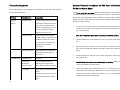
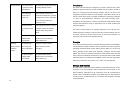
No DNA eluted
Poor cell lysis due
Mix thoroughly with Buffer DL prior
Introduction
to improper mixing
to loading HiBind® column.
The Solar® Forensic DNA Kit is designed to provide a rapid and easy method for the
isolation of genomic DNA from forensic samples such as dry blood, buccal swabs, and
with Buffer DL.
Poor cell and/or
Tissue sample must be cut or
protein lysis in
minced into small pieces. Increase
Buffer QTL.
incubation time at 65oC with Buffer
QTL to ensure that tissue is
completely lysed.
sperm for consistent PCR and Southern analysis. This kit can also be used for the
preparation of genomic DNA from mouse tail snips, whole blood, buffy coat, serum, and
plasma. The kit allows single or multiple, simultaneous processing of samples. There is
no need for phenol/chloroform extractions, and time-consuming steps such as
precipitation with isopropanol or ethanol are eliminated. DNA purified using the Solar®
Forensic DNA method is ready for applications such as PCR, Southern blotting, and
restriction digestion.
Absolute ethanol
Before applying sample to column,
not added to Buffer
an aliquot of Buffer DL/ethanol
DL.
must be added. See protocol
The Solar® Forensic DNA Kit is specially designed to work with the OB Specimen
above.
Collection Paper for isolation of genomic DNA from forensic samples such as dry blood
and sperm. This kit can be also used for fresh or frozen tissue samples or mouse tail
No ethanol added
Dilute Buffer PW2 with the
to Buffer PW2
indicated volume of absolute
Concentrate.
ethanol before use.
Washing
Incomplete lysis
Buffer DL is viscous and the
Solar® Forensic DNA Kit uses the reversible binding properties of the HiBind® matrix, a
leaves colored
due to improper
sample must be votexed
mixing with Buffer
thoroughly.
new silica-based material, combined with the speed of mini-column spin technology. A
residue in
column
DL.
snips (call customer service for detailed protocol).
Principle
specifically formulated buffer system allows genomic DNA up to 50 kb to bind to the
matrix. Samples are first lysed under denaturing conditions and then applied to the
No ethanol added
Dilute Buffer PW2 with the
to Buffer PW2
indicated volume of absolute
Concentrate.
ethanol before use.
HiBind® spin columns to which the DNA binds, while cellular debris, hemoglobin, and
other proteins are effectively washed away. High quality DNA is finally eluted in sterile
deionized water or low salt buffer. Each HiBind® column can bind approximately 100 μg
DNA. Use of more than 30 mg tissue or 107 cells is not recommended.
Storage and Stability
All components of the Solar® Forensic DNA Kit, except the Protease K, can be stored at
22oC-25oC. Once reconstituted in water, Protease K must be stored at -20oC. Under
these conditions, performance of all components of the kit is guaranteed at least 18
months. Under cool ambient conditions, a precipitate may form in the Buffer DL. In case
of such an event, heat the bottle at 37oC to dissolve the precipitate. Store Buffer DL at
12
room temperature.
Kit Contents
Improper washing
Product Number
DH194-00
DH194-01
DH194-02
5 Preps
50 Preps
200 Preps
5
50
200
15
150
600
Buffer DL
5 ml
20 ml
60 ml
Buffer QTL
5 ml
20 ml
50 ml
Buffer PW1
5 ml
30 ml
110 ml
Buffer PW2 Concentrate
5 ml
20 ml
3 x 20 ml
ER Buffer
2 ml
30 ml
100 ml
200 ul
1.8 ml
7 ml
3 mg
30 mg
4 x 30 mg
1
1
1
Purification times
HiBind® DNA Mini columns
2 ml Collection Tubes
Buffer PW2 Concentrate must be
diluted with absolute (100%)
ethanol as specified before use.
Low A260/A280
Extended
Resin from the column may be
ratio
centrifugation
present in eluate. Avoid
during elution step.
centrifugation at speeds higher
than specified. The material can
be removed from the eluate by
centrifugation — it will not interfere
with PCR or restriction digests.
Poor cell lysis due
Repeat the procedure, this time
to incomplete
making sere to vortex the sample
mixing with Buffer
with Buffer DL immediately and
DL
completely.
Note: The Solar® Forensic DNA Kit is supplied with enough buffers for the standard
Incomplete cell lysis
Increase incubation time with
protocol. However, due to increased volumes called for in some protocols (such as the
or protein
Buffer QTL and protease. Ensure
buccal swab protocol), fewer preparations may be performed. Also, additional buffers
degradation due to
that no visible pieces of tissue
can be purchased separately from Solar Bio-Tek. See the Accessories section in the
insufficient
remain.
catalog or call customer service for price information.
incubation.
Protease Storage Buffer
Protease K
User Manual
Before Starting
IMPORTANT
1 Reconstitute Protease K in 150 μl (Trial Kit) or 1.5 ml
(50 and 200 preps) Protease Storage Buffer. Vortex
Samples are rich in
After applying to column, wash
protein.
with 300 μl of a 1:1 mixture of
Buffer DL and ethanol and then
with Buffer PW2.
vial briefly prior to use.
2 Buffer PW2 Concentrate must be diluted with
absolute ethanol(96-100%) as follows:
DH194-00
Add 20 ml absolute ethanol
DH194-01
Add 80 ml absolute ethanol
DH194-02 Add 80 ml absolute ethanol / bottle
All centrifugation steps must be performed at room temperature.
2
11
Troubleshooting Guide
Standard Protocol for Isolation of DNA from Dried Blood, Body
Use the table below to find solutions to any problems you may have with the Solar®
Forensic DNA Isolation Kit:
Fluids and Sperm Spots
Dried blood, body fluids, and sperm samples on filter paper can be processed using the
following method. We recommend using Specimen Paper for spotting blood, as this
Problem
Possible Cause
Suggestions
Clogged
Incomplete lysis
Extend incubation time of lysis with
Column
Buffer QTL and protease. Add the
correct volume of Buffer DL and
incubate for specified time at 70oC.
unique filter paper disintegrates when incubated in aqueous buffers, allowing for the
efficient recovery of DNA. This kit can also be used for samples collected by using other
specimen collection papers.
1.
μl of blood can be used for each spot.) Tear or cut filter into small pieces and place
It may be necessary to extend
into a microfuge tube.
incubation time by 10 min.
Sample too large
Cut or punch out the blood spot (or other sample) from the filter paper. (Up to 200
Note: Use 3-4 punched cycles (3mm diameter) for each DNA isolation.
If using more than 30 mg tissue,
increase volumes of Protease K or
Proteinase K, Buffer QTL, Buffer
2.
Add 200 μl Buffer QTL and incubate at 60oC for 15 minutes. Vortex every 2 min to
mix.
DL, and ethanol. Pass aliquots of
lysate through one column
successively.
Sample too viscous
3.
60oC with occasional mixing. Briefly centrifuge to remove any droplets from inside
Divide sample into multiple tubes,
the lid.
adjust volume to 250 μl with 10
mM Tris-HCl.
Low DNA yield
Clogged column
See above
Poor sample
Incubate the OB specimen
release from
collection paper longer in SQL
collection paper
buffer. Shake the tubes frequently.
Poor elution
Repeat elution or increase elution
volume.
Incubation of column at 70oC for 5
min with ER Buffer may increase
Add 25 μl Protease K solution and mix by votexing. Incubate for 45 minutes at
4.
Add 225 μl Buffer DL and votex to mix. Briefly centrifuge to remove any droplets
from inside the lid.
5.
Add 225 μl absolute ethanol and mix thoroughly by vortexing. Briefly centrifuge to
remove any droplets from inside the lid.
6.
Insert each HiBind® DNA Minicolumn into a 2 ml collection tube (provided).
Transfer the entire sample from Step 5 into the column, including any precipitate
that may have formed. Centrifuge at 8,000 x g for 1 min to bind DNA. Discard
collection tube and flow-through liquid.
yields.
10
3
7.
8.
Place each column into a second 2 ml tube and wash by pipetting 500 μl of Buffer
7.
Insert the column in a 2 ml collection tube. Then centrifuge 1 minute to dry the
PW1 into column. Centrifuge at 8,000 x g for 1 min. Dispose of flow-through liquid
column. Drying the column is critical for removal of residual ethanol that might
and re-use the collection tube.
otherwise interfere with downstream applications.
Place each column into a same 2 ml tube from step 7 and wash by pipetting 700 μl
of Buffer PW2 diluted with ethanol into column. Centrifuge at 8,000 x g for 1 min.
8.
Place the column in a nuclease-free 1.5 ml microcentrifuge tube and add 30-50μl
TE or water. Allow to stand for 1-2 minutes, then centrifuge 1 minute to elute DNA.
Dispose of collection tube and flow-through liquid.
Note: Buffer PW2 is provided as a concentrate and must be diluted with absolute
ethanol as indicated on the bottle label. If refrigerated, the diluted Buffer PW2
must be brought to room temperature before use. Refrigeration is NOT
recommended.
9.
Using a new collection tube, wash the column a second time with 700 μl of Buffer
PW2 and centrifuge as above. Discard flow-through and re-use the collection
tube.
10.
Using the same 2 ml collection tube, centrifuge at maximum speed (>10,000 x g)
for 2 minutes to dry the column. This step is critical for removal of residual ethanol
that might otherwise interfere with downstream applications.
11.
Determination of Yield and Quality
The total DNA yield can be determined by a spectrophotometer using deionized water,
Tris-HCl buffer, or ER Buffer as blank. DNA concentration is calculated as:
[DNA] = (Absorbance260) x (0.05 μg/μl) x (Dilution factor)
The quality of DNA can be assessed by measuring absorbance at both 260 nm and at
280 nm. A ratio of (A260/A280) of 1.7-1.9 corresponds to 85%-95% purity.
Expected yields vary with both amount, and type of tissue used. 30 mg of fresh tissue
will yield 10-40 μg DNA with two elution (each 200 μl).
Place the column into a nuclease-free 1.5 ml microfuge tube and add 50-100 μl of
ER Buffer preheated to 70oC. Allow the tube to sit for 3 minutes at room
temperature.
12.
To elute DNA from the column, centrifuge at 8,000 x g for 1 min. Repeat the
elution with a second volume of 50-100 μl ER Buffer.
Note: Incubation at 70oC rather than at room temperature will give a modest
increase in DNA yield per elution. Alternatively, use of the first eluate for second
elution will increase DNA concentration.
Blood spots from finger pricks usually contain no more than 50 μl blood and yield
approximately 500 ng to 1 μg DNA. This is sufficient for PCR analysis. To obtain
4
9
available, vortex the sample every 20-30 minutes. Lysis time depends on
higher DNA concentrations, elute with 50 μl preheated ER Buffer or TE and repeat
amount and type of samples, but is usually under 3 hours. One can allow lysis
with the first eluate.
to proceed overnight.
Protocol for Isolation of Genomic DNA from Sperm:
4.
Centrifuge at 15,000 x g for 5 min and transfer the supernatant into a new
tube.
This protocol can be used for fresh or frozen semen samples with equal efficiency.
Frozen samples must to be thawed thoroughly before use. Note that lysis time will vary
5.
Follow the standard protocol from Step 4.
Forensic DNA Kit Vacuum/Spin Protocol
depending on the size and density of the source material.
Make Buffer RS before starting
20 mM Tris-HCl (pH 8.0)
20 mM EDTA
Note: Please read through previous sections of this manual before using this protocol.
200 mM NaCl
80 mM DTT
4% SDS
1.
2.
Prepare samples by following the standard protocol in previous sections (Steps
DTT oxidizes quickly in aqueous solutions and should also be added just before use.
1-5).
Store the DTT stock solution (1 M) at -20℃.
Prepare the vacuum manifold according to manufacturer s instructions and
1.
connect the V-Spin column to the manifold.
3.
Load the sample/Buffer DL/ethanol mixture into the column.
Add 1-100 μl of sperm to a 1.5 ml microcentrifuge tube. Bring the volume up to
100 μl with ER Buffer.
2.
Add 100 μl Buffer RS and 25 μl Protease K. Vortex to mix and incubate at 55oC in
a shaking waterbath to affect complete lysis. If no shaking waterbath is available,
4.
5.
Switch on vacuum source to draw the sample through the column; then turn off the
vortex the sample every 20-30 minutes. Lysis time depends on amount and type
vacuum.
of tissue, but is usually under 1 hour.
Wash the column by adding 500 μl Buffer PW1. Draw Buffer PW1 through the
3.
column by turning on the vacuum source.
6.
Wash the column by adding 700 μl Buffer PW2. Draw the Buffer PW2 through the
column by turning on the vacuum source. Repeat this step with another 700 μl
Add 200 μl Buffer DL and 210 μl absolute ethanol to the sample and mix by
vortexing.
4.
Follow the standard Solar forensic DNA protocol from Step 6, (i.e. apply sample to
the HiBind DNA Mini column).
Buffer PW2.
8
5
elution will increase DNA concentration.
Protocol For Isolation of Genomic DNA From Buccal Swabs:
This protocol has been tested for the following swab types: cotton, C.E.P. (Life Science).
Protocol for Isolation of Bacterial DNA From Biological Fluids:
Typical yields from these swabs are 0.5 - 3 μg DNA.
1.
Scrape the swabs firmly against the inside of each cheek 6 -7 times. Air or vacuum
dry the swabs for 2 hours after collection. The person providing the sample should
not eat or drink for at least 30 minutes prior to the sample collection.
2.
Separate the swab from the stick. Place the buccal swab into a 2.0 mL
microcentrifuge tube and add 550 μl PBS to the tube.
3.
Add 25 μl Protease K solution and 550 μl Buffer DL to the sample. Mix
1. Pellet bacteria by centrifuging 10 minutes at 8,000rpm.
2. Resuspend bacterial pellet with 200 μl SQL buffer.
3. Follow the standard protocol from Step 3.
Protocol For Isolation of Genomic DNA From Eye, Nasal, And
Other Swabs:
immediately by votexing for 30 seconds. Incubate 30 min at 60oC with occasional
mixing. Briefly centrifuge to remove any droplets from inside the lid.
4.
Add 550 μl absolute ethanol and mix thoroughly by vortexing. Briefly centrifuge to
remove any droplets from inside the lid.
5.
Insert the HiBind DNA Minicolumn into a 2 ml collection tube (provided). Carefully
apply 600 μl of the mixture from Step 4 into the column. Centrifuge at 8,000 x g for
1 min to bind DNA. Discard flow-through liquid and reuse the collection tube for
the next step.
6.
Insert the column into a new 2 ml collection tube. Carefully apply remaining
1.
Collect the sample and put into 2 ml PBS. Incubate 2-3 hours at 30oC.
2.
Pellet bacteria by centrifuging 10 minutes at 8,000rpm.
3.
Resuspend bacterial pellet with 200 μl SQL buffer.
4.
Follow the standard protocol from Step 3.
Protocol For isolation of Genomic DNA from other Forensic
Sample
volume (about 500 μl) of the mixture from Step 4 into the column. Centrifuge at
8,000 x g for 1 min to bind DNA. Discard the flow-through liquid.
7.
1.
Collect the sample and put into tube.
the HiBind DNA spin column).
2.
Resuspend sample with 200 μl SQL buffer.
Note: Incubation at 70oC rather than at room temperature will give a modest
3.
Add 25 μl Protease K solution and mix by votexing. Incubate at 55oC in a
Follow the standard Solar forensic DNA protocol from Step 7 (i.e. apply sample to
increase in DNA yield per elution. Alternatively, use of the first eluate for second
6
shaking waterbath to affect complete lysis. If no shaking waterbath is
7