1
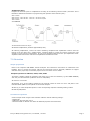
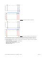
Devyser HFE
For IVD performance evaluation only
Instructions for Use
1. Introduction to Devyser HFE
Intended use
The Devyser HFE kit is intended for IVD performance evaluation for identification of mutations in the HFE
gene in clinical samples.
Included in the kit
The Devyser HFE kit contains ready-to-use reagents for specific PCR amplification of genetic regions in the
HFE gene.
Test procedure
DNA extraction – The Devyser HFE kit has been validated using QIAamp DNA Blood Mini Kit (Qiagen,
cat#51104) for extraction of DNA from human whole blood.
Amplification – The Devyser HFE kit has been validated using ABI GeneAmp® Systems 9700.
Detection - Applied Biosystems Genetic Analyzers (ABI PRISM® 310, 3100, 3130, 3500) that support
detection of Devyser Dye-Set DEV-5 or ABI Dye-Set D (DS-30).
Principle of the Procedure
HFE Hereditary Hemochromatosis (HFE) is an autosomal recessive disorder of iron metabolism wherein the
body accumulates excess iron. Accumulation of iron in a variety of organs leads to impairment and severe
damage in the affected organs. Common complications include liver cirrhosis, diabetes, hepatomas, arthritis
and cardiomyopathies. Early onset of treatment is of great importance since severe effects of the disease
usually do not appear until after decades of progressive iron loading. Removal of excess iron by therapeutic
phlebotomy decreases morbidity and mortality if instituted early in the course of the disease. Early
identification of presymptomatic HFE patients thus represents a major chronic disease prevention opportunity.
HFE is one of the most common inherited diseases among individuals of Northern European decent with up to
1 in 200 affected and an estimated carrier frequency of up to 1 in 8 individuals.
Classical, or type 1, HFE is caused by mutations in the HFE gene on chromosome 6p21.3. More than 80% of
all HFE patients are homozygous for the C282Y mutation. In addition, increased risk of disease is related with
the appearance of the compound heterozygotes C282Y/H63D and S65C/C282Y.
Devyser HFE, Investigational Use, 7-A020-EN, v10-2011
Page 1
2. Warnings and Precautions
A.
Devyser HFE has been validated using a total PCR reaction volume of 25 µL. Changing the reaction volume
will compromise the kit performance.
B.
Avoid microbial contamination of reagents when removing aliquots from reagent vials. The use of sterile
disposable aerosol barrier pipette tips is recommended.
C.
Do not pool reagents from different lots or from different vials of the same lot.
D.
Do not use a kit after its expiry date.
E.
Do not use opened or damaged kit reagent vials.
F.
Work flow in the laboratory should proceed in a uni-directional manner, beginning in the reagent
preparation area and moving to the DNA extraction area and then to the amplification area and finally to the
detection area. Pre-amplification activities should begin with reagent preparation and proceed to DNA
extraction. Reagent preparation activities and DNA extraction activities should be performed in separate
areas. Supplies and equipment should be dedicated to each activity and not used for other activities or
moved between areas. Gloves should be worn in each area and should be changed before leaving that area.
Equipment and supplies used for reagent preparation should not be used for DNA extraction activities or for
pipetting or processing amplified DNA or other sources of target DNA. Amplification and detection supplies
and equipment should remain in the amplification and detection area at all times.
G.
Handling of kit components and samples, their use, storage and disposal should be in accordance with the
procedures defined by national biohazard safety guidelines or regulations.
H.
Wear powder free disposable gloves, laboratory coats and eye protection when handling specimens and kit
reagents. Wash hands thoroughly after handling specimens and kit reagents.
Devyser HFE, Investigational Use, 7-A020-EN, v10-2011
Page 2
3. Symbols used on Labels
Lot or batch number
Expiry date
Manufacturer
For IVD performance evaluation only
Number of tests
Store below temperature shown
4. Required Material
4.1 Included in the Devyser HFE kit (#8-A030)
Configuration
The Devyser HFE kit contains reagents for analysis of maximum 25 samples.
Components
Cap
Colour
Tube
Colour
Label
Art.Nr.
Kit Content
Orange
Clear
PCR Activator
4-A018
1x25 Tests
White
Amber
Devyser HFE Mix
4-A131
1x25 Tests
Devyser HFE, Investigational Use, 7-A020-EN, v10-2011
Page 3
4.2 Required but Not Provided
Reagent Preparation
Consumables for the Thermal Cycler.
Micropipette/dispenser with aerosol barrier tips or displacement tips (500 µL).
Disposable protective gloves (powder free).
DNA Extraction
Reagents and equipment according to manufacturer instructions for use.
Micropipette/multipipette with aerosol barrier tips.
Amplification
Thermal Cycler: ABI GeneAmp® PCR System 9700 using 9600 mode. For use of alternative thermal cyclers
the following ramping rates must be applied: heating @ 0.8 C/s, cooling @ 1.6 C/s.
Micropipette/dispenser with aerosol barrier tips or displacement tips (5, 20 µL).
Detection
Applied Biosystems Genetic Analyzer (ABI PRISM® 310, 3100, 3130, 3500).
Performance optimized polymers: POP-4 or POP-7
Hi-Di Formamide, Genetic Analysis Grade (ABI cat.#4311320).
1x Genetic Analyzer Buffer
Micropipette/multipipette/dispenser with aerosol barrier tips or displacement tips (1,5 uL, 15 uL).
Size Standard:
Devyser Dye Set DEV-5: 560 SIZER ORANGE (Devyser cat.# 8-A402)
ABI Dye Set D: Gene-Scan-500 ROX Size Standard (ABI cat.#401734/#4310366).
4.3 Dye Set Calibration:
ABI3100/3130/3500/3730:
DEV-5 Dye Set MultiCap kit (Devyser cat# 8-A401 )
or
DS-30 (Dye set D) Matrix Standard Kit (ABI cat.#4345827).
ABI310:
DEV-5 Dye Set SingleCap kit(Devyser cat# 8-A400)
Run with module file “GS STR POP4 (1 mL) G5.md5”
or
ABI Dye Set D: 6FAM™ (a), HEX (a), ROX™ (a) and NED™ (b).
Run with module file “GS STR POP4 (1 mL) D.md4”
(a): Fluorescent Amidite Matrix Standards [6FAM™, TET, HEX, TAMRA™, ROX™] (ABI cat# 401546)
(b) NED™ Matrix Standard (ABI cat # 402996)
Devyser HFE, Investigational Use, 7-A020-EN, v10-2011
Page 4
5. Storage and Handling Requirements
A.
Store all components below -18°C.
B.
Reaction Master Mix (prepared by addition of Devyser HFE Mix to PCR Activator) may be stored at +2 to
+8°C for at least 7 days and at below –18 C for at least 90 days. Avoid repeated freeze-thawing.
C.
Dispose of unused reagents and waste in accordance with country, federal, state and local regulations.
D.
Do not mix reagents from different kit lot numbers.
6. Sample Requirements
Clinical Samples
The Devyser HFE kit is for use with human genomic DNA.
Procedure and Storage
According to manufacturer instructions for use.
Controls
It is recommended that suitable controls such as normal DNA and negative control (no DNA) are included in
each run (see section 8).
7. Instructions for Use
Run Sizes
Each Devyser HFE kit (Art # 8-A030) contains reagents for 25 samples.
It is recommended that the activated reaction mix is dispensed into appropriate PCR reaction vials after
preparation. Before dispensing ensure that the activated reaction mix is properly mixed (see section 7.1).
Dispense in 20 µL aliquots and store at below – 18 °C.
To avoid contamination always use un-opened vials. Any reagents left in opened vials should be discarded.
7.1 Workflow Devyser HFE kit (Art # 8-A030)
The Reaction Master Mixes should be prepared before preparing the samples, if the complete process is
performed in one day. Only if the samples are prepared the day before amplification or earlier, the opposite
order is advisable.
Devyser HFE, Investigational Use, 7-A020-EN, v10-2011
Page 5
The Reaction Master Mix is prepared by adding the Devyser HFE Mix to the PCR Activator.
Devyser HFE has been validated using a total PCR reaction volume of 25 µL. Changing the reaction volume
will compromise the kit performance.
Needed reagents from the kit are PCR Activator and Devyser HFE Mix.
Reagent Preparation Area:
1. Centrifuge each tube briefly to collect the content. Do not vortex the tubes at this step!
2. Carefully add 500 µL Devyser HFE Mix to the PCR Activator tube.
NOTE! > 3. Mix manually by pipetting 300-500 µL several times from the bottom of the tube.
4. Vortex the Reaction Master Mix vial and centrifuge briefly to collect the content.
5. Add 20 µL of Reaction Master Mix to separate PCR reaction tubes (8-strip of tubes or micro well plate).
6. Cap the reaction tubes and centrifuge briefly to collect the contents.
The Reaction Master Mix is stable at +2-8°C for at least 7 days and at below –18 C for at least 90 days.
Avoid repeated freeze-thawing.
7.2. Sample Preparation And PCR amplification
DNA Extraction
According to manufacturer’s instructions for use.
DNA concentration and DNA purity are important factors for successful testing using the Devyser HFE kit.
DNA should be free from contaminating protein and salts. Poor quality DNA may result in increased
background or amplification failure. Addition of too much or too little DNA to the PCR reaction can cause
amplification failure.
It is recommended that alternative DNA extraction methods and sample materials are thoroughly evaluated
with the Devyser HFE kit prior to the results being used for diagnostic use.
For recommended PCR conditions and analysis settings (see below), results are consistently obtained at DNA
concentrations between 10 and 30 ng genomic DNA/µL of sample. We recommend the use of DNA solutions
with a concentration of 20 ng/µl. The use of excess amounts of DNA can lead to the appearance of false
positive peaks.
Addition of Sample
Samples should be added in a dedicated area separated from reagent preparation, amplification and detection
areas.
1. Add 5 µL of clinical sample (10-30 ng genomic DNA/µL of sample) to dedicated PCR reaction tubes
containing Reaction Master Mix (from step 7.1)
2. Cap the tubes and centrifuge briefly to collect the content.
Amplification
Turn on the Thermal Cycler at least 30 minutes prior to amplification.
For ABI GeneAmp® System 9700 set “ramp speed” to “9600 mode”.
For use of alternative thermal cyclers the following ramping rates must be applied: heating @ 0.8 C/s, cooling
@ 1.6 C/s.
Devyser HFE, Investigational Use, 7-A020-EN, v10-2011
Page 6
Amplification Area:
Program the Thermal Cycler for amplification according to the following Thermo Profile (consult the User´s
Manual for additional information on programming and operation of the thermal cycler):
95°C 15 min
94°C 30 sec - 60°C 60 sec - 72°C 60 - sec for 3 cycles
94°C 30 sec - 64°C 60 sec - 72°C 60 - sec for 27 cycles
72°C 15 min
4°C FOREVER
1. Set reaction volume to 25 µL.
2. Start the amplification (duration approximately 2hrs).
3. Following amplification, remove the tubes containing completed PCR amplification reaction from the
thermal cycler and place into a suitable holder. Centrifuge briefly to collect the content. Remove the caps
carefully to avoid aerosol contamination. Do not bring amplified material into the pre-amplification areas.
Amplified material should be restricted to amplification and detection areas.
7.3. Detection
Sample preparation
Refer to the respective ABI PRISM® Genetic Analyzers User Manual for instructions on maintenance and
handling. Prior to running the Devyser HFE kit, the instrument must be spectrally calibrated to support
detection of the proper dye-set with the polymer used. See section 4.3 for details.
Sample Preparation for ABI 310/ 3100/ 3130 /3500
1. Prepare a loading cocktail by combining and mixing 2 µL of the size standard (e.g. 560 SIZER ORANGE)
with 100 µL Hi-Di Formamide (sufficient mix for 6 wells/tubes).
2. Vortex for 15 seconds.
3. Dispense 15 µL of the loading cocktail into the required number of wells of a micro well plate or into
individual tubes (ABI310) to be placed on the Genetic Analyzer.
4. Add 1,5 µL of the sample PCR product to the corresponding well/tube containing loading cocktail.
5. Seal the plate/tubes.
Instrument Preparation
Create a sample sheet using the data collection software with the following settings:
» Sample ID.
» ABI Prism: Dye Set: DEV-5 or D.
» Recommended run Module: See below for different polymers and instruments.
Devyser HFE, Investigational Use, 7-A020-EN, v10-2011
Page 7
Run Modules
ABI 310
Run Parameters
Capillary length
POP-4
47 cm
Run temperature
60
Injection voltage
8 kV
Injection time
Run voltage
Run time
2s
15 kV
30 min
ABI 3100/3130
Run Parameters
Capillary length
POP-4/POP-7
36 cm
Run temperature
60
Injection voltage
1,5 kV
Injection time
10 s
Run voltage
15 kV
Run time
1800 s
ABI 3500
Run Parameters
Capillary length
POP-7
50 cm
Run temperature
60
Injection voltage
1,6 kV
Injection time
Run voltage
8s
19,5 kV
Run time
1300 s
The amount of PCR product injected into the capillaries can be adjusted by increasing/decreasing the
injection time and/or injection voltage.
8. Results and Analysis
Principle of Detection
Mutation and wild type alleles are selectively amplified by PCR. By the use of fluorescently labeled primers the
visualization and identification of the PCR products are performed using a Genetic Analyzer with associated
software. Wild type alleles are detected in the BLUE channel. Mutation alleles are detected in the GREEN
channel
Data Analysis
The allele specific peaks in the electrophoretic trace should be identified according to their PCR fragment
length and fluorescent label as presented in the allele overview table below.
Detected alleles in the HFE Gene
Wildtype (Wt)
Codon
Mutation (Mut)
Base Change
Fragment lengths and colour*
HFE-63
Wt: His63
Mut: Asp63 (H63D)
CAT>GAT
Wt: 205 bp BLUE
Mut: 202 bp GREEN
HFE-65
Wt: Ser65
Mut: Cys65 (S65C)
AGT>TGT
Wt: 197 bp BLUE
Mut: 194 bp GREEN
Wt: Cys282
TGC>TAC
Mut: Tyr282 (C282Y)
*Based on observed fragment lengths using ABI3130 and POP7.
HFE-282
Devyser HFE, Investigational Use, 7-A020-EN, v10-2011
Wt: 140 bp BLUE
Mut: 136 bp GREEN
Page 8
Flourescence Criteria
ABI 310/3100/3130: The acceptable range for peak height is between 150 and 6000 relative fluorescence
units (rfu). If peak height exceeds 6000 rfu it is recommended that the sample is re-injected with reduced
injection time and/or injection voltage.
ABI 3500: The acceptable range for marker peak height is between 150 and 25000 relative fluorescence units
(rfu). If peak height exceeds 25000 rfu it is recommended that the sample is re-injected with reduced
injection time and/or injection voltage.
Control samples
Determine that the control samples are approved prior to analyzing the clinical samples.
Negative Control
No specific peaks should be present within the range 100-250 bp.
Normal DNA Control (homozygous for the wild type alleles)
Three wild type specific peaks in the BLUE channel must be detected.
No peaks higher than 150 rfu should be present within the range 100-250 bp in the GREEN channel.
Clinical Samples
Determine the presence or absence of wild type and mutation specific allele peaks:
Homozygous wild type: A sample is considered a homozygous wild type for a specific codon when the wild
type signal in the BLUE channel is within the acceptable length and fluorescence criteria ranges and no GREEN
signal higher than 150 rfu for the same codon is detected in the sample. See Figure 1 for example.
Homozygous mutation: A sample is considered to be homozygously mutated for a specific codon when the
mutation specific signal in the GREEN channel is within the acceptable length and fluorescence criteria ranges
and no BLUE signal higher than 150 rfu for the same codon is detected in the sample. See Figure 2 for
example.
Heterozygous mutation: A sample is considered to be heterozygously mutated for a specific codon when both
the wild type specific BLUE and the mutation specific GREEN signals for the same codon are detected in the
sample. See Figure 3 for example.
Result examples
Figure 1. Homozygous wildtype.
Devyser HFE, Investigational Use, 7-A020-EN, v10-2011
Page 9
Figure 2. Homozygous mutation in codon 282.
Figure 3. Heterozygous mutations in codons 282 and 63.
If the electrophoretic traces are of poor quality (peak smears/aberrant sizing or electrophoretic spikes) it is
recommended that the sample is re-injected using fresh reagents for electrophoresis (Polymer, buffer etc.).
If a
»
»
»
»
»
»
marker displays inconclusive results or a single marker is not detected a number of reasons are possible:
Primer site deletion
Contaminating DNA: second genotype, PCR amplicons.
Primer site polymorphism/mutation.
DNA concentration used is too high or too low.
DNA used in PCR is degraded.
Electrophoretic spike
Devyser HFE, Investigational Use, 7-A020-EN, v10-2011
Page 10
9. Procedural Limitations
A.
Use of this product should be limited to personnel trained in the techniques of PCR.
B.
Devyser HFE has been validated using the ABI Thermal Cycler GeneAmp® 9700. It is recommended that
alternative thermo-cycler instruments are thoroughly evaluated with the Devyser HFE kit prior to the results
being used for diagnostic use.
C.
The Devyser HFE kit has been validated using QIAamp DNA Blood Mini Kit for extraction of DNA from human
whole blood. Performance with other matrices and DNA extraction kits has not been validated and may
result in false negative or false positive results.
D.
Devyser HFE should be used only for identification of patients carrying mutations in the HFE gene according
to the instructions for use. The assay has not been validated for diagnosis of Hereditary Hemochromatosis.
Results obtained with Devyser HFE can only be directly applied to the tissue or specific sample material
tested.
E.
The Devyser HFE kit has been validated for qualitative mutation analysis only. The kit is not validated for
quantitative or relative mutation analysis.
10. Notice to Purchaser
Results from Devyser HFE should be interpreted with consideration of the overall picture obtained from
clinical and laboratory findings. Devyser AB will not accept responsibility for any clinical decisions taken.
Purchase of this product does not provide a license to perform PCR under patents owned by any third party.
11. Contact Information
Devyser AB
Instrumentvägen 19
SE-126 53 Hägersten
SWEDEN
Phone: +46-8-562 15 850
Homepage: www.devyser.com
Technical Support
Phone: +46-8-562 15 850
E-mail: [email protected]
Devyser HFE, Investigational Use, 7-A020-EN, v10-2011
Page 11