1
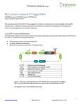
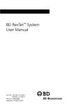
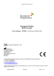
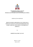
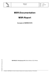
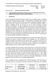
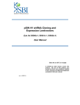
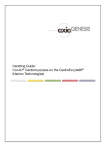
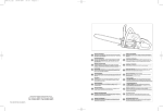
PLATINUM Select MLP Retroviral shRNA-mir
TRH1000, TRM1001, TRHS1000, TRMS1001, TRHK1000, TRMK1001, TRH5000, TRM5000
Overview
PLATINUM Select MLP Retroviral microRNA-adapted short hairpin RNA (shRNA-mir) are next
generation vector-based RNAi triggers generated using the proprietary shERWOOD Algorithm
developed in Dr. Greg Hannon’s laboratory at Cold Spring Harbor Laboratories. Based on the
functional testing of 270,000 shRNA sequences using a high-throughput sensor assay (Fellman
et al 2011 and unpublished data) the shERWOOD algorithm has been trained to select shRNAmir designs that are rare and consistently potent even at single copy representation in the
genome. The new SHERWOOD algorithm predicted sequences have been applied to the
creation of new PLATINUM Select MLP Retroviral shRNA-mir collections targeting human,
mouse and rat genomes.
Platinum Select MLP Retroviral shRNA-mir collections are available as individual constructs,
target gene sets, gene families as well as pathways and genome libraries.
Table 1: shRNA-mir clone formats available
Format
Description
Cat #
Individual clones
Human retroviral shRNA-mir
Mouse retroviral shRNA-mir
TRH1000
TRM1001
Target gene set:
3-4 clones targeting a
gene
Human retroviral shRNA-mir
Mouse retroviral shRNA-mir
TRHS1000
TRMS1001
Starter kit:
3-4 clones targeting a
gene, controls and
transfection reagent
Gene families and
pathway sets
Human retroviral shRNA-mir
Mouse retroviral shRNA-mir
TRHK1000
TRMK1001
Human retroviral shRNA-mir
TRH3004, TRH3006,
TRH3007, TRH3010,
TRH3005, TRH3001,
TRH3002, TRH3003
Genome library
Human retroviral shRNA-mir
TRH5000
Mouse retroviral shRNA-mir
TRM5000
Table 2: shRNA-mir clone formats available
The information and protocols contained in this manual have been developed for the use of all the above formats.
www.transomic.com
866-833-0712
1
[email protected]
shRNA-mir design and vector information
PLATINUM Select shRNA-mir have the following characteristics:
Hairpin design
shRNA-mir sequences are embedded in a primary microRNA-30 context so as to undergo
processing via the endogenous microRNA pathway, which has been shown to minimize
competition with endogenous microRNAs by the exogenous sequences (Castanotto 2007).
Simple stem loop hairpins have been shown to be disruptive to normal microRNA function and
processing while microRNA-adapted shRNA (shRNA-mir) display reduced toxicity and off-target
effects when compared with simple stem loop hairpin sequences. (Grimm et al. 2006, McBride
et al. 2008, Beer et al. 2010, Pan et al. 2011). The shRNA-mir expression cassette consists of a
22-nucleotide target-gene-specific sequence, a 19-nucleotide loop, and another 22-nucleotide
reverse complementary sequence, thus forming a hairpin.
shERWOOD algorithm designs have been created per target gene according to the following
criteria:
• Hairpin sequences target the coding regions of Refseq annotated genes.
• Multiple transcript genes were targeted by designs that spanned across all transcripts.
In the few cases (less than 0.5%) where there was insufficient sequence design space
that spanned across all transcripts - each transcript has a separate design.
pMLP vector information
The pMLP shRNA-mir expression vector has a number of features allowing both transient and
stable transfection; as well as the stable delivery of the shRNA-mir expression cassette into host
cells via a replication-incompetent retrovirus. The shRNA-mir expression cassette; sense, loop
and antisense elements are all under the control of the viral LTR promoter.
The shRNA-mir sequences have been cloned in to the pMLP vector, which has the following
characteristics:
• MSCV-based retroviral vector for delivery and expression in most mammalian cell lines
including murine or human hematopoietic and embryonic stem (ES) cells.
• shRNA-mir constructs are expressed from the retroviral LTR promoter.
• The ability to select stable integrants using puromycin selection.
• TurboGFP serves as a marker for retroviral integration.
The pMLP vector contains both 5’ and 3’ LTRs of MSCV. Upon transfection of the plasmids into a
packaging cell line, replication-incompetent high titer virus can be obtained and used to infect
target cells. The pMLP vector is identical to the Murine Stem Cell Virus (MSCV) vectors derived
from the Murine Embryonic Stem Cell Virus (MESV) and the LN retroviral vectors (Hawley et al.
1994, Grez et al. 1990, Miller and Rosman 1989).
Once transfected or transduced in to the experimental cell line, the pMLP vectors achieve
stable, high-level gene expression in hematopoietic and embryonic stem cells through a
specifically designed 5' long terminal repeat (LTR). This LTR is from the murine stem cell PCMV
www.transomic.com
866-833-0712
2
[email protected]
virus, and it differs from the MoMuLV LTR used in other retroviral vectors by several point
mutations and a deletion. These changes enhance transcriptional activation and prevent
transcriptional suppression in embryonic stem and embryonal carcinoma cells. As a result, the
LTR drives high-level constitutive expression of the shRNA-mir transcript in stem cells or other
mammalian cell lines. The murine phosphoglycerate kinase (PKG) promoter controls expression
of the puromycin resistance gene (Puromycin resistance) for antibiotic selection in eukaryotic
cells. pMLP also contains the pUC origin of replication and E. coli Ampicillin resistance gene for
propagation and antibiotic selection in bacteria.
The unique sequence specificity of the potent, mir-based PLATINUM Select shRNA-mir; placed
in the versatile, efficient pMLP retroviral vector, provides a highly successful method to knock
down a gene of interest either transiently or stably to allow gene function analysis at a new and
improved level.
5’ LTR
miR 30 flanks
PGK
Puro
IRES
TurboGFP
3’ LTR
Ψ+
Beta-lactamase
pUC Ori
Modified MSCV LTR for enhanced expression
Create a natural substrate for microRNA processing
Strong promoter across many cell lines
Mammalian selection marker
Internal ribosomal entry site
Fluorescent marker with high photostability
Transcriptional stop
Viral packaging signal
Ampicillin antibiotic resistance genes
High-copy origin of replication for propagation in E. coli
Figure 1: Cartoon depicting elements of the pMLP vector containing the shRNA-mir sequence. The vector
elements table describes the utility of the various elements shown.
See appendix 1 for a more detailed vector map.
www.transomic.com
866-833-0712
3
[email protected]
Replication protocols
Materials for individual and plate replication
Catalog #
LB-Lennox Broth (low salt)
Glycerol
Carbenicillin
96-well plates
Aluminum seals
Disposable replicators
VWR EM1.00547.0500
VWR EM-4760
VWR 97063-144
VWR 62407-174
VWR 29445-082
Genetix X5054
Grow all pMLP retroviral shRNA-mir clones at 30°C for 30 hours or until even growth is observed in all wells.
Autoclave broth to sterilize and allow cooling to 60°C before adding the antibiotic.
Individual shRNA-mir clones
E. coli carrying pMLP retroviral shRNA-mir are best propagated in LB broth or LB broth 8%
glycerol for freezing.
1. Grow the shRNA-mir clone culture in LB broth with the ampicillin or carbenicillin
(100 μg/ml).
2. 2-10 ml starter cultures for plasmid purification can be inoculated using 2 to 10 µl.
Alternatively
1. Pick a single starter colony from a freshly streaked LB agar plate containing the
antibiotic and inoculate into the desired volume of LB broth for plasmid
purification.
2. Grow for 30 hours at 30°C for with vigorous shaking (~300 rpm).
Replication of plates
Prepare target plates by dispensing ~200 μl of LB-Lennox media supplemented with 8% glycerol
and 100 μg/ml carbenicillin. If a lower-volume 96-well plate is substituted, then fill each well
~50% with media. Glycerol can be omitted from the media if you are culturing for a plasmid
DNA extraction.
Prepare source plates
1. Remove foil seals while the source plates are still frozen. This minimizes crosscontamination.
2. Wipe any condensation underneath the lid with a paper wipe dampened with ethanol.
3. Thaw the source plates with the lid on.
Replicate
1. Gently place a disposable replicator in the thawed source plate and lightly move the
replicator around inside the well to mix the culture. Make sure to scrape the bottom of
the plate of the well.
www.transomic.com
866-833-0712
4
[email protected]
2. Gently remove the replicator from the source plate and gently place in the target plate
and mix in the same manner to transfer cells.
3. Dispose of the replicator.
4. Place the lids back on the source plates and target plates.
5. Repeat steps 1-4 until all plates have been replicated.
6. Return the source plates to the -80°C freezer.
7. Place the inoculated target plates in a 30°C incubator for 30 hours or until even growth
is observed in all wells.
Minimize thawed condition of plates where possible.
Always store plates at -80°C. It is recommended that an archival copy is made as soon as possible.
Glycerol stocks kept at -80°C are stable indefinitely as long as freeze/thaw cycles are kept to a minimum.
Plasmid preparation
For transfection and transduction (infection) experiments the pMLP plasmid DNA will first have
to be extracted from the bacterial cells. When transforming directly into an experimental cell
line, either a standard plasmid mini-preparation can be used or one that yields endotoxin free
DNA. When extracting plasmid DNA to make virus for transduction, more DNA is required and
using and endotoxin free kit will generally yield higher viral titers
Restriction digest
To carry out a diagnostic restriction digest on pMLP retroviral shRNA-mir vectors it is
recommended that the plasmid is digested with NcoI. NcoI cuts pMLP three times on either side
of the LTRs to generate three bands of 423bp, 3618bp and 3761bp in length.
Digest approximately 500ng of plasmid and allow the reaction to proceed for 3 hours.
Load half the digestion mixture on a 0.7% gel to visualize the resultant bands.
1
2
3
4
5
6
7
NEB TriDye 2-log DNA Ladder
Sample 1 uncut
Sample 2 cut (NcoI)
Sample 3 uncut
Sample 4 cut (NcoI)
Sample 5 uncut
Sample 6 cut (NcoI)
Figure 2: Agarose gel (0.7%) depicting pMLP samples contain shRNA-mir undigested or digested with NcoI
www.transomic.com
866-833-0712
5
[email protected]
Puromycin selection (puromycin kill curve)
The pMLP retroviral vector has a puromycin resistance marker for selection in mammalian cells.
To establish stable cell lines, once transfection/transduction has occurred, the cells can be
placed on puromycin to select for stable integrants. Since each cell line potentially has a
different sensitivity to puromycin, the optimal concentration of puromycin (pretransfection/transduction) should be determined. Below is the protocol for titrating puromycin
(kill curve), using as an example a 24 well tissue culture dish.
1. Prepare appropriate cell culture media and puromycin dilutions for antibiotic titration.
Using a stock solution of puromycin (1.25µg/µl stock recommended) make dilutions in
the media of the antibiotic ranging from 0 to 10 µg/ml (final concentration) in 0.5µg/ml
increments.
Table 3: Dilutions and volumes required for establishing optimal puromycin concentration
Volume of Puromycin Stock
Solution Added (µl)
0
0.2
0.4
0.6
0.8
1
1.2
1.6
2
3
4
Total Volume of Media plus
Antibiotic per 24 Well
500 µl
500 µl
500 µl
500 µl
500 µl
500 µl
500 µl
500 µl
500 µl
500 µl
500 µl
Final Concentration
(µg/ml)
0
0.5
1
1.5
2
2.5
3
4
5
7.5
10
2. Split/dilute cells from a confluent well of a 24 well plate (~ 5 x 10⁴ cells per 500µl
media).
3. Plate ~ 5 x 10⁴ cells per well in media without puromycin and allow enough time for cells
to attach (less than 24 hours for most cell lines) before adding antibiotic.
4. Begin antibiotic selection the following day by replacing with media containing the
appropriate concentrations of puromycin (see Table 3).
5. Incubate cells at 37°C, or use conditions normal for your target cells.
6. Observe the cells for approximately 7 days.
7. Replace the media every 3 days with media containing the correct puromycin
concentration for each well.
8. To minimize duration to achieve fully selected stable, cell populations one should
choose the lowest concentration of drug that begins to give substantial cell death in 3
days and kills 100% of the cells within 5 days.
www.transomic.com
866-833-0712
6
[email protected]
Transfection
OMNIfect transfection reagent for delivery of plasmid DNA
Use the following procedure to transfect plasmid DNA into mammalian cells in a 24-well
format. For other plate formats, scale up or down the amounts of DNA and OMNIfect reagent
proportionally to the total transfection volume (Table 4).
Adherent cells: One day before transfection, plate 80,000 cells/well in 500 μl of growth medium
without antibiotics so that cells will be 70–95% confluent at the time of transfection.
Suspension cells: On the same day of transfection just prior to preparing transfection complex
plate 160,000/well cells in 500 μl of growth medium without antibiotics.
Transfection complex preparation (Figure 3):
Volumes and amounts are for each well to be transfected.
1. Plasmid DNA preparation: Dilute 0.5 µg of plasmid DNA in a microfuge tube containing
Opti-MEM® I Reduced Serum Media*** up to a total volume of 25 µl.
2. OMNIfect reagent preparation: In a separate microfuge tube, add 1 µL of OMNIfect into
24 µl Opti-MEM® I Reduced Serum Media*** for a total volume of 25 µl.
3. Final transfection complex: Transfer the diluted DNA solution to the diluted OMNIfect
reagent (total volume = 50 µl). Mix gently and incubate at room temperature for 10
minutes.
Adding transfection complex to wells:
1. Add the 50 µl of transfection complex to each well containing cells and medium.
2. Incubate cells at 37°C in a CO2 incubator for 24-48 hours.
3. After 24-48 hours of incubation, assay cells for gene activity.
*** serum-free DMEM medium can also be used.
www.transomic.com
866-833-0712
7
[email protected]
Figure 3: Transfection protocol for 24 well plates (volumes indicated are per well). To transfect the entire plate
multiply all volumes and DNA amount by 24.
Table 4: Suggested amounts of DNA, medium and OMNIfect for transfection of plasmid DNA into adherent and
suspension cells.
Tissue
Culture
Plates
Surface
Area per
Well (cm2)
µl Plating
Medium per
Well
6- well
9
2000
µg Plasmid DNA
per Well
µl OMNIfect per
Well
µl Transfection
Complex per
Well†
2
4
200
(in 100 µl
(in 100 µl
Opti-MEM® I)
Opti-MEM® I)
12-well
4
1000
1
2
100
(in 50 µl
(in 50 µl
Opti-MEM® I)
Opti-MEM® I)
24-well
2
500
0.5
1
50
(in 25µl
(in 25µl
Opti-MEM® I)
Opti-MEM® I)
96-well
0.3
200
0.1
0.2
10-20
(in 10µl
(in 10µl
Opti-MEM® I)
Opti-MEM® I)
† Total volume of the transfec-on complex is made up of equal parts of DNA solution and OMNIfect
solution.
www.transomic.com
866-833-0712
8
[email protected]
Optimizing your transfection:
It is important to optimize transfection conditions to obtain the highest transfection efficiency
with lowest toxicity for various cell types. The optimal ratio of OMNIfect to DNA is relatively
consistent across many cell types. For further optimization try the following steps in order.
1. Use the recommended ratio of DNA: transfection reagent (at 1 μg DNA:2 μl OMNIfect),
but vary the volume.
a. Start with a range of volumes that cover +20% to -20%.
For example, in a 24-well plate a range of 40 μl to 60 μl of transfection complex
would be added to the well. (The plating media would remain the same.)
2. If further optimization is needed, transfection efficiency and cytotoxicity may be altered
by adjusting the ratio of DNA (μg) to OMNIfect reagent (μl). A range of ratios from
1:1.5 to 1:2.5 is recommended.
Note: If transfection conditions result in unacceptable cytotoxicity in a particular cell line the
following modifications are recommended:
1. Decrease the volume of transfection complex that is added to each well.
2. Higher transfection efficiencies are normally achieved if the transfection medium is not
removed. However, if toxicity is a problem, aspirate the transfection complex after 6
hours of transfection and replace with fresh growth medium.
3. Increase the cell density in your transfection.
4. Assay cells for gene activity 24 hours following the addition of transfection complex to
cells.
Puromycin selection of transfected cells
1. After 24-72 hours of incubation, examine the cells microscopically for TurboGFP
expression.
a. Note: When visualizing TurboGFP expression, if less than 90% of all cells are
green, it is recommended in these cases to utilize puromycin selection in order
to reduce background expression of your gene of interest from untransfected
cells.
b. The working concentration of puromycin varies between cell lines. We
recommend you determine the optimal concentration of puromycin required to
kill your host cell line prior to selection for shRNAmir transfectants (see
puromycin selection protocol). Typically, the working concentration ranges from
1-10 µg/ml. You should use the lowest concentration that kills 100% of the cells
in 1-4 days from the start of puromycin selection.
2. If selecting for stably transfected cells (optional), change the medium on the cells to that
containing puromycin for selection. It is important to wait at least 24 hours before
beginning selection.
3. Begin the antibiotic selection by replacing the medium with complete medium
supplemented with the optimal puromycin concentration. Incubate.
www.transomic.com
866-833-0712
9
[email protected]
4. Approximately every 2-3 days replace with freshly prepared selective media. Monitor
the cells daily and observe the percentage of surviving cells. At some time point almost
all of the cells surviving selection will be harboring the shRNA-mir construct. Optimum
effectiveness should be reached in 3-6 days with puromycin. Observe the cells for
approximately 7 days until you see single colonies surviving the selection. The negative
control should have no surviving cells.
Packaging retrovirus
Using the recommended packaging system (See the Recommended Packaging System section),
pMLP and an env expressing vector are cotransfected into the GP2-293 Packaging Cell Line. gag
and pol genes, which are necessary for viral replication, are integrated and stably expressed
from the packaging cell line genome. While all of the components needed to produce virus are
present in the packaging cell during this process, only the RNA from the pMLP vector is
packaged into the viral particles and transferred to the target cell. These viral particles are
capable of transduction, however without the ability to express the gag, pol or env genes they
are incapable of producing new viral particles in the targeted cells. These are referred to as
replication-incompetent retroviral particles.
Separating the essential viral structural genes during the packaging process ensures a high level
of safety by limiting the opportunities for recombination during cell division and thereby
minimizing the chance of producing replication competent virus. Examples of similar packaging
systems or those where the gag and pol are provide in cis-acting plasmids are referenced here
Mann et al. 1983, Miller and Buttimore 1986, Morgenstern and Land 1990, Miller and Chen
1996.
Note: The viral supernatants produced by this retroviral vector could, depending on your cloned insert, contain
potentially hazardous recombinant virus. Due caution must be exercised in the production and handling of
recombinant retrovirus. Appropriate NIH, regional, and institutional guidelines apply.
www.transomic.com
866-833-0712
10
[email protected]
Figure 4: Schematic depicting retroviral packaging of pMLP retroviral vectors
A retroviral transfer vector (pMLP) is co-transfected with the desired helper vector encoding
the env protein into a packaging cell line. The gag and pol genes, essential for virus production,
are stably integrated into the cell line’s genome and constitutively expressed. gag, pol and env
provide the proteins necessary for viral assembly and integration. The transfer vector contains
the shRNA-mir and selection cassette that will integrate into the target cell’s genome. Viral
particles are released from the packaging cell and can be harvested from the supernatant of the
packaging cell. This virus can be concentrated or used as is.
www.transomic.com
866-833-0712
11
[email protected]
Recommended Packaging System
The pMLP retroviral shRNA-mir vector can be packaged and viral particles produced in 48 hours
using commercially available packaging kits. The Retro-XTM Universal Expression System from Clontech
(Cat. No. 631530) has been shown to produce high titer virus with the pMLP retroviral vector . The
packaging system provides GP2-293 Packaging Cell Line and all four commonly used envelopes
on separate vectors (VSV-G, eco, ampho and 10A1) to allow you to choose the tropism that is
most appropriate for your target cells. In combination with the Retro-X Concentrator (Cat. No.
7
631455/6) titers of 1 x 10 can be achieved.
Retro-X Packaging kits and Concentrator can be purchased directly from transOMIC
technologies. For more information see : http://transomic.com/Delivery-and-Packaging
Supporting data for Retro-XTM Universal Expression System
Three KRAS Platinum Select shRNA-mir and a negative control (shRNA-mir against firefly
luciferase) were packaged into VSV-G pseudotyped retrovirus particles using the Retro-X
Universal Packaging System (Cat# 631530). Crude retroviral supernatants, as well as
supernatants concentrated using Retro-X Concentrator (Cat#631455/6), were titered to
determine colony forming units (CFU) by puromycin selection or infectious units (IFU) by green
fluorescence (FACS).
Average titers for unconcentrated virus were 2.8x10⁵ CFU/ml and 4.7x10⁶ IFU/ml. While both
methods are considered to be functional titers, variations in expression of turboGFP or
puromycin can cause differences in the titer. The average difference between the two
titrations is typical for these methods.
Concentration of the retroviral supernatants resulted in a 10-fold average increase in titer for a
20X volume concentration. Concentration can be magnified if the fold volume change is
increased. The final concentrated titer was 2.96x10⁶ on puromycin selection and 4.5x10⁷
IFU/ml.
Packaging shRNA-mir viruses
While viruses carrying shRNAs are packaged almost identically to viruses carrying proteinencoding genes, one point is worth noting. Since the host RNAi biogenesis machinery
efficiently cleaves shRNAs, this can reduce the level of viral genomic RNAs and consequently
viral titers. Therefore, one can enhance titers by co-transfecting the viral plasmid with a siRNA
targeting a core microRNA biogenesis component, DGCR-8/Pasha. The following siRNA has
been shown to be effective in increasing titers by several fold.
Pasha siRNA - CGGGTGGATCATGACATTCCA, dXdY overhang, all annealed, no modification
Safety and handling of retroviruses
The protocols in this user manual require producing, handling, and storing infectious retrovirus.
A thorough understanding of safe laboratory practices and potential retroviral hazards is
essential.
MSCV does not naturally infect human cells; however, viruses packaged from the MSCV-based
www.transomic.com
866-833-0712
12
[email protected]
vectors are capable of infecting human cells if packaged in system with the correct tropism. The
viral supernatants produced by these retroviral systems could, depending on your retroviral
insert, contain potentially hazardous recombinant virus.
For these reasons, exercise due caution when producing and handling recombinant retrovirus.
Appropriate NIH, regional, and institutional guidelines apply, as well as specific guidelines for
other countries. Contact your on-site safety officer for specific requirements. In the United
States, NIH guidelines require that retroviral production and transduction be performed in a
Biosafety Level 2 (BL2) facility. A brief description of BL2 is given below. More information
about BL2 guidelines is available in Appendix 4.
Practices
• Perform work in a limited access area
• Post biohazard warning signs
• Minimize production of aerosols
• Decontaminate potentially infectious wastes before disposal
• Take precautions with sharps
Safety equipment
• Use a laminar flow hood with a HEPA filter
• Wear protective laboratory coat, face protection, and double gloves Facilities
• Autoclave for decontamination of solid and liquid waste
• Use un-recirculated exhaust air
• Stock chemical disinfectants for spills
Establishing viral titers
Establishing viral titers is an important step in optimizing an experiment. Knowing the viral titers allows
you to do the following:
1. Determine the efficiency of the packaging procedure.
2. Optimize conditions for infection of your target cell line.
a. Infection efficiency differs based on target cell line and media.
3. Determine the number of cells that can be infected with your current stock of virus.
Recommendations for titering pMLP shRNA-mir retroviral particles
When measuring the amount of virus you have produced it is important to understand the different
methods for measuring the titer. Physical titers measure the presence of viral proteins. These methods
are often fast, but detect functional and non-functional viral particles. Methods that measure only
those viral particles that can integrate and express in a target cell are more accurate and are referred to
as functional titers.
www.transomic.com
866-833-0712
13
[email protected]
The pMLP vector includes two markers for functional titers. TurboGFP expression can be measured with
fluorescent microscope or FACS analysis or colonies can be counted following puromycin selection.
Functional titers may differ depending on the method used to confirm integration of the virus. For
example, cell lines that are particularly sensitive to puromycin selection maybe turboGFP positive but
die during puromycin selection. Therefore, for the most precise functional titer we recommend using
the selection method that will be adopted in your experiment.
Transduction mammalian cells with MLP retroviralshRNA-mir viral particles
Reagents
Cell line to be infected
Appropriate culture media
Polybrene solution
Tissue culture plates and plastic-ware
Polybrene solution
Suspend 8 mg Hexadimethrine Bromide into 1ml H2O for an 8mg/ml stock
Filter through 0.2um syringe filter
Equipment
Tissue culture incubators
Platform rocker
Low-speed centrifuge and adapters that can hold multi-well plates or desired culture plates
Method
General considerations:
Growth media of the target cell can affect transduction efficiency if it is differs from that of the
packaging cell line. Here, the investigator should experiment with exchanging the media on the
packaging cells with that used for the target cell type 12 hours before virus harvest.
Alternatively, the target cells might remain unaffected by a brief exposure to virus-containing
media while still being infected efficiently.
The method is given for 10 cm plates. Infection can be carried out in any standard tissue culture
vessel, scaling all amounts based upon surface area.
www.transomic.com
866-833-0712
14
[email protected]
Prepare cells
1. Cells should be plated at least one day prior to infection such that they are 60-70%
confluent at the time of initial virus exposure. The optimal density can vary for different
cell lines and should be determined empirically.
a. Note: Most cell lines require 12-24 hours to recover after plating for optimal
infection efficiency. This is thought to be in part due to loss of cell surface
receptors, used by the virus, during the trypsinization step when recovering
adherent cell lines.
2. Exchange medium 3-4 hours prior to infection.
Infect cells
3. Remove medium and then add a combination of viral supernatant and fresh medium for
a total volume of 10ml. The ratio depends upon the desired infection rate and the viral
titer. For most cells to have a single integrant, use an infection rate around 30% (MOI of
0.3).
4. Add polybrene to a final concentration of 8µg/ml.
a. This aids in infection; however, some cells are sensitive to polybrene exposure.
Therefore, this step can be omitted or the level of polybrene can be reduced to
4-6 µg/ml.
5. Optional step. Load tissue culture plates into a low speed centrifuge and spin for 1 hour
at 800xg. Be sure to turn braking off to avoid spilling media. This step will significantly
increase infection for some cell types but is not recommended for viruses that can infect
human cells because of the risks of contamination of the centrifuge and the exposure of
investigators to potentially hazardous viruses.
6. Transfer cells to a tissue culture incubator. If viruses are pseudotyped with VSV-G, cells
can be incubated at 37°C immediately. However, for viruses bearing ecotropic or
amphotropic envelope proteins, increased infection can often be obtained by incubating
for the first 12 hours at 32°C.
7. After 12-24 hours, it is often advisable to exchange media. This is not necessary if the
viral supernatant corresponded to only a small percentage of the medium used to infect
cells.
8. Assess infection rates, for example by counting GFP-positive cells, 48-96 hours postinfection.
SELECTION
Relative transduction efficiency
As cell types vary in their transduction efficiency it is recommended that a functional titer be
established for the experimental cell line to be used. In order to do this, virus of known titer
produced from one of the negative controls or control virus purchased from transOMIC
www.transomic.com
866-833-0712
15
[email protected]
technologies (See Table 5 below) should be used to establish their transduction efficiency.
Following the titering protocol above, transduce a known number of cells with varying dilutions
of the control virus. Obtain the TU/ml in the experimental cell and calculate the transduction
efficiency as follows:
TU/ml of control in experimental cell line =
Known TU/ml of control virus
Relative transduction efficiency
To proceed with experimentation in the experimental cell line, all viral titers established in
HEK293(T) or NIH323 will have to be multiplied by the relative transduction efficiency to
establish how much virus needs to be added to this cell line to achieve the percentage
transduction desired.
Controls and validation
There are 3 types of MLP retroviral shRNA-mir controls available - these are negative and
positive controls, as well as an empty vector.
Table 5: Categories and uses of controls used RNAi experiments.
Type of control Product
Non-targeting Platinum Select MLP
controls
Retroviral shRNAmir
non-targeting controlFF Luciferase
Platinum Select MLP
Retroviral shRNA-mir
non-targeting controlRFP
Positive
Platinum Select MLP
controls
Retroviral shRNA-mir
positive control-GAPDH
(Hs, Mm)
Empty vector
Platinum Select MLP
control
Retroviral empty vector
Use
Changes in the mRNA or protein levels in cells
treated with negative or non-targeting controls
reflect non-specific responses in cells and can be
used as a baseline against which specific knock
down can be measured.
Used to demonstrate that your RNAi experimental
assay is functional and the shRNA-mir construct is
successfully activating the RNAi pathway.
To detect changes unrelated to the RNAi
component - cellular toxicity or changes in gene
expression due to transfection or transduction
alone.
Measuring knock down
There are a number of ways to establish the level of knock down of a gene after transfection or
transduction with shRNA. These include quantitative PCR (QPCR) of the mRNA, western blots or
assays specifically designed for the function of the gene of interest. Generally, the first tests
done to establish efficiency of knock down are via QPCR on RNA extracted from the treated
cells. Percentage knock down is determined relative to negative controls or empty vectors,
www.transomic.com
866-833-0712
16
[email protected]
transfected or transduced in the same experimental system. Western blots and other assays
would be evaluated in a similar way.
QPCR can be carried out on mRNA or total RNA depending on what the internal control used for
the real-time PCR is (a constitutive mRNA or an 18s rRNA for example).
To obtain reproducible, dependable data; samples and controls should be run in duplicate or
even triplicate. Real time PCR should be done in triplicate to control variation between QPCR
reactions. This means that for each experimental point there are between six and nine PCR
samples.
The location of the QPCR primer/probe set, relative to where the shRNA binds does not
influence the measurement of knock down. However when choosing a primer/probe set make
sure it detects the all the transcripts of the mRNA. In the rare cases that the shRNA is only
against one transcript, ensure that this is the one being detected by the primers.
Western blots are used to establish the knock down of a target gene’s protein. An antibody
against the target protein needs to purchased and verified in the experimental cells to ensure
the effectiveness of the antibody as well as that the cells do produce the protein of interest.
Protein lysates from the knock down experiment will then be detected on a western blot,
comparing the signal from the transfected/transduced cells with that of the negative controls.
Effective gene silencing will result in a lower signal (or no signal) from cells transfected with the
shRNA. Immunofluorescent procedures require the same attention to antibody quality and
expression, as well incorporation of the appropriate controls.
Controls are essential as a reference for target gene expression. The negative controls provided
are ideal for this purpose. The empty vector and an untreated control are also good to have as
experimental data points. The knock down data will be presented as the amount of the gene’s
mRNA/protein in the knocked down cell lines with respect to the negative control.
It is worth noting that any untransfected or untransduced cells in an experiment also contribute
to the relevant mRNA/protein in the QPCR/protein analysis; thus it is advisable to exclude these
untransduced/untransfected cells. A higher transfection, MOI or puromycin selection will insure
that the cells used for QPCR/protein analysis all contain at least one shRNA-mir construct.
Table 6: Methods for knock down verification
Biological
molecule
measured
Method
Advantages
Disadvantages
mRNA level
Real Time PCR (QPCR)
A rapid and sensitive
way to determine
pre-translational
knock down via the
mRNA. This is
recommended as the
first step in
PCR does not
measure the effect of
the shRNA on the
protein levels.
Reduction in the
amount the mRNA
does not always
www.transomic.com
866-833-0712
17
[email protected]
evaluating gene
silencing.
Protein Level:
Direct
Western blotting
Epitope tagging
To determine the
effect of gene knock
down on protein
expression levels.
Protein level:
Cell functional
assay
Cell proliferation
assays
Cytotoxicity assays
Apoptosis assays
ELISAs
Cellular function
assays measure for
general effects on
proliferation,
apoptosis,
cytotoxicity, or other
complex changes
including phenotypic
change. Commercial
kits are available
some of these and
phenotypic changes
often require
specialized
equipment, or
indirect assays.
www.transomic.com
866-833-0712
compare with the
remaining protein
levels in the cell,
especially when the
protein has a long
half-life.
Thus protein knock
may need to
evaluated as well to
establish at what
time point the
protein reflects the
effects of gene knock
down. Experimental
time points would
then have to be
adjusted accordingly
to see the expected
phenotype or
experimental out-put.
A good antibiotic may
not be available
against the protein of
interest. Here a
epitope-tagged
protein may be more
informative
Where the difference
in protein levels is
minimal in certain
experiments, western
blots, while sensitive,
are not sensitive
enough.
Many genes and their
protein are not
covered by a known
assay. Many of the
assays are not
available
commercially.
18
[email protected]
www.transomic.com
866-833-0712
19
[email protected]
Appendices
Appendix 1 – the pMLP vector information
Element
5' LTR
MESV Psi
3' MIR30A context
Mir30 Loop
5' MIR30A context
PGK promoter
IRES
3' LTR
ORI
AmpR
SV40 promoter
SV40 ori
Start
1
581
1423
1574
1614
1770
2954
4300
5973
7004
7396
7563
Stop
517
922
1552
1591
1742
2269
3524
4814
5385
6144
7712
7698
Sequencing primer for shRNAmir
Copy sequence here:
5’ TGTTTGAATGAGGCTTCAGTACTTTACAG 3’
Figure 5: Detailed map of the pMLP vector, vector element table and sequenceing primer
www.transomic.com
866-833-0712
20
[email protected]
Appendix 4 – References
shRNA–mir and design
Fellman et al., 2011. Functional identification of optimized RNAi triggers using a massively parallel
sensor assay. Mol Cell. 18; 41(6):733-46.
Castanotto, 2007.Combinatorial delivery of small interfering RNAs reduces RNAi efficacy by selective
incorporation into RISC. Nucleic Acids Res. 35(15):5154-64.
Grimm et al., 2006. Fatality in mice due to oversaturation of cellular microRNA/short hairpin RNA
pathways. Nature 441, 537-541.
McBride et al., 2008. Artificial miRNAs mitigate shRNA-mediated toxicity in the brain: Implications for
the therapeutic development of RNAi. PNAS USA 2008, 105; 15, 5868-5873
Beer et al., 2010. Low-level shRNA Cytotoxicity can contribute to MYC-induced hepatocellular carcinoma
in adult mice. Mol Ther 18(1):161-170.
Pan et al., 2011. Disturbance of the microRNA pathway by commonly used lentiviral shRNA libraries
limits the application for screening host factors involved in hepatitis C virus infection. FEBS Lett. 2011
6;585(7):1025-1030.
pMLP vector
Grez et al., 1990. Embryonic stem cell virus, a recombinant murine retrovirus with expression in
embryonic stem cells. Proc. Natl. Acad. Sci. USA 87: 9202–9206.
Miller and Rosman, 1989. Improved Retroviral Vectors for Gene Transfer and Expression. BioTechniques
7: 980–990.
Hawley et al., 1994. Versatile retroviral vectors for potential use in gene therapy Gene Ther. 1:136–138.
Packaging
Mann et al., 1983. Construction of a retrovirus packaging mutant and its use to produce helper-free
defective retrovirus. Cell 33:153–159.
Miller and Buttimore, 1986. Redesign of retrovirus packaging cell lines to avoid recombination leading
to helper virus production. Mol. Cell. Biol. 6:2895–2902.
Morgenstern and Land, 1990. Advanced mammalian gene transfer: high titre retroviral vectors with
multiple drug selection markers and a complementary helper-free packaging cell line. Nucleic Acids Res.
18:3587–3590.
www.transomic.com
866-833-0712
21
[email protected]
Miller and Chen, 1996. Retrovirus packaging cells based on 10A1 murine leukemia virus for production
of vectors that use multiple receptors for cell entry. J. Virol. 70:5564–5571.
Safety guidelines for working with retrovirus
http://oba.od.nih.gov/rdna/nih_guidelines_oba.html
www.fda.gov/downloads/AdvisoryCommittees/.../UCM232592.pdf
Biosafety in Microbiological and Biomedical Laboratories, Fourth Edition (May 1999) HHS Pub. No. (CDC)
93-8395. U.S. Department of Health and Human Services, PHS, CDC, NIH.
Controls and validation
Nature Editorial. 2003. Whither RNAi? Nature Cell Biology 5, 489 – 490.
Christophe et al., 2006. High-throughput RNAi screening in cultured cells: a user's guide. Nature Reviews
Genetics 7, 373–384
Infection (transduction) of mammalian cells with retroviral shRNAs
Brummelkamp et al., 2002. A System for Stable Expression of Short Interfering RNAs in Mammalian
Cells. Science; 296:550-553.
Paddison et al.,2002. Short hairpin RNAs (shRNAs) induce sequence-specific silencing in mammalian
cells. Genes Dev. 16(8):948-958.
Zeng et al., 2002. Both Natural and Designed Micro RNAs Can Inhibit the Expression of Cognate mRNAs
When Expressed in Human Cells. Molecular Cell. 9: 1327-1333.
Stegmeier et al., 2005. A lentiviral microRNA-based system for single-copy polymerase II-regulated RNA
interference in mammalian cells. PNAS; 102:37; 13212-13217.
Dickens et al., 2005. Probing tumor phenotypes using stable and regulated synthetic microRNA
precursors, Nature Genetics; Vol 37, No 11 1289-1295.
Limited use licenses
This product is covered by several limited use licenses. For updated information please refer to
www.transomic.com/support/productlicenses
www.transomic.com
866-833-0712
22
[email protected]