1
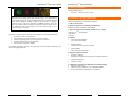
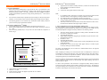
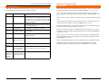
AntibodyArrayTM Instruction Manual
AntibodyArrayTM Instruction Manual
TABLE OF CONTENTS
Introduction ……………………………………………………………………….
I.
List of Components ……………………………………………………………….
II.
2
Additional Materials Required …………………………………………………..
5
Methods ……………………………………………………………………………
6
III.
IV.
A.
B.
C.
D.
E.
F.
G.
H.
I.
AntibodyArray Staining
Instruction Manual
TM
V.
VI.
VII.
General considerations
6
Protocol for AntibodyArrayTM staining
6
Some considerations for observing and analyzing staining results ………….
7
Protocol I for fixation and permeabilization of cultured cells
9
Protocol II for fixation and permeabilization of cultured cells
10
Protocol I for fixation of frozen sections and cell preps ……………………..
10
Protocol II for fixation of frozen Sections and cell preps
10
Steps to take if tissue keeps falling off slides………………………………...
10
Troubleshooting …………………………………………………………………...
12
References …………………………………………………………………………
13
Appendix I. Antibody List by Positions …………………………………………
14
Staining AntibodyArray
18
Hypromatrix, Inc.
9
Protocol for preparing tissue sections ………………………………………..
TM
www.hypromatrix.com
5
I …………………………………………………
14
Trial AntibodyArrayTM……………………………………………………….
14
VIII. Appendix II. Products from Hypromatrix ………………………………………
16
Hypromatrix, Inc.
11
www.hypromatrix.com
AntibodyArrayTM Instruction Manual
AntibodyArrayTM Instruction Manual
I. INTRODUCTION
Immunochemical staining is a versatile technique for determining both the presence and localization of an antigen (Harlow and Lane, 1988). This information is of immense value to biomedical research and clinical medicine. Most of the current methods, all of which involve incubating cells with an antibody solution, only allow cell staining with one or a few antibodies at a
time. These methods are not suitable for applications in which the expressions and sub-cellular
localizations of a large number of different proteins need to be examined.
a.
b.
Fig. 2: Comparison of protein expressions between ME180 cells (left) and
A431cells (right) using antibody arrays with 240 antibodies. Alkaline phosphatase-labeled secondary antibodies were used and the staining was visualized
by color reaction with BCIP/NBT as substrates
d.
Knowledge of a protein’s sub-cellular localization can provide important information about
the protein’s functional state. For example, change of a transcriptional factor’s location from
cytoplasm to nucleus often suggests its activation. When observed under a fluorescence microscope, AntibodyArrayTM staining can reveal sub-cellular localizations of many proteins
simultaneously.
c.
Fig. 1: Overview of AntibodyArrayTM staining technology. a, make contact
between staining antibody array and adherent cells; b, incubate arrays and cells
to allow antibodies to bind to their antigens; c, remove array support; d, detect
bound antibodies.
The AntibodyArrayTM staining method takes advantage of “Dissociable Protein Array” technology, which allows the delivery of a large number of proteins to their targets in a positionaddressable manner. In staining AntibodyArrayTMs, the antibodies are immobilized on a membrane in such a manner that when they make contact with cells fixed on another support, the
antibodies can bind to their respective antigens. When the array support is separated from the
cells, the antibodies will be dissociated from the support and remain bound to the antigens.
Therefore, the method enables the staining of multiple antibodies simultaneously, each at a predetermined position.
AntibodyArrayTM staining technology offers a high-throughput method for examining in vivo
protein activities. It has many applications, including:
1.
2.
Examining protein expressions
Revealing protein sub-cellular localizations
Measuring protein expressions has applications in a variety of fields, including biomedical research, disease diagnosis, and drug discovery. Antibody array-staining provides an unique approach to reveal protein expression patterns. It is useful in comparing the expressions of a large
number of proteins between different biological samples (see Fig. 2).
www.hypromatrix.com
22
Hypromatrix, Inc.
Fig. 3: Fluorescent staining with staining AntibodyArrayTM. An array of 200
rabbit polyclonal antibodies were used and the staining at four positions are
shown here as representatives. a, b, transcriptional factor IRF1. c, d, signaling
molecule 14-3-3 β. e, f, cell adhesion protein β-catenin. g, h, transcriptional factor Ets-1. Low magnification (a, c, e, g, and i) shows the stained cells and surrounding non-stained area; and high magnification shows the detailed nuclei localization of IRF1 (b), cytoplasmic staining of 14-3-3 β (d) and Ets-1 (h), and
membrane staining of β-catenin at cell-cell contacts (d). Scale bar in a, 300 µm;
Scale bar in b, 30 µm.
Simultaneous staining of two proteins (double staining) is a unique tool for studying two
functionally related proteins. For example, evidence of protein interactions often includes the
demonstration that the proteins co-localize in the same cellular structure. Double staining is
also useful when the protein of interest is only expressed in small number of target cells
among a heterogeneous cell population, and the protein of interest needs to be observed together with a protein marker which is used to denote the target cells. Array staining is unique
in that it allows the examination of multiple proteins individually as well as simultaneously
in the same cell preparation.
Hypromatrix, Inc.
3
17
www.hypromatrix.com
AntibodyArrayTM Instruction Manual
AntibodyArrayTM Instruction Manual
II. LIST OF COMPONENTS
Staining AntibodyArrayTM
Store at 4 oC. Stable for at least 4 weeks.
Fig. 4: Fluorescent double staining with AntibodyArrayTM. a, Low magnification
of A431 cells staining with an array containing rabbit anti-YY1 antibodies (left in
green), mouse anti-p130cas antibodies (middle in red), and both YY1 (right in
green) and p130cas (right in red) antibodies at neighboring positions. Goat antirabbit Cy2-labeled secondary antibodies and goat anti-mouse Cy3-labeled secondary antibodies were used. b, Enlarged view of the double staining of YY1
(green) and p130cas (red) from a.
Hypromatrix’s Staining Antibody Microrrays are designed for the following applications:
1.
2.
3.
4.
5.
Revealing a protein’s novel functions;
Understanding molecular mechanisms of a protein’s function.
Dissecting signaling pathways activated by specific stimulations;
Screening cellular effects of drug candidates;
Discovering novel diagnostic markers.
III. ADDITIONAL MATERIALS REQUIRED
The following materials are needed but not supplied:
A. Cells or tissue sections
B. Reagents required for cell fixation permeabilization
Methanol, Acetone, Formaldehyde, Glutaldehyde
C. Materials required for performing AntibodyArrayTM staining
AntibodyArrayTM staining apparatus
Filter papers
Filter pads
D. Solutions
1. Cell fixation/permeabilization:
50% Aceton/50% Methanol.
For additional information regarding other applications and custom-tailored staining arrays,
please contact Hypromatrix, Inc.
2.
Blocking solution
5% BSA (bovine serum albumin) in PBS
3.
Washing buffer:
PBS (phosphate buffered saline)
E. Reagents required for detection
1. Secondary antibodies:
fluorophore-conjugated goat anti-mouse and anti-rabbit antibodies.
3.
4.
Anti-bleaching reagent
Mounting medium
F. Equipment required for observing fluorescent immunostaining
1. Fluorescence microscope
2. Motorized stages: Hypromatrix Cat. # HM8020 & HM8021
3. Stage control Software: Hypromatrix Cat. #HM8010
www.hypromatrix.com
4
16
Hypromatrix, Inc.
Hypromatrix, Inc.
53
www.hypromatrix.com
AntibodyArrayTM Instruction Manual
AntibodyArrayTM Instruction Manual
8.
IV. METHODS
A. General considerations
1. Before using staining AntibodyArrayTM, tests should be done to get information on the
suitability of the cells for AntibodyArrayTM staining. Some cells may fall off the support
during the staining procedure. Cell culture conditions and fixation conditions should be
carefully examined before experiments. Testing Staining AntibodyArrayTM can be used for
this purpose.
Apply fluorescence-labeled secondary antibodies (a mixture of anti-rabbit and antimouse antibodies); and incubate for an hour.
9. Wash with PBS.
10. Visualize the staining under fluorescent microscope.
Note:
1.
2.
One cell fixation condition may be optimal only for the staining of a subset of the proteins.
Therefore, several conditions may be needed to obtain proper staining for all the antigens.
2.
3.
3.
The following protocols are for reference only. Researchers should carefully study their
systems and select proper experimental conditions.
4.
B. Protocol for AntibodyArrayTM staining
1. Fix and permeabilize cells to exposure antigens (for detail see Protocols D to I).
2. Block staining AntibodyArrayTM and fixed cells in PBS solution containing 5% BSA for
0.5—1 hour.
3. Place the staining AntibodyArrayTM on top of the cells and assemble the components as
shown in Fig. 4.
C. Some considerations for observing and analyzing staining results
For semi-quantitative analysis of protein expressions, array staining can be detected with enzyme-conjugated secondary antibodies and visualized via color reaction. For example, one may
use alkaline phosphatase conjugated secondary antibodies in the following manner:
1.
2.
3.
4.
5.
6.
Screw
7.
Moving plate
Pad
Staining AntibodyArray
When comparing two stainings, the overall intensities must be normalized first. This may be
accomplished by equalizing the intensities of the corresponding reference spots whose expression is identical in control and experimental conditions. Then the numerical intensities of the
corresponding spots may be calculated and compared.
Fixed cells
Filter paper
Apparatus frame
Because an antibody will bind both its intended characterized antigen(s) and some unintended
proteins which have similar structure to the immunogen (non-specific binding), there may be
some false positive signals.
Filter paper
To obtain a more quantitative result, fluorescence conjugated secondary antibody should be
used and array staining should be observed under a fluorescence microscope. The problem of
non-specific binding can be minimized since both the fluorescence intensity and subcellular
localization of the antigens can be detected and analyzed.
Fig. 4: Assembly of various staining components
6.
7.
Apply pressure and secure the assembly.
Stain for 1- to 2-hour.
Remove the array from the cell support and put the cells in a container.
Rinse the cells with PBS.
www.hypromatrix.com
46
After array staining, place the coverslip in a washing container. Remember the orientation of the coverslip in the container.
Wash with PBS three times.
Apply alkaline phosphotase-conjugated goat anti-rabbit and goat anti-mouse secondary antibodies to the cells.
Incubate for 1 hour.
Wash with PBS three times.
Add phosphotase substrate 3-bromo-4-chloro-5-indolyl phosphate/nitro blue tetrazolium (BCIP/NBT).
Incubate until a brown color develops. When proper intensity is reached, stop the
reaction by washing the substrate with PBS.
The image of the StainingArray can be scanned on a standard flatbed scanner. The digitized
images can then be analyzed.
Pad
4.
5.
For comparing protein expressions, all samples were processed in parallel and the
enzymatic reactions can be stopped by washing off substrates with PBS.
Care should be taken to avoid trapping air between cells and arrays.
Arrays should be carefully aligned with cells and the alignment should be recorded to
avoid any confusion at later steps.
After arrays contact cells, any movement between them should be avoided. The interaction between antibodies and antigens begins in minutes
Hypromatrix, Inc.
Hypromatrix, Inc.
7
15
www.hypromatrix.com
AntibodyArrayTM Instruction Manual
A general procedure for fluorescent AntibodyArray staining is:
1.
2.
3.
4.
5.
After array staining, place the coverslip in a washing container. Remember the orientation of the coverslip.
Wash with PBS three times.
Apply Alexa 488-conjugated goat anti-rabbit and goat anti-mouse secondary antibodies to the cells.
Wash with PBS three times.
Mount the coverslip on a glass slide and observe under a fluorescence microscope.
AntibodyArrayTM Instruction Manual
With a motorized stage:
Array staining is best observed under a fluorescence microscope equipped with a motorized
ArrayStage from Hypromatrix, which also comes with software specifically written for this
purpose. ArrayStage from Hypromatrix is compatible with the following manufacturers’ fluorescent microscopes:
Manufacturer
Nikon
Model
Upright E400, E600, E800, E1000, Optiphot I and II
Inverted TE200, 300, TE2000, TS100
Without a motorized stage:
Because of the large number of antigens needed to be examined for each array staining, the
staining is best observed under a fluorescence microscope with a motorized stage, which allows
the movement to each spot automatically. However, the staining can also be observed without a
motorized stage.
Olympus
Leica,
Upright DMLB, DMR, DMLM, Dialux, Laborlux, Diaplan
Inverted DMIR, DMIL
When a motorized stage is not available, the AntibodyArray should be aligned with the cells on
a coverslip as good as possible. The orientation of the staining (e.g., flips, rotations) during the
staining and washing processes should also be recorded. This is important for later steps when
the staining is observed under a microscope.
Zeiss
Upright Universal, Axioskop 1 and 2, Axioplan 1 and 2, Axiophot 1 and 2
Inverted Axiovert 1 and 2
A general procedure to follow when observing a StainingArray without a motorized stage is:
After staining
1. Mount the coverslip with cells on a slide such that the cells are facing up.
2. Align the coverslip with the slide correctly;
3. Apply mount medium and overlay another coverslip to cover the cells. Try not to
move the cells. If you did, you can correct the movement by inserting a piece of paper or a razor blade between the slide and the overlaying coverslip to move the coverslip with the cells. Be careful to avoid introducing air bubble.
4. Place the slide on the stage of a microscope, with the coverslip facing the objective
lens. Record the orientation.
5. Examine the slide under low magnification (e.g., 10X objective) first. When you
move around the field, you should see many bright areas, about 1mm apart, separated
by dark non-stained areas. Exam all of the spots quickly first and check the orientation of the staining image relative to the staining AntibodyArray.
6. Find the reference spots: go to the approximate position of a reference spot, locate
the reference spot by its unique staining pattern (e.g., nuclear staining of Histone
H1). Then find additional reference spots.
7. Go to the spot of interest by moving the microscope stage. You can use the ruler in
the microscope eyepiece to measure the moving distance. In most cases you should
be able to identify the next spot by the fluorescence which is lacking outside the
stained areas.
www.hypromatrix.com
148
Hypromatrix, Inc.
Upright BH, BX40,50,60, BX41,51,61, BX45
Inverted IX50, 70, IX51, 71, 81, GX51, 71
For compatibility with other microscopes, please ask.
With a motorized stage and its control software, you can observe one array staining at a time or
several array stainings (e.g., staining of different samples with the same staining microarrays)
together.
For detailed information on ArrayStage and its control software, please contact Hypromatrix,
Inc.
There are generic motorized stages from several manufactures, which can also be used for observing array staining. Please contact individual manufacturer for specifications and control
software.
D. Protocol I for fixation and permeabilization of cultured cells
1. Grow cells on cover slips until desired state.
2. Aspire the medium.
3. Fix the cells by 50% Methanol and 50% Acetone (cold -20 degree).
4. Wash for three times and then put into freezer for 10 to 20 minutes.
5. Rinse with PBS for 3 times.
6. Blocking in 5% BSA (in PBS) at 4 degree for 1 hour.
E. Protocol II for fixation and permeabilization of cultured cells
1. Grow cells on a coverslip in a 6-well.
2. Wash cells 1x with media without serum and 2x PBS.
3. Block the cells 15' in PBS-BSA.
4. Wash 2x with PBS.
5. Fix the cells 20' in 4% paraformaldehyde at room temperature.
6. Wash 3x with PBS.
7. Permeabilize the cells 10' with 0.2 % Triton-X-100.
8. Wash 3x with PBS.
9. Block the cells 15' in PBS-BSA.
10. Wash 2x with PBS.
Hypromatrix, Inc.
95
www.hypromatrix.com
AntibodyArrayTM Instruction Manual
F. Protocol for preparing tissue sections
1. Fix the tissue in 10% formalin at 4°C overnight.
2. Paraffin-embed the fixed tissue.
3. Mount tissue sections on slides.
5. Clear the paraffin with xylene for ten minutes; move slides to a fresh dish of xylene
for an additional ten minutes. (NOTE: Perform all xylene washes in a fume hood!)
6. Rinse the slides twice for 2 minutes in 100% alcohols (18:1:1 100% ethanol:100%
methanol:100% isopropanol).
7. Rinse the slides twice for 2 minutes in a 95% solution of the 100% alcohols.
8. Place slides in an 80% solution of the 100% alcohols for 2 minutes, followed by deionized water for 5 minutes.
9. Rinse slides several times with fresh deionized water followed by another fiveminute wash using fresh water.
10. SDS Antigen Retrieval: place slides face-up in incubation tray and cover each section
with 1% SDS in TBS (100mM Tris pH 7.4, 138mM NaCl, 27mM KCl). Incubate for
five minutes at room temperature, followed by three five minute washes with TBS.
11. Blocking: immerse slides in a dish containing blocking buffer (serum from host species of secondary antibody to be used, diluted 1:10 in TBS). Incubate at 37°C for one
hour.
AntibodyArrayTM Instruction Manual
4.
When lifting the tissue on to the slide out of the water-bath, at no point before or
during this process should you touch the flat surface of the slide. Hold the slides by
the edges or by the front end.
G. Protocol I for fixation of frozen sections and cell preps
1. Place section or cell prep on slide.
2. Air dry: 1 hour for cell preps and from 2 hrs. to overnight for sections.
3. Place in acetone for 5 - 10 minutes
4. Air dry for 10 minutes.
5. Rehydrate by placing 5 minutes in each of three separate solutions of PBS (with 2%
BSA to reduce background).
6. IMPORTANT: Use of methanol or other alcohols at any stage of this process may
prevent staining with certain antibodies.
H. Protocol II for fixation of frozen sections and cell preps
1. Obtain frozen sections, 4 or 8-well chamber slides or cover-slips containing cultured
cells.
2. Fix in 4 % paraformaldehyde in PBS for 30 min at room temperature or min
(chamber-slides or cover-slips) in PBS containing 0.05%-0. 1% Triton X-100.
3. For examination of cell surface components, methanol or Triton X-100 extraction
should not be used.
4. After a 30-min incubation in PBS containing 0.05% Twen-20 (PBST) and 3%-BSA,
I. Steps to take if tissue keeps falling off slides
1. Cut no thicker than 4 microns. (3 is ideal)
2. Dry 2-4 hours at 58° C in oven (4 hours minimum for breast tissue or other fatty tissue). You can also dry overnight at 37° C. Finally, there are microwave methods of
drying tissue, but the protocol depends on your microwave and the number of
slides. Generally, a 1000W microwave can dry up to 100 slides in about 3 minutes. You should examine each slide to make sure the paraffin has melted uniformly
across the tissue section.
3. Make sure you use positively charged slides. If the tissue adhesion problem coincides with the use of a newly received shipment of slides, you may have a bad lot of
slides.
www.hypromatrix.com
610
Hypromatrix, Inc.
Hypromatrix, Inc.
11
13
www.hypromatrix.com
AntibodyArrayTM Instruction Manual
AntibodyArrayTM Instruction Manual
V. TROUBLESHOOTING
VI. REFERENCES
If the results you obtained are different from what you have expected, use the following guide
for troubleshooting. For further help, please contact Hypromatrix.
Brett D. Martin, Bruce P. Gaber, Charles H. Patterson, and David C. Turner (1998) Direct Protein Microarray Fabrication Using a Hydrogel "Stamper", Langmuir, 14 (15), 3971 -3975, .
Observations Possible Causes
Ge H., UPA, (2000) a universal protein array system for quantitative detection of proteinprotein, protein-DNA, protein-RNA and protein-ligand interactions. Nucleic Acids Res. 28(2):
e3
No signal
Staining is
weak
Solutions
Cells fell off the support. Check cells under microscope to make sure that most
cells are still there after the staining. Change cell fixation conditions or use an additional step to further
immobilize cells on the support. Use properly treated
support to grow cells.
Improper 2nd antibodies. Check the 2nd antibodies to use correct anti-mouse or
anti-rabbit antibodies. Use correct enzyme- or fluorescent-conjugated antibodies.
Improper use of Staining Make sure that the correct side of the array is conAntibodyArrays
tacted with the cells. Follow the instructions.
Secondary Antibody is Use high-quality enzyme- or fluorescent-conjugated
not sensitive enough.
2nd antibodies.
Bleaching.
Use anti-bleaching mounting medium and use less
bleaching flurophore.
Insufficient incubation
Increase the incubation time.
time between arrays and
cells.
Too much
background
Positive
staining at
spots which
should be
negative
Harlow and Lane, (1988) Antibodies, a laboratory manual. Cold Spring Harbor Laboratory
Press.
Lueking A, Horn M, Eickhoff H, Bussow K, Lehrach H, Walter G (1999) Protein microarrays
for gene expression and antibody screening. Anal. Biochem. 270(1):103-111
MacBeath G. and Schreiber SL, (2000) Printing Proteins as Microarrays for High-Throughput
Function Determination, Science 289(5485): p. 1760-1763.
Mendoza LG, McQuary P, Mongan A, Gangadharan R, Brignac S, Eggers M. (1999 ) Highthroughput microarray-based enzyme-linked immunosorbent assay (ELISA). Biotechniques 27
(4):778-80, 782-6, 788.
Wang, Y., Wu, T.R., Cai, S., Welte, T., and Chin, Y.E. (2000) Stat1 as a component of tumor
necrosis factor alpha receptor 1-TRADD signaling complex to inhibit NF-kappaB activation.
Mol. Cell Biol. 20(13), 4505-12.
Wang, Y (2003) Immunostaining with dissociable antibody microarrays. Proteomics. In press.
de Wildt RM, Mundy CR, Gorick BD, Tomlinson IM. (2000) Antibody arrays for highthroughput screening of antibody-antigen interactions. Nat Biotechnol. 18(9):989-994
Blocking is not complete Block the cells overnight.
Antibodies contain reac- Pre-incubate the antibodies with the blocking retivities to some compo- agents. Add blocking reagents in wash solution.
nents in blocking reagents
Antibodies recognize
Contact Hypromatrix and request some control.
proteins other than the
supposed antigens in the
cells.
www.hypromatrix.com
12
12
Hypromatrix, Inc.
Hypromatrix, Inc.
13
7
www.hypromatrix.com
www.hypromatrix.com
c-Abl
14-3-3
Histon H1
ERK2
IL 1 R
Lck
p130Cas
RAIDD
sp1
Histon H1
Cdk2
ERK1
IKKb
JNK1,2,3
NF-kappa B
p65
c-Raf-1
Histon H1
1
2
3
4
5
6
7
8
Fas/CD95/
APO-1
c-Fgr
Annexin VI
D
814
Stat3
Rb p107
p45 skp2
Max
Stat4
RbP130
(Rb2)
p53
MDM2
IL4R a
FGFR4
CREB
Bak
E
Stat5a
Rel B
P63 (KET)
Histon H1
Integrin a V
Flt-3/2
Cyclin B
BRCA1
F
TRADD
Rho A
PCNA
MEK1
Integrin b 1
GRK 2
Cyclin E
gammacatenin
G
Hypromatrix, Inc.
Hypromatrix, Inc.
Histone H1
p130cas
ERK1
Histone H1
2
3
15
11
Histone H1
IRF-1
Histone H1
K
ZAP70
Kinase
Smad4
Rab3
NF2
Jak1
IFN-g R a
Egr-3
Cdk1/Cdc2
Note: the spots of Histone H1 shown in red are recommended reference spots.
YY1
Ets 1
r-catenin
14-3-3 beta
Histone H1
1
D
YY1
Smad1
(1/2/3)
PTP2 (SH)
c-Myc
ISGF3
gamma
Id1
Egr-1
Cdc6
J
Histon H1
Sos1/2
RACK1
NF-kappa B
50
Jak3
Ikappa B-b
erb2 (Neu,
Her 2)
Histon H1
L
A. Staining AntibodyArray I
Catalog Number
HM8100
C
Vav
Sam68
PTP1 (SH)
Met
IRF1
HSP-70
E2F1
CBP
I
TM
B
TRAF2
Rsk-1
PSD-95
MEKK2
IRAK
GSK-3 alpha
Dynamin
c-Cbl
H
APPENDIX I. ANTIBODY LIST BY POSITIONS
A
Note: the spots of Histone H1 shown in red are recommended reference spots.
Stat1
Rap1
p38 MAPK
Mad-1
IL 2 R alpha IL 2 R, beta
Ets-1/2
Cdk6
C
B
A
AntibodyArrayTM Instruction Manual
AntibodyArrayTM Instruction Manual
D. Trial Staining AntibodyArrayTM
Catalog Number
HM8900
www.hypromatrix.com
AntibodyArrayTM Instruction Manual
AntibodyArrayTM Instruction Manual
APPENDIX II. PRODUCTS FROM HYPROMATRIX, INC
Hypromatrix, Inc.
A. Staining AntibodyArrayTMs
1. Staining AntibodyArrayTM I
Catalog Number
HM8100
Hypromatrix, Inc.
25 Winthrop Street
Worcester, MA 01604
http://www.hypromatrix.com
2. Trial Staining AntibodyArrayTM
Catalog Number
HM8900
B. AntibodyArrayTMs
1. Signal Transduction AntibodyArrayTM
Catalog Number
HM3000
Telephone: (508) 797-1700
Toll free: (800) 742-6522
Fax:
(508) 302-0748
Email: [email protected]
2. Apoptosis AntibodyArrayTM
Catalog Number
HM4000
3. Cell Cycle AntibodyArrayTM
Catalog Number
HM5000
4. Custom AntibodyArrayTM
Catalog Number
HM6000
C. Antibodies
1. Primary antibodies
Hypromatrix offers a variety of high quality antibodies. For a complete list of
antibodies and their specificities, please visit our web site at www.hypromatrix.com.
International Distributors
2. Secondary antibodies
Alexa 488 conjugated Goat anti-rabbit secondary antibody
Catalog Number
HM2101
Germany
France
Alexa 488 conjugated Goat anti-mouse secondary antibody
Catalog Number
HM2105
D. Others
1. AntibodyArrayTM staining apparatus
Catalog Number
HM8000
2. Motorized ArrayStage for upright microscopes
Catalog Number
HM8020
Clinisciences SA
147, Avenue Henri Ginoux
92120 Montrouge, France
www.clinisciences.com
BioCat GmbH
Im Neuenheimer Feld 581
D-69120 Heidelberg Germany
www.biocat.de
Japan
United Kingdom
Cambridge Bioscience
24-25 Signet Court
Newmarket Road
Cambridge CB5 8LA
United Kingdom
www.bioscience.co.uk
Funakoshi Co., Ltd.
9-7 Hongo 2-Chome
Bunkyo-ku
Tokyo 113-0033
Japan
www.funakoshi.co.jp
3. Motorized ArrayStage for inverted microscopes
Catalog Number
HM8021
4. ArrayStage Control program
Catalog Number
HM8010
Limit liability claims
All of the antibodies work under conditions tested by Hypromatrix. They may not work in
other conditions. Hypromatrix may change antibodies without notification. Returns will be
only accepted with Hypromatrix's authorization. Hypromatrix, Inc. shall have no liability for
any damage as the result of improper using of its products
www.hypromatrix.com
16
10
Hypromatrix, Inc.
Hypromatrix, Inc.
9
www.hypromatrix.com