1
AntibodyArrayTM Instruction Manual
AntibodyArrayTM Instruction Manual
I. INTRODUCTION
Immunochemical staining is a versatile technique for determining both the presence and localization of an antigen (Harlow and Lane, 1988). This information is of immense value to biomedical research and clinical medicine. Most of the current methods, all of which involve incubating cells with an antibody solution, only allow cell staining with one or a few antibodies at a
time. These methods are not suitable for applications in which the expressions and sub-cellular
localizations of a large number of different proteins need to be examined.
a.
b.
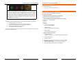
Fig. 2: Comparison of protein expressions between ME180 cells (left) and
A431cells (right) using antibody arrays with 240 antibodies. Alkaline phosphatase-labeled secondary antibodies were used and the staining was visualized
by color reaction with BCIP/NBT as substrates
d.
Knowledge of a protein’s sub-cellular localization can provide important information about
the protein’s functional state. For example, change of a transcriptional factor’s location from
cytoplasm to nucleus often suggests its activation. When observed under a fluorescence microscope, AntibodyArrayTM staining can reveal sub-cellular localizations of many proteins
simultaneously.
c.
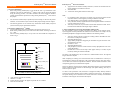
Fig. 1: Overview of AntibodyArrayTM staining technology. a, make contact
between staining antibody array and adherent cells; b, incubate arrays and cells
to allow antibodies to bind to their antigens; c, remove array support; d, detect
bound antibodies.
The AntibodyArrayTM staining method takes advantage of “Dissociable Protein Array” technology, which allows the delivery of a large number of proteins to their targets in a positionaddressable manner. In staining AntibodyArrayTMs, the antibodies are immobilized on a membrane in such a manner that when they make contact with cells fixed on another support, the
antibodies can bind to their respective antigens. When the array support is separated from the
cells, the antibodies will be dissociated from the support and remain bound to the antigens.
Therefore, the method enables the staining of multiple antibodies simultaneously, each at a predetermined position.
AntibodyArrayTM staining technology offers a high-throughput method for examining in vivo
protein activities. It has many applications, including:
1.
2.
Examining protein expressions
Revealing protein sub-cellular localizations
Measuring protein expressions has applications in a variety of fields, including biomedical research, disease diagnosis, and drug discovery. Antibody array-staining provides an unique approach to reveal protein expression patterns. It is useful in comparing the expressions of a large
number of proteins between different biological samples (see Fig. 2).
www.hypromatrix.com
22
Hypromatrix, Inc.
Fig. 3: Fluorescent staining with staining AntibodyArrayTM. An array of 200
rabbit polyclonal antibodies were used and the staining at four positions are
shown here as representatives. a, b, transcriptional factor IRF1. c, d, signaling
molecule 14-3-3 β. e, f, cell adhesion protein β-catenin. g, h, transcriptional factor Ets-1. Low magnification (a, c, e, g, and i) shows the stained cells and surrounding non-stained area; and high magnification shows the detailed nuclei localization of IRF1 (b), cytoplasmic staining of 14-3-3 β (d) and Ets-1 (h), and
membrane staining of β-catenin at cell-cell contacts (d). Scale bar in a, 300 µm;
Scale bar in b, 30 µm.
Simultaneous staining of two proteins (double staining) is a unique tool for studying two
functionally related proteins. For example, evidence of protein interactions often includes the
demonstration that the proteins co-localize in the same cellular structure. Double staining is
also useful when the protein of interest is only expressed in small number of target cells
among a heterogeneous cell population, and the protein of interest needs to be observed together with a protein marker which is used to denote the target cells. Array staining is unique
in that it allows the examination of multiple proteins individually as well as simultaneously
in the same cell preparation.
Hypromatrix, Inc.
3
17
www.hypromatrix.com