1
AntibodyArrayTM Instruction Manual
AntibodyArrayTM Instruction Manual
8.
IV. METHODS
A. General considerations
1. Before using staining AntibodyArrayTM, tests should be done to get information on the
suitability of the cells for AntibodyArrayTM staining. Some cells may fall off the support
during the staining procedure. Cell culture conditions and fixation conditions should be
carefully examined before experiments. Testing Staining AntibodyArrayTM can be used for
this purpose.
Apply fluorescence-labeled secondary antibodies (a mixture of anti-rabbit and antimouse antibodies); and incubate for an hour.
9. Wash with PBS.
10. Visualize the staining under fluorescent microscope.
Note:
1.
2.
One cell fixation condition may be optimal only for the staining of a subset of the proteins.
Therefore, several conditions may be needed to obtain proper staining for all the antigens.
2.
3.
3.
The following protocols are for reference only. Researchers should carefully study their
systems and select proper experimental conditions.
4.
B. Protocol for AntibodyArrayTM staining
1. Fix and permeabilize cells to exposure antigens (for detail see Protocols D to I).
2. Block staining AntibodyArrayTM and fixed cells in PBS solution containing 5% BSA for
0.5—1 hour.
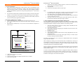
3. Place the staining AntibodyArrayTM on top of the cells and assemble the components as
shown in Fig. 4.
C. Some considerations for observing and analyzing staining results
For semi-quantitative analysis of protein expressions, array staining can be detected with enzyme-conjugated secondary antibodies and visualized via color reaction. For example, one may
use alkaline phosphatase conjugated secondary antibodies in the following manner:
1.
2.
3.
4.
5.
6.
Screw
7.
Moving plate
Pad
Staining AntibodyArray
When comparing two stainings, the overall intensities must be normalized first. This may be
accomplished by equalizing the intensities of the corresponding reference spots whose expression is identical in control and experimental conditions. Then the numerical intensities of the
corresponding spots may be calculated and compared.
Fixed cells
Filter paper
Apparatus frame
Because an antibody will bind both its intended characterized antigen(s) and some unintended
proteins which have similar structure to the immunogen (non-specific binding), there may be
some false positive signals.
Filter paper
To obtain a more quantitative result, fluorescence conjugated secondary antibody should be
used and array staining should be observed under a fluorescence microscope. The problem of
non-specific binding can be minimized since both the fluorescence intensity and subcellular
localization of the antigens can be detected and analyzed.
Fig. 4: Assembly of various staining components
6.
7.
Apply pressure and secure the assembly.
Stain for 1- to 2-hour.
Remove the array from the cell support and put the cells in a container.
Rinse the cells with PBS.
www.hypromatrix.com
46
After array staining, place the coverslip in a washing container. Remember the orientation of the coverslip in the container.
Wash with PBS three times.
Apply alkaline phosphotase-conjugated goat anti-rabbit and goat anti-mouse secondary antibodies to the cells.
Incubate for 1 hour.
Wash with PBS three times.
Add phosphotase substrate 3-bromo-4-chloro-5-indolyl phosphate/nitro blue tetrazolium (BCIP/NBT).
Incubate until a brown color develops. When proper intensity is reached, stop the
reaction by washing the substrate with PBS.
The image of the StainingArray can be scanned on a standard flatbed scanner. The digitized
images can then be analyzed.
Pad
4.
5.
For comparing protein expressions, all samples were processed in parallel and the
enzymatic reactions can be stopped by washing off substrates with PBS.
Care should be taken to avoid trapping air between cells and arrays.
Arrays should be carefully aligned with cells and the alignment should be recorded to
avoid any confusion at later steps.
After arrays contact cells, any movement between them should be avoided. The interaction between antibodies and antigens begins in minutes
Hypromatrix, Inc.
Hypromatrix, Inc.
7
15
www.hypromatrix.com