1
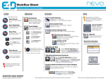
Elements Training Workbook & Resources Table of Contents PlanScan System & Equipment Overview ......................................................................................................................................................4 Exercise 1 - Premolar Crown with Buccal Bite.................................................................................................................................................6 Exercise 2 - Molar Crown with Buccal Bite.................................................................................................................................................... 17 Exercise 3 - Onlay Restoration ......................................................................................................................................................................... 22 Exercise 4 - Pre-op Crown ................................................................................................................................................................................. 27 Optional Exercise - Anterior Crown with Buccal Bite ................................................................................................................................. 31 Information Resources ...................................................................................................................................................................................... 37 Registration .......................................................................................................................................................................................................... 37 Customer Support Information....................................................................................................................................................................... 38 CDD Program ....................................................................................................................................................................................................... 38 Prep Guidelines & Materials ............................................................................................................................................................................. 39 Material Selection............................................................................................................................................................................................... 40 Integration Day & Starter Kit............................................................................................................................................................................ 48 CAD/CAM Supplies ............................................................................................................................................................................................. 50 Faculty Contact List Gary Severance, DDS Chief Marketing Officer Jeremy Hiser, RDA, CDD Manager of E-Learning and Documentation [email protected] Elizabeth Pastrana, RDA, CDD CAD/CAM Special Markets Manager [email protected] Angie Heick Senior Technical Writer [email protected] Marcie Howard, CDD Education Coordinator [email protected] Joshua Bradley, CDD Clinical Educator [email protected] Matt Dunn, RDA, CDD Clinical Educator [email protected] Leia VanHoozer, RDA, CDD Clinical Educator [email protected] Windy Winland, RDA, CDD Clinical Educator [email protected] Kristi Curtis Customer Account Specialist [email protected] Alice DeLuna Customer Solutions Specialist [email protected] For the Education Department, call 800.537.6070 option 4. TEACHING STYLE At Planmeca University we incorporate the Tell, Show, Do teaching style. Tell - Listen to the instructor giving detailed instructions on the upcoming material. Show - Watch the instructor demonstrate the proper technique. Do - Perform the action and ask questions. HOW TO USE THIS WORKBOOK This section covers equipment & guidelines This section covers scanning & scanning techniques This section is all about design This section is for milling and materials This section has resources and information for your success This workbook is yours to keep; don’t be scared to write in it! Words in BOLD are icons or choices within the software. You’ll also find quick tips and time savers throughout. Hover over icons in the software to reveal their names. PlanScan System & Equipment Overview PlanScan System & Equipment Overview PlanScan Laptop 1. Powering ON and OFF the laptop 2. Windows 8 - Tiles and accessing the desktop 3. Care and general maintenance Connecting the Thunderbolt™ Adapter Properly connecting and disconnecting the scanner prevents damage to your devices. 1. Insert the Thunderbolt adapter into the adapter slot on the side of the laptop. (The adapter should remain attached, even when not in use.) 2. After opening the PlanCAD software, connect the red FireWire connector of the scanner into the white Thunderbolt adapter. The laptop gives an audible signal to confirm that the connection is fully seated. To remove the scanner, hold the red end with one hand and with the other hand grasp the Thunderbolt adapter. Gently pull apart to disconnect. Leave the white Thunderbolt adapter attached to the computer. Disconnecting the Thunderbolt Adapter If you wish to remove the adapter from the laptop: 1. Disconnect the scanner and exit Romexis to the Windows desktop. 2. Navigate to the Eject Media icon in the lower left corner of the desktop. 3. Click the icon and choose Eject IEEE 1394 Controller. 4. Remove the Thunderbolt adapter from the laptop. Failure to follow the Thunderbolt Adapter procedure may result in an inoperable scanner. For additional questions or concerns please contact Customer Support at 800.537.6070. PlanScan Scanner 4 1. Scanning Tips 2. Cradle 3. Scanner Cable; connecting and disconnecting the scanner are in a later section. n. ©2015 E4D Technologies All Rights Reserved. EDU1110.N PlanScan System & Equipment Overview Connecting the Scanning Tip (If scanning intraorally, disinfect the tip before connecting it to the base. See the User Manual for full instructions or the insert that is inside the scanning tip box.) 1. Grasp the body of the scanner with one hand. 2. Use the other hand to press the scanning tip onto the scanner as shown. A locking click is heard once the tip is fully seated. Disconnecting the Scanning Tip 1. Grasp the body of the scanner with one hand. 2. With your other hand depress the green button on the underside of the scanner. Gently pull the tip from the scanner. To prevent damage to the scanner, pull the pieces apart gently and without twisting or bending. When the scanner is not in use, place the non-functional protective scanner tip on the scanner. (Included with the scanner during shipping.) Failure to follow this procedure may result in damage to the scanner and scanning tip. Always follow the manufacturer’s instructions for disinfection. PlanMill 40 Maintenance of the PlanMill 40 is covered in the Elements course and in the User Manual. • Each week or every 3 hours of milling, the mill fluids need to be replaced. • Every other week or every 10 hours of milling, the Collets and Spindle Caps require maintenance. For Celebration customers - If your mill is inoperative when you return to your office, check the back of your Job Server for a dongle that is taped to the back. Plug the dongle into the back of your Job Server to enable the communications between the machines. ©2015 E4D Technologies All Rights Reserved. EDU1110.N 5 Exercise 1 - Premolar Crown with Buccal Bite Exercise 1 - Premolar Crown with Buccal Bite Tooth #5 (1-4 ISO) Romexis 1. On the main screen of Romexis, click Add Patient. 2. Add your name in the patient demographics screen, complete the options in bold. 3. Click Save Patient at the bottom of the screen. 4. Click CAD/CAM in Romexis options to the left of the screen. 5. Under Scan & Design New Restoration click New Scan and Design. This will take you to the Setup Tab. Setup Tab Enter the setup information for this case, then proceed to the Scan Tab. 1. Select Tooth 5 (1-4 ISO); the tooth will highlight and turn orange as you move the cursor away. 2. Choose the restoration type Crown. 3. Select the bite option Buccal/Opposing. 4. Choose Library A. 5. Pick the material Empress CAD LT. 6. Select shade A1. Scan Tab Overview Navigation Scan Options Viewing Options Model Alignment Scanner Status Scan Data Options 6 ©2015 E4D Technologies All Rights Reserved. EDU1110.N Exercise 1 - Premolar Crown with Buccal Bite Connecting the PlanScan Scanner 1. After opening the PlanCAD software, connect the red FireWire connector of the scanner into the white Thunderbolt adapter. 2. Verify the scanner status in the Scan Tab; wait until the scanner tip is warming before attempting to scan intraorally. 3. Activate the scanner with the Space Bar of the laptop or the Power button on top of the Scanner. 4. Disconnect the scanner (red firewire connector) after moving to the Margin Tab Holding the PlanScan Scanner Hold the scanner close to the tip like a handpiece or overhanded. Rest the neck of the scanner on the adjacent teeth. The tip of the scanner must point toward the distal of the preparation. If you scan in the incorrect orientation, you will need to delete those scans and start over. Scanning Live View and Model Indicators Live View Model View Scanninig stops when the Live View turns Sepia. Return back to a known area. CAPTURING DATA THROUGHOUT SCALE Touching the surface ©2015 E4D Technologies All Rights Reserved. EDU1110.N Floating away from surface 7 Exercise 1 - Premolar Crown with Buccal Bite Basic Scanning Pattern Begin scanning directly over the occlusal surface of the preparation. Move in a gradual, continuous motion toward the mesial neighbor. Transition from the occlusal, cusp, axial wall, to gingival surfaces. The scanner should be held close to 90° while scanning parallel to the buccal surface. Goals of Prep Scanning 100% of the prep Interproximal contact point 90% of the adjacent teeth Good axial data for design 2-3 mm gingival tissue on buccal and lingual Keep your eyes on the screen and use the model and live view to track your progress and current position. Evaluate the model 1. Use the mouse to rotate, move, and zoom in and out to evaluate the model. Left Click Right Click Scroll Wheel Select - position the cursor on an item and click the left button to select. Rotate Model - press and hold the right button while dragging the mouse on the desktop. Zoom - rotate the scroll wheel to change the size of the model. Move - press and hold the wheel to move the model. It’s important to practice using the mouse. Ensure you are comfortable moving the model and zooming in/out. 2. 8 Rotate the model to look for low data areas in key areas: the preparation, interproximal contacts, etc.. ©2015 E4D Technologies All Rights Reserved. EDU1110.N Exercise 1 - Premolar Crown with Buccal Bite 3. Fill in any required missing data by activating the scanner. Use the fill in techniques. Mesial Tip - Rest the neck of the scanner tip on the mesial neighbor, rock the scanner to point the blue laser into the distal interproximal area. Distal Tip - Rest the end of the scanner tip on the distal neighbor; rock the scanner to point the blue laser into the mesial interproximal area. Ensure your model has 100% of the preparation, the interproximal contact areas, and at least 90% of the adjacent teeth and full cusps. Focus on the contact zone, not the gingival contours. 4. Erase any interfering data such as extra teeth, tongue, cheek, and cotton rolls. 5. Click Generate Model or press M on the keyboard to finish building the model. Buccal Bite and Opposing The opposing teeth are scanned to acquire bite information for the proposal. The buccal bite is scanned to align the preparation model with the opposing model. Scan the teeth that are opposing the teeth in the preparation scan. Identify the three teeth directly opposing those in the prep scan. Note: Many clinical operators scan the Opposing while the patient is being anesthetized. ©2015 E4D Technologies All Rights Reserved. EDU1110.N 9 Exercise 1 - Premolar Crown with Buccal Bite Scan Opposing 1. Click Opposing in the scan options on the left of the screen. 2. Starting with the distal tooth, scan the occlusal data. 3. Transition to the buccal and scan the buccal surface. Include 2-3 mm of gingival data. (Cusp tip, axial wall, gingival) Lingual data is not necessary. Goals of Opposing Scans 100% of the occlusal and buccal surfaces 2-3 mm gingival tissue on the buccal surface Lingual data not necessary 4. Erase interfering data such as tongue, cheek, and cotton rolls. Scan Buccal 1. Click Buccal in the scan options on the left of the screen. 2. Close the articulated model gently. If it shifts during the scanning, the alignment may be incorrect. 3. Scan the buccal surfaces of the teeth that were captured in the preparation and opposing models. Ensure some gingival data is captured. Goals of Buccal Bite Capture the buccal surface of the dentition in the prep and opposing 2-3 mm gingival data No rotations necessary Note: Be sure to verify the status of the buccal alignment. In most cases, alignment is done automatically by the software. A green dot in the Buccal icon indicates a successful alignment. Always verify the alignment before continuing with the next step. 10 ©2015 E4D Technologies All Rights Reserved. EDU1110.N Exercise 1 - Premolar Crown with Buccal Bite Evaluate and Adjust the Orientation 1. Click the Margin tab. 2. Evaluate and adjust the Orientation using View Controls to change the point of view. A. In the Occlusal View, balance the model from buccal to lingual. A B. In the Distal View, align the buccal cusps of the neighbors. B C. In the Buccal View, evaluate marginal ridge alignment. C 3. Click the Orientation icon to accept the current position. ©2015 E4D Technologies All Rights Reserved. EDU1110.N 11 Exercise 1 - Premolar Crown with Buccal Bite Mark the Margin 1. Use the scroll wheel to zoom in on the preparation. 2. Click Trace and click on the inside of the margin. 3. Moving in small increments, click as you move around the preparation. Don’t worry if you make a mistake while drawing the margin. 4. The margin is finished when the original point (blue dot) is clicked to finish the circle. 5. Practice adjusting the margin with both Move Margin and Add Segments. Move Margin is used for minor adjustments. Add Segments is used to redraw a section of the margin. Design Please reference the CAD/CAM workflow for design. We will use this form throughout the design process. PlanCAD Workflow HAVE QUESTIONS? Contact Support @ 800.537.6070 SCAN Verifying the amount of scan data will ensure a better fitting restoration. Scan Prep Erase Brush 100% of Prep and contact zones; begin on the occlusal surface of the prep. Tongue, Cheek, Fingers or Extra data Scan Opposing S Scan Buccal 1 100% Occlusal and buccal gingival data g Capture all teeth in prior scans Bite Alignment Verify before moving to the Margin tab. MARGIN Orientation Trace Margin Verify Alignment Automatically active; balance the model from the occlusal view to mark the margin. From the occlusal view, mark the margin on the shoulder Verify the orientation of the model with the Green Preview Tooth. Orientation can be deactivated or activated at any time using this icon. Use Move Margin to adjust placement DESIGN Use Add Segments to redraw a portion Follow the design flow. Return to a section and repeat any necessary steps when needed. Fit Generate proposal is the first step in the design process. Customizing the proposed restoration Refine contours, marginal ridges, to fit in its place over the prep. embrasures and cusps. AutoGenesis OFF - Resize, Reposition, Apply Some cases may require orientation to be adjusted. Form Use Incremental Change Tools to adjust overall fit. Click an arrow next to the desired adjustment. Twist, Rotate, or Expand to fit in place. Use Freeform Change Tools to make small adjustments to contour. Rubber Tooth can be used to adjust: Axial Walls, Marginal Ridges, Occlusal table, and Embrasures. Use Move Feature for cusps, Smooth Surface to blend, and Dropper to add material when needed to modify the proposal. Function Prepare for Milling Use these tools to adjust the bite and occlusion to ideal parameters. Ensure the restoration will mill properly. Adjust bite with View Bite Registration (x2), View Contacts, & Contact Refinement. Goal: White, Brown, Black Adjust interproximal contacts with Freeform Change Tools. Use Hide Model and Smooth Surface to refine contacts. Goal: Light Green/ Aqua surrounded by Dark Blue Material Thickness: Occlusal table 1.5 - 2 mm Axial walls 1 - 1.5 mm Ideal margins are Yellow. If red/orange verify placement with Move Margin. Use Dropper as needed to add material MILL 12 Sprue Position Block Size Selection Mill SIM Adjust the sprue away from margins, contacts, and occlusion when needed. Select the block size for the chosen restoration and material. Check the internal fit of your restoration before milling. Look for internal interferences in the intaglio surface of the restoration and use Slice Plane when needed to evaluate. Overmill Intaglio View ©2015 E4D Technologies All rights reserved EDU1103.D DRAFT Generate AutoGenesis ON - Click Apply Use the Green Preview Tooth as a guide to align the model after activating Orientation ©2015 E4D Technologies All Rights Reserved. EDU1110.N Exercise 1 - Premolar Crown with Buccal Bite Navigation Sets of Tools Viewing Options Model Orientation Tool Options 1. Click the Design tab. The Tooth Libraries tools automatically appear. 2. Click Apply to have Autogenesis generate the proposal. 3. Click Incremental Change Tools to evaluate the proposal for large adjustments. Use the tool options to make changes where needed. Click Apply before continuing. 4. Click Freeform Change Tools and Material Thickness (in view options) to evaluate the proposal. The proposal should be blue/green with a yellow margin. 5. Click Rubber Tooth and adjust the axial walls, marginal ridges, and embrasures. Adjust axial wall contours by pulling material away from or toward the preparation. Adjust central grooves and cusps by pulling material up or down. Note: Rotate the model to adjust circumferentially. From the occlusal view, adjust the embrasure shapes. ©2015 E4D Technologies All Rights Reserved. EDU1110.N Check the emergence profile and adjust. 13 Exercise 1 - Premolar Crown with Buccal Bite 6. Click View Bite Registration to see the opposing dentition model above the proposal. Click View Bite Registration a second time to make the template transparent. Adjust the translucency of the opposing model. 7. Click View Contacts. Use Contact Refinement to adjust to White, Brown, Black. Intraoral adjustment will be needed. Ideal occlusion, slight adjustment may be needed. 8. Click View Bite Registration again to deactivate the template. 9. Click Hide Model to remove the model from view. 10. Rotate the proposal to view the interproximal contacts. Adjust interproximal contacts as needed with Smooth Surface in Freeform Change Tools. No Contact Ideal Contact Zone Adjustments Needed The goal is dark blue with a hint of aqua. Broadness of Contact (Dark Blue) Floss Snap (Aqua) Before Goal 11. Deactivate Hide Model. 12. Deactivate View Contacts. 14 ©2015 E4D Technologies All Rights Reserved. EDU1110.N Exercise 1 - Premolar Crown with Buccal Bite 13. Click Material Thickness. The desired material thickness is based on the block manufacturer’s recommended thickness for your restoration type. The desired material thickness for a crown is 1-1.5 mm along the axial walls and 1.5-2 mm on the occlusal table. 14. Evaluate the margin. The material thickness should be yellow around the margin with no red or orange. Good example - yellow margin Poor example - Red or orange along the margin 15. If there is red or orange around the margin, click Move Margin to evaluate the margin for accurate placement. Adjust the margin, if needed. Going back to the Margin tab and making changes to the margin will result in losing your design. 16. If the margin is placed accurately and is still red/orange, use the Dropper tool to add material thickness. Congratulations on your first design with the PlanScan system! Review the CAD/CAM Workflow before continuing to the Mill tab. ©2015 E4D Technologies All Rights Reserved. EDU1110.N 15 Exercise 1 - Premolar Crown with Buccal Bite Milling 1. Click the Mill tab 2. Evaluate your design and review the material thickness indicators. 3. Check the sprue position and use the Sprue Placement wheel to adjust when needed. 4. Select the block size (also based on sprue positioning) High, away from margin and contact areas. Too close to the margin 5. Click Mill Sim. 6. Evaluate the simulation. 7. Click Send to Mill, click OK The system defaults to the setting for the restoration type. Standard - Full Coverage Crowns Detailed - Inlay, Onlay, and Veneers 16 ©2015 E4D Technologies All Rights Reserved. EDU1110.N Exercise 2 - Molar Crown with Buccal Bite Exercise 2 - Molar Crown with Buccal Bite Tooth #30 (4-6 ISO) with bite registration Some images in this exercise may vary from the physical model. The same work flow will be used. Setup Enter the setup information for this case: • Tooth 30 (4-6 ISO) • Crown • Buccal/Opposing • Library A • IPS e.max CAD LT • Select shade B1 Scan Prep Scan prep using the basic scan method for a single unit posterior case. Goals of Prep Scanning 100% of the prep and Interproximal contact point 90% of the adjacent teeth and good axial data for design 2-3 mm gingival tissue on buccal and lingual Scan Opposing 1. Click Opposing. Goals of Opposing Scanning 100% of the occlusal and buccal surfaces 2-3 mm gingival tissue on the buccal surface Lingual data not necessary 2. Erase any interfering data such as tongue, cheek, and cotton rolls. ©2015 E4D Technologies All Rights Reserved. EDU1110.N 17 Exercise 2 - Molar Crown with Buccal Bite Scan Buccal 1. Click Scan Buccal. 2. Scan the buccal surfaces of the teeth that were captured in the preparation and opposing models. Ensure some gingival data is captured. Use the same scanner orientation as the other two scans. Goals of Buccal Bite Scanning Capture the buccal surface of the dentition in the prep and opposing 2-3 mm gingival data Note: Be sure to verify the status of the buccal alignment. No rotations necessary In most cases, alignment is done automatically by the software. A green dot in the Buccal icon indicates a successful alignment. Always verify the alignment before continuing with the next step. Evaluate and Adjust the Orientation 1. Click the Margin tab. 2. Evaluate and adjust the Orientation using View Controls to change the point of view. 3. Click the Orientation icon to accept the current position. Mark the Margin 1. Use the scroll wheel to zoom in on the preparation. 2. Click Trace and click on the inside of the margin. 3. Moving in small increments, click as you move around the preparation. Don’t worry if you make a mistake while drawing the margin. 4. 18 The margin is finished when the original point (blue dot) is clicked to finish the circle. ©2015 E4D Technologies All Rights Reserved. EDU1110.N Exercise 2 - Molar Crown with Buccal Bite Design Please reference the CAD/CAM workflow for design. We will use this form throughout the design process. 1. Click the Design tab. The Tooth Libraries tools automatically appear. 2. Turn Autogenesis OFF and click Apply to generate the proposal. 3. Click Incremental Change Tools to evaluate the proposal for large adjustments. Use the tool options to make changes where needed. Click Apply before continuing. 4. Click Freeform Change Tools and Material Thickness (in view options) to evaluate the proposal. The proposal should be blue/green with a yellow margin. 5. Click Rubber Tooth and adjust the axial walls, marginal ridges, and embrasures. 6. Click View Bite Registration to see the opposing dentition model above the proposal. Click View Bite Registration a second time to make the template transparent. Adjust the translucency of the opposing model. 7. Click View Contacts. 8. Click View Contacts. Use Contact Refinement to adjust to White, Brown, Black. Intraoral adjustment will be needed. Ideal occlusion, slight adjustment may be needed. 9. Click View Bite Registration again to deactivate the template. 10. Click Hide Model to remove the model from view. ©2015 E4D Technologies All Rights Reserved. EDU1110.N 19 Exercise 2 - Molar Crown with Buccal Bite 11. Rotate the proposal to view the interproximal contacts. Adjust interproximal contacts as needed with Smooth Surface in Freeform Change Tools. The goal is dark blue with a hint of aqua. No Contact Ideal Contact Zone Adjustment Needed Broadness of Contact (Drk. Blue) Floss Snap (Aqua) 12. Deactivate Hide Model. 13. Deactivate View Contacts. 14. Click Material Thickness. The desired material thickness is based on the block manufacturer’s recommended thickness for your restoration type. The desired material thickness for a crown is 1-1.5 mm along the axial walls and 1.5-2 mm on the occlusal table. 20 ©2015 E4D Technologies All Rights Reserved. EDU1110.N Exercise 2 - Molar Crown with Buccal Bite 15. Evaluate the margin. The material thickness should be yellow around the margin with no red or orange. Good example - yellow margin Poor example - Red or orange along the margin 16. If there is red around the margin, click Move Margin to evaluate the margin for accurate placement. Adjust the margin if needed. Going back to the Margin tab to make changes will result in losing your design. If the margin is placed accurately and is still red/orange, use the Dropper tool to add material thickness. Congratulations on your molar crown with the PlanScan system! Review the CAD/CAM Workflow before continuing to the Mill tab. Milling 1. Click the Mill tab 2. Evaluate your design and review the material thickness indicators. 3. Check the sprue position and use the Sprue Placement wheel to adjust when needed. 4. Select the block size (also based on sprue positioning) 5. Click Mill Sim. 6. Evaluate the simulation. 7. Click Send to Mill, click OK ©2015 E4D Technologies All Rights Reserved. EDU1110.N 21 Exercise 3 - Onlay Restoration Exercise 3 - Onlay Restoration Onlay Tooth #14 (2-6 ISO) Setup Enter the setup information for this case: • Tooth 14 (2-6 ISO) • Onlay • Buccal/Opposing • Library A • Lava Ultimate LT • Select shade A1 Scan Prep 1. Click the Scan tab. 2. Follow the basic scan pattern. 3. Evaluate the preparation model. The same basic scan pattern is used for partial restorations. Tooth 13 (2-5 ISO) is also a preparation , but we are not designing it at this time. The opposing dentition is a preparation, so we are not going to scan the buccal and opposing. Orientation The pictures for this case are of a different onlay. The procedure is the same. 1. Click the Margin tab. 2. Set the Orientation for the onlay. Use the remaining anatomy of the prepped tooth to aid your orientation. Mark the Margin 22 1. Use the scroll wheel to zoom in on the preparation. 2. Click Trace and click on the inside of the margin. 3. Moving in small increments, click as you move around the preparation. 4. The margin is finished when the original point (blue dot) is clicked to finish the circle. ©2015 E4D Technologies All Rights Reserved. EDU1110.N Exercise 3 - Onlay Restoration Once the margin is drawn for an inlay or onlay, a notification screen appears. Note: If this screen doesn’t appear, click Selection Area. 5. Click Take Me There to go to the Selection Area screen. 6. Click Add to Selection and circle Tooth 14 (2-6 ISO). Using Selection Area will define the area of the tooth structure for design. Over selection an area will cause a poor proposal. Good Selection Poor Selection 7. Complete the Selection Area and return to the Margin Tool screen. 8. Click Hide Model to isolate the preparation and to evaluate and adjust the margin with Move Margin and Add Segments as needed. Please reference the CAD/CAM workflow for design. We will use this form throughout the design process. ©2015 E4D Technologies All Rights Reserved. EDU1110.N 23 Exercise 3 - Onlay Restoration Design 1. Click the Design tab. 2. Ensure Autogenesis is ON and click Apply. Autogenesis creates a proposal based on the Selection Area designated. 3. Click Incremental Change Tools to evaluate the proposal for large adjustments. Use the tool options to make changes where needed. Click Apply before continuing. 4. Click Freeform Change Tools and Material Thickness (in view options) to evaluate the proposal. The proposal should be blue/green with a yellow margin. 5. Click Rubber Tooth and adjust the axial walls, ridges, and embrasures. Activate Move Feature to adjust surfaces incrementally. 6. Click View Contacts. Since this case does not have any occlusal contact information, we will skip to the interproximal contact. 7. Rotate the proposal to view the interproximal contacts. Adjust interproximal contacts as needed with Smooth Surface in Freeform Change Tools. The goal is dark blue with a hint of aqua. No Contact Ideal Contact Zone Intraoral Adjustment Needed Broadness of Contact (Drk. Blue) Floss Snap (Aqua) 24 8. Deactivate Hide Model. 9. Deactivate View Contacts. ©2015 E4D Technologies All Rights Reserved. EDU1110.N Exercise 3 - Onlay Restoration 10. Click Material Thickness. 11. Evaluate the margin. The material thickness should be yellow around the margin with no red or orange. Good example - yellow margin Poor example - Red or orange along the margin 12. If there is red around the margin, click Move Margin to evaluate the margin for accurate placement. Adjust the margin if needed. Going back to the Margin tab to make changes will result in losing your design. 13. If the margin placement is accurate, use the Dropper tool to add material thickness. In some situations, it will be difficult to attain ideal occlusal contact strength and reach minimum material thickness. In the example below, the red material thickness around the margin indicates the margin is too thin. 14. Click Dropper and add material thickness. This will result in adequate material thickness strength but may create a strong contact with the opposing dentition. This can be corrected intraorally. Congratulations on your first partial restoration with the PlanScan system! Review the CAD/CAM Workflow before continuing to the Mill tab. ©2015 E4D Technologies All Rights Reserved. EDU1110.N 25 Exercise 3 - Onlay Restoration Mill 1. Click the Mill tab. 2. Evaluate your design and review the material thickness indicators. 3. Click Hide Model and check the sprue position and use the Sprue Placement wheel to adjust when needed. 4. If no sprue is visible, the sprue is positioned on the internal aspect of the restoration. Move the sprue to an external position. 5. Ensure the total circumference of the sprue is visible. 6. Select the block size (also based on sprue positioning). 7. Click Mill Sim. 8. Evaluate the simulation. 9. Click Send to Mill, click OK. The system defaults to the setting for the restoration type. Standard - Full Coverage Crowns Detailed - Inlay, Onlay, and Veneers 26 ©2015 E4D Technologies All Rights Reserved. EDU1110.N Exercise 4 - Pre-op Crown Exercise 4 - Pre-op Crown Pre-op Crown Tooth #30 (4-6 ISO) Setup Enter the setup information for this case: • Tooth 30 (4-6 ISO) • Crown • Buccal/Opposing • Pre-op • IPS e.max • Select shade A1 Scan Prep 1. Click the Scan tab. 2. Click Preop. 3. Follow the basic scan pattern. 4. Evaluate the pre-op model. Goals of Pre-Op Scanning 100% of the pre-op tooth 90% of the adjacent teeth Good axial data for design 2-3 mm gingival tissue on buccal and lingual 5. Click Prep. 6. A Time Saver prompt appears. Click Yes. ©2015 E4D Technologies All Rights Reserved. EDU1110.N 27 Exercise 4 - Pre-op Crown The pre-op model is duplicated as the preparation model. 7. Click Erase. 8. Erase the pre-op tooth and the marginal ridges of the neighboring teeth. 9. Click the Erase tool again to accept the changes. 10. Press the button on the scanner and start scanning over one of the neighboring teeth. When the system recognizes the same data on your prep model, it will begin scanning. Move over the preparation and the system will fill in the areas that you erased. 11. Fill in the preparation data and the interproximal areas. 12. Evaluate the model for any missing data. 13. Generate the model. 28 ©2015 E4D Technologies All Rights Reserved. EDU1110.N Exercise 4 - Pre-op Crown Orientation 1. Click the Margin tab. 2. Evaluate and adjust the Orientation using View Controls to change the point of view. 3. Click the Orientation icon to accept the current position. Mark the Margin 1. Use the scroll wheel to zoom in on the preparation. 2. Click Trace and click on the inside of the margin. 3. Moving in small increments, click as you move around the preparation. 4. The margin is finished when the original point (blue dot) is clicked to finish the circle. Edit the Pre-op 1. Click Pre-Op Editing. 2. Use the Trace tool to designate the area of the model that you want to use as the Pre-op library surface. Stay away from rough areas and the margin. 3. Click on the blue dot to finish the pre-op area. 4. Use Move Curve and Add Segments to edit the Pre-op if needed. ©2015 E4D Technologies All Rights Reserved. EDU1110.N 29 Exercise 4 - Pre-op Crown Design 1. Click the Design tab. Note that the Library at the bottom of the screen now includes Pre-op. 2. Click Apply. Autogenesis creates a proposal based on the Pre-op area that you designated and Library A. 3. Follow the normal design workflow. 4. Click View Pre-op to see the combination of the pre-op model and the prep model. Speckled areas are where the pre-op is in close proximity to the proposal. Solid stone color shows where the pre-op is above the proposal. Solid proposal color is where the proposal is above the pre-op. Click View Pre-op a second time to make the pre-op model translucent. 5. Use Slice Plane and Rubber Tooth to make adustments if needed. Milling 30 1. Click the Mill tab 2. Evaluate your design and review the material thickness indicators. 3. Check the sprue position and use the Sprue Placement wheel to adjust when needed. 4. Select the block size (also based on sprue positioning). 5. Click Mill Sim. 6. Evaluate the simulation. 7. Click Send to Mill, click OK ©2015 E4D Technologies All Rights Reserved. EDU1110.N Optional Exercise - Anterior Crown with Buccal Bite Optional Exercise - Anterior Crown with Buccal Bite Tooth #9 (2-1 ISO) with bite registration Setup Enter the setup information for this case: • Tooth 9 (2-1 ISO) • Crown • Buccal/Opposing • Library A2 • Empress CAD Multi • Select shade A1 Scan Prep Scan prep using the basic scan method for a single unit anterior case. The tip of the wand should face the highest tooth number. Begin scanning directly over the occlusal surface of the preparation. Move in a gradual, continuous motion toward the neighbor. Transition from the incisal, axial wall, and to the gingival surfaces. The scanner should be held at close to 90° while scanning parallel to the axial surface. Watch as your model builds to see any areas that might require a different rotation or angle. Goals of Prep Scanning 100% of the prep and interproximal contact point 90% of the adjacent teeth and good axial data for design 2-3 mm gingival tissue on buccal and lingual Scan Opposing 1. Click Opposing. 2. Starting in the same scanner orientation as the prep scan, scan the incisal data and rotate to the buccal. Goals 100% of the occlusal and buccal surfaces 2-3 mm gingival tissue on the buccal surface Lingual data not necessary 3. Erase any interfering data such as tongue, cheek, and cotton rolls. ©2015 E4D Technologies All Rights Reserved. EDU1110.N 31 Optional Exercise - Anterior Crown with Buccal Bite Scan Buccal 1. Click Buccal. 2. Scan the buccal surfaces of the teeth that were captured in the preparation and opposing models. Ensure some gingival data is captured. Use the same scanner orientation as the other two scans. Goals Capture the buccal surface of the dentition in the prep and opposing 2-3 mm gingival data Note: Be sure to verify the status of the buccal alignment. No rotations necessary Margin 1. Click the Margin tab. 2. Evaluate and adjust the Orientation. Orientation may have to be adjusted a second time after marking the margin. Align the incisal edge with the bottom of the design screen 3. Click Orientation to accept the current position. 1. Use the scroll wheel to zoom in on the preparation. 2. Click Trace and click on the inside of the margin. 3. Moving in small increments, click as you move around the preparation. 4. The margin is finished when the original point (blue dot) is clicked to finish the circle. 5. Adjust with Move Margin and Add Segments as needed. Mark the Margin 32 ©2015 E4D Technologies All Rights Reserved. EDU1110.N Optional Exercise - Anterior Crown with Buccal Bite Design Please reference the CAD/CAM workflow for design. We will use this form throughout the design process. 1. Click the Design tab, the Tooth Libraries tools automatically appear. Review the available libraries for best fit. Available tooth libraries for design. Yellow highlight indicates current selection. 2. If the library tooth is not a good size in relation to the neighbors, use the ALT + (Up or Down) Arrows on the keyboard to resize the library tooth. 3. If the library tooth needs to be moved, left click and drag the green tooth to ideal position. 4. Turn OFF Autogenesis and click Apply. 5. Click Incremental Change Tools to evaluate the proposal for large adjustments. Use the tool options to make changes where needed. Click Apply before continuing. 6. Click Freeform Change Tools and Material Thickness (in view options) to evaluate the proposal. The proposal should be blue/green with a yellow margin. 7. Click Rubber Tooth and adjust the axial walls, ridges, and embrasures. Activate Move Feature to adjust surfaces incrementally. Adjust axial wall contours by pulling material away from or toward the preparation. 8. Click View Bite Registration to see the opposing dentition model above the proposal. Click View Bite Registration a second time to make the template transparent. Adjust the translucency of the opposing model. ©2015 E4D Technologies All Rights Reserved. EDU1110.N 33 Optional Exercise - Anterior Crown with Buccal Bite 9. Click View Contacts. Use Contact Refinement to adjust to White, Brown, Black. Adjustments needed. Ideal occlusion, slight adjustment may be needed. 10. Click View Bite Registration again to deactivate the template. 11. Click Hide Model to remove the model from view. 12. Rotate the proposal to view the interproximal contacts. Adjust interproximal contacts as needed with Smooth Surface in Freeform Change Tools. The goal is dark blue with some green. Final adjustments will be made after the restoration is milled. No Contact Ideal Contact Zone Intraoral Adjustment Needed Broadness of Contact (Dark Blue) Leave a little green in the contact area to finalize after testing the fit. 13. Deactivate Hide Model. 14. Deactivate View Contacts. 15. Click Material Thickness. The desired material thickness is based on the block manufacturer’s recommended thickness for your restoration type. The desired material thickness for a crown is 1-1.5 mm along the axial walls (bright green - dark green) and 1.5-2 mm on the incisal (dark green - blue). 34 ©2015 E4D Technologies All Rights Reserved. EDU1110.N Optional Exercise - Anterior Crown with Buccal Bite 16. Evaluate the margin. The material thickness should be yellow around the margin with no red or orange. Good example - yellow margin Poor example - Red or orange along the margin 17. If there is red around the margin, click Move Margin to evaluate the margin for accurate placement. Adjust the margin if needed. Going back to the Margin tab to make changes will result in losing your design. If the margin is placed accurately and is still red/orange, use the Dropper tool to add material thickness. Congratulations on your second design with the PlanScan system! Review the CAD/CAM Workflow before continuing to the Mill tab. Milling 1. Click the Mill tab 2. Evaluate your design and review the material thickness indicators. 3. Check the sprue position and adjust when needed. 4. Select the block size (also based on sprue positioning) ©2015 E4D Technologies All Rights Reserved. EDU1110.N 35 Optional Exercise - Anterior Crown with Buccal Bite 5. For this exercise, IPS Empress Multiblock was selected as the material. The amount of chroma and translucency can be adjusted. Use the Restoration Positioning arrows to move the restoration up or down to change its value. Translucent Use Restoration Positioning to move the proposal up or down within the block. Chroma 6. Click Mill Sim. 7. Evaluate the simulation. 8. Click Send to Mill, click OK The system defaults to the setting for the restoration type. Standard - Full Coverage Crowns Detailed - Inlay, Onlay, and Veneers 36 ©2015 E4D Technologies All Rights Reserved. EDU1110.N Registration INFORMATION RESOURCES There are many resources available for gathering information. • Download documentation and marketing materials - www.e4d.com/material-bank. Printed copies are available for a fee and can be ordered by emailing [email protected]. • Videos - www.e4d.com/videos • Education opportunity details - www.e4d.com/education Visit CadCamCan.com for additional videos and resources. Subscription needed for some sections. Please note that cadcamcan.com is a separate site. To post on their forums, you will need to Create an Account on the cadcamcan. com website. The registration invitation code is PlanScan (case sensitive). Newsletters, Chairside Chat, and update information are communicated via email. When you create your account in class, you may choose to be added to our email list. You may unsubscribe at any time. Registration To register, go to www.e4d.com/register. This is usually done while you are at the Elements class in Dallas. 1. Doctor is the default selection. If you are not a dentist, click Team Member. It is important that you fill out your information under the correct tab. 2. Fill out the information carefully. Note: the fields are different for Dentists and Team Members. The dentist information will be used as your listing in Dentist Finder. 3. At the bottom of the registration are several email subscription checkboxes. 4. • Weekly Video Tutorials • Send me Product Updates • Dentist Finder (on the Dentist registration only) • CDD Registration (on the Team Member registration only) Click Submit. ©2015 E4D Technologies All Rights Reserved. EDU1110.N 37 CDD Program CUSTOMER SUPPORT INFORMATION PlanScan System support E4D Customer Support 1.800.537.6070 866.361.1333 corporate phone 972.234.3557 corporate fax [email protected] 7am-7pm Central Time Mon-Thurs 7am-6pm Central Time Friday CDD PROGRAM The self-paced Certified in Digital Dentistry Program (CDD) provides motivated operators with the opportunity to gain professional recognition and establish credibility in proficiency with the latest dental CAD/CAM technology. Go to e4d.com/cdd to learn more. 38 ©2015 E4D Technologies All Rights Reserved. EDU1110.N Prep Guidelines & Materials 3UHS*XLGHOLQHV 6 to 10 Degree Taper Tapered Sides 3UHS*XLGHOLQHV Tapered Sides Rounded Internal Angles 3UHS*XLGHOLQHV Tapered Sides Rounded Internal Angles Equi/Supra Gingival Margins ©2015 E4D Technologies All Rights Reserved. EDU1110.N 39 3UHS*XLGHOLQHV 1.5 to 2 mm 1 mm 1 to 1.5 mm Tapered Sides Rounded Internal Angles Equi/Supra Gingival Margins Adequate Reduction 3UHS*XLGHOLQHV Tapered Sides Rounded Internal Angles Equi/Supra Gingival Margins Adequate Reduction Tissue Management $92,' 40 ©2015 E4D Technologies All Rights Reserved. EDU1110.N A copy is provided for each practice in your blue take away bag and included in the User Manual Electronic versions are available online $GGLWLRQDO5HVRXUFHV&DQ%H)RXQG$W +DYLQJWURXEOHYLHZLQJWKLVHPDLO" &OLFNKHUH ©2015 E4D Technologies All Rights Reserved. EDU1110.N 41 Material Selection %ORFN6L]H6HOHFWLRQ Materials come in a variety of sizes. The size of the designed restoration and sprue position will determine the available size to mill. ,36(PSUHVV&$' E\,YRFODU9LYDGHQW Beautiful Esthetics IPS Empress CAD offers over 100 combinations of block size, shades, and translucencies. Multi Shade & Translucency Cut back and layer esthetics in a monolithic block Multiple translucencies create the most natural looking, esthetic restoration Control incisal translucency and gingival color 42 ©2015 E4D Technologies All Rights Reserved. EDU1110.N ,36(PSUHVV&$' E\,YRFODU9LYDGHQW High Translucency Excellent chameleon effect Blends easily with existing tooth structure Inlays virtually “disappear” 20% more translucent than the Low Translucency Block Low Translucency Higher value “Block out” capability. Higher opacity level ,36(PSUHVV&$' E\,YRFODU9LYDGHQW Efficient IPS Empress CAD offers the option to simply mill and polish for maximum efficiency or glaze fire for up to a 50% increase in strength*. *Clinician’s Report – October 2009, Volume 2 Issue 10 ,36(PSUHVV&$' E\,YRFODU9LYDGHQW Multi A1, A2, A3, A3.5, B1 BL1, BL3 HT (High Translucency) A1, A2, A3, A3.5, B1, B2, B3, C2, D3 LT (Low Translucency) A1, A2, A3, A3.5, B1, B2, B3, C2, D3 BL1, BL2, BL3, BL4 ©2015 E4D Technologies All Rights Reserved. EDU1110.N 43 ,36HPD[ &$' E\,YRFODU 9LYDGHQW Beautiful Esthetics IPS e.max CAD offers a wide range of shades, sizes, and translucencies to allow the dental professional to provide beautiful esthetics and the durability to ensure clinical success for all indications The Highlights True-to-nature shade behavior for highly esthetic solutions Versatile use and comprehensive range of indications Lifelike esthetics, irrespective of the shade of the preparation ,36HPD[ &$' E\,YRFODU 9LYDGHQW Benefits Durable restorations due to the high strength Adhesive, self-adhesive or conventional cementation depending on the indication Bridge Materials C16 ± Ideal for longer dentition and large restorations B32 ± Up to three-unit bridges up to the second premolar as the abutment tooth ,36HPD[ &$' E\,YRFODU 9LYDGHQW HT (High Translucency) A1, A2, A3, A3.5, A4, B1, B2, B3, B4, C1, C2, C3, C4, D2, D3, D4 BL1, BL2, BL3, BL4 LT (Low Translucency) A1, A2, A3, A3.5, A4, B1, B2, B3, B4, C1, C2, C3, C4, D2, D3, D4 BL1, BL2, BL3, BL4 C16 & B32 Blocks A1, A2, A3, A3.5, B1, B2, C1, C2, D2 BL1 44 ©2015 E4D Technologies All Rights Reserved. EDU1110.N ,36HPD[ &$',PSXOVH E\,YRFODU 9LYDGHQW Value blocks – various brightness values The Value blocks feature different brightness values: 1 is the lowest and 3 the highest. Opal blocks – lifelike opalescence effect The Opal blocks exhibit a decreasing opalescence and increasing brightness value from 1 to 2. The Opal blocks can be used as an “enamel replacement” material. Aesthetic and minimally invasive restorations – thin veneers in particular. ,36HPD[ &$',PSXOVH E\,YRFODU 9LYDGHQW Advantages Lithium disilicate glass-ceramic (LS2) with a strength of 360 MPa Opal blocks for highly esthetic, minimally invasive veneers with a minimum thickness of 0.4 mm Value blocks for lifelike brightness value in crowns 7HOLR &$' E\,YRFODU 9LYDGHQW Strength and Endurance Long term temporary bridge material (12 mo.). Flexural strength of 130 MPa Esthetic Polyacrylate material technology allows for beautiful esthetic results simply by polishing or with the option to apply stains and glaze for a customized appearance. Shades A1, A2, A3, A3.5, B1 BL3 ©2015 E4D Technologies All Rights Reserved. EDU1110.N 45 /DYD8OWLPDWH E\0 Nano Technology High flexural strength (200 MPa) adds durability to posterior restoration Excellent wear resistance Brilliant and long-lasting polish Excellent stain resistance for color stability Shades A1, A2, A3, A3.5, B1, C2, D2 BL 3DUDGLJP0= E\0 Versatile and Easy Enamel-like wear characteristics are superior to that of ceramic blocks Easy to finish and polish Easy to repair intraorally Shades A1, A2, A3, A3.5, B3 Enamel =LUOX[ )& E\=DKQ'HQWDO Advantages of Full Contour Zirconia Flexural strength of 1100 MPa Simple stain and glaze technique High translucency pre-shaded zirconia Predictable aesthetic outcome Excellent alternative to PFM’s Low wear on opposing dentition To prevent contamination it is required to perform maintenance between milling different materials. A sintering oven is required for Zirlux FC2. 46 ©2015 E4D Technologies All Rights Reserved. EDU1110.N %XUQRXW%ORFN%2% E\('7HFKQRORJLHV Advantages Ideal for the lost wax technique allowing the optimal design of the restoration to be used for lost-wax casting or pressing techniques for additional material and restoration utilization 2SWLRQVWRWKLQNDERXW« Bridges? Anteriors? Choose the best option for your patient Call your manufacturer representative for more details View manufacturer websites for more specific indications and uses Implants? Remember to always follow the manufacturer instructions provided with each type of material. For additional information regarding the content in this presentation. Please contact the manufacturer for the product in question. ©2015 E4D Technologies All Rights Reserved. EDU1110.N 47 Integration Day & Starter Kit ,QWHJUDWLRQ'D\ Day starts at 7:30am and ends 3pm 3 Pre-prepared, Single Unit, Posteriors (premolar, molar) Schedule: Patients at 8am, 10am, and 1pm Allow 3 hours for the first appointment that may overlap the second 2 hour appointments are needed for the second and third patients Lunch and Learn Mill maintenance DDX Setup Discuss how to continue with your education No other patients scheduled Focused on those who attended the Elements of Success course in Texas 3UHPLHU 6WDUWHU.LW 1 Diamond Twist Paste Kit 1 Traxodent Sample 2 Sample Prep Burs 1 Milling Tools Sample Pack - 2 Ellipsoidal - 2 Conical - 2 Tapered 1 Sample Knit-Pak Cord ,YRFODU 6WDUWHU.LW Telio CAD: IPS Empress CAD: 4 Telio CAD Blocks Telio CS Link Transparent Telio CS Desensitizer 5g OptraPol Test Pack 4 IPS Empress CAD Blocks 2 Empress Shades 1 Empress Stain 1 Empress Glaze 1 Empress Glaze Liquid IPS e.max CAD: Misc. items: 4 e.max CAD Blocks 2 e.max Shades 1 e.max Stain 1 e.max Glaze Paste 1 e.max Glaze Liquid 1 e.max Crystallization Tray 48 2 Multilink Primer 1 Monobond Plus 1 Ceramic Etching Gel 1 Multilink Automix Trans 1 Optrastick 1 Optrafine Promo Pack 1 Object Fix Putty Cementation Navigation DVD ©2015 E4D Technologies All Rights Reserved. EDU1110.N 0 6WDUWHU.LW Lava Ultimate: 5 A2 LT C14 Blocks 5 A2 HT C14 Blocks Misc. items: 1 RelyX Ultimate Adhesive (A1) 1 Scotchbond Universal Adhesive 1 3M ESPE Retraction Capsule 1 CoJet Sand Blast Coating Agent 1 Lava Ultimate Guide 6WDUWHU.LW Mill Coolant Defoaming Solution :KDW¶V1H[W" Contact your local representative today: o Order blocks in shade values for upcoming patients o Order mill tools: 1 sleeve of each; Ellipsoidal and Tapered o Stains and Shades for characterization o Spray Glaze and speed tray for e.max (depending on order) o Infection Control Procedures for Scanner Tips (HLD or Autoclave?) o Lens tissues (KimWipes) o Lab handpiece and Finishing Kit o Sand blaster (if using Lava Ultimate) o Prep Kits (recommended, not required) ©2015 E4D Technologies All Rights Reserved. EDU1110.N 49 CAD/CAM SUPPLIES The materials listed below are all items used at Planmeca University. They are grouped by item type. For new documentation, go to www.e4d.com/material-bank. Documentation Name Vendor User Manual E4D Technologies Milling Center Quick Reference E4D Technologies Infection Control Name Vendor Item Number Alcohol Prep Pads Schein 1048298 MaxiCide Plus w/ Activator Schein 102-5796 (Qt) 102-2865 (Gallon) MetriTest Strips Schein 602-3437 Distilled Water Schein 395-0139 Gloves Schein X-Small 5654510 Small 5658087 Medium 5657431 Large 5659481 X-Large 5651575 Allrap Cover Film 4x6 Clear Schein 1273240 Steri-Soaker Schein 6581402 Name Vendor Item Number Two Striper Full Crown Kit Schein Premier 3780210 Schein Premier 3780213 2013582 Name Vendor Item Number Earth Stone - Quick Set Stone Schein 9662932 Orban 1/2 Perio Blade for trimming bite registration Premier 1004751 Preparation Design Two Striper Inlay/Onlay Kit 2013581 Impression and Model Materials Scanning 50 Name Vendor Item Number Scanning Tips (HLD pack of 3) Schein 6314915 Scanning Tips Autoclave Standard Schein 4457436 Scanning Tips Autoclave Landscape Schein 4454531 Scanning Tips Autoclave Portrait Schein 4452695 Optical Wipes - Kimwipes Schein 1017070 Ergotron Cart (smaller) Schein 1276580 Enovate Cart (larger) Schein 6310850 ©2015 E4D Technologies All Rights Reserved. EDU1110.N Milling Center Name Vendor Item Number Coolant Schein 6311524 Defoaming Solution Schein 6318999 Schein Premier 3781031 Schein Premier 3780560 Schein Premier 3786546 Schein Premier 3780206 Name Vendor Item Number Two Striper Finishing Kit Schein Premier 3780201 Name Vendor Item Number Accufilm I Single Sided Red Articulating Paper Schein 1865309 Two Striper E4D Mill Diamonds (Burs) Conical Ellipsoidal Tapered Assorted 2016002 2016001 2016000 2016004 Restoration Finishing 2013553 Articulating Paper Clinical materials and accessories (cements, adhesives, stains & glaze, etc.) Ivoclar Vivadent 3M ESPE Rebecca Spillman, MS Bill McGlynn Ivoclar Vivadent 175 Pineview Drive Amherst, NY 14228 716.691.2248 phone [email protected] 3M ESPE 3M Center Bldg. 275-2SE-03 St. Paul, MN 55144-1000 651.733.9078 phone [email protected] Premier Dental Products Company John Bonner Premier Dental Products Company 1710 Romano Drive Plymouth Meeting, PA 19462 610.239.6022 888.773.6872 Ex. 1022 [email protected] ©2015 E4D Technologies All Rights Reserved. EDU1110.N 51 NOTES 52 ©2015 E4D Technologies All Rights Reserved. EDU1110.N ©2015 E4D Technologies All Rights Reserved. EDU1110.N 53 Burn Out Block (BOB) Zirlux FC2 Telio CAD IPS e.max CAD HT IPS e.max CAD LT IPS e.max CAD Impulse IPS Empress CAD HT IPS Empress CAD LT IPS Empress Multi Paradigm MZ100 Lava Ultimate Provisional Only Anterior Veneer FOR CAST OR PRESSED INDICATIONS ONLY Provisional Only Anterior Full Crown Manufacturer Specifications for Materials Provisional Only Posterior Full Crown Provisional Only Anterior Only Bridge With manufacturer caution Indicated Provisional Only Provisional Only Implant ©2015 E4D Technologies All rights reserved EDU1119.E Provisional Only Inlay/Onlay Restoration Type Block Recommendation Chart CAD/CAM Materials 54 ©2015 E4D Technologies All Rights Reserved. EDU1110.N and firing Oven program 3 of IPS e.max Characterization 2 restoration Preparing the 1 Sunset Mahogany (occlusal pit) White (fluorosis, cusps and ridges) Incisal (enhance cusps, translucency) Sunset (occlusal shading) Shade 1 (gingival shading) Crystal Glaze Browse between programs and settings Navigation bar Indicates the selected firing program, firing progress, and other menu options Main screen Information bar Indicates current furnace temp and selected furnace programs Incisal After characterization place the restoration onto the crystallization tray for firing. Note there is an additional Speed Crystallization Tray for IPS e.max Flow (shown) will be used to affix the restoration to the firing pin for characterization and firing. Object Fix - Putty can also be used P4 - Empress P3 - Speed crys. spray P2 - Corrective firing P1 - IPS e.max Program Information Shade 1 White Mahogany Crystallization Tray Object Fix Crystal Glaze Liquid IPS e.max CAD Characterization Process ©2015 E4D Technologies All Rights Reserved. EDU1110.N 2-3 mm gingival tissue on buccal and lingual Good axial data for design Rest the scanner on the proximal dentition and perpendicular to the arch To achieve 100% of the interproximal contact area, a slight rotation of the scanner will be needed 100% of the prep and interproximal contact areas 90% of the adjacent teeth Interproximal Preparation Goals & Patterns Lingual and gingival data not necessary 2-3 mm gingival tissue on the buccal side 100% of the cusps Opposing Scanning Technique Note: Information on scanning Bite Registration material can be found in the User Manual No rotations necessary © 2014 E4D Technologies All Rights Reserved EDU1107.C DRAFT 2-3 mm gingival tissue on buccal and lingual Good axial data for design 90% of the adjacent teeth 100% of the prep and interproximal contact areas Capture the buccal surface of the dentition in the prep and opposing 2-3 mm gingival tissue Impressions Buccal Bite NOTES 55 56 ©2015 E4D Technologies All Rights Reserved. EDU1110.N Block Size Selection Select the block size for the chosen restoration and material. Adjust the sprue away from margins, contacts, and occlusion when needed. Click an arrow next to the desired adjustment. Twist, Rotate, or Expand to fit in place. Sprue Position MILL Some cases may require orientation to be adjusted. AutoGenesis OFF - Resize, Reposition, Apply Use Incremental Change Tools to adjust overall fit. Check the internal fit of your restoration before milling. Mill SIM Use Move Feature for cusps, Smooth Surface to blend, and Dropper to add material when needed to modify the proposal. Rubber Tooth can be used to adjust: Axial Walls, Marginal Ridges, Occlusal table, and Embrasures. Use Freeform Change Tools to make small adjustments to contour. Customizing the proposed restoration Refine contours, marginal ridges, to fit in its place over the prep. embrasures and cusps. AutoGenesis ON - Click Apply Fit Generate proposal is the first step in the design process. Form Use the Green Preview Tooth as a guide to align the model after activating Orientation Verify before moving to the Margin tab. Bite Alignment Look for internal interferences in the intaglio surface of the restoration and use Slice Plane when needed to evaluate. Overmill Adjust interproximal contacts with Freeform Change Tools. Use Hide Model and Smooth Surface to refine contacts. Goal: Light Green/ Aqua surrounded by Dark Blue Adjust bite with View Bite Registration (x2), View Contacts, & Contact Refinement. Goal: White, Brown, Black Use these tools to adjust the bite and occlusion to ideal parameters. Function Intaglio View Ideal margins are Yellow. If red/orange verify placement with Move Margin. Use Dropper as needed to add material Material Thickness: Occlusal table 1.5 - 2 mm Axial walls 1 - 1.5 mm Ensure the restoration will mill properly. Prepare for Milling Follow the design flow. Return to a section and repeat any necessary steps when needed. Use Add Segments to redraw a portion Capture all teeth in prior scans Verify the orientation of the model with the Green Preview Tooth. Verify Alignment 1 100% Occlusal and buccal gingival data g Scan Buccal Verifying the amount of scan data will ensure a better fitting restoration. Scan Opposing S Generate DESIGN Use Move Margin to adjust placement From the occlusal view, mark the margin on the shoulder Orientation can be deactivated or activated at any time using this icon. Trace Margin Automatically active; balance the model from the occlusal view to mark the margin. Tongue, Cheek, Fingers or Extra data Erase Brush Orientation MARGIN 100% of Prep and contact zones; begin on the occlusal surface of the prep. Scan Prep SCAN HAVE QUESTIONS? Contact Support @ 800.537.6070 PlanCAD Workflow ©2015 E4D Technologies All rights reserved EDU1103.D DRAFT