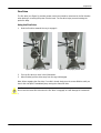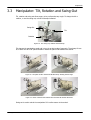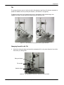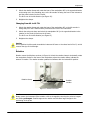
1
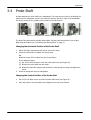
Leica Angle Two™ Computer-Assisted Stereotaxic System User Manual Living up to Life Intended Use Statement The Angle Two™ system is designed for stereotaxic surgery on small animals only and is not intended for use on humans. Trademarks Leica, Leica Microsystems and logo are trademarks of Leica Microsystems GmbH HRB 5187 and used under license. Angle Two is a trademark of Leica Biosystems St. Louis, LLC 61-1561098. Other trademarks are the property of their owners. Copyright Leica Biosystems St. Louis, LLC owns the copyright on this document and any associated software. Leica Biosystems St. Louis is part of the Leica Microsystems group of companies. Under law, our written permission is required before either the documentation or the software is copied, reproduced, translated, or converted to electronic or other machine-readable form, in whole or in part. Doc. 95.8808 Rev A01 © Leica Biosystems St. Louis, LLC 2009 Important Information for All Users The term "Leica Microsystems" when used in text in this document refers to Leica Biosystems St. Louis, LLC. Due to a policy of continuous improvement, Leica Microsystems reserves the right to change specifications without notice. Warranty claims can be made only if the system has been used for the specified application and operated according to the instructions in this document. Damage resulting from inappropriate handling and/or misuse of the product will invalidate the warranty. Leica Microsystems cannot assume liability for any such damage. Persons operating the Angle Two must be adequately trained and warned of any potential hazards or hazardous procedures before operating the instrument. Only trained staff are to remove any parts from the instrument, and only if instructed within this manual. Repairs must only be carried out by qualified service personnel authorized by Leica Microsystems. Safety Safety notices in this manual are cautions. Cautions are notifications of hazards that could lead to damage to the instrument or the animal undergoing surgery. Cautions use symbols with a yellow border, as illustrated below: Revision Record Rev. Issued Sections affected Detail A01 September 2009 All First release Contacting Leica Microsystems For enquiries related to the Angle Two™ stereotaxic system: Phone: +1 (866) 444 0944 +1 (314) 522 0300 Email: [email protected] Web site: www.myneurolab.com For other Leica Microsystems products see the web site: www.leica-microsystems.com Leica Angle Two™ User Manual Rev A01 © Leica Biosystems St. Louis, LLC 2009 2 Contents Leica Angle Two™ . . . . . . . . . . . . . . . . . . . . . . . . . . . . . . . . . . . . . . . . . . . . . . . . . . . . . . . . . . 1 Intended Use Statement . . . . . . . . . . . . . . . . . . . . . . . . . . . . . . . . . . . . . . . . . . . . . . . . . . . . . . . Trademarks . . . . . . . . . . . . . . . . . . . . . . . . . . . . . . . . . . . . . . . . . . . . . . . . . . . . . . . . . . . . . . . . Copyright. . . . . . . . . . . . . . . . . . . . . . . . . . . . . . . . . . . . . . . . . . . . . . . . . . . . . . . . . . . . . . . . . . Important Information for All Users . . . . . . . . . . . . . . . . . . . . . . . . . . . . . . . . . . . . . . . . . . . . . . . Safety . . . . . . . . . . . . . . . . . . . . . . . . . . . . . . . . . . . . . . . . . . . . . . . . . . . . . . . . . . . . . . . . . . . . Revision Record . . . . . . . . . . . . . . . . . . . . . . . . . . . . . . . . . . . . . . . . . . . . . . . . . . . . . . . . . . . . . Contacting Leica Microsystems. . . . . . . . . . . . . . . . . . . . . . . . . . . . . . . . . . . . . . . . . . . . . . . . . . . 1 3 4 5 . . . . . . . . . . . . . . . . . . . . . . . . . . . . . . . . . . . . . . . . . . . . . . . . . . . . . . . . . . . . . . . .2 .2 .2 .2 .2 .2 .2 Introduction . . . . . . . . . . . . . . . . . . . . . . . . . . . . . . . . . . . . . . . . . . . . . . . . . . . . 5 1.1 2 . . . . . . . Basic Steps of Use. . . . . . . . . . . . . . . . . . . . . . . . . . . . . . . . . . . . . . . . . . . . . . . . . . . . . . . . . 5 Instrument Installation . . . . . . . . . . . . . . . . . . . . . . . . . . . . . . . . . . . . . . . . . . . 6 2.1 Checklist . . . . . . . . . . . . . . . . . . . . . . . . . . . . . . . . . . . . . . . . . . . . . . . . . . . . . . . . . . . . . . . 6 2.2 Assembly . . . . . . . . . . . . . . . . . . . . . . . . . . . . . . . . . . . . . . . . . . . . . . . . . . . . . . . . . . . . . . . 7 Components . . . . . . . . . . . . . . . . . . . . . . . . . . . . . . . . . . . . . . . . . . . . . . . . . . . . 9 3.1 U Frame . . . . . . . . . . . . . . . . . . . . . . . . . . . . . . . . . . . . . . . . . . . . . . . . . . . . . . . . . . . . . . . 10 3.2 Manipulator: Linear Movements . . . . . . . . . . . . . . . . . . . . . . . . . . . . . . . . . . . . . . . . . . . . . . 13 3.3 Manipulator: Tilt, Rotation and Swing-Out . . . . . . . . . . . . . . . . . . . . . . . . . . . . . . . . . . . . . . . 16 3.4 Probe Shaft . . . . . . . . . . . . . . . . . . . . . . . . . . . . . . . . . . . . . . . . . . . . . . . . . . . . . . . . . . . . 20 3.5 Vernier Scales . . . . . . . . . . . . . . . . . . . . . . . . . . . . . . . . . . . . . . . . . . . . . . . . . . . . . . . . . . . 22 3.6 Computer . . . . . . . . . . . . . . . . . . . . . . . . . . . . . . . . . . . . . . . . . . . . . . . . . . . . . . . . . . . . . . 23 3.7 Ancillary Products . . . . . . . . . . . . . . . . . . . . . . . . . . . . . . . . . . . . . . . . . . . . . . . . . . . . . . . . 23 Software Overview . . . . . . . . . . . . . . . . . . . . . . . . . . . . . . . . . . . . . . . . . . . . . . 24 4.1 Atlas . . . . . . . . . . . . . . . . . . . . . . . . . . . . . . . . . . . . . . . . . . . . . . . . . . . . . . . . . . . . . . . . . 25 4.2 Modes . . . . . . . . . . . . . . . . . . . . . . . . . . . . . . . . . . . . . . . . . . . . . . . . . . . . . . . . . . . . . . . . 26 4.3 Initial Settings. . . . . . . . . . . . . . . . . . . . . . . . . . . . . . . . . . . . . . . . . . . . . . . . . . . . . . . . . . . 29 4.4 Coordinates . . . . . . . . . . . . . . . . . . . . . . . . . . . . . . . . . . . . . . . . . . . . . . . . . . . . . . . . . . . . 31 Alignment . . . . . . . . . . . . . . . . . . . . . . . . . . . . . . . . . . . . . . . . . . . . . . . . . . . . . 33 5.1 Checklist . . . . . . . . . . . . . . . . . . . . . . . . . . . . . . . . . . . . . . . . . . . . . . . . . . . . . . . . . . . . . . 33 5.2 Procedure. . . . . . . . . . . . . . . . . . . . . . . . . . . . . . . . . . . . . . . . . . . . . . . . . . . . . . . . . . . . . . 34 6 Using the Instrument . . . . . . . . . . . . . . . . . . . . . . . . . . . . . . . . . . . . . . . . . . . . 42 7 Cleaning and Maintenance . . . . . . . . . . . . . . . . . . . . . . . . . . . . . . . . . . . . . . . . 47 8 Specifications . . . . . . . . . . . . . . . . . . . . . . . . . . . . . . . . . . . . . . . . . . . . . . . . . . 48 Software Licence Terms . . . . . . . . . . . . . . . . . . . . . . . . . . . . . . . . . . . . . . . . . . . . . . . . . . . . 50 Leica Angle Two™ User Manual Rev A01 © Leica Biosystems St. Louis, LLC 2009 3 4 1 Introduction The Leica Angle Two™ stereotaxic system is designed to perform surgery at any angle and position. Reaching the target site is easier and more accurate than manual systems as the Angle Two is computer assisted – all calculations are done for you – meaning no user errors and no pilot studies are needed. Probe position is set with three linear movements (in anterior/posterior, medial/lateral and dorsal/ventral axes) and two rotary movements (tilt and rotation). Encoders on the manipulator measure these positions and relay them to the Angle Two software. Using this information the software calculates the exact distance the manipulator must move to reach the target site at any tilt or rotation. The Virtual Skull Flat™ feature in the software uses the bregma and lambda reference points to calculate skull tilt. You don’t need to move the animal to align the skull horizontally or perform difficult calculations to incorporate tilt into the target coordinates – the software does it for you, automatically setting appropriate “distance to target” figures. To use the Angle Two system choose one of the fully integrated animal atlases available and then select the coronal plate that shows the target site. Click on the screen to mark the site or alternatively use previously saved target coordinates. Once the target site is entered move the probe to known reference points (typically bregma and lambda) which the software uses to align the atlas plate with the animal’s brain. Then simply move the manipulator drives and watch the AP, ML and DV displays count down to zero – indicating you have reached the target site. 1.1 Basic Steps of Use After unpacking and installing the instrument: 1. Align the instrument (see “Alignment” on page 33) 2. Select the target site (see “Selecting the Target Site” on page 43) 3. Position the animal (see “Positioning the Animal” on page 43) 4. Locate the zero reference points (see “Locating Bregma and Lambda” on page 44) 5. Move the manipulator over the target site (see “Locating the Target Site” on page 45) and 6. Drill and insert the probe (see “Probe Placement” on page 46). Leica Angle Two™ User Manual Rev A01 © Leica Biosystems St. Louis, LLC 2009 5 2 Instrument Installation The Leica Angle Two™ system needs to be assembled before use. Use the information below to check you have received all parts and to assemble the system. 2.1 Checklist The list below has the standard items supplied in the Angle Two system. Additional or alternative items may be included depending on the configuration ordered. Angle Two instrument U frame with 45° and 18° ear bars and species adaptor One or two manipulators with one standard probe holder each Stereotaxic Alignment Kit (dial indicator, T-bar, clamp and Allen wrench) A brain atlas in book form appropriate to the species you have selected ‘Angle Two Support’ DVD Computer (with Windows XP, Angle Two software v 3.0.0, and a rat and/or mouse brain atlas installed) Monitor Keyboard Mouse Leica Angle Two™ User Manual Rev A01 © Leica Biosystems St. Louis, LLC 2009 6 Instrument Installation 2.2 Assembly When unpacked the Angle Two instrument has either one or two manipulators folded over the U frame. 1. Unlock the tilt clamp and gently lift each manipulator upright into position. The rotation encoder will point down and the tilt encoder to the left, away from the U frame. Figure 1. Unlock the tilt clamp (highlighted) and gently lift the manipulator upright into position Leica Angle Two™ User Manual Rev A01 © Leica Biosystems St. Louis, LLC 2009 7 Instrument Installation 2. Connect the five instrument cables into the ports at the rear of the computer. Each cable and its corresponding port has the same label. If you have a second manipulator, insert the cables into the second card provided. Figure 2. Angle Two cables and computer ports 3. Connect the mouse and keyboard into the computer’s USB ports. 4. Connect the monitor and computer power cables into an outlet. 5. Turn on the computer. The Angle Two icon is displayed on the desktop. Figure 3. The Angle Two desktop shortcut Caution Do not pull or attempt to lift the instrument by any of its cables. Caution The computer must be connected to an earthed mains power outlet socket. Leica Angle Two™ User Manual Rev A01 © Leica Biosystems St. Louis, LLC 2009 8 3 Components This chapter describes the parts of the Leica Angle Two™ system and their functions. The components are divided into the following sections: “U Frame” on page 10 – includes the ear bars, nose clamp and incisor bar. “Manipulator: Linear Movements” on page 13 – the three coarse drives that move the manipulator in the AP, ML and DV directions and the fine drive for small movements in the DV direction. “Manipulator: Tilt, Rotation and Swing-Out” on page 16 – how to tilt and rotate the probe, and swing-out for moving the probe away in order to get clear access to the animal. “Probe Shaft” on page 20 – how to change the shaft’s vertical and horizontal positions and attach a probe. “Vernier Scales” on page 22 – how to use the vernier scales. “Computer” on page 23 – lists the hardware and software provided. “Ancillary Products” on page 23 – other products available from Leica Microsystems for stereotaxic surgery. Leica Angle Two™ User Manual Rev A01 © Leica Biosystems St. Louis, LLC 2009 9 Components 3.1 U Frame Figure 4 shows the Angle Two U frame with a rat adapter and 45° ear bars attached. Some of the part names in the figures below link to sections of the manual where the part is discussed in more detail. Nose Clamp Incisor Bar Ear Bars U Frame Figure 4. The Angle Two U frame The animal is held in the instrument U frame with the head at the closed end of the ‘U’ and the tail at the open end. The ear bars, nose clamp and incisor bar attached to the U frame secure the animal during surgery. Adapters for a range of species are available (see www.myneurolab.com). Incisor Bar The animal’s incisor teeth are placed over the incisor bar. The bar can be adjusted horizontally and vertically using the thumbscrews indicated below. Vertical control Incisor Bar Horizontal control Figure 5. The incisor bar can be moved vertically and horizontally Leica Angle Two™ User Manual Rev A01 © Leica Biosystems St. Louis, LLC 2009 10 Components Caution Do not use the rat incisor bar on a mouse. A mouse adaptor is available from Leica Microsystems Note: Hamsters and gerbils can use the rat ear bars and incisor bar. Nose Clamp The nose clamp is placed over the animal’s nose and gently lowered using the thumbscrew indicated. The nose clamp helps to keep the animal in position when injectable anesthesia is used. Nose clamp Vertical control Removes the nose clamp Figure 6. The nose clamp If using gas anesthesia with a nose cone, remove the nose clamp by undoing the thumbscrew and insert the nose cone. Ear Bars The ear bars are tapered at one end to fit deep inside the ear canal. The instrument comes with two rat ear bars (18° and 45°). Other animal ear bars and adaptors can be purchased from Leica Microsystems. After the bars are positioned in the animal’s ears, the animal is centered in the middle of the U frame using the scales on each bar. Use this measurement to reproduce the exact position of the animal for repeat studies. Leica Angle Two™ User Manual Rev A01 © Leica Biosystems St. Louis, LLC 2009 11 Components The ear bars are locked into position using the locking levers. The right lever has a right-hand thread, and the left lever a left-hand thread. The levers only need a quarter turn to lock the ear bars. Figure 7. Turn the lever to lock the ear bar into place Caution Use 45° ear bars if post-surgical testing is planned. Do not use 18° ear bars as they can easily break the tympanic membrane of an animal. Caution Do not use the 45° or 18° ear bars on a mouse. The mouse’s breathing passages run between the ears, and as the skull is very thin, the mouse can easily suffocate if too much pressure is applied by the ear bars. Leica Angle Two™ User Manual Rev A01 © Leica Biosystems St. Louis, LLC 2009 12 Components 3.2 Manipulator: Linear Movements The manipulator is attached to the U frame and is used to move the probe. The manipulator has three linear movements: AP (Anterior/Posterior): moves horizontally along the length of the instrument ML (Medial/Lateral): moves horizontally across the width of the instrument DV (Dorsal/Ventral): moves vertically towards the animal. AP ML DV Figure 8. The three linear movements of the manipulator: AP, ML and DV Leica Angle Two™ User Manual Rev A01 © Leica Biosystems St. Louis, LLC 2009 13 Components Each linear movement is controlled by a coarse drive and, in the DV direction, by an additional fine drive. When locating the target site, first use the ML and AP coarse drives to move the probe into place above the target. Then lower the probe until it reaches the brain using the coarse DV drive. Finally, engage the fine drive to continue DV movements to the target within the brain. DV drive ML drive Fine drive AP drive Figure 9. The AP, ML, DV and fine drives move the probe in linear movements Caution Do not continue to turn the drives at the end of their movement range, as it will damage the nut below. Leica Angle Two™ User Manual Rev A01 © Leica Biosystems St. Louis, LLC 2009 14 Components Fine Drive The fine drive (see Figure 9) provides greater control and smoother movement in the DV direction when placing or removing the probe from the brain. The fine drive helps prevent inserting the probe too deep. Using the Fine Drive 1. Push the fine drive towards the cog to engage it. Figure 10. Engaging the fine drive Figure 11. Disengaging the fine drive 2. Turn the fine drive to move in the DV direction. 3. When finished, pull the drive away from the cog to disengage. Note: When engaging the fine drive, if no click is heard, slowly turn the coarse DV drive until you hear it click into place. Immediately stop moving the coarse DV drive. Caution Do not use the coarse DV drive when the fine drive is engaged, as it will damage the instrument. Leica Angle Two™ User Manual Rev A01 © Leica Biosystems St. Louis, LLC 2009 15 Components 3.3 Manipulator: Tilt, Rotation and Swing-Out Tilt, rotation and swing-out allow surgery to be performed at any angle. To change the tilt or rotation, or use the swing-out, unlock the clamps indicated. Swing-Out Tilt Rotation Figure 12. The swing-out, rotation and tilt clamps Tilt changes the manipulator angle and in turn the probe’s angle of approach. The primary tilt can be in either the AP or ML planes. Rotation rotates the plane of tilt between AP and ML. Figure 13. The plane of tilt in the AP and ML directions, showing the tilt range Figure 14. Plane of the tilt with rotations from the basic AP and ML directions Swing-out is used to swivel the manipulator 90° to allow access to the animal. Leica Angle Two™ User Manual Rev A01 © Leica Biosystems St. Louis, LLC 2009 16 Components Tilt The primary tilt plane can be in either the AP or ML directions (see Figure 15). Always remember to update the software with the primary tilt direction (“Tilt Direction” on page 29). To adjust the tilt loosen the tilt clamp and move the manipulator to the desired angle, then retighten the clamp. A scale below the clamp shows the amount of tilt. Figure 15. Tilt movement in the AP (left) and ML (right) directions Changing From AP to ML Tilt 1. Unlock the swing-out clamp and swivel the manipulator 90° so the probe faces the front of the instrument (see Swing-Out). Swing-out clamp Tilt encoder Rotation clamp Figure 16. The manipulator swung-out to the front of the instrument Leica Angle Two™ User Manual Rev A01 © Leica Biosystems St. Louis, LLC 2009 17 Components 2. Unlock the rotation clamp and rotate the base of the manipulator 90° in the opposite direction to the swing-out in the preceding step, so the tilt encoder is facing the front of the instrument and the probe is back over the U frame. The tilt is now in the ML direction (see Figure 15). 3. Retighten the clamps. Changing From ML to AP Tilt 1. Unlock the rotation clamp and rotate the base of the manipulator 90° so the tilt encoder is facing away from the U frame and the probe faces the front of the instrument. 2. Unlock the swing-out clamp and swivel the manipulator 90° (in the opposite direction to the rotation) so the probe is back over the U frame. The tilt is now in the AP direction (see Figure 15) 3. Retighten the clamps. Caution Do not position the tilt encoder towards the instrument U frame or the closed end of the ‘U’, as this reverses the sign of the tilt angle. Rotation Rotation moves the tilt plane as shown in Figure 14. Unlock the rotation clamp to horizontally rotate the manipulator. Scales on the base of the manipulator, above the rotation clamp, indicate the amount of rotation. The rotation encoder updates the software with the instrument’s position. Rotation clamp Rotation encoder Figure 17. Rotation movement Caution Always rezero the instrument if the rotation or tilt are changed by returning the probe to bregma and clicking at Bregma. Small changes to the rotation and tilt cause large changes to the positioning of the probe Leica Angle Two™ User Manual Rev A01 © Leica Biosystems St. Louis, LLC 2009 18 Components Swing-Out Use swing-out to change the primary tilt plane by 90°, between the ML and AP directions (see Figure 13) or to remove the probe from the area within the U frame and return it later to the same position. To change the primary tilt plane see “Tilt” on page 17. To swing out the manipulator: 1. Unlock the swing-out clamp and push the manipulator up with your thumb just enough to clear the raised edge on the base of the manipulator. Swing-out clamp Figure 18. Raising the manipulator using swing-out 2. Swivel the manipulator around 90° then lower it back down in its new position. 3. Retighten the swing-out clamp. Leica Angle Two™ User Manual Rev A01 © Leica Biosystems St. Louis, LLC 2009 19 Components 3.4 Probe Shaft A clamp attaches the probe shaft to the manipulator. The clamp has two holes for positioning the clamp onto the manipulator, and you can rotate the clamp to the left or right of the manipulator arm to give a total of four possible probe positions in the AP direction. Figure 19. Four positions for attaching the probe shaft to the manipulator The Angle Two comes with a standard probe holder. Contact Leica Microsystems if you require different probe holders (see “Contacting Leica Microsystems” on page 2). Changing the Horizontal Position of the Probe Shaft 1. Unlock the clamp thumbscrew and remove it from the clamp. 2. Select the second hole to reattach the clamp screw OR Rotate the clamp 180° to select from two new positions. If you rotate the clamp: (i) Use a 3/32” Allen wrench to undo the probe shaft screw (see Figure 20). (ii) Remove the probe shaft from the clamp. (iii) Rotate the shaft 180° degrees and reposition it back through the clamp and tighten the screw. 3. Screw the clamp back onto the manipulator. Changing the Vertical Position of the Probe Shaft 1. Use a 3/32 inch Allen wrench to undo the probe shaft screw (see Figure 20). 2. Move the probe to the new position and retighten the screw when finished. Leica Angle Two™ User Manual Rev A01 © Leica Biosystems St. Louis, LLC 2009 20 Components Probe clamp screw Figure 20. The probe shaft screw Attaching a Probe At the bottom of the standard probe shaft is a plastic block with three grooved faces and one flat face. Insert the probe into one of the grooves (grooves are 1 mm apart) and tighten the metal clamp to secure. Probe shaft Clamp screw Grooves on the plastic block Metal clamp Figure 21. The probe holder at the bottom of the probe shaft Leica Angle Two™ User Manual Rev A01 © Leica Biosystems St. Louis, LLC 2009 21 Components 3.5 Vernier Scales All Leica stereotaxic instruments have vernier scales for manual measurements. While the instrument software makes all calculations for you, some manual measurements might be needed, for example to record the exact position of the ear bars for repeat studies. To use the scales follow the example below. The main scale, graduated in millimeters, is on the ear bar, and the vernier is on the U frame. To measure the position of the ear bar: 1. Locate the zero on the vernier scale and read below on the ear bar scale to get the first measurement – in the example it measures 1.2 cm. 2. Use the vernier scale to measure the second decimal place. To do this locate the line of the vernier scale that directly aligns with a millimeter line on the main scale below (only one can align, the rest are slightly off). The third line of the vernier scale aligns with the main scale beneath, therefore the measurement is 1.23 cm. The third line of the vernier scale on the U frame aligns with the ear bar scale First measurement on the ear bar scale is 1.2 cm Figure 22. Using the vernier scale Leica Angle Two™ User Manual Rev A01 © Leica Biosystems St. Louis, LLC 2009 22 Components 3.6 Computer The computer consists of a desktop case, monitor, mouse and keyboard. The computer comes with the Angle Two software that includes an integrated rat or mouse brain atlas. The computer should only be used to operate the Angle Two instrument. If your computer is to be networked we recommend installing antivirus or firewall software only. If you require additional species atlases contact Leica Microsystems (see “Contacting Leica Microsystems” on page 2). Note: if you do not want to have a keyboard connected, you can use the virtual keyboard. Click start\ All Programs\ Accessories\ Accessibility\ On-Screen Keyboard. 3.7 Ancillary Products Leica Microsystems has a wide range of accessories available for use with the Angle Two instrument. Some useful accessories include: Video microscope for Angle Two, Product #39464678 Rat Autoclip™ suture clips, Product #39465004 Manual drill with stop, Product #39463018 Large probe holder, Product #39464350 Other instruments that can be used in conjunction with the Angle Two are: Perfusion Two™ automated pressure perfusion system Nanoinjector, motorized stepper for stereotaxic use Impact One™ stereotaxic impactor for CCI Spinal holders For details of these and a full line of gas anesthesia equipment, probes and species adapters, see www.myneurolab.com. Leica Angle Two™ User Manual Rev A01 © Leica Biosystems St. Louis, LLC 2009 23 4 Software Overview This chapter describes the Angle Two™ software and its functions. The topics covered are: “Atlas” on page 25 – how to select an atlas and view the coronal plates. “Modes” on page 26 – explains what Set Target, To Target, Atlas and Vertical are used for. “Initial Settings” on page 29 – describes how to set the tilt direction, zero the rotation and tilt angles, enter offsets and indicate bregma and lambda to the software. “Coordinates” on page 31 – how to save and export coordinates, and how to use saved coordinates. Figure 23 shows the software interface, with the atlas coronal plate in the center and the right and left manipulator controls on either side. Clicking anywhere on the left or right side of the screen activates the respective manipulator’s controls for use. If you have only one manipulator, then only one side of the screen will be active. Atlas Modes Initial Settings Coordinates Figure 23. The Angle Two screen with the left manipulator attached Leica Angle Two™ User Manual Rev A01 © Leica Biosystems St. Louis, LLC 2009 24 Software Overview 4.1 Atlas The instrument comes with either one mouse or one rat brain atlas installed. Contact Leica Microsystems to see what other animal atlases are available. Click Select Atlas to choose an atlas. The atlas consists of a series of coronal plates displayed in the center of the screen. The position of the current plate relative to the rest of the brain is shown in the top left corner. Click Rostral and Caudal to move through the coronal plates. Plate position in the brain Coronal plate Atlas selection Rostral button Caudal button Figure 24. The coronal plate is displayed with an insert in the top left corner showing its position in the brain Leica Angle Two™ User Manual Rev A01 © Leica Biosystems St. Louis, LLC 2009 25 Software Overview 4.2 Modes Four modes are used during surgery: Set Target, To Target, Atlas and Vertical. Each mode is used for a specific function and has its own set of coordinate boxes that appear beneath the mode selection area. Click the appropriate mode to activate it for use. Modes Coordinate boxes for the selected mode Figure 25. The four modes and coordinate boxes Set Target Select Set Target mode to enter the position of the target site – where the probe will go. The target site appears as a red crosshair on the coronal plate and its ML, AP and DV coordinates are displayed in the Target (Red) coordinate boxes (see Figure 25). Target coordinates can be edited at any time and saved. To save target coordinates click Save Named Point (see “Saving” on page 31). There are three ways to enter the coordinates: 1. Select the coronal plate showing the target site and click on a position in the map to set it as the target. 2. Use saved coordinates – double click on saved coordinates located across the bottom of the screen. The coordinates are automatically entered and a red crosshair appears (see Figure 26). Leica Angle Two™ User Manual Rev A01 © Leica Biosystems St. Louis, LLC 2009 26 Software Overview 3. Manually enter the coordinates into the Target (Red) boxes. Target coordinates Target position Saved coordinates Figure 26. Target coordinates can be entered manually into the coordinate boxes, loaded from saved coordinates or by positioning the mouse on the screen and clicking at the position If performing bilateral surgery, after the first target site has been located and probe placed, click +/- beside the ML coordinates box. The target site is moved to the opposite side of the coronal plate and the To Target coordinates are automatically updated. To Target The To Target mode shows the distance that the probe must move in the ML, AP and DV axes to reach the target site. The probe’s current position is shown as a blue crosshair, and the target site as a red crosshair. The To Target coordinate values reduce as the manipulator moves closer to the target site, until they read zero – indicating the target site has been reached. Probe position Distance to target Target site Figure 27. The distance the probe needs to move to reach the target site is shown in To Target mode Leica Angle Two™ User Manual Rev A01 © Leica Biosystems St. Louis, LLC 2009 27 Software Overview Atlas The Atlas mode shows the exact position of the probe including the rotation, tilt and the deviation of the animal’s skull from 0° (skull flat). The Skull value is only determined once the bregma and lambda points have been located. To save the atlas coordinates, click Export Position (see “Exporting” on page 32). Vertical Vertical mode is available for users wanting to measure the vertical position from the top of the brain rather than the top of the skull. In this mode the tilt and rotation must be zero, and the ‘virtual skull flat’ feature is unavailable. To locate the vertical zero point a user locates bregma, and from here moves the probe directly over the target site – which must have already been determined in pilot studies. The target site is marked on the skull and a hole drilled. The probe tip is carefully lowered through the hole towards the brain until the blood fluid, visible in the hole, dips downwards. This indicates that the probe is putting pressure on the dura membrane. This is the vertical zero point. To mark the location click Z to zero the instrument’s coordinates. Leica Angle Two™ User Manual Rev A01 © Leica Biosystems St. Louis, LLC 2009 28 Software Overview 4.3 Initial Settings Before locating the target site, you must do the following on the Angle Two software: Select the tilt direction Zero the tilt and rotation angles Enter any probe offsets Enter the locations of the reference points bregma and lambda Zero tilt and rotation and set offsets Bregma button Lambda button Tilt direction Figure 28. The initial settings buttons Tilt Direction After aligning the instrument, click ll To ML or ll To AP to indicate the primary plane of the tilt as either in the ML or AP direction (see “Tilt” on page 17). Zero Rotation and Tilt After aligning the instrument and entering the tilt direction, the software’s tilt and rotation values must be zeroed. Click Setup and Zero Angles in the Set Up dialog, check both angles read 0.00°. Figure 29. The set up dialog and Zero Angles button Leica Angle Two™ User Manual Rev A01 © Leica Biosystems St. Louis, LLC 2009 29 Software Overview Enter Offsets Offsets are used when performing surgery on animals that do not fall within the weight range of the atlas being used. Offsets are a distance in millimeters that is added or subtracted from the probe’s path to the target site. The offsets are determined in pre-experiments by locating the target site using the atlas coordinates. The amount that the atlas coordinates miss the actual target site is determined and used as the offset values. Once the offsets are entered, and the bregma and lambda points have been located, the offset values are automatically factored into the To Target coordinates, and the display is updated to show the probe approaching the correct target site. To enter offsets, click Use Zero Offsets in the Set Up dialog and enter the values. Click Close when finished. Bregma and Lambda Typically, the bregma and lambda points of the skull are used as zero reference points to determine the location of the target site. Once entered into the software, they are used in the skull flat feature which automatically incorporates any tilt in the animal’s skull into the calculation of the “to target” values. To set the bregma point, carefully place the probe tip on the bregma point of the animal’s skull and click at Bregma. To set the lambda point, gently lower the probe tip on the lambda point of the animal’s skull and click at Lambda. Always locate the bregma point first. Leica Angle Two™ User Manual Rev A01 © Leica Biosystems St. Louis, LLC 2009 30 Software Overview 4.4 Coordinates Coordinates can be saved or exported at any time while the instrument is being used. Double click on saved coordinates to load the target coordinates from a previous study, rather than relocating the target position. Saving coordinates Exporting coordinates Saved Coordinates Figure 30. Saving coordinates, exporting coordinates and saved coordinates Saving The target site, probe position or any other coordinates can be saved by clicking Save Named Point (see Figure 30). In the Saved Name Point dialog: Figure 31. The Saved Name Point dialog Leica Angle Two™ User Manual Rev A01 © Leica Biosystems St. Louis, LLC 2009 31 Software Overview 1. Enter a name and description. 2. In Choose Source Point select which coordinates you want to save: 3. Target Position – saves the current target site Device Position – saves the current position of the probe Input – saves a new target position, which you must provide coordinates for. Click Save. Once the coordinates are saved they will appear in the area beneath the atlas with the entered save name. Exporting To save the probe’s current position to an external location on the computer, click Export Position. In the Export dialog the coordinates are displayed – including the instrument’s tilt, rotation and the deviation of the animal’s head from skull flat (0°). Enter a description of the coordinates and click Save As. By default the coordinates are saved to folders located at c:\Program Files\AngleTwo\Targetpoints. If using the rat brain atlas the coordinates are saved in the Rat Coronal folder. Figure 32. The Export dialog Saved Coordinates Saved coordinates are displayed across the bottom of the interface. Double-clicking saved coordinates loads them into the Set Target boxes. Right-click on saved coordinates to open a menu with the following options (see Figure 33): Load Target – loads the coordinates into the Set Target boxes. Edit Target – opens Notepad which displays the coordinates, which you can edit. Export Target – exports the coordinates to an external location on the computer. When selected, the Export dialog opens. Enter in a description of the coordinates and click Save. Delete Target – deletes the coordinates from the saved area. Click Yes to confirm. Figure 33. Options for saved coordinates Leica Angle Two™ User Manual Rev A01 © Leica Biosystems St. Louis, LLC 2009 32 5 Alignment Align the instrument whenever the software is started. Alignment is necessary to check the horizontal (ML) and vertical (DV) planes are perpendicular. To align the instrument the rotation and tilt must be set so the scales read zero. The alignment kit is installed onto the instrument, with the dial indicator positioned where the probe would normally be. Then the dial indicator is moved across and up the T bar, mounted in the ear bar slots. If the dial indicator reading remains unchanged then the instrument is aligned and ready for surgical use. However, if the dial indicator reading changes, then the instrument is not aligned and the rotation or tilt must be adjusted. If you are unable to align the instrument contact Leica Microsystems. 5.1 Checklist Figure 34 shows the alignment kit contents, check that you have all five items: Alignment shaft Dial indicator Clamp Clamp adaptor T-bar Figure 34. The alignment kit Leica Angle Two™ User Manual Rev A01 © Leica Biosystems St. Louis, LLC 2009 33 Alignment 5.2 Procedure The primary tilt of the instrument can be in either the ML or AP direction so there are two procedures for the vertical alignment. Select the correct instruction set for your instrument set up. Set the Rotation and Tilt The instrument’s rotation and tilt must be moved so that the scales read zero. 1. Unlock the tilt and rotation clamps, and move the instrument so the scales read zero, as shown in Figure 35. 2. Relock the clamps when finished. Tilt clamp Zero tilt Zero rotation Rotation clamp Figure 35. Both the tilt and rotation are at zero Installing the Alignment Kit The alignment kit needs some assembly before being installed. 3. Double-click the ‘Angle Two’ icon on the desktop to start the program. 4. Remove the ear bars and probe shaft from the instrument. Leica Angle Two™ User Manual Rev A01 © Leica Biosystems St. Louis, LLC 2009 34 Alignment 5. Attach the clamp adaptor to the dial indicator by sliding the raised area of the dial indicator into the groove of the adaptor. Clamp adaptor Dial indicator Figure 36. The raised area of the dial indicator slides into the groove of the clamp adaptor 6. Slide the clamp covering towards the middle of the clamp to reveal the clamp hole. Push the top of the clamp adaptor into the clamp hole and release the covering to lock the adaptor in position. Clamp screw Clamp covering Figure 37. Slide the clamp covering to the middle to insert the clamp adaptor inside 7. Attach the clamp to the bottom of the alignment shaft and tighten the clamp screw so that it cannot move. Leica Angle Two™ User Manual Rev A01 © Leica Biosystems St. Louis, LLC 2009 35 Alignment 8. Attach the other end of the alignment shaft into the manipulator, and tighten the thumbscrew. Figure 38. The dial indicator attached to the Angle Two 9. Remove the ear bars and install the T-bar. Lock it into position. Figure 39. The T-bar locked into position Leica Angle Two™ User Manual Rev A01 © Leica Biosystems St. Louis, LLC 2009 36 Alignment Horizontal Alignment The ML horizontal path of the probe is controlled by the rotation of the instrument. This part of the alignment procedure checks the ML horizontal path is perpendicular. To test this the dial indicator is moved horizontally along the length of the T-bar. 10. Use the AP drive to position the dial indicator spring against the T-bar. AP drive Dial indicator spring T-bar Figure 40. The dial indicator spring against the T-bar 11. Continue to move the AP drive so that the dial indicator arm completes one full rotation. Stop moving the drive when the dial reads zero. Figure 41. Dial indicator at zero Leica Angle Two™ User Manual Rev A01 © Leica Biosystems St. Louis, LLC 2009 37 Alignment 12. Use the ML drive to move the spring horizontally along the T-bar. Move the indicator to one side and then across the total length of the bar to the other side. Figure 42. Move the dial across the T-bar to check the horizontal alignment The instrument is horizontally aligned if the dial arm only moves several divisions back and forth, but stays roughly in the same position. The instrument is not horizontally aligned if the dial arm continuously moves in one direction. To select a new horizontal position: (i) Loosen the rotation clamp (see Figure 35) and carefully adjust the manipulator into a new position. (ii) Retighten the clamp and repeat steps 1- 3. NOTE: if you cannot horizontally align the instrument contact Leica Microsystems. Vertical Alignment The vertical path of the probe is controlled by the tilt of the instrument. This part of the alignment procedure is to confirm that the vertical path is straight. To test this the dial indicator is moved vertically up and down the T-bar. As the primary plane of the tilt can be either in the AP or ML direction, select the appropriate instruction set below: For AP tilt (tilt is along the length of the instrument) see “AP Tilt” on page 39. For ML tilt (tilt is along the width of the instrument) see “ML Tilt” on page 40. Leica Angle Two™ User Manual Rev A01 © Leica Biosystems St. Louis, LLC 2009 38 Alignment AP Tilt 13. If the tilt is in the AP direction use the ML drive to position the dial indicator spring in the middle of the T-bar. Do not move the AP drive. Figure 43. Tilt (1) is in the AP direction (2) 14. Move the spring to the bottom of the bar and then all the way up using the DV drive. Figure 44. Move the dial up the T-bar to check the vertical alignment The instrument is vertically aligned if the dial arm only moves several divisions back and forth, but stays roughly in the same position. The instrument is not vertically aligned if the dial arm continuously moves in one direction. To select a new vertical position: (i) Loosen the tilt clamp (see Figure 35) and carefully move the manipulator into a new position. (ii) Retighten the clamp and repeat step 14. Leica Angle Two™ User Manual Rev A01 © Leica Biosystems St. Louis, LLC 2009 39 Alignment 15. When the AP tilt is aligned, select ll To AP on the computer screen (see Figure 28). 16. Click Setup and click Zero Angles to zero the Tilt and Rotation. Figure 45. The set up dialog and Zero Angles button NOTE: if you cannot vertically align the instrument contact Leica Microsystems. ML Tilt 13. If the tilt is in the ML direction position the dial indicator spring against the side face of the Tbar (see Figure 47). Figure 46. Tilt (1) is in the ML direction (2) Leica Angle Two™ User Manual Rev A01 © Leica Biosystems St. Louis, LLC 2009 40 Alignment 14. Move the spring to the bottom and then up using the DV drive. Figure 47. Vertical alignment when the tilt is in the ML direction The instrument is aligned in the vertical axis if the dial arm only moves several divisions back and forth, but stays roughly in the same position. The instrument is not vertically aligned if the dial arm continuously moves in one direction. To select a new vertical position: (i) Loosen the tilt clamp (see Figure 35) and slightly adjust the manipulator into a new position. (ii) Retighten the clamp and repeat step 14. 15. When the ML tilt is aligned, select ll To ML on the computer screen (see Figure 28). 16. Click Setup and click Zero Angles to zero the Tilt and Rotation. NOTE: if you cannot vertically align the instrument contact Leica Microsystems. Leica Angle Two™ User Manual Rev A01 © Leica Biosystems St. Louis, LLC 2009 41 6 Using the Instrument This chapter describes the whole process of using the Angle Two™ system to perform surgery; from starting the software to successfully placing the probe. This chapter has the following sections: “Preparation” on page 43 “Selecting the Target Site” on page 43 “Positioning the Animal” on page 43 “Locating Bregma and Lambda” on page 44 “Locating the Target Site” on page 45 “Probe Placement” on page 46 Leica Angle Two™ User Manual Rev A01 © Leica Biosystems St. Louis, LLC 2009 42 Using the Instrument Preparation 1. Ensure that the instrument is clean (see “Cleaning and Maintenance” on page 47). 2. Turn the computer on and start the Angle Two software (see Figure 3). 3. Align the instrument using the alignment kit (see “Alignment” on page 33). Selecting the Target Site The software opens in Set Target mode. 4. Enter the target site into the Target (Red) boxes beneath using one of the following options (see “Set Target” on page 26): (i) Select the site on the atlas (ii) Open saved coordinates (iii) Manually type in the coordinates 5. If using offsets (see “Initial Settings” on page 29) click Setup and check the Use Zero Offsets box (see Figure 45), and enter the values. 6. Unlock the rotation and tilt clamps and move the manipulator to the angle you want to use to approach the target site. Relock the clamps when positioned. Positioning the Animal If repeat surgery is planned the position of the animal must be easily reproducible. Always use ear bars and an incisor bar suitable for the animal. Installing the Ear Bars 7. Set the left ear bar tip 1 cm left of the center position, and lock it in place. 8. Remove the right ear bar and leave the lock ‘open’. Locked Unlocked Figure 48. The left ear bar locked 1 cm from the center position and the right ear bar removed with the lock ‘open’ 9. Unscrew the incisor bar’s horizontal screw (see Figure 5) and slide the incisor bar to the rear of the instrument. 10. Hold the animal’s body with one hand and the head with the other hand. Move the animal so that the left ear bar is inside the ear canal. Point the animal’s nose slightly to the right to help with insertion. Leica Angle Two™ User Manual Rev A01 © Leica Biosystems St. Louis, LLC 2009 43 Using the Instrument 11. Support the head and insert the right ear bar into the right ear. 12. Lower the right ear bar into the instrument and lock it in place. 13. Check that both ear bar scales have the same readings. If not, loosen both ear bar locks and move the animal until both ear bar scales agree, then relock the ear bars. 14. Gently move the animal’s nose from side to side and up and down to check the ear bars are correctly positioned: If the nose moves reposition the ear bars further inside the ear canal. Ensure the ear bars have the same reading. Installing the Incisor Bar and Nose Clamp 15. Slide the incisor bar into the animal’s mouth and hook the incisor teeth over the bar. 16. Push the animal’s nose down, and slide the bar away from the animal so that the bar is directly behind the incisor teeth. 17. Tighten the horizontal thumbscrew (see Figure 5), so the bar cannot move. 18. If using an injectable anesthetic lower the nose clamp so it is secure against the animal’s nose. If using gas anesthetic remove the nose clamp and install the nose cone. Locating Bregma and Lambda After the skull is exposed locate the bregma and lambda points and mark them. We recommend using the video microscope or a stereomicroscope to accurately determine each point and reduce error. The bregma and lambda points are used by the software to calculate the distance the manipulator must move along each axis to reach the target site. As well, the bregma and lambda points provide the variance angle of the animal’s skull from flat (0°), which is corrected for by the software so no manual adjustment of the instrument is needed. NOTE: Always position the probe at the bregma point first. To identify the bregma and lambda points to the software: 19. Place the probe on the marked bregma point and click ‘at Bregma’. The Atlas mode will open showing the probe’s coordinates. 20. Reposition the probe tip at lambda and click at Lambda. The Skull box displays the skull’s angle of variance from flat (0°). Leica Angle Two™ User Manual Rev A01 © Leica Biosystems St. Louis, LLC 2009 44 Using the Instrument Locating the Target Site 21. Click To Target (see “To Target” on page 27) to view the distances to the target site. 22. Use the ML and AP drives to position the probe directly ‘above’ the target. As the target site is approached the ML and AP distances count down. 23. Continue to move the probe until the AP and ML axis displays read zero. A line appears on screen showing the path of descent (DV) towards the target. Probe crosshair ML and AP at zero Line of descent Target site position Figure 49. When the ML and AP coordinates read zero the probe crosshair (blue) shows a line of descent to the target crosshair (red). 24. Stop moving the drives. Note: you can preview the probe’s path through the brain to the target site by removing the probe and lowering the shaft using the DV drive. Drilling 25. Lower the probe using the DV drive so that the probe is just above the skull and mark the spot on the skull. 26. Unlock the swing-out clamp (see “Swing-Out” on page 19) and move the manipulator out of the way. Using the swing-out ensures the probe is returned to its exact position later, so you do not have to relocate the target site. 27. Set up the drill and select the drill bit. We recommend using a manual drill with settable depth stops. This reduces the risk of drilling too far and into the brain tissue below. Select the smallest possible drill bit. 28. Drill into the skull at the marked spot. Gently rotate the drill taking care not to drill too far. Caution Drill bits have a spherical head so the center of the drilled area is deeper than the edges. Be careful that the center of the drill does not go too far and strike a blood vessel beneath the bone. Caution After drilling the hole, bone might remain hidden around the outer edges of the drill site. Be careful when inserting the glass probe, as the bone can easily break the probe tip. Leica Angle Two™ User Manual Rev A01 © Leica Biosystems St. Louis, LLC 2009 45 Using the Instrument Probe Placement 29. Unlock the swing-out clamp and reposition the manipulator and probe over the animal. 30. Relock the swing-out clamp. 31. Use the coarse DV drive to approach the target site. 32. Engage the fine drive when about to enter the brain. A single blue circle appears around the blue crosshairs when the probe is 0.05 mm from the target site (see Figure 50). 33. Stop moving the probe once a second blue circle appears around the blue crosshairs. This indicates that the target site has been reached. Figure 50. A single blue circle appears when the probe is 0.05 mm from the target. A second blue circle appears when the probe is at the target site. 34. Perform the operation, and when complete withdraw the probe using the fine drive until the probe is clear of the brain. If you are performing bilateral surgery: (i) Select Set Target mode and click +/- (see Figure 25), to change the sign of the ML coordinate. (ii) Move the probe to the target site and repeat steps 22 to 32. 35. Remove the animal from the instrument and close up the incision. Leica Angle Two™ User Manual Rev A01 © Leica Biosystems St. Louis, LLC 2009 46 7 Cleaning and Maintenance Caution Do not autoclave. The bushings and probe holder block can be damaged. Caution Do not lubricate the axis rod slides or threaded drive. The plastic bushings can absorb oil and cause the instrument to seize. Caution Do not use alcohol to clean the surface of the probe holder block, as it can cause cracking. Wash the instrument with a cloth or paper towel, dampened with water or dishwasher detergent. If sterilization is needed, use Cidex® or a similar glutaraldehyde based sterilizer. Leica Angle Two™ User Manual Rev A01 © Leica Biosystems St. Louis, LLC 2009 47 8 Specifications Instrument Dimensions and Weight Base width 253 mm (10.0”) Base length 355 mm (14.0”) Base height 20 mm (0.8”) U frame width 165 mm (6.5”) U frame length 200 mm (8.0”) U frame height 55 mm (2.1”) Base and manipulator height 381 mm (15”) Base weight (no manipulator) 6.1 kg (13.5 lb.) Resolution, Linear Scales Vernier 0.1 mm Digital display 0.01 mm Internal scale 0.005/rev Drive Advancement per Revolution Linear axis drive (screw pitch) 5 mm/rev. (0.2”/rev.) Fine drive 0.25 mm/rev. (0.01”/rev.) Manipulator Movement AP 80 mm (3.1”) ML 80 mm (3.1”) DV 62.5 mm (2.5”) Tilt ±90° ML or AP axis, or combined Rotation 360° Rotary encoder resolution 1000 lines/rev. (0.36°) Leica Angle Two™ User Manual Rev A01 © Leica Biosystems St. Louis, LLC 2009 48 Specifications Tooth Bar Movement Ranges DV 20 mm (0.8”) AP 44.5 mm (1.8”) Probe Holder Diameter 7.88 mm (0.31”) Shaft length 200 mm (7.9”) Probe holder block 11 mm (0.4”) square (10 grooves spaced at 1 mm on 3 sides) Standard clamp holds 0.2 to 2 mm shaft (0.008” to 0.08”) Side clamp holds 0.2 to 4.5 mm shaft (0.008” to 0.2”) “C” clamp holds 0.2 to 4.5 mm shaft (0.008” to 0.2”) Set screw on shaft holder: 3/32” Leica Angle Two™ User Manual Rev A01 © Leica Biosystems St. Louis, LLC 2009 49 Software Licence Terms Software Licence Terms The following License Terms govern your use of the accompanying software unless you have a separate signed agreement with Leica Microsystems. License Grant Leica Microsystems grants you a license to Use one copy of the Software. "Use" means storing, loading, installing, executing or displaying the Software. You may not modify the Software or disable any licensing or control features of the Software. Ownership The Software is owned and copyrighted by Leica Microsystems. Your license confers no title to, or ownership in, the Software and is not a sale of any rights in the Software. Copies and Adaptations You may only make copies or adaptations of the Software for archival purposes or when copying or adaptation is an essential step in the authorized Use of the Software. You must reproduce all copyright notices in the original Software on all copies or adaptations. You may not copy the Software onto any public network. No Disassembly or Decryption You may not disassemble or decompile the Software unless Leica Microsystem's prior written consent is obtained. In some jurisdictions, Leica Microsystem's consent may not be required for limited disassembly or decompilation. Upon request, you will provide Leica Microsystems with reasonably detailed information regarding any disassembly or decompilation Transfer Your license will automatically terminate upon any transfer of the Software. Upon transfer, you must deliver the Software, including any copies and related documentation, to the transferee. The transferee must accept these License Terms as a condition to the transfer. Termination Leica Microsystems may terminate your license upon notice for failure to comply with any of these License Terms. Upon termination, you must immediately destroy the Software, together with all copies, adaptations and merged portions in any form. Export Requirements You may not export or re-export the Software or any copy or adaptation in violation of any applicable laws or regulations. Any person to whom this software is transferred should be aware that use of the software is subject to any Leica Microsystems software license terms distributed with the software. Use of the software indicates acceptance of these terms. If the user does not accept these terms, the software should be returned. Disclaimer of Warranty Leica Microsystems and its licensors disclaim all warranties, express, implied and statutory including, without limitation, the implied warranties of merchantability, fitness for a particular purpose, title and non-infringement of third party rights. No oral or written information or advice given by Leica Microsystems, its dealers, distributors, agents or employees shall in any way increase the scope of this warranty. Some jurisdictions do not allow the limitation or exclusion of implied warranties or how long an implied warranty may last, so the above limitations may not apply to you. Limitation of Warranty Leica Microsystems shall not be liable for any incidental, special, consequential or indirect damages of any kind (including damages for interruption of business, procurement of substituted goods, loss of profits, or the like) regardless of the form of action whether in contract, tort (including negligence), strict product liability or any other legal or equitable theory, even if such party has been advised of the possibility of such damages. In no event will Leica Microsystems aggregate cumulative liability for all claims out of or related to this agreement and software exceed the amounts you paid for software. Some jurisdictions do not allow the exclusion or limitation of incidental or consequential damages so this limitation and exclusion may not apply to you. Warranty disclaimer and limited liability are fundamental elements of the basis of the bargain between Leica Microsystems and you. Leica Microsystems would not be able to provide the software without such limitations. Leica Angle Two™ User Manual Rev A01 © Leica Biosystems St. Louis, LLC 2009 50