1
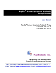
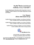
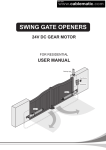
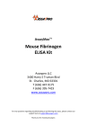
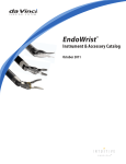
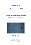
RayBio® Rat Cytokine Antibody Array
Patent Pending Technology
User Manual (Revised 06/14/09)
RayBio® Rat Cytokine Antibody Array 1 (Cat# AAR-CYT-1)
RayBio® Rat Cytokine Antibody Array 2 (Cat# AAR-CYT-2)
RayBio® Custom Ray Cytokine Antibody Array (Cat# AAR-CUST)
RayBio® Rat Cytokine Antibody Array Service (Cat# AAR-SERV)
Please read manual carefully before starting experiment
RayBiotech, Inc.
We Provide You with Excellent
Protein Array Systems and Service
Tel:(Toll Free) 1-888-494-8555 or 770-729-2992; Fax: 1-888-547-0580;
Website: www.raybiotech.com Email: [email protected]
HU
UH
HU
U
RayBiotech, Inc.
RayBio® Rat Cytokine Antibody Array Protocol
TABLE OF CONTENTS
U
I.
Introduction……..………………………………2
How It Works…………………………………. .4
II.
Materials Provided…………………………… 5
Additional Materials Required………………… 5
III. Overview and General Considerations……… 6
A. Preparation of Samples…………………… 6
B. Handling Array Membrane……………… .6
C. Incubation………………………………….6
IV. Protocol……………………………………… 7
A. Blocking and Incubation…………………. 7
B. Detection………………………………… 9
V.
Interpretation of Results…………………… 10
VI. Troubleshooting Guide……………………… 13
VII. Reference List ……………………………… 14
Cytokine protein arrays are RayBiotech patent-pending technology.
RayBio® is the trademark of RayBiotech, Inc.
RayBio® Rat Cytokine Antibody Array Protocol
1
I. Introduction
0B
All cell functions, including cell proliferation, cell death and differentiation,
as well as maintenance of health status and development of disease, are
controlled by a multitude of genes and signaling pathways. New techniques
such as cDNA microarrays have enabled us to analyze global gene
expression 1-3. However, almost all cell functions are executed by proteins,
which cannot be studied simply through DNA and RNA techniques.
Experimental analysis clearly shows a disparity between the relative
expression levels of mRNA and their corresponding proteins 4. Therefore,
analysis of the protein profile is critical. Currently, two-dimensional
polyacrylamide SDS page coupled with mass spectrometry is the mainstream
approach to analyzing multiple protein expression levels 5,6. However, the
requirement of sophisticated devices and the lack of quantitative
measurements greatly limit their broad application. Thus, effective study of
multiple protein expression levels has been complicated, costly are timeconsuming until now.
Our RayBio® Rat Cytokine Antibody Array is the first commercially
available cytokine protein array system 7-11. By using the RayBiotech system,
scientists can rapidly and accurately identify the expression profiles of
multiple cytokines in several hours inexpensively.
The RayBiotech kit provides a simple format and highly sensitive approach
to simultaneously detect multiple cytokine expression levels from
conditioned media, patient’s sera, cell lysate, tissue lysates and other sources.
The RayBio® Mouse Cytokine Antibody Array C series 1000 can detect 96
mouse cytokines in single experiment. RayBiotech also provides RayBio®
Human Cytokine Antibody Array C series 4000 which is the only product
available in the market that can detect 274 human cytokines in single
experiment.
Traditionally, cytokines are detected by using ELISA (enzyme-linked
immunsorbent assays); however, RayBiotech’s approach has several
RayBio® Rat Cytokine Antibody Array Protocol
2
advantages over ELISA. First, and most important, our approach can
simultaneously detect many cytokines. Secondly, the sensitivity is higher.
With this approach, most cytokines can be detected at pg/ml levels. As little
as 10 pg/ml of human IL-2 can be detected in the protein array format.
Furthermore, the detection range is much greater than ELISA. For example,
the detection range of human IL-2 varies from 10 to 100,000 pg/ml, whereas
the detection range varies only within 100-1000 fold in a typical ELISA.
Therefore, the detection range with protein arrays is greater than ELISA.
Additionally, variability is far lower in comparison ELISA. As determined
by densitometry, the variation between two spots ranged from 0 to 10% in
duplicated experiments. In contrast, variation (about 20%) in ELISA is much
higher. Finally, the system is much quicker and much easier to adapt to highthroughput techniques.
Pathway-specific array systems allow investigators to focus on the specific
problem and are becoming an increasingly powerful tool in cDNA
microarray systems. RayBiotech’s first protein array system, known as
RayBio® Rat Cytokine Antibody Array, is particularly useful in comparison
with the mouse cytokine cDNA microarray system. Besides the ability to
detect protein expression, RayBiotech’s system is a more accurate reflection
of active cytokine levels because it only detects secreted cytokines, and no
amplification step is needed. Cytokines play an important role in innate
immunity, apoptosis, angiogenesis, cell growth and differentiation 12. They
are involved in most disease processes, including cancer and cardiac
diseases. The interaction between cytokines and the cellular immune system
is a dynamic process. The interactions of positive and negative stimuli, and
positive as well as negative regulatory loops are complex and often involve
multiple cytokines.
Without doubt, simultaneous detection of multiple cytokines provides a
powerful tool to study cytokines.
RayBio® Rat Cytokine Antibody Array Protocol
3
Here’s how it works
Samples
Array support
Incubation of Sample
with arrayed antibody
supports
1-2 hrs
Cocktail of
Biotin-Ab
Incubation with
Biotinylated Ab
1-2 hrs
Labeledstreptavidin
Incubation with
labeled-Streptavidin
Detection of
signals
Data analysis
and graph
RayBio® Rat Cytokine Antibody Array Protocol
4
1 hrs
II. Materials Provided
Upon receipt, all components of the RayBio® Rat Cytokine Antibody Array
kit should be stored at -200C to -800C. At -200C to -800C the kit will retain
complete activity for up to 6 months. Once thawed, the array membranes
and 1X Blocking Buffer should be kept at –200C and all other components
should be stored at 40C. After thawing the reagents, the kit must be used
within three months, and please use the kit within six months of purchase.
• RayBio® Rat Cytokine Antibody Array membranes (2/4/8 membranes)
• Biotin-Conjugated Anti-Cytokines (1/2/4 vials) (each vial is for two
array membranes)
• 1,000X HRP-Conjugated Streptavidin (50 μl)
• 1X Blocking Buffer (15/25ml)
• 20X Wash Buffer I (10/20ml)
• 20X Wash Buffer II (10/20ml)
• 2X Cell Lysis Buffer (10/20ml)
• Detection Buffer C (1.5/2.5ml)
• Detection Buffer D (1.5/2.5ml)
• Eight-Well Tray (1 each)
• Manual
Additional Materials Required
1B
• Small plastic boxes or containers
• Orbital shaker
• Plastic sheet protector or SaranWrap
• Kodak x-omat AR film (REF 165 1454) and film processor
or
Chemiluminescence imaging system
RayBio® Rat Cytokine Antibody Array Protocol
5
III. Overview and General Considerations
2B
A. Preparation of Samples
• Use serum-free conditioned media if possible.
• If serum-containing conditioned media is required, use the serum as a
control since many types of sera contain cytokines.
• For cell lysates and tissue lysates, we recommend using 1X Cell Lysis
Buffer to extract proteins from cell or tissue (e.g. using homogenizer).
After extraction, spin the sample down and save the supernatant for
your experiment. Determine the protein concentration. Dilute 2X Cell
Lysis Buffer with H2O (we recommend adding proteinase inhibitors to
Cell Lysis Buffer before use).
• We recommend using
1 ml of Conditioned media
or
1 ml of original or 10-fold diluted sera
or
50-500 μg of protein for cell lysates and tissue lysates.
4BU
If you experience high background, you may further dilute your sample.
B. Handling Array Membranes
• Always use forceps to handle membranes, and grip the membranes by
the edges only.
• Never allow the array membranes to dry during experiments.
5BU
C. Incubation
• Completely cover the membranes with sample or buffer during
incubation, and cover the eight-well tray with a lid to avoid drying.
• Avoid foaming during incubation steps.
• Perform all incubation and wash steps under gentle rotation.
• Several incubation steps such as step 2 (blocking), step 3 (sample
incubation), step 7 (biotin-Ab incubation) or step 10 (HRP-streptavidin
incubation) may be done at 40C for overnight.
6BU
RayBio® Rat Cytokine Antibody Array Protocol
6
IV. Protocol
3B
A. Blocking and Incubation
U
1. Place each membrane into the provided eight-well tray (- means the
antibody printed side).
2. Add 2 ml 1X Blocking Buffer and incubate at room temperature for 30
min to block membranes.
Note: incubation may be done at 40C for overnight.
3. Incubate membranes with 1ml of sample at room temperature for 1 to 2
hours. Dilute sample using 1X Blocking Buffer if necessary.
Note: We recommend using 1 ml of Conditioned media or 1 ml of
original or 10-fold diluted sera or plasma or 50-500 μg of protein for cell
lysates and tissue lysates. Dilute the lysate at least 10 folds with 1 X
blocking buffer.
Note: The amount of sample used depends on the abundance of cytokines.
More of the sample can be used if the signals are too weak. If the signals
are too strong, the sample can be diluted further.
Note: Incubation may be done at 40C for overnight.
4. Decant the samples from each container, and wash 3 times with 2 ml of
1X Wash Buffer I at room temperature with shaking. Please allow 5
min per wash. Dilute 20X Wash Buffer I with H2O.
5. Wash 2 times with 2 ml of 1X Wash Buffer II at room temperature with
shaking. Allow 5 min per wash. Dilute 20X Wash Buffer II with H2O.
6. Prepare working solution for primary antibody.
RayBio® Rat Cytokine Antibody Array Protocol
7
Add 100 μl of 1X blocking buffer to the Biotin-Conjugated AntiCytokines tube. Mix gently and transfer all mixture to a tube containing
2 ml of 1X blocking buffer.
Note: the diluted biotin-conjugated antibodies can be stored at 40C for 2-3
days.
7. Add 1 ml of diluted biotin-conjugated antibodies to each membrane.
Incubate at room temperature for 1-2 hours.
Note: incubation may be done at 40C for overnight.
8. Wash as directed in steps 4 and 5.
9. Add 2 ml of 1,000 fold diluted HRP-conjugated streptavidin (e.g. add 2
µl of HRP-conjugated streptavidin to 1998 µl 1X Blocking Buffer) to
each membrane.
Note: Mix the tube containing 1,000X HRP-Conjugated Streptavidin
well before use since precipitation may form during storage.
10. Incubate at room temperature for 2 hours.
Note: incubation may be done at 40C for overnight.
11. Wash as directed in steps 4 and 5.
RayBio® Rat Cytokine Antibody Array Protocol
8
B. Detection
U
U
* Do not let the membrane dry out during detection. The detection
process must be completed within 40 minutes without stopping.
1. Proceed with the detection reaction.
Add 250 μl of 1X Detection Buffer C and 250 μl of 1X Detection Buffer
D for one membrane; mix both solutions. Drain off excess wash buffer
by holding the membrane vertically with forceps. Place membrane
protein side up (“-“ mark is on the protein side top left corner) on a clean
plastic sheet (provided in the kit). Pipette the mixed Detection Buffer
onto the membrane and incubate at room temperature for 2 minutes.
Ensure that the detection mixture is completely and evenly covering the
membrane without any air bubbles.
2. Drain off any excess detection reagent by holding the membrane
vertically with forceps and touching the edge against a tissue. Gently
place the membrane, protein side up, on a piece of plastic sheet (“-“ mark
is on the protein side top left corner). Cover with another piece of plastic
sheet on the array. Gently smooth out any air bubbles. Avoid using
pressure on the membrane.
3. Expose the array to x-ray film (we recommend to use Kodak x-omat
AR film) and detect signal using film developer. Or the signal can be
detected directly from the membrane using a chemiluminescence
imaging system.
Expose the membranes for 40 seconds and then re-expose the film
according to the intensity of signals. If the signals are too strong
(background too high), reduce exposure time (e.g. 5-30 seconds). If the
signals are too weak, increase exposure time (e.g. 5-20 min or overnight).
Or re-incubate membranes overnight with 1x HRP-conjugated
streptavidin, and redo detection in the second day.
4. Save membranes in –20 0C to –80 0C for future reference.
RayBio® Rat Cytokine Antibody Array Protocol
9
V. Interpretation of Results:
The following figure shows RayBio® Rat Cytokine Antibody Array
membranes probed with conditioned media from two different cell lines.
Membranes were exposed to Kodak x-omat film at room temperature for 1
minute. The biotin-conjugated IgG produces positive signals, which can
be used to identify the orientation and to compare the relative expression
levels among the different membranes.
One important parameter is background. To obtain the best results, we
suggest that several exposures be attempted. We also strongly recommend
using a negative control in which the sample is replaced with an
appropriate mock buffer according to the array protocol, particularly
during your first experiment.
Typical results using RayBio® Cytokine Antibody arrays
By comparing the signal intensities, relative expression levels of
cytokines can be made. The intensities of signals can be quantified by
densitometry. The positive control can be used to normalize the results from
the different membranes being compared. The signals also can be detected
and quantified by using a chemiluminescence-imaging device.
The RayBio® Analysis Tool is a program specifically designed for
analysis of RayBio® Cytokine Antibody Arrays. This tool will not only assist
in compiling and organizing your data, but also reduces your calculations to
a “copy and paste.” Call RayBiotech, Inc. at 770-729-2992 for ordering
information.
RayBio® Rat Cytokine Antibody Array Protocol
10
RayBio® Rat Cytokine Antibody Array 1
(Detect 19 rat cytokines in one experiment)
a
b
c
d
e
f
g
h
1
Pos
Pos
Neg
Neg
CINC-2
CINC-3
CNTF
Fractalkine
2
Pos
Pos
Neg
Neg
CINC-2
CINC-3
CNTF
Fractalkine
3
GM-CSF
IFN-γ
IL-1α
IL-1β
IL-4
IL-6
IL-10
LIX
4
GM-CSF
IFN-γ
IL-1α
IL-1β
IL-4
IL-6
IL-10
LIX
5
Leptin
MCP-1
MIP-3α
β-NGF
TIMP-1
TNF-α
VEGF
BLANK
6
Leptin
MCP-1
MIP-3α
β-NGF
TIMP-1
TNF-α
VEGF
BLANK
7
BLANK
BLANK
BLANK
BLANK
BLANK
BLANK
BLANK
Pos
8
BLANK
BLANK
BLANK
BLANK
BLANK
BLANK
BLANK
Pos
RayBio® Rat Antibody Array 2
(Detect 34 cytokines in one experiment)
A
B
C
D
E
F
G
H
I
J
K
L
1
POS
POS
NEG
NEG
Activin A
Agrin
B7-2/CD86
beta-NGF
CINC-1
CINC-2alpha
CINC-3
CNTF
2
POS
POS
NEG
NEG
Activin A
Agrin
B7-2/CD86
beta-NGF
CINC-1
CINC-2alpha
CINC-3
CNTF
3
Fas Ligand
Fractalkine
GM-CSF
ICAM-1
IFN-gamma
IL-1alpha
IL-1beta
IL-1 R6
IL-2
IL-4
IL-6
IL-10
4
Fas Ligand
Fractalkine
GM-CSF
ICAM-1
IFN-gamma
IL-1alpha
IL-1beta
IL-1 R6
IL-2
IL-4
IL-6
IL-10
5
IL-13
Leptin
LIX
L-Selectin
MCP-1
MIP-3alpha
MMP-8
PDGF-AA
Prolactin R
RAGE
Thymus
Chemokine-1
TIMP-1
6
IL-13
Leptin
LIX
L-Selectin
MCP-1
MIP-3alpha
MMP-8
PDGF-AA
Prolactin R
RAGE
Thymus
Chemokine-1
TIMP-1
7
TNF-alpha
VEGF
BLANK
BLANK
BLANK
BLANK
BLANK
BLANK
BLANK
BLANK
BLANK
POS
8
TNF-alpha
VEGF
BLANK
BLANK
BLANK
BLANK
BLANK
BLANK
BLANK
BLANK
BLANK
POS
* For use with serum, plasma, condition medium, urine, other body fluids, cell lysates and certain tissue lysates samples
RayBiotech, Inc., the protein array pioneer company, strives to research and develop
new products to meet demands of the biomedical community. RayBio’s patent-pending
technology allows detection of over 180 cytokines, chemokines and other proteins in a
single experiment. Our format is simple, sensitive, reliable and cost effective. Products
include: Cytokine Arrays, Chemokine Arrays, ELISA kits, Phosphotyrosine kits,
Recombinant Proteins, Antibodies, and custom services.
1. Antibody arrays
Cytokine antibody array
RayBio® Rat Cytokine Antibody Array Protocol
11
Human cytokine antibody arrays
Mouse cytokine antibody arrays
Rat cytokine antibody arrays
Pathway- or disease-focused antibody arrays
Inflammation antibody array
Angiogensis antibody array
Chemokine antibody array
Growth factor antibody array
MMP antibody array
Atherosclerosis antibody array
Quantibody arrays for quantitative measurement of cytokine and other protein
concentraton
Phosphorylation antibody arrays
Biotin label-based antibody arrays for high density antibody arrays.
Antibody analysis tool, software
2.
3.
4.
5.
6.
7.
ELISA
Cell-based phosphorylation assay
Custom antibody arrays
Antibody
Recombinant protein
Protein arrays
RayBiotech also provides excellent custom service:
1. Antibody arrays
2. Protein arrays
3. Peptide synthesis
4. Production of recombinant protein and antibody
5. Peptide arrays
6. Phosphorylation arrays
7. ELISA
Just simply send your samples and we will do the assay for you.
Technology transfer program
Have you developed technologies or reagents interested to the scientific and research
community? RayBiotech can help you commercialize your technologies, reagents and
dream.
RayBio® Rat Cytokine Antibody Array Protocol
12
VI. Troubleshooting guide
Problem
Cause
Weak signal or no 1. Taking too much time
signal
for Detection.
2. Film developer does
not work properly.
3. Did not mix HRPstreptavidin well before
use.
Recommendation
1. The whole Detection process must be
completed in 30 min.
2. Fix film developer.
3. Mix tube containing 1,000X HRPConjugated Streptavidin well before use since
precipitation may form during storage.
4. Sample is too dilute.
4. Increase sample volume, (e.g. using undilute
sample) or using more cells (e.g. seed 2 million
cells. After 1 or 2 days, change complete
medium with low serum medium and collect
conditioned medium 2 day later. Use about 1
to 2 ml of conditioned medium for
experiment).
5. Other.
1. Reduce blocking concentration by diluting
in 1X Wash Buffer II.
2. Slightly increase HRP concentrations.
3. Slightly increase biotin-antibody
concentrations.
4. Expose film for overnight to detect weak
signal.
Uneven signal
1. Bubbles formed
1. Remove bubble during incubation.
during incubation.
2. Membranes were not 2. Completely cover membranes with solution.
completely covered by
solution.
High background 1. Exposure to x-ray file 1. Decrease exposure time.
is too long.
2. Membranes were
2. Completely cover membranes with solution
allowed to dry out during during experiment.
experiment.
3. Sample is too
concentrated.
RayBio® Rat Cytokine Antibody Array Protocol
3. Use more diluted sample.
13
Reference List
7B
1. HIV-1-mediated apoptosis of neuronal cells:
Proximal molecular mechanisms of HIV-1-induced encephalopathy.
Yan Xu, Joseph Kulkoshy, Roger j. Pomerantz. PNAS. 2004 May 4,
2004 Vol. 101 No. 18.
H
2. Veto-like activity of mesenchymal stem cells: functional discrimination
between cellular responses to alloantigens and recall antigens.
Rameshwar P. Journal of Immunology. 2003 Oct 1;171(7):3426-34.
3. Cytokine responses elicited in endothelial cells after treatment with a
specific toxin. Jaya Pandey. BioCompare Product Review. May 13,
2004
H
H
4. Proteomic Characterization of the Interstitial Fluid Perfusing the Breast
Tumor Microenvironment. A Novel Resource for Biomarker and
Therapeutic Target Discovery. Julio E. Celis, Pavel Gromov, Teresa
Cabezón, José M. A. Moreira, Noona Ambartsumian, Kerstin Sandelin,
Fritz Rank, and Irina Gromova. Molecular Cellular Proteomics. April
2004; 11(3):328-39.
H
H
5. Increased Expression and Secretion of Interleukin-6 in Patients with
Barrett’s Esophagus.. Katerina Dvorakova, Harinder Garewal Clinical
Cancer Research. 2004 Mar 15;10(6):2020-8.
H
H
6. Antibody array-generated profiles of cytokine release from THP-1
leukemic monocytes exposed to different amphotericin B formulations.
Turtinen LW, Prall DN, Bremer LA, Nauss RE, Hartsel SC.
Antimicrobial Agents Chemotherapy. 2004 Feb;48(2):396-403.
7. Reduced T-cell and dendritic cell function is related to cyclooxygenase2 overexpression and protaglandin e(2) secretion in patients with breast
cancer". Pockaj BA, Basu GD. Annals of Surgical Oncology. 3:327344, 2004.
H
H
8. Inhibition of macrophage migration inhibitory factor decreases
proliferation and cytokine expression in bladder cancer cells. Katherine
H
RayBio® Rat Cytokine Antibody Array Protocol
14
L Meyer-Siegler, BMC Cancer. 2004, 4:34.
H
9. The malaria metabolite hemozoin initiates proinflammatory signaling
via a MyD88- dependent pathway.International Congress of
Immunology. 2004 July W23-81.
H
H
10. In Vivo Proteomic Analysis of Cytokine Expression in Laser CaptureMicrodissected Urothelial Cells of Obstructed Ureteropelvic Junction
Procured by Laparoscopic Dismembered Pyeloplasty. Journal of
Endourology. 2003 June; Volume:17 Number:5 Page:333--336.
11. Cytokine Antibody Arrays: A Promising Tool to Identify Molecular
Targets for Drug Discovery. Huang, Combinatorial Chemistry &
High Throughput Screening. 2003, 6,79-99
H
H
H
H
12. K.S. Rosenthal, N.Goel, R. Singavarapu, D.H. Zimmerman. Cel-1000
protects mice against HSV-1 challenge by stimulating IL-2 production.
Abstract published in abstract book of Interscience Conference on
Antimicrobial Agents and Chemotherapy held in Chicago during
Sept14-17, 2003.
H
13. Y. Lin, Ruochun Huang, Li-Pai Chen, Henry Lisoukov, Zhen-Hai Lu,
Shiyong Li, Cheng C. Wang and R.-P. Huang. (2003) Profiling of
cytokine expression by biotin-labeled-based protein arrays.
Proteomics.3: 1750-1757.
H
H
14. C. C. Wang, R.-P. Huang, H. Lisoukov, M. Sommer, R. Huang, Y.
Lin and J. Burke. (2002) Array-based multiplexed screening and
quantitation of human cytokines and chemokines. J. Proteome Res.
1:337-343.
H
15. R. Huang, Y. Lin, C. C. Wang, J. Gano, B. Lin, Q. Shi, A. Boynton, J.
Burke and R.-P. Huang. (2002) Connexin suppresses human
glioblastoma cell growth by down-regulation of monocyte chemotactic
protein 1, as discovered using protein array technology. Cancer
Research. 62 : 2806-2812.
H
RayBio® Rat Cytokine Antibody Array Protocol
15
16. R.-P. Huang, R. Huang, Y. Fan and Y. Lin. (2001). A novel method
for high-throughput protein profiling from conditioned media and
patient’s sera. Ana. Biochem. 294(1):55-62.
H
H
RayBio® Rat Cytokine Antibody Array Protocol
16
Note:
RayBio® is the trademark of RayBiotech, Inc.
Cytokine protein arrays are RayBiotech patent-pending technology.
This product is intended for research only and is not to be used for clinical
diagnosis. Our produces may not be resold, modified for resale, or used to
manufacture commercial products without written approval by RayBiotech,
Inc.
Under no circumstances shall RayBiotech be liable for any damages arising
out of the use of the materials.
Products are guaranteed for three months from the date of purchase when
handled and stored properly. In the event of any defect in quality or
merchantability, RayBiotech’s liability to buyer for any claim relating to
products shall be limited to replacement or refund of the purchase price.
ECLTM is the trademark of Amersham Pharmacia Biotech.
This product is for research use only.
©2008 RayBiotech, Inc.
RayBio® Rat Cytokine Antibody Array Protocol
17