1
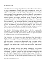
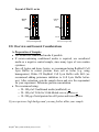
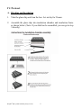
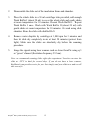
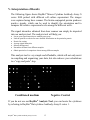
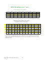
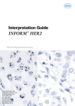
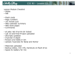
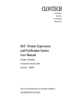
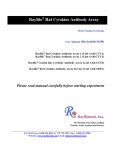
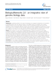
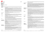
RayBio® Rat Cytokine Antibody Array
G Series
Patent Pending Technology
User Manual (Revised February 18, 2008)
RayBio® Rat Cytokine Antibody Array G Series 1 (Cat# AAR-CYT-G1-4)
RayBio® Rat Cytokine Antibody Array G Series 2 (Cat# AAR-CYT-G2-4)
RayBio® Rat Cytokine Antibody Array G Series 1 (Cat# AAR-CYT-G1-8)
RayBio® Rat Cytokine Antibody Array G Series 2 (Cat# AAR-CYT-G2-8)
RayBio® Custom Rat Cytokine Antibody Array G Series 1 (Cat# AAR-CUST-G)
RayBio® Rat Cytokine Antibody Array G Series 1 Service (Cat# AAR-SER-G)
Please read the manual carefully before you start your experiment
RayBiotech, Inc.
We Provide You With Excellent
Protein Array Systems And Service
Tel:(Toll Free) 1-888-494-8555 or 770-729-2992; Fax: 1-888-547-0580;
Website:www.raybiotech.com Email: [email protected]
RayBiotech, Inc.
RayBio® Rat Cytokine Antibody Array G Series Protocol
TABLE OF CONTENTS
I.
Introduction……..……………………………...
2
How It Works………………………..…………
5
Materials Provided……………………………..
6
Additional Materials Required…………………
6
III. Overview and General Considerations…………
7
A. Preparation of Samples………………………
7
B. Handling Glass Chips ……………………….
8
C. Incubation……………………………………
8
IV. Protocol…………………………………………
9
A. Blocking and Incubation…………………….
9
B. Detection…………………………………….
12
Interpretation of Results………………………..
13
VI. Troubleshooting Guide…………………………
18
VII. Selected References Using RayBiotech Products
19
II.
V.
Cytokine Antibody Arrays are RayBiotech patent-pending technology.
RayBio® is the trademark of RayBiotech, Inc.
RayBio® Rat Cytokine Antibody Array G series
1
I. Introduction
All cell functions, including cell proliferation, cell death and differentiation,
as well as maintenance of health status and development of disease, are
controlled by many genes and signaling pathways. New techniques such as
cDNA microarrays have enabled us to analyze the global gene expression 1-3.
However, almost all cell functions are executed by proteins, which cannot be
studied by DNA and RNA alone. Experimental analysis clearly shows a
disparity between the relative expression levels of mRNA and their
corresponding proteins 4. Therefore, it is critical to analyze the protein
profile. Currently, two-dimensional polyacrylamide SDS page coupled with
mass spectrometry is the mainstream approach to analyzing multiple protein
expression levels 5,6. However, the requirement of sophisticated devices and
the lack of quantitative measurements greatly limit its broad application.
Thus, no simple, cost effective, and rapid method of analysis of multiple
protein expression levels has been available to researchers until now.
Our RayBio® Rat Cytokine Antibody Array is the first commercially
available rat cytokine antibody array system 7-11. By using the RayBiotech
system, scientists can rapidly and accurately identify the expression profiles
of multiple cytokines in several hours inexpensively.
The RayBiotech kit (G series) is a glass chip format. The kit provides a
highly sensitive approach to simultaneously detect multiple cytokine
expression levels from cell culture supernatant, patient’s serum, tissue lysate
and other sources. The arrays are manufactured using non-contact arrayer.
The experimental procedure is simple and can be performed in any
laboratory. The signals from G series arrays are detected using a laser
scanner.
Besides the products listed in this manual, RayBiotech also provides
RayBio® Mouse Cytokine Antibody Array G series 2000 for simultaneous
detection of 144 mouse cytokines in single experiment. RayBiotech also
provides RayBio® Human Cytokine Antibody Array G series 4000 which is
the only product available in the market that can detect 274 human cytokines
in single experiment. We also provide quantitative antibody arrays which can
RayBio® Rat Cytokine Antibody Array G series
2
quantitatively measure multiple protein expression in antibody array
technology.
Pathway-specific array systems allow investigators to focus on the specific
problem and are becoming an increasingly powerful tool in cDNA
microarray system. RayBiotech’s first protein array system, known as
RayBio® Rat Cytokine Antibody Array, is particularly useful compared with
the Rat cytokine cDNA microarray system. Besides the ability to detect
protein expression, RayBiotech’s system is a more accurate reflection of
active cytokine levels because it only detects secreted cytokines, and no
amplification step is needed. Furthermore, it is much simpler, faster,
environmentally friendlier, and more sensitive.
Simultaneous detection of multiple cytokines undoubtedly provides a
powerful tool to study cytokines. Cytokines play an important role in innate
immunity, apoptosis, angiogenesis, cell growth and differentiation 12.
Cytokines are involved in most disease processes, including cancer and
cardiac diseases. The interaction between cytokines and the cellular immune
system is a dynamic process. The interactions of positive and negative
stimuli, and positive as well as negative regulatory loops are complex and
often involve multiple cytokines.
Reference List
1. LPS induces the interaction of a transcription factor, LPS-induced TNF-a factor,
and STAT6(B) with effects on multiple cytokines. Tang X, Marciano DL,
Leeman SE, Amar S. PNAS. 2005 ;102(14): 5132-5137.
2. HIV-1-mediated apoptosis of neuronal cells: Proximal molecular mechanisms of
HIV-1-induced encephalopathy. Xu Y, Kulkoshy J, Pomerantz RJ. PNAS. 2004
;101(18): 7071-7075.
3. Synergistic increases in intracellular Ca(2+), and the release of MCP-1,
RANTES, and IL-6 by astrocytes treated with opiates and HIV-1 Tat. El-Hage N,
Gurwell JA, Singh IN, Knapp PE, Nath A, Hauser KF. GLIA. 2005;50(2):91106.
RayBio® Rat Cytokine Antibody Array G series
3
4. Bone Marrow Stroma Influences Transforming Growth Factor-β Production in
Breast Cancer Cells to Regulate c-myc Activation of the Preprotachykinin-I Gene
in Breast Cancer Cells. Oh HS, Moharita A, Potian JG, Whitehead IP, et al.
Cancer Res. 2004; 64:6327-6336.
5. Recombinant Herpes Simplex Virus Type 1 (HSV-1) Codelivering Interleukin12p35 as a Molecular Adjuvant Enhances the Protective Immune Response
against Ocular HSV-1 Challenge. Osario Y, Ghiasi H. J Virology 2005;
79(6):3297-3308.
6. Dysregulated Inflammatory Response to Candida albicans in a C5-Deficient
Mouse Strain. Mullick A, Elias M, Picard S, Gros P. Infect Immunity. 2004;
.72(10):5868-5849.
7. Leukotriene B4 Strongly Increases Monocyte Chemoattractant Protein-1 in
Human Monocytes. Huang L, Zhao A, Wong F, Ayala JM, Cui J. Arterioscler
Thromb Vascul Biol. 2004;24:1783-1788
8. Giroux M, Denis F. Human CD1d-unrestricted NKT cells release chemokines
upon Fas engagement. Blood. 2005;105(2):703–710.
9. Monitoring the response of orthotopic bladder tumors to granulocyte macrophage
colony-stimulating factor therapy using the prostate-specific antigen gene as a
reporter. Wu Q, Esuvaranathan K, Mahendran R. Clin Cancer Res. 2004;
10(20):6977-84.
10. Neuroglial activation and neuroinflammation in the brain of patients with autism
(p NA). Vargas DL, Nascimbene C, Krishnan C, Zimmerman AW, Pardo CA.
Ann Neurol. 2005; 57(1): 67 – 81.
11. Cytokine profiling of macrophages exposed to Porphyromonas gingivalis, its LPS
or its FimA. Zhou Q, Desta T, Graves DT, Amar S. Infect Immunity. 2005;
73(2):935-43.
12. Veto-like activity of mesenchymal stem cells: functional discrimination between
cellular responses to alloantigens and recall antigens. Rameshwar P. Journal of
Immunology. 2003;171(7):3426-34.
13. The promise of cytokine antibody arrays in drug discovery process. Huang RP,
Yang W, Yang Y, Flowers L, et al. Expert Opinion on Drug Discovery. (2005)
9:601-615.
RayBio® Rat Cytokine Antibody Array G series
4
Here’s how it works
Array
support
Sample
Incubation of Sample
With arrayed antibody
Supports
Cocktail of
Biotin-Ab
Incubation with
Biotinylated Ab
1-2 hrs
1-2 hrs
Labeled –
streptavidin
Incubation with
Labeled- streptavidin
2 hrs
Detection of signals
Data analysis and graph
RayBio® Rat Cytokine Antibody Array G series
5
II. Materials Provided
Upon receipt, all components of the RayBio® Rat Cytokine Antibody Array
kit should be stored at -20°C. At -20°C the kit will retain complete activity
for up to 6 months. Once thawed, the glass chips, Fluorescent dyestreptavidin, Internal Control and 2X Blocking Buffer should be kept at –
20°C and all other component should be stored at 4°C. Use within three
months after reagents have been thawed. Please use within six months of
purchase.
• RayBio® Rat Cytokine Antibody Microarray slides (1 slide with 4 or 8
subarrays each)
• Biotin-Conjugated Anti-Cytokines (1 tube/4 subarrays, 1 or 2 tubes)
• 1,500X Fluorescent Dye-conjugated Streptavidin (Cy3 equivalent, 1
tube)
• 2X Blocking Buffer (10 ml)
• 20X Wash Buffer I (30 ml)
• 20X Wash Buffer II (30 ml)
• 2X Cell Lysis Buffer (10 ml)
• RayBio® G series antibody array accessory (including slide incubation
chamber, Gasket, Protective cover, Snap-on sides and adhesive film)
• 30 ml tube
• Manual
Additional Materials Required
•
•
•
•
•
Orbital shaker
Laser scanner for fluorescence detection
Aluminum foil
Distilled water
Plastic box
RayBio® Rat Cytokine Antibody Array G series
6
Layout of Rat G series
Antibody
Array
Antibody
Array
Blank
Blank
Barcode
Barcode
8 arrays in one glass chip
4 arrays in one glass chip
III. Overview and General Considerations
A. Preparation of Samples
• Use serum-free conditioned media if possible.
• If serum-containing conditioned media is required, use uncultured
media as a negative control sample, since many types of sera contain
cytokines.
• For cell lysates and tissue lysates, we recommend using RayBio® Cell
Lysis Buffer to extract proteins from cell or tissue (e.g. using
homogenizer). Dilute 2X RayBio® Cell Lysis Buffer with H2O (we
recommend adding proteinase inhibitors to Cell Lysis Buffer before
use). After extraction, spin the sample down and save the supernatant
for your experiment. Determine protein concentration.
• We recommend using:
o 50–100 µl of Conditioned media (undiluted), or
o 50–100 µl of 2-fold to 5-fold diluted sera or plasma, or
o 10–200 µg of total protein for cell lysates and tissue lysates.
If you experience high background, you may further dilute your sample.
RayBio® Rat Cytokine Antibody Array G series
7
B. Handling glass chips
• The microarray slides are sensitive, do not touch the surface. Hold the
slides by the edges only.
• Handle all buffers and slides with powder-free gloves.
• Avoid breaking glass slide.
• Handle glass chip in clean environment.
C. Incubation
• Completely cover array area with sample or buffer during incubation,
and cover the incubation chamber with adhesive film or plastic sheet
protector to avoid drying.
• Avoid foaming during incubation steps.
• Perform all incubation and wash steps under gentle rotation.
• Cover the incubation chamber with adhesive film during incubation,
particularly when incubation is more than 2 hours or 50 µl of sample or
reagent is used.
• Avoid cross-contamination from overflowing solution to neighboring
wells.
• Several incubation steps such as step 3 (blocking), step 4 (sample
incubation), step 9 (biotin-Ab incubation) or step 12 (Fluorescent dyestreptavidin incubation) may be done at 4°C for overnight. Please make
sure to cover the incubation chamber tightly to prevent evaporation.
RayBio® Rat Cytokine Antibody Array G series
8
IV. Protocol
A. Blocking and Incubation
1. Take the glass chip out from the box. Let air dry for 2 hours.
2. Assemble the glass chip into incubation chamber and incubation frame
as shown below. (Note: if you slide has be assembled, you can go to step
3 directly).
RayBio® Rat Cytokine Antibody Array G series
9
3. Add 100 µl 1 X Blocking Buffer into each well and incubate at room
temperature for 30 min to block slides. Dilute 2X Blocking Buffer with
H2O. Make sure no bubbles are in the well.
Note: only add reagents to wells printed with antibodies.
4. Decant Blocking Buffer from each well. Add 50 to 100 µl of each
sample to array III and array IV. Incubate arrays with sample at room
temperature for 1 to 2 hours. Dilute sample using 1X Blocking Buffer if
necessary.
Note: Incubation may be done overnight at 4°C.
Note: We recommend using 50 to 100 µl of undiluted conditioned media or 50 to 100 µl
of 2-5 fold diluted serum or plasma or 10-200 µg of protein for cell lysates and
tissue lysates. Dilute the lysate at least 10 fold with 1X blocking buffer to make a
total volume of 50 to 100 µl. Make sure there are no bubbles in the wells.
Note: The amount of sample used depends on the abundance of cytokines. More
concentrated sample can be used if signals are too weak. If signals are too strong,
the sample can be diluted further.
5. Decant the samples from each well, and wash 3 times with 150 µl of 1X
Wash Buffer I at room temperature with gentle shaking. 2 min per wash.
Dilute 20X Wash Buffer I with H2O. Completely remove wash buffer I
in each wash step.
Note: avoid solution flowing into neighboring wells.
6. Put the glass chip with frame into a box with 1X Wash Buffer I (cover
the whole glass slide and frame with Wash Buffer I), and wash 2 times at
room temperature with gentle shaking for 10 min per wash.
7. Decant the 1X Wash Buffer I from each well, Put the glass chip with
frame into the box with 1X Wash Buffer II (cover the whole glass slide
and frame with Wash Buffer II), and wash 2 times at room temperature
with gentle shaking for 5 min per wash. Remove all of Wash Buffer II in
the well. Dilute 20X Wash Buffer II with H2O.
RayBio® Rat Cytokine Antibody Array G series
10
8. Prepare working solution for biotin-conjugated antibodies. After brief
spin to force antibody aliquot to bottom of tube, add 300 µl of 1x
blocking buffer to the Biotin-Conjugated Antibody tube. Mix gently.
Note: the diluted biotin-conjugated antibodies can be stored at 4°C for 2-3 days.
9. Add 70 µl of diluted biotin-conjugated antibodies to each subarray.
Incubate at room temperature for 2 hours.
Note: incubation may be done at 4°C for overnight.
10. Wash as directed in steps 5 and then wash 3 times with 150 µl of 1X
Wash Buffer II at room temperature with shaking. 2 min per wash.
Completely remove wash buffer II in each wash step.
11. Add 70 µl of 1,500 fold diluted Fluorescent dye-conjugated streptavidin
(after brief spin to force liquid to bottom of tube, add 1.5 ml of Blocking
Buffer to Fluorescent dye-conjugated streptavidin tube) to each subarray.
Cover the incubation chamber with Adhesive film. Cover the plate with
aluminum foil to avoid exposure to light or incubate in dark room.
12. Incubate at room temperature for 1 to 2 hours.
Note: incubation may be done at 4°C for overnight.
13. Wash with Wash Buffer I twice as directed in steps 5.
B.
Fluorescence Detection
1.
Decant excess Wash Buffer from wells.
RayBio® Rat Cytokine Antibody Array G series
11
2.
Disassemble the slide out of the incubation frame and chamber.
3.
Place the whole slide in a 30 ml centrifuge tube provided, add enough
Wash Buffer I (about 20 ml) to cover the whole slide and gently shake
at room temperature for 10 minutes. Decant Wash Buffer I. Repeat
Wash Buffer I once. Wash with Wash Buffer II (about 20 ml) with
gentle shake at room temperature for 10 minutes. Or wash using slide
chamber. Rinse the slide with distilled H2O.
4.
Remove water droplets by centrifuge at 1,000 rpm for 3 minutes and
then let slide dry completely in air at least 20 minutes (protect from
light). Make sure the slides are absolutely dry before the scanning
procedure.
5.
Image the signals using laser scanner such as Axon GenePix using cy3
or “green” channel (Excitation frequency 532 nm).
Note: we recommend scanning slides right after experiment. You also can store the
slide at –20°C in dark for several days. If you do not have a laser scanner,
RayBiotech can provide service for you. Just simply send your slide to us and we will
take care of it.
RayBio® Rat Cytokine Antibody Array G series
12
V. Interpretation of Results:
The following figure shows RayBio® Mouse Cytokine Antibody Array G
series 1000 probed with different cell culture supernatant. The images
were captured using laser scanner. The biotin-conjugated protein produces
positive signals, which can be used to identify the orientation and to
compare the relative expression levels among the different wells.
The signal intensities obtained from laser scanner can simply be imported
into our analysis tool. The analysis tool will help you:
•
•
•
•
•
•
•
Locate your signal intensities to antibody array map
Link the protein to website for more detailed information on the particular protein
Protein list sorting
Average signal intensities
Subtract background
Normalize the data from different samples
Obtain protein level comparison charts among different samples
This analysis tool is very simple and affordable, which will not only assist
in compiling and organizing your data, but also reduces your calculations
to a “copy and paste” step.
Conditioned medium
Negative Control
If you do not use our RayBio® Analysis Tool, you can locate the cytokines
by referring to RayBio® Rat cytokine Antibody Array G series 1.
RayBio® Rat Cytokine Antibody Array G series
13
Normalization and comparison
For biomarker discovery or for analysis of large number of arrays, great
attention must be paid to the normalization. Our antibody array design
includes several controls for normalization and comparison of arrays
performing in different membranes and different experiments (for more
information please read the reference 13 in the introduction).
Positive control. Positive control is biotinylated protein. It can be used to
normalize the streptavidin incubation step. If the positive signals from
different array membranes are similar, positive control is a simple and
effective way for normalization.
Negative control. Negative control is BSA. Normally, it should only give a
background reading.
Data Extraction Tips:
• Ignore any comet tails
• Define the area for signal capture for all spots as 110-120 micron diameter,
using the same area for every spot.
• Use median signal value, not the total or the mean
• Use local background correction (also median value).
• Exclude obvious outlier data in its calculations.
Using these guidelines, along with using PMT, brightness and contrast
settings that reduce the background as much as possible, we get very good
results with interassay and intraassay CV <20%, even with some
imperfections in the antibody spots.
Threshold of significant difference in expression: Any ≥1.5-fold increase or
≤0.65-fold decrease in signal intensity for a single analyte between samples,
provided that both signals are well above background (Mean background + 2
standard deviations, accuracy ≈ 95%).
RayBio® Rat Cytokine Antibody Array G series
14
RayBio® Rat Antibody Array G - Series 1
Detect 19 cytokines in one experiment.
a
b
c
d
e
f
g
h
1
Pos 1
Pos 2
Pos 3
Neg
Neg
CINC-2
CINC-3
CNTF
Fractalkine
2
Pos 1
Pos 2
Pos 3
Neg
Neg
CINC-2
CINC-3
CNTF
Fractalkine
3
GM-CSF
IFN-γ
IL-1α
IL-1β
IL-4
IL-6
IL-10
LIX
Leptin
4
GM-CSF
IFN-γ
IL-1α
IL-1β
IL-4
IL-6
IL-10
LIX
Leptin
5
MCP-1
MIP-3α
β-NGF
TIMP-1
TNF-α
VEGF
Neg
Neg
Pos 1
6
MCP-1
MIP-3α
β-NGF
TIMP-1
TNF-α
VEGF
Neg
Neg
Pos 1
RayBio® Rat Cytokine Antibody Array G series 2
(for simultaneous detection of 34 rat cytokines)
A
B
C
D
E
F
G
H
I
J
K
L
1
POS 1
POS 2
POS 3
NEG
Activin A
Agrin
B7-2/CD86
beta-NGF
CINC-1
CINC-2alpha
CINC-3
CNTF
2
POS 1
POS 2
POS 3
NEG
Activin A
Agrin
B7-2/CD86
beta-NGF
CINC-1
CINC-2alpha
CINC-3
CNTF
3
Fas Ligand
Fractalkine
GM-CSF
ICAM-1
IFN-gamma
IL-1alpha
IL-1beta
IL-1 R6
IL-2
IL-4
IL-6
IL-10
4
Fas Ligand
Fractalkine
GM-CSF
ICAM-1
IFN-gamma
IL-1alpha
IL-1beta
IL-1 R6
IL-2
IL-4
IL-6
IL-10
Thymus
Chemokine-1
Thymus
Chemokine-1
5
IL-13
Leptin
LIX
L-Selectin
MCP-1
MIP-3alpha
MMP-8
PDGF-AA
Prolactin R
RAGE
6
IL-13
Leptin
LIX
L-Selectin
MCP-1
MIP-3alpha
MMP-8
PDGF-AA
Prolactin R
RAGE
7
TNF-alpha
VEGF
NEG
NEG
NEG
NEG
NEG
NEG
NEG
NEG
NEG
POS 1
8
TNF-alpha
VEGF
NEG
NEG
NEG
NEG
NEG
NEG
NEG
NEG
NEG
POS 1
TIMP-1
TIMP-1
Abbreviations: IP-10, Interferon-inducible protein-10; LAP, latency associated peptide (TGF-β1); LIF, leukocyte
inhibitory factor. MMP, Matrix Metalloproteinase; Pos, positive control; Neg, negative control. All other are used
standard abbreviations.
RayBio® Rat Cytokine Antibody Array G series
15
RayBiotech, Inc., the protein array pioneer company, strives to research and
develop new products to meet demands of the biomedical community.
RayBio’s patent-pending technology allows detection of over 180 cytokines,
chemokines and other proteins in a single experiment. Our format is simple,
sensitive, reliable and cost effective. Products include: Cytokine Arrays,
Chemokine Arrays, ELISA kits, Phosphotyrosine kits, Recombinant
Proteins, Antibodies, and custom services.
1. Antibody arrays
Cytokine antibody array
Human cytokine antibody arrays
Mouse cytokine antibody arrays
Rat cytokine antibody arrays
Pathway- or disease-focused antibody arrays
Inflammation antibody array
Angiogensis antibody array
Chemokine antibody array
Growth factor antibody array
MMP antibody array
Atherosclerosis antibody array
Quantibody arrays for quantitative measurement of cytokine and other protein
concentraton
Phosphorylation antibody arrays
Biotin label-based antibody arrays for high density antibody arrays.
Antibody analysis tool, software
2.
3.
4.
5.
6.
7.
8.
9.
ELISA
Cell-based phosphorylation assay
Custom antibody arrays
Antibody
Recombinant protein
Peptide
Protein arrays
EIA
RayBiotech also provides excellent custom service:
1. Antibody arrays
RayBio® Rat Cytokine Antibody Array G series
16
2.
3.
4.
5.
6.
7.
8.
Protein arrays
Peptide synthesis
Production of recombinant protein and antibody
Peptide arrays
Phosphorylation arrays
ELISA
EIA
Just simply send your samples and we will do the assay for you.
Technology transfer program
Have you developed technologies or reagents interested to the scientific and research
community? RayBiotech can help you commercialize your technologies, reagents and
dream.
RayBio® Rat Cytokine Antibody Array G series
17
VI. Troubleshooting guide
Problem
Weak signal
High background
Cause
Recommendation
Inadequate detection
Check laser power
and PMT parameters
Inadequate reagent volumes
or improper dilution
Check pipetters and
ensure correct preparation
Short incubation times
Ensure sufficient incubation
Time and change sample
incubation step to overnight
Too low protein concentration
in sample
Don’t make too low dilution
Or concentrate sample
Improper storage of kit
Store kit at suggested
temperature
Make sure correct
amount of antibodies
Excess of biotinylated antibodies
Excess of streptavidin
Make sure correct
amount of streptavidin
Inadequate detection
Check laser power
And PMT parameters
dust
Work in clean environment
Increase wash time and
use more wash buffer
Insufficient wash
Uneven signal
Bubbles formed during
incubation
Avoid bubble formation
during incubation
Arrays are not completed
Covered by reagent
Completely cover arrays
with solution
RayBio® Rat Cytokine Antibody Array G series
18
VII. Selected References Using RayBiotech Products
Human AB Serum and Thrombin-Activated Platelet-Rich Plasma Are
Suitable Alternatives to Fetal Calf Serum for the Expansion of
Mesenchymal Stem Cells from Adipose Tissue. Kocaoemer A, Kern S,
Kluter H, Bieback K. Stem Cells. 2007; 25:1270-1278
ATP Binding by Monarch-1/NLRP12 Is Critical for Its Inhibitory Function.
Ye Z, Lich JD, Moore CB, Duncan JA, Williams KL, Ting JP-Y. Mol Cell
Biol. 2008;28:1841-1850.
Infiltration of CD4+ lymphocytes into the brain contributes to
neurodegeneration in a mouse model of Parkinson disease. Brochard V,
Combadière B, Prigent A, Laouar Y, et al. J Clin Invest. 2009; 119 :182192.
Transferrin-derived synthetic peptide induces highly conserved proinflammatory responses of macrophages. Haddad G, Belosevic M. Mol
Immunol. 2009; 46:576-586.
Robinson K, Vona-Davis L, Riggs D, Jackson B, McFadden D. Peptide YY
Attenuates STAT1 and STAT3 Activation Induced by TNF-alpha in Acinar
Cell Line AR42J. J Am Coll Surg. 2006; 2002:788-796.
Stokes L, Surprenant A. Purinergic P2Y2 Receptors Induce Increased
MCP-1/CCL2 Synthesis and Release from Rat Alveolar Peritoneal
Macrophages. J Immunol. 2007;179:6016-6023.
Struzynska L, Dabrowska-Bouta B, Koza K, Sulkowski G. InflammationLike Glial Response in Lead-Exposed Immature Rat Brain. Toxicol Sci.
2007;95(1):156-162.
Watanabe M, Guo W, Zou S, Sugiyo S, Dubner R, Ren K. Neurosci Lett.
2005;382:128-133.
RayBio® Rat Cytokine Antibody Array G series
19
Newman MB, Willing AE, Manresa JJ, Davis-Sanberg C, Sanberg PR.
Stroke-induced Migration of Human Umbilical Cord Blood Cells: Time
Course and Cytokines. Stem Cells Devel. 2005; 14:576-586.
Moon SK, Woo J-I, Lee H-Y, Park R, et al. Toll-Like Receptor 2Dependent NF-kappa-B Activation Is Involved in Nontypeable
Haemophilus influenzae-Induced Monocyte Chemotactic Protein 1 UpRegulation in the Spiral Ligament Fibrocytes of the Inner Ear. Infect
Immun. 2007; 75(7): 3361-3372.
Dave B, Eason RR, Geng Y, Su Y, Badger TM, Simmen RCM. Tp53Associated Growth Arrest and DNA Damage Repair Gene Expression Is
Attenuated in Mammary Epithelial Cells of Rats Fed Whey Proteins. J
Nutr. 2006;136:1156-1160.
Tögel F, Hu Z, Weiss K, Issac J, Lange C, Westenfelder C. Administered
mesenchymal stem cells protect agains ischemic acute renal failure through
differentiation-independent mechanisms. Am J Physiol Renal Physiol.
2005;289:F31-F42.
Ahmed N, Dreier R, Gopferich A, Grifka J, Grassel S. Soluble Signalling
Factors Derived from Differentiated Cartilage Tissue Affect Chondrogenic
Differentiation of Rat Adult Marrow Stromal Cells. Cell Physiol Biochem.
2007;20:665-678.
Fu Y-S, LinY-Y, Chou S-C, Tsai T-H, et al. Tetramethylpyrazine inhibits
activities of glioma cells and glutamate neuro-excitotoxicity: Potential
therapeutic application for treatment of gliomas. Neuro Oncol. 2008
10(2):139-152.
RayBio® Rat Cytokine Antibody Array G series
20
Note:
RayBio® is the trademark of RayBiotech, Inc.
Cytokine protein arrays are RayBiotech patent-pending technology.
This product is intended for research only and is not to be used for clinical
diagnosis. Our produces may not be resold, modified for resale, or used to
manufacture commercial products without written approval by RayBiotech,
Inc.
Under no circumstances shall RayBiotech be liable for any damages arising
out of the use of the materials.
Products are guaranteed for three months from the date of purchase when
handled and stored properly. In the event of any defect in quality or
merchantability, RayBiotech’s liability to buyer for any claim relating to
products shall be limited to replacement or refund of the purchase price.
HiLytePlus™ is a trademark of AnaSpec, Inc.
This product is for research use only.
©2009 RayBiotech, Inc.
RayBio® Rat Cytokine Antibody Array G series
21