1
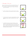
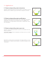
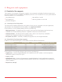
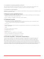
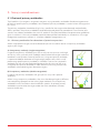
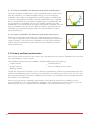
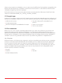
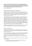
User Guide
Duolink® In Situ –
Fluorescence
sigma.com
The protocols in this manual are compatible with all Duolink In Situ PLA probes, Duolink In Situ Detection Reagents
Green (Cat. No. DUO92014), Orange (Cat. No. DUO92007), Red (Cat. No. DUO92008) and Far Red (Cat. No. DUO92013)
Duolink® In Situ – Fluorescence
Table of Contents
1. Introduction ....................................................................................................................................4
2. Principle of the assay .................................................................................................................5
3. Applications .....................................................................................................................................6
3.1 Detect and quantify protein interactions .........................................................6
3.2 Detect and quantify protein modifications .....................................................6
3.3 Detect and quantify protein expression ...........................................................6
4. Reagents and equipment .......................................................................................................7
4.1 Duolink In Situ reagents ..............................................................................................7
4.2 Reagents to be supplied by the user ..................................................................8
4.3 Equipment needed ........................................................................................................8
4.4 Duolink ImageTool – software for image analysis .......................................8
5. Assay considerations ..................................................................................................................9
5.1 Choice of primary antibodies ..................................................................................9
5.2 Primary antibody optimization ............................................................................10
5.3 Sample type .....................................................................................................................11
5.4 Pre-treatment ..................................................................................................................11
5.5 Controls ...............................................................................................................................13
6. Reagent preparation ...............................................................................................................14
6.1 Duolink In Situ reagents ...........................................................................................14
6.2 Duolink In Situ Wash Buffers ..................................................................................15
6.3 Samples ..............................................................................................................................15
6.4 Reaction volume ...........................................................................................................15
7. Assay protocol .............................................................................................................................16
7.1 PLA probe protocol – CUSTOM solutions .....................................................16
7.2 PLA probe protocol – Duolink In Situ Solutions ........................................17
7.3 Detection protocol ......................................................................................................18
8. Results ..............................................................................................................................................19
8.1 Typical results ..................................................................................................................19
8.2 Image acquisition .........................................................................................................20
8.3 Duolink ImageTool – image analysis ................................................................21
9. Troubleshooting ........................................................................................................................22
Appendix A – Wash Buffers formulas .....................................................................................24
Appendix B – Duolink In Situ in chamber slides .............................................................25
Appendix C – Duolink In Situ on cover slips ......................................................................26
References ...............................................................................................................................................27
3
1. Introduction
Duolink® In Situ reagents from Olink® Bioscience enable detection, visualization and quantification of
individual proteins, protein modifications and protein interactions in tissue and cell samples prepared for
microscopy. The target is detected using one or two primary antibodies depending on the application. In
the case that two primary antibodies are used, they must have been raised in different species.
The Duolink In Situ reagents are based on in situ PLA®, which is a proximity ligation assay technology.
A pair of oligonucleotide labeled secondary antibodies (PLA probes) generates a signal only when the
two PLA probes have bound in close proximity, either to the same primary antibody or two primary
antibodies that have bound to the sample in close proximity. The signal from each detected pair of PLA
probes is visualized as an individual fluorescent spot. These PLA signals can be quantified (counted) and
assigned to a specific subcellular location based on microscopy images.
4
2. Principle of the assay
Typical starting materials are adherent cells, cytospin preparations or tissue
sections on a glass slide, fixed, pre-treated and blocked with a blocking
reagent according to the requirements of the primary antibodies used.
A.The samples are incubated with primary antibodies that bind to the
protein(s) to be detected.
A.
B.Secondary antibodies conjugated with oligonucleotides (PLA probe
MINUS and PLA probe PLUS) are added to the reaction and incubated.
B.
C.The Ligation solution, consisting of two oligonucleotides (illustrated as
red bands) and Ligase, is added and the oligonucleotides will hybridize
to the two PLA probes and join to a closed circle if they are in close
proximity.
C.
D.The Amplification solution, consisting of nucleotides (not shown)
and fluorescently labeled oligonucleotides, is added together with
Polymerase. The oligonucleotide arm of one of the PLA probes acts as a
primer for a rolling-circle amplification (RCA) reaction using the ligated
circle as a template, generating a concatemeric (repeated sequence)
product. The fluorescently labeled oligonucleotides will hybridize to the
RCA product. The signal is easily visible as a distinct fluorescent spot and
analyzed by fluorescence microscopy.
D.
Fig 1. Assay principle
5
3. Applications
3.1 Detect and quantify protein interactions
Detection and quantification of interacting proteins can be done using two
different primary antibodies against each of the two proteins of interest.
Fig 2. Protein interactions
3.2 Detect and quantify protein modifications
Detection and quantification of a protein and its specific post-translational
modification can be done using two different primary antibodies, one
directed against the target protein and one against a modification site on
the same protein.
Fig 3. Protein modifications
3.3 Detect and quantify protein expression
Detection and quantification of protein expression can be done with two
different approaches.
(A) To detect and quantify one single protein with high sensitivity, use only
one primary antibody.
Fig 4. (A) Single recognition
(B) To detect and quantify one single protein with high specificity, use two
different primary antibodies directed against two different epitopes on the
same protein.
Fig 5. (B) Double recognition
6
4. Reagents and equipment
4.1 Duolink In Situ reagents
The Duolink In Situ reagents are generic reagents using secondary antibodies for detecting presence
of analyte-specific primary antibodies provided by the user. To run a Duolink In Situ assay you need the
following Duolink In Situ components.
• PLA probe MINUS
• PLA probe PLUS
• Detection Reagents
• Wash Buffers A and B
• Mounting Medium with DAPI
4.1.1 Duolink In Situ PLA probes
The choice of PLA probes depends on the species of your primary antibodies and your application,
see section 3. For a complete list of PLA probes, visit sigma.com/duolink
Each Duolink In Situ PLA probe product contains the following:
• Blocking Solution – For blocking of the sample if you have not already optimized your primary
antibody with another blocking solution. Supplied in ready-to-use concentration
• Antibody Diluent – For dilution of PLA probes and in some cases the primary antibodies
(section 5.4.3). Supplied in ready-to-use concentration
• PLA probe (5x) stock – Secondary antibody conjugated with a PLA oligonucleotide
4.1.2 Duolink In Situ Detection reagents
Duolink In Situ Detection Reagents are available with four different fluorophores.
Detection Reagents
Excitation (nm)
Emission (nm)
Filters for image acquisition
Detection Reagents Green
495
527
same filters as e.g. Cy2 or FITC
Detection Reagents Orange
554
579
same filters as e.g. Cy3
Detection Reagents Red
594
624
same filters as e.g. Texas Red
Detection Reagents Far Red
644
669
same filters as e.g. Cy5
Each Duolink In Situ Detection Reagents box contains the following:
• Ligation (5x) – Contains oligonucleotides that hybridize to the PLA probes and all components
needed for ligation except the Ligase
• Ligase (1 U/μl)
• Amplification (5x) – Contains all components needed for Rolling Circle Amplification except the
Polymerase. Included are also oligonucleotide probes labeled with a fluorophore that hybridize to
the RCA product
• Polymerase (10 U/μl)
7
4.1.3 Duolink In Situ Mounting Medium with DAPI
Optimal mounting medium for preservation of the PLA signals. It also contains DAPI nuclear stain
(excitation 360 nm and emission 460 nm). Aqueous mountant which does not solidify.
4.1.4 Duolink In Situ Wash Buffers
Duolink In Situ Wash Buffers contains 3 pouches of Wash Buffer A and one pouch of Wash Buffer B, to be
dissolved in high purity water.
4.2 Reagents to be supplied by the user
• Reagents required for fixation and antigen retrieval of the sample (according to your own protocol,
specific for each antigen and antibody used)
• Primary antibody or antibodies (matching a set of PLA probes)
4.3 Equipment needed
• Fluorescence microscope equipped as follows:
– Excitation/emission filters compatible with fluorophore and nuclear stain excitation/emission
– Camera and software for image acquisition (optional)
• Staining jars
• Pen or mask for delimitation of reaction area (grease pen or silicon mask, e.g. ImmEdge™ Pen from
Vector Laboratories)
• Shaker
• Humidity chamber (moist chamber)
• Freeze block for enzymes
• Incubator, +37 °C
• Pipettes (covering the range from 1 µl to 1000 µl)
• Cover slips compatible with fluorescence microscopy
• High purity water (sterile filtered, Milli-Q® or similar)
4.4 Duolink ImageTool – Software for Image Analysis
The result from a Duolink In Situ experiment is typically a number of distinct fluorescent spots, PLA
signals, of sub-micrometer size in various locations of the studied cells. Use Duolink ImageTool to
obtain objective quantification of PLA signals. The nuclei are automatically detected and cytoplasm
size estimated, enabling single cell statistical analysis of expression levels in tissue or cell populations.
Furthermore, regions of interest can be defined, a feature of particular relevance when studying tissue
samples. Raw imaging data can be imported directly from the four major microscope vendors (Olympus,
Leica, Nikon and Zeiss). The results data can be exported into an Excel sheet for further evaluation.
8
5. Assay considerations
5.1 Choice of primary antibodies
The Duolink In Situ reagents are generic reagents using secondary antibodies for detecting presence
of analyte-specific primary antibodies. Your choice of primary antibodies is crucial when setting up the
PLA assay.
The primary antibodies should be of IgG-class, specific for the target to be detected and preferably
affinity purified. The primary antibodies could be either polyclonal or monoclonal. To maximize your
success rate, choose antibodies that are IHC and/or IF classified and follow the optimization guidelines
given in section 5.2. Primary antibodies could also be qualified individually in a Duolink In Situ single
recognition experiment (section 5.1.1) before a double recognition assay.
5.1.1 Primary antibodies for detection of protein expression
When single protein targets are to be detected you can use either one or two primary antibodies
against your target.
A) One primary antibody (single recognition)
Usage of one primary antibody gives you an easy-to-set-up assay with high
sensitivity. In this case only one antibody needs to bind the target under the
conditions used (fixation, retrieval, buffer etc.). Using one primary antibody
is recommended for detection of single target proteins only when a well
performing specific primary antibody is available. If you use an unspecific
antibody you will see high background due to the sensitivity of the assay.
Using only one primary antibody is more efficient than using two primary
antibodies when detecting low abundant targets.
Fig 6. (A) Single recognition
B) Two primary antibodies (double recognition)
Usage of two primary antibodies will give you an assay with superior
specificity.
When using two primary antibodies, they must be directed against different,
non-competing epitopes on the same target molecule. The two primary
antibodies must have been raised in different species. Also, both primary
antibodies must bind to the target under the same conditions (fixation,
retrieval, buffer etc.).
Fig 7. (B) Double recognition
9
5.1.2 Primary antibodies for detection of protein modifications
Detection of protein modifications, such as phosphorylations, often suffers
from low specificity. Our recommended strategy is to use two primary
antibodies, one against the target protein and one against a modification
site on the same protein. Preferably a specific modification site is targeted,
but it is also possible to use a generic antibody against a modification site.
Using two primary antibodies will give you superior specificity for your
modification assay compared to using one single primary antibody. The two
primary antibodies must be raised in different species. Also, both primary
antibodies must bind to the target under the same conditions (fixation,
retrieval, buffer etc.).
Fig 8. Protein modifications
5.1.3 Primary antibodies for detection of protein interactions
Duolink In Situ provides an excellent way to detect protein interactions.
This is done using two primary antibodies, each directed against one of the
targets of interest. The two primary antibodies must be raised in different
species and must bind to the target under the same conditions (fixation,
retrieval, buffer etc.).
Fig 9. Protein interactions
5.2 Primary antibody optimization
You will save time and increase your success rate if you optimize your primary antibodies to be sure that
they work properly in your material.
The conditions for your primary antibodies should be optimized with respect to:
• Sample fixation
• Antigen retrieval
• Blocking solution
• Antibody diluent
• Primary antibody concentration
Sample pre-treatment for Duolink In Situ is identical to procedures used for immunohistochemical (IHC)
and/or immunofluorescence (IF) staining. If you already have a working assay for IHC or IF, use the same
pre-treatment protocol. The primary antibody concentration may need to be titrated and optimized for
the Duolink In Situ conditions.
When using two primary antibodies, optimization is best done for one primary antibody at a time
using the Duolink In Situ single recognition approach. These conditions can then be used as a start for
optimizing your assays using both primary antibodies in the same Duolink In Situ assay.
10
When using two primary antibodies it may in some cases be difficult to find conditions compatible with
both of the selected primary antibodies. Under these circumstances it is advisable to search for other
antibodies recognizing alternative epitopes.
For further advice on immunostaining optimization we recommend the Education Guide from Dako
(http://www.dako.com/08002_ihc_staining_methods_5ed.pdf ).
5.3 Sample type
Duolink In Situ reagents can be used with a broad range of sample types depending on the preferences of
your primary antibodies. Duolink In Situ has so far shown to be successful with the following sample types.
• Adherent cell cultures
• Formalin fixed paraffin embedded tissue
sections
• Cytospin preparations
• Formalin fixed paraffin embedded cell samples • Frozen tissue sections
• Fresh tissue sections
5.4 Pre-treatment
Before you start, your samples should be deposited on glass slides and sufficiently pre-treated with
respect to fixation, retrieval and permeabilization. It is crucial for the performance of the assay to
optimize the conditions for the primary antibodies. Also, the choice of wash buffer may be dependent
on your primary antibodies. Use the recommendations from the vendor of your primary antibodies, if
available. If you are using chamber slides or cover slips, follow the guidelines in Appendix B or C.
5.4.1 Fixation
Different fixations will influence the antigenicity and thus the performance of your primary antibodies.
If there is no fixation recommendation for your antibody you must optimize this for your application.
For cell materials like cells grown on slides or cytospin preparations you should choose the fixation
most suitable for your primary antibody. The best choice of fixation may vary depending on your cell
material, antibody and the type of protein you want to study. Duolink In Situ is compatible with all
fixations typically used for IHC including:
• Ethanol
• Acetone
• Paraformaldehyde (PFA)
• Zinc
• Formalin
11
5.4.2 Antigen/Epitope Retrieval
Antigen/epitope retrieval is mostly used for formalin fixed paraffin embedded (FFPE) material. Formalin
fixation is masking the epitopes which then can be un-masked by antigen/epitope retrieval. Different
methods for antigen/epitope retrieval will influence the performance of your primary antibodies and
must be optimized for your application. Duolink In Situ is compatible with all antigen retrieval methods
typically used for IHC including:
• Heat Induced Epitope Retrieval (HIER)
• Enzyme treatment with Proteinase K, Pepsin, Trypsin etc.
For HIER it might be necessary to try a few different solutions of various pH.
5.4.3 Blocking solution and Antibody diluent
It is essential to use a proper blocking solution and antibody diluent. Use the recommendations from
the vendor of your primary antibodies, if available. If you have previously optimized your assay in for
example IHC, use the same conditions for Duolink In Situ.
There are two different protocols depending on whether you use your own blocking solution and
antibody diluent or the Duolink In Situ Blocking Solution and Antibody Diluent.
PLA probe protocol – CUSTOM solutions
• Use this protocol if you use your own blocking and antibody diluent (recommended),
see section 7.1
The same agent used for blocking of the sample should be present also in the antibody diluent,
preferably together with some detergent to reduce background staining, as well as in the diluent for
the PLA probes.
Note: Ensure that you do not use bulk IgG from the same species as your primary antibodies as blocking reagent as this will cause false
signals from the PLA probes.
PLA probe protocol – Duolink In Situ Solutions
• Use this protocol if you have no previous experience, or no recommendation from the antibody
vendor, regarding blocking and antibody diluents, see section 7.2
Note: The Duolink In Situ Antibody Diluent contains salt, blocking agent and detergents. All to prevent unspecific binding of the
antibodies. It is important that you first verify the function of your primary antibodies, one at a time, using Duolink In Situ single
recognition or possibly IF or IHC. Use proper controls to verify that your individual antibodies do bind the correct target under the
conditions used.
12
5.5 Controls
To be able to properly evaluate your results it is advisable to include both biological and technical
controls. Examples of suitable controls are given below.
5.5.1 Positive control
Ideally you should include a cell or tissue type that you know contains your target protein and that
your primary antibodies should bind to. If you are using two primary antibodies, the primary antibodies
should be known to bind in close proximity. This also enables you to verify the Duolink In Situ procedure.
5.5.2 Negative control
Ideally you should include a negative control with a cell line or tissue that does not express one or both
of your targets. Such a control will give you information on the specificity of your primary antibodies.
If this control gives you background signals in excess of signal obtained from omitting the primary
antibody/antibodies, it means that your primary antibodies, under the present conditions, are binding
to other targets than expected.
If facing problems with signals in your negative cell line/tissue you might need to titrate your primary
antibodies or look for more specific antibodies.
5.5.3 Negative control, technical
By omitting primary antibodies you will get a hint of how the PLA probe background looks like in your
system. Alternatively, if you are using an approach with two primary antibodies, you can also choose to
omit only one of your primary antibodies.
If facing problems with background signals with the primary antibodies omitted, consult the
troubleshooting guide, see section 9.
13
6. Reagent preparation
6.1 Duolink In Situ reagents
Some Duolink In Situ reagents are supplied as concentrated stocks. Dilute required volumes of the stocks.
Note: Do not store diluted reagents.
Blocking Solution – store at +4 °C
• Vortex before use
• Ready-to-use blocking solution, one drop equals approximately 40 µl
Antibody Diluent – store at +4 °C
• Vortex before use
• For dilution of primary antibodies and PLA probes
PLA probe (5x) – store at +4 °C
• Vortex before use
• Dilute 1:5 in Antibody Diluent or custom diluent immediately before use and vortex the solution
before addition to the sample
Ligation (5x) – store at -20 °C
• Thaw at room temperature and vortex before use
• Dilute 1:5 in high purity water immediately before use
Note: The buffer contains DTT that may precipitate during freezing. Vortex to dissolve/homogenize.
Ligase (1 U/μl) – store at -20 °C
• The Ligase should be kept at -20 °C at all times. Use a freezing block (-20 °C) when removing the
enzyme from the freezer
• Add the Ligase to the reaction mix at a 1:40 dilution immediately before addition to the sample
• Ensure that the Ligation-Ligase reaction solution is thoroughly vortexed before addition to the sample
Amplification (5x) – store at -20 °C
• Thaw at room temperature and vortex before use
• Dilute 1:5 in high purity water immediately before use
Polymerase (10 U/μl) – store at -20 °C
• The Polymerase should be kept at -20 °C at all times. Use a freezing block when removing the
enzyme from the freezer
• Add the Polymerase to the reaction mix at a 1:80 dilution immediately before addition to the sample
• Ensure that the Amplification-Polymerase reaction solution is thoroughly vortexed before addition to
the sample
14
6.2 Duolink In Situ Wash Buffers
The washing steps should be performed in a staining jar, with a minimum volume of 70 ml, on a
shaker (cradle) with gentle orbital shaking. Washing after the primary antibodies should be performed
in Duolink In Situ Wash Buffer A or in the wash buffer optimal for your primary antibodies. In all
subsequent wash steps ensure to use Duolink In Situ Wash Buffers.
Wash Buffer A – store at room temperature
• To prepare a 1x buffer, dissolve the content of one pouch in high purity water to a final volume of
1000 ml
• Store pouches at room temperature. 1x solutions may be kept at room temperature for short
time storage (one week or less). For long time storage store at +4 °C. Bring the solutions to room
temperature before use
• Alternatively make your own Duolink In Situ Wash Buffer A according to Appendix A
Wash Buffer B – store at room temperature
• To prepare a 1x buffer, dissolve the content of one pouch in high purity water to a final volume of
1000 ml
• Store pouches at room temperature. 1x solutions may be kept at room temperature for short
time storage (one week or less). For long time storage store at +4 °C. Bring the solutions to room
temperature before use
• For the final wash step, prepare 0.01x Wash Buffer B by diluting 1x buffer 1:100 in high purity water
• Alternatively make your own Duolink In Situ Wash Buffer B according to Appendix A
6.3 Samples
Before you start the Duolink In Situ protocol, the sample should have been deposited on a glass
slide, sufficiently pre-treated to fit your primary antibodies with respect to fixation, retrieval and
permeabilization, see section 5.2. Your reaction area must be delimited, with e.g. a grease pen or silicon
mask. If you are using chamber slides or cover slips, follow the guidelines in Appendix B or C.
6.4 Reaction volume
Area
Total Reaction Volume
• Use open droplet reactions
• Perform the incubations without a cover slip
• Perform all incubations in a pre-heated
0.2 cm2
15 μl
1 cm2
40 μl
• Use volumes corresponding to your delimited
humidity chamber
reaction area. Never use less than 15 μl of total
reaction volume, see Table 1 or the Reaction
Volume Guide at sigma.com/duolink.
Note: It is important that all incubations are performed in a
humid environment to prevent excessive evaporation. If the
sample goes dry, this will give rise to severe artifacts.
2 cm2
80 μl
3 cm2
120 μl
4 cm2
160 μl
6 cm2
240 μl
8 cm2
320 μl
10 cm2
400 μl
Table 1. Suitable reaction volume for different reaction areas.
15
7. Assay protocol
7.1 PLA probe protocol – CUSTOM solutions
Follow this protocol if you use your own blocking solution and antibody diluent that you know work
with your primary antibodies.
Before you start, your samples should be deposited on glass slides and pre-treated with respect to
fixation, retrieval and/or permeabilization. If you are using chamber slides or cover slips, follow the
guidelines in Appendix B or C.
Use open droplet reactions without a cover slip and perform all incubations in a humidity chamber.
Use volumes corresponding to your delimited reaction area, see the Reaction Volume Guide at
sigma.com/duolink or section 6.4.
1.
Blocking
Use your previously tested blocking solution.
a)
Add blocking solution to each sample.
b)
Incubate the slides.
2.
Primary Antibodies
Use your previously tested buffer to dilute your primary antibodies.
a)
Dilute your primary antibody to a suitable concentration in your custom antibody diluent. If using two primary
antibodies dilute the two antibodies in the same diluent.
b)
Tap off the blocking solution from the slides. Try to obtain an equal residual volume on each slide as this will
affect reproducibility. Do not allow the samples to dry before adding the primary antibodies as this will cause
background.
c)
Add the primary antibody solution to each sample.
d)
Incubate in a humidity chamber. Use temperature and time optimal for your primary antibodies.
3.
PLA probes
Use the same buffer as for your primary antibodies or use Duolink In Situ Antibody Diluent.
Note: The buffer should contain the blocking agent that was used for blocking the samples.
a)
Mix and dilute the two PLA probes 1:5 in your chosen buffer. Allow the mixture to sit for 20 min at room
temperature. E.g. for a 40 µl reaction take 8 µl of PLA probe MINUS stock, 8 µl of PLA probe PLUS stock and 24 µl of the
antibody buffer.
b)
Tap off the primary antibody solution from the slides.
c)
Wash the slides in a wash buffer suitable for your primary antibodies.
Washing should be performed in a staining jar, with a minimum volume of 70 ml on a shaker (cradle) with gentle
orbital shaking. Bring the wash buffers to room temperature before use.
d)
Add the diluted PLA probe solution.
e)
Incubate the slides in a pre-heated humidity chamber for 1 h at +37 °C.
Proceed to the Detection protocol in section 7.3 on page 18.
16
7.2 PLA probe protocol – Duolink In Situ Solutions
Follow this protocol if you use the Duolink In Situ Blocking Solution for blocking of your sample and
Duolink In Situ Antibody Diluent for dilution of your primary antibodies.
Before you start, your samples should be deposited on glass slides and pre-treated with respect to
fixation, retrieval and/or permeabilization. If you are using chamber slides or cover slips, follow the
guidelines in Appendix B or C.
Use open droplet reactions without a cover slip and perform all incubations in a humidity chamber. The
volume examples are based on 40 μl reaction volume suitable for 1 cm2. Use volumes corresponding to
your delimited reaction area, see the Reaction Volume Guide at sigma.com/duolink or section 6.4.
1.
Blocking
a)
Add one drop of Blocking Solution per 1 cm2.
Ensure to cover the entire reaction area with Blocking Solution.
b)
Incubate the slides in a pre-heated humidity chamber for 30 min at +37 °C.
2.
Primary Antibodies
a)
Dilute your primary antibody or antibodies to suitable concentration in the Antibody Diluent.
b)
Tap off the Blocking Solution from the slides. Try to obtain an equal residual volume on each slide as this will
affect reproducibility.
Do not allow the samples to dry before adding the primary antibodies as this will cause background.
c)
Add the primary antibody solution to each sample.
d)
Incubate in a humidity chamber.
Use the optimal incubation temperature and time for your primary antibodies.
3.
PLA probes
a)
Dilute the two PLA probes 1:5 in Antibody Diluent.
E.g. for a 40 µl reaction take 8 µl of PLA probe MINUS stock, 8 µl of PLA probe PLUS stock and 24 µl of Antibody Diluent.
b)
Tap off the primary antibody solution from the slides.
c)
Wash the slides in 1x Wash Buffer A or in a wash buffer suitable for your primary antibody,
preferably 2 x 5 min. Washing should be performed in a staining jar, with a minimum volume of 70 ml on a shaker
(cradle) with gentle orbital shaking. Bring the wash buffers to room temperature before use.
d)
Add the PLA probe solution.
e)
Incubate the slides in a pre-heated humidity chamber for 1 h at +37 °C.
Proceed to the Detection protocol in section 7.3 on page 18.
17
7.3 Detection protocol
Proceed from step 3e of the PLA probe protocols. Use open droplet reactions without a cover slip and
perform all incubations in a humidity chamber. The volume examples are based on 40 μl reaction
volume, suitable for 1 cm2 reaction area. Adjust the volumes corresponding to your specific delimited
reaction area, see the Reaction Volume Guide at sigma.com/duolink or section 6.4.
4.
Ligation
a)
Dilute the Ligation stock 1:5 in high purity water and mix.
Wait to add the Ligase until immediately before addition to the samples. Take the addition of Ligase into account
when calculating the amount of water added.
E.g. for a 40 µl reaction take 8 µl of the 5x Ligation stock and 31 µl of high purity water.
b)
Tap off the PLA probe solution from the slides.
c)
Wash the slides in 1x Wash Buffer A for 2 x 5 min under gentle agitation.
d)
Remove the Ligase from the freezer using a freezing block (-20 °C).
Add Ligase to the Ligation solution from step a) at a 1:40 dilution and vortex.
E.g. for a 40 µl reaction add 1µl of Ligase to 39 µl of Ligation solution.
e)
Add the Ligation-Ligase solution to each sample.
f)
Incubate the slides in a pre-heated humidity chamber for 30 min at +37 °C.
5.
Amplification
Note: Light sensitive reagents.
a)
Dilute the Amplification stock 1:5 in high purity water and mix.
Wait to add the Polymerase until immediately before addition to the sample. Take the addition of Polymerase into
account when calculating the amount of water added.
E.g. for a 40 µl reaction take 8 µl of the 5x Amplification stock and 31.5 µl of high purity water.
b)
Tap off the Ligation-Ligase solution from the slides.
c)
Wash the slides in 1x Wash Buffer A for 2 x 2 min under gentle agitation.
Tap off all wash solution after the last washing.
d)
Remove the Polymerase from the freezer using a freezing block (-20 °C).
Add Polymerase to the Amplification solution from step a) at a 1:80 dilution and vortex.
E.g. for a 40 µl reaction add 0.5 µl of Polymerase to 39.5 µl of Amplification solution.
e)
Add the Amplification-Polymerase solution to each sample.
f)
Incubate the slides in a pre-heated humidity chamber for 100 min at +37 °C.
6.
Final wash step
Note: Light sensitive reagents. Wash and dry the slides protected from light. Ensure to have 1x and 0.01x Wash Buffer B available.
a)
Tap off the Amplification-Polymerase solution from the slides.
b)
Wash the slides in 1x Wash Buffer B for 2 x 10 min.
c)
Wash the slides in 0.01x Wash Buffer B for 1 min.
d)
Let the slides dry at room temperature in the dark.
7.
Preparation for imaging
Mount your slides with a cover slip using a minimal volume of Duolink In Situ Mounting Medium with DAPI,
ensuring no air bubbles get caught under the cover slip. Nail polish can be used to seal the edges. Wait for
approximately 15 min before analyzing in a fluorescence or confocal microscope, using at least a 20x objective.
After imaging, store the slides at -20 °C in the dark.
Duolink In Situ Mounting Media with DAPI is aqueous and does not solidify.
18
8. Results
8.1 Typical results
The result from a Duolink In Situ experiment is typically a number of discrete fluorescent spots, PLA
signals, in various locations of the studied cells, see Figure 10. In some cases, when studying highly
expressed proteins, the density of signals may be so high that the signals coalesce, see Figure 11.
A
B
Fig 10. Detection of EGFR in cytospin preparations of A431 cells using Duolink In Situ with one primary antibody where the primary
antibody has been titrated to a very low concentration to give individual signals. The pictures show a maximum intensity projection of the
raw image based on 20 z-planes. PLA signals are shown in red and the nuclei in blue. The nucleus image was acquired in one z-plane.
A) Positive reaction. B) Negative control without primary antibodies.
A
B
Fig 11. Detection of Her2 in FFPE preparations of SKBR-3 high expression cells using Duolink In Situ with two primary antibodies. In this
case the antibodies have been titrated to give signals in low expression cell lines. The pictures show a maximum intensity projection of the
raw image based on 20 z-planes. PLA signals are shown in red and the nuclei in blue. The nucleus image was acquired in one z-plane.
A) Positive reaction. B) Negative control without primary antibodies.
19
8.2 Image acquisition
The PLA signal is recognized as a fluorescent spot, see Figure 10, in a fluorescence microscope using
the appropriate filters for the detection fluorophore used. An individual signal is of sub-micrometer size.
For images taken in one focal plane, several signals can be either above or below the current focus. A
true PLA signal is easy to “go through” by changing the focus making it appear and disappear. This does
not apply to coalesced signals which may occur for highly expressed proteins, see below. To detect
all PLA signals it is thus necessary to obtain images throughout the entire thickness of the sample.
However, you may acquire images in one plane as long as all images to be compared are acquired in a
similar position within the sample.
As with all fluorescent imaging it is important to keep settings constant during an experiment with
regard to exposure time and filters used etc. It is recommended to get a feeling for the setting by use of
one acquisition time needed for your specific microscope setting by use of one positive slide and one
negative slide. For proper positive and negative controls please refer to section 5.5 Controls on page
13. It is important to remember that a negative slide without primary antibodies but with PLA probes
is likely to contain a few signals (one or two signals every tenth cell) and therefore the image from the
negative reaction should not necessarily be completely blank. The same setting should be used for all
the images. If the number of PLA signals is large, it could happen that they coalesce all over the image
or in certain cells. Care must then be taken to choose for how to set the acquisition time, whether to
show the heavily expressed areas or to show all PLA signals, even individual ones.
20
8.3 Duolink ImageTool – image analysis
To analyze the results, use Duolink ImageTool to obtain objective quantification of PLA signals. By using
the software it is possible to obtain either the number of signals and cells per image, allowing average
measurements, or to allocate each individual signal to a specific cell using single cell analysis. The nuclei
are automatically detected and cytoplasm size estimated, enabling single cell statistical analysis of
expression levels in tissue or cell populations.
A result can typically look as shown in Figure 12.
Fig 12. Analysis of an image from a fluorescence microscope.
Nuclei are blue, the red spots are the PLA signals representing the protein target of interest. PLA signals
marked with white circles and nuclei outlined in yellow are quantified at analysis. The green outlines
represent the user defined cytoplasm size.
When studying highly expressed proteins the density of PLA signals may be so high that it is impossible
to discern the individual signals, see Figure 11. Duolink ImageTool can not quantify regions of merged
signals; only separable signals can be counted. For images taken with fluorescence microscopy, a
quality control function displays a warning if signals are merged. In this case the data is best analyzed by
fluorescence intensity measurement or area fraction of sample with signal, using software for traditional
fluorescence analysis. The primary antibodies used in the assay can be titered down to reduce the
number and incidence of merged signals.
21
9. Troubleshooting
Some general guidelines are given below.
Problem
Probable cause
Suggested solution
No or few
signals in
positive
samples
No or insufficient
binding of primary
antibodies
Optimize your primary antibodies individually with the Duolink In Situ
single recognition approach, IHC or IF in positive control samples. Optimize
parameters such as fixation, retrieval protocol, blocking and buffer conditions
etc for the subsequent Duolink In Situ double recognition assay. Evaluate and
optimize fixative, retrieval, incubation temperature, time, concentration and
buffer composition.
Insufficient reaction
volume
Ensure that your reaction area corresponds to the reaction volume. The
droplet must cover the reaction area. Encircle your reaction area using a
hydrophobic barrier and incubate the slides in a humidity chamber to prevent
evaporation. Do NOT use a cover slip to disperse the droplet.
Mounting media
The intensity of the PLA signals will decrease quickly in some mounting
media. Duolink In Situ Mounting Medium with DAPI is optimized for
preserving the PLA signals.
Wrong filter used for
acquisition
The fluorophore in the Amplification Green has an excitation wavelength of
495 nm and an emission wavelength of 527 nm and can be detected using
the same filters as for e.g. Cy2 or FITC.
The fluorophore in the Amplification Orange has an excitation wavelength of
554 nm and an emission wavelength of 579 nm and can be detected using
the same filters as for e.g. Cy3.
The fluorophore in the Amplification Red has an excitation wavelength of 594
nm and an emission wavelength of 624 nm and can be detected using the
same filters as for e.g. Texas Red.
The fluorophore in the Amplification Far Red has an excitation wavelength of
644 nm and an emission wavelength of 669 nm and can be detected using
the same filters as for e.g. Cy5.
Make sure that you use appropriate filters.
22
Inefficient ligation
Keep ligation incubation time and temperature. Ensure that no excessive
amount of wash solution remains on slide before addition of ligation reagents.
Ensure that the Ligase is active (i.e. has been kept at -20 °C) and that correct
dilution of the reagents have been used. Prepare fresh dilutions just before use;
do not allow mix, with enzyme, to stand for more than five minutes before use.
Inefficient
amplification
If the signals are very weak they can appear to be few, as only a fraction
reaches above detection threshold. Ensure that no excessive amount of
wash solution remains on the slide before addition of amplification reagents.
Keep amplification time and temperature. Ensure Polymerase is active (i.e.
has been kept at -20 °C) and that correct dilution of the reagents have been
used. Prepare fresh dilutions just before use; do not allow mix, with enzyme, to
stand for more than five minutes before use.
Problem
Probable cause
Suggested solution
High
background –
general cause
Incomplete
deparaffinization
If you use paraffin embedded samples, incomplete removal of paraffin can
cause background. Use fresh solutions if necessary and ensure that correct
times during deparaffinization have been used.
Custom blocking
solution
If you have used your own blocking solution, allow primary antibody and
PLA probes to be in contact with your blocking reagent before addition to
the sample.
Insufficient washing Ensure sufficient washing under gentle agitation, a minimum volume of
of slides
70 ml is recommended. Use fresh washing solutions and jars.
Drying of sample
Ensure good humidity during all incubation steps and never let slides dry out
after washes and before addition of reagents.
Unspecific
binding of primary
antibodies
Titrate primary antibodies (IHC, IF or PLA) with respect to concentration,
temperature, time and buffer and ensure appropriate fixation protocol
was used.
Large amounts
of nonamplification
dependent
background
Dust, salt or fixation
precipitates cause
highly fluorescent
particles
Discard wash solutions and use new solutions and washing jars. If the
problem remains, sterile filter all washing solutions.
Fluorescent
mist over
entire slide
Ink-type pen has
been used
Never label slides with ink-type pens. Use diamond or graphite pen.
Inappropriate wash
buffers
Use Duolink In Situ Wash Buffers A and B during the Detection protocol.
Inappropriate
mounting medium
Use Duolink In Situ Mounting Medium with DAPI for mounting of the slides.
Contamination in
wash solutions
Discard wash solutions and use new solutions and washing jars.
Red
fluorescent
nuclei
Inappropriate wash
buffers
Use Duolink In Situ Wash buffer B in the final washing step after the
Amplification.
Inappropriate
mounting medium
Use Duolink In Situ Mounting Medium with DAPI for mounting of the slides.
Large variation
of signals
over slide
(individual
reaction)
Uneven spread of
reagents during
different steps
Ensure that entire area of investigation is covered during each step.
Partial drying of
parts of the area
during incubation
Check before each new step that no region has dried during incubation.
If so, use a better humidity chamber.
The reaction
spreads over
a too large
surface/is
unable to
cover sample
No or insufficient
Use a grease-pen or alike to encircle reaction area.
delimitation around
reaction area
Inappropriate
reaction volume
Adjust reaction volume according to your reaction area. For guidance on
reaction volumes please refer to section “Reagent Preparation” or the separate
Reaction Volume Guide.
No nuclear
staining is
observed
Mounting without
mounting media
containing DAPI
Ensure using Duolink In Situ Mounting Medium with DAPI.
Wash your cells at least twice to ensure that the culture medium is removed
before adding the fixative.
Wrong filter used for Nuclear staining is performed with DAPI, excitation 360 nm and emission
acquisition
460 nm. Correct filters must be used.
If problems remain, contact us at
sigma-aldrich.com/techservice or (800) 325-5832
23
Appendix A – Wash Buffers formulas
Duolink In Situ Wash Buffer A
Alternative 1 – use Duolink In Situ Wash Buffer A (Cat. No. DUO82047)
To prepare a 1x buffer, dissolve the content of one pouch in high purity water to a final volume of 1000 ml.
Store pouches at room temperature. Expiry date is marked on each individual lot. 1x solutions may be
kept at room temperature for short time storage (one week or less). For long time storage store at +4 °C.
Bring the solutions to room temperature before use.
Alternative 2 – make your own Duolink In Situ Wash Buffer A
Dissolve 8.8 g NaCl, 1.2 g Tris base and 0.5 ml Tween 20 in 800 ml high purity water. Adjust pH to 7.4
using HCl. Add high purity water to 1000 ml (final concentrations 0.01 M Tris, 0.15 M NaCl and 0.05%
Tween 20). Filter the solution through a 0.22 µm filter and store at +4 °C. Bring the solutions to room
temperature before use.
Duolink In Situ Wash Buffer B
Alternative 1 – use Duolink In Situ Wash Buffer B (Cat. No. DUO82048)
To prepare a 1x buffer, dissolve the content of one pouch in high purity water to a final volume of 1000 ml.
Store pouches at room temperature. Expiry date is marked on each individual lot. 1x solutions may be
kept at room temperature for short time storage (one week or less). For long time storage store at +4 °C.
Bring the solutions to room temperature before use.
Alternative 2 – make your own Duolink In Situ Wash Buffer B
Dissolve 5.84 g NaCl, 4.24 g Tris base and 26.0 g Tris-HCl in 500 ml high purity water. Adjust pH to 7.5
using HCl. Add high purity water to 1000 ml (final concentrations 0.2 M Tris and 0.1 M NaCl). Filter the
solution through a 0.22 µm filter and store at +4 °C. Bring the solutions to room temperature before use.
24
Appendix B – Duolink In Situ in chamber slides
The following protocol describes how to wash and fix cells that are placed on chamber slides, before
using them in a Duolink In Situ assay. In order to avoid drying of the samples, prepare just a few slides
at a time.
Culture and treat your cells in a container of your choice, e.g. flasks or chamber slides. Before fixation,
cells should be plated in chamber slides and allowed to grow to desired confluency. Fixation should be
done as soon as possible after harvesting the cells, in order to minimize degradation of proteins. If cells
are grown too dense, it may be difficult to separate nuclei clusters in the final image analysis.
Washing before fixation
1.
Pour out the cell culture medium by quickly inverting the chamber slides. Quickly add cold wash
buffer of your choice, e.g. 1xPBS, so the wells are completely filled to prevent the cells from drying.
2.
First wash: Take one slide at a time and pour out the wash buffer by inverting the slide.
Fill up immediately with fresh wash buffer.
3.
Second wash: Repeat step 2.
4.
Third wash: Remove the chambers while the wash buffer remains in the wells. Do not let the cells
dry out. If your slide contains a silicon barrier, remove it as well. Quickly place the slide in a Coplin
jar with cold wash buffer and shake it manually for a few seconds.
Fixation
1.
Transfer the slides from the Coplin jar with wash buffer to a new Coplin jar with a fixative of your
choice, e.g. freshly thawed 3% PFA in 1xPBS. Fix the slides according to your protocol, preferably
with gentle agitation.
2.
Rinse three times with wash buffer, e.g. cold 1xPBS, 2 min per rinse (for PFA fixation, add 200 µl
1M glycine/Coplin jar in the second wash).
3.
Wash quickly in MilliQ water once to remove salt.
4.
Use the slides directly in your Duolink In Situ assay, or dry and store the slides at -20 °C. When
drying the slides, try to speed up the drying process before freezing in order to avoid degradation
of proteins at room temperature.
5.
Delimit the reaction area by using a grease pen before proceeding with the Duolink In Situ
assay protocol.
25
Appendix C – Duolink In Situ on cover slips
Place the cover slips in a multiwell plate when running the Duolink In Situ assay. Mark the edges of
the cover slip with a hydrophobic pen to delimit the reaction area. Use the Reaction Volume Guide at
sigma.com/duolink for guidelines on how much reaction volume is needed per reaction area.
Incubation steps
Make sure that the cells do not dry out and that the temperature is kept at 37 °C during the incubation
steps. In order to improve heat transfer and ensure an even temperature throughout the plate, use the
Duolink In Situ Microplate Heat Transfer Block (Cat. No. DUO82065) pre-heated to 37 °C.
Wash steps
Perform the washing steps with the cover slips still in the multiwell plate. Put the plate on a shaker
during the washing steps, and add as much wash buffer as possible. Use the same washing times as
recommended in the Duolink In Situ assay protocol. Make sure that the cells do not dry out when
changing wash buffer.
Mounting
Mount each cover slip by turning it upside down on a regular microscope slide using a minimal volume
of Duolink In Situ Mounting Medium with DAPI in between. More details are found in section 7.3,
chapter 7, in the Duolink In Situ User Manual.
26
References
Jarvius M, Paulsson J, Weibrecht I, Leuchowius KJ, Andersson AC, Wählby C, Gullberg M, Botling J, Sjöblom T, Markova B, Östman A,
Landegren U, Söderberg O. In situ detection of phosphorylated platelet-derived growth factor receptor β using a generalized proximity
ligation method. Molecular and Cellular Proteomics, 6, 1500-1509 (2007).
Söderberg O, Gullberg M, Jarvius M , Ridderstråle K, Leuchowius KJ, Jarvius J, Wester K, Hydbring P, Bahram F, Larsson LG, and Landegren U.
Direct observation of individual endogenous protein complexes in situ by proximity ligation. Nat Methods, 3, 995-1000 (2006).
Gullberg M, Gustafsdottir SM, Schallmeiner E, Jarvius J, Bjarnegård M, Betsholtz C, Landegren U, and Fredriksson S. Cytokine detection by
antibody-based proximity ligation. Proc Natl Acad Sci USA, 101, 8420‑24 (2004).
Fredriksson S, Gullberg M, Jarvius J, Olsson C, Pietras K, Gustafsdottir SM, Östman A, and Landegren U. Protein detection using
proximity-dependent DNA ligation assays. Nat Biotechnol, 20, 473-77 (2002).
27
All third party trademarks are the property of their respective owners.
This product is covered by several patents and patent applications including US 6,511,809, US 6,558,928, US 6,8785,15, US 7,074,564,
US 5,665,539 and related US and foreign patents.
This product is for research use only. Not for use in human diagnostic or therapeutic procedures.
This product includes a license for non-commercial use of the Duolink product. Commercial users will require additional licenses.
Please contact Olink AB for details.
There are no warranties, expressed or implied, which extend beyond this description. Olink AB is not liable for property damage,
personal injury, or economic loss caused by this product.
Order/Customer Service (800) 325-3010 • Fax (800) 325-5052
Technical Service (800) 325-5832 • sigma-aldrich.com/techservice
Development/Custom Manufacturing Inquiries
(800) 244-1173
Safety-related Information sigma-aldrich.com/safetycenter
World Headquarters
3050 Spruce St.
St. Louis, MO 63103
(314) 771-5765
sigma-aldrich.com
© 2013 Sigma-Aldrich Co. LLC. All rights reserved. SAFC, SIGMA and SIGMA-ALDRICH are trademarks of Sigma-Aldrich Co. LLC, registered in the US and other countries.
Where bio begins is a trademark of Sigma-Aldrich Co. LLC. Content and images are © 2012 Olink AB and used with permission. Duolink and PLA are trademarks of Olink AB.
80021
1033