1
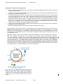
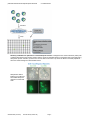
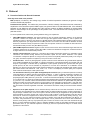
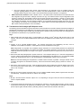
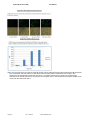
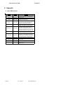
Pluripotency Reporters Cat. #SR2007x Series User Manual Store at -20ºC A limited-use label license covers this product. By use of this product, you accept the terms and conditions outlined in the Licensing and Warranty Statement contained in this user manual. pGreenFire Plasmid and Virus Response Reporter Constructs Cat. # SR20070-SR20071 Contents I. Introduction and Background A. B. C. D. E. F. G. Overview Lentiviral Expression System Production of Pseudotyped Viral Particles Delivery of Packaged Vector Construct into Target Cells List of Components and Available Reporters Additional Required Materials Safety Guidelines 2 2 4 4 6 6 6 II. Protocol A. Procedure Outline and General Comments B. Transduction of Packaged pGF1 Reporter Vector 8 9 III. Troubleshooting 11 IV. References 12 V. Appendix A. pGF1-mCMV-Puro Features B. Properties of dscGFP Fluorescent Protein C. Related Products D. Technical Support 13 14 14 14 VI. Licensing and Warranty Statement 16 ** This Product shall be used by the purchaser for internal research purposes only and distribution is strictly prohibited without written permission by System Biosciences. 888-266-5066 (Toll Free) 650-968-2200 (outside US) Page 1 System Biosciences (SBI) User Manual I. Introduction and Background A. Overview This manual provides information describing how to use the packaged VSV-G pseudotyped lentiviral pGreenFire™ Transcriptional Reporter (TR) Constructs to generate stable cell lines with the reporter constructs integrated into the host genome. Before using the reagents and material supplied with this product, please read the entire user manual. B. Lentiviral Transcriptional Reporter System Eukaryotic gene expression is regulated by a wide variety of developmental and environmental stimuli. First, an extracellular signaling molecule binds to a specific receptor. The signal is then transmitted through a series of molecular cascades, which activate or deactivate specific transcription factors (TFs) that regulate gene expression. The expression of any given gene is controlled by multiple transcription factors, which in turn are modulated by multiple signal transduction pathways. Many of these signal transduction pathways converge at transcription factors that bind to specific transcriptional response elements (TREs) or regions found in the promoters of various genes and modulate the transcription of these genes. The activation of a signal transduction pathway (e.g. by growth factors, drugs, etc.) can therefore be monitored by the expression level of the reporter gene controlled by a promoter containing these response sequences. The commonly used plasmid-based transcriptional reporter vectors containing different reporter genes can be delivered by transient transfection to the nucleus of target cells to monitor the activation of signal transduction pathways converging at a specific response element. Lentiviral expression vectors are the most effective vehicles for delivering genetic material to almost any mammalian cell—including non-dividing cells and to model organisms. As with standard plasmid vectors, it is possible to introduce lentiviral Transcriptional Reporter (TR) constructs in plasmid form into the cells with low-to-medium efficiency using conventional transfection protocols. However, by packaging the lentiviral TR vector construct in pseudoviral particles, you can obtain highly efficient transduction and heritable expression of transcriptional reporter constructs—even with the most difficult-to-transfect cells, like primary, stem, and differentiated cells. In comparison to retroviral delivery systems, lentivectors enter the cell nucleus without requiring cell replication. Page 2 ver. 3-140110 www.systembio.com pGreenFire Plasmid and Virus Response Reporter Constructs Cat. # SR20070-SR20071 Advantages of lentivector technology include: Ready-to-use pre-packaged constructs with a wide range of Transcriptional Response Elements (TREs) or regions for multiple transcriptional factors. Lentiviral reporter constructs can efficiently transduce nearly all cell types, even those that are difficult-to-transfect such as primary or non-dividing mammalian cells. Our lentiviral-based reporter system is a novel approach to study transcriptional regulation and offers many advantages over current transcription reporter systems. TR constructs will integrate into the genome and therefore be subject to chromatin regulation (Leung, T.H., et.al., 2004). Expression of the reporter gene indicates activation of a given transcriptional response element (TRE) by the cognate transcriptional factor in the natural chromosomal environment rather than in the episomal state in the nucleoplasm as is the case for conventional plasmid-based TR vectors. Tandem copies of integration can be avoided, thus allowing for faithful promoter regulation. Copy number of reporter constructs can be controlled by varying the multiplicity of infection (MOI). Construction of stable reporter cell lines is possible with TR lentivectors in just several days with a built-in constitutively expressing Puro or Neo resistance gene. Monitoring of signaling pathways by flow cytometry (FACS) is enabled by GFP reporters. SBI’s pGreenFire1 lentivectors are based on the traditional HIV (Human immunodeficiency virus) vector backbone. To address biosafety issues, SBI uses a third generation HIV lentiviral vector. (Dull, T., et.al., 1998, Miyoshi, H., et.al., 1998, Zufferey, R., et.al.,1999, Ramezani, A., et.al. 2000). In spite of improved biosafety features, third generation HIV cloning vectors still pose a potential biohazard risk due to the possible recombination with endogenous viral sequences to form a self-replicating HIV virus. In addition, the pGF1 cloning vectors developed at SBI are self-inactivating as a result of a deletion in U3 region of 3’LTR. Upon integration into the genome, the 5’ LTR promoter is inactivated, which prevents formation of replicationcompetent viral particles. The pGF1 vectors express dscGFP (destabilized) reporter gene and Firefly luciferase under the control of a minimal CMV (mCMV) promoter linked to transcriptional regulatory elements (TRE) or sequences such as Oct4 conserved region 4. The WPRE element enhances the expression level of the reporter gene. Constitutively expressed Puro/Neo resistant gene allows efficieny selection of stable transfectants. Figure 1 shows a map of the pGF1mCMV vector that expresses green fluorescent protein and Firefly luciferase from the mCMV promoter and can be used as a negative control. In order to facilitate the use of the lentivector-based GF system, SBI offers a wide range of transcriptional reporter constructs packaged in VSV-G pseudoviral particles. Fig. 1. Map of the HIV-based pGreenFire Pluripotency Response Reporter Vector. Essential components of the pGF1 vector system are, Response Elements (arrow), mCMV promoter (purple), destabilized copGFP (green), the self-cleaving T2A peptide (yellow) and the Firefly luciferase gene (red), and a selection resistance genes Puro or Neo (Marker). 888-266-5066 (Toll Free) 650-968-2200 (outside US) Page 3 System Biosciences (SBI) User Manual C. Production of Pseudotyped Viral Particles Currently, the most efficient technology for producing a high titer of infectious, replication-incompetent lentiviral particles is based on transient, coordinated expression of a viral effector expression construct and plasmids carrying all the necessary packaging proteins delivered into packaging (also called “producer”) cells (like HEK 293T) by simultaneous transfection with lentiviral expression and packaging plasmids. For more details on the packaging procedure see the pPACK-H1 Lentivector Packaging Kit user manual (available on SBI’s website at www.systembio.com). The pGF1 Response Reporter vectors can be packaged into VSV-G pseudotyped viral particles by co-transfection with the pPACK-H1 Lentiviral Packaging Plasmid Mix into HEK 239T cells (SBI, 293TN Producer Cell Line, Cat. # LV900A-1). Following transfection, the media contains the pseudoviral particles and can be concentrated by PEGit precipitation (or ultracentrifugation) using the protocol described in the Lentivector Packaging Kit (Cat. # LV500A-1). When expressed, the hybrid RSV/5’ LTR (HIV based) drives high level transcription of the lentiviral construct and produces a transcript that contains all the necessary functional elements (i.e., Psi, RRE, and cPPT) for efficient packaging. When this construct is expressed in HEK 293 cells that also express viral coat proteins (i.e., a packaging cell line), the lentiviral transcripts are efficiently packaged into pseudoviral particles. After isolation, these pseudoviral particles containing the RNA version of the TR cassette can be efficiently transduced into any mammalian target cells. Following transduction into the target cells, this expression cassette is reverse transcribed and integrated into the genome of the target cell. D. Delivery of Packaged Vector Construct into Target Cells Pantropic VSV-G pseudotyped viral particles containing the RNA copy of the pGF1 construct can be efficiently used to deliver and express reporter gene in a wide range of mammalian target cells. Pseudotyped packaged pGF1 constructs can effectively transduce both dividing and non-dividing established cell lines, including primary and differentiated cells, and numerous cell types ( neuronal, dendritic, endothelial, retinal, pancreatic, hepatic, aortic smooth muscle, airway epithelia, skin fibroblast, macrophage cells, etc). Lentivectors have been successfully used for direct in vivo delivery and expression of transgenic constructs in hamster muscle, mouse and primate brain, rabbit and mouse airway epithelium, and mouse liver. For a more complete list of cells or tissues which have been successfully transduced with lentivectors, please see the Reference Section. The pseudoviral particles can infect (or transduce) target cells and express reporter gene but cannot replicate within target cells because the viral structural genes are absent. Following transduction into the target cells, the GF cassette is reverse transcribed and integrated into the genome of the target cell. After integration, the GF cassette is under the transcriptional control of the transcriptional regulatory response elements and dscGFP/luciferase expression can be activated under the appropriate conditions. Page 4 ver. 3-140110 www.systembio.com pGreenFire Plasmid and Virus Response Reporter Constructs Cat. # SR20070-SR20071 Summary of Workflow for pGreenFire1 Lentiviral Reporter Vectors. Packaged virus is used to transduce (infect) cells of interest and introduce the reporter construct into the cell as an integrated provirus. Transduced cells can be split and 1) assayed for reporter activation, 2) frozen as a stock of cells for future use or 3) cloned to obtain monoclonal populations with more uniform background and activation levels. Sample GFP data for both the Oct 4 CR4 and Sox2 SSR2 Response reporters in Human iPS Cells 888-266-5066 (Toll Free) 650-968-2200 (outside US) Page 5 System Biosciences (SBI) User Manual E. List of Components The pGreenFire Response Reporter vectors are available as either plasmid DNA or pre-packaged VSV-G pseudotyped lentiviral particles. Premade reporters for a variety of signaling pathways are available and come complete with positive (pGF1-CMV) and negative (pGF1-mCMV) controls. Ten micrograms of each plasmid DNA is provided. IMPORTANT: Please note that pGreenFire lenti-reporter constructs must be transduced into target cells as a packaged virus in order for the constructs to function properly. Transfection of the constructs into a target cell keeps the constitutive RSV promoter intact, thus overriding the pathway-specific TRE promoters and leading to nonspecific expression of the reporter genes. Pre-packaged pGF1 constructs are provided as frozen pseudoviral particles. The total number of infection units (ifu) and concentration (the titer) were determined using HT1080 cells and may vary for different lots of each packaged reporter vector. The exact ifu, titer, and volume for each packaged reporter construct are indicated on its corresponding Product Analysis Certificate. For pre-packaged virus, the positive control construct with full CMV or MSCV promoters. The CMV promoter does Not work in stem cells, choose the MSCV option if you are working with stem cells. The negative control construct with mCMV promoter (TR010VA-1) are available separately. The Packaged Lentiviral Reporter Vectors are shipped on dry ice and should be immediately stored at –70C upon receipt. Avoid thawing and refreezing of pseudoviral particles! Properly stored pseudoviral particles are stable for 6 months from the date received. F. Additional Required Materials Dulbecco’s Modified Eagle’s Medium (D-MEM) (high glucose with sodium pyruvate and glutamine; Invitrogen, Cat. # 11995073) Fetal Bovine Serum (Invitrogen, Cat. # 16000036) Penicillin/Streptomycin (Invitrogen, Cat. # 15070063) Trypsin-EDTA (Sigma, Cat. # T3924) Polybrene® (hexadimethrine bromide; Sigma, Cat. # H9268) Millex-HV 0.45 m PVDF filters (Millipore, Cat. # SLHVR25LS) Tissue Culture Plates and Related Tissue Culture Supplies 293TN Human Kidney Producer Cell Line (SBI, Cat. # LV900A-1) Lipofectamine and Plus Reagent (Invitrogen) Third generation HIV-based packaging plasmids (such as SBI pPACK-H1 LV500A-1) G. Safety Guidelines Work with HIV-based lentiviral vectors falls within NIH Biosafety Level 2 criteria. For a detailed description of laboratory biosafety level criteria, consult the following pages on the Centers for Disease Control Office of Health and Safety Web site: http://www.cdc.gov/od/ohs/biosfty/bmbl4/bmbl4s3.htm http://www.cdc.gov/od/ohs/biosfty/bmbl4/bmbl4toc.htm Also, you should consult the health and safety guidelines and officers at your institution regarding use and handling of the lentiviral system. In addition, although the system itself has been designed to minimize possible risk, specific recombinant lentivector constructs may be potentially hazardous, depending on the nature of introduced insert (such as oncogenes, toxins, Transcriptional Response Element to tumor suppressor genes, etc.). For these reasons, it is critical to exercise due caution while working with recombinant lentiviruses. To ensure safe laboratory handling, you should thoroughly understand the biology of the lentiviral vectors and the specific modifications and design features of the SBI system with which you are working. Page 6 ver. 3-140110 www.systembio.com pGreenFire Plasmid and Virus Response Reporter Constructs Cat. # SR20070-SR20071 SBI’s pGF1 lentivectors together with the pPACKH1 packaging plasmids comprises a third-generation HIV-1-based cloning vector system. These lentivectors are based on the vectors developed for gene therapy applications by Dr. J. G. Sodroski (US patent #5,665,577 and # 5,981,276). This system is designed to maximize its biosafety features including: Deletion in the enhancer of U3 region of 3’LTR ensures self-inactivation of lentiviral construct after transduction and integration into genomic DNA of the target cells. RSV promoter upstream of 5’LTR in pGF1 expression vector allows efficient Tat-independent production of viral RNA, reducing the number of genes from HIV-1 that are used in this system. Number of HIV-1 viral genes necessary for packaging, replication and transduction is reduced to three (gag, rev and pol), and these genes are expressed from different plasmids lacking packaging signals and significant homology to pGF1 expression vector, VSV-G expression vector, or each other to prevent generation of recombinant replicationcompetent virus. None of the HIV-1 genes (gag, pol, rev) will be present in the packaged viral genome, as they are expressed from packaging plasmids lacking packaging signal—therefore, the lentiviral particles generated are replicationincompetent. Pseudoviral particles will carry only the expression construct of your target gene. To avoid any possible contamination and maintain a clean laboratory environment we also recommend following these standard safety practices: Wear double gloves, face protection, and lab coat at all times. Perform work in a limited access area in a Biological Safety Cabinet Class II and post biohazard warning signs. Minimize splashes or aerosols with careful pipetting. Take precautions with needles, blades, etc. Decontaminate work surfaces at least once a day and after any spill of viable material. Decontaminate all cultures, stocks, and other biological wastes before disposal using approved decontamination methods, such as autoclaving. Before decontamination the biological materials should be placed in a sealed, durable, leak-proof container for transport from the laboratory. 888-266-5066 (Toll Free) 650-968-2200 (outside US) Page 7 System Biosciences (SBI) User Manual II. Protocol A. Procedure Outline and General Comments Some key terms used in the protocol: MOI (multiplicity of infection)—the average copy number of lentiviral expression constructs per genome of target cell in the infected cell population. Pseudoviral titer (ifu/ml): The relative titer (concentration, infection units/ml) of lentiviral constructs, measured by amplification of the lentivector-specific WPRE region from genomic DNA of HT1080 infected cells. As a calibration standard, we use DNA from cells infected with a GFP reporter construct at different multiplicity of infection (MOI) based on FACS analysis of the percentage of GFP-positive cells. The Pseudoviral Titer is always specific to a particular cell line. To ensure optimal results, follow these general guidelines during your experiments: pGF1-CMV Reporter Construct: This plasmid should be used to estimate transduction efficiency of the lentiviral expression construct into target cells, select the cell type with highest infection efficiency, and to optimize the transduction protocol. Moreover, the presence of dscGFP-positive cells indicates that the lentiviral construct can be efficiently expressed in your target cells from the CMV promoter. The construct can also used for calibration of FACS machine for maximum intensity of expression. Please note, this reporter construct is not suitable for stem cells due to the inactivation CMV promoter. Use pGF1-MSCV promoter. pGF1-mCMV Reporter Construct: Negative control construct which can be used to transduce target cells under the conditions optimized for the positive control pGF1-CMV construct and determine “background” of GFP fluorescence of target cells with a non-activated CMV promoter. Infection with Polybrene®: Polybrene is a polycation that increase transduction efficiency by neutralizing charge interactions and increase binding between the pseudoviral capsid and the cellular membrane. The optimal concentration of Polybrene depends on cell type and may need to be empirically determined (usually in the range of 4-8 g/ml). Excessive exposure to Polybrene (>12 hr) can be toxic to some cells. Pseudoviral titer: The titer of packaged pGF1 reporter constructs can be determined using a PCR based approach such as SBI’s UltraRapid titer kit (LV960A-1). Following transduction of pseudoviral particles into HT1080 cells and analysis of percentage of infected cells by amplification of pGF1-specific products from genomic DNA with gene specific primers, the titer can be estimated by comparison to a known set of standards. As a calibration control standard, we used the genomic DNA control template isolated from HT1080 cells transduced with positive control pGF1-CMV-construct and infected at 1:100, 5:100, 1:10, 1:4 and 1:2 multiplicity of infection (MOI), and compared these results to FACS analysis of GFP-positive cells. The transduction efficiency of the pGF1 Packaged Reporter Construct (and your lentiviral expression construct) varies significantly for different cells and experimental conditions. In order to optimize transduction conditions, we recommend that you use HT1080 (or similar) cells as a positive control in parallel with your target cells and use prepackaged pGF1-CMV (TR011VA-1) from SBI. To determine the desired multiplicity of infection (MOI) appropriate for your target cells, you should do several transductions with packaged pGF1 pseudoviral particles at different MOI’s (e.g. from 0.1 to 5). Results of these test transductions should be used to determine an optimal MOI that yields the optimal percentage of infected cells based on the percentage of cells expressing the GFP marker. Note that some cell types, such as primary tissue cultures of differentiated cells (e.g. epithelial cells attached to a membrane, etc.) may be resistant to infection regardless of the MOI. Conventional cell cultures usually do not have infection-resistant cells. Expression of the pGF1 Reporter can be measured directly at about 48-72 hours after transduction. At this time, GF constructs are integrated into the genomic DNA resulting in stably transduced reporter cell lines. Depending on the percentage of infected cells, one or more copies of the GF construct may be integrated into the genomic DNA of each transduced cell. These reporter cells can be cloned in order to obtain a uniform population of the GF cell line where the GF construct is integrated into a defined chromosomal location. Some actively dividing cells (e.g., 293, HT1080, HeLa, etc.) may express the reporter construct in 80-90% of the cells after transduction at an MOI of 1-2. For these “easy-to-transduce” cells, most biological assays can be performed at 48-72 hours after transduction. However, some primary cells may only express the pGF1 construct in 10-50% of cells, even when transduced at high MOI’s. For these “difficult-to-transduce” cells, it is probably desirable to select the cells stably expressing the control pGF1-CMV vector (or your lentiviral expression construct) by FACS for experimental assays or to obtain cloned populations of TR cell lines. Page 8 ver. 3-140110 www.systembio.com pGreenFire Plasmid and Virus Response Reporter Constructs Cat. # SR20070-SR20071 Due to the specificity of the pGF1 reporter, GFP expression is only expected to occur in conditions where the particular signaling pathway is active. For instance, the pGF1-NF-κB reporter is silent in infected cells until an activating stimulus is given such as TNF-α, then both dscGFP and firefly luciferase is co-expressed. SBI’s lentiviral GF constructs contain a deletion in the 3’ LTR which leads to self-inactivation of the lentiviral vector after integration into genomic DNA. Although more than one copy of a lentiviral construct may be integrated into the genome of a single cell, the lentiviral construct cannot produce infectious viral particles. However, in spite of these safety features, please remember that you are working with transducible pseudoviral particles. Although the particles are replication-incompetent, they are infection-competent, so the lentiviral expression cassette which they carry will infect, integrate, and express in any mammalian cells. Please follow the recommended guidelines for working with BSL-2 class viruses (see Section I.G for more details). B. Transduction of the Packaged pGF1 Reporter Vector The following protocol describes the general procedure for the transduction of the pGF1 Reporter Constructs packaged in pseudotyped viral particles into HT1080 cells. This protocol assumes that you will use these guidelines in order to perform transduction of your target cells in parallel using HT1080 cells as a positive control and can be used as a starting point for the optimization for transduction of your particular cell-type. Day 1. 1. Plate HT1080 cells (and target cells) in a 48-well plate at a density of 1x10⁴ cells per well 24 hours prior to viral infection. Add 0.5 ml of complete D-MEM medium (with serum and antibiotics) and incubate cells at 37°C with 5% CO2 overnight. Day 2. 2. Prepare 10 ml of complete D-MEM medium. For extremely fast-growing and metabolizing cell lines, such as HEK293T, use 3% FBS in the medium. Add Polybrene to a final concentration of 5-8 g/ml. Note: Polybrene® is a polycation that neutralizes charge interactions to increase binding between the pseudoviral capsid and the cellular membrane. The optimal concentration of Polybrene depends on cell type and may need to be empirically determined (usually in the range of 2-10 g/ml). Excessive exposure to Polybrene (>12 hr) can be toxic to some cells. 3. Remove test tube with packaged pGF1 reporter vector from –70°C and place directly in ice. Thaw the viral particles for 5-10 min. Dilute an appropriate amount (depending on the optimal MOI) of viral particles in 0.1 ml of complete D-MEM medium with Polybrene (from step 2). Use several dilutions of packaged pGF reporter vector if necessary. Note: We recommend diluting the viral particles in a smaller volume of medium, if possible. The higher the titer of virus in solution, the higher is the transduction efficiency. Mix the virus with the medium gently by rotation or inversion. Do not vortex. 4. Remove the culture medium from cells. Infect HT1080 (and target cells) by adding the viral stock dilutions to the wells. For one well (mock well control) add 0.1 ml of D-MEM medium with Polybrene (from Step 2). Incubate cells at 37°C with 5% CO2 overnight. Day 3. 5. Remove the culture medium and replace with 0.5 ml of complete D-MEM medium (without Polybrene but with serum and antibiotics). Incubate the cells at 37°C with 5% CO2 overnight. Day 4. 6. By day 4, the culture will be confluent. Split it 1:3 to 1:5, depending on the type of cells, and continue incubating for 48 hours in complete D-MEM. Day 6. 7. The infected HT1080 cells (target cells) can be analyzed for expression of the pGF1-CMV and pGF1 reporter construct by fluorescent microscopy or by performing a luciferase assay. Figure 2 demonstrates the activation of the NF-κB specific reporter (pGF1-NF-κB, TR012A-1) following infection of 293T cells and activation with TNF-α. 888-266-5066 (Toll Free) 650-968-2200 (outside US) Page 9 System Biosciences (SBI) User Manual Fig. 2. The top panel shows the results of an NF-κB reporter cell line created by infection with the pGreenFire1-NF-κB reporter virus. Transduced cells were plated in 12 well dishes and then treated with the indicated amounts of TNF-α. GFP fluorescence was assessed by fluorescence microscopy. The bottom panel shows the results of a luciferase assay performed on the 293TN cells shown in the top panel. As a negative control, cells transduced with the pGF1-mCMV control vector were also treated with TNF-α. Page 10 ver. 3-140110 www.systembio.com pGreenFire Plasmid and Virus Response Reporter Constructs Cat. # SR20070-SR20071 III. Troubleshooting A. Why are my cells green following transfection for viral packaging? In order to produce the viral genomic RNA that will be packaged into viral particles, a strong promoter is included in the 5’ LTR to drive its expression. Due to the mechanism of lentiviral replication, this promoter in the 5’ LTR is not present in the genomic RNA or the resulting integrated provirus. Therefore, GFP is expressed from the viral 5’LTR following transfection, however, following transduction, transcription should only occur from the reporter gene when activated. B. 1. Inefficient Transduction of Packaged pGF1 Reporter Vector into Target Cells Poor infection efficiency Target cells have too high or too low density Plate fewer or more cells in order to have about 50% confluency at infection stage. Target cell line may be difficult to transduce Use a higher concentration (less fold dilution) of pseudoviral particles. Optimize the transduction protocol and use as positive control cells H1299 cell line. ® Wrong amount of Polybrene added during infection stage ® ® If Polybrene is toxic to the target cells, optimize Polybrene concentration in the range of 1-5 g/ml. Loss of pseudoviral titer during storage Ensure storage of the Packaged Reporter Vector at –70°C. Each freeze-thaw cycle causes reduction of the titer by 2030%. Use a fresh stock for transduction. Do not keep the stock longer than 6-12 months. Volume of infecting supernatant is too high Keep the volume as low as possible to achieve maximal adsorption of viral particles to the cells. 2. Transduction affects target cell viability Packaged Reporter Vector affects target cell growth Use a shorter transduction time to minimize the toxic effect to the target cells. Try replacing with a similar target cell type. ® Polybrene is toxic for target cells Optimize the concentration and exposure time to Polybrene® during the transduction step. 3. No Expression of positive control pGF1-CMV reporter in target cells The CMV promoter is not functional in target cells It is a very rare case, but the only way to solve this problem is to change the type of target cells. 888-266-5066 (Toll Free) 650-968-2200 (outside US) Page 11 System Biosciences (SBI) User Manual IV. References Okumura-Nakanishi S, Saito M, Niwa H, Ishikawa F. Oct-3/4 and Sox2 regulate Oct-3/4 gene in embryonic stem cells. J Biol Chem. 2005 Feb 18;280(7):5307-17. Tomioka M, Nishimoto M, Miyagi S, Katayanagi T, Fukui N, Niwa H, Muramatsu M, Okuda A. Identification of Sox-2 regulatory region which is under the control of Oct-3/4-Sox-2 complex. Nucleic Acids Res. 2002 Jul 15;30(14):3202-13. Buchschacher, G.L., and Wong-Staal, F. (2000) Development of lentiviral vectors for gene theraphy for human diseases. Blood. 95:2499-2504. Burns, J.C., Friedmann, T., Driever, W., Burrascano, M., and Yee, J.K. (1993) Vesicular stomatitis virus G glycoprotein pseudotyped retroviral vectors: concentration to a very high titer and efficient gene transfer into mammalian and non-mammalian cells. Proc. Natl. Acad. Sci. USA. 90:8033-8034. Cann, A.J.(ed). (2000) RNA Viruses. A Practical Approach. Oxford Univ. Press. Dull, T., Zufferey, R., Kelly, M., Mandel, R.J., Nguyen, M., Trono, D., and Naldini, L. (1998) A third-generation lentivirus vector with a conditional packaging system. J. Virol. 72:8463-8471. Gould, D.J. and Favorov, P. (2003) Vectors for the treatment of autoimmune diseases. Gene Therapy 10:912-927. Morgan, R.A., Cornetta, K. and Anderson, W.F. (1990) Application of the polymerase chain reaction in retroviral-mediated gene transfer and the analysis of gene-marked human TIL cells. Hum. Gene Ther. 1:135-149. Pfeifer, A., Kessler, T., Yang, M., Baranov, E., Kootstra, N., Cheresh, D.A., Hoffman, R.M. and Verma, I.M. (2001) Transduction of liver cells by lentiviral vectors: Analysis in living animals by fluorescence imaging. Mol. Ther. 3:319-322. Qin, X.F., An, D.S., Chen, I.S., and Baltimore, D. (2003) Inhibiting HIV-1 infection in human T cells by lentiviral-mediated delivery of small interfering RNA against CCR5. Proc. Natl. Acad. Sci. USA 100:183-188 Quinn, T.P., and Trevor, K.T. (1997) Rapid quantitation of recombinant retrovirus produced by packaging cell clones. Biotechniques 23:1038-1044. Sui, G., Soohoo, C. Affar, E.B., Gay, F., Forrester, W.C., and Shi, Y. (2002) A DNA vector-based RNAi technology to suppress gene expression in mammalian cells. Proc. Natl. Acad. Sci. U.S.A 99:5515-5520 Curran MA, Nolan GP. Nonprimate lentiviral vectors. Curr Top Microbiol Immunol. 2002; 261: 75-105. Curran MA, Nolan GP. Recombinant feline immunodeficiency virus vectors. Preparation and use. Methods Mol Med. 2002; 69: 335-50 Loewen N, Barraza R, Whitwam T, Saenz DT, Kemler I, Poeschla EM. FIV Vectors. Methods Mol Biol. 2003; 229: 251-71. Naldini L. Lentiviruses as gene transfer agents for delivery to non-dividing cells. Curr Opin Biotechnol. 1998 Oct; 9(5): 457-63. Sauter SL, Gasmi M. FIV vector systems. Somat Cell Mol Genet. 2001 Nov; 26(1-6): 99-129. Alisky JM, Hughes SM, Sauter SL, Jolly D, Dubensky TW Jr, Staber PD, Chiorini JA, Davidson BL. Transduction of murine cerebellar neurons with recombinant FIV and AAV5 vectors. Neuroreport. 2000 Aug 21; 11(12): 2669-73. Brooks AI, Stein CS, Hughes SM, Heth J, McCray PM Jr, Sauter SL, Johnston JC, Cory-Slechta DA, Federoff HJ, Davidson BL. Functional correction of established central nervous system deficits in an animal model of lysosomal storage disease with feline immunodeficiency virus-based vectors. Proc Natl Acad Sci U S A. 2002 Apr 30; 99(9): 6216-21. Crystal RG. Bad for cats, good for humans? Modified feline immunodeficiency virus for gene therapy. J Clin Invest. 1999 Dec; 104(11): 1491-3. Curran MA, Kaiser SM, Achacoso PL, Nolan GP. Efficient transduction of nondividing cells by optimized feline immunodeficiency virus vectors. Mol Ther. 2000 Jan; 1(1): 31-8. Derksen TA, Sauter SL, Davidson BL. Feline immunodeficiency virus vectors. Gene transfer to mouse retina following intravitreal injection. J Gene Med. 2002 Sep-Oct; 4(5): 463-9. Haskell RE, Hughes SM, Chiorini JA, Alisky JM, Davidson BL. Viral-mediated delivery of the late-infantile neuronal ceroid lipofuscinosis gene, TPP-I to the mouse central nervous system. Gene Ther. 2003 Jan; 10(1): 34-42. Page 12 ver. 3-140110 www.systembio.com pGreenFire Plasmid and Virus Response Reporter Constructs Cat. # SR20070-SR20071 Price MA, Case SS, Carbonaro DA, Yu XJ, Petersen D, Sabo KM, Curran MA, Engel BC, Margarian H, Abkowitz JL, Nolan GP, Kohn DB, Crooks GM. Expression from second-generation feline immunodeficiency virus vectors is impaired in human hematopoietic cells. Mol Ther. 2002 Nov; 6(5): 645-52. Stein CS, Davidson BL. Gene transfer to the brain using feline immunodeficiency virus-based lentivirus vectors. Methods Enzymol. 2002; 346: 433-54. Browning MT, Schmidt RD, Lew KA, Rizvi TA. Primate and feline lentivirus vector RNA packaging and propagation by heterologous lentivirus virions. J Virol. 2001 Jun; 75(11): 5129-40. Curran MA, Kaiser SM, Achacoso PL, Nolan GP. Efficient transduction of nondividing cells by optimized feline immunodeficiency virus vectors. Mol Ther. 2000 Jan; 1(1): 31-8. Poeschla EM, Wong-Staal F, Looney DJ. Efficient transduction of nondividing human cells by feline immunodeficiency virus lentiviral vectors. Nat Med. 1998 Mar; 4(3): 354-7. Poeschla, E.M., Looney, D.J., and Wong-Staal, F. (2003) Lentiviral nucleic acids and uses thereof. US Patent NO. 6,555,107 B2 Dull, T., Zufferey, R., Kelly, M., Mandel, R.J., Nguyen, M, Trono, D. (1998) J. Virol.,72, 8463-8471 Miyoshi, H., Blomer, U., Takashi, M., Gage, F.N., Verma, I.M (1998), J.Virol., 72, 8150-8157. Zufferey, R., Donello, J.E., Trono, D., Hope, T.J. (1999), J.Virol., 73, 2886-2892 Ramezani, A., Hawley, T.S., Hawley, R.G. (2000) Mol. Ther., 2, 458-469 Leung, T.H., Hoffmann, A., Baltimore, D. 2004, Cell, v. 118, 453-464 888-266-5066 (Toll Free) 650-968-2200 (outside US) Page 13 System Biosciences (SBI) User Manual V. Appendix A. pGF1-CMV Features Feature Location * Function Hybrid RSV promoter-R/U5 long terminal repeat; required for viral packaging and transcription RSV-5' LTR 1-415 gag 567-919 Packaging signal RRE 1066-1309 Rev response element binds gag and involved in packaging of viral transcripts cPPT 1798-1899 Central polypurine tract (includes DNA Flap region) involved in nuclear translocation and integration of transduced viral genome CMV promoter 1943-2271 DscGFP-T2A-FLuc 2329-4848 WPRE 4859-5399 3' LTR (U3) 5538-5771 SV40 Poly-A 5843-5974 Transcription termination and polyadenylation 5983-6129 Allows for episomal replication of plasmid in eukaryotic cells SV40 Ori pUC Ori AmpR Human cytomegalovirus (CMV)--constitutive promoter for transcription of dscGFP and zeoR Copepod green fluorescent protein (similar to regular EGFP, but with brighter color) as a reporter for the transfected/ transduced cells; a destabilizing (ds) peptide on the C-end shortens the half life time of the mature protein to 1 hour Posttranscriptional regulatory element which enhances the stability of the viral transcripts Required for viral reverse transcription; selfinactivating 3' LTR with deletion in U3 region prevents formation of replication-competent viral particles after integration into genomic DNA 6499-7172(C) Allows for high-copy replication in E. coli 7317-8177(C) Ampicillin resistant gene for selection of the plasmid in E. coli * The notation (C) refers to the complementary strand. Page 14 ver. 3-140110 www.systembio.com pGreenFire Plasmid and Virus Response Reporter Constructs Cat. # SR20070-SR20071 B. Properties of the dscGFP Fluorescent Protein The pGF Vectors contain the full-length copGFP gene with optimized human codons for high level of expression of the fluorescent protein from the CMV promoter in mammalian cells. The copGFP marker is a novel natural green monomeric GFP-like protein from copepod (Pontellina sp.). A unique feature of the dscGFP protein is the presence of an additional destabilizing (ds) peptide on the C-end of the protein which shortens the half life time of the mature protein without additional transcription to 1 hour. The copGFP protein is a non-toxic, non-aggregating protein with fast protein maturation, high stability at a wide range of pH (pH 4-12), and does not require any additional cofactors or substrates. The copGFP protein has very bright fluorescence that exceeds at least 1.3 times the brightness of EGFP, the widely used Aequorea victoria GFP mutant. The copGFP protein emits green fluorescence with the following characteristics: emission wavelength max – 502 nm excitation wavelength max – 482 nm quantum yield – 0.6 -1 -1 extinction coefficient – 70,000 M cm Due to its exceptional properties, copGFP is an excellent fluorescent marker which can be used instead of EGFP for monitoring delivery of lentiviral constructs into cells. C. Related Products PathNet™ Lentiviral TRE Cloning and Delivery Vectors pTRF1-mCMV-dscGFP Vector (Cat. # TR100PA-1) pTRF3-mCMV-Luc Vector (Cat. # TR200PC-1) pTRH1-mCMV-dscGFP Vector (Cat. # TR500PA-1) pTRH5-mCMV-Luc Vector (Cat. # TR600PE-1) These FIV- and HIV-based Lentiviral Transcriptional Reporter cloning vectors allow you to clone transcriptional response elements (TRE) under the mCMV promoter or promoter of your choice and efficiently transduce these constructs in a wide range of cells. PathNet™ Lentiviral Transcriptional Reporter Constructs (in Plasmid form) Many (visit our website at www.systembio.com for a current list of available TR plasmid constructs) These FIV- and HIV-based plasmid reporter constructs, when packaged in pseudoviral particles using the pPACKF1 or pPACKH1 Lentivector Packaging Kit, allow you to transduce and analyze a wide variety of TF-specific constructs in a wide range of cells. Based on the highly efficient lentiviral system, the PathNet™ Transcriptional Reporter Vectors provide a convenient and cost-effective system to deliver and stably integrate sequences of your choice into the host genome. pPACK Lentivector Packaging Kits FIV-Based: pPACKF1 Packaging Kit (Cat. # LV100A-1) HIV-Based: pPACKH1 Packaging Kit (Cat. # LV500A-1) Unique lentiviral vectors that produce all the necessary lentiviral proteins and the VSV-G envelope glycoprotein from vesicular stomatitis virus required to package pGF1 lentiviral constructs into pseudoviral particles. 293TN cells (SBI, Cat. # LV900A-1; or ATCC, Cat. # CRL-11268) transiently transfected with the Lentiviral Packaging Plasmid Mix and one of the pGF1 reporter vectors produce packaged pseudoviral particles containing a pGF1-TRE-mCMV construct. Lentivector UltraRapid Titer PCR Kit (Cat. # LV960A-1 [for human cells], LV961A-1 [for mouse cells]) Measure copy number (MOI) of integrated lentiviral constructs in genomic DNA of target cells after transduction with SBI’s PathNet™, pGreenFire1 vectors or with constructs made in any of SBI’s FIV or HIV-based lentivectors. D. Technical Support For more information about SBI products and to download manuals in PDF format, please visit our web site: http://www.systembio.com For additional information or technical assistance, please call or email us at: 888-266-5066 (Toll Free) 650-968-2200 (outside US) Page 15 System Biosciences (SBI) User Manual System Biosciences (SBI) 265 North Whisman Rd. Mountain View, CA 94043 Phone: (650) 968-2200 (888) 266-5066 (Toll Free) Fax: (650) 968-2277 E-mail: General Information: [email protected] Technical Support: [email protected] Ordering Information: [email protected] Page 16 ver. 3-140110 www.systembio.com