1
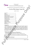
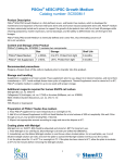
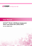
piPS Cells
Cat. #SC801A-1 & SC802A-1
User Manual
Store cells in liquid nitrogen
ver. 1-021111
A limited-use label license covers this
product. By use of this product, you
accept the terms and conditions outlined
in the Licensing and Warranty Statement
contained in this user manual.
piPS Cells
Cats. # SC801A-1, SC802A-1
Contents
I.
Introduction and Background ..................................................2
A.
Evolution of Reprogramming Technology ...........................2
B.
Protein derived iPS cell line ................................................3
II.
Protocols .................................................................................5
A.
Materials..............................................................................5
Gelatin treatment of plates ..............................................................7
Thawing MEF cells ..........................................................................7
Thawing human iPS cells................................................................9
III.
References ........................................................................12
IV.
Technical Support .............................................................13
V.
Licensing and Warranty ........................................................13
888-266-5066 (Toll Free)
650-968-2200 (outside US)
Page 1
System Biosciences (SBI)
I.
User Manual
Introduction and Background
A. Evolution of Reprogramming Technology
The concept of reprogramming was initially demonstrated by
Gurdon et al. in 1958, where they generated adult Xenopus from
somatic cells by nuclear reprogramming. After the discovery of
induced pluripotent stem cells, iPSC technique using defined
transcription factors has been a basis and standard for generation
of pluripotent stem cells. In comparison to somatic cell nuclear
transfer, the iPSC technology offers an unprecedented technical
simplicity and enables generation of patient-specific pluripotent
stem cells with less ethical concerns.
Although iPSC technology provides unprecedented opportunities
in biomedical research and regenerative medicine, there remains
a great deal to learn about iPSC safety, the reprogramming
mechanisms, the quality of iPSCs from different source cells and
the variations using different reprogramming technology. Since
2006, iPSC technology has evolved from integrated virus
(Retrovirus and lentivirus) to non-integrated virus (Adenovirus)
viral methods, to non-viral methods (plasmids), genetic methods
(DNA or RNA vectors) to non-genetic methods (proteins). These
technical advances provide safer iPSCs for more meaningful
mechanistic studies, iPSC-based disease modeling, and drug
screening.
Year 2006
Page 2
Present
ver. 1-020711
www.systembio.com
piPS Cells
Cats. # SC801A-1, SC802A-1
B. Protein derived iPS cell line
SBI offers the human protein iPS cell lines highlighted in Cell Stem
Cell, Generation of Human Induced Pluripotent Stem Cells by
Direct Delivery of Reprogramming Proteins. Kim D, et al., 2009,
4:472-476. The piPS cell lines were derived from the most
commonly used source cells, newborn human fibroblasts.
To date, all methods to generate iPSCs require the use of genetic
materials and/or potentially mutagenic chemicals. Using protein
engineering technology, stable piPSCs were induced from human
fibroblasts by directly delivering four reprogramming proteins
(Oct4, Sox2, Klf4, and c-Myc) fused with a cell-penetrating
peptide. These piPSCs exhibited similarities to human embryonic
stem cells in morphology, proliferation, global gene expression,
DNA methylation patterns, and expression of characteristic
pluripotency markers.
PiPSC lines produced with recombinant proteins were successfully
maintained for more than 35 passages and differentiated into
derivatives of all three embryonic germ layers during the formation
of embryoid body (in vitro) and teratomas (in vivo), the most
stringent tests for the quality of human iPS cells. This protein
reprogramming system eliminates the potential risks associated
with the viruses, DNA transfection, and potentially harmful
chemicals. Therefore, it provides a promising safe source of
patient-specific cells for the future regenerative medicine.
888-266-5066 (Toll Free)
650-968-2200 (outside US)
Page 3
System Biosciences (SBI)
Page 4
User Manual
ver. 1-020711
www.systembio.com
piPS Cells
Cats. # SC801A-1, SC802A-1
II.
Protocols
A. Materials
Human ESC medium
Component
Knockout serum
replacement
Glutamax-1
Nonessential amino
acid
2-mercaptoethanol
penicillin and
streptomycin
bFGF
KO DMEM/F12
Cat. #
Final
concentration
Source
10828028
20%
Invitrogen
35050061
2 mM
11140050
M7522
15140122
233-FB025
12660012
Invitrogen
-4
Invitrogen
-4
1 x 10 M
50 U and 50
µg /ml
Sigma
10 ng/ml
R&D
1 x 10 M
Invitrogen
Invitrogen
MEF medium
Component
Cat. #
FBS
16000077
Glutamax-1
penicillin and
streptomycin
DMEM
35050061
888-266-5066 (Toll Free)
15140122
Final
concentration
10%
2 mM
50 U and 50 µg
/ml
11995065
650-968-2200 (outside US)
Source
Invitrogen
Invitrogen
Invitrogen
Invitrogen
Page 5
System Biosciences (SBI)
User Manual
Other Required Reagents
Component
Cat. #
Final concentration
Source
Rock
Inhibitor Y27632
Y0503
10 µM/ml
Sigma
G1890
Dissolve 0.5 g of
gelatin from porcine
skin in 500 ml DPBS
and autoclave. Stable
for 1 yr at room
temperature
Sigma
SCR005
Aliquot in 10 ml and
store in -20°C. Dilute
1:1 with DPBS before
use.
Millipore
0.1% (w/v)
Gelatin
Accutase
Human ES freezing medium
90% FBS plus 10% DMSO, with 10 µM ROCK inhibitor Y-27632
NOTE: This protocol is for growing piPS cells on MEF feeder cells.
These should already be growing before you plate your piPS cells.
Page 6
ver. 1-020711
www.systembio.com
piPS Cells
Cats. # SC801A-1, SC802A-1
B. Protocol for Human iPS Cell Culture
Growth condition for mouse fibroblasts (for feeder layers)
Gelatin treatment of plates
1. Add enough sterile/autoclaved 0.1% gelatin to cover the
bottom of the wells.
Approximate amounts: 10cm - 5ml
6 well - 1.5ml/well
24 well - 0.5 ml/well
96 well - 200 µl/well
2. Incubate the gelatin-coated dishes for at least 15 min at
37 °C.
3. Aspirate excess gelatin solution before using.
Thawing MEF cells
To insure the highest level of viability, be sure to warm medium to
37 °C before using it on the cells. Cells should be plated at a
minimum cell density of 1 x 10⁴ cells/cm2.
1. Remove the vial from liquid nitrogen and thaw quickly in
37 °C water bath.
2. Remove the vial from the water bath as soon as the cells
are half way thawed, and sterilize by spraying with 70%
ethanol.
3. Transfer the cells with 10 ml of MEF medium to a 15-cm
conical tube and pellet the cells by centrifugation at 200x g
for 5 min.
4. Discard the supernatant and resuspend the cells with 10
ml fresh MEF medium and plate the cells at seed density
of 1 x 10⁴ cells/cm2.
888-266-5066 (Toll Free)
650-968-2200 (outside US)
Page 7
System Biosciences (SBI)
User Manual
5. Incubate at 37 °C with 5% CO2 in air atmosphere, until the
cells reach 80-90% confluency.
6. Change medium twice a week or when pH decreases.
Passage of MEF cells
Cells should be split when they reach confluency. A split based on
seed density of 0.5 x 10⁴ cells /cm2 is recommended.
1. Discard the medium and wash the cells twice with PBS.
2. Aspirate PBS, and add 1 ml per T75 flask of 0.25%
trypsin-EDTA, and incubate for 1 min.
3. Add 5 ml of MEF medium, and break up the cell clumps by
gently pipetting up and down several times.
4. Transfer cells into a conical tube and centrifuge at 200 g
for 5 min.
5. Discard the supernatant, and resuspend the cell pellet in
10 ml MEF medium.
6. Count the number of cells, plate cells at 0.5 x 10⁴
cells/cm2, and incubate at 37 °C with 5% CO2.
Freezing MEF cells
1. Follow steps 1-4 from the Passage of Cells above.
2. Discard the supernatant, and resuspend the pellet in MEF
medium. Add approximately 1 ml for each T75 flask.
3. Count the number of cells and dilute the cell suspension to
1 x 10⁷ cells/ml.
4. Add an equal volume of cold 2X Freezing Media
(containing 20% DMSO and 80% FBS) to the cell
suspension.
5. Aliquot 1 ml of suspension into each cryovial (5 x 10⁶
cells/vial).
Page 8
ver. 1-020711
www.systembio.com
piPS Cells
Cats. # SC801A-1, SC802A-1
6. Place the vials in a cell-freezing container and keep it at 80 °C overnight.
7. Transfer the vials to a liquid nitrogen tank for long-term
storage.
Mitomycin C treatment of MEF
At confluence, MEF cells are treated with mitomycin C to halt the
division of the cells when they are still able to condition the
medium as the feeder layers for human iPS cells.
1. Add 6 mL of fresh MEF medium containing 50 μl of
mitomycin C solution (1mg/ml) to one T75 flask of
confluent MEF cells, and swirl it briefly.
2. Incubate at 37 °C for at least 3 h.
3. After incubation, aspirate the mitomycin C-containing
medium off the cells, and wash the cells twice with 10 ml
of PBS.
4. Aspirate off PBS, add 1 ml of 0.25% trypsin-EDTA, swirl to
cover the entire surface, and incubate for 1 min at room
temperature.
5. Add 5 ml of MEF medium, and break up the cells to a
single cell suspension by pipetting up and down. Count
the number of cells. Seed the cells on gelatin-coated
6
5
dishes (1x 10 cells per 100-mm dish, or 1.5 x 10 cells
per well of 6-well plate).
6. Cells should be ready to use by the next day.
Growth condition for human iPS cells
Thawing human iPS cells
To insure the highest level of viability, be sure to warm medium to
37 °C before using it on the cells. Due to the low survival rate of
cryopreserved human iPS cells, the recovery is expected to take
at least one week.
888-266-5066 (Toll Free)
650-968-2200 (outside US)
Page 9
System Biosciences (SBI)
User Manual
1. Remove the vial from liquid nitrogen and thaw quickly in
37 °C water bath.
2. Remove the vial from the water bath as soon as the cells
are half way thawed, and sterilize by spraying with 70%
ethanol.
3. Transfer the cells with 10 ml of human ES medium to a
15-cm conical tube and pellet the cells by centrifugation at
200x g for 5 min.
4. While centrifuging, remove MEF medium from the 6-well
plate with MEF feeder cells, wash the well twice with 1 ml
of KO DMEM/F12, and add 1 ml of human ES medium
supplemented with ROCK inhibitor Y-27632 (10 µM).
5. Discard the supernatant of the tube containing human iPS
cells, resuspend the cells with 1 ml of fresh human ES
medium with ROCK inhibitor Y-27632, and plate the cells
in the wells of 6-well plate with MEF feeder cells.
6. Incubate at 37 °C with 5% CO2 in air atmosphere, until the
cells reach 80% confluency.
7. Change the medium everyday.
Note: Y-27632 is not necessary for regular human iPS cell
culture.
Maintenance of human iPS cells
It is important to note that do NOT keep human iPS cells in culture
for long periods in order to maintain the pluripotency.
1. Aspirate the medium, and wash the cells twice with 1 ml of
PBS.
2. Remove PBS completely, add 0.5 ml of 1:1 Accutase to
each well of a 6-well plate, and incubate at room
temperature for 1 min.
3. While incubating, remove a 6-well plate with MEF feeder
cells from the incubator. Aspirate MEF medium, wash with
1 ml of KO DMEM/F12 twice for each well, and add 1 ml
of human ES medium with 10 µM ROCK inhibitor to each
well.
Page 10
ver. 1-020711
www.systembio.com
piPS Cells
Cats. # SC801A-1, SC802A-1
4. When the edge of the human iPS cell colonies have fold
up, aspirate the Accutase solution and wash the cells
three times with DMEM/F12 medium.
5. Add 1 ml per well of human ES medium, and dislodge the
cell colonies by using cell scraper.
6. Transfer the contents into a 15 ml conical tube with 5 ml of
pre-warmed human ES medium. Use another 1 ml of
medium to wash the well one more time and combine it to
the same tube.
7. Centrifuge at 200x g for 5 minutes at room temperature.
8. Carefully aspirate the overlaying medium, then gently
finger tap the tube bottom to dislodge the cell pellet.
9. Gently add 3 to 6 ml of fresh human ES medium with 10
µM ROCK inhibitor, and resuspend the cells by gently
pipetting up and down.
10. Add 1 ml of the human iPS cell suspension to each well of
the 6-well plate. Right after plating iPS cells, gently swirl
the plate back-and-forth and side-to-side to evenly
distribute the cells, and incubate at 37°C.
11. The ES media must be changed every day and human
iPS cells sub-cultured every 5-7 days when the
undifferentiated colonies are big enough. Track passage
number of iPS cells.
Note: human iPS cells could also be grown on HFF feeders.
Freezing human iPS cells
1. Grow human iPS cells to the exponential phase in a 6-well
plate, and pre-treat cells with 10 µM Y-27632 for one hour
prior to freeze.
2. Remove PBS completely, add 0.5 ml of 1:1 Accutase, and
incubate at room temperature for 1 min.
888-266-5066 (Toll Free)
650-968-2200 (outside US)
Page 11
System Biosciences (SBI)
User Manual
3. While incubating, remove a 6-well plate with MEF feeder
cells from the incubator. Aspirate MEF medium, wash with
1 ml of KO DMEM/F12 twice for each well, and add 1 ml
of human ES medium to each well.
4. When the edge of the human iPS cell colonies fold up,
aspirate Accutase solution and wash the cells three times
with KO DMEM/F12 medium.
5. Add 1 ml per well of human ES medium, and dislodge the
cell colonies using cell scraper.
6. Transfer the contents into a 15 ml conical tube with 5 ml of
pre-warmed human ES medium. Use another 1 ml of
medium to wash the well one more time and combine it to
the same tube.
7. Centrifuge at 200x g for 5 minutes at room temperature.
8. Carefully aspirate the overlaying supernatant, then gently
finger tap the tube bottom to dislodge the cell pellet.
9. Gently add 2 ml of human ES freezing medium
supplemented with 10 µM Y-27632, resuspend the cells
by gently pipetting up and down, and aliquot it at 1 ml per
vial.
10. Put the vials in a cell-freezing container, and store the
vials at –80°C overnight.
11. Transfer the vials to liquid nitrogen for long-term storage.
III.
References
Kim D et al., Generation of Human Induced Pluripotent Stem Cells
by Direct Delivery of Reprogramming Proteins. Cell Stem Cell.
2009; 4:472-476.
Fangjun Jia et al. A nonviral minicircle vector for deriving human
iPS cells. Nature Methods 2010;7(3):197-9.
Page 12
ver. 1-020711
www.systembio.com
piPS Cells
Cats. # SC801A-1, SC802A-1
IV.
Technical Support
For more information about SBI products and to download
manuals in PDF format, please visit our web site:
http://www.systembio.com
For additional information or technical assistance, please call or
email us at:
Phone: (650) 968-2200
(888) 266-5066 (Toll Free)
Fax:
(650) 968-2277
E-mail:
General Information:
[email protected]
Technical Support:
[email protected]
Ordering Information: [email protected]
System Biosciences (SBI)
265 North Whisman Road
Mountain View, CA 94043
V.
Licensing and Warranty
Use of the P-iPS cell lines (i.e., the “Product”) is subject to the
following terms and conditions. If the terms and conditions are not
acceptable, return all components of the Product to System
Biosciences (SBI) within 7 calendar days. Purchase and use of
any part of the Product constitutes acceptance of the above terms.
The purchaser of the Product is granted a limited license to use
the Product under the following terms and conditions:
The Product shall be used by the purchaser for internal research
purposes only. The Product is expressly not designed, intended,
888-266-5066 (Toll Free)
650-968-2200 (outside US)
Page 13
System Biosciences (SBI)
User Manual
or warranted for use in humans or for therapeutic or diagnostic
use.
The Product may not be resold, modified for resale, or used to
manufacture commercial products without prior written consent of
SBI.
This Product should be used in accordance with the NIH
guidelines developed for recombinant DNA and genetic research.
** This Product shall be used by the purchaser for internal research purposes
only and distribution is strictly prohibited without written permission by
System Biosciences.
Limited Warranty
SBI warrants that the Product meets the specifications described
in the accompanying Product Analysis Certificate. If it is proven to
the satisfaction of SBI that the Product fails to meet these
specifications, SBI will replace the Product or provide the
purchaser with a refund. This limited warranty shall not extend to
anyone other than the original purchaser of the Product. Notice of
nonconforming products must be made to SBI within 30 days of
receipt of the Product.
SBI’s liability is expressly limited to replacement of Product or a
refund limited to the actual purchase price. SBI’s liability does not
extend to any damages arising from use or improper use of the
Product, or losses associated with the use of additional materials
or reagents. This limited warranty is the sole and exclusive
warranty. SBI does not provide any other warranties of any kind,
expressed or implied, including the merchantability or fitness of the
Product for a particular purpose.
SBI is committed to providing our customers with high-quality
products. If you should have any questions or concerns about any
SBI products, please contact us at (888) 266-5066.
© 2011 System Biosciences (SBI), All Rights Reserved.
Page 14
ver. 1-020711
www.systembio.com