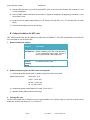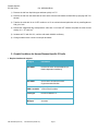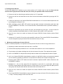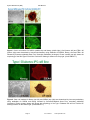
1
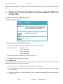
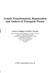
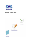
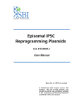
Disease Specific iPS Cells Cat #SC60XA Series User Manual Store in Gas Phase of Liquid Nitrogen ver. 1-031011 A limited-use label license covers this product. By use of this product, you accept the terms and conditions outlined in the Licensing and Warranty Statement contained in this user manual. Disease Specific iPS Cell Lines Cat. # SC60xA/B-xxx Contents I. Human Disease-Specific iPS Cells .........................................2 A. Description ..........................................................................2 B. Human Wild-type iPSC cell line (Cat# SC600A/B-WT) ......2 C. Type I Diabetes iPS cell line (Cat# SC602A/B-DTI) ...........3 D. Metachromatic Leukodystrophy (MLD) iPS cell line (Cat# SC601A/B-MLD) E. Amyotrophic Lateral Sclerosis iPS cell line (Cat# SC603A/B-ALS) F. Muscular Dystrophy (MD) iPS cell line (Cat# SC604A/B-MD) 3 G. Glioblastoma Multiforme (GBM) iPS cell line (Cat# SC605A/B-GBM) H. II. 3 3 Parkinson's Disease (PD) iPS cell lines (SC607A/B-PD & SC608A/B-PD) 3 Culture and Growth Conditions for Disease-Specific iPSC and Feeder Cells 4 A. Culture Conditions for MEF feeder cells .............................4 B. Culture Conditions for HFF cells .........................................6 C. Growth Conditions for Human Disease-Specific iPS cells ..7 III. Validation Data ....................................................................9 IV. References ........................................................................16 V. Technical Support.............................................................16 VI. Licensing and Warranty Statement ...................................16 888-266-5066 (Toll Free) 650-968-2200 (outside US) Page 1 3 System Biosciences (SBI) User Manual List of Components Each disease specific iPS cell line set comes as one vial with 2 x 10⁵ cells. The product is shipped on dry ice and should be immediately stored in the gas phase of liquid nitrogen. In general, iPS cells are challenging to culture and should only be operated by researchers experienced in the intricacies of human embryonic stem (hES) cell culture. The methods for culture are nearly identical to hES cell culture, although more care and increased maintenance will be required. Disease-specific iPS cells must be grown on feeder cells for culture. I. Human Disease-Specific iPS Cells A. Description SBI and DV Biologics have partnered to develop novel human iPS cell lines from patient-derived sources. Utilizing iPS cell lines from these disorders represents an opportunity to recapitulate both normal and pathological tissue formation in vitro for the ideal drug development screening cell line, to facilitate new therapeutic discovery and disease modeling. Human disease-specific induced pluripotent stem cells (iPSCs, cat.# SC60xA/B-xxx) were generated by transducing source cells with retroviruses individually encoding the four human transcription factors (Oct4, Sox2, Klf4, and c-Myc) that have been shown to induce the reprogramming of somatic cells to a pluripotent state. The cells were derived using morphological selection criteria and without the use of fluorescent markers or drug selection. When cultured under standard human ES cell culture conditions, the morphology of SBI human disease-specific iPSCs is identical to that of human ES cells. The cells also express the pluripotency markers Nanog, SSEA3, TRA-1-60, etc., and demonstrate a strong endogenous AP activity. Human iPS cells must be grown on a feeder cell layer. Appropriate feeder cells for human iPS cells are mouse embryonic fibroblasts or human foreskin fibroblasts. B. Human Wild-type iPSC cell line (Cat# SC600A/B-WT) The SC600A/B-WT human iPS cell line is derived from cultured skin explants from dermal fibroblasts from a single donor. The donor is a 64 year old Caucasian male with no known disease. The human wild-type iPS cell Page 2 ver. 2-093013 www.systembio.com Disease Specific iPS Cell Lines Cat. # SC60xA/B-xxx line was generated and cultured in the same conditions as the diseased iPS cell lines. It is an ideal control cell line for the disease and patient-specific iPS cell lines. C. Type I Diabetes iPS cell line (Cat# SC602A/B-DTI) Type I Diabetes is an autoimmune condition where the pancreatic beta cells are destroyed. Patients display diabetes-related autoantibodies. The cause of type I diabetes is polygenic, and chronic insulin therapy is required for survival. The type I diabetes human iPS cells were derived from human patient fibroblasts from cultured skin explants from a single donor. The patient was a 29-year old female of Hispanic-Caucasian descent, who was diagnosed with Type I Diabetes at age 12. D. Metachromatic Leukodystrophy (MLD) iPS cell line (Cat# SC601A/B-MLD) Metachromatic leukodystrophy (MLD) is a group of disorders marked by storage buildup in the white matter of the central nervous system, in the peripheral nerves, and to some extent in the kidneys. Similar to Krabbé disease, MLD affects the myelin that covers and protects the nerves. This autosomal recessive disorder is caused by a deficiency of the enzyme arylsulfatase A. Both males and females are affected by this disorder and death generally occurs within 6 to 14 years after onset of symptoms. MLD has a juvenile onset that is marked by proximal and distal weakness, hypotonia, decreased or absent tendon reflexes, and mild distal sensory loss. The MLD iPS cell line were drived from human patient fibroblasts cultured from skin explants from a single donor. They can be used as a model to study other polyneuropathies as well. E. Amyotrophic Lateral Sclerosis iPS cell line (Cat# SC603A/B-ALS) ALS is the most common form of motor neuron disease. Sporadic forms (of unknown cause) account for about 90-95 percent of ALS cases. The human patient dermal fibroblasts are derived from cultured skin explants from a single human donor. The patient was a 61 year old female of Latino-Caucasian descent, who was diagnosed with agressive and sporatic ALS. F. Muscular Dystrophy (MD) iPS cell line (Cat# SC604A/B-MD) Muscular dystrophies are a group of genetic conditions characterized by progressive muscle weakness and wasting (atrophy). The Duchenne and Becker types of muscular dystrophy primarily affect the skeletal muscles, which are used for movement, and the muscles of the heart. Mutations in the Dystrophin, X-linked, gene cause Duchenne and Becker muscular dystrophy. The human patient dermal fibroblasts are derived from cultured skin explants from a single donor. The patient was one year old male of Latino-Caucasian descent, who had a deletion of Dystrohpin gene at exons 3-6. G. Glioblastoma Multiforme (GBM) iPS cell line (Cat# SC605A/B-GBM) Glioblastoma multiforme (GBM) is the most common and most aggressive malignant primary brain tumor in humans. GBM is the most invasive type of glial tumor that grows rapidly and spreads to nearby tissues. The human patient dermal fibroblasts are derived from cultured skin explants from a single donor. The patient was 41 year old male of Latino-Caucasian descent, who was diagnosed by both MRI and CT-SCAN at age 40. H. Parkinson's Disease (PD) iPS cell lines (SC607A/B-PD & SC608A/B-PD) Parkinson's disease (PD) is the result from the progressive death of dopamine-generating cells in the substantia nigra in the midbrain. Early in the course of the disease, the most prevalent symptoms are movement-related, including tremors/shaking, rigidity, slowness of movement and difficulty with walking. As Parkinson's disease progresses, cognitive and behavioral problems may develop and dementia commonly occurs in the advanced stages of the disease. The human patient dermal fibroblasts are derived from cultured skin explants from a single donor. The patient with late onset Parkinson’s disease was 81 year old Hispanic-Caucasian male, who was diagnosed with 888-266-5066 (Toll Free) 650-968-2200 (outside US) Page 3 System Biosciences (SBI) User Manual Parkinson's disease at age 79. The patient with early onset Parkinson’s disease was 41 year old Hispanic-Caucasian male. II. Culture and Growth Conditions for Disease-Specific iPSC and Feeder Cells A. Culture conditions for MEF feeder cells 1. Required media and reagents Reagent Information MEF Medium DMEM containing 10% FBS, 2 mM glutamine, 1x10⁻⁴ M nonessential amino acids and 50 U and 50 µg/ml penicillin and streptomycin. 2x Cold Freezing Media 20% DMSO and 80% FBS Mitomycin C solution 1 mg/ ml 2. Gelatin treatment of plates for MEF feeder cells 1) Add enough sterile/ autoclaved 0.1% gelatin to cover the bottom of the wells. Approximate amounts: 10cm plate – 5 ml 6 well – 1.5 ml/ well 24 well – 0.5 ml/ well 96 well – 200 µl/ well 2) Incubate the gelatin-coated dishes for at least 15 min at 37°C. 3) Aspirate excess gelatin solution before use. 3. Thawing MEF cells To insure the highest level of viability, be sure to warm medium to 37°C before using it on the cells. Cells should be plated at a minimum cell density of 1 x 10⁴ cells/ cm2. 1) Remove the vial from liquid nitrogen and thaw quickly in a 37°C water bath. 2) Remove the vial from the water bath as soon as the cells are half-thawed, and sterilize the tube by spraying with 70% ethanol. 3) Transfer the cells with 10 ml of MEF medium to a 15 cm conical tube and pellet the cells by centrifugation at 200 g for 5 min. Page 4 ver. 2-093013 www.systembio.com Disease Specific iPS Cell Lines Cat. # SC60xA/B-xxx 4) Discard the supernatant and resuspend the cells with 10 ml fresh MEF medium and plate the cells at seed density of 104 cells/ cm2. 5) Incubate at 37°C with 5% CO2, until the cells reach 80-90% confluency. 6) Change medium twice a week or when pH decreases. 4. Passaging MEF cells Cells should be split when they reach confluency. We recommend splitting the cells based on 0.5x10⁴ cells/ cm2. 1) Discard the medium and wash the cells twice with PBS. 2) Aspirate PBS, and add 1 ml per T75 flask of 0.25% trypsin-EDTA, and incubate for 1 min. 3) Add 5 ml of MEF medium, and break up the cell clumps by gently pipetting up and down several times. 4) Transfer cells into a conical tube and centrifuge at 200 g for 5 min. 5) Discard the supernatant and resuspend the cell pellet in 10 ml MEF medium. 6) Count the number of cells, plate cells at 0.5x10⁴ cells/ cm2 and incubate at 37°C with 5% CO2. 5. Freezing MEF cells 1) Follow steps 1-4 from the Passaging MEF cells protocol (above). 2) Discard the supernatant, and resuspend the pellet in MEF medium. Add approximately 1 ml for each T75 flask. 3) Count the number of cells and dilute the cell suspension to 1 x 10⁷ cells/ ml. 4) Add an equal volume of cold 2X Freezing Media to the cell suspension. 5) Aliquot 1 ml of suspension into each cryovial (5 x 10⁶ cells/ vial). 6) Place the vials in a cell-freezing container and keep it at -80°C overnight. 7) Transfer the vials to a liquid nitrogen take for long-term storage. 6. Mitomycin C treatment of MEF Mitomycin C acts to halt the division of MEF cells so that they can be used to condition the medium for human iPS cells. MEF cells should be at confluence when treated with mitomycin C. 1) Add 6 ml of fresh MEF medium contain 50 µl of mitomycin C solution (1 mg/ ml) to one T75 flask of confluent MEF cells, and swirl it briefly. The final concentration of mitomycin C is 8 µg/ ml. 2) Incubate at 37°C for at least 3 hrs. 3) Aspirate the mitomycin C-containing medium off the cells and wash the cells twice with 10 ml PBS. 888-266-5066 (Toll Free) 650-968-2200 (outside US) Page 5 System Biosciences (SBI) User Manual 4) Aspirate PBS and add 1 ml of 0.25% trypsin-EDTA, swirl to cover the entire surface, and incubate for 1 min at room temperature. 5) Add 5 ml MEF medium and break up the cells to a single-cell suspension by pipetting up and down. Count the number of cells. 6) Seed the cells on gelatin-coated dishes (2 x 10⁶ cells per 100-mm dish, or 3 x 10⁵ cells per well of a 6-well plate). 7) Cells should be ready to use by the next day. B. Culture Conditions for HFF cells HFF cells for feeder cells can be obtained from SBI (Cat # PC502B-HFF). The HFF cells provided in the iPSC kits are not suitable for use as feeder cells. 1. Required media and reagents Reagent Information HFF Medium DMEM containing 10% FBS, 2mM glutamine, 0.1 M nonessential amino acids, and 50 U and 50 µg/ml penicillin and streptomycin. 2x Cold Freezing Media 20% DMSO and 80% FBS 2. Gelatin treatment of plates for HFF feeder cells (optional) 1) Add enough sterile/ autoclaved 0.1% gelatin to cover the bottom of the wells. Approximate amounts: 10cm plate – 5 ml 6 well – 1.5 ml/ well 24 well – 0.5 ml/ well 96 ell – 200 µl/ well 2) Incubate the gelatin-coated dishes for at least 15 min at 37°C. 3) Aspirate excess gelatin solution before using 3. Thawing HFF cells To insure the highest level of viability, be sure to warm medium to 37°C before using it on the cells. Cells should be plated at a minimum cell density of 1 x 10⁴ cells/ cm2. Page 6 ver. 2-093013 www.systembio.com Disease Specific iPS Cell Lines Cat. # SC60xA/B-xxx 1) Remove the vial from liquid nitrogen and thaw quickly in 37°C. 2) Remove the vial from the water bath as soon as the cells are half-thawed, and sterilize by spraying with 70% ethanol. 3) Transfer the cells with 10 ml of HFF medium to a 15 cm conical tube and pellet the cells by centrifugation at 200 g for 5 min. 4) Discard the supernatant and resuspend the cells with 10 ml fresh HFF medium and plate the cells at seed density of 1 x 10⁴ cells/ cm2. 5) Incubate at 37°C with 5% CO2, until the cells reach 80-90% confluency. 6) Change medium twice a week or when pH decreases. C. Growth Conditions for Human Disease-Specific iPS cells 1. Required media and reagents 888-266-5066 (Toll Free) Reagent Information SC100M-1 Human ESC/iPSC Growth Medium (for feeder-dependent conditions) SC150M-1 CRYO-GOLD Human ESC/iPSC Cryopreservation Medium ZRD-Y-01/05/25 Y27632 ROCK Inhibitor Invitrogen Knockout DMEM/F12 Millipore Accutase 650-968-2200 (outside US) Page 7 System Biosciences (SBI) User Manual 2. Thawing human iPS cells To insure the highest level of viability, be sure to warm medium to 37°C before using it on the cells. Due to the low survival rate of cryopreserved human iPS cells, the recovery is expected to take at least one week. 1) Remove the vial from liquid nitrogen and thaw quickly in a 37°C water bath. 2) Remove the vial from the water bath as soon as the cells are half-thawed, and sterilize by spraying with 70% ethanol. 3) Transfer the cells with 10 ml of Human ESC/iPSC Growth Medium to a 15 ml conical tube and pellet the cells by centrifugation at 200g for 5 min. 4) While centrifuging, remove MEF/ HFF medium from the feeder cell plates, and wash the wells twice with Knockout DMEM/F12. Then add 1 ml of Human ESC/iPSC Growth Medium with 10 µM ROCK inhibitor (Y27632). 5) Discard the supernatant from the human iPS cells, and resuspend the cells with 1 ml fresh human ES medium containing 10 µM ROCK inhibitor (Y-27632). Plate the cells on MEF or HFF feeder cells. 6) After 24 hours, remove the media and replace with Human ESC/iPSC Growth Medium (without ROCK inhibitor). 7) Incubate at 37°C with 5% CO2 until the cells reach 80% confluency. The Human ESC/iPSC Growth Medium must be changed every day and human iPS cells subcultured every 5-7 days. 3. Maintenance and Passage of human iPS cells In order to maintain pluripotency, it is important not to keep human iPS cells in culture for long periods of time. 1) Aspirate the medium and wash the cells twice with 1 ml of PBS. 2) Remove PBS completely and add 0.5 ml of 1x Accutase and incubate for 2 min at room temperature. 3) Tap the bottom of the plate to dislodge the cells from the bottom of the plate. Then aspirate the accutase. 4) Add 1 ml of DMEM/F12 to the plate and carefully wash the feeder cells, and aspirate the medium. Repeat. 5) Add 1 ml of Human ESC/iPSC Growth Medium containing 10 µM ROCK inhibitor to the plate and suspend the cell colonies by pipetting up and down. It is important not to break up the colonies into single cells. 6) Remove a plate of MEF or HFF feeder cells from the incubator. Aspirate the MEF medium. Wash once with KO DMEM/F12 medium. 7) Distribute 0.2 – 0.3 ml of the human iPS cell suspension to each well of a 6-well plate. Add Human ESC/iPSC Growth Medium with ROCK inhibitor to a final volume of 2 ml per well. Right after plating the iPS cells, gently swirl the plate back-and-forth and side-to-side and incubate at 37°C. 8) After 24 hours, remove the media and replace with Human ESC/iPSC Growth Medium (without ROCK inhibitor). 9) The Human ESC/iPSC Growth Medium must be changed every day and human iPS cells subcultured every 5-7 days. Track the passage number of the iPS cells. Page 8 ver. 2-093013 www.systembio.com Disease Specific iPS Cell Lines Cat. # SC60xA/B-xxx 4. Freezing human iPS Cells 1) Grow cells to the exponential phase in a 6-well plate. 2) Aspirate the medium and wash the cells twice with 2 ml PBS. 3) Add 0.5 ml of 1x accutase and incubate 2 min at 37°C. Aspirate the accutase 4) Add 1 ml of Knockout DMEM/F12 to the plate and carefully wash the feeder cells. 5) Add 1 ml of Human ESC/iPSC Growth Medium to the plate. Scrape the colonies off using a cell scraper. 6) Transfer the cell suspension to a 15 ml conical tube and spin the cells at 200 g for 5 min. 7) Discard the supernatant and resuspend the cells with 1 ml CRYO-GOLD Human ESC/iPSC Cryopreservation Medium. 8) Aliquot it at 1 ml per vial. 9) Put the vials in a cell-freezing container, and store the vials at -80°C overnight. 10) Transfer the vials to liquid nitrogen for long-term storage. III. Validation Data 888-266-5066 (Toll Free) 650-968-2200 (outside US) Page 9 System Biosciences (SBI) User Manual Figure 1. Stem cell markers for SSEA3 (middle left) and Nanog (middle right), Oct4 (bottom left) and TRA-1-60 (bottom right) were determined by immunocytochemistry using antibodies for SSEA3, Nanog, Oct4 and TRA-1-60 followed by fluorescent-labeled Alexa Fluor secondary antibodies (Invitrogen). Phase contrast (top left) shows the morphology of the Wild Type iPS cell line. Positive AP staining is shown on the top right. (Cat # SAB-KIT-1) Figure 2. Stem cell markers for Nanog (top left) and SSEA4 (top right) were determined by immunocytochemistry using antibodies for SSEA4 and Nanog followed by fluorescent-labeled Alexa Fluor secondary antibodies (Invitrogen). Phase contrast (bottom left) shows the morphology of the Type I Diabetes iPS cell line. Positive AP staining is shown on the bottom right. (Cat # SAB-KIT-1) Page 10 ver. 2-093013 www.systembio.com Disease Specific iPS Cell Lines Cat. # SC60xA/B-xxx Figure 3. Stem cell markers for Nanog (top left) and SSEA4 (top right) were determined by immunocytochemistry using antibodies for SSEA4 and Nanog followed by fluorescent-labeled Alexa Fluor secondary antibodies (Invitrogen). Phase contrast (bottom left) shows the morphology of the MLD iPS cell line. Positive AP staining is shown on the bottom right. (Cat # SAB-KIT-1) 888-266-5066 (Toll Free) 650-968-2200 (outside US) Page 11 System Biosciences (SBI) User Manual Figure 4. Stem cell markers for Nanog (top left), SSEA3 (top right) and TRA-1-60 (bottom right) were determined by immunocytochemistry using antibodies for Nanog, SSEA3 and TRA-1-60 followed by fluorescent-labeled Alexa Fluor secondary antibodies (Invitrogen). Phase contrast (middle left) shows the morphology of the ALS iPS cell line. Positive AP staining is shown on the middle right. (Cat # SAB-KIT-1) Page 12 ver. 2-093013 www.systembio.com Disease Specific iPS Cell Lines Cat. # SC60xA/B-xxx Figure 5. Stem cell markers for Nanog (top left), SSEA3 (top right), Oct4 (bottom left) and TRA-1-60 (bottom right) were determined by immunocytochemistry using antibodies for Nanog, SSEA3, Oct4 and TRA-1-60 followed by fluorescent-labeled Alexa Fluor secondary antibodies (Invitrogen). Phase contrast (middle left) shows the morphology of the MD iPS cell line. Positive AP staining is shown on the middle right. (Cat # SAB-KIT-1) 888-266-5066 (Toll Free) 650-968-2200 (outside US) Page 13 System Biosciences (SBI) User Manual Figure 6. Stem cell markers for Nanog (top left), SSEA3 (top right) and TRA-1-60 (bottom right) were determined by immunocytochemistry using antibodies for Nanog, SSEA3 and TRA-1-60 followed by fluorescent-labeled Alexa Fluor secondary antibodies (Invitrogen). Phase contrast (middle left) shows the morphology of the ALS iPS cell line. Positive AP staining is shown on the middle right. (Cat # SAB-KIT-1) Figure 7. Stem cell markers for Nanog (top left), SSEA3 (top right), TRA-1-60 (bottom left) were determined by immunocytochemistry using antibodies for Nanog, SSEA3 and TRA-1-60 followed by fluorescent-labeled Alexa Fluor secondary antibodies (Invitrogen). Positive AP staining is shown on the bottom right. (Cat # SAB-KIT-1) Page 14 ver. 2-093013 www.systembio.com Disease Specific iPS Cell Lines Cat. # SC60xA/B-xxx Figure 8. Stem cell markers for Nanog (top left), Oct4 (top right), TRA-1-60 (bottom left) were determined by immunocytochemistry using antibodies for Nanog, SSEA3 and TRA-1-60 followed by fluorescent-labeled Alexa Fluor secondary antibodies (Invitrogen). Positive AP staining is shown on the bottom right. (Cat # SAB-KIT-1) 888-266-5066 (Toll Free) 650-968-2200 (outside US) Page 15 System Biosciences (SBI) IV. User Manual References Rossant, J. 2007. Stem cells: The magic brew. Nature 448, 260–262. Takahashi, K. and Yamanaka, S. 2006. Induction of pluripotent stem cells from mouse embryonic and adult fibroblast cultures by defined factors. Cell 126: 663–676. Takahashi K. et al. 2007. Induction of pluripotent stem cells from adult human fibroblasts by defined factors. Cell. 131: 861–72. Park, IH et al. 2008. Reprogramming of human somatic cells to pluripotency with defined factors. Nature. 451:141–6. Baker, Monya 2007. Adult cells reprogrammed to pluripotency, without tumors. Nature Reports Stem Cells. 2007– 12–11. Nakagawa, M et al. 2008. Generation of induced pluripotent stem cells without Myc from mouse and human fibroblasts. Nature Biotechnology. 26: 101–106. Okita, K, et al. 2007. Generation of germline-competent induced pluripotent stem cells. Nature. 448:313–7 Yu, J. et al. 2007. Induced Pluripotent Stem Cell Lines Derived from Human Somatic Cells. Science 318: 1917–1920. V. Technical Support For more information about SBI products or to download manuals in PDF format, please visit our website: http://www.systembio.com For additional information or technical assistance, please call or email us at: [email protected], 650-968-2200. VI. Licensing and Warranty Statement Limited Use License Use of the Disease Specific iPS cells (i.e., the “Product”) is subject to the following terms and conditions. If the terms and conditions are not acceptable, return all components of the Product to System Biosciences (SBI) within 7 calendar days. Purchase and use of any part of the Product constitutes acceptance of the above terms. The purchaser of the Product is granted a limited license to use the Product under the following terms and conditions: The Product shall be used by the purchaser for internal research purposes only. The Product is expressly not designed, intended, or warranted for use in humans or for therapeutic or diagnostic use. The Product may not be resold, modified for resale, or used to manufacture commercial products without prior written consent of SBI. This Product should be used in accordance with the NIH guidelines developed for stem cell research. SBI has pending patent applications related to the Product. For information concerning licenses for commercial use, contact SBI. Purchase of the product does not grant any rights or license for use other than those explicitly listed in this Licensing and Warranty Statement. Use of the Product for any use other than described expressly herein may be covered by Page 16 ver. 2-093013 www.systembio.com Disease Specific iPS Cell Lines Cat. # SC60xA/B-xxx patents or subject to rights other than those mentioned. SBI disclaims any and all responsibility for injury or damage which may be caused by the failure of the buyer or any other person to use the Product in accordance with the terms and conditions outlined herein. Limited Warranty SBI warrants that the Product meets the specifications described in this manual. If it is proven to the satisfaction of SBI that the Product fails to meet these specifications, SBI will replace the Product or provide the purchaser with a refund. This limited warranty shall not extend to anyone other than the original purchaser of the Product. Notice of nonconforming products must be made to SBI within 30 days of receipt of the Product. SBI’s liability is expressly limited to replacement of Product or a refund limited to the actual purchase price. SBI’s liability does not extend to any damages arising from use or improper use of the Product, or losses associated with the use of additional materials or reagents. This limited warranty is the sole and exclusive warranty. SBI does not provide any other warranties of any kind, expressed or implied, including the merchantability or fitness of the Product for a particular purpose. SBI is committed to providing our customers with high-quality products. concerns about any SBI products, please contact us at (888) 266-5066. © 2013 System Biosciences (SBI). 888-266-5066 (Toll Free) 650-968-2200 (outside US) Page 17 If you should have any questions or