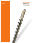
1
Technique Guide
COBLATION™
PROCISE™ MLW
Laryngeal Wand
COBLATION™ plasma technology for
laryngeal lesions and tracheal procedures
The PROCISE™ MLW laryngeal Wand provides “pinpoint” ablation
and coagulation capabilities
Surgical technique
COBLATOR™ II surgery system setup
1 Set up the COBLATOR II system and the PROCISE MLW laryngeal
Wand according to the Controller User’s Manual and Wand Instructions
for Use (IFU).
Note: This guide is not intended to replace the COBLATOR II User’s Manual or Wand IFU.
Thoroughly review the User’s Manual and Wand IFU before installing or operating this system.
2Default settings: Coblate 7, Coag 3. Adjust as needed and per surgeon preference.
3Connect the Wand’s suction tubing to an OR suction source separate from any other suction
instrumentation. Suction should be set to approximately 250mmHg.
4Connect the Wand’s saline tubing to bag of normal saline and adjust saline flow to a minimum
intermittent drip.
Patient/other preparation
1 To ensure optimal visualization throughout the procedure, the Wand can be used with standard
laryngoscopes and microscopes. Use the largest laryngoscope that can be accommodated.
Those with proximally and distally adjustable blades are particularly useful.
2Standard cuffed microlaryngeal tubes are adequate for protection of the lower airways from
any excess saline. This can be aided by gentle packing above the balloon with wet cottonoids.
Chance contact of the COBLATION Wand with the tube will not cause damage to the airway tube.
Note: Recent studies about airway fires suggest that using COBLATION technology in place of
traditional electrosurgical or laser devices during oropharyngeal surgery significantly reduces
the risk of igniting an airway fire due to the low heat generated and the lack of spark or ignition
medium under normal operating circumstances.1,2,3 Special endotracheal tubes used with lasers
are not necessary.
Venturi ventilation has also been used successfully in conjuntion with COBLATION technology.
A “head down” (Trendelenberg) position should be utilized to ensure any excess saline flows into
the pharynx and not in the trachea.
The ultra-slim,
extended Wand
shaft of the
PROCISE MLW
Wand provides
access to the trachea,
making it well-suited
for operating on small
anatomy.
Procedure
1 To ablate tissue, position Wand tip in close proximity to target tissue.
Caution: Care should be taken in monitoring the targeted tissue during ablation
to ensure consistent and controlled tissue removal is achieved. Care should
also be taken to ensure surrounding tissue is properly monitored. Due to the
smaller anatomies of certain patients, carefully monitor the surrounding tissues
to ensure tissue ablation is localized to the targeted tissue.
Before COBLATION™ technology treatment
After COBLATION technology treatment
2Press the ablation pedal (yellow) of the Foot Control to activate the Wand.
Make brief contact (1-2 seconds) with target tissue through the use of a
dabbing motion. Continue ablation briefly after removing the tip from target
tissue to allow “digestion” of any tissue on the electrode surface. Deactivate
the Wand by taking your foot off the foot pedal.
Caution: Keep the active electrode directly facing target tissue. User should
always pay close attention to the position of the PROCISE™ MLW Wand, and
to its proximity to surrounding untargeted tissue. Ensure non-targeted tissue
does not contact the exposed metal of active or return electrodes. Do not
bend the Wand shaft or rub the Wand tip against target tissue during ablation
as this could result in clogging of the suction line. Do not use the Wand
suction line as a standard surgical suction device to clear debris from the
surgical field as this could result in clogging of the suction line.
3To coagulate tissue, position Wand tip directly over the source of bleeding and
depress the Coagulation pedal (blue).
Histology (animal model)5
Thermal effect4
Default COBLATE setting,
Day 3
600
Depth (µm)
500
Default COBLATE setting,
Day 21
400
300
200
100
0
Default coblate
Maximum coblate
Default coag
Device settings
At Day 21, all vocal-fold lesions were 100% epithelialized
in canine model. Gross appearance of vocal-fold lesions
was fully healed with no exudates present.
References
1 Smith LP, Roy S. Operating room fires in otolaryngology: risk factors and prevention. Am J Otolaryngol.
Article in press (Epub 2010 Apr 14).
2 Roy S, Smith LP. Device-related risk of fire in oropharyngeal surgery: a mechanical model. Am J
Otolaryngol. 2010 Sept;31(5):356-359. This article references preclinical non-human data. As such, results
may not necessarily be the same in human procedures.
3 Matt BH, Cottee LA. Reducing risk of fire in the operating room using COBLATION™ technology. Otolaryngol
Head Neck Surg. 2010 Sept;143(3):454-5.
4 Data based on tongue tissue in an animal model; results may not be the same in humans. Data on file –
report # 35764-01.
5 Data based on vocal fold study in an animal model; results may not be the same in humans. Data on file –
report # 35766-01.
ArthroCare Corporation
7000 West William Cannon Drive
Austin, TX 78735
USA
www.smith-nephew.com
Order Entry: 1-800-797-6520
Order Entry Fax: 1-888-994-2782
© 2014 Smith & Nephew, Inc.
™Trademark of Smith & Nephew. Reg. US Pat. & TM Office.
P/N 45403 Rev. C 11/14