1
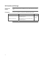
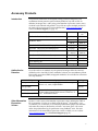
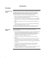
pcDNA™6/myc-His A, B, and C Catalog no. V221-20 Rev. date: 9 November 2010 Manual part no. 25-0233 MAN0000076 Corporate Headquarters Invitrogen Corporation 1600 Faraday Avenue Carlsbad, CA 92008 T: 1 760 603 7200 F: 1 760 602 6500 E: [email protected] For country-specific contact information visit our web site at www.invitrogen.com User Manual ii Table of Contents Kit Contents and Storage..................................................................................................................................... iv Accessory Products ............................................................................................................................................... v Introduction ................................................................................................................... 1 Overview .................................................................................................................................................................1 Methods ......................................................................................................................... 2 Cloning into pcDNA™6/myc-His A, B, and C.....................................................................................................2 Transfection and Analysis.....................................................................................................................................7 Creating Stable Cell Lines ...................................................................................................................................10 Appendix...................................................................................................................... 14 pcDNA™6/myc-His Vector..................................................................................................................................14 pcDNA™6/myc-His/lacZ.....................................................................................................................................16 Technical Support.................................................................................................................................................17 Purchaser Notification .........................................................................................................................................18 References..............................................................................................................................................................19 iii Kit Contents and Storage Shipping and Storage pcDNA™6/myc-His vectors are shipped on wet ice. Upon receipt, store vectors at –20°C. Kit Contents All vectors are supplied as detailed below. Store the vectors at –20°C. Vector ™ Composition pcDNA 6/myc-His A, B, and C 40 μl of 0.5 μg/μl vector in 10 mM Tris-HCl, 1 mM EDTA, pH 8.0 pcDNA™6/myc-His/lacZ 40 μl of 0.5 μg/μl vector in 10 mM Tris-HCl, 1 mM EDTA, pH 8.0 Amount 20 μg 20 μg iv Accessory Products The products listed below are designed to help you detect and purify your recombinant fusion proteins expressed from pcDNA™6/myc-His vectors. In addition, Invitrogen has a wide variety of mammalian expression vectors, many of which can be utilized with pcDNA™6/myc-His to express multiple proteins in the same cell (see next page). For more information, www.invitrogen.com or contact Technical Support (see page 17). Introduction Amount Catalog no. 6 purifications K850-01 50 ml R801-01 150 ml R801-15 100 preps K2100-03 25 preps K2100-04 Electrocomp TOP10F’ 2 × 20 rxns 6 × 20 rxns C665-11 C665-24 One Shot™ TOP10F’ (chemically competent cells) 20 × 50 μl C3030-03 -Gal Assay Kit 1 kit K1455-01 -Gal Staining Kit 1 kit K1465-01 50 mg R210-01 Product ProBond™ Purification System ProBond™ Resin ™ PureLink HiPure Plasmid Miniprep Kit ™ PureLink HiPure Plasmid Midiprep Kit ™ Blasticidin If you do not have an antibody to your protein, Invitrogen offers the Anti-myc antibodies or the Anti-His(C-term) antibodies to detect your recombinant protein. Horseradish peroxidase (HRP)-conjugated antibodies are available for convenient one-step detection. Antibodies for Detection Antibody Anti-myc Anti-myc-HRP Anti-His(C-term) Anti-His(C-term)-HRP Other Mammalian Expression Vectors Epitope Catalog no. Detects 10 amino acid epitope derived from c-myc (Evans et al., 1985): EQKLISEEDL R950-25 Detects the C-terminal polyhistidine tag (requires the free carboxyl group for detection) (Lindner et al., 1997): HHHHHH-COOH R930-25 R951-25 R931-25 We have a wide variety of mammalian expression vectors utilizing the CMV or EF-1 promoters. Vectors are available with the Xpress™ (N-terminal), c-myc (C-terminal), V5 (C-terminal), or C-terminal polyhistidine epitopes for detection and either the neomycin, blasticidin, or Zeocin™ resistance genes. All vectors utilize the polyhistidine tag for purification using ProBond™ resin. For more information on the mammalian expression vectors available, see our website (www.invitrogen.com) or call Technical Support (page 17). v Introduction Overview Description of the System pcDNA™6/myc-His A, B, and C are 5.1-kb vectors designed for overproduction of recombinant proteins in mammalian cell lines. Features of the vectors allow purification and detection of expressed proteins (see pages 14–15 for more information). High-level stable and transient expression can be carried out in most mammalian cells. The vectors contain the following elements: Human cytomegalovirus immediate-early (CMV) promoter for high-level expression in a wide range of mammalian cells. Three reading frames to facilitate in-frame cloning with a C-terminal peptide encoding the myc (c-myc) epitope and a polyhistidine (6xHis) metal-binding tag. Blasticidin resistance gene (bsd) for selection of stable cell lines (Kimura et al., 1994). Episomal replication in cell lines that are latently infected with SV40 or that express the SV40 large T antigen (e.g., COS7). The control plasmid, pcDNA™6/myc-His/lacZ, is included for use as a positive control for transfection, expression, and detection in the cell line of choice. Experimental Outline 1 Use the following outline to clone and express your gene of interest in pcDNA™6/myc-His. Consult the multiple cloning sites described on pages 3–5 to determine which vector (A, B, or C) should be used to clone your gene in-frame with the C-terminal myc epitope and the polyhistidine tag. Ligate your insert into the appropriate vector and transform into E. coli. Select transformants on 50–100 μg/ml ampicillin or 50 μg/ml blasticidin. Analyze your transformants for the presence of the insert by restriction digestion. Select a transformant with the correct restriction pattern and use sequencing to confirm that your gene is cloned in-frame with the C-terminal peptide. Transfect your construct into the cell line of choice using your own method of transfection. Test for expression of your recombinant gene by western blot analysis or functional assay. For antibodies to the myc epitope or the C-terminal polyhistidine tag, see the next page. To purify your recombinant protein, you may use metal-chelating resin such as ProBond™. ProBond™ resin is available separately (see page v). Methods Cloning into pcDNA™6/myc-His A, B, and C General Molecular Biology Techniques For help with DNA ligations, E. coli transformations, restriction enzyme analysis, purification of single-stranded DNA, DNA sequencing, and DNA biochemistry, refer to published references (Ausubel et al., 1994; Sambrook et al., 1989). E. coli Strain Many E. coli strains are suitable for the propagation of pcDNA™6/myc-His vectors, including TOP10F´, DH5-F’™, JM109 and INVF’. We recommend that you propagate vectors containing inserts in E. coli strains that are recombinant deficient (recA) and endonuclease A-deficient (endA). For your convenience, TOP10F’ is available as chemically competent or electrocompetent cells from Invitrogen (see page v). Transformation Method You may use any method of your choice for transformation. Chemical transformation is the most convenient for most researchers. Electroporation is the most efficient and the method of choice for large plasmids. Maintenance of pcDNA™6/myc-His To propagate and maintain the pcDNA™6/myc-His vectors, use the supplied 0.5 μg/μl stock solution in TE, pH 8.0 to transform a recA, endA E. coli strain like TOP10F’, DH5™, JM109 or equivalent. Select transformants on LB plates containing 50–100 μg/ml ampicillin or 50 μg/ml blasticidin. Be sure to prepare a glycerol stock of each plasmid for longterm storage (see page 6 for recipe). Cloning Considerations Your insert should contain a Kozak consensus sequence with an ATG initiation codon for proper initiation of translation (Kozak, 1987; Kozak 1990). An example of a Kozak consensus sequence is provided below. Other sequences are possible, but the G or A at position –3 and the G at position +4 (shown in bold) illustrates the most commonly occurring sequence with strong consensus. Replacing one of the two bases at these positions provides moderate consensus, while having neither results in weak consensus. The ATG initiation codon is shown underlined. (G/A)NNATGG If you wish to express your protein WITHOUT the C-terminal peptide, be sure to include a stop codon. Continued on next page 2 Cloning into pcDNA™6/myc-His A, B, and C, Continued Multiple Cloning Site of Version A Below is the multiple cloning site for pcDNA™6/myc -His A. Restriction sites are labeled to indicate the cleavage site. The boxed nucleotides indicate the variable region. Note that there is a stop codon between the BamH I site and the BstX I site. The multiple cloning site has been confirmed by sequencing and functional testing. The sequence of pcDNA™6/myc -His A is available for downloading from our website at www.invitrogen.com or from Technical Support (page 17). T7 promoter/priming site Hind III Acc65 I Kpn I BamH I 861 ATTAATACGA CTCACTATAG GGAGACCCAA GCTGGCTAGT TAA GCT TGG TAC CGA GCT CGG Ala Trp Tyr Arg Ala Arg BstX I* EcoR I Pst I EcoR V BstX I* Not I 922 ATC CAC TAG TCC AGT GTG GTG GAA TTC TGC AGA TAT CCA GCA CAG TGG CGG CCG Ile His *** Ser Ser Val Val Glu Phe Cys Arg Tyr Pro Ala Gln Trp Arg Pro Xho I Xba I Apa I BstB I myc epitope 976 CTC GAG TCT AGA GGG CCC TTC GAA CAA AAA CTC ATC TCA GAA GAG GAT CTG AAT Leu Glu Ser Arg Gly Pro Phe Glu Gln Lys Leu Ile Ser Glu Glu Asp Leu Asn Polyhistidine tag Age I Pme I 1030 ATG CAT ACC GGT CAT CAT CAC CAT CAC CAT TGA GTTTAAACCC GCTGATCAGC Met His Thr Glu His His His His His His *** BGH Reverse priming site 1083 CTCGACTGTG CCTTCTAG *Note that there are two BstXI sites in the polylinker. Continued on next page 3 Cloning into pcDNA™6/myc-His A, B, and C, Continued Multiple Cloning Site of Version B Below is the multiple cloning site for pcDNA™6/myc -His B. Restriction sites are labeled to indicate the cleavage site. The boxed nucleotides indicate the variable region. The multiple cloning site has been confirmed by sequencing and functional testing. The sequence of pcDNA™6/myc -His B is available for downloading from our website at www.invitrogen.com or from Technical Support (page 17). T7 promoter/priming site 861 Hind III Kpn I BamH I ATTAATACGA CTCACTATAG GGAGACCCAA GCTGGCTAGT TAAG CTT GGT ACC GAG CTC GGA Leu Gly Thr Glu Leu Gly Pst I EcoR V BstX I* EcoR I 923 Acc65 I BstX I* Not I TCC ACT AGT CCA GTG TGG TGG AAT TCT GCA GAT ATC CAG CAC AGT GGC GGC CGC Ser Thr Ser Pro Val Trp Trp Asn Ser Ala Asp Ile Gln His Ser Gly Gly Arg Xho I Xba I Apa I Sac II BstB I myc epitope 977 TCG AGT CTA GAG GGC CCG CGG TTC GAA CAA AAA CTC ATC TCA GAA GAG GAT Ser Ser Leu Glu Gly Pro Arg Phe Glu Gln Lys Leu Ile Ser Glu Glu Asp 1028 CTG AAT ATG CAT ACC GGT CAT CAT CAC CAT CAC CAT TGA GTTT AAACCCGCTG Leu Asn Met His Thr Gly His His His His His His *** Age I Polyhistidine tag Pme I BGH Reverse priming site 1081 ATCAGCCTCG ACTGTGCCTT CTAGTTGCCA *Note that there are two BstXI sites in the polylinker. Continued on next page 4 Cloning into pcDNA™6/myc-His A, B, and C, Continued Multiple Cloning Site of Version C Below is the multiple cloning site for pcDNA™6/myc -His C. Restriction sites are labeled to indicate the cleavage site. The boxed nucleotides indicate the variable region. The multiple cloning site has been confirmed by sequencing and functional testing. The sequence of pcDNA™6/myc -His C is available for downloading from our website at www.invitrogen.com or from Technical Support (page 17). T7 promoter/priming site 861 BstX I* Xho I BstE II EcoR I Pst I EcoR V BstX I* BstB I myc epitope GCG GCC GCT CGA GGT CAC CCA TTC GAA CAA AAA CTC ATC TCA GAA GAG GAT Ala Ala Ala Arg Gly His Pro Phe Glu Gln Lys Leu Ile Ser Glu Glu Asp Age I 1020 Kpn I TCG GAT CCA CTA GTC CAG TGT GGT GGA ATT CTG CAG ATA TCC AGC ACA GTG Ser Asp Pro Leu Val Gln Cys Gly Gly Ile Leu Gln Ile Ser Ser Thr Val Not I 969 Acc65 I ATTAATACGA CTCACTATAG GGAGACCCAA GCTGGCTAGT TA AGC TTG GTA CCG AGC Ser Leu Val Pro Ser BamH I 918 Hind III Polyhistidine tag Pme I CTG AAT ATG CAT ACC GGT CAT CAT CAC CAT CAC CAT TGA GTTTAAACCC Leu Asn Met His Thr Gly His His His His His His *** BGH Reverse priming site 1069 GCTGATCAGC CTCGACTGTG CCTTCTAGTT GC *Note that there are two BstXI sites in the polylinker. Continued on next page 5 Cloning into pcDNA™6/myc-His A, B, and C, Continued MEND ION AT RECOM E. coli Transformation Preparing a Glycerol Stock Transform your ligation mixtures into a competent recA, endA E. coli strain (e.g. TOP10F´, DH5™) and select on LB plates containing 50–100 μg/ml ampicillin or 50 μg/ml blasticidin. Select 10–20 clones and analyze for the presence and orientation of your insert. We recommend that you sequence your construct with the T7 Forward and BGH Reverse primers to confirm that your gene is fused in frame with the myc epitope and the C-terminal polyhistidine tag. Once you have identified the correct clone, be sure to purify the colony and make a glycerol stock for long-term storage. It is also a good idea to keep a DNA stock of your plasmid at –20°C. • Streak the original colony out on an LB plate containing 50 μg/ml ampicillin or 50 μg/ml blasticidin. Incubate the plate at 37°C overnight. • Isolate a single colony and inoculate into 1–2 ml of LB containing 50 μg/ml ampicillin. • Grow the culture to mid-log phase (OD600 = 0.5–0.7). • Mix 0.85 ml of culture with 0.15 ml of sterile glycerol and transfer to a cryovial. Store at –80°C. 6 Transfection and Analysis Introduction Once you have confirmed that your construct is in the correct orientation and fused in frame with the C-terminal peptide, you are ready to transfect your cell line of choice. We recommend that you include the positive control vector and a mock transfection to evaluate your results. Plasmid Preparation Plasmid DNA for transfection into eukaryotic cells must be very clean and free from phenol and sodium chloride. Contaminants will kill the cells and salt will interfere with lipid complexing, decreasing transfection efficiency. We recommend isolating plasmid DNA using the PureLink™ HiPure Miniprep Kit or the PureLink™ HiPure Midiprep Kit (see page v). Methods of Transfection For established cell lines (e.g., HeLa), consult original references or the supplier of your cell line for the optimal method of transfection. It is recommended that you follow exactly the protocol for your cell line. Pay particular attention to medium requirements, when to pass the cells, and at what dilution to split the cells. Further information is provided in Current Protocols in Molecular Biology. Methods of transfection include calcium phosphate (Chen & Okayama, 1987; Wigler et al., 1977), lipid-mediated (Felgner et al., 1989; Felgner & Ringold, 1989) and electroporation (Chu et al., 1987; Shigekawa & Dower, 1988). For high efficiency transfection in a broad range of mammalian cells, we recommend using Lipofectamine™ 2000 Reagent available from Invitrogen. For more information on Lipofectamine™ 2000 and other transfection reagents available, visit our website at www.invitrogen.com or contact Technical Support (page 17). Positive Control pcDNA™6/myc-His/lacZ is provided as a positive control vector for mammalian cell transfection and expression (see page 16) and may be used to optimize transfection conditions for your cell line. The gene encoding -galactosidase is expressed in mammalian cells under the control of the CMV promoter. A successful transfection will result in -galactosidase expression that can be easily assayed (see below). Assay for galactosidase Activity You may assay for -galactosidase expression by activity assay using cell-free lysates (Miller, 1972) or by staining the cells for activity. Invitrogen offers the -Gal Assay Kit and the -Gal Staining Kit for fast and easy detection of -galactosidase expression (see page v). Continued on next page 7 Transfection and Analysis, Continued Detection of Fusion Proteins Several antibodies are available from Invitrogen to detect expression of your fusion protein from pcDNA™6/myc-His (see page v). To detect fusion protein by Western blot, you will need to prepare a cell lysate from transfected cells. We recommend that you perform a time course to optimize expression of the fusion protein (e.g., 24, 48, 72 hours, etc. after transfection). To lyse cells: Cell Lysis Buffer 1. Wash cell monolayers (~106 cells) once with phosphate-buffered saline (PBS). 2. Scrape cells into 1 ml PBS and pellet the cells at 1,500 × g for 5 minutes. 3. Resuspend in 50 μl Cell Lysis Buffer (see recipe below). Other lysis buffers may be suitable. 4. Incubate cell suspension at 37°C for 10 minutes to lyse the cells. 5. Centrifuge the cell lysate at 10,000 × g for 10 minutes to pellet nuclei and transfer the supernatant to a fresh tube. Assay the lysate for protein concentration. Note: Do not use protein assays utilizing Coomassie® Blue or other dyes. NP-40 interferes with the binding of the dye with the protein. 6. Add SDS-PAGE sample buffer to a final concentration of 1X and boil the sample for 5 minutes. 7. Load 20 μg of lysate onto an SDS-PAGE gel and electrophorese. Use the appropriate percentage of acrylamide to resolve your fusion protein. 50 mM Tris, pH 7.8 150 mM NaCl 1% Nonidet P-40 1. This solution can be prepared from the following common stock solutions. For 100 ml, combine: 1 M Tris base 5 M NaCl Nonidet P-40 5 ml 3 ml 1 ml 2. Bring the volume up to 90 ml with deionized water and adjust the pH to 7.8 with HCl. 3. Bring the volume up to 100 ml. Store at room temperature. Note: Protease inhibitors may be added at the following concentrations: 1 mM PMSF 1 μg/ml pepstatin 1 μg/ml leupeptin Continued on next page 8 Transfection and Analysis, Continued The C-terminal peptide containing the myc epitope and the polyhistidine tag will add approximately 3 kDa to the size of your protein. Purification 9 You will need 5 × 10⁶ to 1 × 10⁷ transfected cells for purification of your protein on a 2-ml ProBond™ column (or other metal-chelating column). Refer to the manufacturer's instructions before attempting to purify your fusion protein. To prepare cells for lysis, refer to the protocol on page 13. Creating Stable Cell Lines Introduction The pcDNA™6/myc-His vectors contain the blasticidin resistance gene for selection of stable cell lines using blasticidin. We recommend that you test the sensitivity of your mammalian host cell to blasticidin, as natural resistance varies among cell lines. General information and guidelines are provided below for your convenience. Blasticidin Blasticidin S HCl is a nucleoside antibiotic isolated from Streptomyces griseochromogenes that inhibits protein synthesis in both prokaryotic and eukaryotic cells (Takeuchi et al., 1958; Yamaguchi et al., 1965). Resistance is conferred by expression of either one of two blasticidin S deaminase genes: bsd from Aspergillus terreus (Kimura et al., 1994) or bsr from Bacillus cereus (Izumi et al., 1991). These deaminases convert blasticidin S to a non-toxic deaminohydroxy derivative (Izumi et al., 1991). Molecular Weight, Formula and Structure The formula for blasticidin is C17H26N8O5-HCl and the molecular weight is 458.9. The diagram below shows the structure of blasticidin. NH2 N N HOOC Handling Blasticidin NH N NH O -HCl CH3 H2N O NH2 O Always wear gloves, mask, goggles, and protective clothing (e.g., a laboratory coat) when handling blasticidin. Weigh out blasticidin and prepare solutions in a hood. Continued on next page 10 Creating Stable Cell Lines, Continued Preparing and Storing Stock Solutions Possible Sites for Linearization Blasticidin may be obtained from Invitrogen in 50-mg aliquots (see page v). Blasticidin is soluble in water. Water is generally used to prepare stock solutions of 5–10 mg/ml. • Dissolve blasticidin in sterile water and filter-sterilize the solution. • Aliquot in small volumes suitable for one-time use (see last point below) and freeze at –20°C for long-term storage or store at +4°C for short term storage. • Aqueous stock solutions are stable for 1–2 weeks at +4°C and 6–8 weeks at –20°C. • The pH of the aqueous solution should not exceed 7 to prevent inactivation of blasticidin. • Do not subject stock solutions to freeze/thaw cycles (do not store in a frostfree freezer). Upon thawing, use what you need and discard the unused portion. To obtain stable transfectants, you may choose to linearize your vector before transfection. While linearizing your vector may not improve the efficiency of transfection, it increases the chances that the vector does not integrate in a way that disrupts the gene of interest. The table below lists unique sites that may be used to linearize your construct prior to transformation. Other restriction sites are possible. Note that the cleavage site is indicated for versions A, B, and C of pcDNA™6/myc-His. Be sure that your insert does not contain the restriction enzyme site you wish to use to linearize your vector. Enzyme Restriction Site (bp) (A,B,C) Location Supplier Bgl II 13 Upstream of CMV promoter Many Mfe I 162 Upstream of CMV promoter New England Biolabs Mlu I 229 5´ end of CMV promoter Many Bst1107 I 2932 (A), 2936 (B), 2928 (C) End of SV40 poly A AGS*, Fermentas, Takara, BoehringerMannhiem Eam1105 I 4204 (A), 4208 (B), 4200 (C) Ampicillin gene AGS*, Fermentas, Takara Fsp I 4426 (A), 4430 (B), 4422 (C) Ampicillin gene Many Sca I 4684 (A), 4688 (B), 4680 (C) Ampicillin gene Many Ssp I 5008 (A), 5012 (B), 5004 (C) Ampicillin gene Many * Angewandte Gentechnologie Systeme Continued on next page 11 Creating Stable Cell Lines, Continued Selection in Mammalian Cell Lines Selection of Stable Integrants To generate a stable cell line expressing your protein, you need to determine the minimum concentration of blasticidin required to kill your untransfected host cell line. Typically, concentrations between 2 and 10 μg/ml blasticidin are sufficient to kill the untransfected host cell line. Test a range of concentrations (see below) to ensure that you determine the minimum concentration necessary for your cell line. • Seed cells (2 × 10⁵ cells/60 mm plate) for each time point and allow the cells to adhere overnight. • The next day, substitute culture medium with medium containing varying concentrations of blasticidin (e.g., 0, 1, 3, 5, 7.5, and 10 μg/ml). • Replenish the selective medium every 3–4 days. Cells sensitive to blasticidin will round up and detach from the plate. Dead cells will accumulate in the medium. Count the number of viable cells at regular intervals to determine the appropriate concentration of blasticidin that prevents growth. Once the appropriate blasticidin concentration is determined, you can generate a stable cell line with your construct. Colonies can generally be identified in 7–10 days with complete selection and expansion in 2 weeks. Transfect your cells using the appropriate protocol for your cell line. Include a sample of untransfected cells as a negative control. After transfection, wash the cells once with 1X PBS and add fresh medium to the cells. 48 hours after transfection, split the cells into fresh medium containing blasticidin at the appropriate concentration for your cell line. Split the cells such that they are no more than 25% confluent. Replenish selective medium every 3–4 days until blasticidin-resistant colonies are detected. Pick and expand colonies. Continued on next page 12 Creating Stable Cell Lines, Continued Preparing Cells for Lysis Lysis of Cells Use the procedure below to prepare cells for lysis prior to purification of your protein on ProBond™. You will need 5 × 10⁶ to 1 × 10⁷ cells for purification of your protein on a 2-ml ProBond™ column (see ProBond™ Purification manual). 1. Seed cells in either five T-75 flasks or two-to-three T-175 flasks. 2. Grow the cells in selective medium until they are 80–90% confluent. 3. Harvest the cells by treating with trypsin-EDTA for 2–5 minutes or by scraping the cells in PBS. 4. Inactivate the trypsin by diluting with fresh medium (if necessary) and transfer the cells to a sterile microcentrifuge tube. 5. Centrifuge the cells at 1,500 rpm for 5 minutes. Resuspend the cell pellet in PBS. 6. Centrifuge the cells at 1,500 rpm for 5 minutes. You may lyse the cells immediately or freeze in liquid nitrogen and store at –80°C until needed. If you are using ProBond™ resin, refer to the ProBond™ Purification manual for details about sample preparation for chromatography. If you are using other metal-chelating resin, refer to the manufacturer's instruction for recommendations on sample preparation. 13 Appendix pcDNA™6/myc-His Vector Age I The figure below summarizes the features of the pcDNA™6/myc-His vectors. The sequences for pcDNA™6/myc-His A, B, and C are available for downloading from our website (www.invitrogen.com ) or from Technical Support (see page 17). V CM P f1 C CMV promoter: bases 209-863 T7 promoter/priming site: bases 863-882 Multiple cloning site: bases 902-999 myc epitope: bases 997-1026 Polyhistidine tag: bases 1042-1059 BGH reverse priming site: bases 1082-1099 BGH polyadenylation signal: bases 1081-1295 f1 origin: bases 1358-1771 SV40 promoter and origin: bases 1813-2121 EM-7 promoter: bases 2169-2224 Blasticidin resistance gene: bases 2249-2641 SV40 polyadenylation signal: bases 2799-2929 pUC origin: bases 3312-3985 Ampicillin resistance gene: bases 4130-4991 EM-7 pU or i bs d n pcDNATM6/ myc-His A, B, C 5.1 kb Term ri 40 o SV A m p i c i l li Comments for pcDNATM6/myc-His A 5126 nucleotides BGH pA 6xHis SV40 pA T7 Hind III Acc65 I Kpn I BamH I BstX I EcoR I Pst I EcoR V BstX I Not I Xho I BstE II* Xba I* Apa I* Sac II** BstB I myc epitope Pme I Map of pcDNA™6/myc-His *There is a unique BstE II site, but no Xba I or Apa I sites in version C. **There is a unique Sac II site between the Apa I site and the BstB I site in version B only. Continued on next page 14 pcDNA™6/myc-His Vector, Continued Features of pcDNA™6/myc-His pcDNA™6/myc-His A (5,126 bp), pcDNA™6/myc-His B (5,130 bp), and pcDNA™6/myc-His C (5,122 bp) contain the following elements. All features have been functionally tested. Feature Benefit Human cytomegalovirus (CMV) immediate-early promoter/enhancer Permits efficient, high-level expression of your recombinant protein (Andersson et al., 1989; Boshart et al., 1985; Nelson et al., 1987). T7 promoter/priming site Allows for in vitro transcription in the sense orientation and sequencing through the insert. Multiple cloning site in three reading frames Allows insertion of your gene and facilitates cloning in frame with the myc epitope and polyhistidine C-terminal tag. myc epitope Allows detection of your recombinant protein with (Glu-Gln-Lys-Leu-Ile-Ser-Glu-Glu- the Anti-myc Antibody or Anti-myc-HRP Antibody (Evans et al., 1985). Asp-Leu) 15 C-terminal polyhistidine tag Permits purification of your recombinant protein on metal-chelating resin such as ProBond™. In addition, the C-terminal polyhistidine tag is the epitope for the Anti-His(C-term) Antibody (Lindner et al., 1997) and the Anti-His (C-term)-HRP Antibody. BGH reverse priming site Permits sequencing through the insert. Bovine growth hormone (BGH) polyadenylation signal Efficient transcription termination and polyadenylation of mRNA (Goodwin and Rottman, 1992). f1 origin Allows rescue of single-stranded DNA. SV40 early promoter and origin Allows efficient, high-level expression of the blasticidin resistance gene and episomal replication in cells expressing the SV40 large T antigen. EM-7 promoter Synthetic promoter based on the bacteriophage T7 promoter for expression of the blasticidin resistance gene in E. coli. Blasticidin resistance gene (bsd) Selection of stable transfectants in mammalian cells (Kimura et al., 1994). SV40 polyadenylation signal Efficient transcription termination and polyadenylation of mRNA. pUC origin High-copy number replication and growth in E. coli. Ampicillin resistance gene (-lactamase) Selection of transformants in E. coli. pcDNA™6/myc-His/lacZ pcDNA™6/myc-His/lacZ is a 8173-bp control vector containing the gene for -galactosidase. This vector was constructed by ligating a 3,932-bp BamH I-Stu I fragment containing the blasticidin resistance gene from pcDNA™6/myc-His B to a 4,240-bp BamH I-Stu I fragment containing the lacZ gene, myc epitope, and polyhistidine tag from pcDNA™3.1/myc-His/lacZ. Map of pcDNA™6/mycHis/lacZ The figure below summarizes the features of the pcDNA™6/myc-His/lacZ vector. Age I The nucleotide sequence for pcDNA™6/myc-His/lacZ is available for downloading from our website (www.invitrogen.com) or by contacting Technical Support (see page 17). V CM P BGH pA Term or i ri 40 o SV EM-7 bs d pU C SV40 pA CMV promoter: bases 209-863 T7 promoter/priming site:bases 863-882 LacZ ORF: bases 963-4019 myc epitope: bases 4044-4073 Polyhistidine tag: bases 4089-4106 BGH reverse priming site: bases 4129-4146 BGH polyadenylation signal: bases 4132-4359 f1 origin: bases 4444-4857 SV40 promoter and origin: bases 4922-5246 EM-7 promoter: bases 5216-5271 Blasticidin resistance gene: bases 5296-5688 SV40 polyadenylation signal: bases 5846-5976 pUC origin: bases 6359-7032 Ampicillin resistance gene: bases 7177-8038 f1 pcDNATM6/ myc-His/lacZ 8.2 kb n 6/myc-His/lacZ A m p i c i l li TM Comments for pcDNA 8173 nucleotides lacZ 6xHis Pme I T7 Not I Xho I BstE II BstB I Hind III Acc65 I Kpn I BamH I Pst I myc epitope 16 Technical Support Web Resources Contact Us Visit the Invitrogen website at www.invitrogen.com for: Technical resources, including manuals, vector maps and sequences, application notes, MSDSs, FAQs, formulations, citations, handbooks, etc. Complete technical support contact information Access to the Invitrogen Online Catalog Additional product information and special offers For more information or technical assistance, call, write, fax, or email. Additional international offices are listed on our website (www.invitrogen.com). Corporate Headquarters: 5791 Van Allen Way Carlsbad, CA 92008 USA Tel: 1 760 603 7200 Tel (Toll Free): 1 800 955 6288 Fax: 1 760 602 6500 E-mail: [email protected] Japanese Headquarters: LOOP-X Bldg. 6F 3-9-15, Kaigan Minato-ku, Tokyo 108-0022 Tel: 81 3 5730 6509 Fax: 81 3 5730 6519 E-mail: [email protected] European Headquarters: Inchinnan Business Park 3 Fountain Drive Paisley PA4 9RF, UK Tel: +44 (0) 141 814 6100 Tech Fax: +44 (0) 141 814 6117 E-mail: [email protected] MSDS Material Safety Data Sheets (MSDSs) are available on our website at www.invitrogen.com/msds. Certificate of Analysis The Certificate of Analysis provides detailed quality control and product qualification information for each product. Certificates of Analysis are available on our website. Go to www.invitrogen.com/support and search for the Certificate of Analysis by product lot number, which is printed on the box. Limited Warranty Invitrogen (a part of Life Technologies Corporation) is committed to providing our customers with high-quality goods and services. Our goal is to ensure that every customer is 100% satisfied with our products and our service. If you should have any questions or concerns about an Invitrogen product or service, contact our Technical Support Representatives. All Invitrogen products are warranted to perform according to specifications stated on the certificate of analysis. The Company will replace, free of charge, any product that does not meet those specifications. This warranty limits the Company’s liability to only the price of the product. No warranty is granted for products beyond their listed expiration date. No warranty is applicable unless all product components are stored in accordance with instructions. The Company reserves the right to select the method(s) used to analyze a product unless the Company agrees to a specified method in writing prior to acceptance of the order. Invitrogen makes every effort to ensure the accuracy of its publications, but realizes that the occasional typographical or other error is inevitable. Therefore the Company makes no warranty of any kind regarding the contents of any publications or documentation. If you discover an error in any of our publications, please report it to our Technical Support Representatives. Life Technologies Corporation shall have no responsibility or liability for any special, incidental, indirect or consequential loss or damage whatsoever. The above limited warranty is sole and exclusive. No other warranty is made, whether expressed or implied, including any warranty of merchantability or fitness for a particular purpose. 17 Purchaser Notification Limited Use Label License No. 22: Vectors and Clones Encoding Histidine Hexamer This product is licensed under U.S. Patent Nos. 5,284,933 and 5,310,663 and foreign equivalents from Hoffmann-LaRoche, Inc., Nutley, NJ and/or Hoffmann-LaRoche Ltd., Basel, Switzerland and is provided only for use in research. Information about licenses for commercial use is available from QIAGEN GmbH, Max-Volmer-Str. 4, D-40724 Hilden, Germany. Limited Use Label License No. 51: Blasticidin and the Blasticidin Selection Marker Blasticidin and the blasticidin resistance gene (bsd) are the subject of U.S. Patent No. 5,527,701 sold under patent license for research purposes only. For information on purchasing a license to this product for purposes other than research, contact Licensing Department, Life Technologies Corporation, 5791 Van Allen Way, Carlsbad, California 92008. Phone (760) 603-7200. Fax (760) 602-6500. 18 References Andersson, S., Davis, D. L., Dahlbäck, H., Jörnvall, H., and Russell, D. W. (1989). Cloning, Structure, and Expression of the Mitochondrial Cytochrome P-450 Sterol 26-Hydroxylase, a Bile Acid Biosynthetic Enzyme. J. Biol. Chem. 264, 8222-8229. Ausubel, F. M., Brent, R., Kingston, R. E., Moore, D. D., Seidman, J. G., Smith, J. A., and Struhl, K. (1994). Current Protocols in Molecular Biology (New York: Greene Publishing Associates and WileyInterscience). Boshart, M., Weber, F., Jahn, G., Dorsch-Häsler, K., Fleckenstein, B., and Schaffner, W. (1985). A Very Strong Enhancer is Located Upstream of an Immediate Early Gene of Human Cytomegalovirus. Cell 41, 521530. Chen, C., and Okayama, H. (1987). High-Efficiency Transformation of Mammalian Cells by Plasmid DNA. Molec. Cell. Biol. 7, 2745-2752. Chu, G., Hayakawa, H., and Berg, P. (1987). Electroporation for the Efficient Transfection of Mammalian Cells with DNA. Nucleic Acids Res. 15, 1311-1326. Evans, G. I., Lewis, G. K., Ramsay, G., and Bishop, V. M. (1985). Isolation of Monoclonal Antibodies Specific for c-myc Proto-oncogene Product. Mol. Cell. Biol. 5, 3610-3616. Felgner, P. L., Holm, M., and Chan, H. (1989). Cationic Liposome Mediated Transfection. Proc. West. Pharmacol. Soc. 32, 115-121. Felgner, P. L., and Ringold, G. M. (1989). Cationic Liposome-Mediated Transfection. Nature 337, 387-388. Goodwin, E. C., and Rottman, F. M. (1992). The 3´-Flanking Sequence of the Bovine Growth Hormone Gene Contains Novel Elements Required for Efficient and Accurate Polyadenylation. J. Biol. Chem. 267, 16330-16334. Izumi, M., Miyazawa, H., Kamakura, T., Yamaguchi, I., Endo, T., and Hanaoka, F. (1991). Blasticidin SResistance Gene (bsr): A Novel Selectable Marker for Mammalian Cells. Exper. Cell Res. 197, 229-233. Kimura, M., Takatsuki, A., and Yamaguchi, I. (1994). Blasticidin S Deaminase Gene from Aspergillus terreus (BSD): A New Drug Resistance Gene for Transfection of Mammalian Cells. Biochim. Biophys. ACTA 1219, 653-659. Kozak, M. (1987). An Analysis of 5´-Noncoding Sequences from 699 Vertebrate Messenger RNAs. Nucleic Acids Res. 15, 8125-8148. Kozak, M. (1991). An Analysis of Vertebrate mRNA Sequences: Intimations of Translational Control. J. Cell Biology 115, 887-903. Kozak, M. (1990). Downstream Secondary Structure Facilitates Recognition of Initiator Codons by Eukaryotic Ribosomes. Proc. Natl. Acad. Sci. USA 87, 8301-8305. Lindner, P., Bauer, K., Krebber, A., Nieba, L., Kremmer, E., Krebber, C., Honegger, A., Klinger, B., Mocikat, R., and Pluckthun, A. (1997). Specific Detection of His-tagged Proteins With Recombinant Anti-His Tag scFv-Phosphatase or scFv-Phage Fusions. BioTechniques 22, 140-149. Miller, J. H. (1972). Experiments in Molecular Genetics (Cold Spring Harbor, New York: Cold Spring Harbor Laboratory). Nelson, J. A., Reynolds-Kohler, C., and Smith, B. A. (1987). Negative and Positive Regulation by a Short Segment in the 5´-Flanking Region of the Human Cytomegalovirus Major Immediate-Early Gene. Molec. Cell. Biol. 7, 4125-4129. Sambrook, J., Fritsch, E. F., and Maniatis, T. (1989). Molecular Cloning: A Laboratory Manual, Second Edition (Plainview, New York: Cold Spring Harbor Laboratory Press). Continued on next page 19 References, Continued Shigekawa, K., and Dower, W. J. (1988). Electroporation of Eukaryotes and Prokaryotes: A General Approach to the Introduction of Macromolecules into Cells. BioTechniques 6, 742-751. Takeuchi, S., Hirayama, K., Ueda, K., Sakai, H., and Yonehara, H. (1958). Blasticidin S, A New Antibiotic. The Journal of Antibiotics, Series A 11, 1-5. Wigler, M., Silverstein, S., Lee, L.-S., Pellicer, A., Cheng, Y.-C., and Axel, R. (1977). Transfer of Purified Herpes Virus Thymidine Kinase Gene to Cultured Mouse Cells. Cell 11, 223-232. Yamaguchi, H., Yamamoto, C., and Tanaka, N. (1965). Inhibition of Protein Synthesis by Blasticidin S. I. Studies with Cell-free Systems from Bacterial and Mammalian Cells. J. Biochem (Tokyo) 57, 667-677. ©2009, 2010 Life Technologies Corporation. All rights reserved. For research use only. Not intended for any animal or human therapeutic or diagnostic use. 20 Notes 21 Corporate Headquarters Invitrogen Corporation 5791 Van Allen Way Carlsbad, CA 92008 T: 1 760 603 7200 F: 1 760 602 6500 E: [email protected] For country-specific contact information, visit our web site at www.invitrogen.com User Manual