1
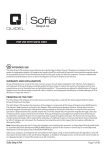
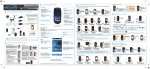
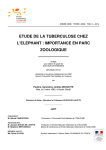
For research purpose only. Not for use in diagnostic procedures for clinical
purposes. For IN VITRO USE ONLY.
ISO 9001/14001 Certified Company
e-MycoTM plus Mycoplasma PCR Detection Kit
Cat. No.
25234
96 Tests
( for 20 µl rxn ]
DESCRIPTION
• Mycoplasma is a genus of bacteria which lack a cell wall. Without a cell wall, they are
unaffected by many common antibiotics such as penicillin or other beta-lactam
antibiotics that target cell wall synthesis. They can be parasitic or saprotrophic.
Several species are pathogenic in humans, including M. pneumoniae and M.
genitalium. Mycoplasma species are often found in research laboratories as
contaminants in cell culture. Mycoplasmal cell culture contamination occurs due to
contamination from individuals or contaminated cell culture medium ingredients.
• Mycoplasma cells are physically small (< 1 µm) and are difficult to detect with a
conventional microscope. Mycoplasmas may induce cellular changes, including
chromosome aberrations, changes in metabolism and cell growth. Severe
Mycoplasma infections may destroy a cell line. Several methods for the detection of
Mycoplasmas have been published. The testing required by the regulatory authorities
is seeding in culture (agar and liquid media). This test is complicated, time
consuming (about 3 ~ 5 weeks), and some Mycoplasma species are difficult to detect
with this method. In recent years, the disadvantages of these methods have been
acknowledged (such as sensitivity, specificity and long and complex procedures),
and use of PCR for the detection of contaminations in cell cultures has become
increasingly widespread.
• e-Myco™ plus Mycoplasma PCR Detection Kit greatly simplify testing and detection
of Mycoplasma contamination in cell cultures. With PCR testing, reliable results are
obtained within a few hours, since the presence of contaminant Mycoplasmas can be
easily and sensitively detected by simply verifying the bands of amplified DNA
fragments after gel electrophoresis. The e-Myco™ plus Mycoplasma PCR Detection
has been shown to be a highly sensitive, specific and rapid method for the detection
of Mycoplasmas contamination in cell cultures.
• Though the gene sequences for 16S rRNA are very similar in most Mycoplasma
species, there are some differences in the sequences of 16S rRNA gene between
certain Mycoplasma species and the other species.
Specific primers set of e-Myco™ plus Mycoplasma PCR Detection Kit was designed
from DNA sequences that are coding for highly conserved 16S rRNA with
considering above point. Thus e-Myco™ plus Mycoplasma PCR Detection Kit can be
used in the detection of more broad range of Mycoplasma species, compared with
any other commercially available PCR-based Mycoplasma detection kit, without
interfering with animal or bacterial DNA.
• An internal control of this product was constructed to identify false negative results in
each reaction. The internal control was designed in such a way that the primers set
was used to amplify the internal control and target DNA, which were differentiated by
size. Furthermore, the sample control was provided with this kit for using in verifying
the effectiveness of template DNA. So, You may easily check your sample
preparation. In addition, the use of 8-methoxypsoralen (8-MOP) was adopted in this
kit. 8-MOP is known to intercalate into double-stranded nucleic acids and form a
covalent inter-strand crosslink after photo-activation by incident light at wavelength
320-400 nm, so it is helpful to prevent cross-contamination by PCR products from
earlier experiments.
• Each tube of the e-Myco™ plus Mycoplasma PCR Detection Kit contains all the
components for PCR except for template: DNA Polymerase, dNTPs, PCR Buffer,
primers set, 8-MOP, and internal control. So, you can just add your templates and
perform the PCR reaction.
CHARACTERISTICS
• Premix Type
- This e-Myco™ plus Mycoplasma PCR Detection Kit contains all the components for
the PCR reaction. You just add a template and DW.
• Wide Range of Detectable Mycoplasmas
- You can detect not only five common cell culture-infecting species of mycoplasma
but also other various species of Mycoplasma over 8 genus 209 species (See
Technical Guide).
• Internal Control
- Internal control embedded in the product prevents misjudgment that possibly arises
from an erroneous PCR test.
• Sample Control
- You can verify easily the effectiveness of template gDNA by checking the
amplification from sample control.
• Species Determination
- You can determine the species of Mycoplasma by sequencing the amplified PCR
products.
• Elimination of Cross-Contamination
- 8-MOP prevents cross-contamination by PCR products.
APPLICATIONS
• The kit is used for the detection of Mycoplasma species that are most commonly
encountered in cell culture, including M. arginini, M. fermentans, M. hyorhinis, M.
orale, and Acholeplasma laidlawii. Furthermore, a most kinds of mycoplasmas can
be detected easily (See Technical Guide).
This kit is covered by patents owned by Abbott Molecular Inc.
(US Pat. No. 5,851,767 and its foreign counterparts)
KIT CONTENTS and STORAGE
Label
Contain
e-Myco™ plus Mycoplasma PCR Detection Kit
96 Tubes
Control DNA
30 µl (20 ng/µl)
(genomic DNA from M. fermentans-infected K562)
DNase/RNase-free Distilled Water
2 ml
Store at -20℃. e-Myco™ plus Mycoplasma PCR Detection Kit is a novel vacuum-dried premix
type. This product has a stable shelf-life of a minimum of 1 year when stored at -20°C and
unopened container.
REACTION TUBE COMPONENT
• PCR Reaction volume
20 µl reaction
e-MycoTM plus Mycoplasma PCR Detection Kit
DNA Polymerase
Chemical Stabilizer
Loading Buffer
dNTPs
Tris-HCl (pH 8.3)
KCl
MgCl2
Mycoplasma Primers Set
Internal Control
8-MOP (dissolved in DMSO)
2.5U
1x
1x
250 mM each
10 mM
50 mM
1.5 mM
Dried under iNtRON’s instruction
[ Overview of Mycoplasma Detection ]
Sample (culture)
(Optional PROTOCOL B
: Cell boiling method
95°C 10min
PROTOCOL A
: Genomic DNA method
Template DNA extraction using i-genomic CTB DNA
Extraction Mini Kit (Cat.No. 17341) for Mycoplasma
detection
e- MycoTM plus Mycoplasma
PCR Detection Kit
1 tube : 20 µl D.W.
(as negative control)
2 tube : 1 µl DNA + 19 µl D.W.
PCR Reaction
Detection on agarose gel
(optional) UV irradiation (10 min)
Discard the PCR product tube
TECHNICAL INFORMATION
• PROTOCOL A : Using the genomic DNA extraction kit
• This e-Myco™ plus Mycoplasma PCR Detection Kit offers a sensitive means to
detect Mycoplasma contamination in cell lines. Under optimal conditions, templates
derived from supernatants of an infected cell culture will yield a maximum signal in
the PCR, whereas an uninfected cell line will yield no PCR products. Undoubtedly,
there will be variations in cell numbers, infection amount, and templates that may
contribute to signal differences in your experiments.
1. Prepare the cultured cell according to 1a or 1b.
1a. Cells grown in suspension ; Transfer the culture fluid into 15 ml or 50 ml of
centrifuge tube and pellet the culture by centrifugation for 5 min at 3,000 rpm.
Remove the supernatant completely and wash the pellet with PBS or fresh media.
Then resuspend the washed cell pellet in appropriate volume of PBS or fresh media.
1b. Cells grown in monolayer ; Cells grown in monolayer can be detached from
culture flask (or plate) by either 1) Trypsinization or 2) Using a cell scraper.
1) To Trypsinize cells : Remove the medium and wash the cells with preheated (at
37℃) PBS. Then aspirate the PBS and add trypsin solution. After cells have become
detached from culture flask (or dish), collect and wash the cells with PBS, then
resuspend the washed cell pellet in appropriate volume of PBS or fresh media.
2) Using a cell scrape, detach cells from culture flask or dish. Collect and wash the
cells with PBS, then resuspend the washed cell pellet in appropriate volume of PBS
or fresh media.
• It is recommended to use the cultured cells for 3~6 days after sub-culturing as a
sample for Mycoplasma detection. You may not detect efficiently Mycoplasma
infection when you use the cells that are not or shortly cultivated.
• The PCR conditions were optimized to obtain the highest level of sensitivity of target
gene detection. So, the internal control band or sample control band may be
sometimes disappeared depending on the efficiency of target gene amplification. The
efficiency of the target gene amplification is dependent upon the amount of template
DNA added to the reaction. Please refer the following table to show the dependency.
2. Determinate the cell number using cell counter (eg. hemocytometer) and
transfer the appropriate number of cells (1 ~ 3 x 10⁶ cells) to a new 1.5 ml
micro-centrifuge tube.
Amount of template DNA
Optimal conditions (Three bands are appeared)
1 ~ 50 ng of template DNA
Masking point of internal control band
above 50 ng of template DNA
Ending point of sample control band
below 1 ng of template DNA
Limit of sensitivity in target gene amplification
6.3 pg of template DNA
3. Pellet the cell by centrifugation for 1 min at 13,000 rpm and discard the
supernatant. Resuspend the cell pellet with the remaining medium by finger
tapping or vortexing.
Note : In order to ensure efficient lysis, it is essential that the cell pellet are
homogeneously resuspended.
* The amount of template DNA is depend on the extent of Mycoplasma infection.
• The efficiency of PCR amplification is varied by the extent of Mycoplasma infection.
Strong Mycoplasma infections are detected in a little as 10~100 cell equivalents,
while weak infections require cell equivalents in the 5,000~50,000 cell range. So, we
recommend to perform PCR reaction with several samples prepared from several cell
numbers. Please refer to Fig. 3.
4. Extract the genomic DNA from the harvested cell. We recommend to use the igenomic CTB DNA Extraction Mini Kit (Cat. No. 17431) in genomic DNA
extraction. The quantity and quality of template DNA used in Mycoplasma
detection is important for reliable and acceptable analysis.
• If you want to do genotyping, excise the target band from the agarose gel, then
isolate the DNA fragment using a gel extraction kit(eg. MEGA-spinTM Agarose gel
DNA Extraction Kit (Cat. No. 17183), MEGAquick-spinTM PCR & Agarose Gel DNA
Extraction Kit (Cat. No. 17282)).
5. Add the genomic DNA purified using the i-genomic CTB DNA Extraction Mini
Kit (Cat. No. 17431) to each tube of e-MycoTM plus Mycoplasma PCR Detection
Kit as a template, and then add sterile water to fill the reaction mix up to 20 µl.
Note: Appropriate amounts of template DNA in a sample: 1 ~ 50 ng
6.After sample mixing, perform PCR according to the following procedure.
Note: We recommend to perform one negative control reaction with sterile water as
a template. We also recommend to perform positive control reaction with 0.1 ~ 1 µl
of control DNA solution.
TRIPLE CHECKING SYSTEM
M N
Sample control (app. 570 bp)
Target band (app. 260 bp)
Internal control (app. 160 bp)
35 cycles
PCR Condition
Temp.
Time
Initial denaturation
94oC
1 min
Denaturation
94oC
30 sec
Annealing
58oC
20 sec
Extension
72oC
1 min
72oC
5 min
Final extension
• Sample control : a parameter indicating the appropriateness in sample preparation
• Target band : a parameter of Mycoplasma infection
• Internal control : a parameter checking any problems that may arise during
amplification
7. For analysis by electrophoresis, use 5 µl of PCR reaction mixture.
8. PCR products should be discarded after UV irradiation (10 min) to prevent
cross-contamination.
Note: Cross-contamination is a common problem with PCR. Please discard the PCR
products after UV irradiation (365 nm) to prevent cross-contamination.
PROTOCOL
You can use this protocol just for detecting the contamination of Mycoplasma. However,
if you want to perform genotyping for the detailed determination of species, please
purify the genomic DNA of suspected Mycoplasma-infected cells using our i-genomic
CTB DNA Extraction Mini Kit (Cat. No. 17341).
[ TECHNICAL TIP ]
1. Use clean, disposable gloves when performing the assay and make sure that the
work area is clean prior to starting the assay setup.
2.
Keep your reagents and PCR mixture tubes on a cold block during reaction setup.
3.
Use positive displacement pipettes.
4.
The amplification and detection areas should be physically separated; i.e., do not
use the same bench area to set up the PCR reactions and run your gels.
[ CAUTIONS ]
• DO NOT expose to UV irradiation, which activates 8-MOP, if you want to determine
the detailed species of Mycoplasma by DNA sequencing analysis.
• (Optional) PROTOCOL B : Using the Boiling Extract Method
1. Prepare cell suspensions from the test cell culture in a 1.5 ml tube. Then count
cell numbers by general counting methods. You need at least 5x10⁴ cells per
test.
Note 1: Harvest adherent cells with trypsin-EDTA solution using standard techniques.
Pipette 1 ml of TE-treated adherent cells. Generally, with suspension cells, such as
K562, you need not treat with TE solution. We recommend that you count the cells.
You should prepare at least 5x10⁴ cells per test (see Technical Guide, >50,000 cells
are needed to complete this protocol)
Note 2: Strong Mycoplasma infections are detected in a little as 20 ~ 100 cells, while
weak infections require cells over 50,000 cells. You can dilute the template according
to the infection rates you suspect. We recommend that you perform the PCR reaction
after preparing serial dilutions of the straight supernatant to obtain optimal results.
2. Transfer the counted cells (over 5x10⁴ cells) to a 1.5 ml tube. Spin the tube in a
centrifuge for 10~15 seconds. Carefully decant the supernatant.
3. Resuspend the cells in 1 ml of sterile PBS or DPBS solution for washing.
4. Spin the tube in a centrifuge for 10~15 seconds. Carefully decant the
supernatant.
Note : (Optional ) Repeat this washing step once more.
e-Myco™ plus Mycoplasma PCR Detection Kit
-2-
T. 0505-550-5600 F. 0505-550-5660
• (Optional) PROTOCOL B : Using the Boiling Extract Method
- continued
5. Resuspend the cell pellets in 100 µl of sterile PBS or DPBS solution.
Note : If you want the best result, use of PBS solution is better than Tris (10 mM, pH
8.5), TE (10 mM Tris, 0.1 mM EDTA), or autoclaved DW.
6. Heat the samples for 10 min, and vortex for 5-10 sec. Then, centrifuge for 2 min
at 13,000 rpm with a tabletop centrifuge at room temperature.
7. Transfer an aliquot of the heated supernatant to a fresh tube. This supernatant
will be used as the template in the PCR.
8. Add 10 µl of the template to each tube of e-Myco™ plus Mycoplasma PCR
Detection Kit, and then add 10 µl of sterile water.
9. After sample mixing, perform PCR reaction according to the procedure
presented in protocol A.
Note : We recommend that you perform one negative control reaction with sterile
water as a template.
10. For analysis by electrophoresis, use 5 µl of PCR reaction mixture.
11. PCR products should be discarded after UV irradiation (10 min) to prevent
cross-contamination.
Note : Cross-contamination is a common problem with PCR. Please discard the
PCR products after UV irradiation (365 nm) to prevent cross-contamination.
EXPECTABLE DATA
◈ Examination of Mycoplasma Infection
M 1 2 3 4 5
Sample control (app 570 bp)
Target (app 260 bp)
Internal control (app 160 bp)
Mycoplasma
Contamination
Free
Free
Contamination
Contamination
4. Presence of amplified product in the negative control
• Check contamination of D.W.
: D.W. can be contaminated. Perform PCR again with fresh sterile water.
• Check contamination of lab instruments and other environments
: We recommend that you use filter tips to reduce contamination and that you use a
pipette after sterilization. All procedures should be done in sterilized conditions.
5. Poor resolution on agarose gel
• We recommend to use a 1.5~2% agarose gel.
• We recommend that electrophoresis is performed for 40 min at 100 V/14 cm using a
6 cm long 2% agarose gel.
DETECTION LIMITS
Target Gene Detection
• K562 cell (M. fermentans-infected) : more than 15 cells
• K562 gDNA (M. fermentans-infected) : more than 6.25 pg
• Concentration of Mycoplasma (in the case with M. fermentans) : more than 20 cfu/ml
Sample Control Detection
• Human / Mammalian-specific DNA sequence
: app. 570 bp length
: Sample control gene is detectable from above 1 ng of template DNA
Internal Control Detection
• Artificial gene (derived from human TNFα gene)
: app. 160 bp length
: Internal control can serve the tools checking any problems that may arise during
amplification. The signal of internal control is mostly appeared. But if the
amount of template added is too high (above 50 ng) the signal of internal control
may be weaken or disappeared by competition with template DNA.
Experimental Data
Fig. 1. Exemplary Data
Lane
1
2
3
4
5
3. No sample control band
• Check template concentration
: Sometimes, the sample control band may disappeared when the concentration of
DNA template is below 1 ng. Check the quantity of DNA template, and adjust the
amount of DNA template in 20 µl PCR reaction to be above 1 ng.
• Check the source of template
: The primers set included in this kit can amplify a mammalian-specific DNA
sequence. If the template source is not mammalian cell (eg. Plant, bacteria or
Mycoplasma culture), the amplification of sample control does not occur.
Test case description
Optimal
Optimal
Excess template
Excess template
Small amount of template
Template amount
1 ~ 50 ng
1 ~ 50 ng
> 50 ng
> 50 ng
1 ng
1) Minimal amount of genomic DNA detectable
½ dilution
M N 1
2 3 4
5
6
7 8 9 10 11 12 13 14 15
Sample control (app 570 bp)
Target (app 260 bp)
Internal control (app 160 bp)
TECHNICAL GUIDE
TROUBLESHOOTING GUIDE
1. No Target band in positive reaction
• Check internal control band
: If internal control band is seen, PCR has been performed properly; it is not a
problem of the product.
• Check the quality or concentration of template
: If the PCR reaction is inhibited by impurities included in DNA preparation, the use of
diluted DNA as a template may be helpful.
: Whereas the signals of sample control (app. 570 bp length) and internal control (app.
160 bp length) are shown, if the target band is not shown, it indicates that the sample
is not infected by Mycoplasma.
• Check a PCR machine
: The problem can be caused by the PCR machine. Please check the temperature
and make sure to check that the machine is working properly.
Fig. 2. Result of determining minimal required amount of genomic DNA per test
To determine the minimal required amount of genomic DNA, genomic DNA was isolated from a
pure culture of M. fermentans-infected K562 cells using i-genomic CTB DNA Extraction Mini Kit
(Cat. No. 17431). The isolated genomic DNA was serially diluted for PCR detection. The result
indicates that the detection limit with this kit is 6.3 pg of genomic DNA per test.
Lane
gDNA
Lane
gDNA
M
N
100 bp DNA Marker 0 ng
7
8
9
1.6 ng
800 pg 400 pg
1
100 ng
10
200 pg
2
50 ng
11
100 pg
3
25 ng
12
50 pg
4
5
6
12.5 ng 6.3 ng 3.2 ng
13
14
15
25 pg 12.5 pg 6.25 pg
2) Minimal cell number required
½ dilution
M
N 1
2 3
4
5
6
7 8
9 10 11 12 13 14 15
2. No internal control band
• Check template concentration
: Competition can occur by using high concentrated DNA template. Please repeat the
PCR with a diluted template. If the concentration of template is above 50 ng, the
signal of internal control may be disappeared by competition with the template.
It does not cause any problem, because the signal of sample control (app. 570 bp
length) can function as a internal control.
• Check the quality of template (possibility of contamination with PCR inhibitors)
: If the PCR reaction is inhibited by impurities included in DNA preparation, the use of
diluted DNA as a template may be helpful. If there is no internal control band, please
inquire with our technical support staff.
• Check the storage condition of product.
Sample control (app 570 bp)
Target (app 260 bp)
Internal control (app 160 bp)
Fig. 3. Result of determining minimal required cell number per test
To determine the minimal required cell number, M. fermentans-infected K562 cells were grown in
pure culture, serially diluted and tested. The result indicates that the detection limit with this kit
is 15 cells per test.
Lane
Cell number
Lane
Cell number
M
100 bp DNA Marker
7
8
3.9x10³ 1.9x10³
N
0
9
9x10²
1
2
3
4
5
6
2.5x10⁵ 1.25x10⁵ 6.25x10⁴ 3.12x10⁴ 1.56x10⁴ 7.8x10³
10
11
12
13
14
15
4.8x10² 2.4x10²
120
60
30
15
3) Minimal Mycoplasma concentration detected
Lane
Copy number
Lane
Copy number
PHYLOGENETIC ANALYSIS
Fig. 4. Result of determining minimal
required concentration of
Mycoplasma per test
To determine minimal required concentration of
Mycoplasma, M. fermentans was selected as a
model Mycoplasma species. M. fermentans was
serially diluted for PCR detection.
Target (app 260 bp)
Internal control (app 160 bp) The result indicates that the detection limit
with this kit is 20 cfu/ml as a Mycoplasma
concentration in culture broth.
½ dilution
M
100 bp DNA Marker
7
8
5.1x10³
2.5x10³
N
0
9
1.28x10³
1
3.4x10⁵
10
6.4x10²
2
1.7x10⁵
11
3.2x10²
3
8.25x10⁴
12
161
4
4.12x10⁴
13
80
5
2.06x10⁴
14
40
M_bovi s
M_prim atum
M_agalact iae
M_leopharyngis
M_m ac ul osum
M_s permatophilum
M_opalesc ens
M_adleri
M_c avi ae
M_f erment ans
M_c alif ornic um
M_.bovigenit alium
M_s im bae
M_phoc irhi nis
M_z alophidermi dis
M_c anim ucos ale
M_li pof aciens
M_c olumbinasale
M_c olumbinum
M_gallinarum
M_meleagridis
M_hyopharyngis
M_li pophilum
M_s phenis ci
M_c anis
M_edwardii
M_c ynos
M_leocapt ivus
M_leocapt ivus
M_bovi rhinis
M_f elis
M_mus telae
M_s ynoviae
M_verecundum
M_c ricet uli
M_oxoniensis
M_c it elli
M_c olum borale
M_s turnidae
M_buteonis
M_buteonis
M_gallopavonis
M_glycophilum
M_c oragypsi
M_pullorum
M_c roc odyli
M_elephantis
M_equigenit alium
M_argi nini
M_gateae
M_c anadens e
M_phoc icerebrale
M_auri s
M_alk alescens
M_s pumans
M_phoc ae
M_equirhinis
M_f alconis
M_anseri s
M_c loacale
Myc oplasa_f aucium
M_oral e
M_t i mone
M_bucc ale
M_s alivarium
M_art hrit idis
M_s ubdolum
M_hyos ynoviae
M_hom inis
M_gypis
M_z alophi
M_c ollis
M_neurolyt icum
M_lagogenit alium
M_molare
M_iguanae
M_dis par
M_f locculare
M_ovipneumoniae
M_hyorhinis
M_vulturi i
M_m oats ii
M_s ualvi
M_m obile
M_agas siz ii
M_c heloniae
M_pulmoni s
As teroleplasma_anaerobium
A_modicum
Ac holeplasma_m orum
M_f elim inut um
Ac holeplasma_granularum
Ac holeplasma_l aidlawii
Ac holeplasma_oculi
A_bact oc las ticum
A_varium
A_abac toclast icum
M_ovis
M_wenyonii
M_haemolam a
M_s uis
M_erythrodidelphis
Haemobartonella_canis
Haemobartonella_f elis
M_c occ oides
M_haemom uris
M_c avipharyngis
M_f ast idiosum
M_ins ons
M_im it ans
M_gallisepticum
M_t est udinis
M_pirum
M_genitalium
M_pneumoniae
M_iowae
M_m uris
M_volis
M_penetrans
U_urealyticum
M_yeat sii
M_c ott ewii
M_putref aciens
M_putref aciens
M_c apric olum
M_mycoides
M_m onodon
M_ellyc hnium
Entom oplasm a_luc ivorax
Entom oplasm a_luminosum
Entom oplasm a_s omnilux
S_taiwanense
S_apis
Entomoplasma_melaleuc ae
Mes oplasma_lactucae
S_cit ri
S_mirum
M_ovipneumoniae
6
1.03x10⁴
15
20
SPECIES DETERMINATION BY SEQUENCING ANALYSIS
• The sequences of PCR products have slight differences among species. You can determine approximately the
Mycoplasma species by sequencing analysis with the following primer. Please refer to the phylogenetic tree on the
right side. For more detailed species analysis, you should perform additional sequencing with your designed primers.
• We provide only the Forward primer sequence. Please synthesize the primer, and then use it in general sequencing.
• Nucleotide sequence of the sequencing primer:
5’ – GGA TTA GAT ACC CTG GTA GTC CAC G– 3’
[Note] The PCR primers set included in this kit differs from the above sequencing primer. We do not list the
sequences of primers comprising the PCR primers set contained in this kit.
DETECTABLE MYCOPLASMA STRAINS (8 Genus / 209 Species)
Genus
Acholeplasma (5)
Anaeroplasma (3)
Asteroleplasma (1)
Entomoplasma (5)
Mycoplasma (182)
Mesoplasma (3)
Spiroplasma (9)
Ureaplasma (1)
Acholeplasma granularum
Anaeroplasma abactoclasticum
Asteroleplasma anaerobium
Entomoplasma lucivorax
M. adleri
M. alligatoris
M. arginini
M. bovis
M. canadense
M. hyopharyngis
M. cloacale
M. columborale
M. crocodyli
M. equigenitalium
M. faucium
M. flocculare
M. gateae
M. haemocanis
Mycoplasma sp. Ms01
M. hyopneumoniae (strain7448)
M. hyosynoviae
M. leocaptivus
M. microti
M. muris
M. mycoides subsp. mycoides SC
M. ovipneumoniae
M. phocirhinis
M. pulmonis
M. maculosum
M. sualvi
M. testudineum
M. wenyonii
Mycoplasma sp. Z61
Mycoplasma sp. 10T4
Mycoplasma sp. 237IAT
Mycoplasma sp. 8790CV
Acholeplasma laidlawii
Anaeroplasma bactoclasticum
Species
Acholeplasma modicum
Anaeroplasma varium
Acholeplasma morum
Acholeplasma oculi
Entomoplasma luminosum
M. agalactiae
M. alvi
M. arthritidis
M. bovoculi
M. canimucosale
M. hyopneumoniae
M. coccoides
M. conjunctivae
M. cynos
M. equirhinis
M. felifaucium
M. gallinaceum
M. genitalium
M. haemofelis
Mycoplasma sp. Ms02
M. insons
M. indiense
M. leonicaptivi
M. moatsii
M. mustelae
M. mycoides sunsp. capri
M. oxoniensis
M. pirum
M. putrefaciens
M. meleagridis
M. subdolum
M. testudinis
M. yeatsii
Mycoplasma sp. SF12
Mycoplasma sp. 11CL2
Mycoplasma sp. 2F1AT
Mycoplasma sp. 94630
Entomoplasma melaleucae
M. agalactiae (strain PG2)
M. amphoriforme
M. auris
M. buccale
M. canis
M. caviae
M. collis
M. corogypsi
M. dispar
M. erythrodidelphis
M. feliminutum
M. gallinarum
M. genitalium G37
M. haemolama
Mycoplasma sp. Ms03
M. hyorhinis
M. iguanae
M. leopharyngis
M. mobile
M. mycoides
M. neurolyticum
M. penetrans
M. pneumoniae
M. salivarium
M. sphenisci
M. suis
M. timone
M. zalophi
Mycoplasma sp. 07SH-h
Mycoplasma sp. 1220
Mycoplasma sp. 34CL
Mycoplasma sp. A1802T
Entomoplasma somnilux
M. agassizii
M. anatis
M. bovigenitalium
M. buteonis
M. capricolum
M. cavipharyngis
M. columbinasale
M. cottewii
M. edwardii
M. falconis
M. felis
M. gallisepticum
M. glycophilum
M. haemomuris
M. hyopneumoniae (strain 232)
M. lagogenitalium
M. iowae
M. lipofaciens
M. molare
M. mycoides subsp. capri
M. opalescens
M. phocicerebrale
M. primatum
M. simbae
M. spumans
M. synoviae
M. verecundum
M. zalophidermidis
Mycoplasma sp. 07SH-p
Mycoplasma sp. 13CL
Mycoplasma sp. 39CL
Mycoplasma sp. ARNO
Entoplasma ellychniae
M. alkalescens
M. anseris
M. bovirhinis
M. californicum
M. capricolum subsp. capricolum
M. citelli
M. columbinum
M. cricetuli
M. elephantis
M. fastidiosum
M. fermentans
M. gallopavonis
M. gypis
M. hominis
Mycoplasma sp. PG50
M. imitans
M. iners
M. lipophilum
M. monodon
M. mycoides subsp. mycoides LC
M. orale
M. phocidae
M. pullorum
M. spermatophilum
M. sturni
M. synoviae (strain 53)
M. vulturii
Mycoplasma sp. Saa1e
Mycoplasma sp. 10T3
Mycoplasma sp. 15CL2
Mycoplasma sp. 50587
Mycoplasma sp. 'bovine group 7'
Mycoplasma sp. C3T
Mycoplasma sp. HRC689
Mycoplasma sp. M209-8
M. capricolum subsp.
capripneumoni
Mycoplasma sp. Saa1c
Mesoplasma entomophilum
Spiroplasma apis
Spiroplasma gladiatoris
Ureaplasma urealyticum
Mycoplasma sp. China-1
Mycoplasma sp. IS2505
Mycoplasma sp. M221-9
M. capricolum subsp.
capripneumoniae
Mycoplasma sp. SF9
Mesoplasma florum
Spiroplasma citri
Spiroplasma mirum
Mycoplasma sp. CSL 4779
Mycoplasma sp. M1
Mycoplasma sp. M222-2
Mycoplasma . sp.
‘ovine/caprine serogroup 11
Mycoplasma sp. CSL 7518-lung
Mycoplasma sp. M200-2
Mycoplasma sp. M222-5
M. hyopneumoniae
(strain J / ATCC 25934)
Mycoplasma sp. VJC358
Mycoplasma sp. M209-7
Mycoplasma sp. M26
Mycoplasma. sp.
'feline hemotropic Switzerland'
Mesoplasma lactucae
Spiroplasma CN-5
Spiroplasma MQ-1
Spiroplasma DU-1
Spiroplasma taiwanense
Spiroplasma DW-1
PARTIAL SEQUENCES OF MAJOR CONTAMINANTS IN CELL CULTURE
The following sequences are partial sequences of major contaminant in general cell culture or pathogenic
Mycoplasma strains. You can determine the species by sequencing analysis.
M. pneumoniae
M. arginini
M. faucium
M. hyorhinis
M. fermentans
U. urealyticum
Me. lactucae
E. lucivorax
A . laidlawii
As. anearobium
24.2
20
e-Myco™ plus Mycoplasma PCR Detection Kit
-4-
15
10
Nuc leotide Substit utions ( x100)
5
0
T. 0505-550-5600 F. 0505-550-5660