1
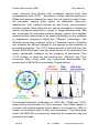
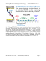
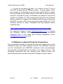
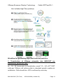
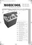
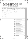
XStamp™ Exosome Targeting Technology Cat #s: XSTPxxPA/VA-1 User Manual Store at -20ºC upon arrival A limited-use label license covers this product. By use of this product, you accept the terms and conditions outlined in the Licensing and Warranty Statement contained in this user manual. XStamp Exosome Display Technology Cat#s XSTPxxxPA-1 Contents I. Introduction ..............................................................................1 A. XStamp Technology Overview............................................1 B. Uses of XStamp Technology ..............................................4 II. XStamp Sample data ...............................................................5 A. XStamp Transfection Protocol ..........................................10 C. XStamp Lentiviral Particle Production...............................12 B. Concentration of Pseudoviral Particles .................................14 C. Transduction of Pseudoviral Particles into Target Cells .......15 I. D. Shipping and Storage Conditions .....................................17 E. Related Products...............................................................17 III. Frequently Asked Questions ..............................................18 IV. References .........................................................................18 V. Technical Support ..............................................................21 VI. Licensing and Warranty information ..................................22 Introduction A. XStamp Technology Overview Exosomes are nanosized membrane vesicles secreted by most cell types in vivo and in vitro. They are produced by the inward budding of multivesicular bodies (MVBs) and subsequently released from the cell into the microenvironment following the fusion of MVBs with the plasma membrane. Exosomes are extracellular nanoshuttles that facilitate communication between cells and organs and are found in various biofluids including blood, 888-266-5066 (Toll Free) 650-968-2200 (outside US) Page 1 System Biosciences (SBI) User Manual urine, amniotic fluid, breast milk, malignant ascites fluid, and cerebrospinal fluid (CSF). Exosomes contain distinct subsets of RNAs and proteins depending upon the cell type from which they are secreted, making them useful for biomarker discovery. Additionally, their natural function as cell-to-cell communication vehicles makes them attractive for use as therapeutic shuttles to deliver biological molecules or drugs to target disease cells. SBI has developed an exosome surface display system that enables desired protein sequences to be placed efficiently on the surfaces of engineered exosomes called the "XStamp" technology. The XStamp technology is based upon a C-terminal fusion of ligands that enables the efficient display of the ligands on the surfaces of secreted exosomes. The C1C2 fusion domain is derived from the human MFG-E8 protein and has been shown to be abundant and nearly exclusively localized on the surfaces of exosomes. This C1C2 domain is what we are terming the “XStamp” tag. Flow cytometry data using cells and exosomes demonstrate the enrichment of MFG-E8 on exosomes (Figure below). Fluorescently-labeled antibodies for MFG-E8, CD58 and CD81 were used in combination for FACs analysis. The CD58 marker is a known cell surface marker that is absent on exosomes. CD81 is known to be present both in cells and exosomes. The FACs data show that MFG-E8 is exclusively detected on exosomes and not present in the cells. Page 2 ver. 1-150804 www.systembio.com XStamp Exosome Display Technology Cat#s XSTPxxxPA-1 XStamp exosome surface engineering To take advantage of the localization of MFG-E8 on exosomes, the C1C2 domain (XStamp domain) of the protein’s gene was cloned into SBI’s MSCV-MCS-EF1-Puro lentivector and a 5’ secretion signal (SS) was placed within the multiple cloning site. The protein ligand chosen to program the exosomes to target cells with a cognate receptor is the “Exposed Ligand” part of the construct. A lentivector plasmid general format is shown below. The XStamp expression cassette is driven by the MSCV promoter that works in most cell lines, including primary cells and stem cells. There is a built-in signal sequence (SS) next to the multicloning site where the desired cDNA (Exposed Ligand) is fused to both the upstream SS and the downstream XStamp C1C2 domain sequence. The SS leader peptide assists the secretion of the fusion protein and is cleaved off during the surface display process and loading on the surfaces of secreted exosomes. If the desired Exposed Ligand already has its own signal sequence, there is an XbaI cloning site 5’ to the SS sequence in the XStamp lentivector where the cDNA encoding the SS-Exposed Ligand can be inserted into the lentivector. The XStamp lentivector also features a downstream EF1-Puromycin cassette for selection and stable cell line development. The lentivector constructs can be used in transient transfection expression studies as well as packaging the construct into lentivirus to stably transduce cells. A more detailed lentivector plasmid map is depicted below. 888-266-5066 (Toll Free) 650-968-2200 (outside US) Page 3 System Biosciences (SBI) User Manual B. Uses of XStamp Technology The XStamp technology allows for cell-mediated generation of ready to use exosomes that display any protein of choice on their surfaces. These exosomes can then be used to efficiently program specific delivery to cells that have a cognate receptor, allowing for more targeted exosome cargo delivery both in vitro and in vivo. Surface display on exosomes can also be used to boost vaccine generation and engineer other drug screening applications. Increasing vaccine potency through exosome antigen targeting. Hartman ZC, Wei J, Glass OK, Guo H, Lei G, Yang XY, Osada T, Hobeika A, Delcayre A, Le Pecq JB, Morse MA, Clay TM, Lyerly HK.Vaccine. 2011 Nov 21;29(50):9361-7. Exosome nanovesicles displaying G protein-coupled receptors for drug discovery.Estelles A, Sperinde J, Roulon T, Aguilar B, Bonner C, LePecq JB, Delcayre A.Int J Nanomedicine. 2007;2(4):751-60. Exosomes as novel therapeutic nanodevices. Delcayre A, Le Pecq JB. Curr Opin Mol Ther. 2006 Feb;8(1):31-8. Review. Page 4 ver. 1-150804 www.systembio.com XStamp Exosome Display Technology II. Cat#s XSTPxxxPA-1 XStamp Sample data A. XStamp-Motilin Motilin is a 22-amino acid polypeptide hormone in the motilin family that, in humans, is encoded by the MLN gene. Motilin is secreted by endocrine M cells that are numerous in crypts of the small intestine, especially in the duodenum and jejunum. The Motilin receptor is a G protein-coupled receptor that binds motilin and is exclusively expressed in the intestine. To test the XStampMotilin construct (catalog# XSTP720PA-1), it was transfected into HEK293 cells and after 48 hours, the exosomes were collected using ExoQuick-TC. The next day, the XStamp-Motilin exosomes were Exo-Fected with a Texas-Red-labeled siRNA to monitor exosome docking and delivery. The transfected XStamp-Motilin exosomes were then added to MDA-MB-231 Breast Cancer Cells (motilin receptor negative) and to HT-29 Colon Cancer Cells (motilin receptor positive). The cells were imaged after 24 hours for uptake of the Texas-Red-labeled siRNA delivery from the XStamped exosomes. The HT-29 colon cancer cells that are motilin receptor positive took up the XStamp-Motilin exosomes at a much higher rate than the MDA-MB-231 Breast Cancer (motilin receptor negative) cells. 888-266-5066 (Toll Free) 650-968-2200 (outside US) Page 5 System Biosciences (SBI) User Manual B. XStamp-NCAM and XStamp-BHP1 The Neural Cell Adhesion Molecule 1 (NCAM) gene encodes a cell adhesion protein which is a member of the immunoglobulin superfamily. The encoded protein is involved in cell-to-cell interactions as well as cell-matrix interactions during development and differentiation. The encoded protein has been shown to be involved in development of the nervous system. The NCAM domains interact with each other during the adhesion process. Page 6 ver. 1-150804 www.systembio.com XStamp Exosome Display Technology Cat#s XSTPxxxPA-1 An XStamp-NCAM fusion was constructed and incorporated the first 300 amino acids (Signal Peptide plus IGc2 domains 1-3) of the mouse NCAM gene translationally fused to the C1C2 XStamp display tag (catalog# XSTP721PA-1). In parallel, a Brain Homing Peptide (BHP1) from “Organ targeting in vivo using phage display peptide libraries.” Pasqualini R, Ruoslahti E. Nature. 1996 Mar 28;380(6572):364-6 was fused to the BHP1 XStamp domain to create the XStamp-NCAM construct (catalog# XSTP722PA-1). 888-266-5066 (Toll Free) 650-968-2200 (outside US) Page 7 System Biosciences (SBI) User Manual To test the XStamp-NCAM construct (catalog# XSTP721PA-1) and the XStamp-BHP1 construct (catalog# XSTP722PA-1) , were transfected separately into mouse MSCs along with SBI’s XPackGFP construct (catalog# XPAK530PA-1) which packages GFP into the interior of exosomes for fluorescent tracking. After 48 hours, the exosomes were collected using ultracentrifugation and then quantitated for exosome protein levels. Equal amounts (100 ug) of Control (No XStamp), XStamp-NCAM or XStamp-BHP1 MSC exosomes (all labeled with XPack-GFP) were added to Neuro2a neuroblastoma cells in culture. The neurons were imaged for phase and GFP signals after a 24 hour incubation with the various exosomes. The cells were imaged after 24 hours for uptake of the XStamp-NCAM coated and BHP1 coated exosomes to deliver the XPack-GFP exosomes. Together, they are known as the “Pack and Stamp” system. Page 8 ver. 1-150804 www.systembio.com XStamp Exosome Display Technology Cat#s XSTPxxxPA-1 The Neuro2a neuroblastoma cells that are NCAM positive took up the XStamp-NCAM and XStamp-BHP1 exosomes at a much higher rate than control XStamp exosomes. NOTE: ExoQuick-TC and Exo-FBS are not provided with the XStamp vectors and can be purchased separately. The following ExoQuick-TC products are recommended for exosome concentration prior to addition to target cells. Cat# Description Size EXOTC10A-1 ExoQuick-TC for Tissue Culture Media and Urine ExoQuick-TC for Tissue Culture Media and Urine Exosome-depleted FBS media supplement - Heat Inactivated 10 ml EXOTC50A-1 EXO-FBSHI50A-1 50 ml 50 ml IMPORTANT NOTE: Be sure to culture your exosome producer cell lines in media that does not contain standard 888-266-5066 (Toll Free) 650-968-2200 (outside US) Page 9 System Biosciences (SBI) User Manual FBS. There are high levels of bovine exosomes present in FBS. Instead, use SBI’s Exo-FBS Heat-Inactivated Exosomedepleted FBS Media Supplement (cat#EXO-FBSHI-50A-1) in place of standard FBS media supplements. A. XStamp Transfection Protocol Transfection of Exosome Producer Cells: 1. Seed exosome producer cells in culture dish of choice to reach 70-80% confluency after 24 hours using media compatible with the cells of choice. Because standard FBS contains high levels of bovine exosomes, be sure to use SBI’s Exosome-depleted FBS Media Supplement to ensure that exosomes isolated after cell transfection are not contaminated by bovine exosomes. Return cells to incubator. 2. 24 hours later, mix XStamp vector with transfection reagent of choice and follow appropriate protocol to achieve transfection of target cells. An example transfection reaction using SBI’s PureFection (Cat# LV750A-1) in a 6-well plate of cells at 70-80% confluency: a. Mix 5 uL PureFection reagent, 2.5 ug XStamp Lentivector, and 200 uL serum-free media in sterile 1.5 mL Eppendorf tube. b. Vortex briefly and incubate at room temperature for 15 minutes. c. Add entire volume to 6-well of cells in a total volume of 2-3 ml media. 3. Change media after 24 hours. 4. Isolate exosomes in 48-96 hour window post-transfection Page 10 ver. 1-150804 www.systembio.com XStamp Exosome Display Technology Cat#s XSTPxxxPA-1 Isolation of XStamp Exosomes and Addition to Target Cells: 1. Remove cell culture media and place in 15 mL or 50 mL centrifuge tube. 2. Spin centrifuge tubes at 3,000 x g for 30 minutes at room temperature or 4°C to get rid of cellular debris. Transfer the supernatant to a new tube. 3. Add ExoQuick-TC at 1:5 the volume of cell culture media. 4. Mix by inversion and incubate at 4°C overnight. 5. Spin centrifuge tubes at 1500 x g for 30 minutes at room temperature or 4°C (temperature does not affect exosome yield). Discard supernatant and resuspend exosome containing pellet in 100 uL PBS. 6. Measure exosome yield using A280 on Nanodrop. Adjust concentration to 1 ug/uL. 7. Add exosomes to cell culture dish containing target cells. For target cells (>1.5x10^5 cells) in a 6 well plate format, 250 ug exosomes is sufficient. The number of exosomes required to discern effects in target cells may vary by cell type and by the specific phenotype being assayed; therefore, optimization of specific experimental conditions may be needed. B. Cloning of Proteins into XStamp MCS: 1. To clone fusions in frame, note that the XStamp lentivectors have a 5’ leader sequence to facilitate secretion and a 3’ C1C2 XStamp domain for exosome display. If your ligand of interest already has an endogenous signal sequence peptide, then use the XbaI site upstream of the built-in leader sequence and replace with cDNA of your choice. 888-266-5066 (Toll Free) 650-968-2200 (outside US) Page 11 System Biosciences (SBI) User Manual 2. It may be necessary to add 1 or 2 bases to the 3’ end of the ORF to generate an in frame fusion. In such cases, count the number of nucleotides from the start of the MCS to where first nucleotide in the initial codon of the ORF-of-interest will be inserted, and add as many nucleotides as needed to make that number a multiple of 3. We recommend using the free plasmid editor software to design the XStamp fusions and ensure the full ORF from the ATG in the leader through cDNA into C1C2 XStamp domain is intact. Download the free plasmid editor software here: http://www.systembio.com/support/resources/online-tools For technical support, email [email protected] to request the XStamp cloning lentivector plasmid editor annotated sequence file. If you need clone design assistance email: [email protected] as well. C. XStamp Lentiviral Particle Production For researchers looking for sustained, long-term expression of the XStamp construct in their desired cell line, the XStamp construct can be transfected into HEK293T producer cells and packaged into pseudo viral particles for infection of a target cell line. The following schematic and the protocol that follows shows the lentiviral production process using the XStamp lentiviral vector. Page 12 ver. 1-150804 www.systembio.com XStamp Exosome Display Technology Cat#s XSTPxxxPA-1 Workflow for generating high-titer lentiviral particles 1. Transfection of XStamp plasmids into HEK293T (or equivalent) producer cells 6 a) 18 - 24 hours prior to transfection, seed 7.0 – 8.0 x10 293T cells per 150mm cell culture plate in standard growth media w/o antibiotics. Cells should be ~80% confluent by next day. 888-266-5066 (Toll Free) 650-968-2200 (outside US) Page 13 System Biosciences (SBI) User Manual b) During transfection day, mix 45 µl of pPACKH1 packaging plasmid mix as provided in the LentiStarter 2.0 Kit and 4.5 µg of XStamp lentivector in 1.6 ml of serum-free DMEM by pipetting. c) Add 55 µl PureFection into the same tube. Vortex for 10 seconds. Note: If using other transfection reagents (e.g. Lipofectamine 2000) please follow suggested guidelines for 150mm plates. d) Incubate mixture at room temperature for 15 minutes. e) Add mixture drop-wise to the dish, and swirl to disperse evenly throughout the plates. f) Change the medium ~12 hours (or next day) after transfection. g) At 48 hours and 72 hours after transfection, collect the medium (which now contains pseudoviral particles) into a 50-ml sterile, capped conical centrifuge tube. Centrifuge at 3000 x g for 15 minutes at room temperature to pellet cell debris. Transfer the viral supernatant into a new tube. Caution: You are working with infectious pseudoviral particles at this stage. Please follow the recommended guidelines for working with BSL-2 biosafety agents. 2. Concentration of Pseudoviral Particles The PEG-it™ Virus Precipitation Solution in the LentiStarter 2.0 Kit provides a simple and highly effective means to concentrate lentiviral particles. PEG-it is a formulation of polyethylene glycol optimized for the precipitation of lentiviral-based particles. The PEG-it Virus Precipitation Solution is provided as a 5x solution. 1. Transfer supernatant containing virus to a sterile vessel and add 1 volume of cold PEG-it Virus Precipitation Solution (4ºC) to every 4 volumes of virus supernatant. (Example: 5ml PEG-it with 20ml viral supernatant). Page 14 ver. 1-150804 www.systembio.com XStamp Exosome Display Technology Cat#s XSTPxxxPA-1 2. Refrigerate overnight (at least 12 hours). Viral supernatants mixed with PEG-it Virus Precipitation Solution are stable for up to 4-5 days at 4°C. 3. Centrifuge supernatant/PEG-it mixture at 1500 × g for 30 minutes at 4ºC. After centrifugation, the virus particles may appear as a beige or white pellet at the bottom of the vessel. 4. Discard the supernatant into a suitable biohazard waste container. Spin down residual PEG-it solution by centrifugation at 1500 × g for 5 minutes. Remove all traces of fluid by aspiration, taking great care not to disturb the precipitated lentiviral particles in pellet. 5. Resuspend lentiviral pellets in 1/500 to 1/1000 of original volume of pooled virus supernatant using cold, sterile Phosphate Buffered Saline (PBS) or DMEM containing 25mM HEPES buffer at 4ºC. For example, if you performed 2 collections from 2 x 150mm plates (20ml per plate), this would be approximately 80ml of media. You would resuspend the resulting pellet in 80-160 µl of 1X PBS or DMEM. 6. Aliquot in cryogenic vials and store at -80°C until ready for use. 7. The resulting pseudoviral particles can be accurately titered using SBI’s UltraRapid Global Titering Kit (Cat #LV961A-1) http://www.systembio.com/lentiviraltechnology/delivery-systems/ultrarapid/overview. C. Transduction of Pseudoviral Particles into Target Cells For efficient transduction of target cells, the negative charges present in the virus envelope protein and the cell surface must be neutralized. SBI’s TransDux reagent (provided in the LentiStarter 2.0 Kit) is a non-toxic, proprietory formulation that promotes cell888-266-5066 (Toll Free) 650-968-2200 (outside US) Page 15 System Biosciences (SBI) User Manual virus contact and subsequent fusion by negating these charges. The following protocol can be utilized for delivery of virus to your target cells. The following protocol is for infection of target cells in a single well of a 24-well plate – if using larger vessels please scale up reagents accordingly. Day 1 1. Plate 75,000 cells per well into a single well of a 24-well plate in cell culture medium. Make sure that cells are well-dispersed and are not clumped together. Include wells for negative (noninfected) cells. Note: If infecting target cells for the first time or an optimal MOI is not known, please titrate virus at varying MOIs (1, 5, 10 and 20, etc.) to optimize transduction using a positive control virus with a fluorescent marker such as SBI’s pre-packaged positive transduction control (Cat #CD511VB-1). Day 2 2. Cells should be between 70-80% confluent. Aspirate medium from cells. 3. Combine culture medium with TransDux to a 1X final concentration. For example, add 2.5 μl of TransDux to 500 µl culture medium and then transfer to each well. If using other types of transduction reagents (e.g. Polybrene) please dilute the reagent to a final working concentration of 2-8 μg/ml. 4. Add XStamp virus at desired MOI to each well and swirl to mix, for negative control wells only add media/viral transduction reagent. Day 3 5. Aspirate off medium and add complete growth medium to cells. Page 16 ver. 1-150804 www.systembio.com XStamp Exosome Display Technology Cat#s XSTPxxxPA-1 Day 5 7. Virus should be integrated into the host cell genome by this time, and should be expressing the XStamp construct for packaging into exosomes. D. Shipping and Storage Conditions The XStamp lentivectors are shipped on either blue ice (4°C) or dry ice and should be stored at -20°C upon arrival. Avoid freezethawing the reagents. Shelf life of either product is 1 year after receipt if stored properly. E. Related Products SBI offers a number of exosome research products. You can review them here: http://www.systembio.com/exosomes • • • • • ExoQuick exosome isolation reagents Exo-FBS exosome-depleted media supplement Detect and quantitate exosomes with ExoAB, ELISA, and EXOCET kits Purify exosome RNA and profile by qPCR with SeraMir Kit Discover novel exoRNA biomarkers with Exo-NGS next-gen sequencing services 888-266-5066 (Toll Free) 650-968-2200 (outside US) Page 17 System Biosciences (SBI) III. User Manual Frequently Asked Questions Q. How long and in what condition should I store exosomes after isolation from an exosome generating cell line? After exosomes are isolated with ExoQuick-TC, the pellet can be stored at -80°C for 1 year. After resuspension in PBS, it can be stored at 4°C for 2 weeks or -20°C for 3 months. Q. How many exosomes should I add to my target cells? 250 ug of exosomes (as determined by A280 on NanoDrop) is sufficient to see efficient delivery of XStamp coated exosomes on target cells in a 6-well plate format. The number of exosomes required in culture dishes of other size can be scaled up or down proportionally to the difference in total cell number relative to one well of a 6 well plate. Example: HEK293T cells 6 well seeding density: 400,000 cells 24 well seeding density: 100,000 cells 100,000/400,000 = ¼ number of cells 250 ug exosomes x ¼ = 62.5 ug exosomes for use in 24 well plate format. IV. References Hartman ZC, Wei J, Glass OK, Guo H, Lei G, Yang XY, Osada T, Hobeika A, Delcayre A, Le Pecq JB, Morse MA, Clay TM, Lyerly HK. Increasing vaccine potency through exosome antigen targeting. Vaccine. 2011 Nov 21;29(50):9361-7. Estelles A, Sperinde J, Roulon T, Aguilar B, Bonner C, LePecq JB, Delcayre A. Exosome nanovesicles displaying G protein-coupled receptors for drug discovery.Int J Nanomedicine. 2007;2(4):75160. Page 18 ver. 1-150804 www.systembio.com XStamp Exosome Display Technology Cat#s XSTPxxxPA-1 Delcayre A, Le Pecq JB. Exosomes as novel therapeutic nanodevices. Curr Opin Mol Ther. 2006 Feb;8(1):31-8. Review. Morse MA, Garst J, Osada T, Khan S, Hobeika A, Clay TM, Valente N, Shreeniwas R, Sutton MA, Delcayre A, Hsu DH, Le Pecq JB, Lyerly HK. A phase I study of dexosome immunotherapy in patients with advanced non-small cell lung cancer. J Transl Med. 2005 Feb 21;3(1):9. Pirjo Laakkonen and Kirsi Vuorinena. Homing peptides as targeted delivery vehicles. Integr. Biol., 2010,2, 326-337. Rountree RB, et al. Exosome targeting of tumor antigens expressed by cancer vaccines can improve antigen immunogenicity and therapeutic efficacy.Cancer Res. 2011 Aug 1;71(15):5235-44. György B, Hung ME, Breakefield XO, Leonard JN. Therapeutic applications of extracellular vesicles: clinical promise and open questions. Annu Rev Pharmacol Toxicol. 2015;55:439-64. doi: 10.1146/annurev-pharmtox-010814-124630. Epub 2014 Oct 3. PubMed PMID: 25292428. van der Meel R, Fens MH, Vader P, van Solinge WW, EniolaAdefeso O, Schiffelers RM. Extracellular vesicles as drug delivery systems: lessons from the liposome field. J Control Release. 2014 Dec 10;195:72-85. doi: 10.1016/j.jconrel.2014.07.049. Epub 2014 Aug 2. Review. PubMed PMID: 25094032. Coleman BM, Hill AF. Extracellular vesicles - Their role in the packaging and spread of misfolded proteins associated with neurodegenerative diseases. Semin Cell Dev Biol. 2015 Feb 20. pii: S1084-9521(15)00034-8.doi:10.1016/j.semcdb.2015.02.007. [Epub ahead of print] Review. PubMed PMID:25704308. 888-266-5066 (Toll Free) 650-968-2200 (outside US) Page 19 System Biosciences (SBI) User Manual Yao Y, Wei W, Sun J, Chen L, Deng X, Ma L, Hao S. Proteomic analysis of exosomes derived from human lymphoma cells. Eur J Med Res. 2015 Jan 29;20(1):8 PubMed PMID: 25631545; PubMed Central PMCID: PMC4329659. Yang J, Wei F, Schafer C, Wong DT. Detection of tumor cellspecific mRNA and protein in exosome-like microvesicles from blood and saliva. PLoS One. 2014 Nov 14;9(11):e110641. doi: 10.1371/journal.pone.0110641. eCollection 2014. PubMed PMID: 25397880; PubMed Central PMCID: PMC4232306. Zhao X, Wu Y, Duan J, Ma Y, Shen Z, Wei L, Cui X, Zhang J, Xie Y, Liu J. Quantitative proteomic analysis of exosome protein content changes induced by hepatitis B virus in Huh-7 cells using SILAC labeling and LC-MS/MS. J Proteome Res. 2014 Dec 5;13(12):5391-402. doi: 10.1021/pr5008703. Epub 2014 Oct 8. PubMed PMID: 25265333. Revenfeld AL, Bæk R, Nielsen MH, Stensballe A, Varming K, Jørgensen M. Diagnostic and prognostic potential of extracellular vesicles in peripheral blood. Clin Ther. 2014 Jun 1;36(6):830-46. doi: 10.1016/j.clinthera.2014.05.008. PubMed PMID: 24952934. Zhang L, Wrana JL. The emerging role of exosomes in Wnt secretion and transport. Curr Opin Genet Dev. 2014 Aug;27:14-9. doi: 10.1016/j.gde.2014.03.006. Epub 2014 May 8. Review. PubMed PMID: 24791688. Drake RR, Kislinger T. The proteomics of prostate cancer exosomes. Expert Rev Proteomics. 2014 Apr;11(2):167-77. doi: 10.1586/14789450.2014.890894. Epub 2014 Feb 25. PubMed PMID: 24564711. Soldevilla B, Rodríguez M, San Millán C, García V, FernándezPeriañez R,Gil-Calderón B, Martín P, García-Grande A, Silva J, Bonilla F, Domínguez G.Tumor-derived exosomes are enriched in ΔNp73, which promotes oncogenic potential in acceptor cells and correlates with patient survival. Hum Mol Genet. 2014 Jan Page 20 ver. 1-150804 www.systembio.com XStamp Exosome Display Technology Cat#s XSTPxxxPA-1 15;23(2):467-78. doi: 10.1093/hmg/ddt437. Epub 2013 Sep 18. PubMed PMID:24067531. Camacho L, Guerrero P, Marchetti D. MicroRNA and protein profiling of brain metastasis competent cell-derived exosomes. PLoS One. 2013 Sep 16;8(9):e73790.doi: 10.1371/journal.pone.0073790. eCollection 2013. PubMed PMID: 24066071; PubMed Central PMCID: PMC3774795. Raimondo F, Morosi L, Corbetta S, Chinello C, Brambilla P, Della Mina P, Villa A, Albo G, Battaglia C, Bosari S, Magni F, Pitto M. Differential protein profiling of renal cell carcinoma urinary exosomes. Mol Biosyst. 2013 Jun;9(6):1220-33. doi: 10.1039/c3mb25582d. Epub 2013 Mar 19. PubMed PMID: 23511837. de Jong OG, Verhaar MC, Chen Y, Vader P, Gremmels H, Posthuma G, Schiffelers RM, Gucek M, van Balkom BW. Cellular stress conditions are reflected in theprotein and RNA content of endothelial cell-derived exosomes. J Extracell Vesicles. 2012 Apr 16;1. doi: 10.3402/jev.v1i0.18396. eCollection 2012. PubMed PMID: 24009886; PubMed Central PMCID: PMC3760650. V. Technical Support For more information about SBI products and to download manuals in PDF format, please visit our web site: http://www.systembio.com For additional information or technical assistance, please call or email us at: System Biosciences (SBI) 265 North Whisman Rd. Mountain View, CA 94043 888-266-5066 (Toll Free) 650-968-2200 (outside US) Page 21 System Biosciences (SBI) User Manual Phone: (650) 968-2200 (888) 266-5066 (Toll Free) Fax: (650) 968-2277 E-mails: General Information: [email protected] Technical Support: [email protected] Ordering Information: [email protected] VI. Licensing and Warranty information Limited Use License Use of the XStamp system (i.e., the “Product”) is subject to the following terms and conditions. If the terms and conditions are not acceptable, return all components of the Product to System Biosciences (SBI) within 7 calendar days. Purchase and use of any part of the Product constitutes acceptance of the above terms. The purchaser of the Product is granted a limited license to use the Product under the following terms and conditions: The Product shall be used by the purchaser for internal research purposes only. The Product is expressly not designed, intended, or warranted for use in humans or for therapeutic or diagnostic use. The Product may not be resold, modified for resale, or used to manufacture commercial products without prior written consent of SBI. This Product should be used in accordance with the NIH guidelines developed for recombinant DNA and genetic research. This Product is a Patented technology under US Patent # 7,704,964 licensed from ExoThera, LLC. Uses of the technology for commercial purposes requires a license from SBI through ExoThera, LLC. Page 22 ver. 1-150804 www.systembio.com XStamp Exosome Display Technology Cat#s XSTPxxxPA-1 Purchase of the product does not grant any rights or license for use other than those explicitly listed in this Licensing and Warranty Statement. Use of the Product for any use other than described expressly herein may be covered by patents or subject to rights other than those mentioned. SBI disclaims any and all responsibility for injury or damage which may be caused by the failure of the buyer or any other person to use the Product in accordance with the terms and conditions outlined herein. Limited Warranty SBI warrants that the Product meets the specifications described in this manual. If it is proven to the satisfaction of SBI that the Product fails to meet these specifications, SBI will replace the Product or provide the purchaser with a credit. This limited warranty shall not extend to anyone other than the original purchaser of the Product. Notice of nonconforming products must be made to SBI within 30 days of receipt of the Product. SBI’s liability is expressly limited to replacement of Product or a credit limited to the actual purchase price. SBI’s liability does not extend to any damages arising from use or improper use of the Product, or losses associated with the use of additional materials or reagents. This limited warranty is the sole and exclusive warranty. SBI does not provide any other warranties of any kind, expressed or implied, including the merchantability or fitness of the Product for a particular purpose. SBI is committed to providing our customers with high-quality products. If you should have any questions or concerns about any SBI products, please contact us at (888) 266-5066. © 2015 System Biosciences (SBI), All Rights Reserved. 888-266-5066 (Toll Free) 650-968-2200 (outside US) Page 23